program and abstracts - link.springer.com · interdisciplinary symposium on osteoporosis 2013...
TRANSCRIPT

Interdisciplinary Symposiumon Osteoporosis 2013
Patient-Centered Care: DevelopingSuccessful Bone Health Teams
PROGRAM AND ABSTRACTS
April 18 – 21, 2013
Fairmont Chicago Millennium Park, Chicago, IL, USA
Osteoporosis InternationalVol. 24 Supplement 2 2013
DOI 10.1007/s00198-0132-2327-4This supplement was not sponsored by outside commercial interests; it was
funded entirely by the Foundation’s own resources.
Osteoporos Int (2013) 24 (Suppl 2):S419–S441DOI 10.1007/s00198-013-2327-4

Interdisciplinary Symposium on Osteoporosis 2013
Program Planning Committee
Deborah T. Gold, MEd, PhD
Duke UniversityCenter for the Study of Aging and Human Development
Andrew Bunta, MDNorthwestern University
Steven T. Harris, MD, FACPUniversity of California, San Francisco
Cheryl Lambing, MDVentura County Health Care Agency
Susan Rawlins, RNC, WHNPNurse Practitioners in Women’s Health
Diane Schneider, MD, MScUniversity of California, San Diego
Pamela Taxel, MDUniversity of Connecticut Health Center
Susan K. Randall, RN, MSN, FNP-BCNational Osteoporosis Foundation — Staff
S420 Osteoporos Int (2013) 24 (Suppl 2):S419–S441

WEDNESDAY, APRIL 18, 2013
PRE CONFERENCE WORKSHOP (1:00 pm–3:30 pm)Bone 101Dolores Shoback, MD, University of California at SanFranciscoSusan Broy, MD, Illinois Bone & Joint Institute, LTD
EXERCISE & REHABILITATION SYMPOSIUM(5:30 pm 8:30 pm)From Evidence to PracticeModerator: Kathy Shipp, PhD, PT, Duke UniversityKathy Gunter, PhD, Oregon State UniversityKate Queen, MD, Haywood Medical CenterKathy Brewer, MPT, Mayo Clinic
NURSING SYMPOSIUM (5:30 pm–8:30 pm)Interdisciplinary Osteoporosis Management Across theHealth Care ContinuumModerator: Susan Williams, MD, MS, CCD, FAAN, FACP,Cleveland Clinic FoundationPatricia Quigley, PhD, MPH, ARNP, CRRN, FAAN,FAANP, James A. Haley VAMCBeth Gard, RN, MSN, ONC, Alegent Creighton HealthNatalie Eddy, DNP, FNP-BC, Lakeshore, Bone and JointInstitute
FRIDAY, APRIL 19, 2013
PLENARY (8:00 am–9:00 am)Medical Teams in the 21st Century: The Bone HealthChallengeModerator: Deborah T. Gold, PhD, Duke UniversityGail Sheehy, Author and CaregiverRichard Dell, MD, Kaiser Permanente, Permanente HealthyBones Program
POSTER SESSION (9:00 am–10:00 am)Poster Presentations
PLENARY (10:10 am–11:10 am)The Year in Osteoporosis and Bone Health: Top ClinicalAdvancesModerator: Robert Recker, MD, Creighton UniversityEthel Siris, MD, New York Presbyterian Hospital, ColumbiaUniversity
PLENARY (11:20 am–12:20 pm)Lawrence G. Raisz, MD Memorial LectureOsteoporosis Challenges and Opportunities: A BasicScientist Visits the Larger WorldModerator: Robert Lindsay, MD, PhD, Helen HayesHospital, Columbia UniversityPaula H. Stern, PhD, Northwestern University Medical School
CONCURRENT SESSIONS (1:30 pm–2:30 pm)Responding to Patient Questions in the Exam Room(Clinician)Moderator: SusanBroy,MD, Illinois Bone& Joint Institute, LTDJeffrey Curtis, MD, University of Alabama-Birmingham
Responding to Patient Questions in the Exam Room(Team Member)Moderator: Andrea Singer, MD, Georgetown UniversityRobin Dore, MD, University of California-Los Angeles
PLENARY (2:40 pm–4:10 pm)Calcium Controversy: Bone Health vs CardiovascularConcerns and MoreModerator: Pamela Taxel, MD, University of ConnecticutHealth CenterMurray Favus, MD, University of ChicagoSusan Williams, MD, Cleveland Clinic FoundationConnie Weaver, PhD, Purdue University
PLENARY (4:20 pm–5:50 pm)Vitamin D Dilemma: Falls and FracturesModerator: Diane Schneider, MD, University of California,San DiegoDouglas Kiel, MD, MPH, Hebrew Senior Life, HarvardMedical SchoolF. Michael Gloth, III, MD, Moorings Park Healthy Living
SATURDAY, APRIL 20, 2013
POSTER SESSION (7:00 am–8:00 am)Breakfast with the ExpertsModerator: Diane Schneider, MD, University of California,San DiegoSusan Rawlins, RNC, WHNP, Nurse Practitioners inWomen’s HealthPamela Taxel, MD, University of ConnecticutRobin Dore, MD, University of California-Los Angeles
Program-at-a-Glance
Osteoporos Int (2013) 24 (Suppl 2):S419–S441 S421

POSTER SESSION (8:00 am–8:50 am)Poster Presentations
PLENARY (9:00 am–10:00 am)Alternative and Complementary Therapies for Osteoporosis—What is the Evidence?Moderator: Christine Simonelli, MD, HealthEast OsteoporosisCareSilvina Levis, MD, University of MiamiSarah Morgan, MD, RD, University of Alabama-Birmingham
CONCURRENT SESSIONS (10:10 am–11:10 am)Case Studies (Clinician Track)Moderator: Jeffrey Curtis, MD, University of Alabama-BirminghamMichael Kleerekoper, MD, University of ToledoTamara Vokes, MD, University of Chicago
Case Studies (Team Members Track)Moderator: Susan Randall, RN, MSN, FNP-BC, NationalOsteoporosis FoundationSusan Rawlins, RNC, WHNP, Nurse Practitioners inWomen’s HealthCheryl Lambing, MD, Ventura County Medical Center
CONCURRENT SESSIONS (11:20 am–12:20 pm)Ensuring Quality of Care — Clinician PerspectiveModerator: Sarah Morgan, MD, University of Alabama-BirminghamJeffrey Curtis, MD, University of Alabama-BirminghamEric MacLaughlin, PharmD, Texas Tech University HealthSciences Center
Ensuring Quality of Care — Team PerformanceModerator: Rick Dell, MD, Kaiser Permanente HealthyBones ProgramLisa Voss, PA, Kaiser PermanenteLaura Frontiero, NP, Kaiser Permanente
PLENARY (1:40 pm–2:40 pm)Misleading Media Headlines: Perspectives for PatientsModerator: Joan Lappe, PhD, RN, FAAN, CreightonUniversityBeth Kitchin, MS, RD, PhD, University of Alabama-Birmingham
PLENARY (2:50 pm–4:20 pm)Exercise (Demonstration) — Safe and SensibleOptions
Moderator: Cheryl Lambing, MD, Ventura County HealthCare AgencyDonald Lein, Jr. PT, PhD, University of Alabama-BirminghamMatthew Taylor, PT, PhD, Dynamic Systems RehabilitationSherry Betz, PT, GCS, TheraPilates
PLENARY (4:30 pm–6:00 pm)Osteoporosis Pharmaceutical Alternatives: Rewards vsRisksModerator: Susan Rawlins, WHNP, Nurse Practitioners inWomen’s HealthSteve Harris, MD, University of California— San FranciscoMichael McClung, MD, Oregon Osteoporosis Center
SUNDAY, APRIL 21, 2013
PLENARY (8:00 am–9:30 am)Secondary Osteoporosis: Diagnosis and Treatment ofCommon ConcernsModerator: Steve Harris, MD, University of California —San FranciscoMishaela Rubin, MD, New York-Presbyterian HospitalColumbia UniversityPaul Miller, MD, Colorado Center for Bone ResearchBeatrice Edwards, MD, University of Texas/MD AndersonCancer Center
PLENARY (9:40 am–11:10 am)Osteoporosis EvaluationModerator: Andrew Bunta, MD, Northwestern UniversityPaul Miller, MD, Colorado Center for Bone ResearchJohn Schousboe, MD, Park Nicollet
PLENARY (11:50 am–1:00 pm)Hot Topics for Team-Based Care: What You Really,Really Need to Know to Manage OsteoporosisPatientsModerator: Andrew Bunta, MD, Northwestern UniversityChristine Simonelli, MD, HealthEast Osteoporosis CareDonald Lein, Jr., PT, PhD, University of Alabama-BirminghamLaura Frontiero, NP, Kaiser PermanenteJill Borchert, PharmD, Midwestern University Chicago
PLENARY (1:30 pm–2:00 pm)Team-Based Care in Osteoporosis: Summary and Wrap-UpModerator: Deborah T. Gold, PhD, Duke University
S422 Osteoporos Int (2013) 24 (Suppl 2):S419–S441

P1MEASUREMENT INVARIANCE OF OSTEOPOROSISHEALTH BELIEF AND SELF-EFFICACY FACTORSIN POST-MENOPAUSAL WOMEN AND OLDERMEN: AN ESEM APPROACH
Peggy A. Doheny, PhD, Kent State University, Strongsville,OH; Carol Sedlak, PhD, Kent State University, Kent, OH;Rosalie Hall, PhD, University of Akron, Akron, OH; AlyciaPerez, PhD, University of Akron, Akron, OH
BACKGROUND: The newly developed technique ofExploratory Structural Equation Modeling (ESEM), whichcombines attributes of exploratory and confirmatory factoranalysis, was used to investigate measurement equivalenceof all subscales of the Horan et al. Osteoporosis Health BeliefScale (OHBS) and the Osteoporosis Self-Efficacy Scale(OSES) in healthy postmenopausal women and older men.METHODS: OHBS and OSES measures were collectedbefore intervention in two longitudinal randomized clin-ical trials designed to study how receipt of personal dualenergy x-ray absorptiometry (DXA) information influ-ences osteoporosis preventing behavior (OPB). A seriesof models was estimated, first establishing fit of a single-group 9-factor model, and then testing nested multi-group models specifying the equivalence of factor load-ings, factor means, and factor covariances across the twogender groups.RESULTS: ESEM analyses demonstrated: (a) factor loadingequivalence across the two samples for the set of 9 factors, asindicated by a non-significant nested chi-square test, SB-scaled Δχ2 (405)=430.076, p=.1874, with additional evi-dence provided by statistically significant (p<.001) factorprofile similarity indices ranging from .62 to .98; (b)significant latent factor mean differences between the twosamples, with men having higher levels of exercise self-efficacy, health motivation and perceived barriers to calcium,and lower levels of perceived osteoporosis susceptibility andseriousness; and (c) equivalence of factor covariance relation-ships in the two samples.CONCLUSIONS: Discussion addresses the implications ofestablishing measurement invariance, benefits of the ESEMapproach, and conceptual explanations and nursing implica-tions for the observed differences in latent factor means forbehavior change.
P2DXA INOLDERMENWITHDOCUMENTEDHEIGHTLOSSCAPTURESA SIGNIFICANTPERCENTAGEOFVULNERABLE HIGH-RISK PATIENTS
Thomas P. Olenginski, M.D., FACP, Geisinger MedicalCenter, Danville, PA; Muhammad Ansar, M.D.,Geisinger Medical Center, Danville, PA; Janet Dennen,None, Geisinger Medical Center, Danville, PA; MattHackenberg, None, Geisinger Medical Center, Danville,PA; Elizabeth Boyer, None, Geisinger Medical Center,Danville, PA; Eric Newman, M.D., Geisinger MedicalCenter, Danville, PA
BACKGROUND: Men represent 20 % of the osteoporosispopulation. While many groups suggest DXA in men, thereis no approved screening code. Our health system uses anintegrated electronic health record (Epic, Madison WI) andwe can document patient height and follow this over time.Realizing that height loss is a code for DXA reimbursement,we designed a QA study, aimed at closing the male ‘DXAscreen’ care gap.METHODS: We met with our ‘caregap’ team and designedour QA analysis. Importantly, we received approval fromPrimary Care Service Line Leadership. An analyst hadaccess to 14,666 patient charts who had multiple clinicvisits, but never had a DXA. From this group, 6147 patientshad documented height loss, of which 2045 lost >1.5 in. andwere age 70 or older. We followed this process: Patientswould be sent a letter, informing them of the reason forDXA, with the approval and consent of their primary carephysicians (PCP). The team sent letters and then calledthose who did not respond. They arranged for a pendedDXA order to be sent to PCP via EHR. In total, 751 patientswere identified and had a DXA order placed after 1/1/2012.DXA order status showed 130 completed DXA’s; 446 or-dered but not scheduled; 166 ordered but cancelled by PCP;and 9 ‘other’. DXA’s were classified with NOF and ACRGIOP guidelines. A patient was High-Risk based on : 1)fragility fracture of spine or hip; 2) T-score<or = −2.5 inpost-menopausal woman or man >50 years old; 3) FRAXmajor osteoporosis fracture risk of 20 % and/or hip fracturerisk of 3 % or more; and 4) ACR GIOP guidelines. Wereport the data on 130 men>age 70 with 1.5 in. or moredocumented height loss who had a completed DXA in EHR.
Abstracts of the Interdisciplinary Symposium on Osteoporosis 2013:Patient-Centered Care: Developing Successful Bone Health Teams
Osteoporos Int (2013) 24 (Suppl 2):S419–S441 S423

RESULTS: 128/130 DXA scans were evaluable. Patientsranged from 70 to 97 years old (mean age 78.6 +/− 5.7 SD).Two DXA reports were unevaluable. Of these patients,56/130 (43 %) men were High-Risk by DXA. Of these 56High-Risk men, 10 (18 %) were High-Risk based on hip orspine fracture; 22 (39 %) based on FRAX; 24 (43 %) basedon T-score. Within this high-risk group, 11 patients (20 %)reported a history of fracture on DXA questionnaire.CONCLUSIONS: Our study documents 43 % of those men70 and older with 1.5 in. or more of documented height losswho had DXA’s were High-Risk. Our study reinforces theclinical application of FRAX as 39 % of our High-Riskpopulation was classified by FRAX. Importantly, the newpayment rate for DXA dropped on 1/1/2013 from a nationalaverage of $56 to $50. The 2007 ISCD Official Positionssupport DXA in men over age 70. Yet, there is no reimburse-ment code. Thus, a continued care gap in male osteoporosiscare exists. The process we used can be modeled by manyUSA health care systems and others abroad. Our study sup-ports efforts to adopt a screening reimbursement code for menover age 70 and may stimulate others to use height loss toidentify men at risk for osteoporosis complications.
P3THE ASSESSMENT OF LOW DENSITY HIP SCANSIN SUBJECTS WITH HIGHER FAT SOFT TISSUECONTENT
Chad A. Dudzek, BS, Norland — a CooperSurgicalCompany, Fort Atkinson, WI; Jing M. Wang, RN, Norland— a CooperSurgical Company, Beijing, China; Felix Rajan,BS, MBA, Siemens Healthcare, Malvern, PA; Kathy M.Dudzek, BS, Norland — a CooperSurgical Company, FortAtkinson, WI; Tom V. Sanchez, BS, Norland — aCooperSurgical Company, Socorro, NM
Bone density assessment by DXA compares attenuation insoft tissue to attenuation in hard tissue data points. Whenexamining hip bone density in subjects with relatively lowbone density and higher fat content, bone point attenuationmay approach attenuation similar to that seen in baselinesoft tissue producing erosion of bone within the study.Analysis software can avoid these errors by making differ-ent regional soft tissue selections. In extreme cases, special-ized setting of the soft tissue region can produce the morecorrect assessment of hip bone density. This study comparedhip bone density analysis in subjects with low bone densityand a higher or lower baseline fat content using standard andspecialized analysis software.Analysis of total hip, trochanter and femur neck bonemineral content, area and bone density and total hip fat
and lean mass was completed in two groups of 20subjects with relatively low bone density. Analysis usedalgorithms that applied a global sample of soft tissue(Alternate-r Enabled) or a more selective sampling ofsoft tissue (Alternate-r Disabled). Group 1 was made upof 20 subjects with a majority of soft tissue being fat(56.2±3.6 %) and Group 2 was made up of 20 subjectswith less soft tissue being fat (41.3±5.3 %). Significantdifference between the analysis modes was determinedby paired t-test analysis of variance.As expected analysis of Group 1 subjects with the Alternate-rEnabled showed erosion of bone below the soft tissue baselinewhile analysis with Alternate-r Disabled allowed better sepa-ration of bone from soft tissue. T-test analysis showed a sig-nificant (p<0.001) difference between all Group 1 analyseswith Alternate-r Enabled and Alternate-r Disabled (Disabledresults being between 127 % and 202 % of Enabled results).When Group 2 subjects were analyzed with the Alternate-rEnabled no subject showed erosion of bone below the softtissue baseline but T-test analysis did show a significant differ-ence in means between the analysis modes for Total BMD (p<0.016), BMC (p<0.018) and Area (p<0.002). Nonetheless,little difference was seen with Disabled results in all Group 2studies being between 99.6 % and 102.5 % of Enabled results.The data show that DXA analysis of bone is sensitive tosurrounding fat tissue and that while in most cases a simpleglobal sampling of soft tissue will produce a reasonable mea-surement some cases will benefit from a more selective sam-pling of soft tissue.
P4Screening for Osteoporosis and Low Bone MineralDensity in HIV-Infected Men
Patsi Albright, MSN, DNP-c, Penn State Hershey MedicalCenter, Harrisburg, PA
Background: HIV-infected patients are living longer andare developing low bone mineral density (BMD) that con-tributes to the development of osteopenia and osteoporosisat an increased rate compared to the general population.Over 70 % of those HIV-infected in the United States aremen. Altered bone metabolism in the HIV-infected is arelatively new phenomenon encountered by clinicians andrepresents a pivotal clinical problem to be addressed in thisaging population.Practice Question: 7Do men aged 21 and over, who areHIV-infected and receive care at Hershey Medical Center(HMC), have low BMD by screening during the course oftheir infection?EBP MODEL: The Larrabee Model for Evidence-BasedPractice Change was used as the framework for this project.
S424 Osteoporos Int (2013) 24 (Suppl 2):S419–S441
SYNTHESIS OF EVIDENCE: A literature search of theprevalence of low BMD in HIV-infected men along with aliterature search pertinent to the use of the OsteoporosisSelf-Screening Tool (OST) and the QuantitativeUltrasound (QUS) in men was performed using CINHAL,Cochrane, and PubMed databases.METHODS: Screen for low BMD by OST and QUS. Referthose men found to be at risk by either or both screeningmethods for a hip and spine dual-energy x-ray absorptiom-etry (DXA). A convenience sample of 222 HIV-infectedmen was selected. All 222 men were screened by the OSTmethod since it is a simple calculation that does not requirethe patient to be present and the information is available inthe patient database. One hundred and seventy-two of thesemen were available for screening using the QUS method.RESULTS: Sixty-three (28 %) of the 222 men screened bythe OST method were found to be at risk for low BMD.Fifty-seven (33 %) of the172 screened by the QUS devicehad low BMD. Only 25 men screened positive by bothmethods. To date 42 men have been screened by DXA. Ofthose, 12 men have osteoporosis, 19 men have osteopeniaand 11 have normal BMD.PRACTICE RECOMMENDATIONS: Include low BMDscreening as a Standard-of-Care for all HIV-infected patientswho receive care at Hershey Medical Center.
P5BUILDING UP EFFECTIVE PARTNERSHIPSBETWEEN HOSPITAL HEALTH PROFESSIONALSAND A MUNICIPALITYACROSS THE CONTINUUMOF OSTEOPOROSIS
Sofoclis Bakides, Director, Molaoi Hospital, Molaoi,Lakonia, Greece; John Grypiotis, Registrar, MolaoiHospital, Molaoi, Lakonia, Greece; John Bakides,Technician Radiologist, Metaxa Hospital, Pireus, Athens,Greece; Konstantina Kavvadia, Resident, Molaoi Hospital,Molaoi, Lakonia, Greece; Panayiotis Tsiverdis, Resident,Molaoi Hospital, Molaoi, Lakonia, Greece; TheodoraDimaresi, Resident, Molaoi Hospital, Molaoi, Lakonia,Greece; George Papageorgiou, Director, Molaoi Hospital,
Molaoi, Lakonia, Greece
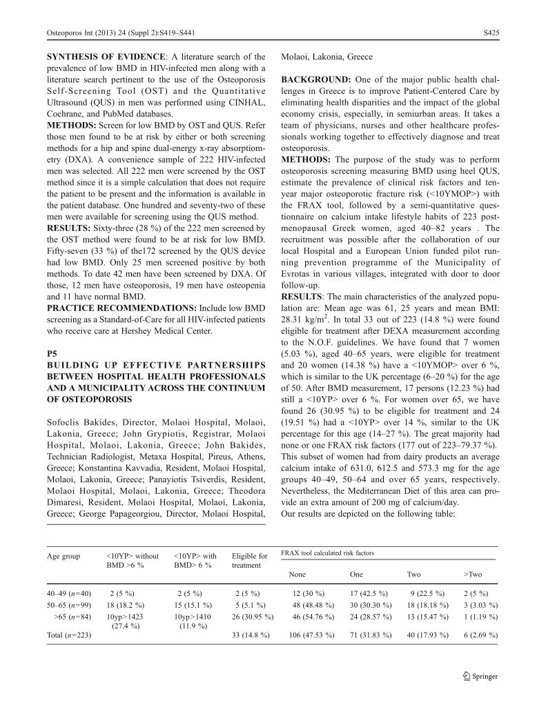
BACKGROUND: One of the major public health chal-lenges in Greece is to improve Patient-Centered Care byeliminating health disparities and the impact of the globaleconomy crisis, especially, in semiurban areas. It takes ateam of physicians, nurses and other healthcare profes-sionals working together to effectively diagnose and treatosteoporosis.METHODS: The purpose of the study was to performosteoporosis screening measuring BMD using heel QUS,estimate the prevalence of clinical risk factors and ten-year major osteoporotic fracture risk (<10YMOP>) withthe FRAX tool, followed by a semi-quantitative ques-tionnaire on calcium intake lifestyle habits of 223 post-menopausal Greek women, aged 40–82 years . Therecruitment was possible after the collaboration of ourlocal Hospital and a European Union funded pilot run-ning prevention programme of the Municipality ofEvrotas in various villages, integrated with door to doorfollow-up.RESULTS: The main characteristics of the analyzed popu-lation are: Mean age was 61, 25 years and mean BMI:28.31 kg/m2. In total 33 out of 223 (14.8 %) were foundeligible for treatment after DEXA measurement accordingto the N.O.F. guidelines. We have found that 7 women(5.03 %), aged 40–65 years, were eligible for treatmentand 20 women (14.38 %) have a <10YMOP> over 6 %,which is similar to the UK percentage (6–20 %) for the ageof 50. After BMD measurement, 17 persons (12.23 %) hadstill a <10YP> over 6 %. For women over 65, we havefound 26 (30.95 %) to be eligible for treatment and 24(19.51 %) had a <10YP> over 14 %, similar to the UKpercentage for this age (14–27 %). The great majority hadnone or one FRAX risk factors (177 out of 223–79.37 %).This subset of women had from dairy products an averagecalcium intake of 631.0, 612.5 and 573.3 mg for the agegroups 40–49, 50–64 and over 65 years, respectively.Nevertheless, the Mediterranean Diet of this area can pro-vide an extra amount of 200 mg of calcium/day.Our results are depicted on the following table:
Age group <10YP> withoutBMD >6 %
<10YP> withBMD> 6 %
Eligible fortreatment
None One Two >Two
40–49 (n=40) 2 (5 %) 2 (5 %) 2 (5 %) 12 (30 %) 17 (42.5 %) 9 (22.5 %) 2 (5 %)
50–65 (n=99) 18 (18.2 %) 15 (15.1 %) 5 (5.1 %) 48 (48.48 %) 30 (30.30 %) 18 (18.18 %) 3 (3.03 %)
>65 (n=84) 10yp>1423(27.4 %)
10yp>1410(11.9 %)
26 (30.95 %) 46 (54.76 %) 24 (28.57 %) 13 (15.47 %) 1 (1.19 %)
Total (n=223) 33 (14.8 %) 106 (47.53 %) 71 (31.83 %) 40 (17.93 %) 6 (2.69 %)
FRAX tool calculated risk factors
Osteoporos Int (2013) 24 (Suppl 2):S419–S441 S425

CONCLUSIONOsteoporosis and relative fragility fractures represent agreat public health problem as they produce elevated so-cial and private costs. Effective primary prevention shouldbe a worldwide public health priority. Local and nationalpolitical support and action is needed for the developmentof targeted screening and intervention programmes throughpartnerships and coordination centres towards a patient-centered approach.
P6OSTEOPOROSIS SCREENING AND FRACTURERISK ASSESSMENT TOOL USAGE AMONGHOUSE STAFF
Jordan Brodsky, M. D., Beth Israel Medical Center,Woodmere, NY; Mehgan Greenfield, M. D., Beth IsraelMedical Center, Woodmere, NY; Erin Patton, M.D. M.P.H,Beth Israel Medical Center, Woodmere, NY
BACKGROUND: Despite increased awareness of the mag-nitude and consequences of osteoporosis and the availabilityof recommendations for screening and treatment by multipleorganizations, osteoporosis is still under diagnosed and in-adequately managed in the United States. It has been esti-mated that 12 million Americans over the age of 50 haveosteoporosis, though only 30 % of eligible women age 65and older had a bone density test. Therefore, identifyingpatients at risk, making a timely diagnosis, implementingprevention measures and initiating pharmacological therapyfor appropriate patients can all help to minimize fracturerisk. Academic hospitals with resident-led outpatient prima-ry care providers are an area where there may be under-utilization of evidence-based fracture risk assessment tools,such as the FRAX score.METHODS: House staff of the Internal Medicine depart-ment at Beth Israel Medical Center, were given an anony-mous questionnaire. The goal was to assess the resident’sknowledge of current practice guidelines and recommenda-tions for osteoporosis and the utilization of the FRAX score.RESULTS: 48 residents of Internal Medicine, levels PGY1, 2 and 3, filled out the questionnaire. 62.5 % of residentsestimated their female patient population was greater than65 years old and 31.25 % of their male patient populationw as greater than 70 years old. 77 % of residents performedage appropriate DEXA scans on their patients. 58.33 % ofresidents had know ledge of what the FRAX score was and47.92 % of resident knew the appropriate use in patient care.62.5 % used the FRAX score to identify patients who metcriteria for the initiation of treatment for osteoporosis.29.17 % could identify the modifiable risk factors and31.25 % identified the no modifiable risk factors whichcalculate the FRAX score. 33.33 % of residents said they
would use the FRAX score on woman less than 65 yearsold. 79.17 % of residents wanted to receive more informa-tion on the FRAX score and its appropriate applications.CONCLUSION: Our study concluded that InternalMedicine residents are following the current guidelines forscreening for osteoporosis with DEXA scans, however, theuse of the FRAX score for the identification of patients athigh risk for fracture requiring the initiation of treatment forosteoporosis, is highly underutilized. There was also a dis-crepancy between the resident’s knowledge of the FRAXscore and its application in clinical practice. Given ourfindings, further training and education regarding osteopo-rosis screening and the use of the FRAX score in a residentled outpatient primary care setting is needed.
P7IMPROVING THE EVALUATION, MANAGEMENT,AND FOLLOW-UP OF OSTEOPOROSIS IN HIPFRACTURE PATIENTS
Heather L. Hofflich, DO, University of California, San Diego,San Diego, CA; Deborah Oh, MD, University of California,San Diego, San Diego, CA; Charlie Choe, MD, University ofCalifornia, San Diego, San Diego, CA; Brian Clay, MD,University of California, San Diego, San Diego, CA;Courtney Tibble, MD, University of California, San Diego,San Diego, CA; Kristi Kulasa, MD, University of California,San Diego, San Diego, CA; Priya Shah, MD, University ofCalifornia, San Diego, San Diego, CA; Edward Fink, MD,University of California, San Diego, San Diego, CA; PaulGirard, MD, University of California, San Diego, San Diego,CA; Sandy Schwartz, MD, University of California, SanDiego, San Diego, CA; Greg Maynard, MD, University ofCalifornia, San Diego, San Diego, CA
BACKGROUND: Less than 30 % of the 400,000 patientsadmitted for osteoporosis-related fractures receive properevaluation and care for osteoporosis, despite multiple effortsamongst physicians and major hospital centers to improveoutcomes.METHODS: We formed a multi-disciplinary team and de-fined definitions for a best practice protocol to assess, treat,document an osteoporosis diagnosis, and triage patientswith fragility fracture, based on best practice recommenda-tions from The Joint Commission and the NationalOsteoporosis Foundation. We established our baseline insti-tutional performance for osteoporosis management via astructured chart review of patients identified by dischargediagnostic codes for hip fracture.The team initiated a pre-authorized osteoporosis consulta-tion from the Endocrinology service for hip fracture pa-tients, “triggered” via a brief query in admission orders orby the orthopedic service nurse practitioner. Osteoporosis
S426 Osteoporos Int (2013) 24 (Suppl 2):S419–S441

consultations utilized a consultation template reflecting ourevidence-based protocol. We reassessed our institutionalperformance using the same structured chart review instru-ment post intervention.RESULTS: After excluding patients on pre-existing oste-oporosis therapy, those unsuitable for long-term osteopo-rosis therapy, and those with fractures attributed to otheretiologies, we analyzed 71 baseline patients and 61inter-vention patients. The groups possessed similar age, gen-der, race, and BMI characteristics. The baseline (on-demand consultation) group suffered from dismal perfor-mance, with only 3–21 % of patients receiving the desiredevaluation, documentation, treatment, or outpatient follow-up. Intervention (triggered consultation) patients improvedmarkedly post-intervention (61–84 % performance) on allparameters except outpatient follow-up, which improvedinsignificantly from 6 % to 15 %.CONCLUSION: While triggered consultation was effec-tive, we suggested using multi-modal layered interven-tions to achieve even better results and address severalidentified barriers.
Table 3 — Performance Results for the Hip Protocol
Baselineperiodn=71
Interventionperiodn=61
%Change
p-value
No. (%) No. (%)
Inpatient consult forosteoporosisPerformed
2 (3 %) 48 (79 %) 76 % p<0.001
Discharge Summarywith Diagnosis ofOsteoporosis
3 (4 %) 41 (67 %) 63 % p<0.001
Dsicharge OsteoporosisFollow-up Plan
4 (6 %) 49 (80 %) 75 % p<0.001
Discharge Prescriptionfor Bisphosphonate
6 (8 %) 37 (61 %) 52 % p<0.001
Dsicharge Prescriptionfor Calcium andVitamin D
10 (14 %) 50 (82 %) 68 % p<0.001
Discharge order forDEXA scan
3 (4 %) 46 (75 %) 71 % p<0.001
Medications initiatedwithin 60 days
15 (21 %) 51 (84 %) 62 % p<0.001
Outpatient OsteoporosisClinic Follow-up visitwithin 60 days
4 (6 %) 9 (15 %) 9 % p=0.096
P8OSTEOSYNTHESIS COMPARED TO HEMIARHROPLASTY FOR OSTEOPOROTIC, UNDISPLACEDAND STABLE FEMORAL NECK FRACTURES
Kaan Irgit, MD, Geisinger Health System, Danville, PA;Raveesh D. Richard, MD, Geisinger Health System,
Danville, PA; Andrew Cornelius, MD, Geisinger HealthSystem, Danville, PA; Thomas R. Bowen, MD, GeisingerHealth System, Danville, PA; Cassondra M. Andreychik,BS, Geisinger Health System, Danville, PA; Daniel S.Horwitz, MD, Geisinger Health System, Danville, PA
BACKGROUND: The incidence of hip fractures in theUnited States and Europe is high and continues to in-crease. The best treatment for femoral neck fractures isstill under debate. The purpose of the study was tocompare the complication, reoperation and mortalityrates of hemiarthroplasty and osteosynthesis in patientswith impacted/stable, osteoporotic, undisplaced femoralneck fractures (AO/OTA 31-B1).METHODS: This study was performed retrospectively atan academic, Level 1 trauma center. 136 patients over60 years of age presenting with stable valgus impacted,and non-impacted undisplaced femoral neck fractures(AO/OTA 31-B1) between 2004 and 2010 qualified forinclusion in this study. We retrospectively compared thecomplication, reoperation and mortality rates betweentwo groups which were matched in age, gender, BMIand ASA scores. All included patients sustained GardenI or II femur neck fractures. 98 patients were treatedusing multiple cannulated screw and 38 patients weretreated with hemiarthroplasty based on surgeon prefer-ence. Osteosynthesis was performed with three parallelcannulated screws. The minimum follow up was24 months. Patient demographics, American Society ofAnesthesiologists (ASA) score, time from injury to sur-gery, duration of surgery, estimated blood loss, treat-ment related complications, length of hospital stay,reoperations, initial total hospital costs and mortalitywere recorded and compared between the internal fixa-tion and hemiarthroplasty groups.RESULTS: The mean age of the 98 patients in theosteosynthesis group was 82 (range, 60–104) and 80 (range,60–90) in the 38 patients treated with hemiarthroplasty.Mean follow up was 44 ± 1.4 months (range, 24–92 months). There were no significant differences in overallcomplication, reoperation and mortality rates for the twogroups. In a logistic regression model analysis, patients overand under 80 years old had similar complication,reoperation and mortality rates. Infection, length of hospitalstay and estimated blood loss were higher afterhemiarthroplasty. Initial hospital costs were higher for thehemiarthroplasty group.CONCLUSION: There were no differences in the surgicaloutcomes, complication, reoperation and mortality rates be-tween the internal fixation and hemiarthroplasty groups.Hemiarthroplasty has no benefit in decreasing complicationsand reoperations for stable femoral neck fractures in theelderly. The costs of surgery and infection rates are higher
Osteoporos Int (2013) 24 (Suppl 2):S419–S441 S427

with hemiarthroplasty as compared to osteosynthesis forthese stable fracture patterns. Internal fixation should bethe initial choice of treatment in patients with osteoporotic,undisplaced femoral neck fractures including those patients,over 80 years of age.
P9TEMPORAL TRENDS IN INCIDENCE OF HIPFRACTURES INVACOMMUNITYLIVINGCENTERS
Tatjana Bulat, MD, VISN 8 Patient Safety Center of Inquiry,Tampa, FL; Gail Powell-Cope, ARNP, PhD, HSRD/RR&DCenter of Excellence, JAH VA Hospital, Tampa, FL; RobertCampbell, PhD, JD, VISN 8 Patient Safety Center ofInquiry, Tampa, FL
Introduction/Objective: We wanted to determine whetherVA national patient safety initiatives (National Falls Toolkitand Hip Protector Toolkit Implementation) have impactedthe incidence of hip fractures in VA community livingcenters (CLC) (aka nursing homes).Design/Methodology: The data were extracted from thehospital discharge datasets available at the AustinInformation and Technology Center (AITC) for FY 2000through 2011. Fractures were identified using ICD-9-CMdiagnosis codes in the 800 through 829 series for the prin-cipal admitting diagnosis (DXPrime). The source of admis-sion was limited to VA CLC (nursing home care units) forthe hip fracture trend analysis. The bed days of care werecomputed from AITC data to allow for a rate of hip fracturesper bed days of care (BDOC) to be calculated for each year.Results: A total of 2, 676 serious fall-related fractures in VAnursing homes resulted in treatment in VA hospitals duringthis time period. There were 1,836 hip fracture dischargesaccounting for 66 % of the total fracture discharges over thistime period. The 311 Intracranial injuries accounted for11 % of the total discharges. Starting in 2005 there was a48 % downward trend in the number of total fracture dis-charges through the end of 2011. There was a 50 % down-ward trend in the number of hip fractures between 2005 and2011. These trends are important given the number of olderveterans served (especially high risk for injury, over 85 yearsof age) has increased in that time period.Conclusion/Discussion: This preliminary analysis estab-lishes that there is a temporal relationship between thepatient safety initiatives implementation in FY 2005–2009and a decrease in rates of hip fractures occurring in VACLCs that were admitted to VA hospitals.
P10PHYSICAL ACTIVITY AND BIOMARKERS OFBONE MINERAL DENSITY IN PERSONS WITHMULTIPLE SCLEROSIS
Paula E. Papanek, PhD, Marquette University, Milwaukee,WI; April Harkins, PhD, Marquette University, Milwaukee,WI; Mary Ellen Csuka, MD, Medical College of Wisconsin,Milwaukee, WI; Benjamin A. Ingraham, BS, MarquetteUniversity, Milwaukee, WI; Brice Cleland, BS, MarquetteUniversity, Milwaukee, WI; Molly Pitluck, BS, MarquetteUniversity, Milwaukee, WI; Alexander V. Ng, PhD,Marquette University, Milwaukee, WI
It has been reported that patients with Multiple Sclerosis(MS) have increased risks of reduced bone mineraldensity (BMD) and fractures. It is unclear whether thisis a primary consequence of the disease or whether it issecondary to low activity, decrease in outdoor activity,low vitamin D 25(OH)D (VITD) levels or other factors(medications). There is emerging evidence that lowVITD levels and reduced physical activity (PA) maynegat ively affec t BMD in MS. Elevated pro-inflammatory cytokines [i.e. IL-6, soluble tumor necro-sis factor II (sTNFRII), and interleukin-10 (IL-10)] andincreased cortisol levels also appear inversely related toBMD in persons without MS.METHODS: In this study, we examined the associations forVITD, PA, endogenous cortisol, and cytokines with BMD inMS patients. Measurements were made in 23 communitydwelling adults volunteers with MS and 21 age-matchedcontrols. The lumbar spine (L2-L4) and femoral neckBMD were measured with dual X-ray absorptiometry(DXA, lunar prodigy) and physical activity was measuredwith accelerometers (average of 7 day recording). VitaminD, cortisol, and cytokines (IL-6, sTNFRII and IL-10) weremeasured by RIA or EIA. Analyses were by unpaired t-testsand Pearson correlations. The results showed that MSsubjects compared with controls had differences in PA(p<0.05), IL-6 (p=0.01), sTNFRII (p=0.001) and meanfemoral neck BMD (p=0.04). No differences were notedin lumbar spine, VITD or cortisol. In our sample (N=23MS), VITD levels were normal and not different fromCN with most of the MS group reporting VITD supple-mentation. VITD levels did not correlate with BMD.Within the MS group alone, PA was correlated to fem-oral BMD (r=0.48, p=0.02) but not lumbar spine (r=−0.14, p=0.56). However, BMD was NOT significantlycorrelated with cortisol, sTNFRII, or IL-10. IL-6 wasinversely correlated to PA within the MS group(r= −0.40, p=0.05).CONCLUSION: In patients with MS who are repletewith VITD, Physical Activity is a major contributor toBMD of the femoral neck. IL-6 levels may be a factor inthe total physical activity of MS patients. Furthermore,low BMD was measured in at least one site in 11 of 23patients with MS (48 %) but in only three control sub-jects (14 %) indicating a need to monitor BMD in this
S428 Osteoporos Int (2013) 24 (Suppl 2):S419–S441

rather young (mean age 41+9 years) patient population.The results also suggest that importance of promotingphysical activity to improve BMD and decrease fracturerisk in persons with MS.
P11THE RELATIVE IMPORTANCE OF 13 DIFFERENTTYPES OF PRESCRIPTION-MEDICATIONINFORMATION TO 1,280 U.S. WOMEN WITHOSTEOPOROSIS
Colleen A. McHorney, PhD, Merck & Co., Inc., NorthWales, PA
BACKGROUND: Chronically-ill patients report significantunmet information needs about their medications (Rx). Only ahandful of studies have been conducted among women withosteoporosis.OBJECTIVE: To assess the importance 1,280 U.S. womenwith osteoporosis attach to 13 types of Rx information.METHODS: A cross-sectional survey of U.S. adults age 40or older with chronic diseases was conducted using theHarris Chronic Disease Panel. A total of 1,280 women withosteoporosis completed the survey. Respondents rated howimportant it would be for them to receive Rx information ifthey were to receive a new osteoporosis Rx: (1) purpose; (2)name; (3) directions; (4) duration; (5) side effects; (6) risks ofside effects; (7) what to do if you experience a side effect; (8)number of refills; (9) effect of food/alcohol with the Rx; (10)Rx cost; (11) drug interactions; (12) Rx benefits; and (13) Rxadherence. Respondents completed 19 questions on their os-teoporosis Rx beliefs: perceived need for osteoporosis Rx(k=11), perceived concerns about osteoporosis Rx (k=6),and perceived affordability of osteoporosis Rx (k=2). Eachinformation-preference item was dichotomized (not at all, alittle, and somewhat important vs. very/extremely important).Logistic regression identified subgroup differences in infor-mation preferences.RESULTS: Age ranged from 40 to 97 (mean=65.7), 96 %was Caucasian, 42 % had a college education, and 53 %earned $50,000 or less annually. Mean importance ratingsranged from a low of 3.80 to a high of 4.56 (mean=4.27 andmedian=4.33). From 65 % to 95 % endorsed that it wouldbe “extremely” or “very important” to receive informationon the 13 items. There was remarkable invariance in osteo-porosis prescription-medication information preferences forall demographic characteristics: the different subgroups ofwomen with osteoporosis did not differ in their preferencesfor osteoporosis prescription-medication information.Osteoporotic women with the highest perceived need forosteoporosis medications preferred more prescription-medication information (median effect of 3.60) as did thosein the middle tertile (median effect of 3.10). For three items
(risk of side effects, common side effects, and duration),osteoporotic women with the most concerns about osteopo-rosis medications desired more information (median effectsize of 3.43). Osteoporotic women with the worst perceivedmedication affordability were 6.4 times more likely to preferinformation about medication costs, name of the medication(OR=1.71), and number of refills (OR=1.68).DISCUSSION: U.S. women with osteoporosis overwhelm-ingly desire information about osteoporosis Rx. and thosepreferences were invariant across demographics. Desire forinformation is a necessary, but not sufficient, condition forwomen’s informed decision making about osteoporosis pre-scription medications.
P12OSTEOPOROSIS-SPECIFICMEDICATION BELIEFS,BUT NOT TIME PERSPECTIVE, DIFFERENTIATEDWOMEN WHO WERE SELF-REPORTED MEDICATION PERSISTERS, NON-PERSISTERS, AND NON-FULFILLERS
Colleen A. McHorney, PhD, Merck & Co., Inc., NorthWales, PA
BACKGROUND: Medication beliefs can be powerful pre-dictors of medication adherence. Researchers have hypoth-esized that patients’ time perspective— their attitudes aboutimmediate vs. future outcomes associated with behavior —could also influence medication-taking.OBJECTIVE: To assess the role of patient’s medicationbeliefs and time perspective in differentiating women withosteoporosis (OP) who were self-reported medication per-sisters, non-persisters, and non-fulfillers.METHODS: A cross-sectional survey of U.S. adults age 40or older with chronic disease was conducted in 2010 using theHarris Chronic Disease Panel. A total of 653 women with self-reported OP completed the survey. Respondents completedquestions about their OP medication (Rx) beliefs: perceivedneed for OP Rx, k=6; perceived concerns about OP Rx, k=2;perceived affordability of OP Rx, k=2; and perceived severityof OP, k=5. They also completed six items on physicianinformation-giving about OP, a single-item measure of patienttrust, and the Consideration of Future Consequences Scale(CFC), an 11-item multi-item scale assessing time perspective.General linear models were used to assess the extent to whichthemeasures differentiated betweenwomenwith OPwhowereself-reported Rx persisters, non-persisters, and non-fulfillers.RESULTS: Of the 653 women with self-reported OP, ageranged from 41 to 87 (mean=63.9), 94 % was Caucasian,38 % had a college education, and 59 % earned $50,000 orless annually. One half (50 %) of the sample were self-reported OP Rx persisters, 44 % were non-persisters, and6 % were non-fulfillers. Neither the CFC present nor future
Osteoporos Int (2013) 24 (Suppl 2):S419–S441 S429

orientation scale statistically distinguished among the OPRx persisters, non-persisters, and non-fulfillers. Perceivedneed for OP Rx most powerfully distinguished among thethree groups (F=160.2, p<.0001), followed by perceivedOP Rx concerns (F=88.4, p<.001), physician information-giving about OP (F=74.2, p<.001), patient trust in physi-cian (F=38.9, p<.001), perceived severity of OP (F=16.1,p<.01), and perceived affordability of OP Rx (F=11.4, p<.001). In all comparisons, OP Rx non-persisters werestatistically indistinguishable from OP non-fulfillers.DISCUSSION: OP-specific Rx beliefs powerfully differen-tiated between U.S. women with OP who were self-reportedmedication persisters, non-persisters, and non-fulfillerswhile time perspective did not. OP Rx non-persisters andnon-fulfillers had suboptimal perceived need for OP Rx,more concerns about them, received less OP informationfrom their providers, had less trust in their physicians, wereless likely to view OP as a chronic condition, and were lesslikely to perceive OP Rx as affordable. Suboptimal Rxbeliefs are accessible and can be ameliorated through effec-tive patient-centered communication about OP and its treat-ment.
P13IN THEIR OWN VOICE: A QUALITATIVE STUDY OFHOW WOMEN WITH OSTEOPOROSIS VIEWDIAGNOSIS AND TREATMENT IN 2012
Colleen A. McHorney, PhD, Merck & Co., Inc., NorthWales, PA
BACKGROUND: Osteoporosis is common and numeroustherapies are available for its treatment. However, signifi-cant under-treatment exists.OBJECTIVES: to understand, from the patient point-of-view, how U.S. women with osteoporosis view the diagno-sis and treatment of osteoporosis in 2012.METHODS: Twelve focus groups with women with self-reported osteoporosis were conducted in Chicago, Atlanta,and Phoenix in November 2012. The transcripts were ana-lyzed using systematic coding via content analysis.RESULTS: A total of 127 women with osteoporosis par-ticipated. Average age was 64.5, and 92 % were Caucasian.Women averaged 2.0 comorbidities in addition to osteopo-rosis. On average, women had the diagnosis of osteoporosisfor 8.1 years. Seven major emerged across the focus groups.
(1) Most women with osteoporosis felt little urgency fortreatment. Women felt that osteoporosis is part of ag-ing. Compared to other diseases, osteoporosis wasviewed as less serious to current health. Many consid-ered osteoporosis to be “out of sight, out of mind”because it was asymptomatic.
(2) Most women perceived their primary care physicians(PCPs) to be “matter-of-fact” about osteoporosis.Women felt that their PCPs minimized osteoporosisrelative to other diseases. PCP’s were often perceivedas blasé and lackadaisical about osteoporosis.
(3) Women did not consider their PCPs to be knowledge-able about osteoporosis. Many women did not considertheir PCP to be “on top” of osteoporosis, and they didnot feel their PCPs were knowledgeable about non-pharmaceutical treatment alternatives.
(4) Most women did not feel knowledgeable themselvesabout osteoporosis.
(5) Women did their own subjective adherence-value prop-osition about initiating and persisting to osteoporosistreatment by weighing the pros and cons of pharmaco-logic treatment. Many women were still treatmentnaïve and an equal proportion had initiated, butdiscontinued, pharmacologic treatment.
(6) Most women did not proactively tell their providerwhen they did not fill a newly-prescribed osteoporosismedication or stopped taking one on their owninitiative.
(7) Women believed there were many things they could dothemselves to control, cure, or minimize osteoporosis.Women believed that over-the-counter calcium andvitamin D supplements were sufficient for treatingosteoporosis. Women believed there was no harm incalcium and vitamin D supplements.
CONCLUSION: In 2012, where there are many options forthe detection and treatment of osteoporosis, women mini-mized the seriousness of osteoporosis, in part because thePCP also did so. Most of the women were under-treated.Women took a “wait and see” attitude about osteoporosis.These results suggest the need for better communicationbetween physician and patient on the seriousness of osteo-porosis and the importance of initiating and continuingtreatment.
P14TIME TO SURGERY FOR HIP FRACTURES USINGA TRAUMA ADMISSION PROTOCOL
Brett P. Frykberg, MD, University of Florida —Jacksonville, Jacksonville, FL; Anthony Bell, MD,University of Florida — Jacksonville, Jacksonville, FL;Anna Acosta, MD, University of Florida — Jacksonville,Jacksonville, FL; Andrew Kerwin, MD, University ofFlorida — Jacksonville, Jacksonville, FL; Michael Suk,MD, JD, MPH, Geisinger Health Systems, Danville, PA
BACKGROUND: There are significant morbidities andmortalities associated with elderly patients presenting with
S430 Osteoporos Int (2013) 24 (Suppl 2):S419–S441

hip fractures. Current guidelines recommend safely getting thepatient from the emergency room to the operating room fordefinitive care in a timely manner in order to decrease themorbidity and mortality associated with these fractures. Theproblem is being able to safely and effectively attain clear-ance from a medical perspective for surgery within a shorttime frame. Particular challenges exist in a Level 1 traumacenter where fewer patients with higher acuity tend toarrive when compared to community hospitals. Traditionalprotocols intended to “clear” patients through a medicalservice often result in delays to surgery secondary to issuessuch as: (1) rounding times for medicine after OR starttimes; (2) attending co-signatures at times that are inconve-nient to the operating service; and (3) turf battles overprimary admission team resulting in dissatisfaction amongemergency room staff. To address these issues a trial pro-tocol for elderly, low energy hip fractures was created. Thisrequired all lower energy hip fractures to be admitted to thesurgical trauma team for appropriate and expeditious timeto surgery. Our hypothesis is that by instituting our proto-col, we will decrease the time between hospital admissionand surgery.METHODS: In 2009, a trauma surgical protocol was put inplace for all low energy hip fractures at our level oneacademic teaching hospital. An IRB was obtained to retro-spectively review charts on 149 patients. Our control groupwas a “pre-protocol” cohort between 2007 and 2009, meet-ing the same criteria. Using chart review analysis, werecorded: time between admission and definitive procedure,morbidities, mortality, and consulted services and comparedthe data between the two groups.RESULTS: Our study demonstrated significantly lowermorbidities in the post-protocol group. Though we didnot show a decrease in time from admission to surgery,there was a trend that did not attain statistical signifi-cance. The overall inpatient mortality rate in our studywas 6 %, with no difference between the two groups.CONCLUSION: Using our trauma admission protocol,we were able to show a significant decrease of morbid-ities in elderly patients with hip fractures as well as adecreased time from admission to surgery.
P15THE IMPACT OF IMPLEMENTATION OF “OWNTHE BONE” IN AN ACADEMIC URBAN HOSPITALSETTING: A PATIENT-CENTERED ANALYSIS OFPROGRAM EFFECTIVENESS
Anthony Essilfie, BA, Columbia University, New York, NY;David Patrick, Jr, BS, Columbia University, New York, NY;Dinaz Irani, MD, Columbia University, New York, NY;Beatrice Edwards, MD, UT MD Anderson Cancer Center,
Houston, TX; William Macaulay, MD, Columbia University,New York, NY
BACKGROUND: The occurrence of fragility fractures isone of the strongest indicators for underlying osteoporosis.With the rate of fragility fractures increasing as much as 20times following a patient’s first fragility fracture, a compre-hensive patient education course on osteoporosis and frac-ture prevention needs to be employed for patient safety. TheAmerican Orthopaedic Association (AOA) initiated an Ownthe Bone™ (OTB) pilot program in 2005 in an attempt toimprove the treatment and prevention of these fragilityfractures. Following a successful pilot program, our institu-tion has maintained its commitment to the OTB protocol asa quality care improvement program for our fragility frac-ture patients. The purpose of this study was to assess theefficacy of the OTB Program in our inpatient, fragilityfracture population.METHODS: Participants were139 fragility fracture patientsthat were identified, educated, and referred for follow up bya fragility fracture liaison. The patient education wasconducted via OTB materials and a letter was sent to PCPsto increase communication between medical disciplines toimprove osteoporosis care. Patients were contacted by tele-phone at an average follow up of 8.4 months after thehospitalization to respond to the OTB Follow-up Survey.RESULTS: Of the 97 (69.8 %) patients that responded tothe survey, 75 (77.3 %) patients had visited their PCP aftersuffering a fragility fracture. Forty-one (42.3 %) patients hada discussion with their PCP regarding their fracture. Thirty-three (34.0 %) patients had a DXA performed after hospitaldischarge. At follow up, 58 (59.8 %) patients were takingvitamin D. Another 58 (59.8 %) patients reported takingcalcium and 15 (15.46 %) patients reported being on phar-macologic osteoporotic medications.CONCLUSION: The OTB program attained comparablevitamin D and calcium supplementation rates relative to otherfragility fracture education programs. However, a gap in med-ical care after “Own the Bone” intervention occurs resulting inlow rates of bone density testing and initiation of pharmaco-logic management by PCP. Further physician education andadherence with guidelines is necessary.
P16USING PREDICTIVE MODELING TO ESTIMATEBONE MINERAL DENSITY IN CHILDREN ANDADULTS WITH PHENYLKETONURIA
Kathryn E. Coakley, MS, RD, Nutrition and Health Sciencesand Molecules to Mankind Programs, Emory University,Atlanta, GA; Teresa D. Douglas, PhD, Metabolic NutritionProgram, Department of Human Genetics, Emory University,Atlanta, GA; Rani H. Singh, PhD, RD, LD, Metabolic
Osteoporos Int (2013) 24 (Suppl 2):S419–S441 S431

Nutrition Program, Department of Human Genetics, EmoryUniversity, Atlanta, GA
BACKGROUND: Phenylketonuria (PKU) is an autosomalrecessive disorder affecting the enzyme phenylalanine hy-droxylase. Elevated concentrations of phenylalanine (phe)result in neurological, behavioral, and physical abnormali-ties. Children and adults with PKU also have a higherprevalence of bone abnormalities and increased fracture riskcompared to non-PKU controls. Bone health is monitoredvia DXA which is expensive and involves radiation.European estimates suggest only 1 in 14 PKU centers mon-itor bone in children while 3 in 5 monitor bone in adults.Frequency of monitoring is unreported in the U.S. Thisstudy aims to use clinical parameters collected in PKUpatients to predict total bone mineral density (BMD).METHODS: Data were collected from early-treated PKUpatients over 4 years of age at baseline of a clinical trial (n=57). Demographic (age, sex, BMI), clinical (phe prescription,medical-food prescription), laboratory (plasma phe and tyro-sine, lipids, vitamin D), genetic (AV sum, a genetic mutationseverity score), and dietary data were included. Correlationcoefficients adjusted for age, sex, BMI, phe, and medical foodintake were calculated between each parameter and totalBMD, a reproducible measure reflecting average density ofmultiple sites. Predictors that correlated significantly withBMD and interactions terms were considered in models.Final models with (1) all data, (2) routine clinic visit data(excluding vitamin D, lipids), and (3) routine+genetic datawere selected considering r-square and MSE. Categories ofactual and predicted BMD z-scores were compared: normal [>−1stadard deviation (SD) from reference], at-risk (−2.5 to−1SD), and low (<−2.5SD). Future studies will collect vari-ables included in models to validate predicted BMD andDXA-measured BMD (total, axial, and peripheral).RESULTS: In the sample (mean age=17.3; 60 % male), 16(28 %) had at-risk BMD; 3 (5 %) had low BMD. BMD wascorrelated with age, BMI, medical food prescription, cho-lesterol, triglycerides, vitamin D, and AV sum (p<0.05). R-square values for final models ranged from 0.75 to 0.86suggesting good fit. Models’ estimated BMD correlatedwith actual BMD [correlation coefficients (1) 0.93, (2)0.87, (3) 0.91; p-value <0.0001] and predicted z-scoresagreed with actual z-scores (kappa=1.00; p-value <0.0001).CONCLUSIONS: Nearly one-third of study participantshad BMD 1 SD below normal, and 3 had BMD at least2.5 SD below normal. Routinely collected parameters canpredict total BMD and z-score category (normal, low, at-risk) in individuals with PKU. Each of the models can beused to identify patients at-risk for bone abnormalities with-out DXA expense and radiation exposure.Partial research support by BioMarin Pharmaceuticals andin part by PHS Grant UL1 RR025008 from the Clinical and
Translational Science Award program, National Institutes ofHealth, National Center for Research Resources
P17DISAGREEMENT IN THE DIAGNOSIS OF OSTEOPENIA/OSTEOPOROSIS BY DUAL ENERGY X-RAYABSORPTIOMETRY MEASUREMENTS WITHNORLAND INSTRUMENTS, BETWEEN DEVICEREFERENCE CURVES AND SELF-DEVELOPEDREFERENCE CURVES, IN THE SPANISH FEMALEPOPULATION
Juan D. Pedrera-Zamorano, PhD, Metabolic Bone DiseasesResearch Group. University of Extremadura, CACERES,Spain; Jesus M. Lavado-Garcia, PhD, Metabolic BoneDiseases Research Group. University of Extremadura,CACERES, Spain; Maria L. Canal-Macias, PhD,Metabolic Bone Diseases Research Group. University ofExtremadura, CACERES, Spain; Julian F. Calderon-Garcia, PhD, Metabolic Bone Diseases Research Group.University of Extremadura, CACERES, Spain; CarmenCosta-Fernandez, RN, Metabolic Bone Diseases ResearchGroup. University of Extremadura, CACERES, Spain; JoseM. Moran, PhD, Metabolic Bone Diseases Research Group.University of Extremadura, CACERES, Spain
The bone mineral density (BMD) reference curve is thereference value used for diagnosing osteopenia/osteoporosisand estimating bone mass changes. Its precision wouldinfluence the correctness of T-score and Z-score rates andthus the credibility of diagnostic results. In this study, wereport the utilization of a new establish BMD referencecurves at diverse skeletal sites in Spanish women and, thecomparison of the diagnostic results with the instrumentreference curves for Spanish women. The CáceresOsteoporosis Reference Database (CAFOR) comprises apopulation of 509 healthy women ranging in age from 18to 39 years; we used a Norland dual X-ray absorptiometry(DXA) bone densitometer (Norland Corp. Fort Atkinson,WI, USA) to measure BMD at the posteroanterior spine(PA; vertebrae L2-L4), followed by a scan of the of thefemoral neck (FN). Device reference curves for theSpanish female population were overall those based on thestudy of Diaz-Curiel et al. in 2001 (Diaz-Curiel et al., MedClin 2001), developed using Hologic instruments (Hologic,Waltham, Mass, USA). An interrater reliability analysisusing the Kappa statistic was achieved to determine consis-tency among reference curves. A total of 2635 women (agerange 40–87) were recruited in the study. The prevalence ofosteoporosis with the device reference curves was of14.99 % (13.68–16.40 % IC 95 %) and osteopenia was of37.76 % (35.93–39.63 % IC 95 %). A lower figure was foundusing the CAFOR reference curves for the osteoporosis
S432 Osteoporos Int (2013) 24 (Suppl 2):S419–S441

prevalence with a 3.07 % (2.48–3.80 % IC 95 %) and higherfigure was found in the diagnosis of osteopenia with a preva-lence of 49.60 % (47.69–51.51 IC 95 %). The 2.70 % (2.11–3.35 % IC 95 %) of the participants were diagnosed as osteo-porosis by the two databases. No one of the participants diag-nosed as osteoporosis, was diagnosed as “normal” by the otherdatabase. The interrater reliability statistic was found to beKappa=0.608 (p<0.001), 95 % CI (0.582, 0.63) showing amoderate agreement within the two reference curves. Based onthe methodology of the Diaz-Curiel study that did not includewomen from our area and used a different DXA device weconsider that we might be overestimating the diagnosis ofosteoporosis within adult Spanish women diagnosed with aNorland instrument. In conclusion, the subject classificationaccording to the WHO standards was significant dissimilarusing the Norland reference values and CAFOR referencevalues for the spine and the femur; the Norland reference valuesclassified a larger proportion of women as osteoporotic (14.99vs. 3.07 %) but less as osteopenic (37.76 vs. 49.60 %) com-pared to CAFOR.
P19REASONS FOR MEDICATION NON-PERSISTENCEAMONG WOMEN WITH OSTEOPOROSIS:TEMPORAL TRENDS FROM 2008 TO 2010
Colleen A. McHorney, PhD, Merck & Co., Inc., NorthWales, PA
BACKGROUND: Persistence with prescription-medicationtherapy for osteoporosis is suboptimal.Only by understandingwomen’s reasons for medication persistence can effectivepatient-centered adherence interventions be developed.OBJECTIVES: To identify self-reported reasons why U.S.women with osteoporosis stop taking a prescription medi-cation without their physician telling them to do so (lack ofmedication persistence).METHODS: Three cross-sectional surveys of U.S. adultsage 40 or older with chronic disease were conducted in2008, 2009, and 2010 using the Harris Chronic DiseasePanel. In 2008, 2009, and 2010, a total of 317, 407, and202 women with osteoporosis, respectively, admitted toosteoporosis medication non-persisters and completed a12-item checklist on reasons for non-persistence. The equal-ity proportions obtained from the three independent sampleswas tested using the Pearson chi-square test to assess theequivalence of reasons for non-persistence between 2008and 2009 and then between 2009 and 2010.RESULTS: As shown in Table 1, across all 3 years, the topfive reasons for osteoporosis non-persistence were experienceor fear of side effects, medication affordability, general med-ication concerns, change in insurance/drug benefits, and thebelief that osteoporosis is not a life-threatening condition. One
statistically-significant difference between 2008 and 2009wasobserved: endorsement of medication affordability as a reasonfor non-persistence dropped significantly following the patentexpiry of alendronate in Spring 2008 (p=.0168). There wereno statistically-significant differences between 2009 and 2010reasons for osteoporosis non-persistence.CONCLUSION: The same top reasons for osteoporosis med-ication non-persistence were observed across three consecutiveyears among U.S. women with osteoporosis. The outcome ofthis research should prove to be informative to clinicians andresearchers who seek to design and evaluate osteoporosis ad-herence interventions consonant with patient-centered reasonsfor medication non-persistence.
Table 1. Reasons for Non-Persistence in Women with Self-Reported Osteoporosis
Spring2008
Spring2009
Spring2010
N=317 N=407 N=202
Experience or fear of side effects 51 % 47 % 48 %
Paying for the osteoporosis Rx was afinancial hardship
43 %* 34 %* 27 %
General concerns about taking theosteoporosis Rx
34 % 27 % 31 %
Change in health insurance/drug benefits 16 % 12 % 7 %
Osteoporosis not a life-threateningcondition
15 % 12 % 15 %
Problems remembering to take it 14 % 17 % 16 %
Did not see clinical evidence theosteoporosis Rx was working
13 % 10 % 13 %
Inconvenient/complex dosing regimen 11 % 13 % 11 %
Did not feel I needed the osteoporosis Rx 9 % 10 % 9 %
Experience or fear of drug interactions 9 % 6 % 9 %
Did not feel osteoporosis Rx would beeffective
6 % 5 % 7 %
Did not understand the purpose of theosteoporosis Rx
2 % 1 % 1 %
* Statistically different between Spring 2008 and Spring2009 at p<.05☨ Statistically different between Spring 2009 and Spring2010 at p<.05
P20LACK OF OSTEOPOROSIS TREATMENT IN REALWORLD HIP FRACTURE PATIENTS
Kelly Krohn, MD, Lilly USA, Indianapolis, IN
PURPOSE: National Osteoporosis Foundation guidelinesrecommend that postmenopausal individuals age 50 andolder presenting with hip fracture should be considered fortreatment with pharmacologic osteoporosis (OP) treatment.This study examined patterns of OP treatment strategies
Osteoporos Int (2013) 24 (Suppl 2):S419–S441 S433
among hip fracture (HFx) patients in real world clinicalpractice.METHODS: Patients aged 50+ with an HFx between1/1/2002 and 12/31/2010 (first observed HFx=index) wereidentified from a large U.S. administrative claims database.Patients included for study had 6+ months of pre-indexcontinuous enrollment (baseline), no baseline evidence ofteriparatide (TPTD), cancer, or Paget’s disease. Patientswere followed for up-to 36 months post-index to observepatterns in pharmacologic OP treatment strategies. Five co-horts were constructed based on pre- and post-index use ofOP treatment: patients with no observed evidence of OPtreatment pre- or post-index (N/N); new bisphosphonate(BP) initiators with no baseline BP (N/BP); BP continuerswith baseline BP (BP/BP); new TPTD initiators with nobaseline BP treatment (N/TPTD); TPTD initiators switchingfrom prior BP (BP/TPTD). Demographics, clinical charac-teristics, and healthcare resource use were compared acrossthe 5 cohorts.RESULTS: Study included 71,115 patients. The majority ofthe sample, 53,634 (75 % of total) patients, was observed tohave no OP treatment (N/N) over a median of 352 days offollow-up; 26,238 of whom had ≥1 year of follow-up. NewBP initiators (N/BP; 9,187 patients) started BP treatmentwithin a median of 117 days. BP continuers (BP/BP; 7,463patients) resumed treatment within a median of 58 days.New TPTD initiators (N/TPTD; 346 patients) startedTPTD treatment within a median of 138 days. TPTD ini-tiators switching from prior BP (BP/TPTD; 485 patients)switched to TPTD treatment within a median of 64 days.Mean ages ranged from 74.0 (BP/TPTD) to 80.5 (N/N)years. The N/N cohort was the oldest (81 vs. 74–79 years),had the highest proportion of males (39 % vs. 8–18 %), andthe lowest baseline use rates of systemic glucocorticoids(13 % vs. 17–30 %) and dual energy X-ray absorptiometryscans (2 % vs. 5–17 %).CONCLUSIONS: In spite of a sentinel event of a hipfracture, which is a known risk factor for future fracture,75 % of patients had no evidence of OP treatment over amedian follow-up of 352 days. These data provide fur-ther evidence of a substantial gap in the management ofOP among patients at very high risk for fractures.
P21UNMET NEED FOR OSTEOPOROSIS TREATMENTIN REAL WORLD KYPHOPLASTY/VERTEBROPLASTY PATIENTS
Kelly Krohn, MD, Lilly USA, Indianapolis, IN
PURPOSE: Postmenopausal men and women aged 50+presenting with vertebral fractures are recommended candi-dates for pharmacologic osteoporosis (OP) treatment
according to National Osteoporosis Foundation (NOF) guide-lines. This study examined real world patterns of OP treatmentstrategies among kyphoplasty/vertebroplasty (KV) patients.METHODS: A large U.S. administrative claims databasewas used to identify patients aged 50+ with a KV between1/1/2002 and 12/31/2010 (first observed KV=index). Allpatients included had 6+ months of pre-index continuousenrollment (baseline), no baseline evidence of teriparatide(TPTD), cancer, or Paget’s disease. Patients were followedfor up-to 36 months post-index to observe patterns in phar-macologic OP treatment strategies. Five cohorts wereconstructed based on pre- and post-index use of OP treatment:patients with no observed evidence of OP treatment pre- orpost-index (N/N); new bisphosphonate (BP) initiators with nobaseline BP (N/BP); BP continuers with baseline BP (BP/BP);new TPTD initiators with no baseline BP treatment (N/TPTD);and TPTD initiators switching from prior BP (BP/TPTD).Demographics, clinical characteristics, and healthcare costswere compared across the 5 cohorts.RESULTS: Study included 23,241 patients. About 50 % ofthe patients (11,667) had no OP treatment (N/N) over amedian of 359 days of follow-up; 5,783 of whom had≥1 year of follow-up. New BP initiators (N/BP; 4,742 pa-tients) started BP treatment within a median of 68 days. BPcontinuers (BP/BP; 5,245 patients) resumed treatment with-in a median of 37 days. New TPTD initiators (N/TPTD; 680patients) started TPTD treatment within a median of 70 days.TPTD initiators switching from prior BP (BP/TPTD; 907patients) switched to TPTD treatment within a median of38 days. Mean ages ranged from 74.2 (N/TPTD) to 77.6(BP/BP) years. The N/N cohort had the highest proportionof males (44 % vs. 14–26 %), and the lowest baseline userates of systemic glucocorticoids (33 % vs. 36–47 %) anddual energy X-ray absorptiometry scans (8 % vs. 13–20 %).Mean baseline healthcare costs were the lowest for the N/BP($13,536) and BP/BP ($12,545) cohorts (vs. $15,059–$16,791).CONCLUSIONS: Despite prominent recommendations forOP treatment in vertebral fracture patients within NOFguidelines, half of studied KV patients had no evidence ofOP treatment over a median follow-up of 359 days. Thesedata suggest substantial unmet need in the management ofOP among high-risk patients.
P22TRANSTHEORETICAL MODEL: FACILITATINGBEHAVIOR CHANGE
Judith Gale, PT, DPT, MPH, Creighton University, Omaha,NE
BACKGROUND: Physical therapists identify the role ofeducator, teacher or facilitator as a large part of their overall
S434 Osteoporos Int (2013) 24 (Suppl 2):S419–S441

responsibilities. During the examination process they arelikely to focus on identifying the problem(s), the patient’sgoals, a working hypothesis, the diagnosis, and an interven-tion plan to be implemented within a limited number ofvisits. Few physical therapists are likely to assess the pa-tient’s beliefs and health behaviors as they relate to adher-ence to the intervention plan. Experienced therapists,however, will talk about “reading the patient” or “connectingwith the patient”. Are such things simply aspects of evaluationand intervention that are part of communicating well, or isthere more to it? Technical competence in assessment andintervention planning, although very important, means littleif patients do not follow the home program or continue un-healthy habits which contribute to their current problems.Experienced physical therapists know that many patients pres-ent with neuromusculoskeletal problems that are the result oflifestyle choices that can put them at risk, for example, ofosteoporotic fractures. The challenge is to negotiate the mostefficacious intervention or prevention plan that the patient willbe motivated to follow.METHOD: A series of case studies will be presenteddescribing use of the Transtheoretical Model of BehaviorChange in patients with osteoporosis. These will demon-strate that while the physical therapist cannot controlwhat the patient does at home, he or she can influencethe patient so there is a greater likelihood that what isprescribed is followed. In the case of osteoporosis, thetreatment plan must become part of everyday life.Behavior change and adherence were facilitated throughpatient-practitioner collaboration and application of theTranstheoretical Model.RESULTS: Designing therapeutic interventions with thehighest likelihood of patient follow-through and adherenceis an essential factor in promoting successful patient out-comes. In the cases presented here, it is apparent thatpatient-practitioner collaboration is important in promotingpatient adherence and that the Transtheoretical Model is auseful tool in moving patients from inaction to action.CONCLUSION: Although knowledge of the condition isimportant, the patient’s initial and long-term motivation arecritical elements in successful prevention and treatment ofosteoporotic fractures. Application of the Transtheoreticalstage-process is one way of facilitating behavior change andadherence to treatment plans.
P23NURSES TAKING INITIATIVE IN PROMOTINGBONE HEALTH: A MULTILEVEL MODEL FORPREVENTING OSTEOPOROSIS
Dianne Travers Gustafson, PhD, Creighton University,Omaha, NE; Joan M. Lappe, Ph.D., Creighton University,Omaha, NE
PURPOSE: To present a working model that will motivateand guide nurses, in any practice setting, to promote bonehealth and prevent osteoporosis.PROPOSAL: Osteoporosis is epidemic and costly to treat,and the incidence is increasing with aging of our population.Osteoporosis is preventable, and promoting bone healththroughout the lifespan is essential for the most effectiveprevention. Nurses in any practice setting, as they interactwith their clients/patients, are well-positioned to decreasethe incidence of osteoporosis by providing education, as-sessment and preventive interventions. The nursing profes-sion in the US has over 3 million members, and workingtowards this common goal, professional nurses can have atremendous impact on reducing the osteoporosis epidemic.Over forty million adults in the U.S. either have osteoporo-sis or are at high risk for the disease due to low bone mass.There is an estimated excessive mortality of 25 % in the firstyear following an osteoporosis-related hip fracture. In addi-tion, osteoporosis is associated with considerable morbidityand economic burden. Estimates are that the annual cost ofosteoporosis will be $25.3 billion by 2025. Osteoporosis iscalled a “silent disease” because many people do not havesymptoms prior to sustaining a fracture and they may nothave had simple screenings that can identify risk. Thus, it iscritically important for nurses to become primary preventionspecialists. Research evidence consistently demonstratesthat $1 invested in primary prevention saves from $3 to$80 in disease and injury treatment costs.The multilevel,working model for promoting bone health and preventingosteoporosis guides nursing practice from individual levelassessment and intervention to building interdisciplinarypartnerships and coalitions to influence policy and legisla-tion. The model clearly identifies strategies for nurses topartner with patients, families, community agencies, otherhealth care providers, health care organizations, and legis-lative bodies to promote population health and reduce thecurrent osteoporosis epidemic.The IOM Future of Nursing: Leading Change,Advancing Health report charges nurses to practice tothe full extent of their education and to participate aspartners in designing health care systems that providequality and safe care. Healthy People 2020, the nation’sguide for health promotion and population health im-provement, asks all health care providers to activelyengage in prevention practices.CONCLUSION: The proposed working model isdesigned to motivate and guide nursing practice initia-tives and shape osteoporosis prevention strategies. Itspurpose is to enable and encourage nurses, as health carepractitioners, to shift the health care system to a primaryprevention approach and, thus, reduce the personal andnational disease incidence and economic burden ofosteoporosis.
Osteoporos Int (2013) 24 (Suppl 2):S419–S441 S435

P24THE RELATIONSHIP AMONG HYPERTENSIONAND HYPERCHOLESTEROLEMIA WITH A LOWBONE MINERAL DENSITY IN SPANISH POSTMENOPAUSALWOMEN
Jose M. Moran, PhD, Metabolic Bone Diseases ResearchGroup. University of Extremadura, CACERES, Spain;Mariana Martinez, RN, Metabolic Bone Diseases ResearchGroup. University of Extremadura, CACERES, Spain;Maria L. Canal-Macias, PhD, Metabolic Bone DiseasesResearch Group. University of Extremadura, CACERES,Spain; Carmen Costa-Fernandez, RN, Metabolic BoneDiseases Research Group. University of Extremadura,CACERES, Spain; Raul Roncero-Martin, PhD, MetabolicBone Diseases Research Group. University of Extremadura,CACERES, Spain; Julian F. Calderon-Garcia, PhD,Metabolic Bone Diseases Research Group. University ofExtremadura, CACERES, Spain; Juan D. Pedrera-Zamorano, PhD, Metabolic Bone Diseases ResearchGroup. University of Extremadura, CACERES, Spain
We aimed to evaluate hypertension (HTA), hypercholester-olemia (HC) and both conditions simultaneously in post-menopausal Spanish women with and without low bonemineral density (BMD) while controlling for the influenceof confounding factors such BMI and age. A total of 1557postmenopausal Spanish women aged 57.67±7.95 yearswere analyzed. Within the studied population, 245 womenhad a diagnosis of HTA, 290 of HC and 221 of bothdiseases. All the women had undergone treatment for theseconditions at least during the last year. The remaining wom-en (n=801) conformed a without treatment group. HTA andHC were included as covariates in a logistic regressionmodel assessing the relationship between these conditionsand both osteoporosis and low BMD while controlling forosteoporosis confounding factors. Specific mean BMDvalues at the femoral neck (FN) and spine provided by theDXA equipment manufacturer (Norland Corp. Fort Atkinson,WI, USA) were used to establish specific low BMD T-scoresand osteoporosis diagnosis according to the WHO T-scorecriteria. Low BMD was defined as T score< −1 and normalBMD was defined as T score > or =−1.HTA and HC were not osteoporosis risk factors (crude oddsratio [OR]=1.076; 95 % CI, 0.798–1.451; P=0.631 for HTA;[OR]=0.849; 95%CI, 0.634–1.136;P=0.271 for HC; [OR]=1.082; 95 % CI, 0.731–1.599; P=0.694 for both). Absence ofsignificance remained after adjustment for potentialconfounding factors (adjusted odds ratio [OR] =1.206; 95 %CI, 0.822–1.767; P=0.338 for HTA; [OR]=0.849; 95 % CI,0.634–1.136;P=0.271 for HC; [OR]=1.082; 95%CI, 0.731–1.599; P=0.694 for both). Without adjustment HTA, HC orboth were not associated with low BMD among Spanish
women (crude odds ratio [OR]=0.837; 95 % CI, 0.670–1.045; P=0.117; [OR]=1.065; 95 % CI, 0.855–1.326;P=0.575; [OR]=0.859; 95 % CI, 0.642–1.149; P=0.306 re-spectively). After adjustment for potential confounding fac-tors, HTA became a protective factor for low BMD (adjusted[OR]=0.737; 95 % CI, 0.565–0.962; P=0.025) but HCremained not significant (adjusted [OR]=0.247; 95 % CI,0.688–1.101; P=0.247). Presence of the two conditions si-multaneously remained as a protective factor (adjusted [OR]=0.683; 95 % CI, 0.429–0.948; P=0.023). Analysis of lowBMD at the FN revealed HC as a risk factor (crude [OR]=1.439; 95 % CI, 1.149–1.802; P=0.002) but after adjustmentthe association remained no longer significant (adjusted [OR]=0.813; 95%CI, 0.607–1.088;P=0.163). No other significantrelationships were observed with the low BMD at the femur orthe spine. As studies have demonstrated that thiazide diureticsare antihypertensives that have positive influence on bonemineral density and, due to the frequent coexistence of hyper-tension and osteoporosis, when selecting a long-term antihy-pertensive therapy the potential effects of antihypertensivedrugs on improvement of BMD should also be considered.
P25SEX AND RACIAL DIFFERENCES OF OSTEOPOROSIS KNOWLEDGE AMONG PATIENTSPRESENTING FOR DXA
Thuy Nguyen, MS, University of Iowa, Iowa City, IA;Stephanie Edmonds, RN, MPH, University of Iowa, IowaCity, IA; Samantha Solimeo, PhD, U.S. Department ofVeterans Affairs, Iowa City, IA; Fredric Wolinsky, PhD,University of Iowa, Iowa City, IA; Douglas Roblin, PhD,Kaiser Permanente, Atlanta, GA; Kenneth Saag, MD,University of Alabama at Birmingham, Birmingham, AL;Peter Cram, MD, University of Iowa, Iowa City, IA
BACKGROUND: In order to motivate patients in theprevention or treatment of osteoporosis and its relatedfracture, health care providers must understand patients’knowledge of osteoporosis. Available evidence on osteo-porosis knowledge is relatively limited and understand-ing of differences in knowledge among key patientsubgroups is relatively unclear. The purpose of this studyis to examine how osteoporosis-related knowledge differsby sex and race.METHODS:We identified patients enrolled in a large NIA-funded randomized controlled trial (the PAADRN Study,Clinical Trials.gov #NCT01507662). We selected adults50 years of age or older who had been administered the10-item ‘Osteoporosis and You’ knowledge scale. Thescale’s summary score ranges from 0 to 10 with a score of10 representing greater knowledge. We compared osteopo-rosis knowledge according to patient sex and race. Linear
S436 Osteoporos Int (2013) 24 (Suppl 2):S419–S441

regression and ANOVA were used to model the bivariaterelationship between osteoporosis knowledge and predictorsalong with covariates such as past history of osteopenia orosteoporosis, age group, and study site.RESULTS: Our cohort consisted of 3,123 patients (mean age67.0 years (±8.6), 82.8 % were female, 77.4 % were White,20.5 % were Black, and 58.8 % had at least some collegeeducation) and 67.8 % had previously undergone DXA.Overall mean knowledge score was 7.6 (±1.9). In bivariateanalysis, mean knowledge for females was 7.6 and for maleswas 7.1 (P<0.0001); alternatively, mean knowledge forWhites was 7.8 and for Blacks was 6.6 (P<0.0001).CONCLUSIONS: Among patients undergoing DXA, menhad significantly lower osteoporosis knowledge than fe-males and Blacks had lower knowledge than Whites.Future research is needed to better understand osteoporosisknowledge among key patient populations.
P26CHOOSING WISELY: EVALUATING THE APPROPRIATE USE OF DEXA IN OSTEOPOROSISSCREENING OF WOMEN 50–64 YEARS OF AGE
Shalu Bansal, MD, Mayo Clinic, Rochester, MN; JenniferL. Pecina, MD, Mayo Clinic, Rochester, MN; Kurt A.Kennel, MD, Mayo Clinic, Rochester, MN; Stephen P.Merry, MD, Mayo Clinic, Rochester, MN; Julie A.Maxson, BA, Mayo Clinic, Rochester, MN; Stephani M.Quigg, MS, Mayo Clinic, Rochester, MN; Tom D. Thacher,MD, Mayo Clinic, Rochester, MN
BACKGROUND: The USPSTF recommends osteoporosisscreening with DEXA in women <65 years old, whose frac-ture risk is equal to or greater than that of a 65 year oldCaucasian woman with no additional risk factors. TheFRAX tool estimates that a 65 year old Caucasian womanwith no other risk factors will have a 9.3 % 10-year risk forany osteoporotic fracture. However, DEXA screening hasbeen identified as one of the top five primary care clinicalactivities that may be inappropriately overused. We evaluatedthe extent of inappropriate DEXA screening for osteoporosisin our primary care setting, based on the USPSTF criteria.METHODS: Data were abstracted from all Mayo ClinicEmployee and Community Health (primary care) femalepatients, aged 50–64 years, who underwent DEXA betweenMarch and August 2012. This data included the demograph-ic and clinical information to calculate fracture risk withFRAX. A calculated fracture risk of 9.3 % or greater or aprior diagnosis of osteoporosis, osteopenia, hyperparathy-roidism, celiac disease, or gastric bypass surgery were con-sidered appropriate DEXA indications.RESULTS: A total of 465 women (mean age 57.4 years)were evaluated; with 53.1 % Family Medicine and 46.9 %
Internal Medicine patients. Consultant, midlevel, and res-ident providers ordered 69.9 %, 21.9 %, and 8.2 % of theDEXAs, respectively. The proportions of women with aDEXA T-score of 2.5 or less (osteoporosis) at the femoralneck and lumbar spine were 11 % and 22 %, respectively.By our criteria, 76.3 % of the DEXA tests were appro-priately ordered, and 23.7 % were inappropriate. Themean age of women with inappropriate DEXA (55.4 y)was significantly lower than that of women with an ap-propriate DEXA (58.0 y, P<0.001). The proportion ofinappropriate DEXA scans was greater in women whohad never had a previous DEXA (52 %) than in thosewith a prior DEXA (11 %, P<0.001). Provider type,primary care specialty, practice site, and BMI were notsignificantly associated with inappropriate DEXA utiliza-tion. The sensitivities of a calculated fracture risk of9.3 % or greater for detecting osteoporosis of the femoralneck and lumbar spine were 53 % and 44 %, respectively.The corresponding specificities for femoral neck and lum-bar spine were 67 % and 69 %, respectively.CONCLUSION: Approximately one quarter of the DEXAtests ordered in women aged 50–64 years were inappropri-ate, based on USPSTF guidelines. The USPSTF-recommended fracture risk threshold of 9.3 % for osteopo-rosis screening may be overly conservative, and a lower riskthreshold or an alternative decision tool could increase thedetection of osteoporosis in this population. FRAX wasdeveloped to predict fracture risk and not to identify thosewith osteoporosis by DEXA. In a time of rising health carecosts and finite resources, primary care providers may ben-efit from tools and systems to identify risk factors thatwarrant osteoporosis screening prior to age 65 years.
P27THE IMPACT OF HEALTH BELIEFS ONOSTEOPOROSIS TREATMENT
Deborah T. Gold, PhD, Duke University Medical School,Durham, NC; Andrew Calderon, BS, Osteoporosis MedicalCenter, Los Angeles, CA; Stuart L. Silverman, MD, CedarsSinai, Los Angeles, CA
INTRODUCTIONThe Health Belief Model helps explain which patients arescreened, evaluated or treated for osteoporosis (OP) (Nadler etal., 2013). Furthermore health beliefs may be an importantfactor in compliance and persistence with OP medications(Schousboe, 2013). Health beliefs include beliefs about OPmedication (risks and benefits) and beliefs about medical care(prefer to self treat vs. prefer to take medication). Little empir-ical research has been done to understand what factors areimportant in the development of health beliefs of postmeno-pausal (PM) women making decisions about their bone health.
Osteoporos Int (2013) 24 (Suppl 2):S419–S441 S437

In analyses reported here, we hypothesized that importantfactors in development of these health beliefs includerace/ethnicity, age, education, SES, and history of prior fracture.MATERIAL AND METHODS: As part of a study ofracial/ethnic differences in patient preferences for OP med-ication, we collected information about OP health and treat-ment beliefs and medication care preferences in 367 PMwomen at risk of OP fractures (mean age=76.7, (SD=7.1);n=100 Caucasian, n=82 Asian, n=85 Hispanic; n=100African American). Health beliefs were measured with theOsteoporosis Health Beliefs Scale (Cadarette et al., 2009)and health care preferences were measured using theMedical Care Preferences Scale (Ganther et al., 2001). Thehealth beliefs scale assesses perceived benefits and risks ofOP treatment while the preferences scale measures personalpreferences along a continuum anchored by self-treatmenton one end versus external care seeking on the other.RESULTS: We found no statistically significant differ-ences in beliefs across race/ethnicity with either thehealth belief scale or the medical care preference scale.However, both scales revealed statistically significantdifferences based on social characteristics including age,with sixth decade women more likely to consider OPtreatment (p=0.039) than older women, and education,where women with less education were more likely toself treat (p=0.01) and less likely to consider OP medi-cation (p<0.001) than those with more education.Patients with prior fracture(s) were more likely to con-sider OP treatment (p=0.04), but prior fractures had noimpact on the medical preferences scale. Individuals withlower SES were more likely to self treat (p<0.0001)according to the preferences scale; however, SES hadno effect on health beliefs about osteoporosis treatment.CONCLUSIONS: The data reported here suggest thathealth beliefs about OP are influenced by age, SES,education and history of prior fracture, although notby race/ethnicity. These findings are consistent withour previously reported findings about OP medicationpreferences (Silverman et al 2012). These findings mayhelp us better understand individual variability in healthbeliefs and medication preferences as well as whichpatients are screened, evaluated or treated for OP.
P28ARE PERIODONTAL ENDPOINTS PREDICTIVE OFTHE FRAX SCORE IN POSTMENOPAUSALWOMEN AND VICE VERSA
Foluke M. Alli, MD, Cleveland Clinic, Cleveland, OH;Gazabpreet K. Bhandal, DDS, Case Western ReserveUniversity, Cleveland, OH; Leena Bahl-Palomo, DDS,MSD, Case Western Reserve University, Cleveland, OH;Holly L. Thacker, MD, Cleveland Clinic, Cleveland, OH
BACKGROUND: The FRAX score has been used to cal-culate the risk of fracture in postmenopausal women 50 yearsor older with low bone mineral density to identify patients athighest risk for fracture who will require treatment.Both periodontitis and osteoporosis constitute significanthealth problems especially in postmenopausal women. It isthought that these are related as they both affect the boneand many of the same factors which increase risk for oste-oporotic fracture are also risk for periodontitis.As such are these periodontal end points also a predictor?Number of teeth lost, clinical attachment loss, gingivalbleeding?AIM: To determine if periodontal endpoints are predictive ofthe FRAX score in postmenopausal women and vice versa.METHOD: This is a cross-sectional study using partici-pants in the NHANES data set that have periodontal datarecorded. Data was obtained on 4207 postmenopausal wom-en who participated in the survey and used to calculate theFRAX score. This was then compared against various peri-odontal end points such as number of teeth lost, clinicalattachment loss and gingival bleeding.RESULT: Increased age was associated with increased os-teoporotic fracture risk P<0.001. As BMI increased, osteo-porotic fracture risk decreased P<0.001.Patients with higher FRAX scores lost more teeth and hadlarger (free gingival margin) FGM to cement enamel junc-tion (CEJ) measurements as well as FGM to sulcus basemeasurements. P<0.001. Univariable analysis showed thatpatients that experienced tooth loss tend to be older and havehigher FRAX scores. Tooth loss was not associated with BMIP 0.84. This pattern held true for mid- facial loss of attachmentand meso-facial loss of attachment. Median attachment lossmeasurements were higher as FRAX scores increased. Butafter controlling for age and BMI, FRAX scores were notassociated with an increased risk in attachment loss.CONCLUSION: If periodontal end points such as numberof teeth lost correlates with FRAX scores, dental profes-sionals are in a position to refer women to women’s healthclinics for fracture risk assessment, counseling and preven-tion and women’s health centers should be referring patientswith increased FRAX scores for dental interventions.
P29CALCIUM AND VITAMIN D DISPARITIES ARERELATED TO GENDER, AGE, RACE, HOUSEHOLDINCOME LEVEL, AND WEIGHT CLASSIFICATIONIN THE UNITED STATES
Taylor C. Wallace, PhD, Council for Responsible Nutrition,Washington, DC
Adequate calcium and vitamin D intakes are critical during allstages of the lifecycle. These nutrients are particularly
S438 Osteoporos Int (2013) 24 (Suppl 2):S419–S441
significant for bone accretion during adolescence and inpreventing bone loss (i.e., osteoporosis) among subpopula-tions such as elderly men and post-menopausal women. Thisstudy aimed to characterize usual intakes of calcium andvitamin D from food and dietary supplements in specificsubpopulations of Americans, and compare those usual in-takes to the established dietary reference intakes for U.S.residents aged ≥4 years using NHANES 2001–2002, 2003–2004, 2005–2006, and 2007–2008 datasets. The NationalCancer Institute method was used to estimate usual in-takes of calcium and vitamin D by source. Calcium andvitamin D disparities may be influenced by a number ofdifferent demographic and/or socioeconomic factors. Ourstudy showed for the first time that calcium and vitaminD intakes from food and dietary supplements combinedwere closely related to an individual’s gender, race,household income, weight classification, and age, partic-ularly adulthood.Calcium and vitamin D intakes from food and dietary supple-ments were not related to an individual’s vegetarian status.Excessive intakes of calcium and vitamin D above the toler-able upper intake level value were low among all studiedpopulations and “overnutrification” did not seem to be widelypresent across these analyses. Age- and gender-specific sup-plementation and modest fortification of foods with calciumand vitamin D may be warranted for targeting certain sub-populations, particularly older adults, post-menopausal wom-en, minorities, and those who are low income and/or obese.
P30PATIENTS’ RESPONSE TOWARD AN AUTOMATEDOSTEOPOROSIS INTERVENTION PROGRAM
Matthew A. Varacallo, BA, Penn State University Collegeof Medicine, Hershey, PA; Ed J. Fox, MD, Penn StateUniversity College of Medicine, Hershey, PA
BACKGROUND: Osteoporosis is overshadowed in an eraof chronic illnesses and a care gap exists between physiciansand patients. Methods for improving the care gap via vari-ous intervention programs have yielded modest success, butmost systems lack automation. The aim of this study was todetermine the effectiveness of implementing an automatedsystem for identifying and enhancing follow-up care forpatients at high risk for osteoporosis.METHODS: Penn State Hershey Medical Center fracturepatients 50 years of age and older were tagged with adiagnostic ICD-9 code upon the ER visit, identifying frac-tures at osteoporosis risk. Hospital encounter screeningidentified these codes and subjects were pre-screened toexclude cases involving trauma/MVA, repeats in the data-base, and individuals already being treated for osteoporosis.103 subjects comprised the final intervention group.
Computer generated letters were sent out to each subjectup to 3 months after the initial visit to the ER indicating thathe or she was at risk for osteoporosis, and these individualswere urged to schedule an appointment with their PCP orwith the bone health clinic at HMC.The control group consisted of 98 subjects. These patientswere not sent a letter, but were contacted via telephone up to3 months after the ER visit to determine whether or not theyhad any follow-up.An Osteoporosis database was created using FileMaker Pro,and some collected data fields included patient age, smokinghistory, and pertinent medications.RESULTS: For the control group, 84 individuals out of thetotal 98 (85.71 %) did not have any follow-up evaluationafter being treated for their fracture, and 14 out of the 98(14.29 %) had some sort of follow-up.For the intervention group, 62 out of 103 (60.19 %) didschedule follow-up, while the remaining 41 out of 103(39.81 %) did not seek follow-up. The data were ana-lyzed using the chi-squared test, yielding a p-value of<0.0001.CONCLUSION: Current literature has demonstrated thelow rate of follow-up care received by patients experiencingfragility fractures (1–25 % without intervention). Researchhas shown the effectiveness of various types of interventionprograms for improving the continuum of care for thesehigh-risk patients, but non-automated intervention programscan have a multitude of human related system failures inidentifying these patients. The results of our study are verysimilar to the current literature demonstrating the success ofthese osteoporosis intervention programs, however, currentstudies lack the implementation of an automated system forthe identification of high-risk patients. Our study success-fully implements such a system that is able to be applied toany hospital with minimal cost and resources.
P35IS HIP FRACTURE RISK ASSESSMENT INDEX(HFRAI), AN ELECTRONIC MEDICAL DATABASEDERIVED TOOL, COMPARABLE TO THE WORLDHEALTH ORGAN I ZAT I ON FRACTUREASSESSMENT TOOL (FRAX)?
Mohammad Albaba, MD, Mayo Clinic, Rochester, MN;Paul Y. Takahashi, MD, Mayo Clinic, Rochester, MN;Stephen S. Cha, Statistician, Mayo Clinic, Rochester, MN
BACKGROUND: The World Health Organization FractureAssessment Tool (FRAX) is a computer-based algorithmthat integrates clinical risk factors and femur neck bonemineral density (FNBMD) to evaluate the fracture risk ofpatients. We have derived and validated the Hip FractureRisk Assessment Index (HFRAI) that uses electronic
Osteoporos Int (2013) 24 (Suppl 2):S419–S441 S439

medical records data to predict hip fracture. HFRAI iscomputed automatically to provide the clinician with areadily available score to assess patient’s risk of hip fracture.It is unknown how HFRAI compares to FRAX. The goal ofthis study was to compare HFRAI to FRAX.METHODS: This was a retrospective cohort study. Werandomly selected 1700 (850 with a known FNBMD and850 without known FNBMD) community-dwelling patientsover 60 years enrolled in a primary care practice in OlmstedCounty, MN on 01/01/2005. We applied FRAX algorithm toobtain 10-year probability of hip fracture for each patients asof 01/01/2005.We created two receiver-operating curves (ROC), one ROCusing the HFRAI scores at 01/01/2005, and the other usingFRAX output of 10-year probabilities for hip fracture. Theprimary outcome was incident hip fracture in the subsequentfour years. We computed the area under the curve (AUC) foreach ROC. We used Mann-Whitney statistics to compareAUCs of the two ROCs.RESULTS: On 01/01/2005 13,457 subjects over 60 yearswere enrolled in the practice. 94 % (12.650) consented tothe study, among which 1953 subjects had FNBMD DEXAscan within the previous 2 years. In our 1700 patients studygroup 62 patients (3.6 %) sustained a hip fracture between01/01/2005 and 12/31/2008 (34 patients with knownFNBMD and 28 patients without known FNBMD). AUCfor HFRAI was 0.75, which was no different than AUC forFRAX of 0.71 (p00.19).CONCLUSION: In our selected cohort HFRAI seemed tobe a comparable tool to FRAX in hip fracture risk stratifi-cation. The AUC trended higher for HFRAI but was nodifferent than FRAX. Both tools integrate several clinicalrisk factors in risk stratification which may explain thesimilarity in our results.
P36EFFECT OF LYCORED ON BIOCHEMICALMARKERS FOR CARDIOVASCULAR PROTECTIONAND OSTEOPOROSIS PROTECTION AT MENOPAUSE: A PARALLEL GROUP PLACEBO CONTROLLED DOUBLE BLIND SUPERIORITY RCT
Meeta Meeta, MD, Tanvir Hospital, Hyderabad, A.P, India
INTRODUCTION: LycoRed® contains bioactive lycopenein its natural bio-environment of associated phytonutrientsas found naturally in the tomato. Lycopene has attractedconsiderable interest in recent years as an important phyto-chemical with a beneficial role in human health due to itspotential as an anti-oxidant and anti-inflammatory therapeu-tic agent. Several recent studies have suggested that dietarylycopene is able to reduce the risk of cardiovascular diseasesand osteoporosis.
OBJECTIVES: To analyze the effect of LycoRed (lycopene)supplementation on biochemical markers for cardiovascular-protection and osteo-protection at menopause.MATERIAL AND METHODS: This multicentric studyrecruited 176 postmenopausal women at 19 centers across 12cities in India. These women were randomly assigned toLycoRed or placebo supplementation. Ethical Committeeclearance for the study was taken and informed consent wasobtained from each subject prior to enrollment. Demographicaldetails andmenopausal symptoms were recorded using a ques-tionnaire. Fasting blood samples were obtained from eachsubject to analyze blood lycopene levels, lipid markers, CADmarker i.e. High sensitivity C-reactive protein (hs-CRP) andbone markers [aminoterminal propeptide of type 1 procollagen(P1NP) and Beta C-terminal telopeptide (β-CTx-1)] at pre andpost supplementation.RESULTS: Out of the 176 women recruited,108 filled theexclusion and inclusion criteria. 57 women in LycoRedgroup and 43 women in placebo group completed theRCT. LycoRed supplementation significantly increased theblood lycopene levels in menopausal women and signifi-cantly lower levels of blood lycopene were found in placebogroup. LycoRed supplementation significantly decreasedthe levels of hs-CRP and P1NP in menopausal women.Moreover, decreased level of β-CTX was also observed inLycoRed group. A significant increase in diastolic BP wasfound in placebo group after 4–6 months supplementation.With regard to menopausal symptoms, LycoRed supplemen-tation significantly improved hot flushes (64 %), sleep disor-der (63 %), depression (70 %), irritability (62 %), anxiety(60 %), sexual problem (67 %), physical/mental exhaustion(74 %), bladder problem (47 %), vaginal dryness (56 %) andjoint & muscular discomfort (48 %).CONCLUSION: Based on these results, it may be conclud-ed that LycoRed supplementation to menopausal women iscardio-protective and osteo-protective. For early preventionof coronary artery disease and osteoporosis, these womenmay benefit from supplementation with lycopene early inlife either through diet or through supplements.
P43THE RECENT BURDEN OF OSTEOPOROSISAND LOW BONE MASS IN THE UNITED STATES
Nicole C. Wright, PhD, University of Alabama atBirmingham; Ann C. Looker, PhD, Centers for DiseaseControl and Prevention; Kenneth G. Saag, MD, MPH,University of Alabama at Birmingham; Jeffrey R.Curtis, MD, University of Alabama at Birmingham;Elizabeth S. Delzell, SD, University of Alabama atBirmingham; Susan Randall, MSN, FNP-BC, NationalOsteoporosis Foundation; Bess Dawson-Hughes, MD,Tufts University
S440 Osteoporos Int (2013) 24 (Suppl 2):S419–S441

BACKGROUND: According to clinical guidelines fromgroups such as the National Osteoporosis Foundation andInternational Society for Clinical Densitometry, osteoporo-sis evaluation should be based on bone mineral density(BMD) at either the hip or spine. However, the clinicalburden of osteoporosis in the US as defined by these guide-lines has not been assessed previously because prior to2005, the National Health and Nutrition ExaminationSurvey (NHANES) only measured BMD at the hip. Theaddition of spine BMD to NHANES 2005-2008 providesthe opportunity to estimate the clinical burden of osteopo-rosis in the US using BMD at either the hip or spine.METHODS: Using the non-institutionalized OP and LBMprevalence data from the 2005-2008 NHANES, we calcu-lated the total number of US residents with OP and LBM.We applied the sex and race/ethnic specific prevalenceestimates from NHANES to the 2010 US Census data tocalculate the overall burden of OP and LBM. UsingCensus projections, we estimated the future OP andLBM burden.RESULTS: The 2010 Census estimated that there wereover 99 million adults 50 years and older in the US.Based on an overall 9.0 % prevalence, we estimated that8.9 million adults have OP. The overall LBM prevalencewas 48.8 %, and we estimated that over 48 millionadults have LBM. Although prevalence of OP increasesnearly 5-fold with age, 5.0 % to 24.9 % from 50-59 to80+, the overall the number of individuals with OPincreased modestly from 2.1 million in those 50-59 to2.8 million in those 80 years and older, reflecting the
population demographics. OP and LBM prevalences werehighest in Mexican Americans, followed by non-HispanicWhites, and non-Hispanic Blacks. Overall, we estimatedthat 6.8 million non-Hispanic White, 0.4 million non-Hispanic Black, and 1.1 million Mexican Americanadults have OP and another 37.4, 3.2, and 4.3 millionhave LBM, respectively (Table). Assuming OP and LBMprevalence remains the same, we project that 10.7 and58.2 million adults will have OP and LBM by 2020 and11.9 and 64.3 million by 2030.CONCLUSIONS: OP and LBM combined are verycommon conditions in the US. Although most of theindividuals with OP or LBM are White women, a sub-stantial number of men and women from otherracial/ethnic groups also suffer from these conditions.
Table. The 2010 Burden of Osteoporosis and Low Bone Mass AmongResidents of the United States 50 Years and Older
Men Women Overall
OP* LBM* OP* LBM* OP* LBM*
Total Population 1.7 16.6 7.4 32.2 8.9 48.3
Race/Ethnicity
Non-Hispanic White 1.2 13.0 5.7 24.7 6.8 37.4
Non-Hispanic Black 0.04 0.9 0.4 2.3 0.4 3.2
Mexican American 0.2 1.7 0.9 2.6 1.1 4.3
*Number in millions
Osteoporos Int (2013) 24 (Suppl 2):S419–S441 S441