program and abstract book - universitätsmedizin berlin · pdf fileprogram and abstract...
TRANSCRIPT

International Workshop on
Spine Loading and Deformation
From Loading to Recovery
Program and Abstract Book
Berlin 2 - 4 July 2015

Table of Contents
Welcome Note of the Congress Chairs 1
Scientific Program 2
Thursday, July 2nd 2
Friday, July 3rd 4
Saturday, July 4th 7
Abstracts 8
Session 1: Intervertebral Discs 9
Session 2: Spinal Motion Segment: Load Sharing and Failure 16
Session 3: Spinal Loads: In vivo Measurements 20
Session 4: Spinal Biomechanics: Loading-Deformation 25
Session 5: Spinal Posture: Deformation 29
Session 6: Spinal Stability: Perturbations 34
Session 7: Spine Biomechanics: Computational Models 39
Session 8: Spine Biomechanics: Implants 44
Index of Authors and Chairs 46
Travel in Berlin 47
General Information 48
General Guidelines for Authors and Presenters 48
Hotels 48

Thursday 2nd July Friday 3rd July Saturday 4th July
9:00–11:00
Pre-Workshop
Clinical Course
8:00–9:40
Session 3
Spinal Loads:
In vivo Measurements
8:30–10:10
Session 7
Spine Biomechanics:
Computational Models
9:40–10:10
Coffee Break
10:10–10:40
Coffee Break
10:10–12:00
Session 4
Spine Biomechanics:
Loading-Deformation
10:40–11:30
Session 8
Spine Biomechanics:
Implants
11:00–13:00
Registration
11:30–12:30
Round Table
13:00–13:20
Welcome
12:00–13:30
Lunch Break
12:30–14:00
Lunch Break
13:20–15:15
Session 1
Intervertebral Discs
13:30–15:10
Session 5
Spinal Posture:
Deformation
14:15 Transfer to ZOB
15:00 Bus departure to
Prague
15:15–15:45
Coffee Break
15:10–15:40
Coffee Break
15:45–17:25
Session 2
Motion Segment:
Load Sharing and Failure
15:40–17:45
Session 6
Spinal Stability:
Perturbations
18:00
Happy Hour
19:00
Dinner

Venue Date
Julius Wolff Institute 2-4 July 2015 Charité - Campus Virchow Klinikum Institutsgebäude Süd Föhrer Straße 15, 13353 Berlin
Organizers
Hendrik Schmidt
Julius Wolff Institute – Charité Berlin, Germany
Saeed A. Shirazi-Adl École Polytechnique Montréal, Canada
Coordinator
Friedmar Graichen [email protected]
Secretary
Barbara Schiller [email protected]

1
Welcome Note of the Congress Chairs
Dear colleagues and friends,
We are pleased to welcome you to the 1st international workshop on Spine
Loading and Deformation: From Loading to Recovery will be held in the beauti-
ful city of Berlin from 2-4 July 2015.
Mechanical loading and deformation in human spine during diurnal activities are
recognized to play a major role in the etiology of back disorders and pain. A
comprehensive knowledge of these loads/deformations is a basic prerequisite
for effective risk prevention and assessment in workplace, sports and rehabilita-
tion, proper management of various disorders, and realistic preclinical testing of
spinal implants.
However despite considerable advancement and numerous investigations,
many crucial issues remain yet unresolved. In vivo, in vitro and computational
model studies are all necessary for tangible progress in this field.
This workshop on spinal loads/deformations aims to bring researchers active in
this field together in order to share and discuss their recent works on related
areas and explore the potentials of their findings. The research topics cover
trunk loads and motions (imaging, sensors and video camera) measure-
ments/predictions during sports, occupational tasks, seated vibration, etc. with
focus on the lumbar and thoracic spines.
We cordially welcome you all to the workshop on Spine Loading and Defor-
mation: From Loading to Recovery and wish you an enriching scientific meeting
and a pleasant stay in Berlin.
Yours,
Hendrik Schmidt
Saeed A. Shirazi-Adl

2
Scientific Program · Thursday, July 2nd
09:00-11:00 Pre-Workshop: Clinical Course
Organizers: Michael Putzier, Matthias Pumberger
Location: Outpatient Clinic, Center for Musculoskeletal Surgery, Charité -
Universitätsmedizin Berlin, Campus Mitte, Luisenstraße 13-16, 10117 Berlin
With this course, we invite interested workshop participants to an unique
opportunity of research-clinician cross-walk. On the basis of individual clinical
cases, the current concepts in spine surgery will be discussed. We provide
clinical cases to demonstrate how the biomechanical assessment of
musculoskeletal pain / disorders complaints can be used to better manage or
treat our patients. Due to logistics constraints, enrollment is limited on the basis
of first come first served.
11:00-13:00 Registration / Coffee / Snack
13:00-13:10
Lecture Hall
Welcome and Workshop Opening Remarks
Hendrik Schmidt, Saeed A. Shirazi-Adl
13:10-13:15
Lecture Hall
Greetings and Opening from the Dean
Axel Pries, Dean, Charité Universitätsmedizin Berlin
13:15-13:20
Lecture Hall
Welcome from the Julius Wolff Institute
Georg Duda, Director, Julius Wolff Institute
13:20-15:15
Lecture Hall
Session 1: Intervertebral Discs
Moderators: Saeed A. Shirazi-Adl, Pieter-Paul A. Vergroesen
13:20 Dynamic Response of Intervertebral Discs under Loading and
Unloading Cycles: On the Role of Fluid Flow
Hendrik Schmidt (Berlin, Germany)
13:45 The Poro-Elastic Behaviour of the Intervertebral Disc: a new
Perspective on Diurnal Fluid Flow
Pieter-Paul A. Vergroesen (Amsterdam, The Netherlands)
14:10 Quantitative T2 Relaxation Time Predict Biomechanical Properties
of Porcine Intervertebral Disc
Jaw-Lin Wang (Taipei, Taiwan)
14:35 Intradiscal Pressure Measurements: An Error-Free Technique?
Maxim Bashkuev (Berlin, Germany)
3
14:50 Biology and Mechanics: Basic Science on Disc Cell Mechanobiology
– the Benefits of Macrobiomechanics, Challenges and Open
Questions
Cornelia Neidlinger-Wilke (Ulm, Germany)
15:15-15:45 Coffee Break
15:45-17:25
Lecture Hall
Session 2: Spinal Motion Segment: Load Sharing and Failure
Moderators: Michael Adams, Judith Meakin
15:45 Variations in Spinal Load-Sharing, and its Clinical Significance
Michael Adams (Bristol, U.K.)
16:10 On the Load-Sharing Along the Ligamentous Lumbosacral Spine:
Finite Element Modeling and Static-Equilibrium Approach
Marwan El-Rich (Alberta, Canada)
16:35 A New Dynamic Disc Loading Simulator Allows Physiological
Loading with High Frequency to Provoke Disc Damage and
Herniations
Hans-Joachim Wilke (Ulm, Germany)
17:00 Seeking for a Description of Spinal Fatigue Loading
Gerd Huber (Hamburg, Germany)
18:00 Happy Hour
Beer, pretzel and live jazz music

4
Scientific Program · Friday, July 3rd
08:00-9:40
Lecture Hall
Session 3: Spinal Loads: In vivo Measurements
Moderators: Navid Arjmand, Marcel Dreischarf
08:00 Leaning with the Free Hand on the Knee Reduces Back Load During
One-Handed Lifting
Idsart Kingma (Amsterdam, The Netherlands)
08:25 Loads on a Vertebral Body Replacement during Different Lifting
Tasks
Antonius Rohlmann (Berlin, Germany)
08:50 Age Related Changes in Mechanical Demands Imposed on the Lower
Back by Manual Material Handling Tasks
Babak Bazrgari (Lexington, USA)
09:15 Estimating 3D Ground Reaction Forces and L5/S1 Moments During
Trunk Bending Using an Inertial/Magnetic Sensor Suit
Gert Faber (Amsterdam, The Netherlands)
9:40-10:10 Coffee Break
10:10-12:00
Lecture Hall
Session 4: Spinal Biomechanics: Loading-Deformation
Moderators: Idsart Kingma, Babak Bazrgari
10:10 Basic Biomechanics of the Spine - What have we Learned in the Past
25 Years?
Thomas Oxland (Vancouver, Canada)
10:45 Estimation of Loads on Human Lumbar Spine - A Critical Review of
Past In Vivo and Computational Model Studies
Marcel Dreischarf (Berlin, Germany), Navid Arjmand (Teheran, Iran)
11:10 Image Driven Subject-Specific Finite Element Models of Spinal Bio-
mechanics
Judith Meakin (Exeter, UK)
11:35 Effects of External Load Magnitude, Elevation, and Orientation on
Trunk Response
Saeed A. Shirazi-Adl (Montreal, Canada)
12:00-13:30 Lunch Break

5
15:40-17:45
Lecture Hall
Session 6: Spinal Stability: Perturbations
Moderators: Gert Faber, Adamantios Arampatzis
15:40 Surgeons and Scientists: Movement Disorders: A Challenge for the
Spinal Biomechanics
Peter Vajkoczy (Berlin, Germany)
16:05 Stumbling Reactions during Perturbed Walking: Neuromuscular
Activity and 3-D Kinematics of the Trunk – A Pilot Study
Juliane Mueller (Potsdam, Germany)
16:30 Effects of a Functional Perturbation Based Intervention on Muscle
Strength and Neuromuscular Control of Spine Stability
Adamantios Arampatzis (Berlin, Germany)
16:55 Effects of Sensorimotor and Strength Training to Enhance Core
Stability: A Randomized Controlled Trial
Steffen Mueller (Potsdam, Germany)
17:20 Paraspinal Muscle Forces, Spinal Loads, and Locus of Pres-
sure Center in Healthy and Low Back Pain Subjects Seated
on a Wobbling Chair
Ali Shahvarpour (Montreal, Canada)
13:30-15:10
Lecture Hall
Session 5: Spinal Posture: Deformation
Moderators: Thomas Oxland, Hendrik Schmidt
13:30 Surgeons and Scientists: Importance of Spinal Alignment on Surgical
Strategy: An Orthopedic Perspective
Matthias Pumberger (Berlin, Germany)
13:55 The Effect of Age and Gender on Lumbar Lordosis, Pelvic Orientation
and the Lumbopelvic Rhythm in the Sagittal Plane in 309 Asympto-
matic Subjects without Low Back Pain
Esther Pries (Berlin, Germany)
14:20 Sagittal Range of Motion of the Thoracic Spine Using Inertial Sensors
Navid Arjmand (Teheran, Iran)
14:45 Age and Gender Differences in Intrinsic Trunk Stiffness
Babak Bazrgari (Lexington, USA)
15:10-15:40 Coffee Break

6
19:00 Departure of the Bus to the Restaurant.
19:30 Social Event: Dinner

7
08:30-10:30
Lecture Hall
Session 7: Spine Biomechanics: Computational Models
Moderators: Mohamad Parnianpour, Thomas Zander
08:30 Muscular Activity Reduces Peak Loads on Intervertebral Discs
Syn Schmitt (Stuttgart, Germany)
08:55 Prediction of the Spine Loading in Scoliotic Subjects by Semi-
Automated 3D Reconstruction of Biplanar Radiographic Images
Fabio Galbusera (Milano, Italy)
09:20 Identification of Muscle Synergies in Performing Static Task: A Com-
putational Study Investigating the Effects of Cost Function and Alter-
native Methods of Input Data Arrangment
Mohamad Parnianpour (Tehran, Iran)
09:45 Thoracolumbar Spine Model with Articulated Ribcage
For the Prediction of Dynamic Spinal Loading
Dominika Ignasiak (Zurich, Switzerland)
10:10-10:40 Coffee Break
10:40-11:30
Lecture Hall
Session 8: Spine Biomechanics: Implants
Moderators: Antonius Rohlmann, Marwan El-Rich
10:40 Sensitivity of Lumbar Spine Loading on Anatomical Parameters
Sebastian Dendorfer (Regensburg, Germany)
11:05 Industry and Scientists: Biomechanical Challenges for Spinal Implant
Development
Christoph Schilling (Tuttlingen, Germany)
11:30-12:30 Round Table Closing Discussions & Remarks
Moderators: Saeed A. Shirazi-Adl, Idsart Kingma, Hendrik Schmidt
Critical assessment of existing approaches, emerging methodologies,
future directions, ...
12:30-14:00 Lunch Break
14:15 Transfer to the bus terminal ZOB
15:00 Bus departure to Prague
Scientific Program · Saturday, July 4th

8
Abstracts

9
Session 1: Intervertebral Discs
Dynamic Response of Intervertebral Discs under Loading and
Unloading Cycles: On the Role of Fluid Flow
Schmidt Ha, Schilling Cb, Bashkuev Ma, Shirazi-Adl Ac, Dreischarf Ma
a Julius Wolff Institute, Charité - Universitätsmedizin Berlin, Berlin, Germany b Aesculap AG, Research & Development, Tuttlingen, Germany
c École Polytechnique, Montréal, Canada
The primary function of the intervertebral disc is mechanical; it supports and
transmits loads from one level to another while providing the spinal compliance
required to perform various tasks. Over the course of daily activities, the disc
fluid content continuously varies depending on loading (arising from the upper
body weight, external loads, inertia and muscle activation), posture and the
internal osmotic pressure (affected by the disc composition). Fluid flow within
the disc is governed by this balance, causing fluctuations in the disc hydration
and hence disc height. The time dependent mechanical behavior of the interver-
tebral disc has extensively been investigated by in vitro studies. Nevertheless, it
remains yet unclear if conditions in in vitro studies properly emulate those in
vivo. In view of long recovery periods in vitro, several studies have raised con-
cerns on the fluid flow through the endplates that might be hampered by
clogged blood vessels post mortem thus impeding diffusion in vitro. Studies on
ovine, porcine, caprine and rat discs show that under constant compression, the
disc height and nucleus pressure decrease due to fluid exudation. The recovery
upon unloading is however very slow in time indicating an insufficient fluid
inflow.
This paper aims to review and discuss the findings of reported studies as well as
our ongoing in vitro investigations on the fluid flow mechanisms in interverte-
bral discs with special focus on creep and recovery in static and dynamic com-
pression. Results of finite element model studies will also be presented and
discussed in order to help identify the underlying mechanisms observed in vitro.

Session 1: Intervertebral Discs
10
The Poro-Elastic Behaviour of the Intervertebral Disc:
a new Perspective on Diurnal Fluid Flow
Vergroesen PPAa,b, van der Veen AJb, Emanuel KSa,b, van Dieën JHb, Smit THa,b
a Department of Orthopaedic Surgery, VU University Medical Center, Amsterdam, The
Netherlands b MOVE Research Institute Amsterdam, Amsterdam, The Netherlands
Daytime spinal loading is twice as long as night time rest, but diurnal disc
height changes due to fluid flow are balanced. A direction-dependent per-
meability of the endplates, favouring inflow over outflow, has been pro-
posed to explain this; however, fluid also flows through the annulus fibro-
sus. This study investigates the poro-elastic behaviour of entire interverte-
bral discs in the context of diurnal fluid flow. Caprine discs were preloaded
in saline for 24 hours under different levels of static load. Under sustained
load, we modulated the disc’s swelling pressure by replacing saline for
demi-water and back again to saline, both for 24h intervals. We measured
the disc height creep and used stretched exponential models to determine
the respective time constants. Reduction of culture medium osmolality
induced an increase in disc height, and the subsequent restoration induced
a decrease in disc height (Fig. 1). Creep varied with the mechanical load
applied. No direction-dependent resistance to fluid flow was observed. In
addition, time constants for mechanical preloading were much shorter than
for osmotic loading, suggesting that outflow is faster than inflow. However,
a time constant does not describe the actual rate of fluid flow: close to
equilibrium fluid flow is slower than far from equilibrium. As time constants
for mechanical loading are shorter and daytime loading twice as long, the
system is closer to the loading equilibrium than to the unloading equilibri-
um. Therefore, paradoxically, fluid inflow is faster during the night than
fluid outflow during the day (Fig. 2).
Figure 1: Results Figure 2: Hypothesis

Session 1: Intervertebral Discs
11
Quantitative T2 Relaxation Time Predict Biomechanical Properties
of Porcine Intervertebral Disc
Lin LY, Wang JL
Institute of Biomedical Engineering, National Taiwan University, Taipei, Taiwan
The disc is an anisotropic biphasic organ. The time-dependent behavior
may result from interactions between the solid and fluid phase of tissue.
The T2 mapping techniques can provide information about the interaction
of water and the collagen networks, hence may be an indicator for the
biomechanical properties of disc. The purpose of this study is to find the
correlation of T2 relaxation time in respect to the rheological and viscoelas-
tic properties of disc.
55 healthy porcine thoracic discs were imaged using a 3T MRI scanner. The
T2 relaxation time of NP and AF were acquired. After MRI scanning, discs
were dissected for mechanical tests. To find the rheological properties of
discs, the creep tests were performed (1 hour 0.8 MPa) and the linear
biphasic model was used to find the aggregate modulus and hydraulic
permeability. The Dynamic Mechanical Analysis tests were conducted to
find the viscoelastic properties. The discs were applied with 0.1~0.8 MPa
compressive stress at frequencies from 0.03 to 10Hz. The phase angle of
discs was acquired after the test. Pearson correlation was performed to
correlate between T2 and disc biomechanical properties. A p-value less
than 0.05 was considered to be significant.
Significant correlations were found between the disc permeability and T2
value of NP, but not in AF. No significant correlations were found between
the aggregate modulus and T2 values of both NP and AF. The phase angle
significantly correlates with the T2 values of AF, particularly at 0.03, 0.1,
and 0.3 Hz.
Acknowledgement: NSC-103-2221-E-002-060-MY3

Session 1: Intervertebral Discs
12
Intradiscal Pressure Measurements: an Error-Free Technique?
Bashkuev Ma, Vergroesen PPAb, Dreischarf Ma, Schilling Cc,
van der Veen AJb, Schmidt Ha, Kingma Id
a Julius Wolff Institute, Charité - Universitätsmedizin Berlin, Berlin, Germany b Dep. of Orthopedic Surgery, MOVE Research Institute Amsterdam, VU University Medical Center
c Aesculap AG, Research & Development, Tuttlingen, Germany d Faculty of Human Movement Sci., MOVE Research Inst. Amsterdam, VU University Amsterdam
As the most direct way to evaluate spinal loads, intradiscal pressure (IDP)
measurements are employed in numerous in vivo and in vitro investiga-
tions. Various sensors differing in size and measurement principles are
currently available, but no data exists regarding inter-sensor reliability in
assessing the IDP. Moreover, despite discs of various species used in animal
studies can strongly vary in size and mechanics, the possible impact of
sensor insertion on the IDP has never been investigated before. This short
communication aims to address the mentioned questions.
Synchronized signals of two differently sized pressure transducers (diame-
ters: 1.33 and 0.36 mm, Fig. 1) during in vitro measurements in two species
(bovine and caprine), as well as their influence on the measured pressure
were compared. First, the discs were subjected to three cycles of 30
minutes loading and unloading and the pressure was measured by two
sensors simultaneously to assess the inter-sensor reliability. In the second
test, the effect of the transducer size was evaluated by alternatingly insert-
ing one transducer into the disc while recording the resulting pressure
change with the second one.
While the sensors yielded similar responses to loading and unloading (ICC:
consistency: 0.968-0.999; absolute agreement: 0.755-0.993) when used
simultaneously, the size of the sensor was found to influence the measured
pressure during the insertion tests. The magnitude of the effect differs
between species (p = 0.001), with pressure increasing drastically, by 76-
177%, when inserting the larger sensor in caprine specimens.
Results suggest that IDP measurements require special attention to the
choice of a proper sensor, in
particular for species with a
small disc size.
Figure 1: The sensors used in measure-
ments are a strain gauge based needle
mounted transducer (top) and a minia-
ture fiber optic pressure sensor based
on the Fabry-Pérot principle (bottom).

Session 1: Intervertebral Discs
13
Basic Science on Disc Cell Mechanobiology - the Benefits of
Macrobiomechanics, Challenges and Open Questions
Neidlinger-Wilke Ca, Urban Jb, Wilke HJa
a Institute of Orthopaedic Research and Biomechanics, Ulm University, Ulm, Germany b University Laboratory of Physiology, Oxford University, Oxford, UK
For understanding disc-degeneration associated back pain and for the
development of suitable biological treatment strategies, it is important to
know how mechanical influences regulate cellular responses and influence
disc physiology and pathology. Intervertebral discs (IVDs) act as the joints of
the spine, providing it with mobility and flexibility, and allowing it to bend
and twist under the variety of high loads acting on the spine. The structure
and biochemical composition of intact discs are perfectly adapted to fulfill
these multiple functions. However, disc structure changes over the whole
human lifetime. During ageing, discs degenerate progressively and disc
structure undergoes changes that impair its biomechanical function; these
degenerative alterations are associated with back pain.
The disc matrix is constantly made and turned over by a small population of
resident cells which respond to the changing mechanical stimuli arising
from applied loads. As the extracellular matrix mediates the mechanical
stimuli to the cells, the stimuli to the cells and their responses depend on
the state of disc maturity and health. Disc mechanobiology investigates
these interactions between mechanical stimuli, state of the matrix and
biologic processes at the cellular, tissue, organ and organism level. Its aim
is to study the impact of mechanical influences on disc physiology and
pathology. In particular, this research aims to improve understanding of
degenerative pathways and possibly to contribute to developing improved
strategies for treatment of disc degeneration.
This review summarizes the most important literature findings on disc
biomechanics from experimental studies and some findings from computa-
tional modeling. It reviews disc mechanobiology studies in animal models,
organ culture and cell culture experiments. Findings from these studies
have increased knowledge of how mechanical loads influences disc matrix
turnover. However transfer of results to the physiological situation in
human discs is limited by age and species differences in animal studies,
while experimental design in cell and tissue culture models tends simplify
loading regimes and does not recreate the physicochemical environment
seen in vivo.

Session 1: Intervertebral Discs
14
This review also stresses that disc degeneration is a multi-factorial problem
where mechanical influences are only one of the factors involved; many
other intrinsic and extrinsic influences such as matrix composition, ageing,
genetic inheritance, and nutrient supply interact with altered loading condi-
tions. To understand the cellular mechanisms underlying these complex
interactions it is important to go beyond current molecular biology studies
and elucidate the cellular signaling pathways involved in the sensing of
mechanical loads by the cells and in conversion of these external signals
into cellular responses. Such studies will aid in uncovering molecular path-
ways of degeneration and in understanding the complex interactions be-
tween loads and biochemical stresses.
In summary, disc mechanobiology is a multidisciplinary research field that
needs the input from biomechanics and FEM studies as well as histology,
molecular biology, biochemistry and medicine. Integration of different
experimental approaches will provide data that increase understanding of
disc physiology and pathology and which may contribute to the develop-
ment of new therapeutic targets and improved strategies for treatment of
disc degeneration.


16
Session 2: Motion Segment: Load Sharing and Failure
Variations in Spinal Load-Sharing, and its Clinical Significance
Adams MA, Dolan P
University of Bristol, Bristol, UK
It is the concentration of mechanical loading that leads to tissue failure and back
pain, rather than its overall magnitude. We consider how the distribution of com-
pressive loading on the lumbar spine can be influenced by several factors.
Compressive loading on the spine is normally resisted by the vertebral bodies and
intervertebral discs, with the fluid-like properties of a healthy disc ensuring an even
distribution of compressive stress on disc and vertebral body alike. However, if an
intervertebral disc is narrowed (by ageing, herniation, or endplate fracture) then
more than half of the compressive force can be transferred to the neural arch, and
concentrations of compressive stress arise in the annulus fibrous, particularly the
posterior annulus. Effects are sensitive to posture. These changes in load-sharing
can explain a great deal of spinal pathology, including osteoarthritic changes in the
apophyseal joints, delamination and collapse of the annulus fibrosus, and anterior
collapse of the vertebral body.
Time-dependent ‘creep’ can play a major role in these processes. Disc creep (which
occurs by reversible fluid-flow) reduces disc height by 10-20% during each day,
altering load-sharing as indicated above. Bone creep (which may not be reversible)
is much slower, but is measureable in cadaveric experiments if bone mineral densi-
ty is low. Bone creep appears to play a major role in vertebral deformity and in
spondylolisthesis in elderly people. Preliminary data suggests that cement augmen-
tation of vertebrae (as used clinically) can restore compressive load-sharing to-
wards normality, and may be particularly effective in reducing bone creep.

Session 2: Spinal Motion Segment: Load Sharing and Failure
17
On the Load-Sharing Along the Ligamentous Lumbosacral Spine:
Finite Element Modeling and Static-Equilibrium Approach
Naserkhaki Sa, Jaremko JLb, Adeeb Sa, El-Rich Ma
a Dept. of Civil and Envir. Engineering, Uni. of Alberta, Edmonton, Alberta, Canada b Dept. of Radiology and Diag. Imaging, Uni. of Alberta, Edmonton, Alberta, Canada
A harmonic synergy between the load-bearing and stabilizing components of the
spine is necessary to maintain its normal function. Dysfunction of any spinal com-
ponent results in system perturbation which may lead to immediate compensation
or long-term adaptation of the other components. Previous studies on spinal load-
sharing used simplified geometry (e.g. sagittally symmetric spine) which neglects
the coupling movement effects or applied superposition by removing the spinal
components one by one which neglects their interaction while carrying load.
To investigate the load-sharing along the ligamentous lumbosacral spine under
flexion/extension moment coupled with follower compressive load.
A 3D nonlinear detailed finite element model with realistic geometry and devel-
oped at tissue level [1] was used. It was subjected to compressive follower load
combined with 7.5Nm flexion/extension. Magnitude of the follower load increased
from 400N at L1 to 575N at L5 which corresponds to the compression forces pro-
duced in the lumbar discs due to an upper body weight of ~40kg [2]. At the de-
formed configuration, a section was created at each segment level and a free body
diagram of the upper vertebra was drawn. The internal forces and moments in the
disc were calculated using the equilibrium equations which included the applied
load and the forces in the ligaments and facet joints provided by the FE analysis.
Load-sharing was expressed as percentage of load carried by spinal components
(Fig. 1).
The load-sharing varied along
the spine. When the spine is
subjected to applied load, the
resulting forces and moments
are mainly resisted by the discs
and ligaments particularly the
capsular ligament respectively.
The facet joints contribute in resisting extension particularly at levels L4-S1. The
effects of spine geometry variations on spinal load-sharing will be studied in the
future using the proposed approach.
[1] Naserkhaki S, et al. "How Does Lumbosacral Spine Geometry Affect Spinal Load-Sharing? Finite Element
Analysis Using Personalized Geometries" 7th World Congress of Biomechanics, Boston, July 6-11, 2014.
[2] El-Rich M, et al. "Muscle Activity, Internal Loads, and Stability of the Human Spine in Standing Postures:
Combined Model and in Vivo Studies" 2004, Spine, 29(23) pp. 2633-2642.

Session 2: Spinal Motion Segment: Load Sharing and Failure
18
A New Dynamic Disc Loading Simulator Allows Physiological Loading
with High Frequency to Provoke Disc Damage and Herniations
Wilke HJa, Berger-Roscher Na, Maile Sa, Rasche Vb,c Kienle Ad
a Institute of Orthopaedic Research and Biomechanics, Ulm University, Ulm, Germany b Department of Internal Medicine II, University Hospital Ulm, Ulm, Germany
c Small Animal MRI, Medical Faculty, Ulm University, Ulm, Germany d SpineServ GmbH & Co. KG, Ulm, Germany
The cause of disc herniation in not well understood yet. It is assumed that heavy
lifting and extreme postures can cause small injuries starting in the inner annulus.
Such injuries are accumulated over years until its structure is weakened and finally
a single loading event leads to a sudden failure of the last few intact lamellae.
However, failure may also start from the outside close to the endplate working its
way into the disc until nucleus material can be extruded through this channel. The
goal of this study is to provoke such disc failure due to repetitive loading to
elucidate the mechanism of disc failure.
Six sheep segments (L3-4, L5-6) were loaded under various combined loading
conditions (5-18° flexion-extension, 3-12° lateral bending, 0-10° axial rotation, 500-
800 N axial compression, up to 1200 loading cycles, 1 Hz) in a newly developed
dynamic six-degree-of-freedom disc loading simulator. Before and after testing the
discs were examined in a ultra highfield µMRI (11.7 Tesla). A three dimensional
reconstruction was performed to visualize the internal disc lesions.
Typical failure patterns and herniations could be provoked with complex
asymmetrical loading protocols. Lateral reversed loading protocol lead to similar
mirror-inverted lesions.
A delamination could be detected between the nucleus and posterior annulus on
the ipsi-lateral side of rotation and ruptures on the anterior-lateral side. These
rupture resulted in something similar to a protrusion on the contra-lateral side of
rotation.
These preliminary tests showed that it is possible to create artificial lesions in the
disc with complex loading protocols. The aim of further tests is to better
understand the mechanisms by which disc failure occurs at the microstructural
level under different loading conditions. Visualization with µMRI at different time
points is a promising method to
investigate the gradual
development of such lesions,
which may finally lead to disc
failure.
Figure 1: Dynamic six-degrees of freedom
disc loading simulator and three-
dimensional reconstruction of provoked
internal disc lesions.

Session 2: Spinal Motion Segment: Load Sharing and Failure
19
Seeking for a Description of Spinal Fatigue Loading
Huber Ga, Nagel Ka, Skrzypiec Db, Klein Ac, Püschel Kc, Morlock MMa
a Institute of Biomechanics, TUHH Hamburg University of Technology, Hamburg, Germany b Institute of Medical and Biological Engineering, University of Leeds, Leeds, UK
c Dept. of Legal Medicine, University Medical Centre Hamburg-Eppendorf, Hamburg, Germany
Wöhler, Weibull and Palmgren-Miner equations are powerful tools to predict
fatigue behaviour of technical systems. For the skeletal system similar knowledge is
hardly available – neither for materials nor for functional units. Improvements in
finite elements modelling increase the demand for such knowledge, if predictions
about the health risk of whole body vibrations or about the potential success of
implants will be done.
In vitro data might fill this gap, but the parameters are difficult to harvest, since
specimens’ characteristics differ largely. This study is aiming to provide descriptions
of fatigue behaviour of functional spinal units.
Functional spinal units (T10/T11 to L4/L5) were exhibited to high numbers of
compression fatigue cycles (n=41, mean age 49yrs) and pooled with those
sustained moderate numbers of loading cycles1 (n=70, mean age 52yrs). A variety
of combinations of the two determining aspects were considered: I) Specimen
specific strength expressed by size and age, II) Type of loading expressed by the
relation between its two peek values and its absolute value.
Product of specimen characteristic: ‘age/area’, loading characteristics: ‘(1-
Fmin/Fmax)’ and the applied loading: ‘Fmean’ enables a basic linear (logarithmic
space) description of the variation (Fig. 1, R²=0.25).
Combinations of specimen-
specific and load-specific
parameters help to match the
experimental findings – even
though the variation remains
high. In conjunction with
numerical modelling, the
derived curves might provide
a step to enable the appraisal
of occupational diseases and
in the following could help to determine the duty cycles for new implants.
Acknowledgment: Funded by FIOSH, Germany, F2059/F2069
[1] Brinckmann, P. et al.: Fatigue fracture of human lumbar vertebrae. Clin Biomech, 3(S1) 1988

20
Session 3: Spinal Loads: In vivo Measurements
Leaning with the Free Hand on the Knee Reduces Back Load
During One-Handed Lifting
Kingma I, Faber GS, van Dieën JH
Research Institute MOVE, Faculty of Human Movement Sciences, VU University Amsterdam,
The Netherlands
When picking objects from the floor, Low Back Pain patients often tend to support
the upper body by leaning with one hand on a knee. While this strategy may reduce
back load, this has not yet been assessed, probably due to the difficulty of measur-
ing the forces between hand and knee.
Ten healthy male subjects lifted a pencil and a crate from the floor, with four lifting
techniques (free, squat, stoop and a Weight Lifters Technique (WLT)), each of
which performed with and without leaning with one hand on the knee. A six De-
grees of Freedom force transducer, with a comfortable surface to support the hand
on, was mounted just above the subjects’ knees. Hand forces, ground reaction
forces, full body kinematics, and trunk EMG were measured during lifting. Using
inverse dynamics and taking the forces between hand and knee into account, we
calculated 3D L5S1 joint moments, and subsequently estimated spine forces using
an EMG-assisted trunk model.
For lifting a pencil, average peak compression forces decreased with hand support,
with effects ranging from 5% for stoop lifting to 27% for the WLT. For lifting a
crate, peak compression forces decreased with 19% (stoop lifting) to 26% (free
lifting) when compared to two-handed lifting. When compared to one-handed
lifting without support, lifting the crate with hand support resulted in average
reductions of 0% (stoop lifting) to 17% (WLT). It is concluded that leaning with a
hand on the knee can lead to substantial reductions of low back loading during
lifting.

Session 3: Spinal Loads: In vivo Measurements
21
Loads on a Vertebral Body Replacement during Different Lifting Tasks
Rohlmann A, Dreischarf M, Graichen F, Bergmann G, Schmidt H
Julius Wolff Institute, Charité – Universitätsmedizin Berlin, Berlin, Germany
Repeated lifting of weights has been identified as a reason for low back pain. It is
still a matter of debate whether squat lifting leads to lower spinal loads than stoop
lifting. Therefore, this study aims to quantify in vivo the loads on a vertebral body
replacement (VBR) during lifting using both lifting techniques. The effect of the
distance between the weights and feet on the VBR loads was also determined.
Moreover, the implant loads were compared for lifting a weight laterally and in
front of the body.
Telemeterized VBR were implanted in five patients. The implant loads were meas-
ured during lifting weights between 4 and 10.8 kg. Lifting was performed with both
hands using the squat and stoop lifting technique and the corresponding trunk and
thigh inclinations were measured. The distance of the lifted weight from the shoe
tips was also varied between 10 and 30 cm. A weight of 10 kg was additionally
lifted laterally with the right hand.
The measured trunk and thigh inclinations were significantly different for squat and
stoop lifting. However, similar maximum implant loads were measured when
weights were lifted in both techniques. The maximum implant loads increased with
increasing distance of the lifted weight. Lifting a weight laterally caused lower
implant forces than lifting it with both hands in front of the body.
The current biomechanical study does not provide evidence for advocating the
squat lifting technique. The anterior-posterior position of the carried weight rela-
tive to the spine strongly affects the spinal load.

Session 3: Spinal Loads: In vivo Measurements
22
Age Related Changes in Mechanical Demands Imposed on the Lower Back
by Manual Material Handling Tasks
Shojaei Ia, Vazirian Ma, Croft ECa, Nussbaum MAb, Bazrgari Ba
a University of Kentucky, Lexington, KY, USA b Virginia Tech, Blacksburg, VA, USA
Aging is becoming an increasingly important risk factors for low back pain (LBP) due
largely to the growing population of older workers. The objective of this study was
to investigate the differences in lower back loading when persons of different age
complete the same manual material handling (MMH) task.
Sixty gender-balanced participants, aged 20 - 70 years, completed two MMH tasks
involving lowering a 5 kg load from upright standing posture to both their knee
height (Task-1) and a fixed height of 50 cm (Task-2), and then lifting back to the
upright posture. Kinematics and kinetics data were respectively collected using
accelerometers and a force platform. Mechanical demands of the MMH task on the
lower back (i.e., net external moment and reaction forces) were estimated using
measured kinematics and kinetics along with an inverse dynamic procedure involv-
ing a rigid multi-segment model of the lower extremities and pelvis.
The maximum net external moment and reaction forces along (axial) and perpen-
dicular (shear) to the spine, as well as corresponding flexion angles of the thorax
and pelvis, are summarized in Tables 1 and 2. Older individuals completed Task-
1/Task-2 with similar/lower net moments, lower/lower axial forces, and high-
er/higher shearing forces imposed on the lower back. Increased lumbar shear
forces were likely due to larger flexion of pelvis and lower flexion of the lumbar
spine older participants and may result in a higher risk of LBP.
Table 1: Measured kinematics and estimated loads in the lower back for Task-1
Age
range
Moment
(N.m)
Axial force
(N)
Shearing force
(N)
Pelvis rotation
(degree)
Thorax rotation
(degree)
22-28 96 500 183 22 82
32-38 105 500 184 22 86
42-48 86 493 232 28 87
52-58 82 466 270 33 83
62-68 90 455 314 38 93
Table 2: Measured kinematics and estimated loads in the lower back for Task-2
Age
range
Moment
(N.m)
Axial force
(N)
Shearing force
(N)
Pelvis rotation
(degree)
Thorax rotation
(degree)
22-28 120 503 200 24 82
32-38 122 501 187 21 83
42-48 108 498 239 28 84
52-58 101 490 240 29 79
62-68 108 486 265 31 76

Session 3: Spinal Loads: In vivo Measurements
23
Estimating 3D Ground Reaction Forces and L5/S1 Moments During
Trunk Bending Using an Inertial/Magnetic Sensor Suit
Faber GSa,b,c, Chang CCc,d, Kingma Ia, Dennerlein JTb,e, van Dieën JHa
a MOVE Research Institute Amsterdam, Faculty of Human Movement Sciences, VU University
Amsterdam, The Netherlands b Department of Environmental Health, Harvard School of Public Health, Boston, MA, USA
c Liberty Mutual Research Institute for Safety, Hopkinton, MA, USA d Department of Industrial Engineering & Engineering Management National Tsing Hua Uni-
versity, Taiwan, ROC e Department of Physical Therapy, Movement, and Rehabilitation Sciences, Northeastern
University, Boston, MA, USA
This study evaluated the performance of a wearable inertial/magnetic motion
capture (IMC) system (Xsens) in the estimation of 3D ground reaction forces (GRF)
and L5/S1 moments during trunk bending tasks.
Ten healthy male subjects performed three tasks, 1) symmetric forward trunk
bending, 2) 45° asymmetric trunk bending and 3) fast forward trunk bending. Using
the ambulatory IMC system, GRF were estimated based on the full-body kinematics
(using Newton’s second law: F=m·a), and L5/S1 moments were estimated based on
the upper-body kinematics using a top-down inverse dynamics analysis. As a gold
standard reference, GRFs were measured with Kistler force plates (FPs), and L5/S1
moments were calculated using a bottom-up inverse dynamics model based on FP
data and lower-body kinematics measured with an optical motion capture system
(Optotrak).
Averaged over subjects, GRF RMS errors remained below 10 N (±1.5% of Body
weight) for the normal symmetric and asymmetric trunk bending and below 20 N
during fast trunk bending. L5/S1 moment RMS errors remained below 10 Nm for all
tasks (±8% of the peak extension moment). This is an encouraging result consider-
ing that these L5/S1 moment errors are in the range of the accuracy of the gold
standard inverse dynamics methods. Only for the lateral flexion component errors
were somewhat larger. It is concluded that, provided absent or known hand forces,
IMC based estimation of L5/S1 moments is a promising method with potential
application for back load monitoring at the workplace.

Aesculap AG | Am Aesculap-Platz | 78532 Tuttlingen | GermanyPhone +49 7461 95-0 | Fax +49 7461 95-2600 | www.aesculap.com
Aesculap – a B. Braun company
A-SP
1400
1
PlasmaporeXP® Stability starts on the surface: Osteoconductive1 porous titanium coating on PEEK-OPTIMA®
CeSPACE®XP Cervical Interbody Fusion System with PlasmaporeXP® coating
PROSPACE®XP PLIF Interbody Fusion System with PlasmaporeXP® coating
ArcadiusXP L® PlasmaporeXP® coated Lumbar Stand-Alone Interbody Device
Redefining Fusion.
1 Boyle C. Cheng, PhD, Biomechanical Pullout Strength and Histology of PlasmaporeXP® Coated Implants: Ovine Multi Time Point Survival Study, Aesculap ART129 12/13

25
Session 4: Spinal Biomechanics: Loading-Deformation
Basic Biomechanics of the Spine -
What have we Learned in the Past 25 Years?
Oxland TRa, Panjabi MMb
a Depts of Orthopaedics and Mechanical Engineering, University of British Columbia, Vancou-
ver, BC, Canada b Depts of Orthopaedics and Mechanical Engineering, Yale University, New Haven CT, USA
The 2nd edition of White & Panjabi's textbook, Clinical Biomechanics of the Spine,
was published in 1990. The third edition is in the final stages of preparation.
Obviously, there has been considerable research on the biomechanics of the spine
over these past 25 years. The focus of this presentation will be to review what we
have learned in regards to the 'basic fundamentals' of spine biomechanics - which
is addressed in Chapter One of the book. We will consider the fundamentals of the
whole spine, the functional spinal unit, and the individual components of the spine
(e.g. vertebra, intervertebral disc, ligaments, spinal cord etc). In these broad
categories, we will revisit our understanding in 1990 and review the main highlights
of new knowledge gained through 25 years of research. We will also note areas
where our understanding is lacking and thereby identify promising topics for future
research. In this presentation, as in the White & Panjabi textbook, our emphasis
will be on experimental research using human material, either in vivo or in vitro.
The insights gained from mathematical models and animal experimentation are
included where relevant. With this presentation, we hope to celebrate the
substantial gains that have been made in the field over these past 25 years and also
identify the work that remains to be done.

Session 4: Spinal Biomechanics: Loading-Deformation
26
Estimation of Loads on Human Lumbar Spine
A Critical Review of Past In Vivo and Computational Model Studies
Dreischarf Ma, Shirazi-Adl Ab, Arjmand Nc, Rohlmann Aa, Schmidt Ha
a Julius Wolff Institute, Charité - Universitätsmedizin Berlin, Berlin, Germany b École Polytechnique, Montréal, Canada
c Department of Mechanical Engineering, Sharif University of Technology, Tehran, Iran
Excessive loads on human lumbar spine during diurnal activities are recognized to
play a major role in the etiology of back disorders and pain. A comprehensive
knowledge of these loads is a prerequisite for proper management of various
spinal disorders, effective risk prevention and assessment in workplace activities,
sports and rehabilitation, realistic testing of spinal implants as well as adequate
loading in in vitro studies. During the last five decades, researchers have used a
variety of in vivo techniques to estimate spinal loads by measuring changes in the
body height, the intradiscal pressure or forces and moments transmitted via in-
strumented implants. In addition, computational models have been employed as
powerful means to directly compute spinal loading under various static and dynam-
ic activities.
This paper aims to systematically review, compare and critically evaluate the exist-
ing literature on in vivo measurement and computational model studies of lumbar
spinal loads. Towards this goal, the paper reviews in separate sections models
dealing with static postures (standing, sitting, lying), slow dynamic activities (walk-
ing, stair climbing, lifting) as well as fast dynamic activities (lifting, sudden pertur-
bations vibrations and impact). The findings are beneficial in many areas in work
place design and ergonomics, biomechanics, and clinical environments.
Session 4: Spinal Biomechanics: Loading-Deformation
27
Image Driven Subject-Specific Finite Element Models
of Spinal Biomechanics
Zanjani-Pour S, Winlove P, Smith C, Meakin JR
Biophysics Research Group, College of Engineering, Mathematics and Physical Sciences, Uni-
versity of Exeter, Exeter, UK
Finite element (FE) modelling is an established technique for investigating spinal
biomechanics. Using image data to produce FE models with subject-specific geome-
try and displacement boundary conditions may extend their use to the assessment
of individuals. The aim of this work was to evaluate the feasibility of this approach
and explore the how it could be developed into a practical method.
Lumbar spine magnetic resonance images from nine individuals in the supine,
standing and sitting postures were obtained. 2D FE models of the vertebral bodies
and discs were created from the supine data. Vertebral bodies were modelled as
rigid bodies and two material models for the discs were assessed (elastic and poro-
elastic). The relative translation and rotation of the vertebral bodies as the individ-
ual moved to standing or sitting was determined and applied to the model. The
resulting stresses and bulging of the L4/L5 disc were determined.
The models predicted that sitting tended to generate higher stresses and bigger
bulges than standing with variation between individuals; this is consistent with
measurements reported in the literature. The results from the two material models
were in broad agreement with each other although the 2D poroelastic model pre-
dicted larger disc bulges than would be expected in vivo.
This study demonstrates the feasibility of using image data to drive subject-specific
FE models of the lumbar spine. Further development of the technique has the
potential to yield a method that may generate new insights into spinal biomechan-
ics for a range of applications.

Session 4: Spinal Biomechanics: Loading-Deformation
28
Effects of External Load Magnitude, Elevation, and Orientation
on Trunk Response
El Ouaaid Za, Shirazi-Adl Aa, Plamondon Ab
a Department of Mechanical Engineering, École Polytechnique, Montreal, Canada b Institut de recherche Robert Sauvé en santé et en sécurité du travail, Montréal, Canada
It is important to evaluate the trunk musculoskeletal response under a wide range
of manual material handling tasks as the external load elevation, orientation, and
magnitude alter. In earlier studies (El Ouaaid et al, 2014a, b), we investigated the
trunk active and passive response under limited load orientations and elevations
that generated identical flexion moments at the L5-S1. In this work, however, we
aim to compute trunk muscle forces, stability and spinal loads under 3 fixed levels
of external forces each applied at 3 elevations and 13 orientations.
Methods: Under an identical upright standing posture and total body weight (68.3
kg)/height (181 cm) (El Ouaaid et al, 2014a, b), the trunk muscle response is com-
puted using a validated kinematics-driven model (El Ouaaid et al, 2014b) as the
external loads of 80, 120, and 160 N are applied symmetrically via both hands. Each
loads is held at 20, 40, and 60 cm heights above the L5-S1 oriented upward (-90°),
inclined upward (-75, -60, -45, -30, -15°), horizontal (0°), inclined downward (15, 30,
45, 60, 75°) and finally downward in gravity direction (90°). In addition, in all ana-
lyses, an antagonist moment of 10 Nm is applied in order to generate abdominal or
extensor antagonist coactivity depending on the load orientation. Abdominal, local
lumbar, and global extensor muscle forces as well as spinal compression/shear
forces at the L5-S1 and L4-L5 lumbar levels are estimated. Finally, the trunk stability
margin is evaluated.
Results and Discussion: Changes in the external load elevation and orientation
substantially influence forces in all muscle groups. Overall, forces in local lumbar
muscles reach their maximum under horizontally oriented loads whereas the global
extensor muscles as well as external moment at the L5-S1 peak under loads in-
clined downward. The spinal compression and shear forces also reach their maxi-
mum under gravity-directed inclined loads and decrease as the external load turns
direction upward reaching their minimum under upward loads. The trunk stability
also increases as the external load turns upward.
Acknowledgement: this work is supported by The IRSST (Quebec) and NSERC (Canada).

29
Session 5: Spinal Posture: Deformation
Importance of Spinal Alignment on Surgical Strategy:
An Orthopedic Perspective
Pumberger M, Putzier M
Centrum für Muskuloskeletale Chirurgie, Charité – Universitätsmedizin Berlin, Berlin, Germany
In 1741 Mr. Andry de Bois-Regard coined the term orthopedics and portrayed a
currently relevant discussion on global balance and alignment (Fig. 1). Over the
past decades, by evolving surgical techniques and spinal fusion strategies, the
outcome of spine surgery has improved dramatically. The correction of overall
coronal and/or sagittal plane decompensation is identified as one of the main
predictors of successful spinal surgical outcome. In daily clinical practice, assess-
ment of spinal alignment is performed by measuring pelvic tilt, sacral slope, pelvic
incidence and sagittal vertical axis. In current literature standard values for all
parameters and formulas of correction have been established to define surgical
strategies.
However, a possible bias can be introduced by numerous factors. All standard
values are measured on conventional plain radiographs, thereby prone to bias, with
influencing factors including the radiologist, the measuring surgeon as well as the
patient. Furthermore, neither spino-pelvic compensatory effects, nor biomechani-
cal relevant structures such as ligaments, tendons or
muscles are considered. Therefore, even in cases of
thoroughly planned deformity correction surgery,
the positive predictive value of surgical outcome
varies significantly.
The solution to overcome the current clinical limita-
tions requires a deeper understanding of the biome-
chanical aspects of the spine. Novel tools allowing an
individualized functional assessment of the spine are
warranted to illustrate its unique biomechanical
characteristics.
Figure 1: Mr. Andry’s first described an approach for the treatment
of spinal scoliosis. The deformity correction should be achieved by
external splinting. Frontispiece of Nicolas Andry de Bois-Regard,
Orthopédie, 1741.
Session 5: Spinal Posture: Deformation
30
The Effect of Age and Gender on Lumbar Lordosis, Sacrum Orientation
and the Lumbopelvic Rhythm in the Sagittal Plane in 309 Asymptomatic
Subjects without Low Back Pain
Pries E, Dreischarf M, Bashkuev M, Schmidt H
Julius Wolff Institute, Charité - Universitätsmedizin Berlin, Berlin, Germany
Frequent upper body bending is associated with low back pain (LBP). The complex
flexion movement, combining lumbar and pelvic motion, is known as the “lum-
bopelvic rhythm“ and can be quantified by means of the L/P ratio (lumbar range of
flexion (ROF) divided by pelvic ROF). This parameter might be helpful to differenti-
ate between asymptomatic subjects and LBP patients, however, the influence of
the factors age and gender is in detail unknown.
In the present study, the Epionics SPINE system, containing strain-gauge technology
and acceleration sensors, was used to assess lumbar angle and sacrum orientation
during upright standing and during upper body flexion in 309 asymptomatic sub-
jects (age: 20-75 yrs; ♂: 134, ♀: 175). The effect of the factors age and gender on
lumbar lordosis, sacrum orientation and subsequently the lumbopelvic rhythm was
investigated.
Aging significantly reduced the lumbar lordosis (8.1°; >50 yrs compared to 20-35
yrs) as well as the sacrum orientation (6.6°) during standing. With aging the lumbar
ROF decreased by 7.5°, while the pelvic ROF compensated this effect and increased
by 7.0°. The L/P ratio decreased from 0.80 to 0.65 during aging; however, this
decrease was only significant in men. Gender influenced sacrum orientation in
standing and in flexion as well as the L/P ratio during the middle and late phase of
flexion.
In this study, the influence of age and gender on lordosis, pelvis and the lumbopel-
vic rhythm could be clearly demonstrated. These findings are of great importance
for individual prevention of LBP and lay the baseline to differentiate symptomatic
from asymptomatic subjects, age- and gender-matched.

Session 5: Spinal Posture: Deformation
31
Sagittal Range of Motion of the Thoracic Spine Using Inertial Sensors
Hajibozorgi Ma, Arjmand Na, Shirazi-Adl Ab
a Department of Mechanical Engineering, Sharif University of Technology, Tehran, Iran b Department of Mechanical Engineering, École Polytechnique de Montréal, Québec, Canada
Evaluation of the range of motion (ROM) of the thoracic spine in the sagittal plane
is valuable in many areas such as the patient discrimination for diagnostic purposes
and the biomechanical modelling for spinal load predictions. Previous cadaveric,
medical imaging, and skin-marker video motion analysis studies have reported
quite different sagittal ROMs varying from ~4 to 32°. The present study aims to
measure sagittal ROM of the thoracic spine using inertial tracking devices as
source-less (no cameras or transmitters required), low-cost, light, and portable
tools for the measurement of human movements in workplaces and clinical cen-
ters.
Eleven young healthy males with no recent back or shoulder complications volun-
teered for the study (22.6 years (SD 0.9), 74.3 kg (SD 13.4), and 177.7 cm (SD 9.9)).
The sensors were securely attached to the spinous processes at the T1, T5, T12,
and S1 levels using double-sided tapes to prevent movements of the sensors with
respect to the underlying skin. Participants were requested to flex forward to their
maximum voluntary rotation and subsequently return to their initial neutral stand-
ing posture with no constraints on the pelvis, their feet shoulder width apart, and
their knees extended.
The means of peak voluntary flexion rotations at the T1-T5, T5-T12, and T1-T12
were 7.5° (SD 3.6), 13.2° (SD 6.4), and 20.7° (SD 7.4), respectively. In addition,
means of peak voluntary flexion rotations at the T1 (total trunk rotation), S1 (total
pelvis rotation), and T12-S1 (total lumbar rotation) were 117.4° (SD 17.3), 47.5° (SD
4.4), and 52.0° (SD 8.3), respectively. Paired t-test analysis indicated significantly
larger contribution from the lower thoracic vertebrae (T5-T12) to the total trunk
rotation when compared to the upper ones (T1-T5) (p<0.001). The measured for-
ward flexion ROM of the thoracic spine (20.7°) in this study is larger than that
measured in vitro in cadaveric studies (4-12°) but smaller than that reported in vivo
in supine position using CT imaging (31.7°).

Session 5: Spinal Posture: Deformation
32
Age and Gender Differences in Intrinsic Trunk Stiffness
Vazirian Ma, Shojaei Ia, Tromp RLa, Nussbaum MAb, Bazrgari Ba
a University of Kentucky, Lexington, KY, USA b Virginia Tech, Blacksburg, VA, USA
Aging is an important risk factor for low back pain (LBP). Given the role of lower
back biomechanics in the development of LBP, it is important to characterize age-
related changes in active and passive aspects of lower back biomechanics. Here, 60
participants in five age groups between 20 and 70 years completed two data collec-
tion sessions involving several experiments related to lower back biomechanics.
Age-related differences in intrinsic trunk stiffness were investigated using a sys-
tems identification approach involving a sudden perturbation paradigm along with
a lumped-parameter model of the lower back. Sudden perturbation tests included
a pseudo-random sequence of anterior-posterior position perturbations (±5mm)
that were completed with two levels of active extension pre-load (i.e., 20% and
30% of maximum voluntary extension effort-MVE). The driving force throughout
the perturbations was measured using a load cell and resulting kinematics were
measured using two laser displacement sensors. Preliminary analyses revealed no
differences in intrinsic trunk stiffness of individuals between 20% and 30% MVE
pre-loads. Hence, mean stiffness across both effort levels was used for subsequent
analyses. Intrinsic stiffness generally increased with age and was larger among
males vs. females. An exception to this general trend was the intrinsic stiffness of
females > 62 years old, and that was lower than among females 42 - 58 years old.
An increase in trunk
intrinsic stiffness with
age suggests that the
higher incidence of LBP
among older individuals
may not be largely re-
lated to spinal instabil-
ity.
Figure 1: Mean values of
intrinsic trunk stiffness

• Excellent biological behavior*
• No known pathogenic reaction to ceramic particles*
• Reduced risk of infection*
• Improved imaging – no artefacts*
BIOLOX®delta – New for spine**
Innovation in Ceramics. BIOLOX®delta Ceramics in Spine Surgery**
* References available on file at CeramTec GmbH** These products are under development and are not approved by any Authority.The shown shoulder is under development and is not approved by any Authority.
B I O L O X ® i s a r e g i s t e r e d t r a d e m a r k.© 2 0 15 C e r a mTe c G m b H w w w . b i o l o x . c o m

34
Session 6: Spinal Stability: Perturbations
Adult Movement Disorders pose a Distinct Neuromuscular Challenge to
Spinal Biomechanics leading to Complex Spine Deformities
Vajkoczy Pa, Faust Ka, Czabanka Ma, Woitzik Ja, Franke Jb
a Department of Neurosurgery, Charité Universitätsmedizin Berlin, Berlin, Germany b Department of Spinal Surgery, Klinikum Dortmund, Dortmund, Germany
Movement Disorders are among the most prevalent neurodegenerative disorders
among adults. The most prominent disease states are Parkinson disease (PD) and
Dystonia. PD for example affects over 1 million people in the US. It is estimated
that the lifetime risk of developing PD is 1.5%. With the aging of the population, the
prevalence of movement disorders will likely continue to grow.
Movement disorders are slowly progressive neurodegenerative disorders with
limited therapeutic possibilities. Among those, the most established ones are
pharmacological neurotransmitter replacement therapy and deep brain stimulation
(DBS). Among the cardinal motor signs of movement disorders are increased,
asymmetric muscle tone, rigidity and gait disorder/postural instability.
Due to these neuromuscular pathological hallmarks, patients with movement
disorders pose significant biomechanical challenges to the spine. This is often
aggravated by the fact that these patients tend to develop marked osteoporosis
with impaired bone quality, postural instability, depression, cognitive impairment,
and a tendency to fall due to instability and equilibrium difficulties. Thus, patients
with movement disorders may present with postural or fixed deformities. For
example, a recent estimate of the prevalence of deformities in PD was 33.5%.
Spinal malalignment might occur both in the sagittal and coronal plane and risk
factors for developing deformities include age, female sex, movement disorder
severity, and back pain.
On the other hand, these patients also pose a significant challenge to the spine
surgeon due to the refractory neuromuscular and biomechanical stress on the
spine. Thus, problems in spine surgery for patients with movement disorders
include high complication and re-operation rates (70-100%), secondary instability
at index or adjacent level (recurrent kyphosis), and hardware failure. General
recommendations encompass restrictive indications, avoidance of destabilizing
maneuvers, such as decompression alone, and the best benefit/risk ratio for
decompression and short instrumentation for compensated deformities. However,
for severe decompensated deformities that necessitate correction, long
instrumentation in order to correct deformtity and spino-pelvic alignment are
unavoidable.
In summary, the growing number of patients with movement disorders will pose a
significant challenge and dilemma to modern spine surgery. A better understanding
of the link between neuromuscular pathology and spinal biomechanics might help
to improve surgical results for these patients.

Session 6: Spinal Stability: Perturbations
35
Stumbling Reactions during Perturbed Walking:
Neuromuscular Activity and 3-D Kinematics of the Trunk – A Pilot Study
Mueller Ja, Mueller Sa, Engel Ta, Reschke Aa, Baur Hb, Mayer Fa
a University Outpatient Clinic, Sports Medicine & Sports Orthopaedics, University of Potsdam,
Germany b Bern University of Applied Sciences, Health, Physiotherapy, Bern, Switzerland
Involvement of reflex activity of lower leg muscles in the compensation of falls is
well investigated. A displacement of the trunk as a compensation strategy is
unclear. Therefore, purpose of the study was the analysis of the kinematics and
muscle activity of the trunk during perturbed walking.
10 physically active subjects (5male/5female; 29±3yrs; 179±11cm; 74±14kg) walked
(velocity: 1m/s) on a split-belt treadmill while randomly 5 right-sided perturbations
(treadmill belt decelerating, 40m/s2, 50ms duration; 200ms after heel contact)
were applied. Trunk muscle activity was assessed with a 12-lead-EMG (6 back/ 6
abdominal muscles; 4000Hz). Trunk kinematics were measured with a 3D-motion
analysis system (8 cameras; 200Hz) using a trunk model consisting of 12 markers (3
segments: upper thoracic area, lower thoracic area, lumbar area). EMG-RMS [%] (0-
200ms after perturbation) was analyzed normalized to RMS of normal walking.
Total range of motion (ROM;[°]) for anterior flexion, lateral flexion and rotation of
each segment were calculated. Individual differences of ROM were built for walking
and stumbling.
Stumbling leads to an increase of ROM for all segments and planes between
1.1±0.2-fold (OTS in R) to 2.6±1.3-fold (UTS in LF) compared to normal walking.
EMG activity of the trunk is increased during stumbling (abdominal: 665±283%;
back: 501±215%).
Artificial stumbling leads to a measurable effect on the trunk quantifiable with a
displacement of trunk kinematics and increase in EMG activity (polysynaptic reflex
activity) compared to normal walking. Higher abdominal muscle activity and ROM
of lateral flexion may indicate a specific (muscular) compensation pattern of the
stumbling.
Acknowledgment: Supported by BISp (IIA1-080102A/11-14)

Session 6: Spinal Stability: Perturbations
36
Effects of a Functional Perturbation based Intervention on Muscle
Strength and Neuromuscular Control of Spine Stability
Arampatzis Aa, Schroll Aa, Moreno-Catalá Ma, Laube Ga, Schüler Sb, Dreinhöfer Kb,c
a Department of Training- and Movement Sciences, Humboldt-Universität zu Berlin, Germany b Medical Park Berlin Humboldtmühle, Germany
c Centrum für Muskuloskeletale Chirurgie (CMSC) Charité Universitätsmedizin Berlin, Germany
Non-specific low back pain (NS-LBP) is an important worldwide problem that
greatly reduces the quality of life of the affected patients. LBP is associated with a
decrease in muscle size and strength as well as in the neuromuscular control of
spine stability (Beneck et al, 2012; Graham et al., 2014). Therefore our purpose in
the current study was to investigate the effects of a novel core rehabilitation
intervention based on functional perturbation training on muscle strength and
neuromuscular control of spine stability in NS-LBP patients.
Fifty-nine NS-LPB patients participated in the study. They were randomly divided
into two experimental (EG) and one control group (KG). EG1 (n=20) performed core
perturbation training using a customized apparatus and exercise under unstable
conditions. EG2 (n=19) performed a traditional muscle strength exercised program
for the trunk muscles. The exercise volume was equal in both interventions (14
weeks, two times per week, 1.5 h per session). In the pre-post measurements we
examined the neuromuscular control of spine stability investigating the trunk
stiffness after quick release experiments and the local dynamic stability (LDS) using
the largest Lyapunov exponents (LLE). Trunk muscle strength was investigated
using several maximal isometric and isokinetic contractions.
Trunk muscle strength increased in both EG to a similar extent (~17%), whereas
only EG1 showed a significant increase in trunk stiffness (~16%). The LLE did not
change in both EG, indicating a no training-induced alteration in LDS of the spine.
The CG did not show any differences between pre- and post-measurements.
The novel intervention based on functional perturbation training resulted in an
improvement of the neuromuscular control of spine stability in NS-LBP patients
without any deficits regarding muscle strength increases compared to traditional
resistance training. Therefore, we can argue that a noise-induced neuromuscular
training may provide a therapeutic benefit to NS-LBP patients.
References: Beneck et al., Arch Phys Med Rehabil, 93:300-306, 2012. Graham et al., J Biomech, 47:1459-
1464, 2014.

Session 6: Spinal Stability: Perturbations
37
Effects of Sensorimotor and Strength Training to Enhance Core Stability:
a Randomized Controlled Trial
Mueller S, Engel T, Mueller J, Stoll J, Mayer F
University of Potsdam, Outpatient Clinic, Potsdam, Germany
Core stability is considered essential to counteract overloading and injury risk.
Moreover, maximum strength and loading capacity are regarded as important
factors in achieving stability and performance of the trunk during physical activity.
Therefore, the purpose was to evaluate the effect of a sensorimotor and strength
training on maximum trunk strength and resistivity to sudden trunk loading.
43 subjects (23f/20m, 29.2±9.8yrs, 71.7k±13.6kg, 174.0±10.1cm, Training:
6.2±6.2h/week) were randomized into sensorimotor (SMT; n=11), strength training
(ST; n=16) and control group (CG; n=16). Intervention was 3 times per week for 6
weeks. At baseline (M1) and after six weeks (M2) maximum strength in trunk
rotation (ROM: 60°) and flexion/extension (ROM: 55°) was tested on an isokinetic
dynamometer in isometric (PTiso), concentric (30°/s; PTcon) and eccentric (30°/s;
PTecc) mode. Furthermore, sudden trunk loading was assessed by eccentric (30°/s)
mode with additional customized dynamometer induced perturbation
(PTecc+pert). Peak torque [Nm] was calculated. Data is presented with mean±SD
and intervention effects were analyzed by two-way ANOVA (α=0.05).
Training interventions showed no difference for maximum strength in isokinetic
trunk rotation or flexion/extension (PTiso, PTcon, PTecc; p>0.05). Load resistivity in
trunk perturbation tests showed higher peak torque after SMT (M1/2 rotation:
157±56/176±47Nm, extension: 328±112/352±130Nm, flexion: 114±50/142±50Nm)
and ST (M1/2 extension: 360±115/395±116Nm, flexion: 145±57/162±56Nm)
compared to CG (p<0.05).
The used sensorimotor and strength training seems valid to improve core stability.
The effect size should be seen in the context of already well trained adults.
Acknowledgment: Supported by BISp (IIA1-080102A/11-14), German Clinical Trials Register (DRKS): DRKS-
ID: DRKS00005229

Session 6: Spinal Stability: Perturbations
38
Paraspinal Muscle Forces, Spinal Loads, and Locus of Pressure Center in
Healthy and Low Back Pain Subjects Seated on a Wobbling Chair
Shahvarpour Aa, Shirazi-Adl Aa, Larivière Cb
a Department of Mechanical Engineering, École Polytechnique, Montreal, Canada b Institut de recherche Robert Sauvé en santé et en sécurité du travail, Montreal, Canada
Stability control on a wobble chair has been the subject of many investigations
both to assess the functional response of trunk paraspinal muscles and to identify
neuromuscular mechanisms involved during stability control tasks. In this study,
muscle active/passive forces, spinal loads, and center of pressure (CoP) positions at
the base were computed based on identical measurements performed on both
healthy and low back pain subjects.
Seventeen subjects with clinical low back pain (nine men and eight women) and
nineteen healthy controls (nine men and ten women) aged between 18 and 65
participated in the in vivo experimental part of this study. The subjects seated on a
wobbling chair with feet resting and thighs and legs attached on a wobble chair. A
ball and socket joint along with 4 spring supports allowed some forward-backward
and lateral movements, while limiting axial rotations of the chair. Two task
difficulties were tested by altering the distance of the springs (height: 4.5- cm;
stiffness: 8467 N/m) from its pivot. Subjects were instructed to keep the hands on
the chest during 60 seconds of each trial. Marker clusters and Optotrak camera
system were used to record the chair, pelvis and trunk kinematics in all planes. A
force-plate under the chair recorded the CoP. The recorded rotations were then
prescribed into an iterative nonlinear transient finite element kinematics-driven
trunk model (Shahvarpour et al, 2015) and muscle forces, spinal loads, and CoP
positions were computed for a number of healthy and low-back pain subjects.
Models were personalized with the recorded kinematics and body weight of each
subject. The seated posture was simulated by additional flattening of the lumbar
lordosis, especially at the distal L4-S1 levels.
The results for a subject (healthy male; easier task) revealed that the largest
compression load (at the L5-S1) varied with time from 568 N to 1153 N. The
average of A-P and M-L shear loads at the L5-S1 was, respectively, 264 N and 21 N.
Due to the spatial kinematics involved, both extensor and abdominal muscles
showed concurrent activities (albeit at rather small levels) that maintained the
subject overall stability. Active (stationary and reflexive) muscle forces had larger
contribution in trunk stability with respect to passive tissues, as the spine
deformation was relatively small. Results are expected to delineate the
neuromuscular strategies in such unstable conditions in both healthy versus low
back pain subjects.

39
Session 7: Spine Biomechanics: Computational Models
Muscular Activity Reduces Peak Loads on Intervertebral Discs
Schmitt Sa,b, Bayer Aa,b, Rupp TKa,b, Rissler Jc, Haeufle DFBa, Günther Ma
a Universität Stuttgart, Institut für Sport- und Bewegungswissenschaft, Stuttgart, Germany b Universität Stuttgart, Stuttgart Research Centre for Simulation Technology, Stuttgart,
Germany c Institut für Arbeitsschutz der Deutschen Gesetzlichen Unfallversicherung, Sankt Augustin,
Germany
For calculating internal, spinal loads the crucial force-bearing structures and
mechanical plus physiological boundary conditions must be validly modelled. In this
study, we predict the transmission of mechanical excitations to seated humans
with a musculoskeletal rigid body computer model (mass: 68 kg) including two legs,
the pelvis, a detailed lumbar spine, and the remaining upper body. The five lumbar
vertebrae plus intervertebral discs (IVDs), 58 spinal ligaments, 260 active muscle-
tendon units (MTUs), and force contacts at the backside, the buttocks, and the feet
are implemented. Whole body movements were synthesised by setting model-
based stimulation input to all MTUs and applying either sinusoidal oscillations (1 -
20 Hz, 5 mm amplitude) or impacts (20 ms, 4 … 16 g amplitude) to the seat. We
considered two submaximal muscular stimulation levels (0.01 and 0.10; maximum:
1.0). Compressive forces in the L4/5 IVD during steady-state sitting are
approximately 15% higher for the 0.10 stimulation level as compared to the 0.01-
level (570 N ). As an example, however, in 15 Hz vibrations, the peak compressive
force is reduced by approximately 15% for the 0.10-level as compared to the 0.01-
level (1100 N ). During impact excitations, the load reduction by increasing muscle
activity is even higher (25% decrease for the 0.10-level). We have thus shown, that
humans can diminish externally induced peak loads on the internal structures by
tuning the muscular activity, for example, by co-activation. This may therefore be a
strategy to reduce the damage done to IVDs.
Session 7: Spine Biomechanics: Computational Models
40
Prediction of the Spine Loading in Scoliotic Subjects by Semi-Automated
3D Reconstruction of Biplanar Radiographic Images
Bassani Ta, Ottardi Cb, Costa Fc, Brayda-Bruno Ma, Wilke HJd, Galbusera Fa
a IRCCS Istituto Ortopedico Galeazzi, Milan, Italy b Laboratory of Biological Structure Mechanics, Department of Chemistry, Materials and
Chemical Engineering ‘Giulio Natta’, Politecnico di Milano, Milano, Italy c Department of Neurosurgery, Humanitas Clinical and Research Center, Milano, Italy
d Institute of Orthopaedic Research and Biomechanics, Center of Musculoskeletal Research
Ulm (ZMFU), University of Ulm, Ulm, Germany
Computational mechanical modeling of the human spine can provide useful infor-
mation to better understand spine loads in specific postures and to assist clinicians
in planning the best strategy for managing spinal deformities, e.g. in case of scolio-
sis. The present study proposes a semi-automatic software approach to predict the
intervertebral disc loads.
A 3D subject-specific spine mechanical model was reconstructed from radiographic
digitized images in the frontal and sagittal planes simultaneously acquired with the
EOS Imaging System (EOS Imaging, France). Five adolescent subjects with mild
scoliosis (Cobb angles <24°) were evaluated in standing position. For each vertebra
a set of landmarks were manually identified on radiographic images via an in-house
dedicated software. Coordinates of landmarks were processed allowing calculating
geometrical parameters of vertebral configuration in 3D space: i) placement; ii)
dimensions; iii) rotation around coronal, sagittal and axial axes. Spherical joints
were set between vertebral pairs simulating intervertebral discs. Thoracic joints
were kept unable to move in order to model the high stiffness of trunk, whereas
lumbar joints were not constrained. Body weight distribution, muscles forces and
muscle insertion points on vertebrae were placed according to physiologically-
anatomically appropriate values. Inverse static analysis, able calculating joints
reactions in maintaining assigned spine configuration, was performed with Any-
Body software (AnyBody Technology, Denmark). Joint reaction forces were com-
puted to quantify load forces acting on intervertebral discs, i.e. axial compression,
frontal and lateral shear forces. Although preliminary, results were found compa-
rable with values known from literature revealing the proposed approach as ap-
propriate in describing subject-specific intervertebral disc loads.

Session 7: Spine Biomechanics: Computational Models
41
Identification of Muscle Synergies in Performing Static Task:
A Computational Study Investigating the Effects of Cost Function
and Alternative Methods of Input Data Arrangement
Eskandari Aa, Sedaghatnejad Ea, Arjmand Na, Parnianpour Ma,b
a Department of Mechanical Engineering, Sharif University of Technology, Tehran, Iran b Department of Industrial and Manufacturing Engineering, University of Wisconsin-
Milwaukee, USA
A hallmark of more advanced models is their higher details of trunk muscles repre-
sented by larger number of muscles. The question is whether we control these
muscles individually as independent agents or we control groups of them called
“synergy”. To address this, we solved a biomechanical model of spine with 18 trunk
muscles that satisfied equilibrium conditions at L4-L5, with different cost functions
such as minimum cubed muscle activation, minimum squared activation, and min-
imum of maximum activation subjected to physiological constraints on allowable
muscle activation.
The solutions of 24 tasks sampled in the sagittal-coronal moment space were ar-
ranged in a data matrix and the synergies were computed by using non-negative
matrix factorization (NNMF) algorithms. Variance accounted for was used to evalu-
ate the number of synergies that emerged by the analysis which were used to
reconstruct the original muscle activations.
The VAF reached 97.56% for four synergies and inclusion of additional synergies
caused only incremental increase in VAF for minimum cubed activation. The inclu-
sion of torque data in the data matrix aligned the torques generated by each syn-
ergy along the four cardinal directions in the moment space (i.e. right and left
lateral bending and flexion/extension moments). The synergies were different by
choosing alternative cost functions as expected. In addition, synergies were quite
different when absolute muscle forces were used versus relative muscle activation
in generating the data matrices. The following computational methods show that
the synergies can reduce the complexity of load distributions and allow reduced
dimensional space to evaluate patient and normal performances in clinical settings.

Session 7: Spine Biomechanics: Computational Models
42
Thoracolumbar Spine Model with Articulated Ribcage
for the Prediction of Dynamic Spinal Loading
Ignasiak Da, Dendorfer Sb, Ferguson SJa
a Institute for Biomechanics, ETH Zurich, Zurich, Switzerland b Laboratory for Biomechanics, OTH Regensburg, Regensburg, Germany
Musculoskeletal modeling is a commonly used method for spinal loading estima-
tion. A better understanding of the thoracic spine kinetics could provide an invalu-
able insight into the pathobiomechanics of spinal disorders, such as hyperkyphosis,
scoliosis, vertebral fractures, or upper back pain. Current models of the thoracic
region are not designed for segmental load estimation, or do not include the rib-
cage, despite its potentially important role in load transmission.
We have developed a numerical musculoskeletal model of the thoracolumbar spine
with articulated ribcage, modeled as a system of individual bony segments, elastic
elements and thoracic muscles, based on a previously established lumbar spine
model and data available from the literature. The inverse dynamics simulations of
the model allow for the prediction of spinal loading as well as costal joints kinetics
and kinematics.
The predicted thoracic compressive forces correlated well (R2=0.88) with reported
intradiscal pressure measurements, providing a first validation of the model. The
inclusion of the ribcage did not affect segmental force predictions when the thorac-
ic spine did not perform motion. During thoracic motion tasks, the ribcage had an
important influence on the predicted compressive forces and muscle activation
patterns. The compressive forces were reduced by up to 32%, or distributed more
evenly between thoracic vertebrae, when compared to the predictions of the mod-
el without ribcage, for mild
thoracic flexion and hyperex-
tension tasks, respectively.
The developed musculoskele-
tal model provides a tool for
addressing a number of
research questions focused
on dynamic thoracic spine
loading or load sharing be-
tween vertebral column and
ribcage.

www.silony-medical.com www.best-career.de
Implantate, instruments und good ideas Silony is committed to manufacture spinal and endoprosthetic implants and instruments featuring intelligent details that are tailored exactly to physicans´and hospitals´ work. We partner with clinical experts to develop products from practice, for practice, made in Germany. Top quality and patient safety are absolute priorities of our work.
MEDICAL DEVICESARE NOT FOR EVERYONE,
BUT RATHER FOR
THE BEST.
WE ARE SEEKING CREATIVE
ENGINEERSWHO WANT TO CHANGE THE INDUSTRY FROM THE INSIDE OUTINTERESTED IN JOINING US?
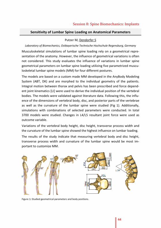
44
Session 8: Spine Biomechanics: Implants
Sensitivity of Lumbar Spine Loading on Anatomical Parameters
Putzer M, Dendorfer S
Laboratory of Biomechanics, Ostbayerische Technische Hochschule Regensburg, Germany
Musculoskeletal simulations of lumbar spine loading rely on a geometrical repre-
sentation of the anatomy. However, the influence of geometrical variations is often
not considered. This study evaluates the influence of variations in lumbar spine
geometrical parameters on lumbar spine loading utilizing five parametrized muscu-
loskeletal lumbar spine models (MM) for four different postures.
The models are based on a custom made MM developed in the AnyBody Modeling
System (ABT, DK) and are morphed to the individual geometry of the patients.
Integral motion between thorax and pelvis has been prescribed and force depend-
ent joint kinematics [LI] were used to derive the individual position of the vertebral
bodies. The models were validated against literature data. Following this, the influ-
ence of the dimensions of vertebral body, disc, and posterior parts of the vertebrae
as well as the curvature of the lumbar spine were studied (Fig 1). Additionally,
simulations with combinations of selected parameters were conducted. In total
3700 models were studied. Changes in L4/L5 resultant joint force were used as
outcome variable.
Variations of the vertebral body height, disc height, transverse process width and
the curvature of the lumbar spine showed the highest influence on lumbar loading.
The results of the study indicate that measuring vertebral body and disc height,
transverse process width and curvature of the lumbar spine would be most im-
portant to customize MM.
Figure 1: Studied geometrical parameters and body positions.

Session 8: Spine Biomechanics: Implants
45
Biomechanical Challenges for Spinal Implant Development
Schilling C, Beger J, Grupp TM
Aesculap AG, Research & Development, Tuttlingen, Germany
This short communication is intended to highlight the biomechanical challenges for
implant development from an industrial perspective.
Hardware related failure of spinal surgical procedures is a common reported
clinical phenomenon. The state of the art implants found in clinical practice are
more or less developed based on empirical findings. Hence clinical problems such
as screw / rod fracture, screw loosening, loss of correction or adjacent segment
disease could be related to an insufficient, non-comprehensive understanding of
the spinal environment regarding acting loads, deformation in combination with
resulting posture under daily life activities.
Besides the number of clinical studies with a high level of evidence for several types
of spinal instrumentation are also very rare in terms of having a differentiated
picture on the correlation of pathological situation, used instrumentation and
patient outcome.
For surgical procedures with novel implant types, e.g. motion preservation
implants, dynamic pedicle screw systems, pedicle screw anchorage in osteoporotic
bone or regenerative treatments it is extremely demanding to predict the clinical
performance with the current biomechanical knowledge. Simulations, in vitro test
methods, or animal models for in vivo investigations are lacking taking into account
the very complex human spinal environment on different scales (from cellular to
whole body level).
Indeed in some areas there is a deep and valuable knowledge available. However
this has to be linked together and sharpened in terms of working in accessible
research clusters on outcome driven, unsolved problems, ideally realizing a closed
loop from research to clinical application.

46
Index of Authors and Chairs
Adams 3, 16
Adeeb 17
Arampatzis 5, 36
Arjmand 4, 5, 26, 31, 41
Bashkuev 2, 9, 12, 30
Bassani 40
Baur 35
Bayer 39
Bazrgari 4, 5, 22, 32
Berger-Roscher 18
Bergmann 21
Brayda-Bruno 40
Chang 23
Costa 40
Croft 22
Czabanka 34
Dendorfer 7, 42, 44
Dennerlein 23
Dolan 16
Dreinhöfer 36
Dreischarf 4, 9, 12, 21, 26, 30
El Ouaaid 28
El-Rich 3, 7, 17
Emanuel 10
Engel 35, 37
Eskandari 41
Faber 4, 5, 20, 23
Faust 34
Ferguson 42
Franke 34
Galbusera 2, 7, 40
Graichen 21
Grupp 45
Günther 39
Haeufle 39
Hajibozorgi 31
Huber 3, 19
Ignasiak 7, 42
Jaremko 17
Kienle 18
Kingma 4, 7, 12, 20, 23
Klein 19
Larivière 38
Laube 36
Lin 11
Maile 18
Mayer 35, 37
Meakin 3, 4, 27
Moreno-Catalá 36
Morlock 19
Mueller J 5, 35, 37
Mueller S 5, 35, 37
Naserkhaki 17
Neidlinger-Wilke 3, 13
Nussbaum 22, 32
Ottardi 40
Oxland 4, 5, 25
Panjabi 25
Parnianpour 7, 41
Plamondon 28
Pries 2, 5, 30
Pumberger 2, 5, 29
Püschel 19
Putzer 44
Putzier 2, 29
Rasche 18
Reschke 35
Rissler 39
Rohlmann 4, 7, 21, 26
Rupp 39
Schilling 7, 9, 12, 45
Schmidt 2, 5, 7, 9, 12, 21, 26, 30
Schmitt 7, 39
Schroll 36
Schüler 36
Sedaghatnejad 41
Shahvarpour 5, 38
Shirazi-Adl 2, 4, 7, 9, 26, 28, 31, 38
Shojaei 22, 32
Skrzypiec 19
Smit 10
Smith 27
Stoll 37
Tromp 32
Urban 13
Vajkoczy 5, 34
van der Veen 10, 12
van Dieën 10, 20, 23
Vazirian 22, 32
Vergroesen 2, 10, 12
Wang 2, 11
Wilke 3, 13, 18, 40
Winlove 27
Woitzik 34
Zander 7
Zanjani-Pour 27

47
Travel in Berlin
The Julius Wolff Institute is located at the Charité Campus Virchow-Klinikum.
Arriving by plane
Airport Berlin-Tegel:
Take the bus TXL and get off at “Turmstraße”. Change to the subway station U9
(direction “Osloer Straße”) and leave the train at “Amrumer Straße”.
Airport Berlin-Schönefeld:
Take the S-Bahn-Line S9 from Berlin-Schönefeld (direction “Pankow”) and get off at
“Ostkreuz”. Change at “Ostkreuz” to the S-Bahn S42 (Ringbahn) and leave the train
at “Westhafen”. Walk across the Putlitzbrücke to the Föhrer Straße.
Arriving by train
Take the train to one of the DB stations
- preferably “Zoologischer Garten”.
Change at “Zoologischer Garten” to the
subway U9 (direction “Osloer Straße”)
and get off at “Amrumer Straße”.
Alternatively, you can take from central
station (“Hauptbahnhof”) the bus 142
(direction “Leopoldplatz”) and get off
at “Amrumer Straße”.
Arriving by car
From the freeway A 100 take the exit
Seestraße. Ample parking is available in
the public parking garage at Seestraße
4. The garage is always open and costs
1 € for every full/partial hour or maxi-
mum 10 € per calendar day. The first
29 minutes are free. Guests who stay
at the Virchow-Gästehaus have free
parking included here.
On the campus the first 59 minutes are
free and every hour afterwards costs
2 €. Disabled parking is available on the
campus on Mittelallee.

48
General Information
Registration
Registration for the workshop is required. Please contact Friedmar Graichen at:
Registration fee for participants without oral presentation is required
Participation in the workshop includes coffee breaks, lunch breaks and happy hour.
Social event includes transport to the restaurant.
Payment and confirmation of payment
An invoice or confirmation of registration will be sent via electronic mail.
Workshop language
The workshop language is English.
WIFI access
Will be provided.
General Guidelines for Authors and Presenters
Submitting your presentation / technical information
Please prepare your presentation in PowerPoint 4:3 aspect ratio. A presentation
notebook with a PDF reader and an MS Office PowerPoint 2010 will be provided.
The use of personal notebooks will not be accepted, it may interrupt the flow of
the program in the lecture hall. A laser pointer will be available at the speaker’s
podium in the lecture hall. A technical supervisor will help you.
Speaker’s preparation
Please hand in your presentation on USB flash drive to our technical staff available
in the room where the talk is scheduled, no later than 90 minutes before the be-
ginning of the session. You may view and/or edit your presentation before.
Hotels
Virchow Gästehaus der Charité
Seestraße 4-5, D-13353 Berlin, Germany
Phone: +49 30 450 578 062
http://gaestehaus.charite.de
Smart Hotel Berlin
Genter Straße 53A, 13353 Berlin, Germany
Phone: +49 30 45486454
http://www.smarthostel-berlin.de

49
Notes

50
Notes

51
Notes

52
Notes

53
Notes

54
Notes

Supported by
We would like to thank everybody who helped us to make this International Workshop
on Spine Loading and Deformation: From Loading to Recovery happen.
Our special thanks to all our sponsors that supported us
SCHM 2572/3-1
