progenitor cell self-renewal and cyclic neutropenia
TRANSCRIPT
1
Progenitor Cell Self-renewal and Cyclic Neutropenia
David Dingli1, Tibor Antal2, Arne Traulsen3, Jorge M. Pacheco4
1 Division of Hematology, College of Medicine, Mayo Clinic, Rochester, MN 55905, USA
2 Program for Evolutionary Dynamics, Harvard University, Cambridge, MA 02138, USA
3 Max Planck Institute for Evolutionary Biology, 24306 Plön, Germany
4 ATP-Group, CFTC & Departamento de Física da Faculdade de Ciências, P-1649-003
Lisboa Codex, Portugal
ABSTRACT
Cyclic neutropenia (CN) is a rare genetic disorder where patients experience regular
cycling of neutrophils and various other hematopoietic lineages. The nadir in the
neutrophil count is the main source of problems due to the risk of life-threatening
infections. Patients with CN benefit from G-CSF therapy although cycling persists.
Mutations in the neutrophil elastase gene (ELA2) have been found in more than half of
the patients with CN. However, neither the connection between phenotypic expression of
ELA2 and CN nor the mechanism of cycling are known. Recently a multi-compartment
model of hematopoiesis that couples stem cell replication with marrow output was
proposed. In the following, we couple this model of hematopoiesis with a linear feedback
mechanism via G-CSF. We propose that the phenotypic effect of ELA2 mutations leads to
a reduction in self-renewal of granulocytic progenitors. The body responds by an overall
relative increase of G-CSF and increasing progenitor cell self-renewal leading to cell
count cycling. The model is compatible with the available experimental data and makes
testable predictions.
2
INTRODUCTION
Cyclic neutropenia (CN) is a rare hematological disorder characterized by
periodic severe neutropenia interspersed by normal or near normal neutrophil counts that
repeat every 19 to 21 days (Dale et al., 1988, Haurie et al., 1998). Although the
neutrophil nadir is the cause of the symptoms in this disorder, almost all cellular elements
in the blood may cycle, being usually out of phase with the neutrophils (Haurie et al.,
1998). Mutations in the gene coding for neutrophil elastase 2 (ELA2) have been identified
in almost half of the patients with this disorder but the exact mechanisms of how these
mutations cause the disease remain unclear (Horwitz et al., 1999, Benson et al., 2003).
Therefore it is not surprising that some scepticism about the role of the ELA2 mutation in
the etiology of CN persists in the field (Germeshausen et al., 2001). The majority of
mutations in the ELA2 gene are base substitutions leading to loss of the splice donor site
in exon four. This forces the use of an upstream cryptic splice donor site that results in
loss of 30 nucleotides during RNA processing (Horwitz et al., 1999). Patients with CN
benefit from pharmacologic doses of granulocyte colony stimulating factor (G-CSF) with
rapid recovery of the neutrophil count although cycling of hematopoiesis persists
(Hammond et al., 1989, Dale et al., 1993).
Several observations are pertinent to set the stage for modelling the dynamics of
this disorder. ELA2 expression is restricted to cells of the granulocyte and monocyte
lineage with the highest levels of expression being in promyelocytes and myelocytes.
Promonocytes express the gene although at levels significantly lower than granulocyte
precursors (Fouret et al., 1989, Campbell et al., 1989). In addition, myeloblasts also
express low levels of the enzyme (Fouret et al., 1989). Many mutations in ELA2
identified in patients with CN often result in the aberrant localization of the enzyme in
sub-cellular compartments (Benson et al., 2003). G-CSF is the main cytokine that
controls the proliferation, survival and differentiation of myeloid precursors (Williams et
al., 1990) and accelerates neutrophil maturation (Price et al., 1996). Granulocyte
progenitors, isolated from patients with CN are less sensitive to G-CSF compared to
normal controls (Hammond et al., 1992). Neutrophils from grey collies with CN express
normal numbers of G-CSF receptors that appear to signal normally (Avalos et al., 1994),
although similar studies on myeloid progenitor and committed cells in which neutrophil
3
elastase (NE) expression is maximal (Fouret et al., 1989) have not been performed. In
vitro, NE antagonizes the action of G-CSF by enzymatic cleavage of the cytokine (El
Ouriaghli et al., 2003) but there is no evidence of release of NE in the bone marrow in
patients with CN. In vivo studies show that the levels of endogenous colony stimulating
factors are highest with the nadir of the neutrophil count (Guerry et al., 1974) and reach a
peak concomitant with the monocyte count. Endogenous G-CSF and other cytokines are
produced by many cells including endothelial cells, fibroblasts and monocytes (Moore et
al., 1974, Barreda et al., 2004). In the grey collie variant of the disease, phlebotomy has
no impact on the cycling of reticulocytes or neutrophils. However, erythrocyte hyper-
transfusion stops reticulocyte but not neutrophil cycling (Adamson et al., 1974).
Neutrophils and their precursors express G-CSF receptors and play an important role in
the clearance of the cytokine from the circulation (Ericson et al., 1997). Finally, apart
from being the most important cytokine controlling granulocyte production, G-CSF is a
growth and survival factor for hematopoietic stem cells (HSC) and granulocyte,
erythroid, monocyte and megakaryocyte colony forming units (CFU-GEMM) (Barreda et
al., 2004) and may increase peripheral blood lymphocyte counts (Hareng et al., 2002).
Despite such a wealth of knowledge about this disorder, an all encompassing
framework to understand its etiology has not been provided. It has been postulated that
the defect must reside in the stem cell compartment (Haurie et al., 1998, Colijn et al.,
2005). However, this is unlikely, since the 19-21 day cycling is not compatible with the
estimated rate of replication of the active stem cell pool (Rufer et al., 1999, Shepherd et
al., 2004). Moreover, studies suggest that the effect of the mutated enzyme is restricted to
cells that express the gene (i.e. myeloblasts and further cells downstream) (Ancliff et al.,
2003). Other investigators have proposed models to explain CN on the basis of a multi-
compartment model of hematopoiesis (Schmitz et al., 1990, Schmitz et al., 1995, Schmitz
et al., 1996). Hematopoiesis is divided into compartments that harbor stem cells,
progenitor cells, more differentiated cells and mature cells that are linked together by
cytokine feedback loops. The model specifically considers feedback loops with G-CSF,
GM-CSF and erythropoietin (EPO) influencing the dynamics of all compartments
downstream of the HSC pool (Wichmann et al., 1983, Schmitz et al., 1990). Schmitz et
al proposed that the main defect in CN is narrowing of the variance in transit times of
4
neutrophil precursors. The authors did not provide a biological basis for such a reduction
in the variance. However, this model could be used to study the effects of therapy with
these cytokines (Schmitz et al., 1995, Schmitz et al., 1996).
Recently, a hierarchical model of hematopoiesis based on steady state fluxes
between successive compartments has been proposed. The process is maintained by the
replication of hematopoietic stem cells that can feed downstream compartments which
metaphorically represent cell lineages at different stages of differentiation (Dingli et al.,
2007). The model postulates that cells in a given compartment j divide at a rate jr that is
characteristic of that compartment. During each replication, cells may differentiate (with
probability ε) such that the two daughter cells shift to compartment 1+j ; with
probability !"1 the daughter cells retain the properties of their parent cell and remain in
compartment j (Figure 1). Under stationary conditions, the size of each compartment is
constant and cells lost from one compartment due to differentiation are replaced by cells
from the upstream compartment 1!j (Dingli et al., 2007). Using this model, it is
possible to estimate the size of each hematopoietic compartment, the rate of replication of
cells and the average time that a given cell type contributes to hematopoiesis (Dingli et
al., 2007). We have shown that in this model, the expected number of cell divisions or
‘compartments’ that link the HSC with circulating blood ( 32=K , Figure 2) is similar to
what has been predicted in other models (Vaziri et al., 1994, Mackey, 2001, Shochat et
al., 2002) as well as experimentally inferred from serial telomere shortening (Vaziri et
al., 1994). It is important to point out that the notion of ‘compartment’ in this model
should not be considered as a quantized image of hematopoiesis where the various
‘compartments’ represent abrupt transitions but rather a continuum between the HSC and
the circulating blood with the ‘compartments’ simply being a tool to keep track of the
number of cell divisions. Cells may divide and move from one compartment to the next
(‘differentiate’), yet morphologically and perhaps functionally may appear to be the
same. For example, the CFU-GEMM cells can be accommodated in compartments 5 to 8.
This is based on experimental evidence of PIG-A mutations in the neutrophils, monocytes
and erythrocytes of healthy adults (Araten et al., 1999). Clearly, these individuals must
have at least 1 mutated CFU-GEMM cell and given the ratio of mutated to normal
neutrophils, one can estimate the minimum and maximum number of CFU-GEMM cells
5
at risk of mutation. The CFU-GM cells span compartments 9 to 11 while CFU-G span
compartments 12 to 16 based on current estimates of the size of the population of cells
(Donohue et al., 1958, Cronkite et al., 1964, Finch et al., 1977, Shochat et al., 2002).
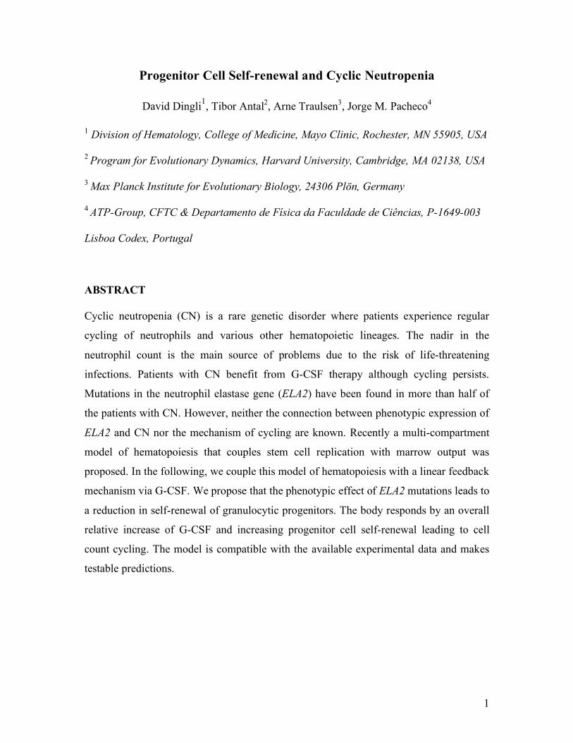
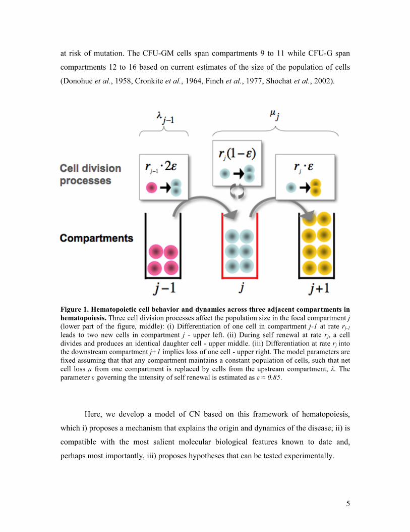
Figure 1. Hematopoietic cell behavior and dynamics across three adjacent compartments in hematopoiesis. Three cell division processes affect the population size in the focal compartment j (lower part of the figure, middle): (i) Differentiation of one cell in compartment j-1 at rate rj-1 leads to two new cells in compartment j - upper left. (ii) During self renewal at rate rj, a cell divides and produces an identical daughter cell - upper middle. (iii) Differentiation at rate rj into the downstream compartment j+1 implies loss of one cell - upper right. The model parameters are fixed assuming that that any compartment maintains a constant population of cells, such that net cell loss µ from one compartment is replaced by cells from the upstream compartment, λ. The parameter ε governing the intensity of self renewal is estimated as ε ≈ 0.85.
Here, we develop a model of CN based on this framework of hematopoiesis,
which i) proposes a mechanism that explains the origin and dynamics of the disease; ii) is
compatible with the most salient molecular biological features known to date and,
perhaps most importantly, iii) proposes hypotheses that can be tested experimentally.
6
THE MODEL
Let us focus on the dynamics of a specific compartment j and investigate the time
dependence of the number of cells in that compartment, jN . In keeping with the model of
hematopoiesis referred to before (Dingli et al., 2007), we consider two elementary
processes: differentiation, ••!o , which occurs with probability ! and self
renewal, ooo! , that occurs with probability !"1 . The symbol odenotes a cell that stays
in compartment j while the symbol • describes a cell that moves to compartment 1+j
(Figure 1). Therefore the behaviour of each cell during any division is stochastic in nature
(Gordon et al., 1994, Abkowitz et al., 1996) although the net effect is that cells tend to
differentiate rather than self-renew ( 5.01 >> ! ), compatible with the view of
hematopoiesis as a linear process with net cell flow from HSC to the circulating
compartment (Donohue et al., 1958, Cronkite et al., 1964, Finch et al., 1977). Within
compartment j , the rate of cell differentiation and transfer into compartment 1+j
is )12( != "µjjr (Dingli et al., 2007). This loss of cells is compensated by the injection
of cells from the upstream compartment, taking place at a rate !" 211 ## =
jjr (Figure 1).
Consequently, we may readily write down the equation for the time dependence of the
number of cells in compartment j:
11 !!+!= jjjj
jNN
dt
dN"µ (1)
The stationary solution for Eq. (1) is given by
0
2
1NCNj
j!
=" where 0
)12(
2>
!=
"
"
rC and
j
j
r
rr
1+= (2)
Equations (1) and (2) hold for all compartments with 311 =!! Kj . In the multi-
compartment model, 400~0N cells and represents the active hematopoietic stem cell
pool that remains constant during adult life (Buescher et al., 1985, Dingli et al., 2006,
Dingli et al., 2007). We have previously shown that the values of ε and r are robust with
respect to changes in the number of active stem cells (0N ) across 4 orders of magnitude
and can be considered as characteristics of normal hematopoiesis (Dingli et al., 2007).
7
In order to model cyclic neutropenia, we investigate how small deviations in the number
of cells in a given compartment ( jN ) alter the flux of cells into and out of that
compartment, restricting the analysis to two adjacent compartments. We introduce a
linear feedback function such that depletion of cells from compartment j leads to
additional influx of cells from compartment j-1. In this way, we obtain a relationship
between the compartment j, the probability of self-renewal (! ) and the replication rate of
the cells (r) that is defined by
0)12)(12(4 422
1 >+!="!
rrrj ## . (3)
A detailed explanation of the model is provided in the Appendix. Under defined
circumstances, it can be shown that oscillations in the number of cells within a given
compartment will occur and persist without damping at a frequency given by
!
"4
j#= (4)
In other words, Eq. (4) captures the relationship between the frequency of the oscillations
and the compartment, j where they start due to a mutation or other insult.
RESULTS
Where is the mutated gene initially expressed?
In the multi-compartment model of hematopoiesis, stem cells replicate at a rate
!
r1
=1 365day-1 (Rufer et al., 1999, Shepherd et al., 2004) and it was assumed that
!
r and
!
" are the same for all compartments (Dingli et al., 2007). The free parameters of the
model are fixed using data from the expansion during polymorphonuclear leukocyte
production, the number of active stem cells and the daily output of the blood system, and
the cell division rates of stem cells and granulocyte precursors. This leads to
!
r =1.26 (cf.
Eq. 2) and 85.0=! (Donohue et al., 1958, Cronkite et al., 1964, Finch et al., 1977).
Given that the period of the cycles in most patients with CN is between 19 and 21 days,
using Eqs. 3 and 4, we can estimate the earliest compartment j where the defect has to
be active to give the required frequency of oscillations. Our model suggests that
21!j (for both values of the period) which localizes the defect downstream of the CFU-
G compartment (cf. Figure 2) (Dingli et al., 2007). For the rare patient with longer cycles
8
(<52 days) (Haurie et al., 1999), the model predicts that 17>j which again places the
defect downstream of the CFU-G compartment. In other words, for a period of 19 to 21
days, compartment 20 will be the earliest compartment with a normal! ( 85.0=! ) and we
predict that 85.0>!
CN
j" in cells of the granulocyte lineage.
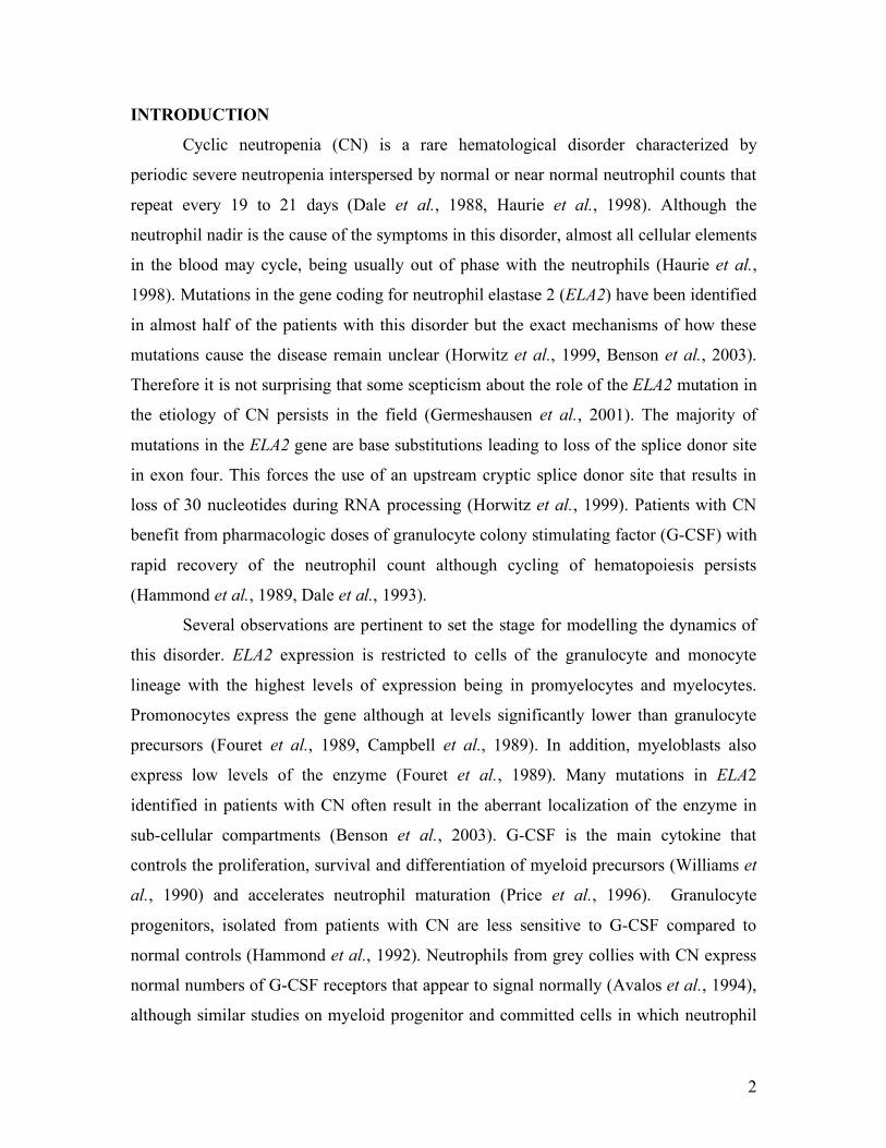
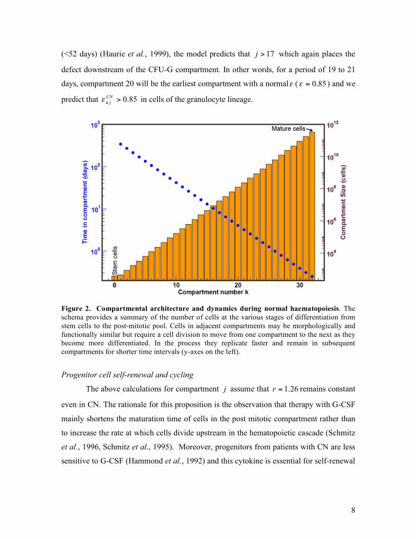
Figure 2. Compartmental architecture and dynamics during normal haematopoiesis. The schema provides a summary of the number of cells at the various stages of differentiation from stem cells to the post-mitotic pool. Cells in adjacent compartments may be morphologically and functionally similar but require a cell division to move from one compartment to the next as they become more differentiated. In the process they replicate faster and remain in subsequent compartments for shorter time intervals (y-axes on the left).
Progenitor cell self-renewal and cycling
The above calculations for compartment j assume that 26.1=r remains constant
even in CN. The rationale for this proposition is the observation that therapy with G-CSF
mainly shortens the maturation time of cells in the post mitotic compartment rather than
to increase the rate at which cells divide upstream in the hematopoietic cascade (Schmitz
et al., 1996, Schmitz et al., 1995). Moreover, progenitors from patients with CN are less
sensitive to G-CSF (Hammond et al., 1992) and this cytokine is essential for self-renewal
9
of the myeloid progenitors and their progeny cells (Barreda et al., 2004, Marley et al.,
2003).
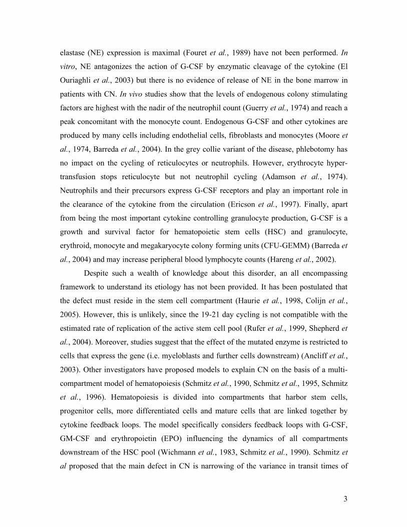
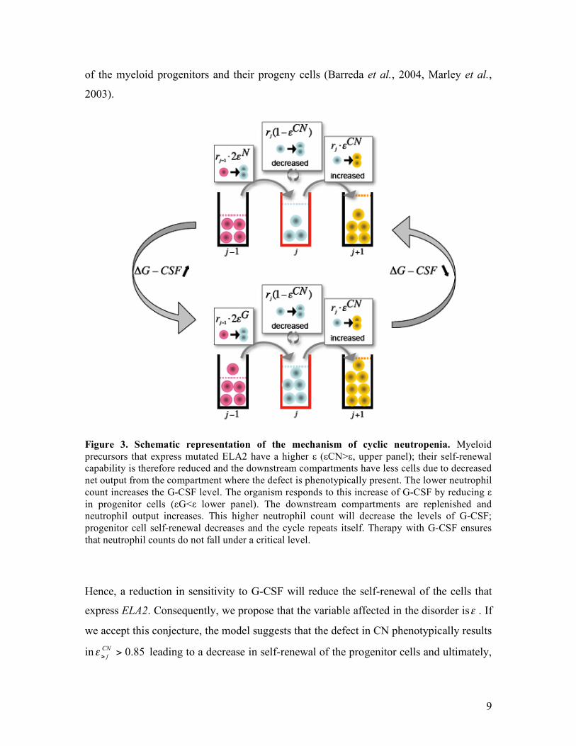
Figure 3. Schematic representation of the mechanism of cyclic neutropenia. Myeloid precursors that express mutated ELA2 have a higher ε (εCN>ε, upper panel); their self-renewal capability is therefore reduced and the downstream compartments have less cells due to decreased net output from the compartment where the defect is phenotypically present. The lower neutrophil count increases the G-CSF level. The organism responds to this increase of G-CSF by reducing ε in progenitor cells (εG<ε lower panel). The downstream compartments are replenished and neutrophil output increases. This higher neutrophil count will decrease the levels of G-CSF; progenitor cell self-renewal decreases and the cycle repeats itself. Therapy with G-CSF ensures that neutrophil counts do not fall under a critical level.
Hence, a reduction in sensitivity to G-CSF will reduce the self-renewal of the cells that
express ELA2. Consequently, we propose that the variable affected in the disorder is! . If
we accept this conjecture, the model suggests that the defect in CN phenotypically results
in 85.0>!
CN
j" leading to a decrease in self-renewal of the progenitor cells and ultimately,

10
to a fall in neutrophil production (Figure 3). Consequently, we expect a decrease in cell
output due to rapid cell transit of neutrophil precursors during hematopoiesis, a prediction
which can be tested. The elevated ! means that neutrophils from patients with CN will
have longer telomeres compared to normal controls since their progenitors undergo less
divisions compared to normal. Therapy with G-CSF reduces ! and CN! and the enhanced
progenitor cell renewal results in an increase of neutrophil production, although with a
delay since upstream compartments must themselves expand before they can replenish
downstream compartments (Figure 3). If this scenario is correct, G-CSF therapy should
lead to shortening of neutrophil telomeres. Although G-CSF shortens the period of the
oscillations, our model is not in conflict with this fact since the major effect of G-CSF is
to shorten the transit time of cells in the post-mitotic pool (Schmitz et al., 1995). In
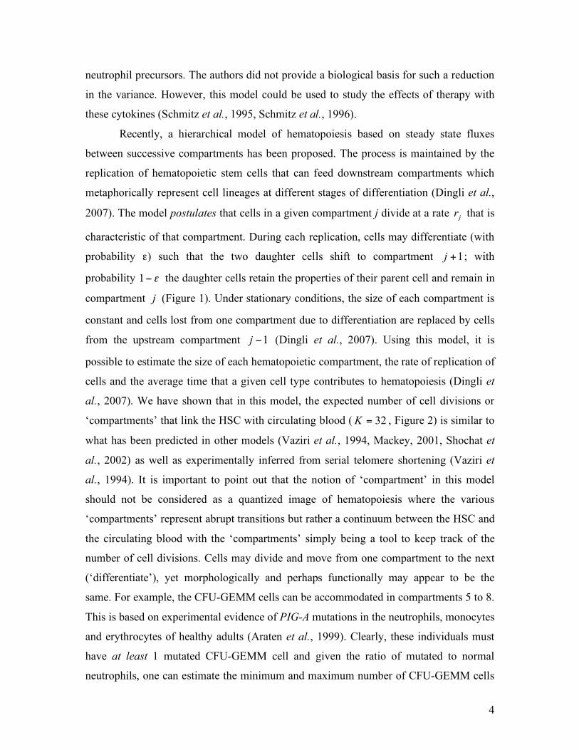
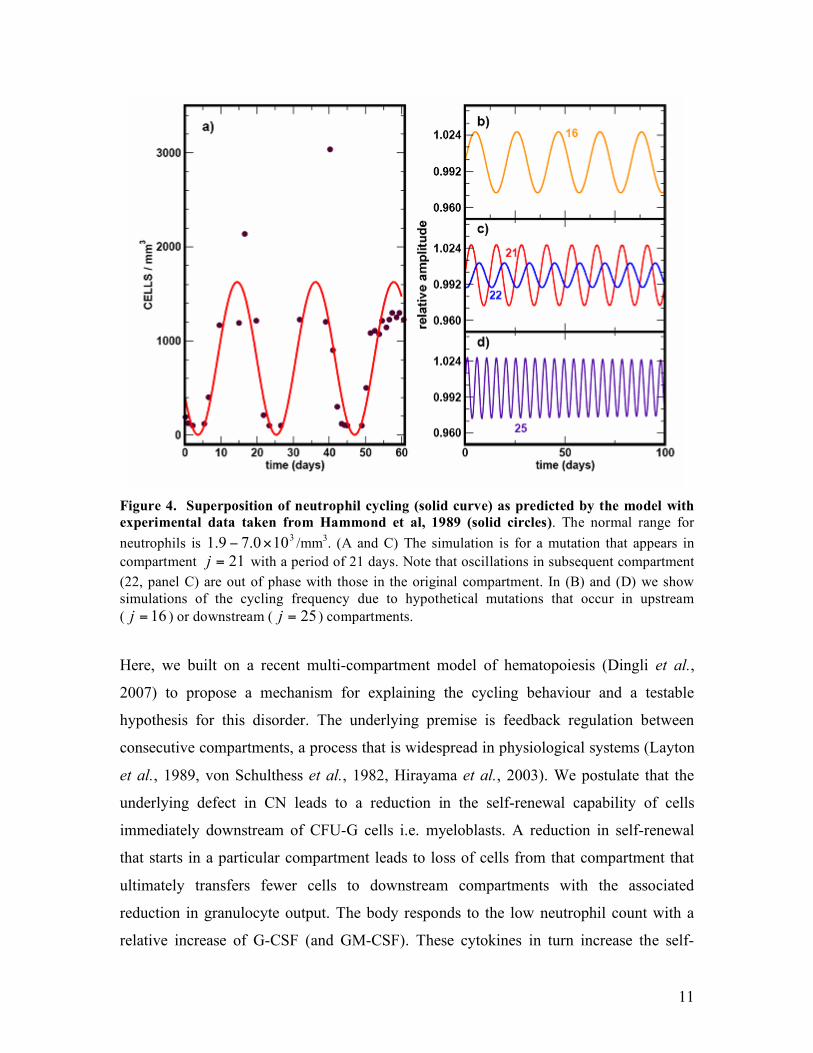
Figure 4, we superimpose serial neutrophil counts observed in a patient with CN with the
cycling predicted by the model. The model captures the overall behaviour without
delving into the detailed self-regulatory mechanisms of hematopoiesis. Finally note that
Eq. (2) can be re-written as 1
2
11
1!
"#
$%&
'!=
(rC ; if 85.0>
CN
j! , then the equilibrium value,
*
jN is reduced and compatible with the observation that the neutrophil count in patients
with CN typically oscillates around a value lower than the average in normal individuals.
We performed numerical simulations to couple additional compartments
downstream of compartment j where the phenotypic effect is first observed in the
neutrophil lineage. All the downstream compartments also oscillate at the same frequency
but with a delay such that a ‘travelling wave’ appears down the compartments. In other
words, the compartments oscillate out of phase. However, the overall frequency of the
oscillations is determined by the location of the compartment where the mutated gene is
initially expressed. If the mutated gene is expressed in compartment 21<j or 21>j the
frequency of the oscillations will decrease and increase respectively (Figure 4B to D).
DISCUSSION
Despite significant progress in elucidating the molecular biology of CN, the etiology of
cycling in this disease has not been adequately explained (Horwitz et al., 2007).
11
Figure 4. Superposition of neutrophil cycling (solid curve) as predicted by the model with experimental data taken from Hammond et al, 1989 (solid circles). The normal range for neutrophils is 3
100.79.1 !" /mm3. (A and C) The simulation is for a mutation that appears in compartment 21=j with a period of 21 days. Note that oscillations in subsequent compartment (22, panel C) are out of phase with those in the original compartment. In (B) and (D) we show simulations of the cycling frequency due to hypothetical mutations that occur in upstream ( 16=j ) or downstream ( 25=j ) compartments.
Here, we built on a recent multi-compartment model of hematopoiesis (Dingli et al.,
2007) to propose a mechanism for explaining the cycling behaviour and a testable
hypothesis for this disorder. The underlying premise is feedback regulation between
consecutive compartments, a process that is widespread in physiological systems (Layton
et al., 1989, von Schulthess et al., 1982, Hirayama et al., 2003). We postulate that the
underlying defect in CN leads to a reduction in the self-renewal capability of cells
immediately downstream of CFU-G cells i.e. myeloblasts. A reduction in self-renewal
that starts in a particular compartment leads to loss of cells from that compartment that
ultimately transfers fewer cells to downstream compartments with the associated
reduction in granulocyte output. The body responds to the low neutrophil count with a
relative increase of G-CSF (and GM-CSF). These cytokines in turn increase the self-
12
renewal capacity of the CFU-GM, CFU-G (Barreda et al., 2004, Marley et al., 2003) and
downstream compartments leading to an overall reduction in ! with an associated
increase in neutrophil output. However, neutrophils express G-CSF receptors that act as
sinks for the cytokine (Ericson et al., 1997). Hence, as the neutrophil count recovers,
cytokine concentrations decrease and the process repeats itself, given the incapacity of !
in the original compartment to return to normal values.
It is clear that although hematopoiesis is scattered in multiple sites of red marrow,
there must be significant synchronization of cell replication for cycling to be observed.
Synchronization of function in the body occurs if cells are coupled together either
structurally or functionally via ion channels or chemically. The separation of
hematopoiesis in space excludes both structural and electrical cellular coupling leaving
chemical coupling as the only alternative. Both G-CSF and GM-CSF could provide such
coupling from their known biology. The reader may wonder why oscillations are not seen
in healthy adults if these inter-compartment feedback loops are present. The explanation
for this is that in normal hematopoiesis, there is no compartment with an abrupt loss of
cells as occurs due to mutations in ELA2. It is this loss of cells that amplifies the feedback
signal (G-CSF) leading to the oscillations. Under normal physiological conditions, the
changes in G-CSF are small enough that the system is essentially always at a steady state.
Interestingly, some patients with myelodysplastic syndrome (Abe et al., 2000) or
myeloproliferative disorders develop cycling hematopoiesis as an acquired phenomenon,
the latter typically after starting therapy with cytoreductive agents such as hydroxyurea
(Steensma et al., 2001). These observations can be rationalized by our model since
patients with myelodysplasia can have defective differentiation of cells (altered ε) with
depletion of specific differentiation compartments in hematopoiesis. Hydroxyurea can
reduce neutrophils and their precursors, effectively increasing the half-life of G-CSF with
amplification of its circulating concentration and the onset of cycling. It would be of
great interest to study the sensitivity of myeloid precursors to G-CSF in such patients.
Our model and the literature
The current model is consistent with the observations that granulocyte committed
progenitor cells have a reduced responsiveness to both G-CSF and GM-CSF (Hammond
et al., 1992). Both cytokines are known to play important roles on growth, survival and
13
self-renewal of progenitor cells (Barreda et al., 2004, Marley et al., 2003). In the canine
variant of the disease (grey collie syndrome), neutrophil progenitors express normal
levels of G-CSF receptors (Avalos et al., 1994) and perhaps the same is true for the
human disorder. However, both G-CSF and GM-CSF share downstream mediators
(Barreda et al., 2004) that could be the target of the abnormal elastase produced in
patients with this disease. Indeed, many mutations in ELA2 found in patients with CN
lead to abnormal cytoplasmic accumulation of the enzyme (Benson et al., 2003, Kollner
et al., 2006) where it may interact with second messengers involved in signalling from
activated type 1 cytokine receptors.
Our model predicts that the disorder cannot arise in progenitor cells earlier than
CFU-GM and contradicts other suggestions that the disorder is due to a defect at the stem
cell level (Haurie et al., 1998, Colijn et al., 2005). The current model fits with the
observation that mutations in ELA2 have a local effect in cells where it is expressed
(Ancliff et al., 2003). In particular, Eq. (7) allows one to identify the compartment in
which the coupling leads to undamped oscillations. Moreover, ELA2 is known to be
expressed in cells at the myeloblasts stage (Fouret et al., 1989) which are the closest cells
to CFU-GM in the hematopoietic cascade. The model is also compatible with the
observation that progenitor cells can also cycle in this disorder (Brandt et al., 1975,
Jacobsen et al., 1979). Cycling at the level of the CFU-GM can also explain monocyte
oscillations that are observed in this disease. Monocytes are also an important source of
G-CSF (Barreda et al., 2004) that can provide the necessary feedback to increase CFU-
GM self-renewal with repopulation of the compartments and increased output of
neutrophils. However, one may question whether our model could explain cycling in
other lineages such as reticulocytes and lymphocytes. Cycling of G-CSF and GM-CSF do
not only influence cells at the CFU-GM stage but can increase self-renewal and
proliferation of CFU-GEMM (Barreda et al., 2004) that can lead to cells of the erythroid
and megakaryocytic lineage with cycling in these cell lineages as well. Finally, G-CSF
can also increase lymphocyte counts (Hareng et al., 2002) and cycling of the cytokine
could lead to cycling of lymphocytes.
In the grey collie syndrome, neutrophils cycle with a shorter period than typical of
the human illness. The rate of HSC replication scales allometrically with the mass of the
14
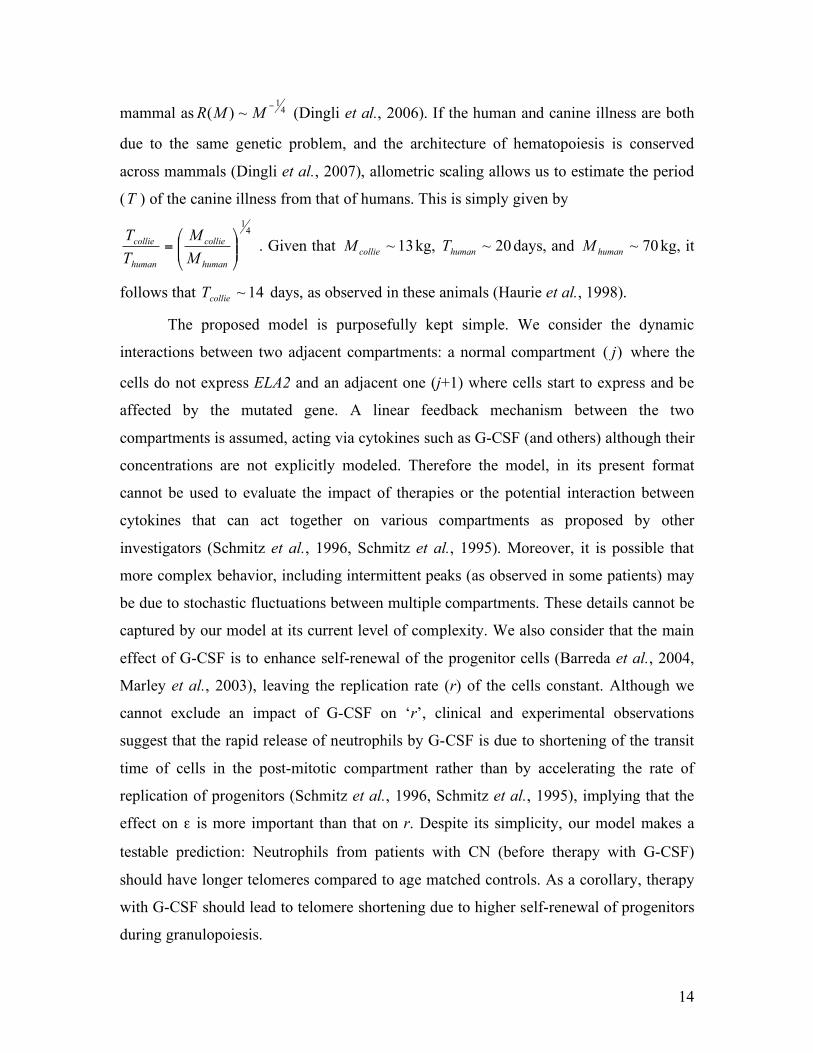
mammal as 41
~)(!
MMR (Dingli et al., 2006). If the human and canine illness are both
due to the same genetic problem, and the architecture of hematopoiesis is conserved
across mammals (Dingli et al., 2007), allometric scaling allows us to estimate the period
(T ) of the canine illness from that of humans. This is simply given by
41
!!"
#$$%
&=
human
collie
human
collie
M
M
T
T . Given that 13~
collieM kg, 20~
humanT days, and 70~
humanM kg, it
follows that 14~collieT days, as observed in these animals (Haurie et al., 1998).
The proposed model is purposefully kept simple. We consider the dynamic
interactions between two adjacent compartments: a normal compartment )( j where the
cells do not express ELA2 and an adjacent one (j+1) where cells start to express and be
affected by the mutated gene. A linear feedback mechanism between the two
compartments is assumed, acting via cytokines such as G-CSF (and others) although their
concentrations are not explicitly modeled. Therefore the model, in its present format
cannot be used to evaluate the impact of therapies or the potential interaction between
cytokines that can act together on various compartments as proposed by other
investigators (Schmitz et al., 1996, Schmitz et al., 1995). Moreover, it is possible that
more complex behavior, including intermittent peaks (as observed in some patients) may
be due to stochastic fluctuations between multiple compartments. These details cannot be
captured by our model at its current level of complexity. We also consider that the main
effect of G-CSF is to enhance self-renewal of the progenitor cells (Barreda et al., 2004,
Marley et al., 2003), leaving the replication rate (r) of the cells constant. Although we
cannot exclude an impact of G-CSF on ‘r’, clinical and experimental observations
suggest that the rapid release of neutrophils by G-CSF is due to shortening of the transit
time of cells in the post-mitotic compartment rather than by accelerating the rate of
replication of progenitors (Schmitz et al., 1996, Schmitz et al., 1995), implying that the
effect on ε is more important than that on r. Despite its simplicity, our model makes a
testable prediction: Neutrophils from patients with CN (before therapy with G-CSF)
should have longer telomeres compared to age matched controls. As a corollary, therapy
with G-CSF should lead to telomere shortening due to higher self-renewal of progenitors
during granulopoiesis.
15
In summary, our linear response model suggests that one potential mechanism to
explain the phenotypic effect of mutations in ELA2 in patients with CN is via a decrease
in self-renewal of the late granulocyte-monocyte progenitors and myeloblasts. The effect
of G-CSF feedback (in response to the low neutrophil counts) is to increase progenitor
cell self-renewal and hence output of neutrophils. Scavenging of G-CSF by the increasing
neutrophil count decreases the progenitor pool leading to a reduction of neutrophil
output. Pharmacologic (therapeutic) doses of G-CSF increase the self-renewal of CFU-
GM and downstream progenitors and increase neutrophil output. Cycling persists due to
the scavenging of the cytokine by the neutrophils, the blunted sensitivity of the
progenitors to this growth factor, and the persistence of the mutated ELA2 and its effect
on ! in the cell compartments where it is expressed.
ACKNOWLEDGEMENTS
This work is supported by Mayo Foundation (DD), “Deutsche Akademie der
Naturforscher Leopoldina” (AT), FCT Portugal (JMP). The Program for Evolutionary
Dynamics is supported by Jeffrey Epstein and NIH grant R01GM078986.
16
REFERENCES
Abe Y, Hirase N, Muta K, Okada Y, Kimura T, Umemura T, Nishimura J, Nawata H (2000). Adult onset cyclic hematopoiesis in a patient with myelodysplastic syndrome. Int J Hematol 71, 40-45.
Abkowitz JL, Catlin SN, Guttorp P (1996). Evidence that hematopoiesis may be a stochastic process in vivo. Nat Med 2, 190-197.
Adamson JW, Dale DC, Elin RJ (1974). Hematopoiesis in the grey collie dog: studies of the regulation of erythropoiesis. J Clin Invest 54, 965-973.
Ancliff PJ, Gale RE, Linch DC (2003). Neutrophil elastase mutations in congenital neutropenia. Hematology 8, 165-171.
Araten DJ, Nafa K, Pakdeesuwan K, Luzzatto L (1999). Clonal populations of hematopoietic cells with paroxysmal nocturnal hemoglobinuria genotype and phenotype are present in normal individuals. Proc Natl Acad Sci U S A 96, 5209-5214.
Avalos BR, Broudy VC, Ceselski SK, Druker BJ, Griffin JD, Hammond WP (1994). Abnormal response to granulocyte colony-stimulating factor (G-CSF) in canine cyclic hematopoiesis is not caused by altered G-CSF receptor expression. Blood 84, 789-794.
Barreda DR, Hanington PC, Belosevic M (2004). Regulation of myeloid development and function by colony stimulating factors. Dev Comp Immunol 28, 509-554.
Benson KF, Li FQ, Person RE, Albani D, Duan Z, Wechsler J, Meade-White K, Williams K, Acland GM, Niemeyer G, Lothrop CD, Horwitz M (2003). Mutations associated with neutropenia in dogs and humans disrupt intracellular transport of neutrophil elastase. Nat Genet 35, 90-96.
Brandt L, Forssman O, Mitelman F, Odeberg H, Olofsson T, Olsson I, Svensson B (1975). Cell production and cell function in human cyclic neutropenia. Scand J Haematol 15, 228-240.
Buescher ES, Alling DW, Gallin JI (1985). Use of an X-linked human neutrophil marker to estimate timing of lyonization and size of the dividing stem cell pool. J Clin Invest 76, 1581-1584.
Campbell EJ, Silverman EK, Campbell MA (1989). Elastase and cathepsin G of human monocytes. Quantification of cellular content, release in response to stimuli, and heterogeneity in elastase-mediated proteolytic activity. J Immunol 143, 2961-2968.
Colijn C, Mackey MC (2005). A mathematical model of hematopoiesis: II. Cyclical neutropenia. J Theor Biol 237, 133-146.
Cronkite EP, Fliedner TM (1964). Granulocytopoiesis. N Engl J Med 270, 1347-1352. Dale DC, Bonilla MA, Davis MW, Nakanishi AM, Hammond WP, Kurtzberg J, Wang W,
Jakubowski A, Winton E, Lalezari P, et al. (1993). A randomized controlled phase III trial of recombinant human granulocyte colony-stimulating factor (filgrastim) for treatment of severe chronic neutropenia. Blood 81, 2496-2502.
Dale DC, Hammond WPt (1988). Cyclic neutropenia: a clinical review. Blood Rev 2, 178-185. Dingli D, Pacheco JM (2006). Allometric scaling of the hematopoietic stem cell pool across
mammals. PLoS ONE, e2. Dingli D, Traulsen A, Pacheco JM (2007). Compartmental architecture and dynamics of
hematopoiesis. PLoS ONE 2, e345. Donohue DM, Reiff RH, Hanson ML, Betson Y, Finch CA (1958). Quantitative measurement of
the erythrocytic and granulocytic cells of the marrow and blood. J Clin Invest 37, 1571-1576.
El Ouriaghli F, Fujiwara H, Melenhorst JJ, Sconocchia G, Hensel N, Barrett AJ (2003). Neutrophil elastase enzymatically antagonizes the in vitro action of G-CSF: implications for the regulation of granulopoiesis. Blood 101, 1752-1758.
17
Ericson SG, Gao H, Gericke GH, Lewis LD (1997). The role of polymorphonuclear neutrophils (PMNs) in clearance of granulocyte colony-stimulating factor (G-CSF) in vivo and in vitro. Exp Hematol 25, 1313-1325.
Finch CA, Harker LA, Cook JD (1977). Kinetics of the formed elements of human blood. Blood 50, 699-707.
Fouret P, du Bois RM, Bernaudin JF, Takahashi H, Ferrans VJ, Crystal RG (1989). Expression of the neutrophil elastase gene during human bone marrow cell differentiation. J Exp Med 169, 833-845.
Germeshausen M, Schulze H, Ballmaier M, Zeidler C, Welte K (2001). Mutations in the gene encoding neutrophil elastase (ELA2) are not sufficient to cause the phenotype of congenital neutropenia. Br J Haematol 115, 222-224.
Gordon MY, Blackett NM (1994). Routes to repopulation--a unification of the stochastic model and separation of stem-cell subpopulations. Leukemia 8, 1068-1072; discussion 1072-1063.
Guerry Dt, Adamson JW, Dale DC, Wolff SM (1974). Human cyclic neutropenia: urinary colony-stimulating factor and erythropoietin levels. Blood 44, 257-262.
Hammond WP, Chatta GS, Andrews RG, Dale DC (1992). Abnormal responsiveness of granulocyte-committed progenitor cells in cyclic neutropenia. Blood 79, 2536-2539.
Hammond WPt, Price TH, Souza LM, Dale DC (1989). Treatment of cyclic neutropenia with granulocyte colony-stimulating factor. N Engl J Med 320, 1306-1311.
Hareng L, Hartung T (2002). Induction and regulation of endogenous granulocyte colony-stimulating factor formation. Biol Chem 383, 1501-1517.
Haurie C, Dale DC, Mackey MC (1998). Cyclical neutropenia and other periodic hematological disorders: a review of mechanisms and mathematical models. Blood 92, 2629-2640.
Haurie C, Dale DC, Mackey MC (1999). Occurrence of periodic oscillations in the differential blood counts of congenital, idiopathic, and cyclical neutropenic patients before and during treatment with G-CSF. Exp Hematol 27, 401-409.
Hirayama Y, Sakamaki S, Tsuji Y, Matsunaga T, Niitsu Y (2003). Cyclic platelet and leukocyte count oscillation in chronic myelocytic leukemia regulated by the negative feedback of transforming growth factor beta. Int J Hematol 77, 71-74.
Horwitz M, Benson KF, Person RE, Aprikyan AG, Dale DC (1999). Mutations in ELA2, encoding neutrophil elastase, define a 21-day biological clock in cyclic haematopoiesis. Nat Genet 23, 433-436.
Horwitz MS, Duan Z, Korkmaz B, Lee H-H, Mealiffe ME, Salipante SJ (2007). Neutrophil elastase in cyclic and severe congenital neutropenia. Blood 109, 1817-1824.
Jacobsen N, Broxmeyer HE (1979). Oscillations of granulocytic and megakaryocytic progenitor cell populations in cyclic neutropenia in man. Scand J Haematol 23, 33-36.
Kollner I, Sodeik B, Schreek S, Heyn H, von Neuhoff N, Germeshausen M, Zeidler C, Kruger M, Schlegelberger B, Welte K, Beger C (2006). Mutations in neutrophil elastase causing congenital neutropenia lead to cytoplasmic protein accumulation and induction of the unfolded protein response. Blood 108, 493-500.
Layton JE, Hockman H, Sheridan WP, Morstyn G (1989). Evidence for a novel in vivo control mechanism of granulopoiesis: mature cell-related control of a regulatory growth factor. Blood 74, 1303-1307.
Mackey MC (2001). Cell kinetic status of haematopoietic stem cells. Cell Prolif 34, 71-83. Marley SB, Lewis JL, Gordon MY (2003). Progenitor cells divide symmetrically to generate new
colony-forming cells and clonal heterogeneity. Br J Haematol 121, 643-648. Moore MA, Spitzer G, Metcalf D, Penington DG (1974). Monocyte production of colony
stimulating factor in familial cyclic neutropenia. Br J Haematol 27, 47-55. Price TH, Chatta GS, Dale DC (1996). Effect of recombinant granulocyte colony-stimulating
factor on neutrophil kinetics in normal young and elderly humans. Blood 88, 335-340.
18
Rufer N, Brummendorf TH, Kolvraa S, Bischoff C, Christensen K, Wadsworth L, Schulzer M, Lansdorp PM (1999). Telomere fluorescence measurements in granulocytes and T lymphocyte subsets point to a high turnover of hematopoietic stem cells and memory T cells in early childhood. J Exp Med 190, 157-167.
Schmitz S, Franke H, Loeffler M, Wichmann HE, Diehl V (1996). Model analysis of the contrasting effects of GM-CSF and G-CSF treatment on peripheral blood neutrophils observed in three patients with childhood-onset cyclic neutropenia. Br J Haematol 95, 616-625.
Schmitz S, Franke H, Wichmann HE, Diehl V (1995). The effect of continuous G-CSF application in human cyclic neutropenia: a model analysis. Br J Haematol 90, 41-47.
Schmitz S, Loeffler M, Jones JB, Lange RD, Wichmann HE (1990). Synchrony of bone marrow proliferation and maturation as the origin of cyclic haemopoiesis. Cell Tissue Kinet 23, 425-442.
Shepherd BE, Guttorp P, Lansdorp PM, Abkowitz JL (2004). Estimating human hematopoietic stem cell kinetics using granulocyte telomere lengths. Exp Hematol 32, 1040-1050.
Shochat E, Stemmer SM, Segel L (2002). Human haematopoiesis in steady state and following intense perturbations. Bull Math Biol 64, 861-886.
Steensma DP, Harrison CN, Tefferi A (2001). Hydroxyurea-associated platelet count oscillations in polycythemia vera: a report of four new cases and a review. Leuk Lymphoma 42, 1243-1253.
Vaziri H, Dragowska W, Allsopp RC, Thomas TE, Harley CB, Lansdorp PM (1994). Evidence for a mitotic clock in human hematopoietic stem cells: loss of telomeric DNA with age. Proc Natl Acad Sci U S A 91, 9857-9860.
von Schulthess GK, Mazer NA (1982). Cyclic neutropenia (CN): a clue to the control of granulopoiesis. Blood 59, 27-37.
Wichmann HE, Loeffler M, Herkenrath P, Gerhardts MD, Wesselborg C, Wulff H (1983). Mathematische modelle in der haematologie Klinische Wochenschrift (Springer) 61, 935-940.
Williams GT, Smith CA, Spooncer E, Dexter TM, Taylor DR (1990). Haemopoietic colony stimulating factors promote cell survival by suppressing apoptosis. Nature 343, 76-79.
19
APPENDIX
Hematopoiesis is a multi-step process whereby cells derived from hematopoietic
stem cells progressively expand and differentiate into all the circulating blood cells.
Conceptually, each cell replication event is considered to be associated with a decision by
the progeny cells to either remain in a given compartment or move to the next where they
become more specialized. However, during normal hematopoiesis the number of cells
within each compartment remains constant. Let us consider the dynamics of a specific
compartment j and investigate the time dependence of the number of cells in that
compartment, jN . In keeping with the multicompartment model of hematopoiesis
previously described (Dingli et al., 2007), we consider two elementary processes:
differentiation, ••!o , which occurs with probability ! and self renewal, ooo! , that
occurs with probability !"1 . The symbol odenotes a cell that stays in compartment j
while the symbol • describes a cell that moves to compartment 1+j (Figure 1). The
behaviour of each cell during any division is stochastic in nature (Gordon et al., 1994,
Abkowitz et al., 1996) although the net effect is such that cells tend to differentiate rather
than self-renew ( 5.01 >> ! ), compatible with the view of hematopoiesis as a linear
process with net cell flow from HSC to the circulating compartment (Donohue et al.,
1958, Cronkite et al., 1964, Finch et al., 1977). Within compartment j , the rate of cell
differentiation and transfer into compartment 1+j is )12( != "µjjr (Dingli et al.,
2007). This loss of cells is compensated by the injection of cells from the upstream
compartment, taking place at a rate !" 211 ## =
jjr (Figure 1). Consequently, we may
readily write down the equation for the time dependence of the number of cells in
compartment j:
11 !!+!= jjjj
jNN
dt
dN"µ (1)
The stationary solution for Eq. (1) is given by
0
2
1NCNj
j!
=" where 0
)12(
2>
!=
"
"
rC and
j
j
r
rr
1+= (2)
20
Equations (1) and (2) hold for all compartments with 311 =!! Kj . In the multi-
compartment model, 400~0N cells and represents the active hematopoietic stem cell
pool that remains constant during adult life (Buescher et al., 1985, Dingli et al., 2006,
Dingli et al., 2007). We have previously shown that the values of ε and r are robust with
respect to changes in the number of active stem cells (0N ) across 4 orders of magnitude
and can be considered as characteristics of normal hematopoiesis (Dingli et al., 2007).
Let us investigate how the time dependence of jN is affected by small deviations from
its stationary value. The dependence of both coefficients of equation (1) on ! makes it
natural to expect that ! may be involved in the response of the hematopoietic system to
external perturbations. Let us assume that, due to a mutation in ELA2 there is a small
deviation of the size of the cell population jN in a specific compartment j with respect to
its stationary value )( *
jN , Eq. (2). To first order we may write for the changes in the rate
coefficients
jjjj
jjjj
naN
naN
!+=
!+=
""
""
11
11
)(~
)(~
##
µµ (3)
where jjj NNn !=* , and 0>a is a parameter that describes the strength of the
response (Figure 3). Such response mechanisms can in principle alter either jr or ! but
the effect is such that whenever jN decreases, there is an increase in the transfer of cells
from compartment 1!j to j . This is based on the well established principle of feedback
that occurs in many physiological processes including hematopoiesis (Layton et al., 1989,
von Schulthess et al., 1982, Hirayama et al., 2003). In the absence of data suggesting a
more complex feedback process, we consider a linear mechanism since it is the simplest.
For two consecutive compartments, we may write
!!"
#$$%
&!!"
#$$%
&
+'
''=!
!"
#$$%
& '
'
''
j
j
jj
j
j
j
n
n
a
a
n
n
dt
d 1
1
11
~
~
µ(
µ (4)
where *
1
~!= jaNa . Depending on the nature of the eigenvalues of the matrix in Eq. (4),
several types of behaviour are possible. Here we are interested in exploring the possibility
21
of sustained (undamped) oscillations. These will be present whenever the eigenvalues
2,1x are purely imaginary. They can be readily computed as
2
~1
2,1
!"±""=
" jja
xµµ
(5)
with ( )211
~~4 aa
jjj+!!=" !! µµ# .
Whenever jja µµ += !1~ , the real parts of the eigenvalues vanish, with ! reading
0)12)(12(4 422
1 >+!="!
rrrj ## . (6)
Hence, oscillations will persist without damping at a frequency given by
!
"4
j#= (7)
In other words, we have established a relationship between the frequency of the
oscillations and the compartment location. For the convenience of the readers, a summary
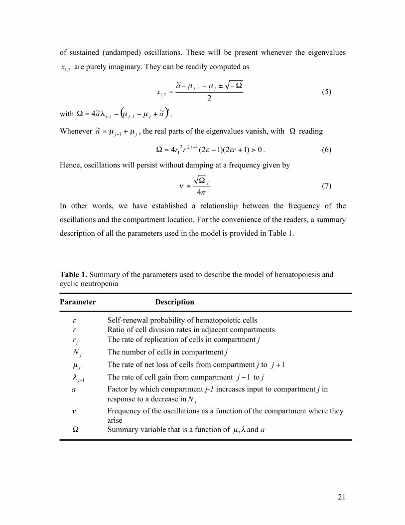
description of all the parameters used in the model is provided in Table 1.
Table 1. Summary of the parameters used to describe the model of hematopoiesis and cyclic neutropenia Parameter Description
! Self-renewal probability of hematopoietic cells
!
r Ratio of cell division rates in adjacent compartments jr The rate of replication of cells in compartment j
jN The number of cells in compartment j
jµ The rate of net loss of cells from compartment j to 1+j
1!j" The rate of cell gain from compartment 1!j to j a Factor by which compartment j-1 increases input to compartment j in response to a decrease in jN ! Frequency of the oscillations as a function of the compartment where they arise ! Summary variable that is a function of !µ, and a