production of recombinant immunoglobulin a in plants for passive
TRANSCRIPT
1
Production of recombinant Immunoglobulin A in plants for passive
immunotherapy
Paloma Juárez Ortega IBMCP
Valencia, January 2014
Advisor Dr. Diego Orzáez Calatayud
Co‐Advisor Prof. Antonio Granell Richart
2
Production of recombinant Immunoglobulin A in plants for passive
immunotherapy
Paloma Juárez Ortega
Advisor Dr. Diego Orzáez Calatayud
Co‐advisor Prof. Antonio Granell Richart
Valencia, January 2014

3
El Dr. Diego Orzáez Calatayud, Científico Titular del Consejo Superior de Investigaciones Científicas y Profesor Asociado de la Universidad Politécnica de Valencia, y el Dr. Antonio Granell Richart, Profesor de investigación del Consejo Superior de Investigaciones Científicas, ambos pertenecientes al instituto de Biología Molecular y Celular de Plantas, CERTIFICAN que la Licenciada en Ciencias Biológicas y Máster en Biotecnología Molecular y Celular de Plantas Paloma Juárez Ortega, ha realizado bajo su dirección en el Instituto de Biología Molecular y Celular de Plantas el trabajo que lleva por título Production of recombinant Immunoglobulin A in plants for passive immunotherapy, y autorizan su presentación para optar al grado de Doctor en Biotecnología. Y para que así conste, expiden y firman el presente certificado en Valencia, a 20 de Enero de 2013 Dr. Diego Orzáez Calatayud Dr. Antonio Granell Richart

4
A mis padres
5
Agradecimientos Hoy, día 18 de Diciembre vuelvo a casa tras de un día de Jornadas de Navidad y papeleos, dejo las
cosas en el sofá y pienso… Cuánto les voy a echar de menos…
Diego, has sido un director de tesis pluriempleado estos años, pues además de jefe has sido un
compañero de labo, un consejero, un mentor y un buen amigo. Me diste la oportunidad de hacer la tesis
contigo cuando todavía tenías bata en vez de despacho y tuve la suerte de poder aprender muchas cosas de
ti, codo con codo en la poyata. Desde esos inicios hasta ahora, jamás has dudado en prestarme tu ayuda
cada vez que la he necesitado (hasta el punto de pasarme correcciones de la tesis por whatsapp desde el
coche!). Te has asegurado de que, de una manera o de otra, todos los trabajos realizados lleguen a buen
puerto, me has llevado a todos los congresos que has podido, me has facilitado el conocer a todos los del
ramo, me has enseñado la importancia de “cerrar” las cosas y sobre todo me has abierto el camino hacia un
mundo que me apasiona y en el cual quiero permanecer sin lugar a dudas. Tengo que agradecer en especial
tu implicación, tu energía y también el que hayas sabido lidiar con mi geniecillo estos años. Gracias por todo.
Puedes estar seguro de que me has enseñado muchísimo y créeme cuando te digo que todas las cosas que
menciono y muchas otras que dejo en el tintero dejan una bonita huella en mí. Has sido un jefe difícil de
igualar.
Toni, un año antes de convertirte en codirector de mi tesis, fuiste el culpable de mi llegada al 2.10
(en aquellos momentos 3.10). Recuerdo aquel primer día de Máster en el que nos preguntaste a todos a qué
nos queríamos dedicar. Yo mencioné que estaba buscando laboratorio para hacer la tesis y al salir, por el
pasillo, me dijiste que me pasara el lunes siguiente por tu despacho. No te imaginas lo largo que fue ese fin
de semana!! Desde entonces siempre has tenido la puerta abierta para mí y un momento para dedicarme
cuando lo he necesitado. Gracias.
Silvia!! Que sería de este labo sin la jefa! Cuánto he aprendido de ti! Y cuantas veces me has reñido
(merecidamente) por dejarme las cosas por medio!! Ha sido un placer trabajar contigo, y viajar contigo más
(con todos vosotros). Me encantará ayudarte a organizar, y por supuesto unirme, a los futuros Labuitjes del
labo. Como he dicho antes, realmente os voy a echar de menos, a los que estáis ahora y a los que estuvisteis
en el pasado. El labo 2.10 ha sido mi segunda casa durante casi 6 años. Marta V., cuántas veces te has
sentado conmigo a echarme una mano con GB... gracias gracias gracias!!. Tienes un Don para las secuencias
(y para muchas otras cosas pequeñaja!). Hemos compartido momentos… digamos que ahora están
catalogados como cómicos. No olvidaré tu cara de no entender nada cuando recién llegada te pasaste toda
la tarde conmigo purificando IgA en la cámara de 4⁰ y cuando ya habíamos acabado, cogí nuestra preciada
6
elución y la tiré por la pila sin pensármelo dos veces. Después de un largo rato mirando a la pila y pensando
inútilmente como recuperarla, terminamos la tarde maquinando que mentirijilla le diríamos a Diego…(Diego,
al final te contamos la verdad). Estefanía, cuida bien de las SiPs ehhh! Seguro que lo harás. Ya lo haces! Si no
fuera por ti esa parte se habría quedado estancada! Mil gracias también por tu ayuda con el Valenciano,
tanto con los intentos fallidos de que aprenda como con las traducciones (como no aprendí me las hicisteis
Jose y tú!!). Jose, gracias a ti también por tu ayuda con el Valenciano y por haber cuidado bien de mi pipeta
de 10 (jijiji). Jose Luis, gracias por tu ayuda con toda la parte de volátiles. He aprendido muchas cosas de ti
estos últimos meses. Flor, tu cuida bien de mis ex pipetas (y ya sabes, escóndelas de las garras de Jose… :P).
Gracias también a Sophie, Cris, Clara, Ale, Pathy y Leandro por todas las veces que me habéis echado una
mano, y por supuesto a todos los estudiantes que han pasado por el labo y me han ayudado tanto. Sara Z,
Sara I, Joaquín, Ana e Irene, una parte de mi tesis os correponde a cada uno de vosotros. Gracias.
Mi querida Asun (mi paraninfa!), llevo toda la tesis diciéndote que te iba a dedicar una página
entera de agradecimientos a ti. Pues bueno, allá va: Primero, gracias por cuidar de mi tan bien estos años. En
gran parte gracias a ti me he sentido en el labo casi tan agusto como en casa. Te has convertido en una gran
amiga (amiga que no se librará de abrirse una cuenta de skype si consigo irme fuera…). Pero no solo has sido
mi amiga, sino también has hecho de hermana mayor. Me has dado consejos valiosísimos y muy variados!
No existen suficientes números para contar las veces que me has ayudado, y no voy a hacer una
recapitulación de todo lo que he aprendido de ti, puesto que si me pongo a pensar, tú has estado
involucrada de una manera o de otra en prácticamente todas las cosas que he aprendido estos años (que
afortunadamente son muchas!). Realmente sin tu ayuda esta tesis no habría sido posible. Aunque no pueda
ponerlo en la tapa, para mi tú has sido codirectora de esta tesis. Y lo digo no solo para que lo sepas tu (que
ya lo sabes), sino para que lo sepan todos (todos los que se traguen mis agradecimientos claro…). Has tenido
una paciencia inagotable conmigo en miles de ocasiones, sobre todo durante estos últimos meses con la
metabolómica, el SimcaP, el Mev… Tu ayuda ha sido infinita, incluso durante los fines de semana. No sé
cómo lo haces pero siempre encuentras respuestas para todo (aunque es cierto que cuentas con la ayuda de
tu google Premium :P). Ya sabes lo que pensamos todos: eres absolutamente indispensable en el labo. Lo
mejor de todo esto es lo bien que lo hemos pasado juntas durante este tiempo. Nos hemos reído como las
que más! He disfrutado de tu compañía tanto en los momentos de trabajo intenso como en los momentos
de ocio. No olvidaré las risas de la noche en que le preparamos junto con Sophie la sorpresa del despacho
nuevo a Diego, con bocadillos, papas y donuts mancillando su inmaculada mesa. Me llevo conmigo una gran
cantidad de buenos recuerdos. Termino este párrafo con la sensación de que te mereces mucho más, y por
ello continuaré agradeciéndote todo lo que has hecho por mí, a diario como siempre.

7
Fuera de las fronteras del 2.10 quiero dar las gracias a todo el conjunto de 4 labos (+ VIP) y en
especial a ciertas personas. Mª Ángeles, que suerte tenemos de tenerte! No solo por todo lo que llevas
entre manos que nos hace la vida más fácil (incluyendo envíos urgentes de tarjetas de crédito camufladas a
puebluchos en medio de las montañas rocosas…), sino por todo lo que te preocupas por nosotros y el cariño
que nos das todos los días. Siempre tienes una palabra amable. No quiero pensar el caos que se va a
organizar cuando te jubiles…
Nora, nos hicimos amigas desde el primer día de Máster. Aunque al principio la comunicación entre
nosotras no era muy fluida por los acentos (podíamos estar medio trayecto en coche para entender dos
frases que nos habíamos dicho), enseguida aprendimos a entendernos. Siempre que te he necesitado has
estado ahí, dándome los mejores consejos. Te voy a echar mucho de menos cuando cada una siga su
camino. Aunque quien sabe si acabaremos en la misma ciudad?! Marta P. y Eugenio, gracias por vuestra
amistad. Ha sido un placer estar rodeada de todos vosotros.
Rafa (invernadero), gracias por cuidar de mis tomateras y tabacos tan bien! Menuda paciencia
tienes! La de veces que me has llamado diciéndome que tenía tomateras casi fosilizando, y siempre con las
mejores palabras y risas. Gracias también por sacar Benthamianas de “la nada” cuando lo he necesitado!
Benito, Teresa y Vicente M., quiero agradeceros todo el trabajo realizado para sacar adelante las
transformaciones de tomate. Javier (Clínico), gracias por permitirme pasar tantos meses en tu laboratorio.
Aprendí muchísimo y además disfruté. Manuel y Noelia (Clínico también) gracias por toda vuestra ayuda y
buenos consejos durante esos meses. Susana (Proteómica), Eugenio y Ana (Secuenciación), Teresa, Erika y
Vicente (Metabolómica), gracias por todo el trabajo realizado y también todo lo que me habéis enseñado.
Mapi, gracias por tu ayuda con la identificación de la masa Mn44 y por tu buen humor que nos alegra a
todos!! María, gracias por mantenerlo todo tan limpio y por esas charraetas de pasillo a diario.
Dejando el IBMCP a parte: Noe, gracias por estar ahí todos los días del año y para todo. Gracias por
estos 12 años de amistad. Ya sabes que para mi eres una hermana (casi gemela!). Tu vida ha dado un giro
inesperado en 2013, a ver que nos depara 2014! Estela y Diego Z., gracias por vuestra amistad, lecciones de
fuerza de voluntad y por haber traído al mundo a Gabriel y Luis!
Papá y mamá, gracias por instruirme en la biología casi a la vez que aprendía a leer (“clasificando”
plantitas con una lupa en Benasque y cerrando placas con parafilm en el IVIA). Por supuesto, gracias también
por vuestro apoyo incondicional, buenos consejos y por hacerme ver siempre el lado positivo de todo. Pepe,
gracias por prestarme tu ayuda cada vez que te la pido y gracias por hacerme la portada de la tesis! Abuelas,
gracias por hacerme sentir importante cuando lo he necesitado. Blas, gracias por ser el ser vivo más
adorable que existe en el planeta. Gracias a todos vosotros por estar ahí.

8
Por último, César, gracias por estar a mi lado. Gracias por tu paciencia que no se acaba nunca.
Gracias por cuidarme todos los días del año, por tener un plato de cena esperándome cada vez que he
llegado tarde de trabajar, por entender que pasara tantas horas en el labo, por hacerme reír cada vez que he
llegado triste. Gracias por todos esos viajes que hemos hecho juntos durante nuestras tesis, los
recordaremos toda la vida. Gracias por hacerme feliz.

9
Summary
Mucosal passive immunization is the transfer of active antibodies from one organism to the mucosal
surfaces of another organism for preventing or treating infectious diseases. Mucosal passive immunization
has a great potential for the prevention and treatment of enteric infections like Rotavirus, which causes
more than 114 million episodes of diarrhoea annually with a death toll of more than 450.000 per year.
However, the high cost of recombinant antibodies with the current manufacturing systems based on
mammalian cells hampers the production of the high antibody quantities required for passive immunization
strategies. Alternative expression platforms such as plants could provide higher scalability and reduced
costs. Moreover, the use of edible plant organs, which are Generally‐Regarded‐As‐Safe (GRAS), could reduce
manufacturing costs even further by easing the requirements for antibody purification. We analyze here the
feasibility of utilizing fruits as inexpensive biofactories of human antibodies that can be orally delivered as
crude extracts or partially purified formulations in mucosal passive immunization strategies.
In the first section of this thesis, the construction of tomato plants producing a model human
Immunoglobulin A (IgA) against rotavirus in their fruits is described. As a result, an elite homozygous line was
obtained whose fruits produced on average 41 g of IgA per gram of fresh weigh, equivalent to 0.69 mg IgA
per gram of dry tomato powder. Minimally processed products derived from IgA‐expressing tomatoes were
shown to strongly inhibit virus infection in an in vitro neutralization assay. Moreover, in order to make IgA‐
expressing tomatoes easily distinguishable from wild‐type tomatoes, they were sexually crossed with a
transgenic tomato line expressing the genes encoding Antirrhinum majus Rosea1 and Delila transcription
factors, which confer purple colour to the fruit. The resulting transgenically‐labelled purple tomatoes
contained not only high levels of recombinant neutralizing human IgA but also increased amounts of
anthocyanins.
In the second section of the thesis the composition of IgA‐expressing tomatoes was analyzed in
search of possible unintended effects that could compromise the GRAS status of the final product. To this
end, transgenic IgA‐tomatoes were compared with wild type tomatoes and also commercial tomato varieties
using proteomic and metabolomic approaches. 2D‐DIGE gels coupled with LC‐MSMS for protein
identification showed that all the uptrend differential proteins detected corresponded only to
immunoglobulin chains or antibody fragments. On the other hand, non‐targeted metabolite data obtained
by UPLC‐MS identified variations between transgenic and non‐transgenic lines, however such variations
could not be associated with the presence of abnormal levels of any particular secondary metabolite in the
IgA fruits. Therefore from the analysis conducted here no sign was obtained that could indicate that tomato‐
IgA formulations are less safe for consumption than their wild type counterparts.

10
The third section of this thesis focused in optimizing the production of the secretory form of the IgA
(sIgA), as this is the most convenient antibody isotype for mucosal passive immunization. SIgA production
requires the co‐expression of four transcriptional units encoding the light chain (LC), heavy chain (HC),
joining chain (JC) and secretory component (SC). In order to optimize its production, sixteen versions of a
human sIgA against rotavirus comprising different antibody chain isotypes with or without retention in the
endoplasmic reticulum were constructed using the GoldenBraid multigene assembly system. Transient
expression in Nicotiana benthamiana of all sIgA versions showed that maximum expression levels were
achieved by the sIgA version containing alpha1 HC, lambda LC and with a KDEL signal linked to the SC, with
an estimated 33% of the total IgA accumulating in the form of a secretory complex.
11
Resumen
La inmunización pasiva de las mucosas se define como la transferencia de anticuerpos activos de un
organismo a las superficies mucosas de otro para la prevención o tratamiento de enfermedades infecciosas.
La inmunización pasiva de las mucosas tiene un gran potencial en la prevención y tratamiento de infecciones
gastrointestinales como las causadas por rotavirus, con más de 114 millones de episodios de diarrea y más
de 450.000 muertes al año. Sin embargo, el elevado coste de los anticuerpos recombinantes que
actualmente se producen en células de mamífero, frena la producción de las grandes cantidades de
anticuerpos requeridas para estrategias de inmunización pasiva. Las plantas, como plataformas de expresión
alternativa, podrían facilitar el escalado y la reducción de costes. Además, el empleo de órganos comestibles,
que están catalogados como Generalmente‐Reconocidos‐Como‐Seguros (GRAS, de sus siglas en inglés),
podría suponer una reducción adicional de los costes al disminuir los requerimientos de purificación de los
anticuerpos. En el presente trabajo se analiza la viabilidad de la utilización de los frutos como biofactorías de
anticuerpos humanos, de modo que puedan ser administrados oralmente como extractos crudos o bien
como formulaciones parcialmente purificadas en estrategias de inmunización pasiva.
En la primera sección de esta tesis se describe la generación de plantas de tomate que producen en
sus frutos una inmunoglobulina A (IgA) modelo frente a rotavirus. Como resultado de este trabajo, se obtuvo
una línea élite homocigota cuyos frutos producían un promedio de 41 μg de IgA por gramo de peso fresco,
equivalente a 0,69 mg de IgA por gramo de polvo de tomate seco. Ciertas formulaciones parcialmente
purificadas, derivadas de tomates con IgA, fueron capaces de inhibir fuertemente la infección viral en un
ensayo de neutralización in vitro. Además, con el propósito de poder distinguir los tomates transgénicos con
IgA de los tomates silvestres, los tomates con IgA se cruzaron con una línea de tomate transgénico que
expresaba los genes de Antirrhinum majus que codifican los factores de transcripción Rosea1 y Delila. Estos
factores de transcripción confieren al fruto un intenso color morado. El tomate morado resultante no solo
presenta elevados niveles de IgA humana neutralizante sino también altos niveles de antocianinas.
En la segunda sección de esta tesis, la composición de los tomates con IgA fue analizada en busca de
posibles efectos no intencionados que pudieran comprometer el estatus GRAS del producto final. Los
tomates transgénicos con IgA fueron comparados con tomates silvestres y también con variedades
comerciales utilizando técnicas de proteómica y metabolómica. Mediante geles 2D‐DIGE y LC‐MSMS para la
identificación de proteínas se demostró que todas las proteínas diferenciales cuyos niveles aumentaban en
las líneas transgénicas correspondían únicamente a cadenas o fragmentos de inmunoglobulinas. Además, un
análisis no dirigido mediante UPLC‐MS permitió identificar variaciones entre líneas transgénicas y no
transgénicas; sin embargo, esas variaciones no se pudieron asociar a la presencia de niveles anormales de

12
ningún metabolito secundario en particular en los frutos con IgA. Por lo tanto, de este análisis no se pudo
deducir que las formulaciones a partir de tomates con IgA fueran menos seguras para el consumo que sus
correspondientes formulaciones con tomates silvestres.
La tercera sección de esta tesis se centró en la optimización de la producción de la forma secretora
de la IgA (sIgA), ya que es el isotipo de anticuerpo más conveniente para la inmunización pasiva de las
mucosas. La producción de sIgA requiere la co‐expresión de cuatro unidades transcripcionales que codifican
la cadena ligera (LC), la cadena pesada (HC), la cadena J (JC) y el componente secretor (SC). Para optimizar
esta producción, se construyeron dieciséis versiones de una sIgA humana frente a rotavirus utilizando el
sistema de ensamblaje multigénico GoldenBraid. Estas 16 versiones consistían en diferentes combinaciones
de cadenas de anticuerpo, incorporando algunas de ellas una señal de retención en el retículo
endoplasmático (KDEL). Mediante expresión transitoria en Nicotiana benthamiana de todas las versiones de
sIgA se observó que se obtenían niveles máximos de expresión con la versión de sIgA formada por la cadena
pesada tipo alfa1, la cadena ligera tipo lambda y una señal KDEL unida al componente secretor. La forma
secretora representó unicamente el 33% del total de la IgA acumulada.
13
Resum
La immunització passiva de les mucoses es defineix com la transferència d’anticossos actius d’un
organisme a les superficies de les mucoses d’un altre organisme per a la prevenció o tractament de malalties
infeccioses. La immunització passiva de les mucoses té un gran potencial en la prevenció i tractament
d’infeccions gatrointestinals com les causades per rotavirus, amb més de 114 milions d’episodis de diarrea y
més de 450.000 morts a l’any. Tanmateix, la producció dels anticossos recombinants suposa una forta
despesa doncs el sistema de producció actual està basat en cèl∙lules de mamífers, fet pel qual relanteix la
producció de grans quantitats d’anticossos per a estratègies de immunització passiva.
Les plantes, com plataforma d’expressió alternativa, podrien facilitar l’escalada i la reducció de les
despeses. A més a més, la utilització com a plataformes d’expressió dels òrgans comestibles de les plantes,
solen estar catalogades com Generalment‐Reconeguts‐Com‐Segurs (GRAS), podrien suposar una reducció
addicional de les despeses al disminuir els requeriments de purificació dels anticossos. En aquest treball
s’analitza la viabilitat de la utilització dels fruits com biofactories econòmiques d’anticossos humans,
aquestos anticossos poden ser administrats oralment com extractes crus o bé com formulacions parcialment
purificades en estratègies de immunització passiva de mucoses.
En la primera secció d’aquesta tesi es descriu la generació de plantes de tomaca que produeixen en
els seus fruits una immunoglobulina A (IgA) model front a rotavirus. El resultat d’aquest treball és l’obtenció
d’una línia elit homocigota, de la qual els seus fruits produeixen una promedi de 41μg d’IgA per gram fresc,
equivalent a 0.69 mg de IgA per gram de pols de tomaca. Certes formulacions, parcialment purificades,
derivades de tomaques amb IgA, foren capaces d’inhibir fortament la infecció viral en un assaig in vitro. A
més, amb el propòsit de poder diferenciar les tomaques transgèniques amb IgA de les tomaques silvestres,
les tomaques transgèniques amb IgA es creuaren amb una línia de tomaca transgènica que expressava els
gens d’Antirrhinum majus que codifiquen per als factors de transcripció Rosea1 i Delila. Aquests factors de
transcripció donen al fruit un intens color morat. El resultat d’aquest encreuament és l’obtenció d’una
tomaca morada que presenta elevats nivells de IgA humana amb, també, alts nivells d’antocians.
En la segona secció d’aquesta tesi, la composició de les tomaques amb IgA sigué analitzada en la
busca de possibles efectes no intencionats que pogueren comprometre l’estatus GRAS del producte final. Les
tomaques transgèniques amb IgA foren comparades amb tomaques silvestres i també amb varietats
comercials utilitzant tècniques de proteòmica i metabolòmica. Mitjançant gels 2D‐DIGE i LC‐MSMS per a la
identificació de proteïnes es demostrà que totes les proteïnes diferencials que augmentaven els seus nivells
en les línies transgèniques corresponien únicament a cadenes o fragments de immunoglobulines. A més, un
análisis no dirigit a través de UPLC‐MS permeté identificar variacions entre línies transgèniques i no
14
transgèniques. No obstant això, aquestes variacions no es pogueren associar a la presència de nivells
anormals de cap metabolit secundari dels fruits amb IgA. Així que, d’aquest anàlisi no es pot concloure que
les formulacions a partir de tomaques amb IgA foren menys segures que les seues corresponents
formulacions amb tomaques silvestres.
La tercera secció d’aquesta tesi es centrà en l’optimització de la forma secretora de la IgA (sIgA),
llavors és l’isòtip d’anticos més rellevant en la imuunització passiva de les mucoses. La producció de sIgA
requereix de la co‐expressió de quatre unitats transcripcionals que codifiquen per a la cadena lleugera (LC),
la cadena pesada (HC), la cadena J (JC) i el component secretor (SC). Per optimitzar aquesta producció, es
construïren setze versions de una sIgA humana front a rotavirus utilitzant el sistema d’ensamblatge
multigènic GoldenBraid. Aquestes setze versions consisteixen en diferents combinacions de cadenes
d’anticos, incorporant alguna d’elles una senyal de retenció al reticle endoplasmàtic. Mitjançant expressió
transitòria en Nicotiana benthamiana de totes les versions de sIgA s’obtingueren nivells màxims d’expressió
amb la versió de sIgA que tenia la cadena pesada de tipus alfa1, la cadena lleugera de tipus lambda i una
senyal KDEL unida al component secretor. La forma secretora respresenta un 33% del total de IgA
acumulada.

15
Table of contents
Introduction ....................................................................................................................................................... 18
1. Plant‐made antibodies. .................................................................................................................. 19
2. Plant‐made antibodies in the context of oral passive immunization. .............................................. 19
3. Edible plant organs as a platform for production of antibodies and other mucosal therapeutics. ... 21
4. Secretory IgA as a target molecule for oral passive immunotherapy. ............................................. 22
5. Practical considerations for antibody production in plants. ............................................................ 24
5.1 Subcellular Localization .......................................................................................................... 24
5.2 Glycosylation .......................................................................................................................... 25
5.3 Antibody Degradation ............................................................................................................ 26
6. Passive immunization against rotavirus in edible fruits as a proof of concept. ................................ 27
7. References ..................................................................................................................................... 30
Objectives .......................................................................................................................................................... 35
Chapter 1 ........................................................................................................................................................... 37
Neutralizing antibodies against rotavirus produced in transgenically labelled purple tomatoes. ................ 37
1. Introduction ................................................................................................................................... 38
2. Results ........................................................................................................................................... 40
2.1 Design and selection of human IgA genes for expression in tomato fruits ............................... 40
2.2 Transgenic fruits accumulate high levels of mAb .................................................................... 41
2.3 Anti‐VP8* activity is maintained in late ripening fruits in the form of Fab’ fragments ............. 43
2.4 Minimally processed tomato‐based products show strong anti‐VP8* activity ......................... 45
2.5 Minimally processed fruit samples show strong rotavirus neutralization activity .................... 47
2.6 Rosea1 and Delila transgenes can be used to confer identity preservation to IgA‐expressing
tomatoes ........................................................................................................................................... 50
3. Discussion ...................................................................................................................................... 52
4. Experimental procedures ............................................................................................................... 57
4.1 DNA constructs and vectors .................................................................................................... 57
4.2 Tomato transformation, plant material and sample preparation ............................................ 58
4.3 VP8* rotavirus surface protein production .............................................................................. 58
4.4 ELISAs for the detection of VP8* binding activity and recombinant immunoglobulin A
determination ................................................................................................................................... 59
4.5 SDS‐PAGE and Western blot analysis ...................................................................................... 60
16
4.6 Protein SSL7 affinity purification ............................................................................................ 60
4.7 Neutralization assays ............................................................................................................. 60
5. Acknowledgements ....................................................................................................................... 61
6. References ..................................................................................................................................... 62
7. Supplementary material ................................................................................................................ 65
Chapter 2 ........................................................................................................................................................... 68
Evaluation of unintended effects in the composition of tomatoes expressing a human immunoglobulin A
against Rotavirus. ............................................................................................................................................. 68
1. Introduction ................................................................................................................................... 69
2. Results ........................................................................................................................................... 72
2.1 Evaluation of IgA content in the fruit. ..................................................................................... 72
2.2 Identification of differentially expressed proteins in IgA‐tomatoes by 2D‐DIGE and LC‐MSMS. 74
2.3 Metabolic profiling for the evaluation of unintended effects by UPLC‐MS. .............................. 77
3. Discussion ...................................................................................................................................... 85
4. Experimental Procedures ............................................................................................................... 88
4.1 Plant material ........................................................................................................................ 88
4.2 Protein extraction, SDS‐PAGE, Western blot and ELISA tests ................................................... 88
4.3 2D DIGE analysis .................................................................................................................... 89
4.4 Protein identification .............................................................................................................. 89
4.5 UPLC‐QTOF ............................................................................................................................. 90
4.6 Statistical data analysis .......................................................................................................... 91
5. Acknowledgements ....................................................................................................................... 92
6. References ..................................................................................................................................... 93
7. Supplementary material ................................................................................................................ 96
Chapter 3 ........................................................................................................................................................... 99
Combinatorial analysis of secretory Immunoglobulin A (sIgA) expression in plants ..................................... 99
1. Introduction ................................................................................................................................. 100
2. Results ......................................................................................................................................... 102
2.1 GoldenBraid‐assisted multigene assembly of 16 versions of secretory IgA ............................ 102
2.2 Transient expression in Nicotiana benthamiana of 16 versions of sIgA against Rotavirus ..... 104
2.3 Detailed characterization and purification of the HCα1‐LCλ‐JC‐SCkdel combination .............. 107
3. Discussion .................................................................................................................................... 110
4. Experimental Procedures ............................................................................................................. 113
4.1 Cloning and assembly of modular parts ................................................................................ 113

17
4.2 Strains and growth conditions .............................................................................................. 114
4.3 Plant transient transformation ............................................................................................. 114
4.4 Plant material and sample preparation ................................................................................ 114
4.5 VP8* Rotavirus surface protein production ........................................................................... 114
4.6 ELISAs for the quantification and detection of VP8* binding activity of IgA and sIgA ............ 115
4.7 SDS‐PAGE and Western blot analysis .................................................................................... 115
4.8 SSL7 affinity purification ....................................................................................................... 116
5. Acknowledgments ....................................................................................................................... 116
6. References ................................................................................................................................... 117
7. Supplementary material .............................................................................................................. 120
General Discussion .......................................................................................................................................... 121
1. IgA expression levels .................................................................................................................... 122
2. Formulation and stability in the mucosa ...................................................................................... 124
3. Safety considerations: Identity preservation and transgenic unintended effects. .......................... 125
4. Production of sIgA in fruits. .......................................................................................................... 126
5. Final remarks ............................................................................................................................... 127
6. References ................................................................................................................................... 129
Conclusions ...................................................................................................................................................... 132

18
Introduction

19
1. Plant‐made antibodies.
Plants have been proposed as advantageous platforms for large‐scale production of antibodies. Plant
Biofactories are economically scalable and sustainable production platforms and their phylogenetic distance
with humans drastically reduces the chances of contamination with unadverted human pathogens (Ko et al.,
2009; Ma et al., 2003). Although mammalian cells (Wurm, 2004) or baculovirus‐infected insect cells (Berger
et al., 2004) are currently the most used production systems for the majority of applications, the number
and types of antibodies expressed in plants have increased incessantly since the first reports in 1989 (Hiatt et
al., 1989), illustrating the versatility of plants as production platforms.
In the last years, some plant‐made antibodies (PMAbs) have been presented as promising
therapeutic solutions. For instance, Large Scale Biology’s manufacturing system for Non‐Hodgkin’s
lymphoma antibodies obtained the US Food and Drug Administration (FDA) approval and conducted a Phase
I clinical trial that demonstrated their safety and immunogenicity (McCormick et al., 2008). CaroRX®, a
mouthwash solution based on antibodies against Streptococcus mutans that prevent from dental caries (De
Muynck et al., 2010; Larrick et al., 2001; Ma, 1988; Wycoff, 2005), was evaluated in phase I and II clinical
trials and has been registered as a medical device in Europe (Larrick et al., 1998).
The use of transgenic plants for the expression of recombinant antibodies is being developed in
leaps and bounds. The approach is especially promising when high amounts of antibodies are required.
Competitive yields, in comparison to those achieved with CHO cells, have been reported not only at a
laboratory scale (Giritch et al., 2006; Petruccelli et al., 2006) but also in prototype industrial setups
(Bendandi et al., 2010; Vezina et al., 2009). Up‐scaling production can be achieved more easily and
economically than with other systems, as for example mammalian cell culture, where scaling up of the
fermentation process requires expensive investment. There are no plant‐made antibodies yet in commercial
production, therefore costs are difficult to estimate. However, it has been reported that, in theory, the costs
of an Immunoglobulin A (IgA) expressed in plants are only 1‐10% compared to the expression in hybridoma
cells (Daniell et al., 2001; Frenzel et al., 2013).
2. Plant‐made antibodies in the context of oral passive immunization.
Passive immunity is the transfer of active antibodies from one organism to another that can prevent or
treat infectious diseases. Naturally, this occurs for instance with the transfer of antibody from mother to
fetus. Maternal antibodies, specifically Immunoglobulin G (IgG), are passed through the placenta to the
20
foetus around the third month of gestation. Natural oral immunization is also required shortly after birth to
prevent infectious diseases. For this, secretory Immunoglobulin A (sIgA) antibodies present in the breast milk
are also transferred to the gut of the newborns, protecting them against infections until they can synthesize
their own antibodies (Raab, 2011). This maternal oral passive immunity (OPI) can be mimicked when
immediate immunity is needed, by orally transferring high levels of ready‐made antibodies produced by
another organism, to non‐immune individuals. OPI is a promising alternative to antibiotics for the
prophylaxis and treatment of a wide variety of infectious diseases (Mohan and Haque, 2003; Rahman et al.,
2013; Vega et al., 2012). There are several options available to deliver antibodies for OPI. Prophylactic
administration of antibody‐ containing protein preparations from immunized eggs (Xu et al., 2011), or
plasma from immunized animals (Marquardt et al., 1999; Niewold et al., 2007) have been successful in
preventing infection in different E. coli challenge experiments with piglets. However, antibodies from
immunized eggs are expensive (65 €/Kg of immunized egg protein) (Chernysheva et al., 2004; Harmsen et al.,
2005) and not exent of contamination risks, whereas the use of animal plasma in feed is strongly
discouraged as there are serious regulatory concerns about its safety. OPI therapies require large amounts of
antibodies to be effective and currently, most recombinant monoclonal antibodies are produced in
mammalian cell platforms in which scaling up can result enormously expensive. Therefore, there is a need
for alternatives in the production of antibodies for PI which satisfies the conditions of being safe, easy to
scale up and inexpensive.
OPI is one of the most promising applications of plant‐made antibodies (PMAb), particularly
considering that species generally regarded as safe (GRAS) may have lower purification requirements for
topic and mucosal applications. Accordingly, several examples in the literature demonstrate the potential of
PMAb as anti‐microbial agents. Zeitlin et al. (1998) compared a humanized anti‐herpes simplex virus 2 (HSV‐
2) mAb expressed in mammalian cell culture with its counterpart expressed in soybean, proving not only the
similarity in their stability in mucosal secretions of the human reproductive tract, but also efficacy for
prevention of vaginal HSV‐2 infection in mouse. Ko et al. (2003) produced a neutralizing anti‐rabies virus IgG
in tobacco plants and demonstrated its effectiveness in vivo. The virus‐neutralizing activity in rabies post‐
exposure prophylaxis was comparable to that of its commercial counterpart. As more recent examples,
Ramessar et al. (2008) developed a PMAb‐based vaginal microbicide to prevent HIV transmission and proved
that the neutralization capability was equal to or superior to its counterpart produced in CHO cells; more
recently Virdi et al. (2013) expressed in Arabidopsis thaliana seeds a new format of antibody by fusing VHHs
against ETEC to the Fc part (constant region) of a porcine immunoglobulin. These antibodies were shown to
efficiently protect ETEC challenged piglets against postweaning diarrhoea.
21
3. Edible plant organs as a platform for production of antibodies and other mucosal therapeutics.
Edible plant organs present the advantage of being safe and palatable for human consumption,
which makes them ideal platforms for production of mucosal therapeutics. Although there are very few
examples of antibodies produced in edible plant tissues, during the last years other edible mucosal
therapeutics have set a precedent.
The concept of edible plant‐made pharmaceuticals was first conceived when in the early 1990s
Charles J. Arntzen came up with the idea of making vaccines in edible fruits. While visiting Bangkok, he saw a
mother soothe a crying baby by offering pieces of banana. The idea of food being genetically engineered to
produce vaccines occurred then to him. The advantages would be enormous: plants grown locally that could
regenerate year after year would certainly reduce a huge amount of costs. Not only that, home‐grown
vaccines would also avoid the transportation over long distances and the need of refrigeration. Last, vaccines
would require no medical personnel and no syringes which, apart from their cost, can be contaminated and
lead to infections (Corthesy, 2013; Mason et al., 1996; Mason et al., 1998; Mason et al., 2002).
Unfortunately, several limitations appeared; among other, the possible development of immunotolerance to
target peptides or proteins, dosage requirements and consistency of dosage from fruit to fruit, plant to plant
and generation to generation. To deal with the second and third problems, the idea of directly consuming
the fruit was substituted by the suggestion of using partially purified formulations, where the dosage could
be standardized while costs would still be reduced (Tokuhara et al., 2013).
During the two last decades, a number of vaccines, antibodies and other mucosal pharmaceuticals
have been produced in edible plant organs to be used for mucosal treatments. A non‐exhaustive list of
examples could start with a vaccine candidate based on the heat‐labile enterotoxin B subunit of
enterotoxigenic E.coli (ETEC) produced in transgenic potato, which demonstrated immunogeneicity against
ETEC challenge in animal studies (Haq et al., 1995; Mason et al., 1998) and also in humans during the Phase I
of the clinical study (Tacket et al., 1998). This ETEC vaccine has also been produced in transgenic corn and
also demonstrated immunogeneicity in humans during clinical trials (Tacket, 2007; Tacket et al., 2004).
Another relevant example is the oral vaccine against cholera named MucoRice, which consists of the antigen
of the cholera toxin B (CTB) accumulated in rice seed storage organelles. Other therapeutic/prophylactic
proteins include the human lactoferrin, which was produced by Meristem Therapeutics in maize and later in
rice seeds when Ventria Biosciences acquired the technology (Bethell and Huang, 2004; Nandi et al., 2005).
This therapeutic protein is currently in clinical development for the prevention of antibiotic‐associated
diarrhoea (AAD) in adults at high risk for this condition (Ventria‐News, 2012). Also, the group of Takeshi
Matsumura in Japan expressed canine interferon using strawberry as platform and completed clinical trials.

22
This work has not been published yet (PBVA, 2013). As a last example, Protalix, in Israel, is developing
glucocerebrosidase (GCD) enzyme (PRX‐112) for the potential treatment of Gaucher disease aimed at oral
delivery. The oral GCD is naturally encapsulated within carrot cells. Protalix has recently initiated a Phase I
clinical trial of PRX‐112 and initial results were announced in October 2013. Overall, oral GCD was found to
be safe and well tolerated in all 12 patients. The presence of the enzyme was detected in patients’ blood
circulation, and the enzyme demonstrated biological activity. Concluding results are expected during the
fourth quarter of 2013 (Protalix, 2013).
Other successful examples of antibodies produced in edible organs are the 2G12 anti‐HIV IgG
produced in maize seeds (Rademacher et al., 2008; Ramessar et al., 2008), although in this case the product
was intended for topical use rather than for oral treatment; more recently a rice‐based llama heavy chain
antibody fragment against Rotavirus named Mucorice‐ARP1 was orally administered to immunocompetent
and immunodeficient mice challenged with Rotavirus, notably decreasing the viral shedding in both
populations (Tokuhara et al., 2013).
4. Secretory IgA as a target molecule for oral passive immunotherapy.
Although immunoglobulin A (IgA) constitutes only 10%‐15% of the total immunoglobulin in serum, it
is the predominant immunoglobulin class in external secretions such as breast milk, saliva, tears and mucus
of the bronchial, genitourinary and digestive tracts. The daily production of secretory IgA (sIgA) is greater
than that of any other immunoglobulin class. Every day, a human secretes from 5 to 15 g of sIgA into mucous
secretions (Goldsby et al., 2003).
SIgA is a multiprotein complex comprising two full IgA molecules dimerized by a short Joining chain
(JC) and surrounded by the secretory component (SC), a polypeptide resulting from the proteolytic cleavage
of the poly‐immunoglobulin receptor (pIgR) present in the surface of most mucosal epithelial cells. Each IgA
is a tetramer of 2 light chains (LC) and 2 heavy chains (HC). HC may occur in two isotype forms, namely HCα1
and HCα2 which differ structurally in the hinge region between the first and second domains. The hinge
region of HCα1 is comprised of 23 residues, while HCα2 has only 10. The greater size of this hinge region
provides HCα1 with an elongated structure which confers to the protein more flexibility and a greater
antigenic reach. On the other hand HCα2 is more compact, and therefore less susceptible to proteolytic
degradation (Furtado et al., 2004). There are also two types of LC designated kappa (κ) and lambda (λ) with
neither structural nor functional differences described between them (Foley et al., 1991). Another way to
describe the structure of the secretory IgA is attending to the fragments in which it can be pieced when the
hinge region is cleaved. These parts are Fab’ fragments which comprise the variable regions and first
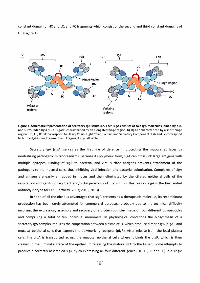
23
U
IgA
JC
SC
Fab
Fc
HC
LC
Variable regions
a
Hinge Region
U
IgA
JC
SC
Fab
Fc
HC
LC
Variable regions
b
Hinge Region
constant domain of HC and LC, and FC fragments which consist of the second and third constant domains of
HC (Figure 1).
Figure 1. Schematic representation of secretory IgA structure. Each sIgA consists of two IgA molecules joined by a JC and surrounded by a SC. a) sIgAα1 characterised by an elongated hinge region; b) sIgAα2 characterized by a short hinge region. HC, LC, JC, SC correspond to Heavy Chain, Light Chain, J‐chain and Secretory Component. Fab and Fc correspond to Antibody‐binding Fragment and Fragment crystallisable.
Secretory IgA (sIgA) serves as the first line of defence in protecting the mucosal surfaces by
neutralizing pathogenic microorganisms. Because its polymeric form, sIgA can cross‐link large antigens with
multiple epitopes. Binding of sIgA to bacterial and viral surface antigens prevents attachment of the
pathogens to the mucosal cells, thus inhibiting viral infection and bacterial colonization. Complexes of sIgA
and antigen are easily entrapped in mucus and then eliminated by the ciliated epithelial cells of the
respiratory and genitourinary tract and/or by peristalsis of the gut. For this reason, sIgA is the best suited
antibody isotype for OPI (Corthesy, 2003; 2010; 2013).
In spite of all the obvious advantages that sIgA presents as a therapeutic molecule, its recombinant
production has been rarely attempted for commercial purposes, probably due to the technical difficulty
involving the expression, assembly and recovery of a protein complex made of four different polypeptides
and comprising a total of ten individual monomers. In physiological conditions the biosynthesis of a
secretory IgA complex requires the cooperation between plasma cells, which produce dimeric IgA (dIgA), and
mucosal epithelial cells that express the polymeric Ig receptor (pIgR). After release from the local plasma
cells, the dIgA is transported across the mucosal epithelial cells where it binds the pIgR, which is then
cleaved in the luminal surface of the epithelium releasing the mature sIgA to the lumen. Some attempts to
produce a correctly assembled sIgA by co‐expressing all four different genes (HC, LC, JC and SC) in a single
(a) (b)

24
mammalian cell type have been successful (Chintalacharuvu and Morrison, 1997), however, to our
knowledge, yields have not been reported and the technology has not been further developed to achieve a
commercially viable product.
As stated before, the first attempt to produce a plant‐made sIgA for passive immunization was the
murine hybrid Guy’s 13 (Ma et al., 1995; 1998); this technology was later acquired by Planet Biotechnology
(Larrick et al., 2001), evaluated in phase I and II clinical trials and registered as a medical device (Weintraub
et al., 2005). Since then, just a few research groups have reported the expression of sIgA in heterologous
systems. Wieland et al. (2005) successfully expressed chicken sIgA against Eimeria acervulina and more
recently Virdi et al. (2013) produced sIgA‐like antibodies against enterotoxigenic E.coli in seeds of
Arabidopsis thaliana. All in all, the production of human sIgA antibodies at a commercial viable scale remains
an unsolved technological problem. When sIgA production is intended for mucosal passive immunotherapy,
plant‐based platforms, and most specially those using edible plant organs, appear to be the most promising
candidates.
5. Practical considerations for antibody production in plants.
The design of a plant biofactory for antibody manufacturing requires multiple considerations
involving not only the expression levels of the antibody but also additional aspects as subcellular targeting,
protein degradation, glycosilation patterns and downstream strategies, all of them influencing the yield,
quality and cost of the final product (Sarrión‐Perdigones et al., 2011). To date, most of these aspects have
been addressed separately, mainly on an empirical basis. However, future optimizations will probably
require designs that integrate all of them following a global approach.
5.1 Subcellular Localization
Although extracellular secretion is the natural route for antibodies in mammals, targeting antibody
chains to specific compartments in the plant cell can result in advantages in terms of stability, yield or
downstream processing (De Muynck et al., 2010). Among the different compartments that have been tested
as destination for recombinant antibodies (chloroplast, plasma membrane, vacuole, etc.), the secretory
pathway seems to be the most convenient route for a correct antibody folding and assembly due to the
oxidizing environment, the low abundance of proteases and the presence of molecular chaperones found in
the endoplasmic reticulum (Ma et al., 2003). Antibody chains are targeted to the secretory pathway using an
appropriate N‐terminal signal peptide, either native (Hiatt et al., 1989b; Sainsbury et al., 2008) or replaced
by a plant one (Düring et al., 1990). Once there, they can either be efficiently retrieved from the cis‐Golgi

25
back to the ER using a C‐terminal H/KDEL retention signal or secreted to the apoplast, following the
secretory pathway (De Muynck et al., 2009; Petruccelli et al., 2006). Although several antibodies have been
reported as strictly apoplastic (De Muynck et al., 2009; De Wilde et al., 1998; Düring et al., 1990), retention
in the ER not only seems a possibility for yield improvement but also for avoiding complex plant Golgi‐
derived glycosylation patterns that could cause immunogenicity in target organisms (Bencurova et al., 2004;
Gomord et al., 2010).
5.2 Glycosylation
One of the main advantages of plants is that, as eukaryotic platforms, they are able to express
glycoproteins. However, final glycopatterns differ between plant and animal cells. Whereas ER glycosylation
patterns are shared, a number of differences occur at the level of the Golgi apparatus. In the plant Golgi, N‐
linked glycan complexes are decorated with β‐1,2 xylose and α‐1,3 fucose residues, whereas the human N‐
glycan contain α‐1,6 fucose. In addition, β‐1,3 galactose and fucose are linked to the terminal N‐
acetylglucosamine of plant N glycans forming a Lewis structure, whereas in mammals a β‐1,4 galactose is
often combined with sialic acid (Ko et al., 2009; Saint‐Jore‐Dupas et al., 2007) (Figure 2).
Figure 2. Schematic representation of plant and human N‐glycosylation patterns. a) plant N‐glycosylation pattern; b) Human N‐glycosylation pattern.
PlantN‐glycosylation HumanN‐glycosylation
Lewis α structure
N‐acetylglucosamine
Xylose
Mannose
Fucose
SialicAcid
Galactose
a b(b) (a)

26
Different strategies have been developed to express recombinant proteins with a mammalian‐like N‐
glycosylation pattern. A first approach to humanize plant N‐glycosylation is the use of RNAi for the
downregulation of endogenous α‐1,3‐fucosyltransferase (FT) and β‐1,2‐xylosyltransferase (XT) which leads to
complex N‐glycosylation lacking immunogenic xylose and fucose epitopes (Schahs et al., 2007; Strasser et al.,
2008). The next step consisted on the expression of a chimeric form of the human β1,4‐galactosyltransferase
(GalT). This was first attempted by Bakker et al. (2001) and later improved by Strasser et al. (2009) who
reported the production of mAbs with a homogeneous galactose structure by targeting GalT to a late Golgi
compartment where the final steps of N‐glycan processing occur.
The last approach to achieve the complete human N‐glycosylation pattern was the addition of
terminal sialic residues, which was achieved by engineering of the full mammalian N‐acetylneuraminic acid
(Neu5Ac) biosynthesis pathway into Arabidopsis thaliana (Castilho et al., 2008). With this system, the
sialylation of the recombinant monoclonal antibody 2G12 in Nicotiana benthamiana retaining full
neutralization activity was reported. Moreover, this sialylated antibody was also free of xylose and fucose, as
it was produced in mutants that lacked plant‐specific N‐glycan residues (Castilho et al., 2010).
A great effort has been made to adapt N‐glycosylation, but it is only recently that attention has been
paid to O‐glycosylation. As stated by Castilho et al. (2012), plants lack endogenous glycosyltransferases that
perform mammalian‐type Ser/Thr glycosylation, which could interfere with the production of defined O‐
glycans. For this reason, they are attractive hosts for engineering of O‐glycosylation steps. Two groups have
reported the first approaches to deal with the engineering of sialylated mucine type O‐glycans to achieve
glycosylation patterns even similar to mammals (Castilho et al., 2012; Yang et al., 2012). This latter step
represents an important milestone in the humanization of plant glycosilation patterns.
It is recommendable to mimic mammal glycosylation in therapeutic proteins to avoid unwanted
antigeneicity and to gain stability. However, this issue is probably not so determining when the therapeutic
proteins are aimed at mucosal treatments. Every day in our diet, we consume plant‐specific glycans and for
this reason, plant glycans attached to these recombinant proteins are not expected to generate antigenicity.
Moreover the concern of regulation agencies on glycosilation patterns has probably decreased significantly
after the commercialization of ELELYSO®, the injectable Glucocerebrosidase from Protalix and Pfizer, which
showed excellent results in clinical trials in spite of carrying Xylose and Fucose residues (Aviezer et al., 2009;
Cox, 2010; Shaaltiel et al., 2007).
5.3 Antibody Degradation
One factor strongly influencing recombinant antibody quality and yield is its stability in a
heterologous environment. Proteases may affect the integrity of antibodies during both protein
accumulation and protein extraction. Antibodies may undergo complete hydrolysis directly reducing the final
27
yield or partial degradation which can alter the integrity and activity of the final product (Benchabane et al.,
2008; Faye et al., 2005). Moreover, it has been proposed that a balanced coexpression of heavy and light
chain is another clue factor for achieving high yield, since unassembled antibody chains, which have been
retained by the ER resident chaperone BiP, could be degraded via plant ERAD‐like systems, in a similar way
as occurs in mammals (Muller et al., 2005; Orzaez et al., 2009).
Gathering together results from various host species and different antibodies, a vast collection of
antibody degradation fragments of different sizes have been reported, ranging from partially assembled
complexes showing molecular weights between 100 and 150 kDa, to smaller ones as partially degraded un‐
associated HC (44 kDa), Fab, (Fab)2 and FC fragments around 44 kDa (De Neve et al., 1993), unassembled LCs
and eventually, smaller degradation fragments difficult to assign.
To date, several strategies can be used to deal with this quality drawback. Although it has not been
fully proved, a balanced expression of antibody chains seems to be the simplest manner to avoid chain
degradation. For this, convenient promoters should be used to boost the expression of antibody chains
which are limiting the generation of complete antibody complexes. Other approaches are the use of tissue
specific promoters to confine transgene expression to compartments with reduced metabolic activity,
targeting proteins to specific cellular organelles (Benchabane et al., 2008), or addition of gelatine as
competitor substrate for peptidases (Wongsamuth and Doran, 1997). Gene knockout or silencing of plant
peptidases is also a tool to take into account if there is a single or only a few target peptidases which are not
essential for plant growth (De Muynck et al., 2010). Last, the co‐expression of recombinant protease
inhibitors interfering with endogenous proteases has also been proposed (Benchabane et al., 2008; Robert
et al., 2013).
6. Passive immunization against rotavirus in edible fruits as a proof of concept.
The enteric pathogen Rotavirus is the most common cause of severe diarrhoea among infants and
young children. By 5 years of age, almost every child has been infected by rotavirus at least once. It is
responsible for more than 114 million episodes of diarrhoea, 25 million clinic visits, 2.4 million hospital
admissions and more than 450,000 deaths per year, most of them in developing countries due to the lack of
safe drinking water, sanitation and hygiene as well as poor overall health and nutritional status (WHO, 1999).
In countries with temperate climates rotaviruses display a seasonal pattern being usually more
frequent in winter, while in the tropics the seasonality of rotavirus diarrhoea is less predictable and
infections can occur during all year. Rotaviruses are spread by the faecal‐oral route and, although young
children are the most susceptible to get infected, it can also occur in adults, especially if they are in contact
28
with children or in the elderly. Once the infection occurs and the viruses pass the gastric barrier, they get
attached to mature enterocytes at the intestinal villi and then they are internalized. The pathophysiology of
rotavirus is not completely understood but involves a deficient absorption of water and nutrients due to the
damage of intestinal epithelium. After an incubation of 1‐3 days, rotavirus gastroenteritis normally begins
with fever and vomiting followed by watery diarrhoea. Symptoms usually last 3 to 8 days, with serious
complications as dehydration, electrolyte and acid‐base disturbance. The most severe symptoms tend to
occur in children between six months to two years of age, and those with compromised or absent immune
system functions (Parashar et al., 2003).
There are two Jennerian vaccines against rotavirus currently in the market: Rotarix and RotaTeq.
However, many times vaccines stimulate the production of IgG antibodies but are poor inducers of mucosal
IgA antibodies which are needed to prevent this type of enteric infections (Lycke, 2012). Moreover, general
malnutrition and chronic diseases, interference from maternal antibodies and co‐infection with other enteric
pathogens might also reduce the effectiveness of vaccines. Financial and logistical challenges can also be an
obstacle as vaccination campaigns are not always possible in developing countries due to the high prices and
transportation of vaccines.
OPI has repeatedly shown to be effective against enteric infections and therefore is a potential
alternative or a valuable complement to vaccination (Monger et al., 2006; Virdi et al., 2013; Zimmermann et
al., 2009). If competitive yields are achieved, edible fruits could be a convenient and inexpensive platform of
production of neutralizing antibodies aimed at OPI. Scaling‐up fruit production is simple by following well‐
established agronomic procedures. Besides, the Generally Regarded As Safe (GRAS) status of palatable plant
organs can significantly reduce cost if exhaustive purification is not needed for oral or mucosal use.
Tomato is a world‐wide cultivated crop, one of the most important in terms of biomass production.
It is edible without thermal treatment, has low allergenicity rates and its allergens are well described (Le et
al., 2006). Tomato crops have an optimum growth in differing conditions. It is a high yield edible crop (max
100 Ton/Ha/Year) and it is also well adapted to greenhouse cultivation under the confined conditions
required for molecular farming. Tomato and other fleshy fruits are commonly discarded as potential
biofactories probably because of their low protein content per fresh weight (Virdi and Depicker, 2013).
However, when water is removed by drying, its protein content reaches 14% (while rice seeds contain aprox.
7%). Most interestingly, it is also a model crop, relatively easy to transform and with many biotech tools
available. Early naïve misconceptions on plant edible vaccines (often depicted as ready‐to‐eat raw fruits
without dose control), raised scepticism and forced many researchers in the field to move towards non‐
edible production platforms intended for antigen purification. Despite of this, it is widely recognized that for
the production of oral/mucosal therapeutics, the use of GRAS organisms constitutes an important
advantage. Although the capacity of tomato fruits to accumulate functional antibodies has not been yet

29
addressed, they have been reported to successfully produce recombinant oral vaccines (Walmsley et al.,
2003; Zhang et al., 2006).
As a proof of this general concept, we decided to engineer tomatoes for the production of anti‐
rotavirus antibodies. As a first approach, the production of a monomeric human IgA directed against the
outer VP8* peptide of the model rotavirus strain SA11 was attempted (chapter 1). To gain further
acceptance for the tomato factory concept, the combination of IgA production with a transgenic labelling
system based in anthocyanins accumulation was also assayed (also described in chapter 1), as it would serve
as an additional colour‐based tracing system for increased security .
The proposal of using tomato as production platform for OPI agents is based on the assumption that
the expression and accumulation of the recombinant therapeutic product will not alter the edibility status
(absence of toxicity) of the wild type fruit. This general assumption has never been contrasted
experimentally, and it will be addressed in chapter 2 by comparing the tomatoes developed in chapter 1
with wild type in‐house grown tomatoes as well as commercial varieties using proteomic and metabolic
approaches.
Once the feasibility and security of fruit‐based IgA production is addressed (as described in chapters
1 and 2), it was necessary to proceed to the production of a full sIgA. As described in this introduction
chapter, there are many considerations regarding the optimization of the production of an antibody in
plants, many of which can only be addressed experimentally. We decided to undertake an exhaustive
comparison of different sIgA configurations, comparing heavy and light chain isotypes as well as different
subcellular localizations. Such exhaustive comparison requires the combinatorial construction of many
multigenic structures, and would not have been possible without the use of a modular cloning system
named GoldenBraid (Sarrión‐Perdigones et al., 2011b), and the use of Nicotiana benthamiana transient
expression methodology. Chapter 3 describes the construction and expression analysis of 16 different sIgA
configurations, the selection of the best performing configuration and the demonstration of the formation of
a sIgA complex in the plant cell.
All together, this work represents an important step forward for the production of a safe and cost‐
effective production of sIgA as an oral immunotherapy agent in edible plant organs.

30
7. References
Aviezer, D., Brill‐Almon, E., Shaaltiel, Y., Hashmueli, S., Bartfeld, D., Mizrachi, S., Liberman, Y., Freeman, A., Zimran, A. and Galun, E. (2009) A plant‐derived recombinant human glucocerebrosidase enzyme‐‐a preclinical and phase I investigation. PLoS One 4, e4792.
Bakker, H., Bardor, M., Molthoff, J.W., Gomord, V., Elbers, I., Stevens, L.H., Jordi, W., Lommen, A., Faye, L., Lerouge, P. and Bosch, D. (2001) Galactose‐extended glycans of antibodies produced by transgenic plants. Proc Natl Acad Sci U S A 98, 2899‐2904.
Bencurova, M., Hemmer, W., Focke‐Tejkl, M., Wilson, I.B. and Altmann, F. (2004) Specificity of IgG and IgE antibodies against plant and insect glycoprotein glycans determined with artificial glycoforms of human transferrin. Glycobiology 14, 457‐466.
Benchabane, M., Goulet, C., Rivard, D., Faye, L., Gomord, V. and Michaud, D. (2008) Preventing unintended proteolysis in plant protein biofactories. Plant Biotechnol J 6, 633‐648.
Bendandi, M., Marillonnet, S., Kandzia, R., Thieme, F., Nickstadt, A., Herz, S., Frode, R., Inoges, S., Lopez‐Diaz de Cerio, A., Soria, E., Villanueva, H., Vancanneyt, G., McCormick, A., Tuse, D., Lenz, J., Butler‐Ransohoff, J.E., Klimyuk, V. and Gleba, Y. (2010) Rapid, high‐yield production in plants of individualized idiotype vaccines for non‐Hodgkin's lymphoma. Ann Oncol 21, 2420‐2427.
Berger, I., Fitzgerald, D.J. and Richmond, T.J. (2004) Baculovirus expression system for heterologous multiprotein complexes. Nature Biotechnology 22, 1583‐1587.
Bethell, D.R. and Huang, J. (2004) Recombinant human lactoferrin treatment for global health issues: iron deficiency and acute diarrhea. Biometals 17, 337‐342.
Castilho, A., Neumann, L., Daskalova, S., Mason, H.S., Steinkellner, H., Altmann, F. and Strasser, R. (2012) Engineering of Sialylated Mucin‐type O‐Glycosylation in Plants. J Biol Chem 287, 36518‐36526.
Castilho, A., Pabst, M., Leonard, R., Veit, C., Altmann, F., Mach, L., Glossl, J., Strasser, R. and Steinkellner, H. (2008) Construction of a functional CMP‐sialic acid biosynthesis pathway in Arabidopsis. Plant Physiol 147, 331‐339.
Castilho, A., Strasser, R., Stadlmann, J., Grass, J., Jez, J., Gattinger, P., Kunert, R., Quendler, H., Pabst, M., Leonard, R., Altmann, F. and Steinkellner, H. (2010) In planta protein sialylation through overexpression of the respective mammalian pathway. J Biol Chem 285, 15923‐15930.
Corthesy, B. (2003) Recombinant secretory immunoglobulin A in passive immunotherapy: linking immunology and biotechnology. Curr Pharm Biotechnol 4, 51‐67.
Corthesy, B. (2010) Role of secretory immunoglobulin A and secretory component in the protection of mucosal surfaces. Future Microbiol 5, 817‐829.
Corthesy, B. (2013) Role of secretory IgA in infection and maintenance of homeostasis. Autoimmun Rev 12, 661‐665.
Cox, T.M. (2010) Gaucher disease: clinical profile and therapeutic developments. Biologics : targets & therapy 4, 299‐313.
Chernysheva, L.V., Friendship, R.M., Dewey, C.E. and Gyles, C.L. (2004) The effect of dietary chicken egg‐yolk antibodies on the clinical response in weaned pigs challenged with a K88+ Escherichia coli isolate. Journal of Swine Health and Production 12, 119‐122.
Chintalacharuvu, K.R. and Morrison, S.L. (1997) Production of secretory immunoglobulin A by a single mammalian cell. Proc Natl Acad Sci U S A 94, 6364‐6368.
Daniell, H., Streatfield, S.J. and Wycoff, K. (2001) Medical molecular farming: production of antibodies, biopharmaceuticals and edible vaccines in plants. Trends in Plant Science 6, 219‐226.
De Muynck, B., Navarre, C. and Boutry, M. (2010) Production of antibodies in plants: status after twenty years. Plant Biotechnol J 8, 529‐563.
De Muynck, B., Navarre, C., Nizet, Y., Stadlmann, J. and Boutry, M. (2009) Different subcellular localization and glycosylation for a functional antibody expressed in Nicotiana tabacum plants and suspension cells. Transgenic Res 18, 467‐482.

31
De Neve, M., De Loose, M., Jacobs, A., Van Houdt, H., Kaluza, B., Weidle, U., Van Montagu, M. and Depicker, A. (1993) Assembly of an antibody and its derived antibody fragment in Nicotiana and Arabidopsis. Transgenic Res 2, 227‐237.
De Wilde, C., De Rycke, R., Beeckman, T., De Neve, M., Van Montagu, M., Engler, G. and Depicker, A. (1998) Accumulation pattern of IgG antibodies and Fab fragments in transgenic Arabidopsis thaliana plants. Plant Cell Physiol 39, 639‐646.
Düring, K., Hippe, S., Kreuzaler, F. and Schell, J. (1990) Synthesis and self‐assembly of a functional monoclonal antibody in transgenic Nicotiana tabacum. Plant Mol Biol 15, 281‐293.
Faye, L., Boulaflous, A., Benchabane, M., Gomord, W. and Michaud, D. (2005) Protein modifications in the plant secretory pathway: current status and practical implications in molecular pharming. Vaccine 23, 1770‐1778.
Foley, R.C., Raison, R.L. and Beh, K.J. (1991) Monoclonal antibody against sheep kappa light chain. Hybridoma 10, 507‐515.
Frenzel, A., Hust, M. and Schirrmann, T. (2013) Expression of recombinant antibodies. Front Immunol 4, 217. Furtado, P.B., Whitty, P.W., Robertson, A., Eaton, J.T., Almogren, A., Kerr, M.A., Woof, J.M. and Perkins, S.J.
(2004) Solution structure determination of monomeric human IgA2 by X‐ray and neutron scattering, analytical ultracentrifugation and constrained modelling: a comparison with monomeric human IgA1. J Mol Biol 338, 921‐941.
Giritch, A., Marillonnet, S., Engler, C., van Eldik, G., Botterman, J., Klimyuk, V. and Gleba, Y. (2006) Rapid high‐yield expression of full‐size IgG antibodies in plants coinfected with noncompeting viral vectors. Proc Natl Acad Sci U S A 103, 14701‐14706.
Goldsby, R., kindt, T., Osborne, B. and Kuby, J. (2003) Immunology. England. Gomord, V., Fitchette, A.C., Menu‐Bouaouiche, L., Saint‐Jore‐Dupas, C., Plasson, C., Michaud, D. and Faye, L.
(2010) Plant‐specific glycosylation patterns in the context of therapeutic protein production. Plant Biotechnol J 8, 564‐587.
Haq, T.A., Mason, H.S., Clements, J.D. and Arntzen, C.J. (1995) Oral immunization with a recombinant bacterial antigen produced in transgenic plants. Science 268, 714‐716.
Harmsen, M.M., van Solt, C.B., Hoogendoorn, A., van Zijderveld, F.G., Niewold, T.A. and van der Meulen, J. (2005) Escherichia coli F4 fimbriae specific llama single‐domain antibody fragments effectively inhibit bacterial adhesion in vitro but poorly protect against diarrhoea. Vet Microbiol 111, 89‐98.
Hiatt, A., Cafferkey, R. and Bowdish, K. (1989a) Production of antibodies in transgenic plants. Nature 342, 76 ‐ 78.
Hiatt, A., Cafferkey, R. and Bowdish, K. (1989b) Production of antibodies in transgenic plants. Nature 342, 76‐78.
Ko, K., Brodzik, R. and Steplewski, Z. (2009) Production of antibodies in plants: approaches and perspectives. Curr Top Microbiol Immunol 332, 55‐78.
Ko, K.S., Tekoah, Y., Rudd, P.M., Harvey, D.J., Dwek, R.A., Spitsin, S., Hanlon, C.A., Rupprecht, C., Dietzschold, B., Golovkin, M. and Koprowski, H. (2003) Function and glycosylation of plant‐derived antiviral monoclonal antibody. Proceedings of the National Academy of Sciences of the United States of America 100, 8013‐8018.
Larrick, J.W., Yu, L., Chen, J., Jaiswal, S. and Wycoff, K. (1998) Production of antibodies in transgenic plants. Res Immunol 149, 603‐608.
Larrick, J.W., Yu, L., Naftzger, C., Jaiswal, S. and Wycoff, K. (2001) Production of secretory IgA antibodies in plants. Biomolecular Engineering 18, 87‐94.
Le, L.Q., Lorenz, Y., Scheurer, S., Fotisch, K., Enrique, E., Bartra, J., Biemelt, S., Vieths, S. and Sonnewald, U. (2006) Design of tomato fruits with reduced allergenicity by dsRNAi‐mediated inhibition of ns‐LTP (Lyc e 3) expression. Plant Biotechnol J 4, 231‐242.
Lycke, N. (2012) Recent progress in mucosal vaccine development: potential and limitations. Nat Rev Immunol 12, 592‐605.
Ma, J.K., Hiatt, A., Hein, M., Vine, N.D., Wang, F., Stabila, P., van Dolleweerd, C., Mostov, K. and Lehner, T. (1995) Generation and assembly of secretory antibodies in plants. Science 268, 716‐719.
32
Ma, J.K., Hikmat, B.Y., Wycoff, K., Vine, N.D., Chargelegue, D., Yu, L., Hein, M.B. and Lehner, T. (1998) Characterization of a recombinant plant monoclonal secretory antibody and preventive immunotherapy in humans. Nat Med 4, 601‐606.
Ma, J.K.C. (1988) Prevention of Colonization of Strep‐Mutans by Local Passive‐Immunization with Monoclonal‐Antibodies in Humans. Journal of Dental Research 67, 643‐643.
Ma, J.K.C., Drake, P.M.W. and Christou, P. (2003) The production of recombinant pharmaceutical proteins in plants. Nature Reviews Genetics 4, 794‐805.
Marquardt, R.R., Jin, L.Z., Kim, J.W., Fang, L., Frohlich, A.A. and Baidoo, S.K. (1999) Passive protective effect of egg‐yolk antibodies against enterotoxigenic Escherichia coli K88+ infection in neonatal and early‐weaned piglets. FEMS Immunol Med Microbiol 23, 283‐288.
Mason, H.S., Ball, J.M., Shi, J.J., Jiang, X., Estes, M.K. and Arntzen, C.J. (1996) Expression of Norwalk virus capsid protein in transgenic tobacco and potato and its oral immunogenicity in mice. Proceedings of the National Academy of Sciences of the United States of America 93, 5335‐5340.
Mason, H.S., Haq, T.A., Clements, J.D. and Arntzen, C.J. (1998) Edible vaccine protects mice against Escherichia coli heat‐labile enterotoxin (LT): potatoes expressing a synthetic LT‐B gene. Vaccine 16, 1336‐1343.
Mason, H.S., Warzecha, H., Mor, T. and Arntzen, C.J. (2002) Edible plant vaccines: applications for prophylactic and therapeutic molecular medicine. Trends in Molecular Medicine 8, 324‐329.
McCormick, A.A., Reddy, S., Reinl, S.J., Cameron, T.I., Czerwinkski, D.K., Vojdani, F., Hanley, K.M., Garger, S.J., White, E.L., Novak, J., Barrett, J., Holtz, R.B., Tuse, D. and Levy, R. (2008) Plant‐produced idiotype vaccines for the treatment of non‐Hodgkin's lymphoma: safety and immunogenicity in a phase I clinical study. Proc Natl Acad Sci U S A 105, 10131‐10136.
Mohan, P. and Haque, K. (2003) Oral immunoglobulin for the prevention of rotavirus infection in low birth weight infants. Cochrane Database Syst Rev, CD003740.
Monger, W., Alamillo, J.M., Sola, I., Perrin, Y., Bestagno, M., Burrone, O.R., Sabella, P., Plana‐Duran, J., Enjuanes, L., Garcia, J.A. and Lomonossoff, G.P. (2006) An antibody derivative expressed from viral vectors passively immunizes pigs against transmissible gastroenteritis virus infection when supplied orally in crude plant extracts. Plant Biotechnol J 4, 623‐631.
Muller, J., Piffanelli, P., Devoto, A., Miklis, M., Elliott, C., Ortmann, B., Schulze‐Lefert, P. and Panstruga, R. (2005) Conserved ERAD‐like quality control of a plant polytopic membrane protein. Plant Cell 17, 149‐163.
Nandi, S., Yalda, D., Lu, S., Nikolov, Z., Misaki, R., Fujiyama, K. and Huang, N. (2005) Process development and economic evaluation of recombinant human lactoferrin expressed in rice grain. Transgenic Res 14, 237‐249.
Niewold, T.A., van Dijk, A.J., Geenen, P.L., Roodink, H., Margry, R. and van der Meulen, J. (2007) Dietary specific antibodies in spray‐dried immune plasma prevent enterotoxigenic Escherichia coli F4 (ETEC) post weaning diarrhoea in piglets. Vet Microbiol 124, 362‐369.
Orzaez, D., Granell, A. and Blazquez, M.A. (2009) Manufacturing antibodies in the plant cell. Biotechnol J 4, 1712‐1724.
Parashar, U.D., Hummelman, E.G., Bresee, J.S., Miller, M.A. and Glass, R.I. (2003) Global illness and deaths caused by rotavirus disease in children. Emerg Infect Dis 9, 565‐572.
PBVA(2013)http://www.meetingsmanagement.co.uk/index.php?option=comcontent&view=article&id=117&Itemid=244 . Plans Based Vaccines, Antibodies & Biologics.
Petruccelli, S., Otegui, M.S., Lareu, F., Tran Dinh, O., Fitchette, A.C., Circosta, A., Rumbo, M., Bardor, M., Carcamo, R., Gomord, V. and Beachy, R.N. (2006) A KDEL‐tagged monoclonal antibody is efficiently retained in the endoplasmic reticulum in leaves, but is both partially secreted and sorted to protein storage vacuoles in seeds. Plant Biotechnol J 4, 511‐527.
Protalix (2013) http://www.protalix.com/development‐pipeline/prx‐112‐oral‐gaucher‐disease.asp. Protalix Biotherapeutics.
Raab, C.P. (2011) Passive immunization. Prim Care 38, 681‐691, viii.

33
Rademacher, T., Sack, M., Arcalis, E., Stadlmann, J., Balzer, S., Altmann, F., Quendler, H., Stiegler, G., Kunert, R., Fischer, R. and Stoger, E. (2008) Recombinant antibody 2G12 produced in maize endosperm efficiently neutralizes HIV‐1 and contains predominantly single‐GlcNAc N‐glycans. Plant Biotechnol J 6, 189‐201.
Rahman, S., Van Nguyen, S., Icatlo, F.C., Jr., Umeda, K. and Kodama, Y. (2013) Oral passive IgY‐based immunotherapeutics: A novel solution for prevention and treatment of alimentary tract diseases. Hum Vaccin Immunother 9.
Ramessar, K., Rademacher, T., Sack, M., Stadlmann, J., Platis, D., Stiegler, G., Labrou, N., Altmann, F., Ma, J., Stoger, E., Capell, T. and Christou, P. (2008) Cost‐effective production of a vaginal protein microbicide to prevent HIV transmission. Proc Natl Acad Sci U S A 105, 3727‐3732.
Robert, S., Khalf, M., Goulet, M.C., D'Aoust, M.A., Sainsbury, F. and Michaud, D. (2013) Protection of recombinant mammalian antibodies from development‐dependent proteolysis in leaves of Nicotiana benthamiana. PLoS One 8, e70203.
Sainsbury, F., Lavoie, P.O., D'Aoust, M.A., Vezina, L.P. and Lomonossoff, G.P. (2008) Expression of multiple proteins using full‐length and deleted versions of cowpea mosaic virus RNA‐2. Plant Biotechnol J 6, 82‐92.
Saint‐Jore‐Dupas, C., Faye, L. and Gomord, V. (2007) From planta to pharma with glycosylation in the toolbox. Trends Biotechnol 25, 317‐323.
Sarrion‐Perdigones A, Juarez P, Granell A and Orzaez D (2011a) Production of Antibodies in Plants, in Antibody Expression and Production (Al‐Rubeai M ed) pp 143‐164: Springer Netherlands.
Sarrion‐Perdigones A, Falconi EE, Zandalinas SI, Juarez P, Fernandez‐del‐Carmen A, Granell A and Orzaez D (2011b) GoldenBraid: an iterative cloning system for standardized assembly of reusable genetic modules. PloS one 6:e21622.
Schahs, M., Strasser, R., Stadlmann, J., Kunert, R., Rademacher, T. and Steinkellner, H. (2007) Production of a monoclonal antibody in plants with a humanized N‐glycosylation pattern. Plant Biotechnol J 5, 657‐663.
Shaaltiel, Y., Bartfeld, D., Hashmueli, S., Baum, G., Brill‐Almon, E., Galili, G., Dym, O., Boldin‐Adamsky, S.A., Silman, I., Sussman, J.L., Futerman, A.H. and Aviezer, D. (2007) Production of glucocerebrosidase with terminal mannose glycans for enzyme replacement therapy of Gaucher's disease using a plant cell system. Plant Biotechnol J 5, 579‐590.
Strasser, R., Castilho, A., Stadlmann, J., Kunert, R., Quendler, H., Gattinger, P., Jez, J., Rademacher, T., Altmann, F., Mach, L. and Steinkellner, H. (2009) Improved virus neutralization by plant‐produced anti‐HIV antibodies with a homogeneous beta1,4‐galactosylated N‐glycan profile. J Biol Chem 284, 20479‐20485.
Strasser, R., Stadlmann, J., Schahs, M., Stiegler, G., Quendler, H., Mach, L., Glossl, J., Weterings, K., Pabst, M. and Steinkellner, H. (2008) Generation of glyco‐engineered Nicotiana benthamiana for the production of monoclonal antibodies with a homogeneous human‐like N‐glycan structure. Plant Biotechnol J 6, 392‐402.
Tacket, C.O. (2007) Plant‐based vaccines against diarrheal diseases. Trans Am Clin Climatol Assoc 118, 79‐87. Tacket, C.O., Mason, H.S., Losonsky, G., Clements, J.D., Levine, M.M. and Arntzen, C.J. (1998)
Immunogenicity in humans of a recombinant bacterial antigen delivered in a transgenic potato. Nat Med 4, 607‐609.
Tacket, C.O., Pasetti, M.F., Edelman, R., Howard, J.A. and Streatfield, S. (2004) Immunogenicity of recombinant LT‐B delivered orally to humans in transgenic corn. Vaccine 22, 4385‐4389.
Tokuhara, D., Alvarez, B., Mejima, M., Hiroiwa, T., Takahashi, Y., Kurokawa, S., Kuroda, M., Oyama, M., Kozuka‐Hata, H., Nochi, T., Sagara, H., Aladin, F., Marcotte, H., Frenken, L.G., Iturriza‐Gomara, M., Kiyono, H., Hammarstrom, L. and Yuki, Y. (2013) Rice‐based oral antibody fragment prophylaxis and therapy against rotavirus infection. J Clin Invest 123, 3829‐3838.
Vega, C.G., Bok, M., Vlasova, A.N., Chattha, K.S., Fernandez, F.M., Wigdorovitz, A., Parreno, V.G. and Saif, L.J. (2012) IgY antibodies protect against human Rotavirus induced diarrhea in the neonatal gnotobiotic piglet disease model. PLoS One 7, e42788.

34
Ventria‐News (2012) http://www.ventria.com/about‐us/news‐press/47‐ventria‐bioscience‐acquires‐meristem‐therapeutics‐recombinant‐lactoferrin‐intellectual‐property. Ventria Bioscience‐News and Press.
Vezina, L.P., Faye, L., Lerouge, P., D'Aoust, M.A., Marquet‐Blouin, E., Burel, C., Lavoie, P.O., Bardor, M. and Gomord, V. (2009) Transient co‐expression for fast and high‐yield production of antibodies with human‐like N‐glycans in plants. Plant Biotechnol J 7, 442‐455.
Virdi, V., Coddens, A., De Buck, S., Millet, S., Goddeeris, B.M., Cox, E., De Greve, H. and Depicker, A. (2013) Orally fed seeds producing designer IgAs protect weaned piglets against enterotoxigenic Escherichia coli infection. Proc Natl Acad Sci U S A.
Virdi, V. and Depicker, A. (2013) Role of plant expression systems in antibody production for passive immunization. Int J Dev Biol 57, 587‐593.
Walmsley, A.M., Alvarez, M.L., Jin, Y., Kirk, D.D., Lee, S.M., Pinkhasov, J., Rigano, M.M., Arntzen, C.J. and Mason, H.S. (2003) Expression of the B subunit of Escherichia coli heat‐labile enterotoxin as a fusion protein in transgenic tomato. Plant Cell Rep 21, 1020‐1026.
Weintraub, J.A., Hilton, J.F., White, J.M., Hoover, C.I., Wycoff, K.L., Yu, L., Larrick, J.W. and Featherstone, J.D. (2005) Clinical trial of a plant‐derived antibody on recolonization of mutans streptococci. Caries Res 39, 241‐250.
WHO (1999) http://www.who.int/infectious‐disease‐report/pages/ch1text.html. World Health Organization. Wieland, W., Lammers, A., Schots, A. and Orzaez, D. (2005) Plant expression of chicken secretory antibodies
derived from combinatorial libraries. Submmited to Plant Biotechnology Journal. Wongsamuth, R. and Doran, P.M. (1997) Production of monoclonal antibodies by tobacco hairy roots.
Biotechnol Bioeng 54, 401‐415. Wurm, F.M. (2004) Production of recombinant protein therapeutics in cultivated mammalian cells. Nat
Biotechnol 22, 1393‐1398. Wycoff, K.L. (2005) Secretory IgA antibodies from plants. Curr Pharm Des 11, 2429‐2437. Xu, Y., Li, X., Jin, L., Zhen, Y., Lu, Y., Li, S., You, J. and Wang, L. (2011) Application of chicken egg yolk
immunoglobulins in the control of terrestrial and aquatic animal diseases: a review. Biotechnol Adv 29, 860‐868.
Yang, Z., Drew, D.P., Jorgensen, B., Mandel, U., Bach, S.S., Ulvskov, P., Levery, S.B., Bennett, E.P., Clausen, H. and Petersen, B.L. (2012) Engineering mammalian mucin‐type O‐glycosylation in plants. J Biol Chem 287, 11911‐11923.
Zeitlin, L., Olmsted, S.S., Moench, T.R., Co, M.S., Martinell, B.J., Paradkar, V.M., Russell, D.R., Queen, C., Cone, R.A. and Whaley, K.J. (1998) A humanized monoclonal antibody produced in transgenic plants for immunoprotection of the vagina against genital herpes. Nat Biotechnol 16, 1361‐1364.
Zhang, X., Buehner, N.A., Hutson, A.M., Estes, M.K. and Mason, H.S. (2006) Tomato is a highly effective vehicle for expression and oral immunization with Norwalk virus capsid protein. Plant Biotechnol J 4, 419‐432.
Zimmermann, J., Saalbach, I., Jahn, D., Giersberg, M., Haehnel, S., Wedel, J., Macek, J., Zoufal, K., Glunder, G., Falkenburg, D. and Kipriyanov, S. (2009) Antibody expressing pea seeds as fodder for prevention of gastrointestinal parasitic infections in chickens. BMC Biotechnology 9, 79.

35
Objectives

36
Mucosal passive immunization is a strategy with a great potential for fighting against enteric infections. The
overall objective of this thesis is to trace a pathway towards the development of fruit‐based antibody
formulations for mucosal treatments against infectious diseases.
To fulfil this general objective, the following specific objectives were proposed:
1. To generate transgenic tomato fruits expressing a model human immunoglobulin A (IgA) with
neutralizing capacity against the enteric pathogen rotavirus.
2. To establish a method to distinguish IgA‐expressing transgenic tomato fruits from wild type fruits
using anthocyanins as labelling traits.
3. To evaluate possible unintended effects in the composition of IgA‐tomato fruits.
4. To select, among different multigenic combinations, the most suitable version of secretory IgA for its
recombinant production in plants.
37
Chapter 1
Neutralizing antibodies against rotavirus produced in transgenically labelled purple tomatoes.
Paloma Juárez, Silvia Presa, Joaquín Espí, Benito Pineda, María T. Antón, Vicente Moreno, Javier Buesa, Antonio Granell and Diego Orzáez. Plant Biotechnology Journal (2012) 10, pp. 341‐352

38
1. Introduction
Fleshy fruit have been traditionally exploited as natural biofactories for orally active, health‐
promoting metabolites (Cipollini and Levey, 1997). Recently, they have also been envisioned as an excellent
vehicle for nutritional enhancement, and considerable efforts have been made to enrich their content of
indigenous and ⁄ or exotic human health‐promoting compounds such as carotenoids, folate, flavonols or
anthocyanins through molecular breeding and metabolic engineering (Apel and Bock, 2009; Bovy et al.,
2002; Bovy et al., 2007; Butelli et al., 2008; Davuluri et al., 2005; Giuliano et al., 2008). Unlike other plant‐
derived products, edible fruit and their derivatives are considered safe and palatable for human
consumption in unheated, minimally processed forms, and their content of antinutrients and toxic
compounds sharply decreases upon ripening. This feature makes edible fruits ideal biofactories for exotic
heat‐labile compounds with mucosal activity such as health‐promoting metabolites and mucosal active
proteins like oral vaccines, immune modulators, biocides and ⁄ or antibodies for artificial passive
immunotherapy.
Transgenic crops have been proposed as advantageous platforms for recombinant antibody
production owing to their productivity, scalability and the low risk of contamination with mammalian
pathogens (De Muynck et al., 2010). The technical feasibility of an artificial passive immunization approach
based on a plant‐made antibody was first demonstrated by the pioneering work of Ma et al. (Ma et al., 1994;
Ma et al., 1995; Ma et al., 1998) who expressed in tobacco a hybrid murine ⁄ human secretory
immunoglobulin G⁄ A (Guy’s 13 sIgG ⁄ A) that is effective against tooth decay. The Guy’s 13 monoclonal
antibody produced in tobacco provided effective protection against Streptococcus mutans infections in
healthy volunteers. The technology to express Guy’s 13 sIgG‐A in plants was licensed by Planet
Biotechnology Inc (Hayward, CA). This was the first plant‐made antibody that was evaluated in phase I and II
clinical trials (Weintraub et al., 2005) as CaroRxTM. In the United States, CaroRxTM is an Investigational New
Drug, and in the European Union, it is a registered Medical Device. More recently, the anti‐HIV monoclonal
IgG 2G12 was successfully produced in corn with the aim of conferring passive protection of vaginal mucosa
against HIV infection (Rademacher et al., 2008). Plant‐derived IgG 2G12 was shown to be effective in viral
inhibition assays and has become a promising cost‐effective source for vaginal gel formulations (Ramessar et
al., 2008).
Antibody purification is still a major component of the total cost of goods in plant‐made
recombinant antibody production (Hood et al., 2002). Although biosafety of any therapeutic ⁄ prophylactic
approach involving complex botanical mixes should be exhaustively investigated, it is expected that
minimally processed extracts from antibody‐producing edible fruits can be used as safe formulations for
mucosal delivery, therefore reducing downstream processing costs. Tomato fruits have been studied as
successful production platforms for recombinant oral vaccines (Walmsley et al., 2003; Zhang et al., 2006a).

39
However, issues such as the capacity of fleshy fruits to accumulate functional antibodies, the evolution of
antibody activity throughout the process of fruit ripening and the formulations that retain neutralizing
activity have not been addressed before. To address these issues, we assessed the recombinant production
in tomato fruit of a phage display–derived human IgA antibody that is effective against the model rotavirus
strain SA11, and tested the ability of minimally processed fruit‐derived products to neutralize rotavirus
infection in vitro.
Besides antibody activity and formulation, an important concern in using edible fruits to produce
recombinant pharmaceuticals stems from the possibility that the fruit expressing pharmaceuticals are mixed
with fruit used for food consumption. To address this issue, it has been recommended that transgenic fruit
should be labelled with distinctive features that help to preserve their identity, facilitate traceability and
avoid the contamination of the food supply. As a proof of this concept, we engineered purple, IgA‐producing
tomatoes, where Antirrhinum majus Rosea1 and Delila transcription factors, known to ectopically activate
anthocyanin biosynthesis in tomato fruits (Butelli et al., 2008), were incorporated into the transgenic IgA
background by sexual crossing to obtain tomato‐made antibodies in transgenically labelled fruit and fruit‐
derived products.
40
2. Results
2.1 Design and selection of human IgA genes for expression in tomato fruits
Antibody repertoires displayed in phage format provide a flexible and adaptable source of
neutralizing antibody fragments that can later be transferred to plant expression systems, either as antibody
fragments or as full‐length antibodies (Wieland et al., 2006). An important limitation of this approach is the
significant heterogeneity of expression levels in the plant, apparently because of differences in stability
conferred by the antibody variable regions, which might be exacerbated by the use of synthetic or
semisynthetic libraries. A possible solution to this is the introduction of a quick selection step for in planta
stability using transient expression analysis, preferably performed in the same plant organ used for the
production of the compound. Based on this rationale, we designed a set of shuttle vectors to facilitate the
transfer of the coding sequences for V regions from a phagemid library into plant expression vectors and
then took advantage of a fruit transient expression assay (Orzaez et al., 2006) to test the expression levels of
a subset of the plant‐produced antibodies against the model rotavirus strain SA11.
DNA fragments that encoded single‐chain antibody fragments (scFv) that had been selected for their
recognition of the VP8* peptide of rotavirus VP4 protein were inserted into plant expression cassettes
carrying the constant regions of human IgA (Figure S1a,b). To gain versatility in the cloning strategy, a
Gateway‐ based set‐up was established. Two Gateway pENTR vectors (pENTR_IgH and pENTR_IgL) were
generated containing the constant regions of IgHa1 and Igk, respectively. Convenient restriction sites were
engineered at the 5’ ends of each constant region to allow for the direct cloning of the variable region of the
heavy chain (VH) and variable region of the light chain (VL) derived from the Griffin.1 phage display library
(Figure S1a). The variable regions from three different anti‐VP8* scFv (namely 2A1, 2E4 and 2B3) were
introduced into the pENTR vectors and transferred to the binary vector pKGW7 through site‐specific
recombination reaction between attL and attR sites (LR recombination). As a result, plant expression
constructs were generated containing either the heavy chain (HC) or the light chain (LC) of a fully
reconstituted IgA antibody under the control of the constitutive 35S promoter.
The in planta expression of each reconstituted antibody was initially tested using an Agrobacterium‐
mediated transient expression assay. Plant expression vectors carrying the genes encoding the HC and the LC
for each antibody to be analysed were transferred to Agrobacterium and infiltrated into tomato fruits. The
high cotransformation efficiency provided by the fruit agroinjection approach allowed for facile monitoring
of the formation of full‐length IgA in fruit cells. As shown in Figure 1a, only the variable regions derived from
41
scFv 2A1 yielded coexpression of HC and LC proteins in planta as detected in Western analysis under
nonreducing conditions. When IgA_2E4 and IgA_2B3 were transiently cotransformed, only LCs were
detected, whereas their respective HCs remained undetectable. A 35SCaMV:GFP construct was included in
all transformations and used to normalize the transformation efficiency in the samples. Transient expression
analysis using KDEL‐tagged versions of the IgA_2A1 HC and LC demonstrated that endoplasmic reticulum
(ER) retention resulted in higher antibody yields (Figure S2). The transient expression data encouraged us to
continue with the stable plant transformation of the IgA_2A1 idiotype using HC and LC fused to a KDEL
peptide as an ERretention signal. For this purpose, an in trans cotransformation strategy was followed
(referred as 35S_IgA): tomato explants were simultaneously co‐inoculated with two Agrobacterium cultures
carrying 35S:IgH2A1‐kdel and 35S:IgL2A1‐kdel expression cassettes, respectively.
In addition to the above‐mentioned in trans cotransformation experiment, an in cis cotransformation
approach was also carried out (referred as NH_IgA). In this case, in order to prevent the duplication of the
35S promoter, LC was placed under the control of the tomato NH promoter. The NH promoter drives high
levels of gene expression predominantly in fruits at mature‐green stage (Estornell et al., 2009). Therefore, a
dual construct was made (as depicted in Figure S1b) carrying 35S:IgH2A1‐kdel and NH:IgL2A1‐kdel
expression cassettes located within the same T‐DNA.
2.2 Transgenic fruits accumulate high levels of mAb
Primary transformants from cis and trans cotransformation approaches were selected by kanamycin
resistance, and the expression of HC and LC was confirmed by Western blots. IgA expression levels in T1
generation were evaluated by ELISA, and the best‐performing lines were self‐pollinated up to T3 for high
antibody titres and homozygosity. The description of two selected T3 lines per construct (NH_1A‐13‐2 and
NH_III6‐2‐6 from the NH_IgA approach and 35S_20A7 and 35S_17A4 from the 35S_IgA approach) is
presented here. Western analysis of NH_IgA lines showed low IgA levels in leaves when compared with fruits
(Figure 1b). Interestingly, when samples were analysed under reducing conditions, it was found that not only
LC but also HC was nearly absent in leaves, suggesting that HC does not accumulate at significant levels in
the absence of its cognate LC. Notably, a significant portion of the total antibody content in NH_IgA samples
under nonreducing conditions was present in the form of a 45‐kDa band, which reacted with both anti‐HC
and anti‐LC antibodies. Furthermore, reducing conditions clearly showed a 25‐kDa degradation band that
was recognized by anti‐HC antibody, while the LC remained intact. Together, these observations indicate
that a significant proportion of total IgA was present in the form of Fab’ fragments, probably as a result of
the cleavage of the full IgA perhaps in the hinge region.

42
Figure 1. Western blot analysis of IgA expression in tomato. (a) Transient expression analysis of scFV‐derived 2E4, 2B3 and 2A1 antibodies using direct fruit‐agroinjection. Fruits were co‐agroinjeted with ER‐retained heavy and light antibody chains, together with GFP as internal control. (b) Western blot analysis of leaves and fruits from best‐permorfing T3 lines from in cis (NH) and in trans (35S) transgenic approaches. Three fruits from each elite T3 plant were pooled in each representative sample. All fruits were labelled at breaker stage and collected bewteen 48 and 72 hours later (c) Comparison between fruit extracts from of 35S_20A7 and wild type (WT) plants. Samples were resolved under either reducing (R) or non‐reducing (NR) conditions and decorated using anti‐heavy chain antibody (HC), anti‐light chain antibody (LC), anti‐GFP antibody (GFP) or by Coomasie staining (CS). HS lane contains control human serum.
IgA production levels were quantified by ELISA in the fruits of the NH_1A‐13‐2 homozygous line,
reaching 15.6 ± 1.2 μg IgA equivalents ⁄ g fresh weight (FW) [1.1 ± 0.1% total soluble protein (TSP)] (Table 1).
The in trans cotransformation strategy (35S_IgA) resulted in 20% (4 of 20) of the T1 kanamycin‐resistant
plants showing both HC and LC expression in vegetative tissues. Following the same minibreeding strategy as
for the NH_IgA plants, lines 35S_20A7 and 35S_17A4 were selected for further characterization. In contrast
to the NH_IgA lines, 35S_IgA plants showed similar expression levels in fruits and vegetative tissues by
Western blot analysis, when gel loads were equalized per total protein content in the sample (Figure 1b). As
in the NH_IgA approach, a significant part of the total antibody content in the leaves and fruits of the
35S_IgA plants was present in the form of Fab’ fragments, resulting from the association between partially
degraded HC (Figure 1b, R ⁄ HC panel) and intact LC (R ⁄ LC panel). In absolute terms, 35S_IgA approach
resulted in higher levels of recombinant protein production, reaching 41.2 ± 2.8 μg of IgA equivalents ⁄ g FW
(a)
GFP
2E4
2B3
2A1
R/HC
R/LC
R/GFP
35S_IgA
35S_IgA
35S_IgA
WT
WT
WT
Leaf Fruit
NH 1A‐13‐2
NH III6‐2‐6
35S 20A7
35S 17A‐4
NH 1A‐13‐2
NH III6‐2‐6
35S 20A7
35S 17A‐4
H.S.
NR/HC
NR/LC
R/HC
R/LC
R/HC R/LCCS
191 ‐
64 ‐51 ‐
191 ‐
64 ‐51 ‐
64 ‐51 ‐
39 ‐
39 ‐
28 ‐
64 ‐
51 ‐
39 ‐
28 ‐
(b)
(c)
kDa
kDa
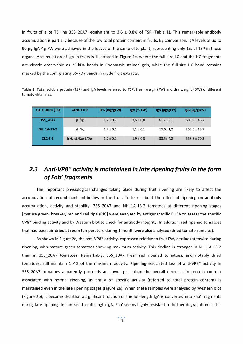
43
in fruits of elite T3 line 35S_20A7, equivalent to 3.6 ± 0.8% of TSP (Table 1). This remarkable antibody
accumulation is partially because of the low total protein content in fruits. By comparison, IgA levels of up to
90 μg IgA ⁄ g FW were achieved in the leaves of the same elite plant, representing only 1% of TSP in those
organs. Accumulation of IgA in fruits is illustrated in Figure 1c, where the full‐size LC and the HC fragments
are clearly observable as 25‐kDa bands in Coomassie‐stained gels, while the full‐size HC band remains
masked by the comigrating 55‐kDa bands in crude fruit extracts.
Table 1. Total soluble protein (TSP) and IgA levels referred to TSP, fresh weigh (FW) and dry weight (DW) of diferent tomato elite lines.
2.3 Anti‐VP8* activity is maintained in late ripening fruits in the form
of Fab’ fragments
The important physiological changes taking place during fruit ripening are likely to affect the
accumulation of recombinant antibodies in the fruit. To learn about the effect of ripening on antibody
accumulation, activity and stability, 35S_20A7 and NH_1A‐13‐2 tomatoes at different ripening stages
[mature green, breaker, red and red ripe (RR)] were analysed by antigenspecific ELISA to assess the specific
VP8* binding activity and by Western blot to check for antibody integrity. In addition, red ripened tomatoes
that had been air‐dried at room temperature during 1 month were also analysed (dried tomato samples).
As shown in Figure 2a, the anti‐VP8* activity, expressed relative to fruit FW, declines stepwise during
ripening, with mature green tomatoes showing maximum activity. This decline is stronger in NH_1A‐13‐2
than in 35S_20A7 tomatoes. Remarkably, 35S_20A7 fresh red ripened tomatoes, and notably dried
tomatoes, still maintain 1 ⁄ 3 of the maximum activity. Ripening‐associated loss of anti‐VP8* activity in
35S_20A7 tomatoes apparently proceeds at slower pace than the overall decrease in protein content
associated with normal ripening, as anti‐VP8* specific activity (referred to total protein content) is
maintained even in the late ripening stages (Figure 2a). When these samples were analysed by Western blot
(Figure 2b), it became clearthat a significant fraction of the full‐length IgA is converted into Fab’ fragments
during late ripening. In contrast to full‐length IgA, Fab’ seems highly resistant to further degradation as it is
ELITE LINES (T3) GENOTYPE TPS (mg/gFW) IgA (% TSP) IgA (µg/gFW) IgA (µg/gDW)
35S_20A7 IgH/IgL 1,2 ± 0,2 3,6 ± 0,8 41,2 ± 2,8 686,9 ± 46,7
NH_1A‐13‐2 IgH/IgL 1,4 ± 0,1 1,1 ± 0,1 15,6± 1,2 259,6 ± 19,7
CR2‐3‐8 IgH/IgL/Ros1/Del 1,7 ± 0,1 1,9 ± 0,3 33,5± 4,2 558,3 ± 70,3

44
0
200
400
600
0
100
200
300
MG BK R RR DRY
Abs 4
92/ mg protein
Abs 4
92 /g FW
Ripening Stages
35S_20A7
R/LC
R/HC
NR/HC
35S_20A7 NH_1A-13-2
MG BK R RR D HSMG BK R RR D HSkDa
64
51
39
28
28
191
64
51
39
28
kDa
64
51
39
28
28
191
64
51
39
28
(a)
(b)
0
100
200
300
0
50
100
150
MG BK R RR DRY
Abs 4
92/ mg protein
Abs 4
92/g FW
Ripening Stages
NH_1A‐13‐2
highlighted by the strong Fab’ band observed in 35S_20A7 tomatoes at the late ripe stage (Figure 2b, lanes
RR and D).
Figure 2. IgA accumulation profile during tomato fruit ripening. (a) Evolution of anti‐VP8* activity in fruit extracts of 35S_20A7 and NH_1A‐13‐2 elite plants referred to fresh weight (light grey) and total protein content (dark grey columns). (b) Western blot analysis of the same fruit samples as in (a). Samples were resolved under either reducing (R) or non‐reducing (NR) conditions and decorated using anti‐heavy chain (HC) or anti‐light chain antibodies (LC). The selected fruit developmental stages were: Mature green fruits (MG), corresponding to fully expanded green fruits (4‐5 weeks after anthesis); Breaker fruits (BK) turning yellow/red, Red fruits (R), harvested between 5 and 12 days after breaker stage; red ripe fruit (RR), harvested between 14 and 21 days after breaker stage; and dry (D) tomatoes, harvested at the red ripe stage and stored at room temperature for one month. HS is a sample of human serum. All samples were obtained by pooling three fruits from each developmental stage and analyzed in triplicate with bars representing standard error.

45
2.4 Minimally processed tomato‐based products show strong anti‐VP8* activity
The anti‐VP8* binding activity of fruit‐derived extracts was determined using fruits from the elite line
35S_20A7. The assays used antigen‐ELISA plates coated with VP8* or with bovine serum albumin (BSA) as a
control. In order to test the resistance of the antibody activity to downstream processing, three tomato
formulations compatible with long‐term conservation and oral delivery were assayed, referred to as ‘juice’,
‘powder’ and ‘dried juice’. ‘Juice’ consisted in a clarified extract from frozen fruit samples ground in liquid
nitrogen with phosphatebuffered saline (PBS) (1 : 3) (w:v). The ‘powder’ format consisted in freeze‐dried
ground tomato powder, later reconstituted in PBS using three volumes of the original FW. ‘Dried juice’
consisted in freeze‐dried ‘juice’ sample, later reconstituted in water using three volumes of the original FW.
All samples were clarified by centrifugation and incubated in serial dilutions in an antigen‐ELISA test. Control
‘juice’, ‘powder’ and ‘driedjuice’ samples from wild‐type Moneymaker fruits also were prepared as described
earlier. For all three products, a strong anti‐ VP8* binding activity was observed, which was not detected in
formulations prepared from wild‐type samples or with control assays that used BSA. Antibody binding titres
showed consistent behaviour, as all three samples reached endpoint titres at 1 ⁄ 512 dilutions (w ⁄ v) (Figure
3). The full conservation of antibody binding activity in ‘powder’ and ‘dried juice’ formats is particularly
promising for oral delivery, as dried formulations also ensure an increase in antibody dosage. In the case of
35S_20A7 tomatoes, because dry weight represents 6% of the total FW, the recombinant antibody levels in
the ‘powder’ samples reach remarkable levels up to 0.68 mg of IgA equivalents per gram of dry weight
(Table 1).

46
0
1
2
3
32 64 128 256 512 1024 2048 4096
Abs 4
92
Dilution Factor
JUICEVP8* + IgA
VP8* + WT
BSA + IgA
0
1
2
3
32 64 128 256 512 1024 2048 4096
Abs 4
92
Dilution Factor
DRIED JUICE VP8* + IgA
VP8* + WT
BSA + IgA
0
1
2
3
32 64 128 256 512 1024 2024 4096
Abs 4
92
Dilution Factor
POWDERVP8* + IgA
VP8* + WT
BSA + IgA
(a)
(b)
(c)
Figure 3. End‐point antigen‐ELISA titering of minimally‐processed fruit‐derived products. “Juice” is a clarified fruit extract; “Dried Juice” is a freeze‐dried “juice”reconstituted in PBS; “Powder” is freeze‐dried tissue reconstituted in PBS. Binding activity refers to the absorbance at 492 nm after 15 min incubation. Samples consisted in 2‐3 pooled fruits from each elite or control plant, harvested 2‐4 days after the breaker stage. All samples were titered in triplicate against VP8* or BSA, with bars representing standard error.

47
2.5 Minimally processed fruit samples show strong rotavirus neutralization activity
Once the VP8* binding activity of the IgA_2A produced in the fruit was determined, the next steps
consisted of (i) testing the rotavirus neutralizing activity of the recombinant antibody expressed in fruit and
(ii) evaluating the possible effect of the context conferred by the remaining components of the tomato
extract in the neutralization capacity. Affinity‐purified recombinant IgA was obtained from 35S_20A7 mature
green fruits using SSL7‐agarose chromatography columns. The SSL7 protein specifically binds the
hydrophobic interface between the Cα2 and Cα3 domains of the Fc portion of the HC. The purification steps
were monitored by antigen‐ELISA and Western blot, and the presence of contaminant proteins in the final
elution was tested with silver‐stained gels. Single‐step affinity purification with SSL7‐agarose resulted in
moderate 3.5x enrichment in antibody activity with respect to the crude starting extracts (Figure 4a). A
significant part of the total activity was detected in the flow‐through fraction. This is probably because the
Fab’ fragments do not bind SSL7 because of the lack of Fc domain, as observed in the Western blot analysis
in Figure 4b. Silver‐stained PAGE of eluted fractions in Figure 4b illustrates the level of purification of the full‐
size recombinant IgA obtained after a single‐step affinity purification with the SSL7 agarose.
Figure 4. SSL7‐based affinity purification of IgA from tomato fruits. (a) Specific Anti‐VP8* activity measured in the different fractions of the purification process. (b) SDS‐PAGE and western analysis of the starting tomato crude extract (CE) and the final SSL7‐agarose eluted fraction (EL). SDS‐PAGE gels were silver‐stained (SS) to asses purity, and western blot analysed to detect heavy chain (HC) and light chain (LC) under both reducing (R) and non‐reducing (NR) conditions.
0
100
200
300
400
C.E. FLOW WASH ELUTE
ABS 4
92/m
g protein
Samples of purification steps
210
71
55
41
64
51
39
28
SS SSNR/HC R/HC R/LC
CE EL CE EL CE ELkDa kDa
(a)
(b)CE EL CE EL

48
Neutralization activity against SA11 rotavirus was determined by means of immunofluorescence
assays on MA104 cell monolayers. The assays were performed by a previous incubation of rotavirus with
serial dilutions of the IgA samples, followed by the infection of the formed monolayer with the resulting
mixture. In a first assay, serial dilutions of the SSL7‐purified IgA were compared with samples treated with
PBS as mock treatment controls. The neutralization capacity of the sample is represented in Figure 5a as the
percentage of foci reduction with respect to the number of foci produced by the control treatment. IgA
dilution series started with 30 μg ⁄ mL, with minimum significant titres (>60%) obtained with 3.75 μg ⁄ mL of
IgA and a calculated IC50 value of 2.5 μg ⁄ mL.
Once the neutralization capacity of the recombinant IgA produced in tomato fruit was
demonstrated, different assays were carried out using different minimally processed tomato fruit extracts,
all of which were suitable candidates for therapeutic delivery. For this purpose, the IgA content of ‘juice’ and
‘powder’ samples from the 35S_20A7 tomatoes was estimated by ELISA. Serial dilutions of the same samples
were assayed for rotavirus neutralization along with equivalent ‘juice’ and ‘powder’ samples from wild‐type
tomatoes as reference controls. Both ‘juice’ and ‘powder’ samples of IgA fruits showed similar neutralization
curves, with IC50 values of 1.8 and 2.0 μg ⁄ mL, respectively (Figure 5b). In these samples, neutralization titres
>60% were obtained with 3.7 μg ⁄ mL of IgA equivalents. This approximately corresponds to a 1 ⁄ 12 dilution
of the original IgA content in fresh fruits and 1 ⁄ 192 (w:w) of the IgA abundance in the fruit powder.
Although IC50 values of purified and unpurified IgA samples are very similar, it is interesting to notice that
‘powder’ and to a lesser extent ‘juice’ samples when employed at low dilutions (equivalent to 7.5 and 15 μg
IgA ⁄ mL) are more effective in foci reduction than equivalent concentrations of purified IgA (Figure 5a). An
image of the effect of tomato IgA on the reduction of rotavirus infective foci is shown in Figure 5c.
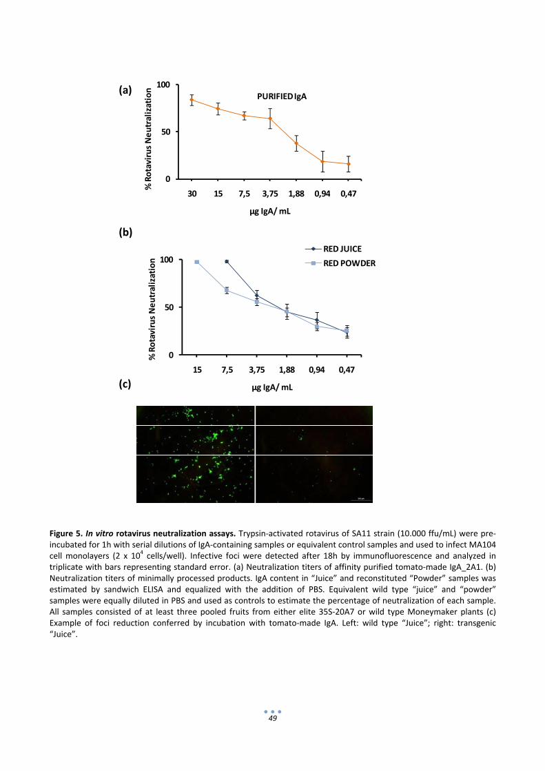
49
Figure 5. In vitro rotavirus neutralization assays. Trypsin‐activated rotavirus of SA11 strain (10.000 ffu/mL) were pre‐incubated for 1h with serial dilutions of IgA‐containing samples or equivalent control samples and used to infect MA104 cell monolayers (2 x 10⁴ cells/well). Infective foci were detected after 18h by immunofluorescence and analyzed in triplicate with bars representing standard error. (a) Neutralization titers of affinity purified tomato‐made IgA_2A1. (b) Neutralization titers of minimally processed products. IgA content in “Juice” and reconstituted “Powder” samples was estimated by sandwich ELISA and equalized with the addition of PBS. Equivalent wild type “juice” and “powder” samples were equally diluted in PBS and used as controls to estimate the percentage of neutralization of each sample. All samples consisted of at least three pooled fruits from either elite 35S‐20A7 or wild type Moneymaker plants (c) Example of foci reduction conferred by incubation with tomato‐made IgA. Left: wild type “Juice”; right: transgenic “Juice”.
0
50
100
30 15 7,5 3,75 1,88 0,94 0,47
% Rotavirus Neutralization
µg IgA/ mL
PURIFIED IgA
0
50
100
15 7,5 3,75 1,88 0,94 0,47
% Rotavirus Neutralization
µg IgA/ mL
RED JUICE
RED POWDER
(a)
(b)
(c)
50
2.6 Rosea1 and Delila transgenes can be used to confer identity preservation to IgA‐expressing tomatoes
Recombinant biofactory strategies involving food crops may benefit from the introduction of
labelling traits. Recently, transgenic Del ⁄ Ros1 MicroTom tomato fruits with an intense purple colour have
been described (Butelli et al., 2008). Several traditionally red tomato cultivars are known to display a bronze
coloration owing to the accumulation of anthocyanins in the peel. In contrast, Del ⁄ Ros1 accumulates large
amounts of anthocyanins not only in the peel of the fruit, but also in the pericarp and jelly tissues. This
feature makes Del ⁄ Ros1 tomatoes ideal for transgenic labelling and makes traceability easier for both fruit
biofactories and their derived ‘juice’ and ‘powder’ products. Del ⁄ Ros1 genotype was initially introduced into
the dwarf, cherry tomato MicroTom cultivar. However, large globe‐type tomatoes are preferable in fruit
biofactory strategies to maximize yields of fruit material. With this goal in mind, purplefruited MicroTom Del
⁄ Ros1 plants (kindly provided by Prof. Martin) were crossed with globe‐type, wild‐type Moneymaker plants.
The offspring showing combined globe‐type and purple phenotypes were single‐seed selected up to the F7
generation. A plant from the F7 generation was sexually crossed with a T1 35S_IgA‐expressing line (35S_3),
and the offspring of this cross were screened in an antigen‐ELISA for IgA expression. Eight of thirteen plants
were found to be IgA positive and were grown to maturity (referred as CR lines). All of them resulted in a
purple‐ coloured fruit phenotype. Moreover, this colour trait was maintained during fruit processing steps,
as ‘juice’ and ‘powder’ samples showed a clearly distinguishable purple colour, in contrast with the light red
colour of wild‐type‐derived products (Figure 6a). Fruits from the F1‐generation CR lines were then tested for
IgA expression, showing maximum antibody levels of 6.2 μg ⁄ g FW (0.8% TSP). CR lines were then self‐
pollinated to F3 generation. As a result, an elite F3 plant with purple fruit, called CR2‐3‐8, showed maximum
IgA expression levels of 33.5 ± 4.2 lg μgA ⁄ g FW, equivalent to 1.9 ± 0.3% TSP, which is a level comparable to
that found in the red‐fruited T3 35S_20A7 line (shown in Table 1). Endpoint ELISA titres of tomato powder
were also conducted, showing titration curves consistent with the estimated IgA levels and confirming the
conservation of the biological activity of the IgA expressed in the purple fruit (Figure 6b). Finally, the
neutralization activity of IgA powder from purple fruit was confirmed in virus neutralization (VN) assays
(Figure 6c). In these assays, disruption of the cell monolayer was observed when samples derived from
purple powder were incubated at low dilutions (up to 1 : 12 w:v, not shown). To dissect any possible effect
of the anthocyanin background on the cell monolayer, VN titres of the CR2‐3‐8 ‘powder’ samples were
performed at dilutions above the cell disruption threshold (1 : 24 w:v onwards) using an IgA‐free sibling
purple‐fruited line CR2‐3‐5 as a control. Purple IgA‐expressing tomato fruit samples also strongly neutralized
rotavirus, with a calculated IC50 value of 1.98 μg ⁄ mL of IgA (Figure 6c), a value similar to that observed
previously in the red tomato ‘powder’.
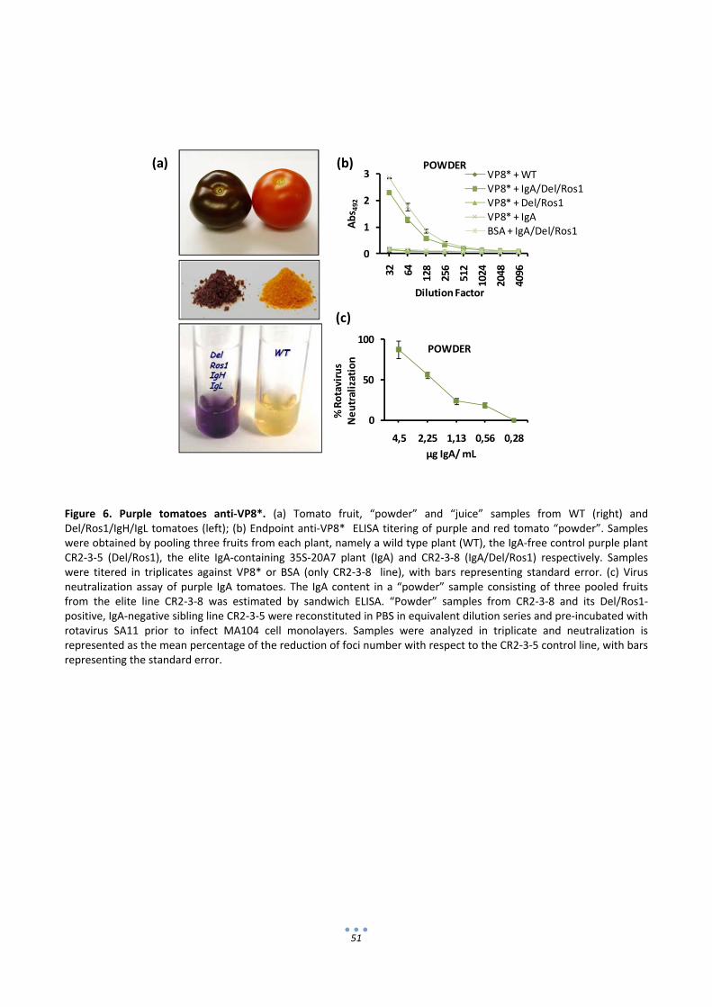
51
Figure 6. Purple tomatoes anti‐VP8*. (a) Tomato fruit, “powder” and “juice” samples from WT (right) and Del/Ros1/IgH/IgL tomatoes (left); (b) Endpoint anti‐VP8* ELISA titering of purple and red tomato “powder”. Samples were obtained by pooling three fruits from each plant, namely a wild type plant (WT), the IgA‐free control purple plant CR2‐3‐5 (Del/Ros1), the elite IgA‐containing 35S‐20A7 plant (IgA) and CR2‐3‐8 (IgA/Del/Ros1) respectively. Samples were titered in triplicates against VP8* or BSA (only CR2‐3‐8 line), with bars representing standard error. (c) Virus neutralization assay of purple IgA tomatoes. The IgA content in a “powder” sample consisting of three pooled fruits from the elite line CR2‐3‐8 was estimated by sandwich ELISA. “Powder” samples from CR2‐3‐8 and its Del/Ros1‐positive, IgA‐negative sibling line CR2‐3‐5 were reconstituted in PBS in equivalent dilution series and pre‐incubated with rotavirus SA11 prior to infect MA104 cell monolayers. Samples were analyzed in triplicate and neutralization is represented as the mean percentage of the reduction of foci number with respect to the CR2‐3‐5 control line, with bars representing the standard error.
(a) (b)
(c)
0
50
100
4,5 2,25 1,13 0,56 0,28
% Rotavirus
Neutralization
µg IgA/ mL
POWDER
0
1
2
3
32 64
128
256
512
1024
2048
4096
Abs 4
92
Dilution Factor
POWDERVP8* + WTVP8* + IgA/Del/Ros1VP8* + Del/Ros1VP8* + IgA BSA + IgA/Del/Ros1

52
3. Discussion
Diarrhoeas caused by enteric pathogens kill an estimated 2.5 million infants worldwide per year
(Kosek et al., 2003), and 500.000 of the fatalities are caused by rotavirus alone (Parashar et al., 2003).
Recently, two Jennerian vaccines against rotavirus have been successfully launched. However, challenges
remain regarding the potential effectiveness of oral live vaccines in developing countries in view of the
prevalence of competing intestinal flora in children, the occurrence of mixed infections, the high levels of
maternally transmitted antibodies and general malnutrition. Recombinant production of rotavirus antigens
in plants has been proposed as an alternative to traditional production platforms (Birch‐Machin et al., 2004;
Choi et al., 2005). In addition, passive oral immunization emerges as a complementary strategy, as this
approach has repeatedly been shown effective against enteric infections (Corthesy, 2002). In this context,
the availability of an inexpensive source of neutralizing antibodies for mucosal protection might serve as a
valuable complement to current vaccination strategies. This work shows that recombinant antibodies
against the model rotavirus strain SA11 accumulate to considerable levels in tomato fruits and that
neutralizing antibody preparations can be obtained from edible fruit tissues using extremely simple
processing steps. Therefore, these results suggest that the production of antibodies in edible fruits can serve
as an economically viable complement for combating enteric diseases, particularly in developing countries.
Oral delivery of minimally processed products derived from edible plant organs has been successfully
assayed in both active and passive immunization models. In active vaccination strategies, the context
provided by the plant tissue has been eventually shown to positively influence protection. This might be
related to the natural antigen encapsulation provided by plant cells and the adjuvant effect provided by
some of the components of the plant extract. The alkaloid tomatine has been proposed as an adjuvant,
potentiating the response observed when tomato‐made norovirus virus‐like particles were used for oral
delivery (Zhang et al., 2006b). In another example, oral boosts with chloroplast‐made plague fusion antigen
F1‐V without standard adjuvants but delivered as crude plant material performed as well as adjuvated
subcutanous boosts (Arlen et al., 2008). More recently, a chloroplast‐based strategy using lettuce as
production platform conferred dual protection against cholera and malaria in mice (Davoodi‐Semiromi et al.,
2010). In the veterinary field, oral passive protection using plant extracts has been sucessfully reported in
pigs for protection against transmissible gastroenteritis virus (Monger et al., 2006) and in poultry for
protection against coccidiosis (Zimmermann et al., 2009). In the latter case, pea‐derived scFv against Eimera
protozoa showed an enhanced prophylactic activity. It is likely that protease inhibitors present in pea seeds
might contribute to the higher stability of the protein and therefore enhanced protection. Herein, we show
that preparations of IgA‐expressing tomato fruit are at least as efficient as purified recombinant IgA in
neutralizing rotavirus infection, indicating that (i) neutralizing activity is not affected by processing and (ii)
53
the Fab’ fragments predominant in fruit tissues are efficient for VN. Moreover, the ‘juice’ and especially the
‘powder’ formulations seemed even more effective than purified IgA in preventing rotavirus infection. This
could be explained by a protective role of the extract in the stability of the antibody or by the contribution of
additional components of the extract acting cooperatively with the antibody in neutralizing the infection.
The effect of the edible plant extracts to enhance VN needs further investigation, as it may boost the
effectiveness of passive protection strategies.
Tomato is a highly productive largely self‐pollinated crop that is well adapted to greenhouse
cultivation under the confined conditions required for molecular farming. Having low protein content per
fresh weigh, fruits have sometimes been discarded as potential biofactories. However, as demonstrated
here, once water is removed from the fruit by drying, the antibody production in fruit approaches highly
competitive levels of gram per kilogram of dry weight. Considering an average production of 6 kg of tomato
(FW) per plant, current IgA yields in fruits would lead to production levels reaching 0.5 g of antibody (IgA
plus Fab’s) per tomato plant, which can be administered as semicrude formulations where the remaining
components are generally regarded as safe. Moreover, downstream processing may benefit from ‘state of
the art’ tomato food ⁄ beverage processing technology. For instance, liquid spry‐drying, a process
increasingly used in antibody formulation (Maa et al., 1998), is also employed in the food industry for the
production of tomato powder (Santos de Sousa et al., 2008). Furthermore, the availability of single‐step
affinity purification systems as the SSL7‐ agarose described here offers an economically relevant tool to
obtain partially purified formulations with increased concentration and ⁄ or safety profile.
Zhou et al (2008) reported an almost complete disappearance of HIV recombinant antigens upon
ripening in transplastomic tomato fruits and therefore proposed the use of green‐fruited tomato varieties
for molecular farming. Ripening seems to affect the protein biosynthesis capacity in plastids more drastically
than in the nucleus ⁄ cytoplasm. We found only a moderate decrease in the activity levels of nuclear‐
encoded antibody in ripened fruit. Moreover, the stability of Fab’ fragments during ripening and postharvest
seems key in the maintenance of the neutralizing activity. The use of green‐fruited tomatoes is,
nevertheless, an interesting proposal also for nuclear‐encoded transgenes, as it may contribute to further
increases in yields. Further pyramiding of ripening‐related genes could lead to new tomato varieties better
adapted to molecular farming. For example, by disrupting ripening‐associated disadvantageous traits (e.g.
protease activity or chromoplast transition), while preserving others (e.g. reduction in antinutrients or
responsiveness of engineered promoters), the deleterious effects of ripening on pharmacological production
may be obviated.
IgA_2A1 made in tomato fruit was engineered for in planta production by inserting the variable
regions of scFv_2A1 into the constant regions of human IgA and by placing the resulting antibody coding
regions under the control of plant promoters. A single‐chain 2A1 antibody fragment was previously isolated

54
in a phage display screening against VP8* peptide, a rotavirus surface antigen known to induce neutralizing
antibody responses (Higo‐Moriguchi et al., 2004). It is generally accepted that broad‐spectrum passive
immunization against intestinal pathogens including rotavirus will benefit from polyclonal mixes of
neutralizing antibodies. This could be achieved either by combining different monoclonal transgenic lines or
by engineering single transgenic lines producing a combination of monoclonal antibodies. In both cases,
phage display constitutes an interesting source of antigen‐binding variable regions for in planta production.
However, we and others (Ballester et al., 2010; Verma et al., 2010; Wieland et al., 2006) have repeatedly
observed strong differences in the stability of different phage display–derived scFv or scFv‐reconstituted full‐
size antibodies when expressed in planta. Therefore, when using phage display as a source for variable
antibody regions, it is important to identify stable antibody idiotypes before undertaking stable
transformation. For this, it is crucial to establish (i) flexible tools for scFv grafting and (ii) fast in planta
expression systems, preferably in the same tissues in which the final production is intended. In our
laboratory, we previously developed a transient expression system for tomato fruits known as fruit
agroinjection, which allows facile construct testing in fruit tissues such as placenta, gel and inner pericarp
(Orzaez et al., 2006). In addition, we have adopted a grafting procedure based on restriction ⁄ ligation and
Gateway recombination that facilitates the cloning of new variable regions into IgA constant regions. The
need for an in planta selection step is illustrated in this work. We took advantage of the aforementioned
tools to select among three anti‐VP8* full‐length antibodies, two of which had to be discarded prior to stable
transformation as they showed very low levels of HC accumulation.
In the final design of the recombinant antibody, a number of additional decisions were made. The
endogenous signal peptide (SP) was substituted with the SP of a pectate lyase expressed in fruit to ensure
processing of the IgA protein in the fruit tissues. IgA was chosen as full antibody isotype, as it will facilitate, if
required for increased efficiency, further conversion into secretory IgA (Crottet and Corthesy, 2001; Ma et
al., 1995). Two transformation approaches were undertaken, involving in trans and in cis cotransformation,
the latter using an additional fruit‐operating NH promoter (Estornell et al., 2009). The NH_IgA approach
resulted, as expected, in very low expression in vegetative tissues compared with fruits. Fruit‐specific
expression may be appreciated as a biosafety measure and may avoid the diversion of synthetic capacity of
the crop towards unprofitable protein production in vegetative tissues. However, in this case, the in trans
strategy employing the same 35S promoter for both HC and LC yielded higher IgA levels in fruits. The 35S
promoter has been previously shown to operate at high levels in fruits during development and ripening
(Estornell et al., 2009) and may have contributed to the high IgA expression levels in spite of a general
decline in transcriptional activity that takes place during late ripening.
The high accumulation of ER‐retained IgA_2A1 observed in transient fruit expression assays
prompted us to use ER retention in stable versions of IgA expression. This effect has been reported
55
repeatedly for plant‐made antibodies (Orzaez et al., 2009) and may be indicative of a lower level of protease
activity in the ER milieu. ER retention prevents complex glycosylation in the Golgi apparatus, particularly the
addition of fucose and xylose plant‐specific residues. It has been argued that the presence of immunogenic
fucose and xylose in plant‐made antibodies could lead to allergic reaction, and ER retention could be used as
a preventive measure (Sriraman et al., 2004). However, it is unlikely that these residues are allergenic
through the mucosal route given the continuous exposure of mucosa to plant glycosylation patterns in food.
Also, in the design of hIgA_2A1, we incorporated the HCaα1 coding region. IgA1 has an extended shape
compared with IgA2, as a result of an elongated hinge region. It has been proposed that the IgA1 shape
facilitates the cross‐linking of viral particles and therefore might provide specialized functions in the
protection against viral pathogens (Bonner et al., 2009).
Edible plant organs provide clear advantages in particular molecular farming approaches. However,
the concerns about contamination of the food chain with engineered contents are often reasonably raised
when pharmaceuticals are produced in food crops. We propose here the use of natural coloured plant
compounds for labelling transgenic crops by means of metabolic engineering. A relatively small amount of
otherwise health‐promoting compounds such as anthocyanins, a natural component in many edible berries,
is sufficient to label IgA‐producing plants. These compounds confer an intense purple colour ensuring the
traceability of transgenic fruits and their derivatives. F1 plants coming from the initial crossing of purple and
IgA‐expressing plants showed considerably lower IgA levels than those measured in the elite T3 35S_IgA‐
expressing lines. This raised the question of a possible dilution effect of the recombinant protein production
in the anthocyanin background. However, when purple IgA plants were bred up to F3, a considerable
increase in IgA content was observed, reaching levels of 33.5 μg IgA ⁄ g FW, close to those reported for red
IgA plants (41.2 μg ⁄ g FW). This indicates that the anthocyanin background has a minimal effect on the levels
of IgA accumulation in fruits. Moreover, IgA from the transgenically labelled purple tomatoes also showed
anti‐VP8* binding and rotavirus neutralizing activities similar to those described for red tomatoes, again
indicating that IgA activity is not affected by the presence of anthocyanins. Disruption of cell monolayers was
observed in VN assays when samples derived from purple ‘powder’ were incubated at low dilutions. It has
previously been reported that some phenolic compounds may react with constituents of certain cell culture
media (Long et al., 2010). These reactions can result in the oxidation of phenol to produce a range of toxic
products such as H2O2. These effects can be regarded as artifactual as they result from the interaction with
the culture media and are not likely to take place in vivo. Moreover, when VN assays were performed at
dilutions above the cell disruption threshold, the neutralizing effect of recombinant IgA was evident.
A further degree of safety can be achieved by genetically linking the antibody and the Del ⁄ Ros1
modules (e.g. including both modules in the same T‐DNA). The same synthetic biology rational can be used
to include additional safety modules as male sterility as well as any other with relevance to antibody
56
production as glycoengineering, decreased protease activity, etc. leading to the design of highly optimized
platforms for molecular farming (Sarrion‐Perdigones et al., 2011a).
This is, to our knowledge, the first report showing viral neutralization activity of a plant‐produced
antibody in minimally processed edible fruit extracts. The results support the need for a detailed
investigation into the biosafety and in vivo effectiveness of fruit‐based passive immunization. Moreover, we
created the first example of a biofortified plant crop additionally displaying prophylactic activity. The model
plant designed here is an example of how gene stacking, either by sexual crossing or by other means, can
lead to new agricultural products with added value and new applications. The introduction of four
transgenes (HC, LC, Ros1 and Del) in a single tomato plant resulted in a completely new product: a
transgenically labelled, orally safe, inexpensive fruit juice with a health‐promoting (antioxidant) activity
(Butelli et al., 2008) and anti‐rotavirus prophylactic potential.

57
4. Experimental procedures
4.1 DNA constructs and vectors
The DNA sequences corresponding to the constant regions of human alpha heavy (HCa) and lambda
light (LCk) antibody chains were obtained from the human library clones EHS1001‐ 9024643 and EHS1001‐
9024145, respectively (Open Biosystems, Huntsville, AL). LCk was PCR‐amplified in a nested reaction using
D06mar01 and D06mar07 oligos as overlapping forward primers and D06mar08 as reverse primer.
D06mar01 and D06mar07 overlapping primers were used to fuse to the 5’end of LCk a number of elements,
namely an attB1 recombination site, a Kozak sequence, the SP from the tomato pectate lyase and a small
polylinker containing ApaLI and NotI restriction sites. In another construct, the D06mar08 primer
incorporated a KDEL peptide, a stop codon and an attB2 recombination site in the 3¢ end of the LCk coding
region (Table S1). The resulting LCk expression cassette still lacking the sequence corresponding to the VL
was BP‐cloned in pDNOR221 vector, generating an ENTRY vector named as pENTR_LCk. In a similar fashion, a
pENTR_HCa vector was created for the convenient cloning of variable regions of the HC into an IgCa frame.
In this case, cloning of HCα was preceded by a step of directed mutagenesis designed to silently remove NcoI
and XhoI sites from HCα coding sequence. For this purpose, HCα was preamplified in three contiguous
fragments using oligonucleotide pairs D03Mar00 ⁄ D03Mar19, D06Mar03 ⁄ D03Mar04 and D03mar20 ⁄
D03Mar06, respectively, and subsequently joined by overlapping PCR. The resulting mutagenized HCα was
reamplified in a nested reaction with D06mar01 and D06mar02 as forward primers, which incorporated
attB1, the pectate lyase SP and NcoI ⁄ XhoI cloning sites. At the 3’ end, the D06Mar06 primer incorporated a
stop codon, the KDEL peptide and attB2. The resulting HCα expression cassette was BP‐cloned into
pDONR221 vector yielding pENTR_HCα vector.
The sequences of the variable antibody regions reacting against VP8* were cloned from a previously
described anti‐VP8 scFv collection selected from the phage display human antibody library Griffin.1
(Monedero et al., 2004). The Griffin.1 library is a large naïve human scFv phagemid library (total diversity of
1.2 x 10⁹) constructed from synthetic V‐gene segments made by recloning the heavy‐chain and light‐chain
variable regions from the lox library vectors (Griffiths et al., 1994) into the phagemid vector pHEN2. VL
fragments (2A1, 2B3 and 2E4) were ApaLI ⁄ NotI‐digested and cloned into pENTRY_LCk ApaLI ⁄ ‐ NotI sites,
generating pENTR_IgL vectors. In parallel, VH fragments (2A1, 2B3 and 2E4) were NcoI ⁄ XhoI excised from
Griffin.1 vector and cloned in the equivalent sites of pENTR_HCa, generating pENTR_IgHa vectors. To
harbour the expression cassettes of the individual antibody chains, pBINJITGW and pKGW7.0 were used as
destination vectors. pKGW7.0 containing 35S promoter and terminator was kindly provided by Karimi et al.
(2002). pBINJGW was engineered by introducing a Gateway cassette (Invitrogen, Carlsbad, CA) in the SmaI
site of pBINJIT plasmid, following manufacturer’s procedures. Binary expression vectors for heavy and light

58
antibody chains were obtained by performing LR reactions between ENTRY vectors (e.g. pENTR_IgL_2A1 and
pENTR_IgHα_2A1) and binary destination vectors, generating plant antibody expression clones (e.g. pEXP_
35SIgL_2A1 and pEXP_ 35SIgHα_2A1).
Heavy and LCs expression cassettes were also combined in a single binary vector under the
constitutive 35S and the fruitoperating NH promoters, respectively. For this purpose, the IgL_2A1 coding
region was PCR‐amplified using primers L07Nov07 and L07Nov08 containing attB4R and attB3R extensions
and BP‐recombined into pDONR221P4r‐P3r (Invitrogen), generating pENTR4R3R_IgL2A1. A triple gateway
recombination reaction was then performed between vectors pENTR1‐4_NH, pENTR3‐2_Tnos, both
belonging to the previously described pENFRUIT collection of combinatorial vectors (Estornell et al., 2009),
and pENTR4r3r_IgL2A1, using pKGW.0 as a destination vector. In this way, an expression cassette was
generated comprising the NH promoter fused to IgL‐2A1 and the Tnos terminator. The whole 4.9‐Kb
expression cassette was PCR‐amplified using primers L07Jun05 and L08Jan01, containing AscI and SalI
recognition sites, respectively. The resulting PCR fragment was digested with AscI ⁄ SalI and cloned into
pEXP_35SIgHα_2A1, generating the binary plasmid pEXP_35SIgH_NHIgL, which combines heavy and light
antibody chains under the control of two different promoters both operating in the tomato fruit.
4.2 Tomato transformation, plant material and sample preparation
Plasmids were transferred to Agrobacterium tumefaciens LBA4404 strain by electroporation and
used for tomato stable transformation (var. Moneymaker), as described previously (Ellul et al., 2003). For
transient expression, plant expression plasmids were transferred to Agrobacterium tumefaciens strain C58
and assayed transiently in MicroTom fruits, as described previously (Orzaez et al., 2006). For sample
preparation, plant tissues (leaves and tomato fruit) were ground with a mortar and pestle to a fine powder
under liquid nitrogen and stored at ‐80º until used. For ‘juice’ samples, proteins were extracted with three
volumes (w ⁄ v) of PBS pH 7.4. After mixing, the suspension was centrifuged twice at 4°C at maximum speed
and the supernatant was immediately used for further analysis. For ‘dried juice’ samples, total protein was
extracted in the same manner, followed by a 24‐h freeze‐drying step. The sample was reconstituted in three
volumes of water (w ⁄ v) for analysis. The ‘powder’ samples were obtained from fine tissue powder after a
24‐h freeze‐drying treatment; total protein was extracted with three volumes of PBS pH 7.4 (w ⁄ v),
centrifuged twice at 4ºC at maximum speed, and the supernatant was used for the analysis.
4.3 VP8* rotavirus surface protein production
Recombinant VP8* was obtained from pQEVP8*‐transformed Escherichia coli M11. Plasmid
pQEVP8* was kindly provided by Dr. Monedero from Instituto de Agroquímica y Tecnología de Alimentos
(IATA, Valencia, Spain). VP8* expression was performed using the QIAexpressionist protocol

59
(http://www.qiagen. com). Frozen cell pellets were thawed in 20 mM Tris–HCl pH 8.0. Cells were lysed by
sonication on ice (6 x 25 s) and washed with buffer A (20 mM Tris–HCl, 0.5 M NaCl, 2 M urea and 2% Triton
X‐100) to isolate inclusion bodies. After 15 min 25.000 g spin, cells were sonicated (6 x 25 s) one more time
and washed again with the same buffer. A last wash with ureafree buffer A was made.
Inclusion bodies were solubilized with 20 mM Tris–HCl, 0.5 M NaCl, 6 M guanidine HCl, 5 mM
imidazole and 1 mM 2‐b‐mercaptoethanol at pH 8.0. The sample was stirred at room temperature for 1 h
and then centrifuged for 15 min at high speed. The remaining particles were removed by passing the sample
through a 0.22‐lm filter. Purification was performed using HisTrap HP affinity columns (GE Healthcare,
Buckinghamshire, UK) following manufacturer’s procedures. Protein refolding in column was performed with
30 mL of a lineal 6–0 M urea gradient at a flow rate of 1 mL ⁄ min, starting with buffer B (20 mM Tris–HCl, 0.5
M NaCl, 6 M urea and 1 mM 2‐mercaptoethanol) and finishing with urea‐free buffer B. The refolded protein
was eluted using a 10‐mL lineal gradient starting with buffer C (20 mM Tris–HCl, 0.5 M NaCl, 20 mM
imidazole and 1 mM 2‐mercaptoethanol) pH 8.0 and ending with the same buffer C supplemented with 500
mM imidazole.
4.4 ELISAs for the detection of VP8* binding activity and recombinant immunoglobulin A determination
Plates (Corning, New York, NY) were coated overnight with 10 lg ⁄ mL of recombinant VP8* for the
detection of VP8* binding activity or 2 μg ⁄ mL of anti‐human IgA a specific (Sigma Aldrich, St. Louis, MO) for
IgA determination, in coating buffer (50 mM carbonate buffer pH 9.8) at 4ºC. Plates were then washed four
times in PBS and blocked with a 2% (w⁄ v) solution of ECL AdvanceTM Blocking agent (GE Healthcare) in PBS‐
T [0.1% (v ⁄ v) Tween 20 in PBS]. Samples were diluted in PBS as required for each assay and incubated for 1
h at room temperature. After incubation, plates were washed four times in PBS and the anti‐human IgA a‐
specific horse radish peroxidase (HRP) 1 : 5000 (Sigma Aldrich) in 5% blocking buffer (GE Healthcare) in PBS‐T
was added and incubated for 1 h at room temperature. After four PBS washes, the substrate (o‐
phenilenediamine from Sigma Aldrich) was added and the reactions were stopped with 3 M HCl. Absorbance
was determined at 492 nm. As a control for specificity, plates were coated with 10 μg ⁄ mL of BSA. A sample
was considered positive when its absorbance was at least three times higher than the absorbance value of
the same sample in the control. Specific VP8* binding activity was estimated using serial sample dilutions,
dividing the absorbance at 492 nm of those dilutions falling within the lineal range by the total protein
content of each sample (mg). A standard curve from 0.8 to 0.1 μg ⁄ mL of commercial IgA from human
colostrum (Sigma Aldrich) was obtained to calculate the concentration of IgA in the different samples. IgA
concentration in each sample was obtained by interpolation with the IgA standard curve. For IgA
60
quantification in elite lines, three samples from each elite line were analysed and the mean ± SD for each
line was calculated.
4.5 SDS‐PAGE and Western blot analysis
Proteins were separated by SDS ⁄ PAGE in 10% and 3%–8% denaturing gels (Invitrogen). Gel staining
was carried out either with Coomassie blue or with silver staining following standard procedures. For
Western blot analysis, blots were incubated with 1 : 20.000 anti‐human IgA a‐specific HRP (Sigma Aldrich) for
the detection of the HC and 1 : 10.000 anti‐human k LC (Sigma Aldrich) followed by 1 : 10.000 μg ⁄ mL HRP‐
conjugated anti‐rabbit IgG (GE Healthcare) for the detection of the LC. Blots were developed with ECL plus
Western blotting Detection System (GE Healthcare).
4.6 Protein SSL7 affinity purification
Protein extracts prepared as explained previously were further clarified using a 0.22‐μm Stericup
(Millipore, Billerica, MA) on ice. The clarified extract was directly used for purification with staphylococcal
superantigen‐like protein 7 (SSL7) agarose columns from InvivoGen (San Diego, CA) following
manufacturer¢s protocol with minor modifications. Wash buffer was substituted by PBS (0.1 M phosphate
and 0.1 M NaCl pH 7.4). Dialysis was not needed as the samples were already in PBS and elution
neutralization was carried out mixing with PBS 1 : 1 (v ⁄ v).
4.7 Neutralization assays
Tomato IgA neutralization of rotavirus SA11 strain was assessed by immunofluorescent focus
reduction assays on MA104 (Health Protection Agency Culture Collection) cell monolayers in 96‐well cell
culture microplates (Corning) infected with rotavirus (Asensi et al., 2006). The plates were coated with 2 x
104 cells per well in minimum essential medium (MEM from Gibco‐ Invitrogen, Carlsbad, CA) supplemented
with 100 U⁄ mL penicillin, 100 lg ⁄ mL streptomycin, 2 mM glutamine, 0.7% CO3H and 10% foetal bovine
serum (FBS) and incubated for 24 h at 37 ⁰C in a CO2 stove until the monolayers were confluent. Rotavirus
suspensions [at two different concentrations, 2 x 10³ and 1 x 10⁴ fluorescing cell‐forming units per mL (f.f.u. ⁄
mL)] in MEM without FBS were activated with 10 μg ⁄ mL of trypsin type IX (Sigma Aldrich) prior to the
incubation with serial dilutions (1 : 2 to 1 : 256) of IgA samples in MEM (FBS‐free), and the resulting mixes
were used to infect the monolayers (1‐h incubation at 37 ⁰C). After incubation, the monolayers were washed
with MEM and incubated for 18 h in MEM containing 1 μg ⁄ mL trypsin at 37⁰C in a CO2 stove. Four different
IgA samples were used in the different assays: ssl‐7 purified recombinant IgA in PBS, 35S_20A7 clarified fruit
extract (‘juice’), freeze‐dried tissue samples from 35S_20A7 and CR2‐3‐8 fruits reconstituted in PBS
(‘powder’). PBS, ‘juice’ and reconstituted ‘powder’ from fruits of wild‐type Moneymaker or IgA‐free purple‐

61
fruited CR2‐ 3‐5 plants were used as mock solutions for comparisons. All fruit samples were collected and
processed between 2 and 4 days after reaching the breaker stage. After incubation, monolayers were fixed
with 100 μL ⁄ well of 1 : 1 (v : v) methanol ⁄ acetone for 15 min and then washed with PBS. An anti‐rotavirus
VP6 monoclonal antibody in PBS 1% BSA (1 : 80) was applied as the primary antibody for 1 h at 37⁰C,
followed by three washes with PBS. The secondary antibody was an antimouse IgG FITC‐labelled (Sigma,
Sigma Aldrich, St. Louis, MO) (1 : 400). The monolayers were washed three more times with PBS and stained
with Evans blue for 5 min, washed again with PBS and 20 μL of mounting fluid (Millipore) was applied to
each well. Images were obtained by excitation with 480‐nm light and acquired at 6.3x magnification factor
using a Leica MZ16F binocular equipped with a Leica DFC300FX digital camera and Leica application suite
software. The 50% inhibiting concentrations (IC50) were calculated by the method of Reed and Muench
(Reed and Muench, 1938), using the concentrations present during the antibody‐virus pre‐incubation step.
5. Acknowledgements
We want to thank Prof. Cathie Martin and Dr. Eugenio Butelli for kindly providing us with Del ⁄ Ros1
plants, Dr. Monedero for the gift of scFv and VP8* clones and Dr. Ann Powell and Dr. Anna N. Stepanova for
carefully correcting the manuscript. This work was supported by the Spanish Ministry of Science, Technology
and Innovation: Grants BIO2008‐03434, AGL2009‐ 13388‐C03‐01 and BIO2010‐15384 and P. Juarez FPU
fellowship.

62
6. References
Apel, W. and Bock, R. (2009) Enhancement of carotenoid biosynthesis in transplastomic tomatoes by induced lycopene‐to‐provitamin A conversion. Plant Physiol 151, 59‐66.
Arlen, P.A., Singleton, M., Adamovicz, J.J., Ding, Y., Davoodi‐Semiromi, A. and Daniell, H. (2008) Effective plague vaccination via oral delivery of plant cells expressing F1‐V antigens in chloroplasts. Infect Immun 76, 3640‐3650.
Asensi, M.T., Martinez‐Costa, C. and Buesa, J. (2006) Anti‐rotavirus antibodies in human milk: quantification and neutralizing activity. J Pediatr Gastroenterol Nutr 42, 560‐567.
Ballester, A.R., Molthoff, J., de Vos, R., Hekkert, B.L., Orzaez, D., Fernandez‐Moreno, J.P., Tripodi, P., Grandillo, S., Martin, C., Heldens, J., Ykema, M., Granell, A. and Bovy, A. (2010) Biochemical and molecular analysis of pink tomatoes: deregulated expression of the gene encoding transcription factor SlMYB12 leads to pink tomato fruit color. Plant Physiol 152, 71‐84.
Birch‐Machin, I., Newell, C.A., Hibberd, J.M. and Gray, J.C. (2004) Accumulation of rotavirus VP6 protein in chloroplasts of transplastomic tobacco is limited by protein stability. Plant Biotechnology Journal 2, 261‐270.
Bonner, A., Almogren, A., Furtado, P.B., Kerr, M.A. and Perkins, S.J. (2009) The nonplanar secretory IgA2 and near planar secretory IgA1 solution structures rationalize their different mucosal immune responses. J Biol Chem 284, 5077‐5087.
Bovy, A., de Vos, R., Kemper, M., Schijlen, E., Pertejo, M.A., Muir, S., Collins, G., Robinson, S., Verhoeyen, M., Hughes, S., Santos‐Buelga, C. and van Tunen, A. (2002) High‐flavonol tomatoes resulting from the heterologous expression of the maize transcription factor genes LC and C1. Plant Cell 14, 2509‐2526.
Bovy, A., Schijlen, E. and Hall, R.D. (2007) Metabolic engineering of flavonoids in tomato (Solanum lycopersicum): the potential for metabolomics. Metabolomics 3, 399‐412.
Butelli, E., Titta, L., Giorgio, M., Mock, H.P., Matros, A., Peterek, S., Schijlen, E.G., Hall, R.D., Bovy, A.G., Luo, J. and Martin, C. (2008) Enrichment of tomato fruit with health‐promoting anthocyanins by expression of select transcription factors. Nat Biotechnol 26, 1301‐1308.
Cipollini, M.L. and Levey, D.J. (1997) Secondary metabolites of fleshy vertebrate‐dispersed fruits: adaptive hypotheses and implications for seed dispersal. Am Nat 150, 346‐372.
Corthesy, B. (2002) Recombinant immunoglobulin A: powerful tools for fundamental and applied research. Trends in Biotechnology 20, 65‐71.
Crottet, P. and Corthesy, B. (2001) Secretory component delays the conversion of secretory IgA into antigen‐binding competent F(ab')2: A possible implication for mucosal defense. The Journal of Immunology 161, 5445‐5453.
Choi, N.W., Estes, M.K. and Langridge, W.H. (2005) Synthesis and assembly of a cholera toxin B subunit‐rotavirus VP7 fusion protein in transgenic potato. Mol Biotechnol 31, 193‐202.
Davoodi‐Semiromi, A., Schreiber, M., Nalapalli, S., Verma, D., Singh, N.D., Banks, R.K., Chakrabarti, D. and Daniell, H. (2010) Chloroplast‐derived vaccine antigens confer dual immunity against cholera and malaria by oral or injectable delivery. Plant Biotechnol J 8, 223‐242.
Davuluri, G.R., van Tuinen, A., Fraser, P.D., Manfredonia, A., Newman, R., Burgess, D., Brummell, D.A., King, S.R., Palys, J., Uhlig, J., Bramley, P.M., Pennings, H.M.J. and Bowler, C. (2005) Fruit‐specific RNAi‐mediated suppression of DET1 enhances carotenoid and flavonoid content in tomatoes. Nature Biotechnology 23, 890‐895.
De Muynck, B., Navarre, C. and Boutry, M. (2010) Production of antibodies in plants: status after twenty years. Plant Biotechnol J 8, 529‐563.
Ellul, P., Garcia‐Sogo, B., Pineda, B., Rios, G., Roig, L. and Moreno, V. (2003) The ploidy level of transgenic plants in Agrobacterium‐mediated transformation of tomato cotyledons (Lycopersicon esculentum Mill.) is genotype and procedure dependent (vol 106, pg 231, 2003). Theoretical and Applied Genetics 107, 190‐190.

63
Estornell, L.H., Orzaez, D., Lopez‐Pena, L., Pineda, B., Anton, M.T., Moreno, V. and Granell, A. (2009) A multisite gateway‐based toolkit for targeted gene expression and hairpin RNA silencing in tomato fruits. Plant Biotechnology Journal 7, 298‐309.
Giuliano, G., Tavazza, R., Diretto, G., Beyer, P. and Taylor, M.A. (2008) Metabolic engineering of carotenoid biosynthesis in plants. Trends Biotechnol 26, 139‐145.
Griffiths, A.D., Williams, S.C., Hartley, O., Tomlinson, I.M., Waterhouse, P., Crosby, W.L., Kontermann, R.E., Jones, P.T., Low, N.M., Allison, T.J. and et al. (1994) Isolation of high affinity human antibodies directly from large synthetic repertoires. EMBO J 13, 3245‐3260.
Higo‐Moriguchi, K., Akahori, Y., Iba, Y., Kurosawa, Y. and Taniguchi, K. (2004) Isolation of human monoclonal antibodies that neutralize human rotavirus. Journal of Virology 78, 3325‐3332.
Hood, E.E., Woodard, S.L. and Horn, M.E. (2002) Monoclonal antibody manufacturing in transgenic plants‐‐myths and realities. Curr Opin Biotechnol 13, 630‐635.
Kosek, M., Bern, C. and Guerrant, R.L. (2003) The global burden of diarrhoeal disease, as estimated from studies published between 1992 and 2000. Bull World Health Organ 81, 197‐204.
Long, L.H., Hoi, A. and Halliwell, B. (2010) Instability of, and generation of hydrogen peroxide by, phenolic compounds in cell culture media. Arch Biochem Biophys 501, 162‐169.
Ma, J.‐K.C., Lehner, T., Stabila, P., Fux, C., I and Hiatt, A. (1994) Assembly of monoclonal antibodies with IgG1 and IgA heavy chain domains in transgenic tobacco plants. European Journal of Immunology 24(1): 131 138 *LHM LIBRARY HOLDINGS MESSAGE: Catalogue record in AGRALIN, 131.
Ma, J.K., Hiatt, A., Hein, M., Vine, N.D., Wang, F., Stabila, P., van Dolleweerd, C., Mostov, K. and Lehner, T. (1995) Generation and assembly of secretory antibodies in plants. Science 268, 716‐719.
Ma, J.K., Hikmat, B.Y., Wycoff, K., Vine, N.D., Chargelegue, D., Yu, L., Hein, M.B. and Lehner, T. (1998) Characterization of a recombinant plant monoclonal secretory antibody and preventive immunotherapy in humans. Nat Med 4, 601‐606.
Maa, Y.F., Nguyen, P.A., Sit, K. and Hsu, C.C. (1998) Spray‐drying performance of a bench‐top spray dryer for protein aerosol powder preparation. Biotechnol Bioeng 60, 301‐309.
Monedero, V., Rodriguez‐Diaz, J., Viana, R., Buesa, J. and Perez‐Martinez, G. (2004) Selection of single‐chain antibodies against the VP8*subunit of rotavirus VP4 outer capsid protein and their expression in Lactobacillus casei. Applied and Environmental Microbiology 70, 6936‐6939.
Monger, W., Alamillo, J.M., Sola, I., Perrin, Y., Bestagno, M., Burrone, O.R., Sabella, P., Plana‐Duran, J., Enjuanes, L., Garcia, J.A. and Lomonossoff, G.P. (2006) An antibody derivative expressed from viral vectors passively immunizes pigs against transmissible gastroenteritis virus infection when supplied orally in crude plant extracts. Plant Biotechnol J 4, 623‐631.
Orzaez, D., Granell, A. and Blazquez, M.A. (2009) Manufacturing antibodies in the plant cell. Biotechnol J 4, 1712‐1724.
Orzaez, D., Mirabel, S., Wieland, W.H. and Granell, A. (2006) Agroinjection of tomato fruits. A tool for rapid functional analysis of transgenes directly in fruit. Plant Physiol 140, 3‐11.
Parashar, U.D., Hummelman, E.G., Bresee, J.S., Miller, M.A. and Glass, R.I. (2003) Global illness and deaths caused by rotavirus disease in children. Emerg Infect Dis 9, 565‐572.
Rademacher, T., Sack, M., Arcalis, E., Stadlmann, J., Balzer, S., Altmann, F., Quendler, H., Stiegler, G., Kunert, R., Fischer, R. and Stoger, E. (2008) Recombinant antibody 2G12 produced in maize endosperm efficiently neutralizes HIV‐1 and contains predominantly single‐GlcNAc N‐glycans. Plant Biotechnol J 6, 189‐201.
Ramessar, K., Rademacher, T., Sack, M., Stadlmann, J., Platis, D., Stiegler, G., Labrou, N., Altmann, F., Ma, J., Stoger, E., Capell, T. and Christou, P. (2008) Cost‐effective production of a vaginal protein microbicide to prevent HIV transmission. Proc Natl Acad Sci U S A 105, 3727‐3732.
Santos de Sousa, A., Vilela Borges, S., Ferreira Magalhaes, N., Vaz Ricardo, H. and Damico Azevedo, A. (2008) Spry‐dried tomato powder: reconstitution properties and colour. Braz Arch Biol Technol 51, 807‐814.
Sarrion‐Perdigones, A., Falconi, E.E., Zandalinas, S.I., Juarez, P., Fernandez‐del‐Carmen, A., Granell, A. and Orzaez, D. (2011) GoldenBraid: an iterative cloning system for standardized assembly of reusable genetic modules. PLoS One 6, e21622.

64
Sriraman, R., Bardor, M., Sack, M., Vaquero, C., Faye, L., Fischer, R., Finnern, R. and Lerouge, P. (2004) Recombinant anti‐hCG antibodies retained in the endoplasmic reticulum of transformed plants lack core‐xylose and core‐alpha(1,3)‐fucose residues. Plant Biotechnology Journal 2, 279‐287.
Verma, D., Kanagaraj, A., Jin, S., Singh, N.D., Kolattukudy, P.E. and Daniell, H. (2010) Chloroplast‐derived enzyme cocktails hydrolyse lignocellulosic biomass and release fermentable sugars. Plant Biotechnol J 8, 332‐350.
Walmsley, A.M., Alvarez, M.L., Jin, Y., Kirk, D.D., Lee, S.M., Pinkhasov, J., Rigano, M.M., Arntzen, C.J. and Mason, H.S. (2003) Expression of the B subunit of Escherichia coli heat‐labile enterotoxin as a fusion protein in transgenic tomato. Plant Cell Rep 21, 1020‐1026.
Weintraub, J.A., Hilton, J.F., White, J.M., Hoover, C.I., Wycoff, K.L., Yu, L., Larrick, J.W. and Featherstone, J.D. (2005) Clinical trial of a plant‐derived antibody on recolonization of mutans streptococci. Caries Res 39, 241‐250.
Wieland, W.H., Lammers, A., Schots, A. and Orzaez, D.V. (2006) Plant expression of chicken secretory antibodies derived from combinatorial libraries. J Biotechnol 122, 382‐391.
Zhang, X., Buehner, N.A., Hutson, A.M., Estes, M.K. and Mason, H.S. (2006a) Tomato is a highly effective vehicle for expression and oral immunization with Norwalk virus capsid protein. Plant Biotechnol J 4, 419‐432.
Zhang, X.R., Buehner, N.A., Hutson, A.M., Estes, M.K. and Mason, H.S. (2006b) Tomato is a highly effective vehicle for expression and oral immunization with Norwalk virus capsid protein. Plant Biotechnology Journal 4, 419‐432.
Zhou, F., Badillo‐Corona, J.A., Karcher, D., Gonzalez‐Rabade, N., Piepenburg, K., Borchers, A.M., Maloney, A.P., Kavanagh, T.A., Gray, J.C. and Bock, R. (2008) High‐level expression of human immunodeficiency virus antigens from the tobacco and tomato plastid genomes. Plant Biotechnol J 6, 897‐913.
Zimmermann, J., Saalbach, I., Jahn, D., Giersberg, M., Haehnel, S., Wedel, J., Macek, J., Zoufal, K., Glunder, G., Falkenburg, D. and Kipriyanov, S. (2009) Antibody expressing pea seeds as fodder for prevention of gastrointestinal parasitic infections in chickens. BMC Biotechnology 9, 79.

65
7. Supplementary material
Supplementary Table 1. Primer Sequences for DNA constructs and Vector.
Primer Sequence
D06Mar01 ggggacaagtttgtacaaaaaagcaggcttccacaatgggcacttcctctgtttttctactattccttctttcttttcttctccttctcccg
D06Mar07 tttcttttcttctccttctcccgagtgcacagtcgacagcggccgccgcccccactgtcactctgttcc
D06Mar08 ggggaccactttgtacaagaaagctgggttctagactatgaacattctgtaggggccactg
D06Mar00 cgagcgcatccccgaccagccccaaggtct
D06Mar19 tgaacctaagagcaggtcctcaagggccgg
D06Mar03 tgcaccgaccggcccttgaggac
D06Mar04 tgaaggtcttcccatgattccaaggc
D06Mar20 ccgggctgtgccgagccttggaatcatggga
D06Mar02 ttcttctccttctcccgtccctccttgccatggccgtcaccgtctcgagcgcatccccgaccagccccaa
D06Mar06 ggggaccactttgtacaagaaagctgggttcaaagttcatctttgtagcaggtgccgtccacct
L07Nov07 ggggacaacttttctatacaaagttgcaatgggcacttcctctgtttttc
L07Nov08 ggggacaactttattatacaaagttgtctaaagttcatcttatgaacat
L07Jun05 cgacaatctgatccaagctcaa
L08Jan01 ccgcgtcatcggcgcgccgggtcataacgtgactccctt
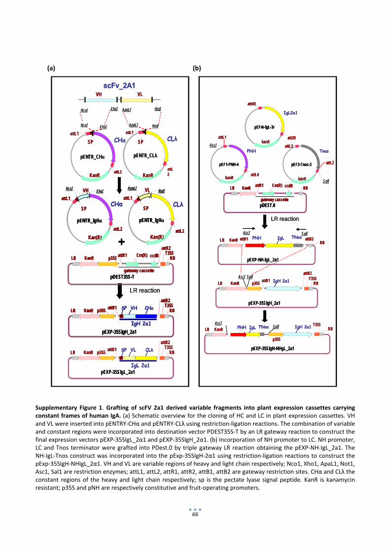
66
(a) (b)
Supplementary Figure 1. Grafting of scFV 2a1 derived variable fragments into plant expression cassettes carrying constant frames of human IgA. (a) Schematic overview for the cloning of HC and LC in plant expression cassettes. VH and VL were inserted into pENTRY‐CHα and pENTRY‐CLλ using restriction‐ligation reactions. The combination of variable and constant regions were incorporated into destination vector PDEST35S‐T by an LR gateway reaction to construct the final expression vectors pEXP‐35SIgL_2α1 and pEXP‐35SIgH_2α1. (b) Incorporation of NH promoter to LC. NH promoter, LC and Tnos terminator were grafted into PDest.0 by triple gateway LR reaction obtaining the pEXP‐NH‐IgL_2a1. The NH‐IgL‐Tnos construct was incorporated into the pExp‐35SIgH‐2α1 using restriction‐ligation reactions to construct the pExp‐35SIgH‐NHIgL_2α1. VH and VL are variable regions of heavy and light chain respectively; Nco1, Xho1, ApaL1, Not1, Asc1, Sal1 are restriction enzymes; attL1, attL2, attR1, attR2, attB1, attB2 are gateway restriction sites. CHα and CLλ the constant regions of the heavy and light chain respectively; sp is the pectate lyase signal peptide. KanR is kanamycin resistant; p35S and pNH are respectively constitutive and fruit‐operating promoters.
scFv_2A1VH VL
XhoINcoI ApaLI NotI
pENTR_CLλ
attL1
attL2
CLλSP
NotIApaLI
pENTR_CHα
attL1
attL2
CHαSP
XhoINcoI
KanR KanR
pENTR_IgHα
VH
Kan(R)
attL2
attL1CHα
SP
NcoIXhoI
pENTR_IgHα
VL
Kan(R)attL2
attL1CLλ
SP
ApaLINotI
+
LR reaction
Cm(R) ccdB
gateway cassette
KanR p35ST35SattR1
attR2
LB RB
pDEST35S-T
IgL 2a1
KanR p35ST35SattB1
attB2
LB RBSP VL CLλ
IgH 2a1
KanR p35ST35SattB1
attB2
LB RBSP VH CHα
pEXP-35SIgH_2a1
pEXP-35SIgL_2a1
scFv_2A1VH VL
XhoINcoI ApaLI NotI
pENTR_CLλ
attL1
attL2
CLλSP
NotIApaLI
pENTR_CHα
attL1
attL2
CHαSP
XhoINcoI
KanR KanR
pENTR_IgHα
VH
Kan(R)
attL2
attL1CHα
SP
NcoIXhoI
pENTR_IgHα
VH
Kan(R)
attL2
attL1CHα
SP
NcoIXhoI
pENTR_IgHα
VL
Kan(R)attL2
attL1CLλ
SP
ApaLINotI
pENTR_IgHα
VL
Kan(R)attL2
attL1CLλ
SP
ApaLINotI
+
LR reaction
Cm(R) ccdB
gateway cassette
KanR p35ST35SattR1
attR2
LB RB
pDEST35S-T
Cm(R) ccdB
gateway cassette
KanR p35ST35SattR1
attR2
LB RB
pDEST35S-T
IgL 2a1
KanR p35ST35SattB1
attB2
LB RBSP VL CLλ
IgH 2a1
KanR p35ST35SattB1
attB2
LB RBSP VH CHα
pEXP-35SIgH_2a1
IgH 2a1
KanR p35ST35SattB1
attB2
LB RBSP VH CHα
IgH 2a1
KanR p35ST35SattB1
attB2
LB RBSP VH CHα
pEXP-35SIgH_2a1
pEXP-35SIgL_2a1
Cm(R) ccdB
gateway cassette
KanRattR1
LB RB
pDEST.0
LR reaction
KanR PNH TNosattB1 attB2LBRB
IgL
pEXP-NH-IgL_2a1
pEF1-PNH-4
attL1
attL4
PNH
kanR
pEF4r-IgL-3r
att4R
att3R
IgL2a1
kanR
pEF3-Tnos-2
attL3
attL2
Tnos
kanR
AscI
SalI
AscI SalI
KanR p35ST35SattB1
attB2
LB RBIgH 2a1
pEXP-35SIgH_2a1
AscI SalI
KanR PNH TNosLB RBIgL
pEXP-35SIgH-NHgL_2a1
AscISalI
p35S
T35SIgH 2a1
Cm(R) ccdB
gateway cassette
KanRattR1
LB RB
pDEST.0
Cm(R) ccdB
gateway cassette
KanRattR1
LB RB
pDEST.0
LR reaction
KanR PNH TNosattB1 attB2LBRB
IgL
pEXP-NH-IgL_2a1
pEF1-PNH-4
attL1
attL4
PNH
kanR
pEF4r-IgL-3r
att4R
att3R
IgL2a1
kanR
pEF3-Tnos-2
attL3
attL2
Tnos
kanR
AscI
SalI
AscI SalI
KanR p35ST35SattB1
attB2
LB RBIgH 2a1
pEXP-35SIgH_2a1
AscI SalI
KanR PNH TNosLB RBIgL
pEXP-35SIgH-NHgL_2a1
AscISalI
p35S
T35SIgH 2a1

67
Supplementary Figure 2. Transient expression of apoplastic and ER‐retained versions of IgA2A1. Heavy (HC) and ligh (LC) chains with (+) or without (‐) KDEL extensions were co‐agroinjected in fruits and analyzed by western blot under non‐reducing conditions (upper panel). A 35S:GFP construct was added to the agroinfiltration mix to serve as a control of transformation efficiency (lower panel).
HC
LC
KDEL
‐191
‐ 64
‐ 51
NR/HC
R/GFP
‐+
‐‐
++
+‐

68
Chapter 2
Evaluation of unintended effects in the composition of tomatoes expressing a human immunoglobulin A against Rotavirus. Paloma Juárez, Asun Fernández‐del‐Carmen, José Luís Rambla, Silvia Presa, Amparo Micó, Antonio Granell and Diego Orzáez.

69
1. Introduction
Antibody‐based treatments aimed at mucosal passive immunisation (MPI) have a great potential in
medicine and veterinary for the prevention of infection diseases. MPI is particularly advantageous for the
treatment of enteric diseases (Monger et al., 2006; Virdi et al., 2013; Zimmermann et al., 2009). For most
applications, passive antibody treatments require large amounts of antibodies to be delivered in the target
mucosa in order to ensure protection (Virdi and Depicker, 2013). Therefore, MPI requires antibody
production platforms which are cost‐effective at a large scale. Recombinant monoclonal antibodies are
excellent candidates for MPI due to their high specificity; however, as most recombinant antibodies are
currently produced in mammalian cell platforms in which up‐scaling leads to increasing production costs,
their application in MPI is seriously hampered.
Low‐cost neutralizing recombinant antibodies aimed at MPI can be inexpensively produced in edible
plant organs. Many seeds, fruits, leaves, tubers and roots are considered safe and palatable for human
consumption in unheated, minimally processed formulations (Tokuhara et al., 2013); therefore, it has been
proposed that antibodies produced in plant organs with Generally‐Regarded‐As‐Safe (GRAS) status could be
delivered as dose‐controlled ingredients in partially processed formulations without the need exhaustive
purification (Virdi and Depicker, 2013). This would certainly reduce the manufacturing costs and would
facilitate scaling‐up, as plant platforms are reportedly easier to scale up than other platforms as e.g. those
based on fermentation (Tokuhara et al., 2013).
The lack of exhaustive purification can be, on the other hand, a double‐edged sword, as the
composition of the final product ultimately results from an event of genetic modification. It could be argued
that, besides inducing the intended production of a recombinant antibody, transgenesis could eventually
lead to unintended effects in the final fruit composition (Gong and Wang, 2013; Rischer and Oksman‐
Caldentey, 2006). Such unintended effects could be derived from the integration of the transgene and/or
from biological interactions caused by transgene‐encoding proteins. With commercialization of GM crops,
unintended effects have become one of the most controversial issues when debating their biological safety
(Herman and Price, 2013). Although during the last 20 years many studies made to assess the compositional
safety of genetically modified crops pointed out the evidence that transgenesis is even less disruptive of crop
composition than traditional breeding, (Di Carli et al., 2009; Herman and Price, 2013; Kusano et al., 2011), to
our knowledge the absence of unintended, deleterious effects in the composition of genetically modified
edible plants organs expressing recombinant antibodies has never been assessed. Therefore, it is important
to perform a systematic evaluation of the possible unintended effects by comparing the genetically modified
plant organs with wild type organs and also with traditional varieties.

70
Recently, our group produced a model human Immunoglobulin A (IgA) for passive protection against
the enteric pathogen rotavirus in transgenic tomato fruits. Dry formulations compatible with oral intake and
long term storage presented a high concentration of active antibodies (≈0,7 mg/g DW), which were shown to
strongly inhibit virus infections in an in vitro virus neutralization assay. Minimally‐processed fruit‐derived
formulations containing recombinant IgA were proposed as potential vehicles for low cost MPI treatments.
The availability of IgA‐producing fruits provides an excellent opportunity to test the possible unintended
effects that the expression of a human antibody could have in the final composition of the fruit, and to infer
the possible deleterious effects to human health that such unintended effects could have.
Safety concerns in tomato fruit composition arise fundamentally from two sources: proteins
(allergens) and secondary metabolites (toxicants). So far, only three tomato protein allergens, Lyc e 1
(profilin), Lyc e 2 (invertase) and Lyc e 3 (nsLTP) are listed in the official IUIS allergen database, although
additional potential allergens have been reported (Welter et al., 2013); Westphal et al. (2004; 2003)
concluded that tomato profilin, Lyc e 1, is a minor human allergen whereas profilin Lyc e 2, beta‐
fructofuranosidase, is an important human allergen. Additionally, lipid transfer protein (Lyc e 3), is a
potentially severe food allergen due mainly to its extreme resistance to pepsin digestion and is, therefore,
considered a pan‐allergen (Asero et al., 2000; OECD, 2008). To investigate the changes the in fruit protein
profile associated with the over‐expression of recombinant proteins, a proteomic analysis is required.
Proteomics is a powerful non‐targeted tool used to detect unintended differences derived from genetic
manipulation. Proteomic based approaches have been used a number of times to compare protein profiles
of transgenic maize (Coll et al., 2011), tomato (Corpillo et al., 2004) or potato (Khalf et al., 2010; Lehesranta
et al., 2005) with their non transgenic counterparts (Herman and Price, 2013).
A second possible source of concern is the presence of toxic metabolites. Fruits are rich in chemically
diverse compounds which can be present in a wide range of different concentrations. The most important
toxicans in tomato are the steroidal glycoalkaloids α‐tomatine and dehydrotomatine. Tomato glycoalkaloids
are synthesized in tomato fruits during early development and then degraded during fruit maturation
(Eltayeb and Roddick, 1984; Eltayeb and Roddick, 1985; Friedman and Levin, 1995; Kozuke et al., 1994).
There is no analytical method capable to extract and detect all metabolites, however, in the last two
decades, several methods have been established for large‐scale analysis and comparison of metabolites in
plant extracts (De Vos et al., 2007; Herman and Price, 2013). Among them, UPLC‐MS specifically allows the
detection and quantification of the kind of semipolar secondary metabolites which are the main concern in
tomato fruit composition.
The goal of this work is to examine a broad set of components in the fruit in order to search for
unintended effects produced by the expression of a human immunoglobulin A, which could eventually affect
the safety of minimally‐processed formulations derived from IgA tomatoes. In this chapter we will perform a

71
proteomic analysis to detect possible changes in the fruit proteome associated with the production of
human IgA. In addition, a metabolomic profile by UPLC‐MS comparing IgA and non‐transgenic tomatoes will
be carried out to detect possible unintended effects in the metabolome.
72
2. Results
2.1 Evaluation of IgA content in the fruit.
In order to evaluate possible unintended effects in fruit composition, two types of IgA‐producing
plants were selected for analysis. The first group was generated in co‐transformation experiments (referred
as 35S_IgA) in which tomato explants were simultaneously co‐inoculated with two Agrobacterium cultures
carrying heavy (HC) and light (LC) antibody chains respectively, both of them expressed under the control of
constitutive 35S promoter. In the second group (referred as NH_IgA), both HC and LC were linked in a same
T‐DNA, with LC placed under the control of the tomato NH promoter (Estornell et al., 2009). In the second
generation (T2), different segregating lines were obtained. Some NH_IgA segregant lines lacking the
transgene (nullizygotes) were selected to serve as appropriate negative controls in subsequent assays. On
the contrary, all 35s_IgA segregants analyzed, coming from a co‐ transformation approach, showed at least
one of the two antibody chains. Consequently no 35S_IgA nullizygote could be included in the analysis. In
total seven independent transgenic events from both groups (NH_IgA and 35s_IgA) showing a broad range
of IgA levels were used in the different comparative analysis (see Table 1). This includes an “elite” T3
homozygous IgA line (line I) previously selected from the 35S_IgA group for its high antibody levels. A
number of IgA‐free control lines were also included in the analysis, which comprised (i) the above mentioned
nullizygote plants from the segregating NH_IgA lines (ii) two wild type Money Maker plants from different
seed batches (iii) three commercial tomato varieties from a local supermarket (Cherry, Plum and Round).
Tomato plants of all non‐commercial lines were grown in the same greenhouse conditions. Fruits
were collected 15 days after breaker stage and frozen tomato powder of each fruit was prepared for further
analysis. A first step in the analysis consisted in assessing the antibody content of the different lines. For this,
tomato powder samples coming from individual fruits were extracted in PBS buffer and the IgA content in
the resulting crude extracts was quantified by means of ELISA analysis. As expected, the “elite” homozygous
35S_IgA line (line I) showed the highest IgA levels, with an average content of 37,5 μg/g FW. The IgA content
of the remaining transgenic lines ranged from 25,5 µg to 1,9 µg IgA/g FW (see Table 1). To check the integrity
of the IgA in the powder samples, a representative number of transgenic fruits was analyzed by Western
blot. As expected, blots revealed specific bands corresponding to the HC (55 kDa) and the LC (28 kDa) in all
the transgenic samples analyzed. A major degradation band earlier characterized as a 28 kDa HC fragment
was also observed in blots developed with anti‐HC antibody. In all cases the relative expression levels
observed in Western blots were consistent with the ELISA quantification. No HC or LC bands were detected
in non‐transgenic control samples (Figure 1).
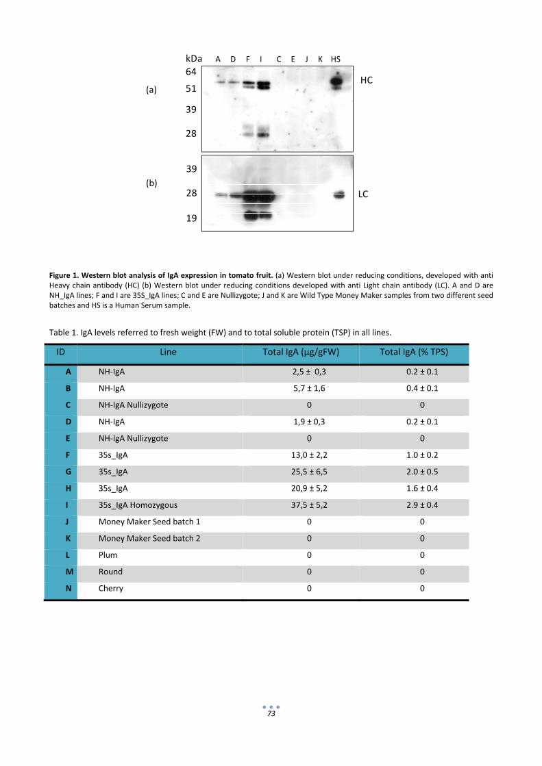
73
64
51
39
28
A D F I C E J K HS
HC
39
28
19
LC
kDa
(a)
(b)
Figure 1. Western blot analysis of IgA expression in tomato fruit. (a) Western blot under reducing conditions, developed with anti Heavy chain antibody (HC) (b) Western blot under reducing conditions developed with anti Light chain antibody (LC). A and D are NH_IgA lines; F and I are 35S_IgA lines; C and E are Nullizygote; J and K are Wild Type Money Maker samples from two different seed batches and HS is a Human Serum sample.
Table 1. IgA levels referred to fresh weight (FW) and to total soluble protein (TSP) in all lines.
ID Line Total IgA (μg/gFW) Total IgA (% TPS)
A NH‐IgA 2,5 ± 0,3 0.2 ± 0.1
B NH‐IgA 5,7 ± 1,6 0.4 ± 0.1
C NH‐IgA Nullizygote 0 0
D NH‐IgA 1,9 ± 0,3 0.2 ± 0.1
E NH‐IgA Nullizygote 0 0
F 35s_IgA 13,0 ± 2,2 1.0 ± 0.2
G 35s_IgA 25,5 ± 6,5 2.0 ± 0.5
H 35s_IgA 20,9 ± 5,2 1.6 ± 0.4
I 35s_IgA Homozygous 37,5 ± 5,2 2.9 ± 0.4
J Money Maker Seed batch 1 0 0
K Money Maker Seed batch 2 0 0
L Plum 0 0
M Round 0 0
N Cherry 0 0
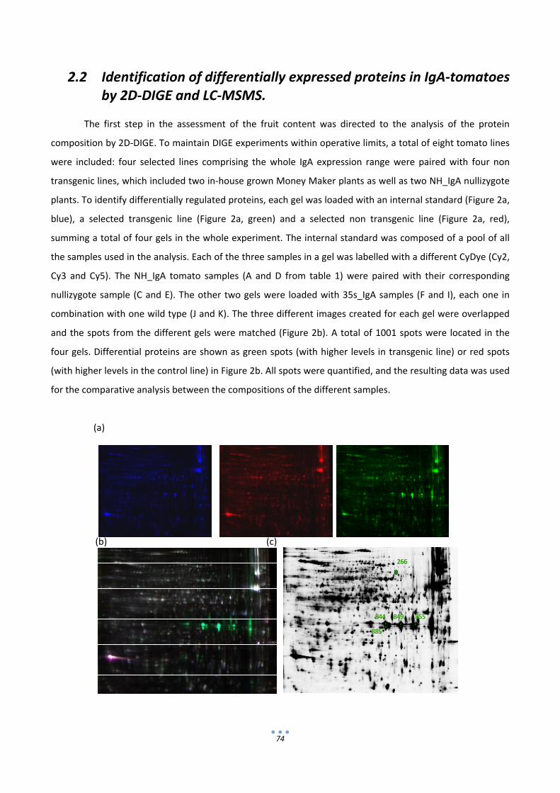
74
266
844
885
849 855
2.2 Identification of differentially expressed proteins in IgA‐tomatoes by 2D‐DIGE and LC‐MSMS.
The first step in the assessment of the fruit content was directed to the analysis of the protein
composition by 2D‐DIGE. To maintain DIGE experiments within operative limits, a total of eight tomato lines
were included: four selected lines comprising the whole IgA expression range were paired with four non
transgenic lines, which included two in‐house grown Money Maker plants as well as two NH_IgA nullizygote
plants. To identify differentially regulated proteins, each gel was loaded with an internal standard (Figure 2a,
blue), a selected transgenic line (Figure 2a, green) and a selected non transgenic line (Figure 2a, red),
summing a total of four gels in the whole experiment. The internal standard was composed of a pool of all
the samples used in the analysis. Each of the three samples in a gel was labelled with a different CyDye (Cy2,
Cy3 and Cy5). The NH_IgA tomato samples (A and D from table 1) were paired with their corresponding
nullizygote sample (C and E). The other two gels were loaded with 35s_IgA samples (F and I), each one in
combination with one wild type (J and K). The three different images created for each gel were overlapped
and the spots from the different gels were matched (Figure 2b). A total of 1001 spots were located in the
four gels. Differential proteins are shown as green spots (with higher levels in transgenic line) or red spots
(with higher levels in the control line) in Figure 2b. All spots were quantified, and the resulting data was used
for the comparative analysis between the compositions of the different samples.
(a)
(b) (c)

75
‐50
‐40
‐30
‐20
‐10
0
10
20
30
40
50
‐60 ‐40 ‐20 0 20 40 60
PC(2) = 18%
PC(1) = 42%
AD
F
I
C
E
J
K
‐60
‐40
‐20
0
20
40
60
‐60 ‐40 ‐20 0 20 40 60
PC(2) = 23%
PC(1) = 28%
A D
C
E
F
I
K
35s_IgA
WT
NH_IgA
Nullizygote
Figure 2. Representative 2D DIGE gel of tomato fruit proteins. (a) Fluorescence images of gel loaded with equal amounts (50 g) of the reference sample (Cy2‐labelled, blue), non transgenic samples (Cy5‐labelled, red) and transgenic samples (Cy3‐labelled, green) were loaded in the same gel. (b) Overlay of the three fluorescence images. Differential proteins appear in green or red, unaffected proteins appear in white. (c) Silver stained 2D DIGE gel. Spots showing significant differences between transgenic and non‐transgenic samples picked for sequencing are outlined in green.
In order to find possible differences between transgenic and non transgenic tomato fruits a Principal
Component Analysis (PCA) was performed with the quantification data extracted from the gels (Figure 3).
The first two principal components of the PCA model explained 42% and 18% of the total variance,
respectively. As depicted in the score scatter plot shown in Fig. 3a, PC1 mainly separated one of the wild
type samples (line J) from the rest of the group. This aberrant separation of line J was most probably due to
sample preparation and therefore it was decided to remove this line from the PCA analysis. Figure 3b shows
the score scatter plot after removing line J. The first two principal components of this alternative PCA model
show 28% and 23% of the total variance, respectively. Here, PC1 slightly separates 35s_IgA lines (F and I)
from the rest of the group which remain together with exception of line K which is not completely grouped
with the rest of the lines. PC2 further discriminates between both 35s_IgA lines (F and I). Samples from
NH_IgA transformation events (both transgenic and Nullizygote) are grouped together in the scores space,
thus not being separated by any of the components of the PCA model.
(a) (b)
Figure 3. PCA of proteomic data of transgenic and non transgenic tomato lines. (a) all samples are included in the plot. (b) sample J is removed from the analysis.
In order to identify those spots which are the main contributors for class separation, a quantitative
comparison between transgenic and non transgenic lines was performed. A total of 37 spots were found
whose average ratio was higher than 2 ( ǀratioǀ > 2). Among them, only 20 showed an increase in both
35s_IgA and NH_IgA lines when considered separately, and only 5 of those were found statistically
significant (p ≤ 0,05) (Figure 2c, outlined in green). All 20 IgA‐associated spots were picked and analyzed by
LC‐MSMS. Table 2 shows the successful identification of the 5 significant spots: spot 266 was identified as
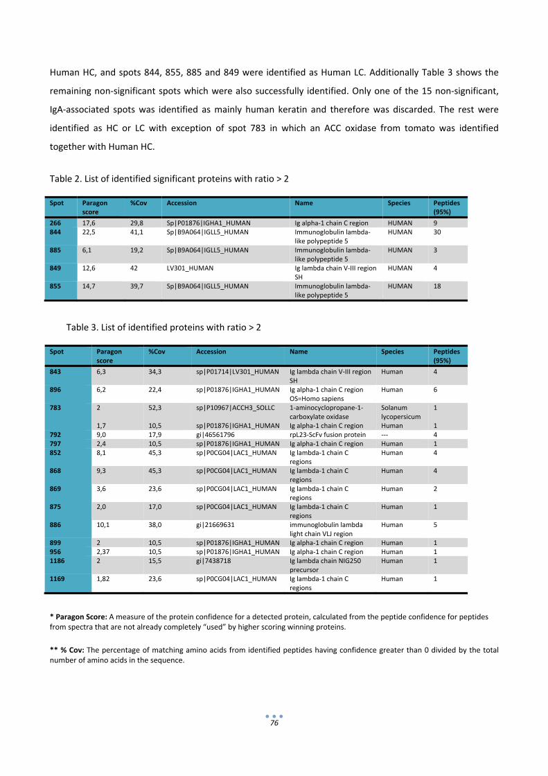
76
Human HC, and spots 844, 855, 885 and 849 were identified as Human LC. Additionally Table 3 shows the
remaining non‐significant spots which were also successfully identified. Only one of the 15 non‐significant,
IgA‐associated spots was identified as mainly human keratin and therefore was discarded. The rest were
identified as HC or LC with exception of spot 783 in which an ACC oxidase from tomato was identified
together with Human HC.
Table 2. List of identified significant proteins with ratio > 2
Table 3. List of identified proteins with ratio > 2
* Paragon Score: A measure of the protein confidence for a detected protein, calculated from the peptide confidence for peptides from spectra that are not already completely “used” by higher scoring winning proteins.
** % Cov: The percentage of matching amino acids from identified peptides having confidence greater than 0 divided by the total number of amino acids in the sequence.
Spot Paragon score
%Cov Accession Name Species Peptides (95%)
266 17,6 29,8 Sp|P01876|IGHA1_HUMAN Ig alpha‐1 chain C region HUMAN 9 844 22,5 41,1 Sp|B9A064|IGLL5_HUMAN Immunoglobulin lambda‐
like polypeptide 5 HUMAN 30
885 6,1 19,2 Sp|B9A064|IGLL5_HUMAN Immunoglobulin lambda‐like polypeptide 5
HUMAN 3
849 12,6 42 LV301_HUMAN Ig lambda chain V‐III region SH
HUMAN 4
855 14,7 39,7 Sp|B9A064|IGLL5_HUMAN Immunoglobulin lambda‐like polypeptide 5
HUMAN 18
Spot Paragon score
%Cov Accession Name Species Peptides (95%)
843 6,3 34,3 sp|P01714|LV301_HUMAN Ig lambda chain V‐III region SH
Human 4
896 6,2 22,4 sp|P01876|IGHA1_HUMAN Ig alpha‐1 chain C region OS=Homo sapiens
Human 6
783 2 1,7
52,3 10,5
sp|P10967|ACCH3_SOLLC sp|P01876|IGHA1_HUMAN
1‐aminocyclopropane‐1‐carboxylate oxidase Ig alpha‐1 chain C region
Solanum lycopersicum Human
1 1
792 9,0 17,9 gi|46561796 rpL23‐ScFv fusion protein ‐‐‐ 4 797 2,4 10,5 sp|P01876|IGHA1_HUMAN Ig alpha‐1 chain C region Human 1 852 8,1 45,3 sp|P0CG04|LAC1_HUMAN Ig lambda‐1 chain C
regions Human 4
868 9,3 45,3 sp|P0CG04|LAC1_HUMAN Ig lambda‐1 chain C regions
Human 4
869 3,6 23,6 sp|P0CG04|LAC1_HUMAN Ig lambda‐1 chain C regions
Human 2
875 2,0 17,0 sp|P0CG04|LAC1_HUMAN Ig lambda‐1 chain C regions
Human 1
886 10,1 38,0 gi|21669631 immunoglobulin lambda light chain VLJ region
Human 5
899 2 10,5 sp|P01876|IGHA1_HUMAN Ig alpha‐1 chain C region Human 1 956 2,37 10,5 sp|P01876|IGHA1_HUMAN Ig alpha‐1 chain C region Human 1 1186 2 15,5 gi|7438718 Ig lambda chain NIG250
precursor Human 1
1169 1,82 23,6 sp|P0CG04|LAC1_HUMAN Ig lambda‐1 chain C regions
Human 1
77
2.3 Metabolic profiling for the evaluation of unintended effects by UPLC‐MS.
Unintended effects on the metabolome of transgenic tomato fruits generated by the expression of
human IgA were also evaluated by non‐targeted metabolomics. Tomato methanol extracts were used for the
analysis, which was carried out by means of reverse‐phase UPLC coupled to mass spectrometry (UPLC‐
QTOF). This technology is specially suited for the detection of semipolar secondary metabolites, primarily
tomato polyphenols and alkaloids (Moco et al., 2006). Between four and five different fruits from seven
transgenic lines containing different levels of IgA, two nullizygote plants, two wild type moneymaker lines
and three commercial varieties (plum, round, and cherry) were analyzed.
The mass spectra obtained from the analysis of tomato methanol extracts by means of UPLC‐QTOF
was first processed with XCMS, an integrated metabolomics analysis platform for the determination of
metabolic profile differences and metabolite identification (Smith et al., 2006). The pre‐processing of the
data generated two discrete data matrices, one for each ionization mode: ESI‐ (electrospray ionization in
negative mode) and ESI+ (electrospray ionization in positive mode). The ESI‐ data set contained 386 mass
variables while 835 peaks were initially identified in the ESI+ mass spectra. In order to remove low quality
data, a first filtering step was done by imposing two different conditions: (i) a mass feature should be
detectable in a minimum of three biological replicates in at least one of the tomato lines, and (ii) its average
day‐coefficient of variation for the reference sample (computed on normalised data) should not be greater
than 40%. Mass peaks that did not fulfil these requirements were considered unreliable and were thus,
excluded from the study. A second filtering step was accomplished by removing the redundant isotopic
features which were identified by the package CAMERA (a collection of algorithms for metabolite profile
annotation with the primary purpose of annotation and evaluation of isotope peaks, adducts and fragments
in peak lists) (Kuhl et al., 2012).The final ESI‐ and ESI+ data sets, with 107 and 249 peaks, respectively, were
joined together in a single matrix for further analysis.
PCA was performed as first step in data exploration to examine the relationship between the
different sample classes. The first two principal components of the PCA model explained 30% and 17% of the
total variance, respectively. As can be seen in the scores scatter plot depicted in Figure 4a, PC1 clearly
separated the commercial varieties from the remaining samples, while PC2 allowed further discrimination
within the commercial group between cherry (N) and plum and round tomatoes (L and M lines). The samples
from IgA‐producing lines and control nullizygote and Moneymaker WT lines, however, grouped together in
the scores space not being separated by any of the components of the PCA model. As the largest variance in
the data set was between commercial and Moneymaker lines, a new PCA was performed in which
commercial lines were excluded from the analysis to thus better explore the differences associated to IgA
production. The obtained PCA model (Figure 4b) did not reveal any clear group structure in the dataset; only

78
NH_IgA
Nullizygote
35s_IgA
WT
Commercial
‐20
‐10
0
10
20
‐20 ‐10 0 10 20 30 40
PC(2) = 17%
PC(1) = 31%
A1 A2
A3
A4A5
B1B2
B3
B4B5
D1
D2
D3
D4
D5
C1
C2C3
C4
C5E1
E2
E3E4E5
F1
F2F3
F4
F5G1
G2G3
G4
G5
H1 H2
H4
H5
I1
I2
I3I4I5
J1J2J3
J4J5
K1
K2
K3
K4
K5
L1
L2
L3
L4
L5
M1
M2 M3
M4
M5
N1
N2
N3
N4
‐10
0
10
‐20 ‐10 0 10 20
PC(2) = 11%
PC(1) = 17%
A1
A2
A3
A4
A5
B1
B2
B3
B4B5
D1
D2
D3
D4
D5C1
C2
C3
C4
C5
E1
E2
E3
E4 E5
F1F2
F3
F4
F5
G1
G2
G3
G4
G5H1H2
H4
H5
I1I2
I3
I4
I5J1
J2
J3
J4
J5
K1
K2
K3
K4
K5
35S_IgA lines F, G, and H, slightly separated from the remaining samples whereas significant confusion
occurred between the homozygous 35S_IgA line I, NH_IgA lines, and the control lines. This does not exclude,
however, changes associated to the expression of the foreign protein, it rather points out that any possible
variation introduced in the metabolome by the expression of IgA in the fruit is smaller than the existing
within‐group variation.
(a) (b)
Figure 4. Principal component analysis score plots of LC‐MS spectra. (a) Complete dataset set of tomato cultivars. (b) Moneymaker lines.
To directly search for specific metabolites that discriminate between IgA‐producing and control lines,
an orthogonal partial least squares discriminate analysis (OPLS‐DA) was set. OPLS‐DA makes use of a priori
information on class membership (e.g. IgA content) to extract class‐related variance in a predictive
component and class‐uncorrelated variation in one or more orthogonal components. Thus, even in the
presence of uncontrolled co‐variates, OPLS‐DA allows a direct identification of class‐discriminant variables.
As 35s_IgA lines and NH_IgA lines contain very different IgA levels, they were treated separately so that each
transgenic group could be compared with its closest control (non‐IgA) group. Thus, three separate
comparisons were made involving 35s_IgA vs. WT, NH_IgA vs. WT, and NH_IgA vs. nullizygote lines (Figure
5). The fitted models comprised one predictive (IgA content) plus 1, 2, and 1 orthogonal components,
respectively. The variance related to class separation explained by the models was 82%, 98%, and 84% with a
cross‐validated predictive ability Q2(Y)=0.31, 0.84, and 0.40, indicating a higher uncertainty in the 35s_IgA vs.
WT and NH_IgA vs. Nullizygote models regarding class separation. The homozygous line I formed a separate
subgroup within the 35s lines as revealed by the first orthogonal component of the model.

79
‐10
‐5
0
5
10
‐10 0 10
To= 15%
Tp = 14%
F1
F2
F3
F4
F5
G1G2
G3
G4 G5
H1
H2
H4
H5
I1
I2
I3
I4
I5
J1
J2
J3
J4
J5
K1
K2
K3K4
K5
‐10
0
10
‐10 0 10
To= 12%
Tp = 12%
A1
A2
A3
A4
A5
B1
B2
B3
B4
B5
D1
D2
D3
D4
D5
J1
J2J3J4
J5
K1
K2
K3
K4
K5
‐10
0
10
‐10 0 10
To= 14%
Tp = 7%
35s_IgA
WT
NH_IgA
Nullizygote
A1
A2
A3
A4A5
B1
B2
B3
B4
B5
D1
D2
D3D4
D5
C1
C2
C3C4
C5
E1
E2
E3
E4
E5
(a) (b)
(c)
Figure 5. OPLS‐DA score plots of the (a) 35S_IgA vs. WT (b) NH_IgA vs. WT, and (c) NH_IgA vs. Nullizygote.
The most relevant metabolite ions for class discrimination were selected, according to the following
rules: the selected ions should present a high variable influence on projection (VIP ≥ 1), a high correlation
with IgA content (| p (corr) | ≥ 0.5), and narrow jack‐knifed 95% confidence intervals of the variable
coefficient (confidence interval not crossing zero). A total of 41, 39, and 27 discriminating mass peaks
(counting 91 different masses) were chosen according to these criteria from the 35s_IgA vs. WT, NH_IgA vs.
WT, and NH_IgA vs. Nullizygote models, respectively. A large proportion of these peaks, however, showed a
very modest variation between the transgenic lines and their counterpart controls with less than half of
them, 42 masses, reaching fold ratios ≥ 2. The complete list of 91 masses is provided in Table S1 as
supplementary material. The three models were then compared by means of SUS‐plots (Shared and Unique
Structures plot, an analysis that combines the p(corr) profiles from two models) constructed with all the
selected variables (Figure 6). The first SUS‐plot showed the correlation between models (35s_IgA vs. WT)
and (NH_IgA vs. WT) (Figure 6a). The second SUS‐plot correlated models (35s_IgA vs. WT) and (NH_IgA vs.
Nullizygote) (Figure 6b). The last SUS‐plot showed the correlation between models (NH_IgA vs. WT) and
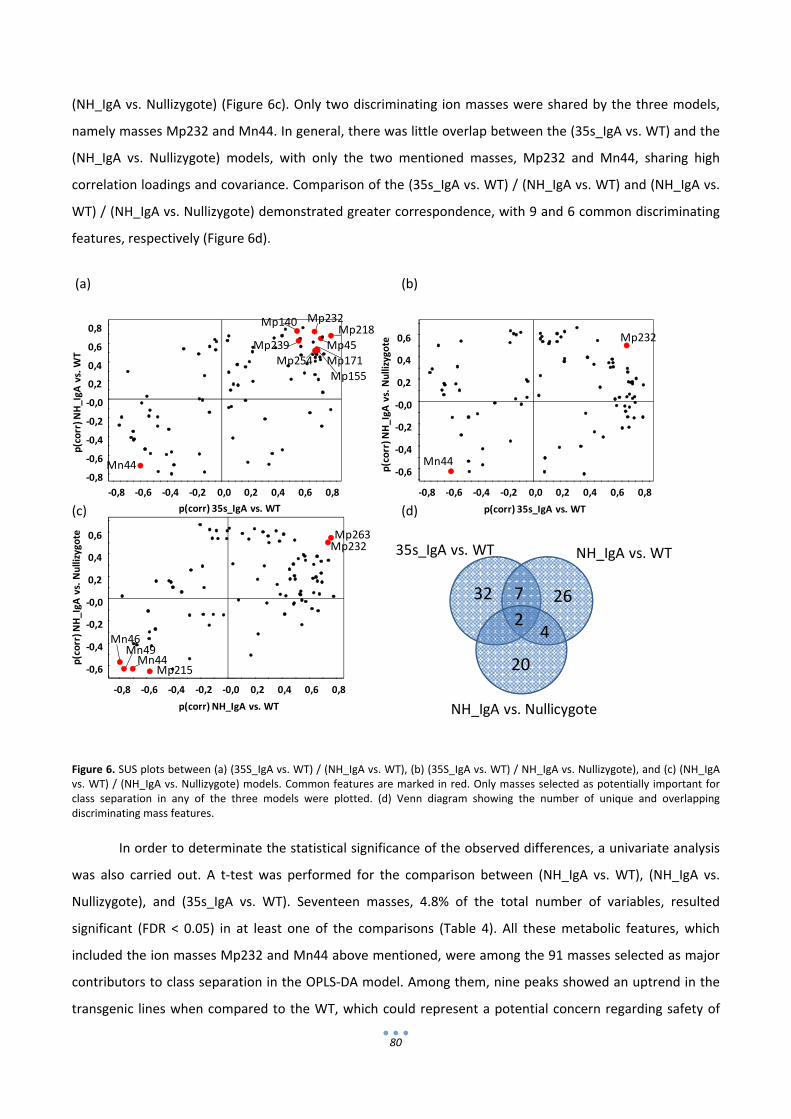
80
32 26
20
7
24
35s_IgA vs. WT NH_IgA vs. WT
NH_IgA vs. Nullicygote
Mn44‐0,8
‐0,6
‐0,4
‐0,2
‐0,0
0,2
0,4
0,6
0,8
‐0,8 ‐0,6 ‐0,4 ‐0,2 0,0 0,2 0,4 0,6 0,8
p(corr) N
H_IgA
vs. WT
p(corr) 35s_IgA vs. WT
Mp140
Mp239
Mp232
Mp45Mp171
Mp155
Mp254
Mp218
‐0,6
‐0,4
‐0,2
‐0,0
0,2
0,4
0,6
‐0,8 ‐0,6 ‐0,4 ‐0,2 0,0 0,2 0,4 0,6 0,8
p(corr) N
H_IgA
vs. Nullizygote
p(corr) 35s_IgA vs. WT
Mn44
Mp232
‐0,6
‐0,4
‐0,2
‐0,0
0,2
0,4
0,6
‐0,8 ‐0,6 ‐0,4 ‐0,2 ‐0,0 0,2 0,4 0,6 0,8
p(corr) N
H_IgA
vs. Nullizygote
p(corr) NH_IgA vs. WT
Mn44Mn49
Mn46
Mp215
Mp263Mp232
(NH_IgA vs. Nullizygote) (Figure 6c). Only two discriminating ion masses were shared by the three models,
namely masses Mp232 and Mn44. In general, there was little overlap between the (35s_IgA vs. WT) and the
(NH_IgA vs. Nullizygote) models, with only the two mentioned masses, Mp232 and Mn44, sharing high
correlation loadings and covariance. Comparison of the (35s_IgA vs. WT) / (NH_IgA vs. WT) and (NH_IgA vs.
WT) / (NH_IgA vs. Nullizygote) demonstrated greater correspondence, with 9 and 6 common discriminating
features, respectively (Figure 6d).
(a) (b)
(c) (d)
Figure 6. SUS plots between (a) (35S_IgA vs. WT) / (NH_IgA vs. WT), (b) (35S_IgA vs. WT) / NH_IgA vs. Nullizygote), and (c) (NH_IgA vs. WT) / (NH_IgA vs. Nullizygote) models. Common features are marked in red. Only masses selected as potentially important for class separation in any of the three models were plotted. (d) Venn diagram showing the number of unique and overlapping discriminating mass features.
In order to determinate the statistical significance of the observed differences, a univariate analysis
was also carried out. A t‐test was performed for the comparison between (NH_IgA vs. WT), (NH_IgA vs.
Nullizygote), and (35s_IgA vs. WT). Seventeen masses, 4.8% of the total number of variables, resulted
significant (FDR < 0.05) in at least one of the comparisons (Table 4). All these metabolic features, which
included the ion masses Mp232 and Mn44 above mentioned, were among the 91 masses selected as major
contributors to class separation in the OPLS‐DA model. Among them, nine peaks showed an uptrend in the
transgenic lines when compared to the WT, which could represent a potential concern regarding safety of
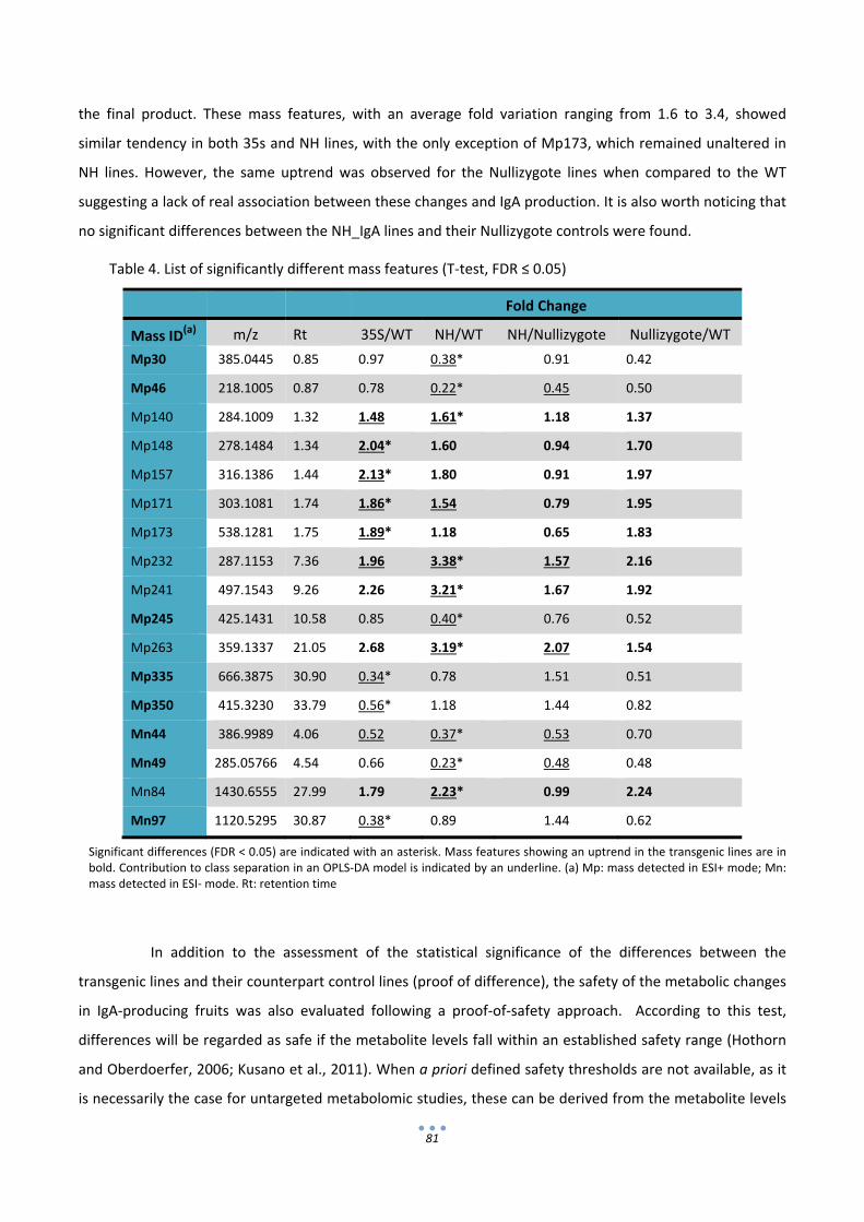
81
the final product. These mass features, with an average fold variation ranging from 1.6 to 3.4, showed
similar tendency in both 35s and NH lines, with the only exception of Mp173, which remained unaltered in
NH lines. However, the same uptrend was observed for the Nullizygote lines when compared to the WT
suggesting a lack of real association between these changes and IgA production. It is also worth noticing that
no significant differences between the NH_IgA lines and their Nullizygote controls were found.
Table 4. List of significantly different mass features (T‐test, FDR ≤ 0.05)
Fold Change
Mass ID(a) m/z Rt 35S/WT NH/WT NH/Nullizygote Nullizygote/WT
Mp30 385.0445 0.85 0.97 0.38* 0.91 0.42
Mp46 218.1005 0.87 0.78 0.22* 0.45 0.50
Mp140 284.1009 1.32 1.48 1.61* 1.18 1.37
Mp148 278.1484 1.34 2.04* 1.60 0.94 1.70
Mp157 316.1386 1.44 2.13* 1.80 0.91 1.97
Mp171 303.1081 1.74 1.86* 1.54 0.79 1.95
Mp173 538.1281 1.75 1.89* 1.18 0.65 1.83
Mp232 287.1153 7.36 1.96 3.38* 1.57 2.16
Mp241 497.1543 9.26 2.26 3.21* 1.67 1.92
Mp245 425.1431 10.58 0.85 0.40* 0.76 0.52
Mp263 359.1337 21.05 2.68 3.19* 2.07 1.54
Mp335 666.3875 30.90 0.34* 0.78 1.51 0.51
Mp350 415.3230 33.79 0.56* 1.18 1.44 0.82
Mn44 386.9989 4.06 0.52 0.37* 0.53 0.70
Mn49 285.05766 4.54 0.66 0.23* 0.48 0.48
Mn84 1430.6555 27.99 1.79 2.23* 0.99 2.24
Mn97 1120.5295 30.87 0.38* 0.89 1.44 0.62
Significant differences (FDR < 0.05) are indicated with an asterisk. Mass features showing an uptrend in the transgenic lines are in bold. Contribution to class separation in an OPLS‐DA model is indicated by an underline. (a) Mp: mass detected in ESI+ mode; Mn: mass detected in ESI‐ mode. Rt: retention time
In addition to the assessment of the statistical significance of the differences between the
transgenic lines and their counterpart control lines (proof of difference), the safety of the metabolic changes
in IgA‐producing fruits was also evaluated following a proof‐of‐safety approach. According to this test,
differences will be regarded as safe if the metabolite levels fall within an established safety range (Hothorn
and Oberdoerfer, 2006; Kusano et al., 2011). When a priori defined safety thresholds are not available, as it
is necessarily the case for untargeted metabolomic studies, these can be derived from the metabolite levels
82
found in commercial varieties. Thus, following the approach of Kusano et al. (2011) symmetric safety limits
were defined as the 90% confidence interval for the ratio to control (WT) of the commercial variety furthest
away from the WT. When, for any given metabolite, the confidence interval for the ratio of the transgenic
fruits to the WT control is within this safety range, the levels of the metabolite can be regarded as safe.
When this analysis was performed, only one mass, Mn52, showed ratios to WT outside the acceptable limits
in the 35s and NH line groups with an average fold variation of 2.4 and 1.7, respectively. When analyzing
lines individually, 42 additional features were identified with ratios outside the established safety range in at
least one of the transgenic tomato lines, but only 22 out of these fell outside the upper boundary and could
therefore represent a potential safety concern (Cases 2 and 3 in Figure 7). This is becouse IgA tomato fruits
are not intended to be used as food product, and therefore safety concerns are constrained to the presence
of new or increased levels of toxicologically relevant compounds, the loss of for example key nutrients being
unimportant. The majority of the 22 remaining masses (86%), showed modest average fold variation which
was in all the cases lower to the established threshold (Case 2 in Figure 7) while the remaining 14% (3
masses, showed in red in table 5) showed an average fold variation above, although sometimes close to the
established threshold (Case 3 in Figure 7) (Table 5). This indicates that strong variance rather than consistent
higher abundance is the reason that makes those masses to fall slightly outside the established safety range.
It should be noticed that in one case safety cannot be concluded for Nullizygote line C. Most importantly, it
should be noticed that for the homozygous elite 35s line I all measured variables were within the acceptable
limits.
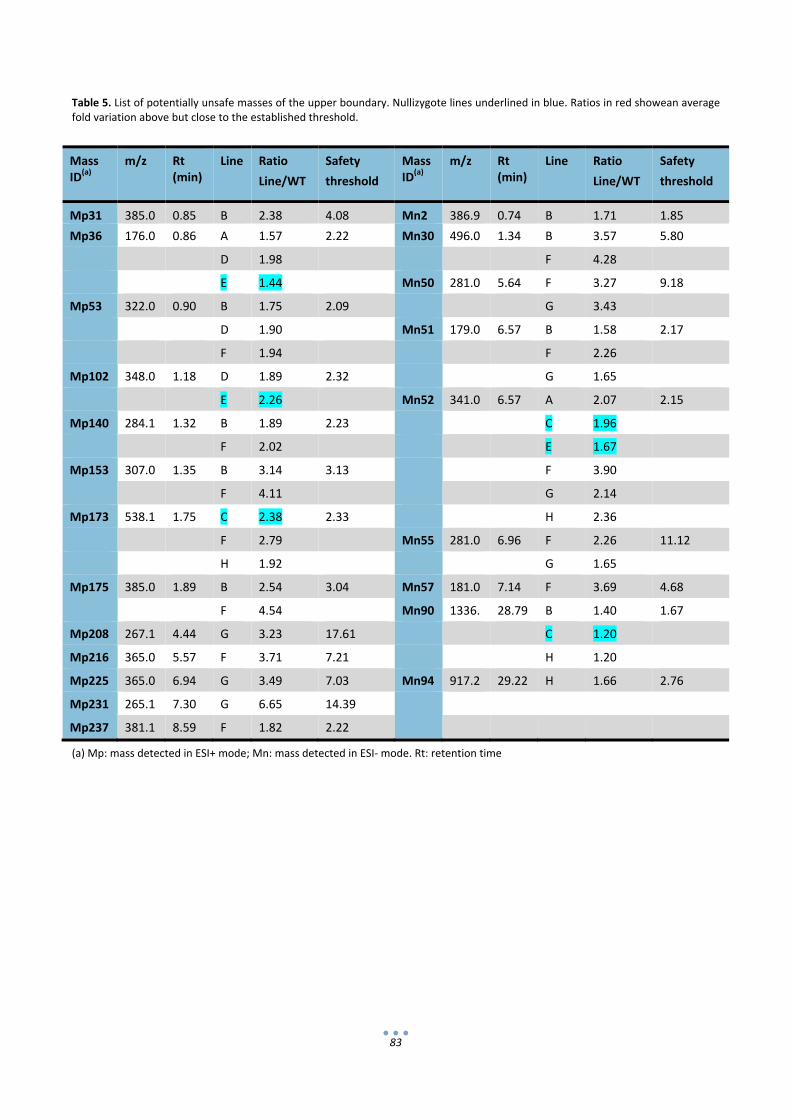
83
Table 5. List of potentially unsafe masses of the upper boundary. Nullizygote lines underlined in blue. Ratios in red showean average fold variation above but close to the established threshold.
(a) Mp: mass detected in ESI+ mode; Mn: mass detected in ESI‐ mode. Rt: retention time
Mass ID(a)
m/z Rt (min)
Line Ratio
Line/WT
Safety
threshold
Mass ID(a)
m/z Rt (min)
Line Ratio
Line/WT
Safety
threshold
Mp31 385.0 0.85 B 2.38 4.08 Mn2 386.9 0.74 B 1.71 1.85
Mp36 176.0 0.86 A 1.57 2.22 Mn30 496.0 1.34 B 3.57 5.80
D 1.98 F 4.28
E 1.44 Mn50 281.0 5.64 F 3.27 9.18
Mp53 322.0 0.90 B 1.75 2.09 G 3.43
D 1.90 Mn51 179.0 6.57 B 1.58 2.17
F 1.94 F 2.26
Mp102 348.0 1.18 D 1.89 2.32 G 1.65
E 2.26 Mn52 341.0 6.57 A 2.07 2.15
Mp140 284.1 1.32 B 1.89 2.23 C 1.96
F 2.02 E 1.67
Mp153 307.0 1.35 B 3.14 3.13 F 3.90
F 4.11 G 2.14
Mp173 538.1 1.75 C 2.38 2.33 H 2.36
F 2.79 Mn55 281.0 6.96 F 2.26 11.12
H 1.92 G 1.65
Mp175 385.0 1.89 B 2.54 3.04 Mn57 181.0 7.14 F 3.69 4.68
F 4.54 Mn90 1336. 28.79 B 1.40 1.67
Mp208 267.1 4.44 G 3.23 17.61 C 1.20
Mp216 365.0 5.57 F 3.71 7.21 H 1.20
Mp225 365.0 6.94 G 3.49 7.03 Mn94 917.2 29.22 H 1.66 2.76
Mp231 265.1 7.30 G 6.65 14.39
Mp237 381.1 8.59 F 1.82 2.22

84
Case 1
UpperBoundary of safety range
Case 2
Case 3
Case 4
Figure 7. Simplified schematic representation of the possible outcomes of the proof of safety assessment. Only the upper boundary limit is considered. Case 1 represents a situation where the metabolite can be regarded as safe. Case 2‐4 represent situations where safety cannot be concluded. Squares show the mean ratios between the transgenic line and WT. Bars represent the confidence intervals.

85
3. Discussion
Plant organs are considered GRAS when at their mature stage they contain no toxicants, allergens or
anti‐nutrients that could result harmful for the consumer. It is expected that recombinant production of IgA
in edible tomatoes does not compromise fruit composition and consequently its edibility. However, this
hypothesis had never been corroborated experimentally before; the most important compounds concerning
edibility in tomato are proteins (allergens) and secondary metabolites (alkaloids) (OECD, 2008). Here, we
analyze proteomic and metabolomic profiles of IgA tomatoes and compare them with non‐transgenic
profiles, as a way to evaluate the changes in tomato composition introduced by IgA production. Whole
proteome was analyzed by means of 2D‐DIGE gels and UPLC‐qTOF was used to analyze secondary
metabolites. The changes observed, or the absence of them, are taken as an indication of the safety of
minimally processed IgA‐tomato products.
Four representative transgenic lines paired with four non‐transgenic lines were chosen for the
proteomic analysis by means of 2D‐DIGE. These included NH_IgA compared with their corresponding
nullizygote fruits as well as 35s_IgA paired with WT samples. Principal component analysis (PCA) from the
quantification data highlighted the presence of an aberrant WT sample. The tomato powder which was used
to prepare this sample was also used for subsequent metabolomic analysis showing no abnormal
performance in this case. This suggests that the aberrant behaviour in proteomic analysis was due to protein
precipitation procedures. Therefore, this sample was removed from PCA analysis.
To quantify the magnitude of the changes between transgenic and non‐transgenic groups, mean
ratios were calculated for each spot between IgA and non IgA samples. In a first arbitrary cut only spots with
ratio >2.0 were selected, as only a higher abundance of a protein can be considered as a potential thread. As
a result, a total of 20 spots were selected for identification by LC‐MSMS, with only 5 of them showing
significant increase in a T‐test analysis (p‐value < 0,05). All the sequenced spots matched human IgA chains
except for spot 783, which showed an overlap between human IgA HC and tomato ACC oxidase, an ethylene‐
forming enzyme involved in climacteric fruit ripening. ACC oxidase is a common protein in climacteric fruits
with no allergenic properties reported, thus, a slight increase in this enzyme probably does not represent a
threat for human consumption. Therefore, it can be deduced from this analysis that no other protein apart
from IgA is substantially changing the composition of the transgenic tomato. However, further analysis of
allergenic proteins could be made in order to further assure the absence of novel allergens in the fruit
composition (Gubesch et al., 2007).
Regarding the analysis of secondary metabolites, PCA including commercial varieties Cherry, Plum
and Round showed a clear separation between these and the remaining Moneymaker samples. This
observation could be due to several reasons. It is possible that inherent variety differences are sufficient to

86
separate the samples, but other possible explanations are the cultivation conditions of each tomato batch,
the post‐harvesting conditions to which they have been subdued, e.g. refrigeration, etc. In any case, those
differences are much more evident and by far, sufficient to mask any differences generated by the
production of an immunoglobulin. For this reason, another PCA was made after removing the commercial
varieties from the data set. This second PCA showed no separation between IgA lines and IgA‐free lines. Both
groups showed a similar distribution, meaning that any environmental component that is affecting the
metabolite composition is more significant than any difference generated by the production of IgA.
Subsequently, OPLS‐DA models were set in order to search for specific unintended effects between
transgenic and non transgenic lines directly related with IgA production. As NH_IgA lines and 35s_IgA lines
presented a drastic difference in IgA levels, they were treated separately. All three OPLS‐DA models were
able to separate transgenic and non‐transgenic lines, however, with exception of the model set for NH_IgA
vs. WT, they presented a low predictive power with values of Q2 < 0,5.
In principle, those masses correlating with IgA levels which are common within the three models are
expected to be truly related with the IgA production. Only two masses, namely Mn44 and Mp232, were
found to fulfil this criterion. Of these, only Mp232 showed an uptrend in transgenic lines, although this
uptrend was only significant when comparing NH_IgA samples with WT but not in the two remaining
comparisons. The fact that Mp232 uptrend is only significant in one of the group comparisons indicates that
it has only minor effects in fruit composition. Moreover the average increase ration is 3.38 fold, and increase
that would only be a concern if high daily doses of tomato‐derived products were required. Nevertheless,
targeted identification of Mp232 could be advisable to better evaluate its possible safety implications.
An additional proof‐of‐safety approach of the metabolic changes in IgA‐producing fruits was
performed following the approach of Kusano et al. (2011). In this analysis, all the measured variables in the
elite line homozygous I line fell within the acceptable limits. However safety could not be concluded for 42
masses in the remaining lines, 22 of which showing an uptrend in transgenic lines. Among those 22 masses,
just three of them showed an average fold variation above the established threshold (case 3 in Figure 7),
none of them being Mp232. This indicates that in most cases a high variability is the reason of these
confidence intervals lying beyond the safety range. It is also important to point out that for 5 of the 22
different masses, nullizygote lines also appeared to be outside the established safety range. Taking together,
these observations suggest a lack of a real association between these changes and IgA production.
It is a common mistake to expect that the composition of a transgenic crop is identical to its non‐
transgenic comparator. As shown here in PCA and OPLS‐DA models, IgA expression leaves an associated
footprint in the fruit composition, which can be detected and partially described using proteomic and
metabolomic profiling. Similarly, a metabolic signature introduced by the recombinant over‐expression of
the taste‐modifying protein miraculin was found before in tomato fruits using a multi‐ platform approach, in
87
the context of a substantial equivalence assessment (Kusano et al., 2011). However, to our knowledge, the
metabolic signature of a therapeutic protein produced in an edible plant organ was not analyzed before.
The over‐expression of xenoproteins in the plant cell can induce sequence‐specific responses in the host,
which range from the suppression of the expression of the foreign gene via post‐transcriptional gene
silencing, to the triggering of a general stress response or the induction of a program of cell death (see
Orzaez et al. (2009) and references therein). Therefore, the accumulation of harmful compounds in response
to IgA expression in fruits could not be initially discarded. By incorporating several independent
transformation events leading to different IgA expression levels, this work tries to evaluate unintended
effects arising from the general strategy of IgA‐based oral passive immunotherapy in tomato. However this
work alone cannot rule out the possibility of harmful unintended effects associated with any strategy
involving IgA production in fruits. At least three potential sources of uncertainty still remain. First, the
unexpected accumulation of harmful compounds other than the “usual suspects”, namely proteins and
semi‐polar secondary metabolites, cannot be discarded with the methodology used here. A single method
enabling complete metabolome analysis does not exist, and therefore additional comprehensive profiling
methods should be considered (Rischer and Oksman‐Caldentey, 2006). Secondly, any new transgenic event
involving IgA production in fruits should evaluate possible unintended effects associated specifically with the
site of insertion, which cannot be evaluated here. Finally, as observed in the proteomic profile, the antibody
itself accumulates in the form of different polypeptides with distinct electrophoretic mobilities, probably
arising from partial proteolytic degradation and/or different glycosylation patterns. Certainly, the pool of
IgA‐related polypeptides present in the fruit is different to those occurring in a natural OPI and therefore the
safety of the plant‐made IgA itself and in the context of the fruit matrix needs to be thoroughly evaluated.
All in all, the data shown here constitutes a first “clearway” for the fruit‐based IgA strategy, by establishing
that not unsafe compositional changes associated with IgA production are detected when un‐targeted
analysis profiling the unsafe “usual suspects” in tomato are performed.
88
4. Experimental Procedures
4.1 Plant material
Seven transgenic lines (A, B, D, F, G, H, I) of tomato (Solanum lycopersicum, cv. Moneymaker)
expressing human immunoglobulin A (IgA) were used in this study along with the parental line – K – used for
genetic transformation. Lines A, B and D were obtained by Agrobacterium mediated transformation of a
plasmid carrying the heavy (HC) and light chains (LC) of human IgA under the cauliflower mosaic virus 35s
constitutive promoter and the tomato NH promoter, respectively. Lines F, G, H, and I were obtained by co‐
transformation with two Agrobacterium cultures, one carrying a plasmid with the 35s:IgA_HC and the
second carrying a plasmid with the 35s:IgA_LC construct. A detailed description of the generation of these
transgenic lines can be found in Juarez et al. (2012). Additionally, two nullizygot segregant lines – C and E –
from the 35S:IgA_HC / NH:IgA_LC transformation, and one Moneymaker line – J – obtained from a different
laboratory were also included as additional controls. Finally, fruits from three widely consumed tomato
types – plum (L), round (M), and cherry (N) – purchased from a local grocery completed the experimental
dataset. Tomato seeds (excluding commercial lines L, M, and N) were grown in pots in a greenhouse with a
light/dark cycle of 16/8 hours. Fruits were harvested 15 days post‐breaker. Five independent samples from
each plant line were collected. After removing gel and seeds, tomato pericarp (with the peel) was ground to
a fine powder in liquid nitrogen and stored at ‐80ºC for further analysis.
4.2 Protein extraction, SDS‐PAGE, Western blot and ELISA tests
For protein analysis, frozen tomato powder samples from each group were pooled together and the
total soluble protein (TSP) was extracted in three volumes (w/v) of ice‐cold PBS (100 mM sodium phosphate,
150 mM NaCl, pH 7.4), followed by 2 x 15 min centrifugation steps in a tabletop centrifuge at 16,000 g and
4ºC.
Protein separation was carried out by SDS‐PAGE on NuPAGE® 10% Bis‐Tris polyacrylamide gels
(Invitrogen, Life Technologies, Paisley, UK). Gels were stained with Coomassie blue. For western blot
analysis, proteins were transferred to PVDF membranes (Amersham HybondTM‐P, GE Healthcare,
Buckinghamshire, UK) by semi‐wet blotting (XCell IITM Blot Module, Invitrogen, Life Technologies) following
the manufacturer’s instructions. Membranes were incubated with 1:20.000 Anti‐Human IgA α‐chain specific
peroxidase conjugated antibody (SIGMA, St. Louis, USA) for the detection of the HC, and with 1: 10.000 anti‐
human λ LC (SIGMA) followed by 1:10.000 μg/mL HRP‐conjugated anti‐rabbit IgG (GE Healthcare) for the
detection of the LC. Blots were developed with the AmershamTM ECL Plus Western Blotting Detection System

89
(GE Healthcare) following the manufacturer’s instructions and were visualized by exposure to X‐ray film
(Fujifilm Coorporation, Tokyo, Japan).
IgA quantification was carried out by sandwich ELISA as previously described (Juarez et al., 2012).
Briefly, 96‐well ELISA plates (Corning, NY, USA) were coated overnight at 4ºC with 2 μg/mL of Anti‐Human
IgA α‐chain specific antibody (SIGMA). Plates were blocked with a 2% (w/v) solution of ECL
AdvanceTM Blocking agent (GE Healthcare), 0.1% Tween‐20 (v/v) in PBS (blocking solution) for 2h at RT.
Tomato extracts, serial two‐fold dilutions in PBS, were applied in triplicate and incubated for 1h at RT. A
human colostrum IgA standard (SIGMA) was included for the construction of a standard curve (0.8 to 0.1
μg/ml‐1 concentration range). A 1:5000 dilution of Anti‐Human IgA α‐chain specific peroxidase conjugated
antibody (SIGMA) in blocking solution was used as detection antibody; SIGMAFASTTM OPD (o‐
phenilenediamine) tablets (SIGMA) was used as the enzymatic substrate. Absorbance was determined at
492 nm. IgA concentration in each sample was obtained by interpolation with the IgA standard curve.
4.3 2D DIGE analysis
TSP extracts were incubated with an equal volume of ice‐cold 20% TCA for 2 h at 4ºC. Proteins were
recovered by centrifugation (16,000 g, 15 min at 4ºC) and the obtained pellets washed three times with cold
acetone before being solubilised in lysis buffer (7M urea, 2M thiourea, 4% CHAPS). Samples were then
cleaned up with the 2D Clean‐up kit (GE Healthcare) following the manufacturer’s instructions. Cleaned
protein precipitates were solubilised in Tris lysis buffer (7M urea, 2M thiourea, 4% CHAPS, 20 mM Tris pH
8.5). Protein concentration was determined by the Bio‐Rad Protein Assay (Bio‐Rad, California, USA) using
bovine serum albumin as standard.
Protein samples were labelled with CyDye DIGE fluor minimal dyes (GE Healthcare) according to
manufacturer’s instructions. IgA producing and control lines were alternatively labelled with Cy3 or Cy5 in
dye‐swap approach. A reference sample (consisting of pooled aliquots of all biological samples within the
experiment) labelled with Cy2 was run on every gel together with the Cy3 and Cy5 experimental samples.
2D electrophoresis was developed as described by (Munoz‐Fambuena et al., 2013) and 2D gels were
scanned using a Typhoon Trio (GE Healthcare). Gel image analysis was performed with DeCyder 2D software
v6.5.
4.4 Protein identification
For picking spots of interest, gels were first Silver stained with Silver Staining Kit, Protein (GE
Healthcare). Spots were manually excised from analytical gels and distained with 2x 5‐min washes with
90
acetonitrile (CAN)/water (1:1, v/v). Subsequently, samples were rehydrated with 50 mM ammonium
bicarbonate for 5 min and 25 mM ammonium bicarbonate in 50% (v:v) CAN for 15 min. Proteins were then
digested with sequencing grade trypsin (Promega) as described elsewhere (Shevchenko et al., 1996). The
digestion was stopped with TFA (1% final concentration), 1 μL of the digestion mixture was spotted onto the
MALDI target plate. A BSA plug was analyzed in the same way to control the digestion process.
Liquid chromatography and tandem mass spectrometry (LC–MS/MS): 5 μl of every sample were
loaded onto a trap column (NanoLC Column, 3μ C18‐CL, 75umx15cm; Eksigent, Radio Rd Redwood City CA)
and desalted with 0.1% TFA at 3μl/min during 5 min. The peptides were then loaded onto an analytical
column (LC Column, 3 μ C18‐CL,75umx25cm, Eksigent) equilibrated in 5% acetonitrile 0.1% FA (formic acid).
Elution was carried out with a linear gradient of 5a35% B in A for 30min. (A: 0.1% FA; B: ACN, 0.1% FA) at a
flow rate of 300 nl/min. Peptides were analysed in a mass spectrometer nanoESI qQTOF (5600 TripleTOF,
ABSCIEX, Framingham, MA). The tripleTOF was operated in information‐dependent acquisition mode, in
which a 0.25‐s TOF MS scan from 350–1250 m/z, was performed, followed by 0.05‐s product ion scans from
100–1500 m/z on the 50 most intense 2‐5 charged ions.
ProteinPilot default parameters were used to generate peak list directly from 5600 TripleTof wiff
files. The Paragon algorithm of ProteinPilot was used to search SwissProt protein database (22470027
proteins searched) with the following parameters: trypsin specificity, cys‐alkylation, no taxonomy restriction,
and the search effort set to rapid. To avoid using the same spectral evidence in more than one protein, the
identified proteins are grouped based on MS/MS spectra by the Protein‐Pilot Pro group algorithm. Thus,
proteins sharing MS/MS spectra are grouped, regardless of the peptide sequence assigned. The protein
within each group that can explain more spectral data with confidence is shown as the primary protein of
the group. Only the proteins of the group for which there is individual evidence (unique peptides with
enough confidence) are also listed, usually toward the end of the protein list.
4.5 UPLC‐QTOF
Frozen tomato powder (500 mg) was extracted by vigorous mixing in 3 volumes cold (‐20ºC)
methanol acidified with 0.125% formic acid. Following centrifugation (10 min, 16,000 x g), the supernatant
was dried (in two separate 750 µl aliquots) in a speed‐vac and stored at ‐80ºC. Prior to analysis, dry extracts
were solubilised in 150 µl methanol‐0.1% formic acid and filtered through a 0.2 µm Anotop 10 membrane
filter (Whatman, Kent, UK). Samples were analysed by UPLC‐MS using an ACQUITY®UPLC‐PDA system
coupled to a Q‐TOF Micro mass spectrometer (Waters Corporation, Milford, MA, USA). Separation was
performed on an ACQUITY BEH C18 column (150 x 2.1 mm i.d., 1.7 µm). The mobile phase consisted of
formic acid:ultrapure water (1:1000 v/v, phase A) and formic acid:acetonitrile (1:1000 v/v, phase B).
Gradient conditions were as follows: from 95 to 90% of A in 14 min, from 90 to 80 % of A in 15 min, from 80

91
to 65% of A in 10 min, from 65 to 57% of A in 1 min, from 57 to 0% of A in 1 min, then held at 100% of B for 3
min, returned to 95% of A in 1 min, and equilibrated for 4 min before next injection. The flow rate was 0.4 ml
min‐1; the column and sample temperature were kept at 40 and 10ºC respectively; the sample injection
volume was 5 µl. UV spectra were acquired between 220 and 800 nm with a 1.2 nm resolution and 20 points
sec‐1 sampling rate. Mass spectrometry analysis was performed by electrospray ionization (ESI) in positive
and negative modes (using one of the extract aliquots for each ionization mode). Q‐Tof parameters were as
follows: capillary voltage 3.0 kv, cone voltage 30 eV (ESI+) or 45 eV (ESI‐), desolvation temperature 300ºC,
source temperature 120ºC; cone gas flow 50 l h‐1, desolvation gas flow 600 l h‐1, collision energy 5 eV. MS
data was acquired in centroid mode in the m/z scan range 100‐1500 with a scan time of 0.5 s and an
interscan time of 0.1 s. Leucine enkephalin was used as the lock mass using a LockSpray exact mass
ionization source. A reference sample (made by pooling equal amounts of all biological sample) was injected
four times (at regular intervals) in each injection series.
MassLynx raw files converted to netCDF format with DataBridge (Waters Corporation, Milford, MA,
USA) were processed with the XCMS package version 1.34.0
(http://bioc.ism.ac.jp/2.11/bioc/html/xcms.html) run under R version 2.15.2 (http://www.r‐project.org/).
The centWave algorithm (Tautenhahn et al., 2008) was used for feature detection; peak grouping and
retention time correction were performed with the density and peakgroups algorithms (Smith et al., 2006),
respectively. Mass features were grouped using the CAMERA algorithm (Kuhl et al., 2012). The area of each
extracted mass feature was first normalised to the total chromatogram area; the reference sample was then
used to normalise the data between different runs (for each mass feature, the test sample area is divided by
the mean area of the reference samples included in that run).
4.6 Statistical data analysis
PCA and OPLS‐DA of the protein and metabolite profiles of tomato fruits were performed with the
software package SIMCA‐P 11 (Umetrics, Sweden). Variables were log‐transformed, mean centred, and
scaled to unit variance prior to analysis.
Two‐sample t‐tests on log transform data were performed using MeV v4.9
(http://www.tm4.org/mev.html), an application of the TM4 Microarray Software Suite (Saeed et al., 2003).
Safety thresholds in the proof‐of‐safety analysis were defined by the symmetric maximum boundaries of the
non parametric 90% confidence intervals for the ratio to WT of the commercial variety furthest away from
the WT. Safety was concluded when the confidence intervals for the ratio to control of the transgenic lines
were within the safety limits. For the calculations of the confidence intervals the R package pairwiseCI was
used.

92
5. Acknowledgements
This work has been funded by Grant BIO2010‐15384 from Plan Nacional I + D of the Spanish Ministry
of Science. Juarez P. is a recipient of a FPU fellowship. The proteomic analysis was carried out in the
Proteomics facility of IBMCP (Instituto de Biologia Molecular y Celular de Plantas UPV‐CSIC) and the protein
identification was performed in the SCSIE_university of Valencia Proteomics Unit, a member of ISCIII
ProteoRed Proteomics Platform. The Metabolomic analysiswas carried out in the Metabolomics facility of
IBMCP. Finally, we want to thank Vicente Monedero in IATA for kindly providing scFv and VP8* clones.

93
6. References
Asero, R., Mistrello, G., Roncarolo, D., de Vries, S.C., Gautier, M.F., Ciurana, C.L., Verbeek, E., Mohammadi, T., Knul‐Brettlova, V., Akkerdaas, J.H., Bulder, I., Aalberse, R.C. and van Ree, R. (2000) Lipid transfer protein: a pan‐allergen in plant‐derived foods that is highly resistant to pepsin digestion. Int Arch Allergy Immunol 122, 20‐32.
Coll, A., Nadal, A., Rossignol, M., Puigdomenech, P. and Pla, M. (2011) Proteomic analysis of MON810 and comparable non‐GM maize varieties grown in agricultural fields. Transgenic Res 20, 939‐949.
Corpillo, D., Gardini, G., Vaira, A.M., Basso, M., Aime, S., Accotto, G.P. and Fasano, M. (2004) Proteomics as a tool to improve investigation of substantial equivalence in genetically modified organisms: the case of a virus‐resistant tomato. Proteomics 4, 193‐200.
De Vos, R.C., Moco, S., Lommen, A., Keurentjes, J.J., Bino, R.J. and Hall, R.D. (2007) Untargeted large‐scale plant metabolomics using liquid chromatography coupled to mass spectrometry. Nat Protoc 2, 778‐791.
Di Carli, M., Villani, M.E., Renzone, G., Nardi, L., Pasquo, A., Franconi, R., Scaloni, A., Benvenuto, E. and Desiderio, A. (2009) Leaf proteome analysis of transgenic plants expressing antiviral antibodies. J Proteome Res 8, 838‐848.
Eltayeb, E.A. and Roddick, J.G. (1984) Changes in the alkaloid content of developing fruits of tomato (Lycopersicon esculentum Mill.). I. Analyses of cultivars and mutants with different ripening characteristics. J. Exp. Bot. 35, 252‐260.
Eltayeb, E.A. and Roddick, J.G. (1985) Biosynthesis and degradation of alpha‐tomaine in developing tomato fruits. Phytochemistry 24, 253‐257.
Estornell, L.H., Orzaez, D., Lopez‐Pena, L., Pineda, B., Anton, M.T., Moreno, V. and Granell, A. (2009) A multisite gateway‐based toolkit for targeted gene expression and hairpin RNA silencing in tomato fruits. Plant Biotechnology Journal 7, 298‐309.
Friedman, M. and Levin, C.E. (1995) R‐Tomatine content in tomato and tomato products determined by HPLC with pulsed amperometric detection. J. Agric. Food Chem. 43, 1507‐1511.
Gong, C.Y. and Wang, T. (2013) Proteomic evaluation of genetically modified crops: current status and challenges. Front Plant Sci 4, 41.
Gubesch, M., Theler, B., Dutta, M., Baumer, B., Mathis, A., Holzhauser, T., Vieths, S. and Ballmer‐Weber, B.K. (2007) Strategy for allergenicity assessment of 'natural novel foods': clinical and molecular investigation of exotic vegetables (water spinach, hyacinth bean and Ethiopian eggplant). Allergy 62, 1243‐1250.
Herman, R.A. and Price, W.D. (2013) Unintended Compositional Changes in Genetically Modified (GM) Crops: 20 Years of Research. J Agric Food Chem.
Hothorn, L.A. and Oberdoerfer, R. (2006) Statistical analysis used in the nutritional assessment of novel food using the proof of safety. Regul Toxicol Pharmacol 44, 125‐135.
Juarez, P., Presa, S., Espi, J., Pineda, B., Anton, M.T., Moreno, V., Buesa, J., Granell, A. and Orzaez, D. (2012) Neutralizing antibodies against rotavirus produced in transgenically labelled purple tomatoes. Plant Biotechnol J 10, 341‐352.
Khalf, M., Goulet, C., Vorster, J., Brunelle, F., Anguenot, R., Fliss, I. and Michaud, D. (2010) Tubers from potato lines expressing a tomato Kunitz protease inhibitor are substantially equivalent to parental and transgenic controls. Plant Biotechnol J 8, 155‐169.
Kozuke, N., Kozuke, E., Yamashita, H. and Fujii, S. (1994) Alpha‐tomatine purification and quantification in tomatoes by HPLC. J. Food Sci. 59, 1211‐1212.
Kuhl, C., Tautenhahn, R., Bottcher, C., Larson, T.R. and Neumann, S. (2012) CAMERA: an integrated strategy for compound spectra extraction and annotation of liquid chromatography/mass spectrometry data sets. Anal Chem 84, 283‐289.
Kusano, M., Redestig, H., Hirai, T., Oikawa, A., Matsuda, F., Fukushima, A., Arita, M., Watanabe, S., Yano, M., Hiwasa‐Tanase, K., Ezura, H. and Saito, K. (2011) Covering chemical diversity of genetically‐modified

94
tomatoes using metabolomics for objective substantial equivalence assessment. PLoS One 6, e16989.
Lehesranta, S.J., Davies, H.V., Shepherd, L.V., Nunan, N., McNicol, J.W., Auriola, S., Koistinen, K.M., Suomalainen, S., Kokko, H.I. and Karenlampi, S.O. (2005) Comparison of tuber proteomes of potato varieties, landraces, and genetically modified lines. Plant Physiol 138, 1690‐1699.
Moco, S., Bino, R.J., Vorst, O., Verhoeven, H.A., de Groot, J., van Beek, T.A., Vervoort, J. and de Vos, C.H. (2006) A liquid chromatography‐mass spectrometry‐based metabolome database for tomato. Plant Physiol 141, 1205‐1218.
Monger, W., Alamillo, J.M., Sola, I., Perrin, Y., Bestagno, M., Burrone, O.R., Sabella, P., Plana‐Duran, J., Enjuanes, L., Garcia, J.A. and Lomonossoff, G.P. (2006) An antibody derivative expressed from viral vectors passively immunizes pigs against transmissible gastroenteritis virus infection when supplied orally in crude plant extracts. Plant Biotechnol J 4, 623‐631.
Munoz‐Fambuena, N., Mesejo, C., Agusti, M., Tarraga, S., Iglesias, D.J., Primo‐Millo, E. and Gonzalez‐Mas, M.C. (2013) Proteomic analysis of "Moncada" mandarin leaves with contrasting fruit load. Plant Physiol Biochem 62, 95‐106.
OECD (2008) Consensus document on compositional considerations for new varieties of tomato: key food and feed nutrients, toxicants and allergens. ENV/JM/MONO(2008)26.
Orzaez, D., Granell, A. and Blazquez, M.A. (2009) Manufacturing antibodies in the plant cell. Biotechnol J 4, 1712‐1724.
Rischer, H. and Oksman‐Caldentey, K.M. (2006) Unintended effects in genetically modified crops: revealed by metabolomics? Trends Biotechnol 24, 102‐104.
Saeed, A.I., Sharov, V., White, J., Li, J., Liang, W., Bhagabati, N., Braisted, J., Klapa, M., Currier, T., Thiagarajan, M., Sturn, A., Snuffin, M., Rezantsev, A., Popov, D., Ryltsov, A., Kostukovich, E., Borisovsky, I., Liu, Z., Vinsavich, A., Trush, V. and Quackenbush, J. (2003) TM4: a free, open‐source system for microarray data management and analysis. Biotechniques 34, 374‐378.
Shevchenko, A., Jensen, O.N., Podtelejnikov, A.V., Sagliocco, F., Wilm, M., Vorm, O., Mortensen, P., Shevchenko, A., Boucherie, H. and Mann, M. (1996) Linking genome and proteome by mass spectrometry: large‐scale identification of yeast proteins from two dimensional gels. Proc Natl Acad Sci U S A 93, 14440‐14445.
Smith, C.A., Want, E.J., O'Maille, G., Abagyan, R. and Siuzdak, G. (2006) XCMS: processing mass spectrometry data for metabolite profiling using nonlinear peak alignment, matching, and identification. Anal Chem 78, 779‐787.
Tautenhahn, R., Bottcher, C. and Neumann, S. (2008) Highly sensitive feature detection for high resolution LC/MS. BMC Bioinformatics 9, 504.
Tokuhara, D., Alvarez, B., Mejima, M., Hiroiwa, T., Takahashi, Y., Kurokawa, S., Kuroda, M., Oyama, M., Kozuka‐Hata, H., Nochi, T., Sagara, H., Aladin, F., Marcotte, H., Frenken, L.G., Iturriza‐Gomara, M., Kiyono, H., Hammarstrom, L. and Yuki, Y. (2013) Rice‐based oral antibody fragment prophylaxis and therapy against rotavirus infection. J Clin Invest 123, 3829‐3838.
Virdi, V., Coddens, A., De Buck, S., Millet, S., Goddeeris, B.M., Cox, E., De Greve, H. and Depicker, A. (2013) Orally fed seeds producing designer IgAs protect weaned piglets against enterotoxigenic Escherichia coli infection. Proc Natl Acad Sci U S A.
Virdi, V. and Depicker, A. (2013) Role of plant expression systems in antibody production for passive immunization. Int J Dev Biol 57, 587‐593.
Welter, S., Lehmann, K., Dolle, S., Schwarz, D., Weckwerth, W., Scheler, C., Worm, M. and Franken, P. (2013) Identification of putative new tomato allergens and differential interaction with IgEs of tomato allergic subjects. Clin Exp Allergy 43, 1419‐1427.
Westphal, S., Kempf, W., Foetisch, K., Retzek, M., Vieths, S. and Scheurer, S. (2004) Tomato profilin Lyc e 1: IgE cross‐reactivity and allergenic potency. Allergy 59, 526‐532.
Westphal, S., Kolarich, D., Foetisch, K., Lauer, I., Altmann, F., Conti, A., Crespo, J.F., Rodriguez, J., Enrique, E., Vieths, S. and Scheurer, S. (2003) Molecular characterization and allergenic activity of Lyc e 2 (beta‐

95
fructofuranosidase), a glycosylated allergen of tomato. European Journal of Biochemistry 270, 1327‐1337.
Zimmermann, J., Saalbach, I., Jahn, D., Giersberg, M., Haehnel, S., Wedel, J., Macek, J., Zoufal, K., Glunder, G., Falkenburg, D. and Kipriyanov, S. (2009) Antibody expressing pea seeds as fodder for prevention of gastrointestinal parasitic infections in chickens. BMC Biotechnology 9, 79, 1‐22.
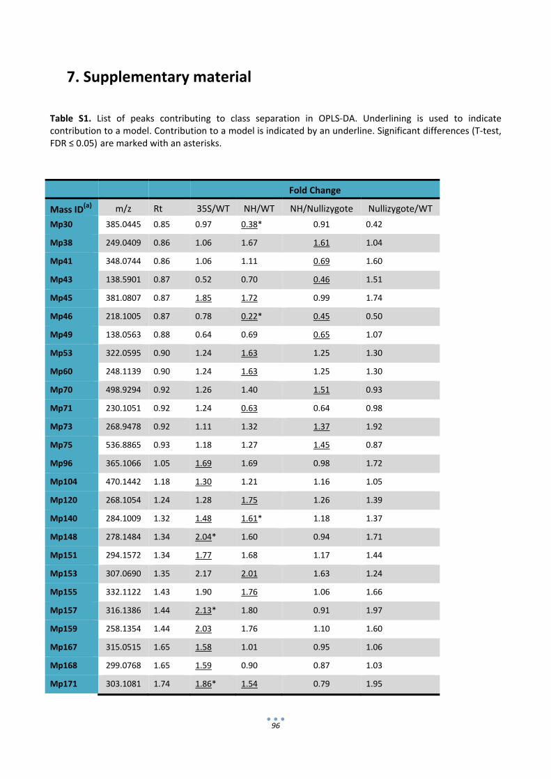
96
7. Supplementary material
Table S1. List of peaks contributing to class separation in OPLS‐DA. Underlining is used to indicate contribution to a model. Contribution to a model is indicated by an underline. Significant differences (T‐test, FDR ≤ 0.05) are marked with an asterisks.
Fold Change
Mass ID(a) m/z Rt 35S/WT NH/WT NH/Nullizygote Nullizygote/WT
Mp30 385.0445 0.85 0.97 0.38* 0.91 0.42
Mp38 249.0409 0.86 1.06 1.67 1.61 1.04
Mp41 348.0744 0.86 1.06 1.11 0.69 1.60
Mp43 138.5901 0.87 0.52 0.70 0.46 1.51
Mp45 381.0807 0.87 1.85 1.72 0.99 1.74
Mp46 218.1005 0.87 0.78 0.22* 0.45 0.50
Mp49 138.0563 0.88 0.64 0.69 0.65 1.07
Mp53 322.0595 0.90 1.24 1.63 1.25 1.30
Mp60 248.1139 0.90 1.24 1.63 1.25 1.30
Mp70 498.9294 0.92 1.26 1.40 1.51 0.93
Mp71 230.1051 0.92 1.24 0.63 0.64 0.98
Mp73 268.9478 0.92 1.11 1.32 1.37 1.92
Mp75 536.8865 0.93 1.18 1.27 1.45 0.87
Mp96 365.1066 1.05 1.69 1.69 0.98 1.72
Mp104 470.1442 1.18 1.30 1.21 1.16 1.05
Mp120 268.1054 1.24 1.28 1.75 1.26 1.39
Mp140 284.1009 1.32 1.48 1.61* 1.18 1.37
Mp148 278.1484 1.34 2.04* 1.60 0.94 1.71
Mp151 294.1572 1.34 1.77 1.68 1.17 1.44
Mp153 307.0690 1.35 2.17 2.01 1.63 1.24
Mp155 332.1122 1.43 1.90 1.76 1.06 1.66
Mp157 316.1386 1.44 2.13* 1.80 0.91 1.97
Mp159 258.1354 1.44 2.03 1.76 1.10 1.60
Mp167 315.0515 1.65 1.58 1.01 0.95 1.06
Mp168 299.0768 1.65 1.59 0.90 0.87 1.03
Mp171 303.1081 1.74 1.86* 1.54 0.79 1.95
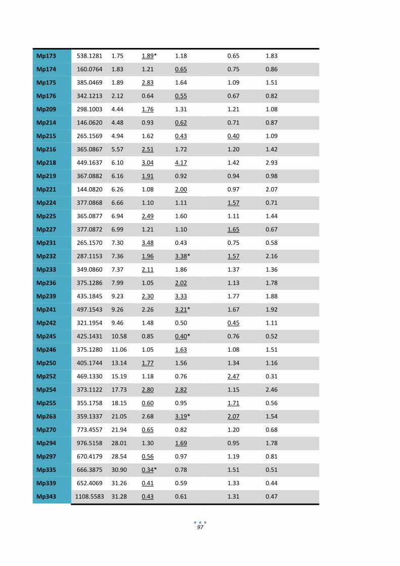
97
Mp173 538.1281 1.75 1.89* 1.18 0.65 1.83
Mp174 160.0764 1.83 1.21 0.65 0.75 0.86
Mp175 385.0469 1.89 2.83 1.64 1.09 1.51
Mp176 342.1213 2.12 0.64 0.55 0.67 0.82
Mp209 298.1003 4.44 1.76 1.31 1.21 1.08
Mp214 146.0620 4.48 0.93 0.62 0.71 0.87
Mp215 265.1569 4.94 1.62 0.43 0.40 1.09
Mp216 365.0867 5.57 2.51 1.72 1.20 1.42
Mp218 449.1637 6.10 3.04 4.17 1.42 2.93
Mp219 367.0882 6.16 1.91 0.92 0.94 0.98
Mp221 144.0820 6.26 1.08 2.00 0.97 2.07
Mp224 377.0868 6.66 1.10 1.11 1.57 0.71
Mp225 365.0877 6.94 2.49 1.60 1.11 1.44
Mp227 377.0872 6.99 1.21 1.10 1.65 0.67
Mp231 265.1570 7.30 3.48 0.43 0.75 0.58
Mp232 287.1153 7.36 1.96 3.38* 1.57 2.16
Mp233 349.0860 7.37 2.11 1.86 1.37 1.36
Mp236 375.1286 7.99 1.05 2.02 1.13 1.78
Mp239 435.1845 9.23 2.30 3.33 1.77 1.88
Mp241 497.1543 9.26 2.26 3.21* 1.67 1.92
Mp242 321.1954 9.46 1.48 0.50 0.45 1.11
Mp245 425.1431 10.58 0.85 0.40* 0.76 0.52
Mp246 375.1280 11.06 1.05 1.63 1.08 1.51
Mp250 405.1744 13.14 1.77 1.56 1.34 1.16
Mp252 469.1330 15.19 1.18 0.76 2.47 0.31
Mp254 373.1122 17.73 2.80 2.82 1.15 2.46
Mp255 355.1758 18.15 0.60 0.95 1.71 0.56
Mp263 359.1337 21.05 2.68 3.19* 2.07 1.54
Mp270 773.4557 21.94 0.65 0.82 1.20 0.68
Mp294 976.5158 28.01 1.30 1.69 0.95 1.78
Mp297 670.4179 28.54 0.56 0.97 1.19 0.81
Mp335 666.3875 30.90 0.34* 0.78 1.51 0.51
Mp339 652.4069 31.26 0.41 0.59 1.33 0.44
Mp343 1108.5583 31.28 0.43 0.61 1.31 0.47
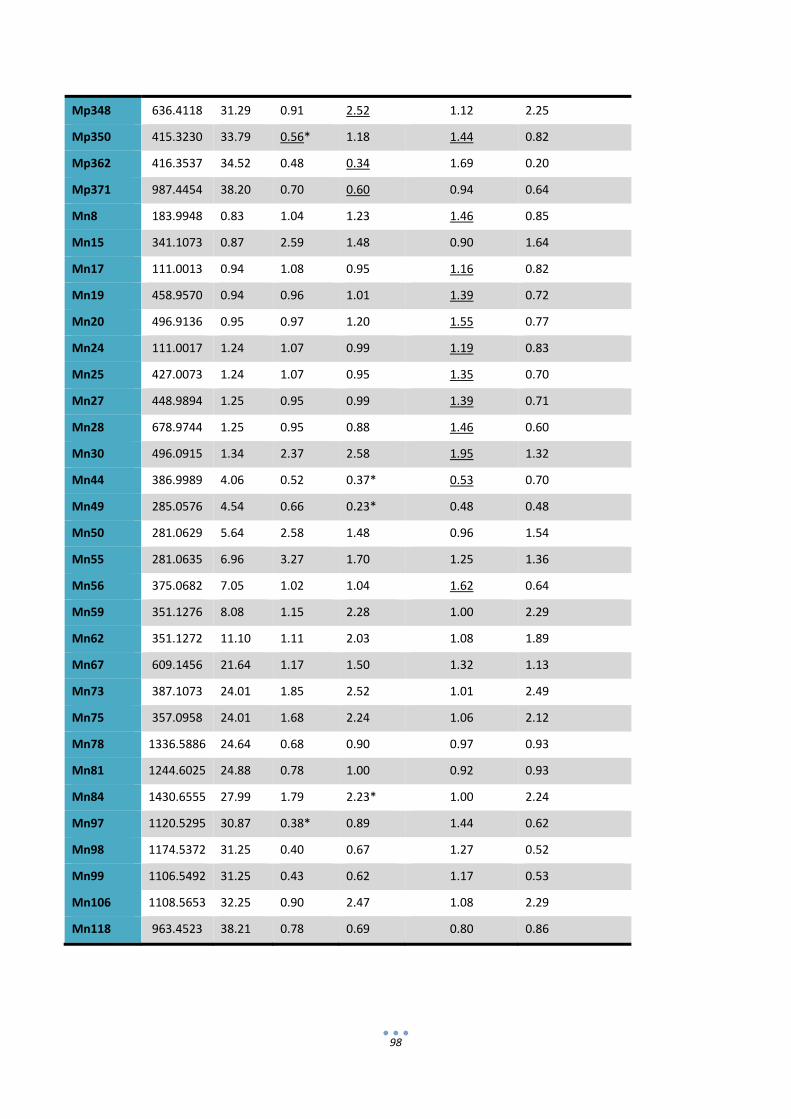
98
Mp348 636.4118 31.29 0.91 2.52 1.12 2.25
Mp350 415.3230 33.79 0.56* 1.18 1.44 0.82
Mp362 416.3537 34.52 0.48 0.34 1.69 0.20
Mp371 987.4454 38.20 0.70 0.60 0.94 0.64
Mn8 183.9948 0.83 1.04 1.23 1.46 0.85
Mn15 341.1073 0.87 2.59 1.48 0.90 1.64
Mn17 111.0013 0.94 1.08 0.95 1.16 0.82
Mn19 458.9570 0.94 0.96 1.01 1.39 0.72
Mn20 496.9136 0.95 0.97 1.20 1.55 0.77
Mn24 111.0017 1.24 1.07 0.99 1.19 0.83
Mn25 427.0073 1.24 1.07 0.95 1.35 0.70
Mn27 448.9894 1.25 0.95 0.99 1.39 0.71
Mn28 678.9744 1.25 0.95 0.88 1.46 0.60
Mn30 496.0915 1.34 2.37 2.58 1.95 1.32
Mn44 386.9989 4.06 0.52 0.37* 0.53 0.70
Mn49 285.0576 4.54 0.66 0.23* 0.48 0.48
Mn50 281.0629 5.64 2.58 1.48 0.96 1.54
Mn55 281.0635 6.96 3.27 1.70 1.25 1.36
Mn56 375.0682 7.05 1.02 1.04 1.62 0.64
Mn59 351.1276 8.08 1.15 2.28 1.00 2.29
Mn62 351.1272 11.10 1.11 2.03 1.08 1.89
Mn67 609.1456 21.64 1.17 1.50 1.32 1.13
Mn73 387.1073 24.01 1.85 2.52 1.01 2.49
Mn75 357.0958 24.01 1.68 2.24 1.06 2.12
Mn78 1336.5886 24.64 0.68 0.90 0.97 0.93
Mn81 1244.6025 24.88 0.78 1.00 0.92 0.93
Mn84 1430.6555 27.99 1.79 2.23* 1.00 2.24
Mn97 1120.5295 30.87 0.38* 0.89 1.44 0.62
Mn98 1174.5372 31.25 0.40 0.67 1.27 0.52
Mn99 1106.5492 31.25 0.43 0.62 1.17 0.53
Mn106 1108.5653 32.25 0.90 2.47 1.08 2.29
Mn118 963.4523 38.21 0.78 0.69 0.80 0.86

99
Chapter 3
Combinatorial analysis of secretory Immunoglobulin A (sIgA) expression in plants Paloma Juárez, Estefanía Huet‐Trujillo, Alejandro Sarrión‐Perdigones, Erica Elvira Falconi, Antonio Granell and Diego Orzáez
International Journal of Molecular Sciences 2013, 14, 6205‐6222
100
1. Introduction
Monoclonal antibodies (mAbs) have been used in research and diagnosis for many years, and their
application in health is increasing rapidly. They play an essential role in cancer therapy (Reichert and Valge‐
Archer, 2007), however topical and oral immunotherapy of mucosal surfaces with mAbs is also of great
interest, as it can block the entry and transmission of human pathogens (Corthesy, 2003; Corthesy, 2009;
Corthesy, 2010).
There are different expression systems available for the production of recombinant antibodies, each
with its advantages and shortcomings. Plants are one of the most interesting platforms for recombinant
antibody production, because they are cost‐effective, highly scalable, have a low risk of contamination with
mammalian pathogens (Fischer et al., 2004; Paul et al., 2011; Sarrion‐Perdigones et al., 2011b; Twyman et
al., 2003) and also can perform post‐translational modifications similar to mammals, as, for example, N‐ and
O‐glycosylation (Saint‐Jore‐Dupas et al., 2007). IgA is the most abundant antibody in mucus, and it forms
part of the first line of defense against infectious agents. IgA can be present in the body fluids in its
monomeric form (mIgA), containing only heavy chain (HC) and light chain (LC) or forming a secretory IgA
(sIgA), a multiprotein structure comprising two full IgA molecules dimerized by a short joining chain (JC) and
surrounded by the secretory component (SC), a polypeptide resulting from the proteolytic cleavage of the
poly‐immunoglobulin receptor (pIgR). IgA and, particularly, sIgA are good candidates for mucosal passive
immunotherapy, having a number of advantages over IgG (e.g., the presence of four antigen binding sites,
increased resistance against proteolysis in the gastrointestinal tract and the blocking of some bacterial
pathogens mediated by carbohydrates, both in the HC and the SC) (Corthesy, 2003; Corthesy, 2009;
Corthesy, 2010). Despite this, most research effort on plant‐made recombinant antibodies has been made
on monoclonal IgG antibodies.
The first attempt to produce a plant‐made sIgA for passive immunization was the murine hybrid
Guy’s 13 sIgG/A (Ma et al., 1995a; Ma et al., 1998), which was evaluated in phase I and II clinical trials
(Weintraub et al., 2005) as CaroxR™. Since then, various groups have expressed sIgA in plants (Larrick et al.,
2001; Nicholson et al., 2005; Wieland et al., 2006; Wycoff, 2004). However, there is a lack of information
about what structural requirements are the best to produce functional, fully human sIgA in plants with
maximum activity.
There are a number of options available when designing a new antibody in sIgA format, which could
lead to completely different products for the same purpose. Therefore, it is important to study how
modifications in the engineered parts will affect antibody function. In this work, we focus on the study of the
in vitro binding activity of the final product, although other functional considerations, such as in vivo activity
or stability, could also be tested with an adequate setup. In the first place, there are several options
concerning structural design: LC may occur in two isotype forms, designated kappa (κ) and lambda (λ), with
101
no functional differences described between them (Foley et al., 1991), and two types of HC, namely the α1
and the α2. In particular, the hinge region differs significantly between the two HC isoforms. The hinge
region of HCα1 is comprised of 23 residues, while HCα2 is made of only 10 residues. The greater number of
amino‐acids in the IgA α1 provides an extended structure and a greater antigenic reach, while IgA α2 is more
compact and, therefore, less susceptible to proteolitic cleavage (Furtado et al., 2004).
Secondly, subcellular localization may also affect the overall efficacy of the antibody. Targeting
antibody chains to specific compartments in the plant cell can improve the stability, yield and/or
downstream processing (De Muynck et al., 2010). The secretory pathway appears to be the most convenient
route for a correct antibody folding and assembly, due to the oxidizing environment of the endoplasmic
reticulum (ER), the low abundance of proteases and the presence of molecular chaperones. Moreover,
protein glycosylation occurs only in the endomembrane system (Ma et al., 2003).
Once in the secretory pathway, there are several possible options; for example, the antibody can be
efficiently retrieved from the cis‐Golgi back to the ER using a C‐terminal H/KDEL retention signal or
deposited in the vacuole (Petruccelli et al., 2006). Several antibodies have also been reported as apoplastic
(De Muynck et al., 2009; Petruccelli et al., 2006); however, in some cases, retention in the ER leads to a yield
improvement (Schouten et al., 1996) and avoids plant complex glycosylation patterns that could cause an
unwanted immune response (Bencurova et al., 2004; Gomord et al., 2010).
An appropriate way to produce the most efficient antibody design is to perform a combinatorial
analysis of several versions of sIgA and select those which accumulate at higher levels and/or present an
improvement on stability and activity. With traditional DNA assembly systems, this can become tedious
work. However, with new standard modular cloning tools, like GoldenBraid (GB) (Sarrion‐Perdigones et al.,
2011a) or MoClo (Weber et al., 2011), to achieve all the combinations should be facilitated. These
technologies open a new way for optimization of antibody production in plants by experimentally testing the
best subcellular targeting and isotype combinations for the expression of a target antibody.
As a proof of this concept, we have followed a combinatorial approach to optimize the plant
production of a sIgA version of an anti‐rotavirus monoclonal antibody (2A1 sIgA). The variable regions of 2A1
antibody were initially selected by phage display against the VP8* peptide of the VP4 protein of the rotavirus
SA11 strain capsid (Monedero et al., 2004). This antibody was previously described in its monomeric format,
showing a strong rotavirus neutralization activity (Juarez et al., 2012).

102
2. Results
2.1 GoldenBraid‐assisted multigene assembly of 16 versions of secretory IgA
A number of standard basic DNA pieces, named GBparts, were used as a starting point in the
building of sIgA multigene structures. Basic GBparts comprised non‐coding DNA regions, as the CaMV 35S
constitutive promoter (35s) and the strong nopaline synthase transcription terminator (Tnos), together with
a number of GB‐adapted coding sequences required for the assembly of a functional sIgA, namely a signal
peptide derived from the tomato pectate lyase gene (SP), the constant regions of the human HCα1 and
HCα2, the constant regions of human LCλ and LCκ, the extracellular region of the human SC and the
complete coding sequence of the human JC. In addition, the heavy and light variable regions of the anti‐
rotavirus VP8* peptide scFv antibody fragment 2A1 were also GB‐adapted and incorporated to the GBpart
collection (Figure 1a). GB‐adaptation, also called GB‐domestication, consisted of: (i) removal of internal BsaI
and BsmBI restriction sites by PCR‐directed mutagenesis; (ii) PCR‐mediated addition of flanking GB standard
bar‐codes, which consist of standard labels of 11 nucleotides that serve to facilitate cloning and, ultimately,
to specify the relative position of each GBpart in the assembly (see Figure 1b and Table S1); and (iii) TA‐
cloning of the resulting domesticated PCR products into pGEM‐T vectors.
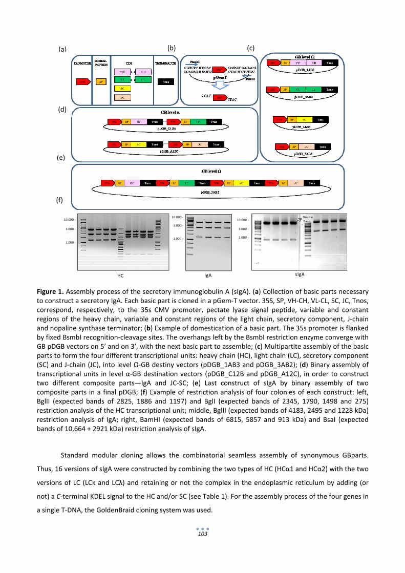
103
Figure 1. Assembly process of the secretory immunoglobulin A (sIgA). (a) Collection of basic parts necessary to construct a secretory IgA. Each basic part is cloned in a pGem‐T vector. 35S, SP, VH‐CH, VL‐CL, SC, JC, Tnos, correspond, respectively, to the 35s CMV promoter, pectate lyase signal peptide, variable and constant regions of the heavy chain, variable and constant regions of the light chain, secretory component, J‐chain and nopaline synthase terminator; (b) Example of domestication of a basic part. The 35s promoter is flanked by fixed BsmbI recognition‐cleavage sites. The overhangs left by the BsmbI restriction enzyme converge with GB pDGB vectors on 5′ and on 3′, with the next basic part to assemble; (c) Multipartite assembly of the basic parts to form the four different transcriptional units: heavy chain (HC), light chain (LC), secretory component (SC) and J‐chain (JC), into level Ω‐GB destiny vectors (pDGB_1AB3 and pDGB_3AB2); (d) Binary assembly of transcriptional units in level α‐GB destination vectors (pDGB_C12B and pDGB_A12C), in order to construct two different composite parts—IgA and JC‐SC; (e) Last construct of sIgA by binary assembly of two composite parts in a final pDGB; (f) Example of restriction analysis of four colonies of each construct: left, BglII (expected bands of 2825, 1886 and 1197) and BglI (expected bands of 2345, 1790, 1498 and 275) restriction analysis of the HC transcriptional unit; middle, BglII (expected bands of 4183, 2495 and 1228 kDa) restriction analysis of IgA; right, BamHI (expected bands of 6815, 5857 and 913 kDa) and BsaI (expected bands of 10,664 + 2921 kDa) restriction analysis of sIgA.
Standard modular cloning allows the combinatorial seamless assembly of synonymous GBparts.
Thus, 16 versions of sIgA were constructed by combining the two types of HC (HCα1 and HCα2) with the two
versions of LC (LCκ and LCλ) and retaining or not the complex in the endoplasmic reticulum by adding (or
not) a C‐terminal KDEL signal to the HC and/or SC (see Table 1). For the assembly process of the four genes in
a single T‐DNA, the GoldenBraid cloning system was used.
HC
10.000 ‐
3.000 ‐
1.000 ‐
sIgA
10.000 ‐
3.000 ‐
1.000 ‐
IgA
10.000 ‐
3.000 ‐
1.000 ‐
a b c
d
e
f
DoubleBand
(a)
(b) (c)
(d)
(e)
(f)

104
Table 1. Screening of 16 versions of sIgA by ELISA test. Anti VP8* binding activity is expressed in terms of Abs492 (Abs, absorbance). +++ refers to Abs > 2.0; ++ refers to Abs > 1.0; − refers to Abs < 0.3.
LCλ/SC LCλ/SCkdel LCκ/SC LCκ/SCkdel
HCα1 ++ ++ − −
HCα1kdel +++ ++ − − HCα2 ++ ++ − −
HCα2kdel ++ ++ − −
A number of steps were followed to construct each sIgA version. First, the so‐called multipartite
assembly reactions were performed. In multipartite reactions, GBparts are assembled together into binary
destination vectors (Ω‐GB vectors) to produce individual transcriptional units (TUs). To this end, GBparts are
simply mixed together in a single tube with Ω‐GB destination vector and subjected to a highly efficient
restriction/ligation reaction, which will orderly assemble all the elements of the transcriptional unit together
(Figure 1c). In GB multipartite assembly, properly assembled structures are distinguished from the initial
destination plasmids using positive blue/white β‐galactosidase selection. Multipartite assemblies comprising
four individual GBparts (e.g., SC TUs) resulted in 95% white colonies on average, whereas 5‐part GBpart
assemblies (e.g., HC TUs) resulted in 85% white colonies on average. In all cases, four individual white
colonies were selected and tested by restriction analysis and a minimum of three out of four colonies were
found correct (see HC panel in Figure 1f as an example).
Once individual transcriptional units were constructed, they were assembled together into 2‐part
TUs structures using level α‐GB destination vectors in a so‐called binary GB reaction. Binary reactions
combining LC + HC produced genetic modules for the expression of full monomeric IgA (mIgA), whereas SC
and JC TUs were also combined in a single genetic module named SC‐JC (Figure 1d). Last, mIgA and SC‐JC
composite parts were assembled together into a final level Ω‐GB destination vector in order to construct the
complete sIgA (Figure 1e). The final size of the sIgA constructs was of 13.6 kb. Binary assemblies ranged
between 5% and 20% white colonies. For each construct, four colonies were selected for digestion, resulting
in 100% correct colonies (see examples in Figure 1f).
2.2 Transient expression in Nicotiana benthamiana of 16 versions of sIgA against Rotavirus
All 16 binary plasmids containing sIgA versions against VP8* (see Table 1) were transferred to
Agrobacterium tumefaciens and transiently transformed in Nicotiana benthamiana by means of
agroinfiltration. For the resulting 16 plant samples, an initial screening was performed by antigen ELISA to
detect anti‐VP8* IgA activity in the clarified crude extracts of agroinfiltrated leaves using an anti‐HC antibody
105
for detection. To avoid potential proteolysis, protease inhibitor, PMSF, was added to every extract. In order
to ensure the accuracy of the comparison among all the combinations, all samples were equalized on the
basis of the luciferase activity of a cotransformed plasmid in which the nopaline synthase promoter drives
the luciferase gene. Antigen ELISA tests showed high anti‐VP8* binding activity in half of the samples (Table
1). Surprisingly, a very low activity was observed in all sIgA versions containing the LCκ. The integrity of
kappa‐sIgA constructs was confirmed by retro‐transformation of Agrobacterium plasmids into E. coli and
subsequent restriction analysis and sequencing. In addition, the low anti‐VP8* activity was confirmed in a
second ELISA experiment that yielded similar results (not shown). Consequently, work with LCκ versions was
discontinued and all further analyses were done with the LCλ‐containing sIgA versions.
A detailed examination of the remaining eight combinations was subsequently performed. The
analysis was completed with TUs expressing monomeric IgA and free SC. Leaf age is a known factor
influencing recombinant protein expression levels. When leaves of three different ages were transiently
transformed and assayed for luciferase expression, a coefficient of variation of 22% was observed.
Therefore, in order to increase the accuracy of the comparison, three independent leaves taken from
different plants (leaves number 4, 5 and 6, counting from the base of the plant) were infiltrated per each
construct. Each leaf was used as an individual biological replicate, and all results were subsequently
normalized using a luciferase reporter system as an internal standard. The anti‐VP8* activity of each
combination was analyzed in detail by antigen‐ELISA using three different detection tools, namely anti‐HC,
anti‐LC and anti‐SC antibodies. Antigen‐ELISA tests against the HC were first carried out in order to give a
first view of total IgA content (including mIgA and sIgA). Considerably high binding activity values were
observed in all eight sIgA combinations, while the SC control remained negative. Figure 2a shows that ER
retention had a positive effect, resulting in significantly higher anti‐VP8* activity (p‐value < 0.01). This effect
is more noticeable when the KDEL signal is added to the SC than to the HC, and the comparison between
these two types of samples yielded also significant differences (p‐value <0.01). The increase in anti‐VP8*
activity observed when the SC is targeted to the ER was the first evidence of the correct formation of a
secretory complex. Interestingly, the ER‐associated activity was shown to increase in a non‐additive fashion,
as the simultaneous targeting of both HC and SC to the ER did not yield significantly higher activity than
individual targeting. A second interesting observation extracted from Figure 2a is that all the combinations
containing HCα1 appear to be significantly more efficient than their HCα2 counterparts (p‐value <0.01). The
above‐described results were also confirmed using anti‐LC as the detecting antibody, confirming the
presence of LC in the functional complexes detected in the previous ELISA test. (Figure 2b).
Finally, antigen‐ELISA tests using anti‐SC as the detecting antibody were developed in order to
specifically detect the anti‐VP8* activity of sIgA complexes, as only SC‐containing, VP8*‐binding antibody
complexes can be detected using this ELISA set up. Interestingly, the activity pattern observed for sIgA

106
combinations was very similar to the one observed for total IgA using anti‐HC (compare Figure 2a,c). In
particular, the positive effect provided by the presence of a KDEL peptide in the SC was also observed using
anti‐SC detecting antibody, further confirming that the retention of the SC in the ER increases the overall
anti‐VP8* IgA activity by specifically stabilizing the sIgA subfraction.
Figure 2. Combinatorial analysis of eight λ versions of sIgA by ELISA tests. (a) ELISA assay developed with anti‐HC; (b) ELISA assay developed with anti‐LCλ; (c) ELISA assay developed with anti‐SC. All plates were coated with VP8* antigen. Three different leaves were infiltrated and tested for each sample. Means of the three biological replicates are represented, with error bars representing the standard deviation. All samples were equalized, with the luciferase reporter system as an internal standard.
0
0,5
1
1,5A
bs4
92Heavy Chain
0
0,5
1
1,5
2
Abs
49
2
Light Chain
0
0,5
1
Abs
492
Secretory Component
a
b
c
(a)
(b)
(c)
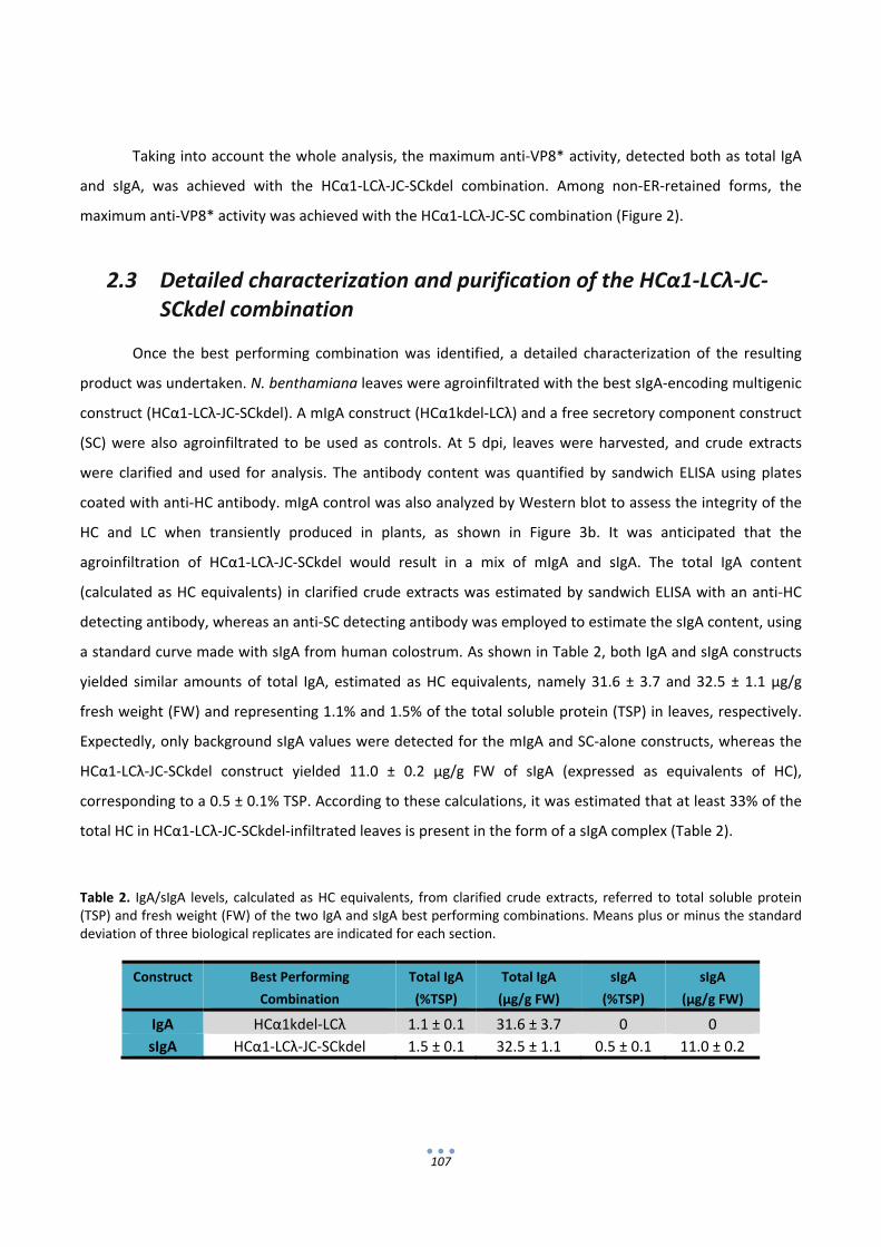
107
Taking into account the whole analysis, the maximum anti‐VP8* activity, detected both as total IgA
and sIgA, was achieved with the HCα1‐LCλ‐JC‐SCkdel combination. Among non‐ER‐retained forms, the
maximum anti‐VP8* activity was achieved with the HCα1‐LCλ‐JC‐SC combination (Figure 2).
2.3 Detailed characterization and purification of the HCα1‐LCλ‐JC‐SCkdel combination
Once the best performing combination was identified, a detailed characterization of the resulting
product was undertaken. N. benthamiana leaves were agroinfiltrated with the best sIgA‐encoding multigenic
construct (HCα1‐LCλ‐JC‐SCkdel). A mIgA construct (HCα1kdel‐LCλ) and a free secretory component construct
(SC) were also agroinfiltrated to be used as controls. At 5 dpi, leaves were harvested, and crude extracts
were clarified and used for analysis. The antibody content was quantified by sandwich ELISA using plates
coated with anti‐HC antibody. mIgA control was also analyzed by Western blot to assess the integrity of the
HC and LC when transiently produced in plants, as shown in Figure 3b. It was anticipated that the
agroinfiltration of HCα1‐LCλ‐JC‐SCkdel would result in a mix of mIgA and sIgA. The total IgA content
(calculated as HC equivalents) in clarified crude extracts was estimated by sandwich ELISA with an anti‐HC
detecting antibody, whereas an anti‐SC detecting antibody was employed to estimate the sIgA content, using
a standard curve made with sIgA from human colostrum. As shown in Table 2, both IgA and sIgA constructs
yielded similar amounts of total IgA, estimated as HC equivalents, namely 31.6 ± 3.7 and 32.5 ± 1.1 μg/g
fresh weight (FW) and representing 1.1% and 1.5% of the total soluble protein (TSP) in leaves, respectively.
Expectedly, only background sIgA values were detected for the mIgA and SC‐alone constructs, whereas the
HCα1‐LCλ‐JC‐SCkdel construct yielded 11.0 ± 0.2 μg/g FW of sIgA (expressed as equivalents of HC),
corresponding to a 0.5 ± 0.1% TSP. According to these calculations, it was estimated that at least 33% of the
total HC in HCα1‐LCλ‐JC‐SCkdel‐infiltrated leaves is present in the form of a sIgA complex (Table 2).
Table 2. IgA/sIgA levels, calculated as HC equivalents, from clarified crude extracts, referred to total soluble protein (TSP) and fresh weight (FW) of the two IgA and sIgA best performing combinations. Means plus or minus the standard deviation of three biological replicates are indicated for each section.
Construct Best Performing
Combination
Total IgA
(%TSP)
Total IgA
(μg/g FW)
sIgA
(%TSP)
sIgA
(μg/g FW)
IgA HCα1kdel‐LCλ 1.1 ± 0.1 31.6 ± 3.7 0 0
sIgA HCα1‐LCλ‐JC‐SCkdel 1.5 ± 0.1 32.5 ± 1.1 0.5 ± 0.1 11.0 ± 0.2

108
In principle, both mIgA and sIgA can be purified using SSL7‐affinity chromatography, which binds the
Cα2/Cα3 domain junction of the Fc (Fragment crystallizable) portion of the HC (Langley et al., 2005). For
affinity purification, clarified crude extracts were passed through SSL7‐agarose columns. The purification
steps were monitored by antigen‐ELISA, coomassie stained SDS‐PAGE and Western blot. Upon PAGE
separation of the elution fractions, a 25 kDa band corresponding to the full size LC and a 55 kDa band
corresponding to the full size HC were observable in coomassie stained gels from IgA and sIgA samples, after
a single step of affinity chromatography (Figure 3a). The 64 kDa coomassie band that would correspond to
the free SC was not detected by coomassie staining, but was readily detectable in Western Blot analysis from
sIgA samples (Figure 3d). In contrast, SSL7 control purifications from SC‐alone and mIgA agroinfiltrations did
not yield any detectable SC band (Figure 3d).
Figure 3. Characterization of SSL7‐purified sIgA. (a) Coomassie stained SDS‐PAGE under reducing conditions of elution fractions corresponding to HCα1‐LCλ‐JC‐SCkdel (sIgA), HCα1kdel‐LCλ (IgA) and free secretory component (SC) purification. Bands correspond to the heavy chain (HC) and light chain (LC); (b) Western blot analysis under reducing conditions of a clarified crude extract (CE) sample of HCα1kdel‐LCλ (IgA) developed with anti‐HC (left) and with anti‐LC (right); (c) ELISA analysis, coated with VP8*, of starting clarified crude extract (CE) and elution fraction (ELU) of three samples: sIgA, IgA and free SC, performed as described in Figure 2. Upper panel developed with anti‐HC, medium panel
a
sIgA IgA SC
Anti-HC
Anti-LC
Anti-SC
C.E. ELU. C.E. ELU. C.E. ELU.
sIgA IgA SC
HC
LC
d
0,0
0,5
1,0
E.C ELU
Bin
ding
Avt
ivit
y
Heavy Chain sIgA
IgA
SC
0,0
0,5
1,0
1,5
E.C ELU
Bin
ding
Act
ivit
y
Light Chain sIgAIgASC
0,0
0,5
1,0
1,5
E.C ELU
Bin
ding
Act
ivit
y
Secretory Component sIgAIgASC
e
c
191
51
28
HC LC
C.E C.Eb
0
0,2
0,4
0,6
0,8
707 450 287 182 116 74 47 30
µg/
mL
kDa
SC
HC
IgA
sIgA
191
51
28
(a) (b)
(c)
(d)
(e)
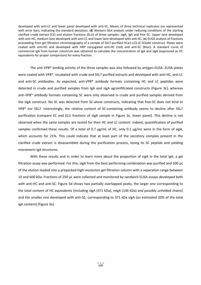
109
developed with anti‐LC and lower panel developed with anti‐SC. Means of three technical replicates are represented with error bars, indicating the standard deviation; (d) Western blot analysis under reducing conditions of the starting clarified crude extract (CE) and elution fractions (ELU) of three samples: sIgA, IgA and free SC. Upper lane developed with anti‐HC, medium lane developed with anti‐LC and lower lane developed with anti‐SC; (e) ELISA analysis of fractions proceeding from gel filtration chromatography of a sample of SSL7‐purified HCα1‐LCλ‐JC‐SCkdel construct. Plates were coated with anti‐HC and developed with HRP conjugated anti‐HC (red) and anti‐SC (blue). A standard curve of commercial IgA from human colostrum was obtained to calculate the concentration of IgA and sIgA (expressed as HC equivalents for proper comparison) for every fraction.
The anti‐VP8* binding activity of the three samples was also followed by antigen ELISA. ELISA plates
were coated with VP8*, incubated with crude and SSL7 purified extracts and developed with anti‐HC, anti‐LC
and anti‐SC antibodies. As expected, anti‐VP8* antibody formats containing HC and LC peptides were
detected in crude and purified samples from IgA and sIgA agroinfiltrated constructs (Figure 3c), whereas
anti‐VP8* antibody formats containing SC were only observed in crude and purified samples derived from
the sIgA construct. No SC was detected from SC‐alone constructs, indicating that free‐SC does not bind to
VP8* nor SSL7. Interestingly, the relative content of SC‐containing antibody seems to decline after SSL7
purification (compare EC and ELU fractions of sIgA sample in Figure 3c, lower panel). This decline is not
observed when the same samples are tested for their HC and LC content. Indeed, quantification of purified
samples confirmed these results. Of a total of 0.7 μg/mL of HC, only 0.1 μg/mL were in the form of sIgA,
which accounts for 21%. This could indicate that at least part of the secretory complex present in the
clarified crude extract is disassembled during the purification process, losing its SC peptide and yielding
monomeric IgA structures.
With these results and in order to learn more about the proportion of sIgA in the total IgA, a gel
filtration assay was performed. For this, sIgA from the best performing combination was purified and 500 μL
of the elution loaded into a prepacked high‐resolution gel filtration column with a separation range between
10 and 600 kDa. Fractions of 250 μL were collected and monitored by sandwich ELISA assays developed both
with anti‐HC and anti‐SC. Figure 3d shows two partially overlapped peaks, the larger one corresponding to
the total content of HC equivalents (including sIgA (371 kDa), mIgA (146 kDa) and possibly unfolded chains)
and the smaller one developed with anti‐SC, corresponding to 371 kDa sIgA (an estimated 20% of the total
IgA content) (Figure 3e).
110
3. Discussion
The selection of the most appropriate isotype for recombinant antibody production in plants is rarely
addressed using experimental approaches, as practical hurdles often override technical or functional
considerations. Moreover, sIgA has been only occasionally considered as a feasible option for mucosal
passive immunotherapy, despite its demonstrated appropriateness (Ma et al., 1995b). Even more, certain
specific decisions concerning antibody design, as the choice for LC and HC isotypes or for subcellular
localization, are rarely made on the basis of an exhaustive experimental analysis. However, it has been
repeatedly observed that antibody expression levels diverge dramatically from case to case and that, e.g.,
the experimental selection of the most stable idiotypes, can bring considerable advantages in terms of yield
(Juarez et al., 2012; Wieland et al., 2006). A technical hurdle that compromises the selection among different
antibody formats is the difficulty to produce multigenic structures that can be assayed in a combinatorial
way. In the case of recombinant production of protein complexes in plants, the assembly of multiple
transcriptional units in a single T‐DNA is an inefficient and tedious task, particularly in the case of sIgA, which
requires the co‐expression of four TUs. As an alternative approach, some labs, including our own, have relied
on trans‐ co‐transformation, even though it implies reduced reproducibility and unwanted heterogeneity in
the expression levels of the different proteins, often impeding the reach of solid conclusions (Sarrion‐
Perdigones et al., 2011a). Here, we demonstrate that the recently developed GB assembly system facilitates
the combinatorial assembly of the four transcriptional units necessary for sIgA expression, ensuring the
coordinated expression of the four genes in transient expression experiments.
In a first screening, it was determined that the use of LCκ severely reduces the activity levels of IgA
2A1. The LCκ used in this assay was codon‐optimized for Nicotiana benthamiana expression, and therefore,
inefficient codon‐usage can be discarded as a possible cause for the observed expression. This observation
was particularly surprising and, although the integrity of the constructs containing LCκ were exhaustively
examined, we could not find any element in the genetic design that could account for the low activity
observed in this set of samples. Provided that LCκ have been successfully employed before in the production
of other plant‐made antibodies (Ramessar et al., 2008; Sack et al., 2007), it is quite possible that this is a
specific feature of the 2A1 variable region. If so, this would highlight the importance of combinatorial
screenings for successful plant‐made antibody expression. Further experiments using variable regions other
than 2A1 will serve to discern if the low activity of kappa light chain is an isotype or idiotype‐specific feature.
Detailed examination of the remaining combinations led to a number of conclusions. First, these
experiments served as a demonstration that a fully human sIgA complex is formed in the plant cell that
retains activity against VP8* antigen. In particular, the detection of anti‐VP8* activity using an anti‐SC
detecting antibody is, to our knowledge, the strongest evidence provided so far in support for the formation
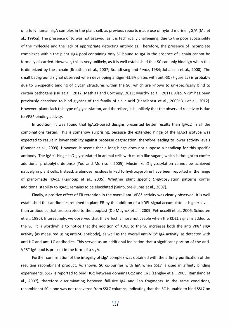
111
of a fully human sIgA complex in the plant cell, as previous reports made use of hybrid murine IgG/A (Ma et
al., 1995a). The presence of JC was not assayed, as it is technically challenging, due to the poor accessibility
of the molecule and the lack of appropriate detecting antibodies. Therefore, the presence of incomplete
complexes within the plant sIgA pool containing only SC bound to IgA in the absence of J‐chain cannot be
formally discarded. However, this is very unlikely, as it is well established that SC can only bind IgA when this
is dimerized by the J‐chain (Braathen et al., 2007; Brandtzaeg and Prydz, 1984; Johansen et al., 2000). The
small background signal observed when developing antigen‐ELISA plates with anti‐SC (Figure 2c) is probably
due to un‐specific binding of glycan structures within the SC, which are known to un‐specifically bind to
certain pathogens (Hu et al., 2012; Mathias and Corthesy, 2011; Murthy et al., 2011). Also, VP8* has been
previously described to bind glycans of the family of sialic acid (Haselhorst et al., 2009; Yu et al., 2012).
However, plants lack this type of glycosylation, and therefore, it is unlikely that the observed reactivity is due
to VP8* binding activity.
In addition, it was found that IgAα1‐based designs presented better results than IgAα2 in all the
combinations tested. This is somehow surprising, because the extended hinge of the IgAα1 isotype was
expected to result in lower stability against protease degradation, therefore leading to lower activity levels
(Bonner et al., 2009). However, it seems that a long hinge does not suppose a handicap for this specific
antibody. The IgAα1 hinge is O‐glycosylated in animal cells with mucin‐like sugars, which is thought to confer
additional proteolytic defense (Yoo and Morrison, 2005). Mucin‐like O‐glycosylation cannot be achieved
natively in plant cells. Instead, arabinose residues linked to hydroxyproline have been reported in the hinge
of plant‐made IgAα1 (Karnoup et al., 2005). Whether plant specific O‐glycosylation patterns confer
additional stability to IgAα1 remains to be elucidated (Saint‐Jore‐Dupas et al., 2007).
Finally, a positive effect of ER retention in the overall anti‐VP8* activity was clearly observed. It is well
established that antibodies retained in plant ER by the addition of a KDEL signal accumulate at higher levels
than antibodies that are secreted to the apoplast (De Muynck et al., 2009; Petruccelli et al., 2006; Schouten
et al., 1996). Interestingly, we observed that this effect is more noticeable when the KDEL signal is added to
the SC. It is worthwhile to notice that the addition of KDEL to the SC increases both the anti VP8* sIgA
activity (as measured using anti‐SC antibody), as well as the overall anti‐VP8* IgA activity, as detected with
anti‐HC and anti‐LC antibodies. This served as an additional indication that a significant portion of the anti‐
VP8* IgA pool is present in the form of a sIgA.
Further confirmation of the integrity of sIgA complex was obtained with the affinity purification of the
resulting recombinant product. As shown, SC co‐purifies with IgA when SSL7 is used in affinity binding
experiments. SSL7 is reported to bind HCα between domains Cα2 and Cα3 (Langley et al., 2005; Ramsland et
al., 2007), therefore discriminating between full‐size IgA and Fab fragments. In the same conditions,
recombinant SC alone was not recovered from SSL7 columns, indicating that the SC is unable to bind SSL7 on
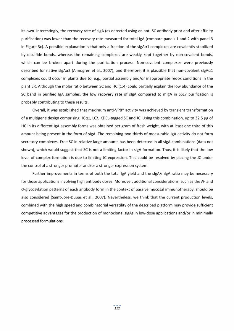
112
its own. Interestingly, the recovery rate of sIgA (as detected using an anti‐SC antibody prior and after affinity
purification) was lower than the recovery rate measured for total IgA (compare panels 1 and 2 with panel 3
in Figure 3c). A possible explanation is that only a fraction of the sIgAα1 complexes are covalently stabilized
by disulfide bonds, whereas the remaining complexes are weakly kept together by non‐covalent bonds,
which can be broken apart during the purification process. Non‐covalent complexes were previously
described for native sIgAα2 (Almogren et al., 2007), and therefore, it is plausible that non‐covalent sIgAα1
complexes could occur in plants due to, e.g., partial assembly and/or inappropriate redox conditions in the
plant ER. Although the molar ratio between SC and HC (1:4) could partially explain the low abundance of the
SC band in purified IgA samples, the low recovery rate of sIgA compared to mIgA in SSL7 purification is
probably contributing to these results.
Overall, it was established that maximum anti‐VP8* activity was achieved by transient transformation
of a multigene design comprising HCα1, LCλ, KDEL‐tagged SC and JC. Using this combination, up to 32.5 μg of
HC in its different IgA assembly forms was obtained per gram of fresh weight, with at least one third of this
amount being present in the form of sIgA. The remaining two thirds of measurable IgA activity do not form
secretory complexes. Free SC in relative large amounts has been detected in all sIgA combinations (data not
shown), which would suggest that SC is not a limiting factor in sIgA formation. Thus, it is likely that the low
level of complex formation is due to limiting JC expression. This could be resolved by placing the JC under
the control of a stronger promoter and/or a stronger expression system.
Further improvements in terms of both the total IgA yield and the sIgA/mIgA ratio may be necessary
for those applications involving high antibody doses. Moreover, additional considerations, such as the N‐ and
O‐glycosylation patterns of each antibody form in the context of passive mucosal immunotherapy, should be
also considered (Saint‐Jore‐Dupas et al., 2007). Nevertheless, we think that the current production levels,
combined with the high speed and combinatorial versatility of the described platform may provide sufficient
competitive advantages for the production of monoclonal sIgAs in low‐dose applications and/or in minimally
processed formulations.
113
4. Experimental Procedures
4.1 Cloning and assembly of modular parts
The DNA sequences corresponding to the constant regions of human alpha1 heavy chain (HCα1),
alpha2 heavy chain (HCα2), lambda light chain (LCλ) and secretory component (SC) were obtained from
Open Biosystems, Huntsville, AL, USA. Kappa light chain (LCκ) and J‐chain (JC) were codon optimized for
Solanum lycopersicum and synthesized by GeneScript, NJ, USA (NCBI Accession numbers KC515402 and
KC515401, respectively). The variable regions against VP8* were obtained from a scFv phage display,
selected as described earlier (Juarez et al., 2012). The DNA module encoding the signal peptide for secretion
was obtained by PCR from tomato SGN‐U212775 unigene. Taking advantage of the property of seamless
assembly, the junctions between the signal peptide and the coding sequences, e.g., HC (PSLLA‐QVQLL), were
tested with the signal P algorithm to ensure a correct processing into mature protein. PCR amplification was
performed by using the Advantage‐2 DNA Polymerase Mix (Clontech, Mountain View, CA, USA). The primers
used for amplification of each basic part were synthesized by IDTdna, Coralville, IA, USA (Table S1). PCR was
analyzed by agarose 1% gel electrophoresis and purified using the QIAquick PCR Purification Kit (Qiagen,
Hilden, Germany). Amplified parts were TA cloned using the pGEM‐T Easy Vector System (Promega,
Madison, WI, USA), and 1 μL of the ligation was transformed into DH5α electrocompetent cells. Plasmid DNA
preparations were obtained by using the E.Z.N.A. Plasmid Mini Kit I (Omega Bio‐Tek, Norcross, GA, USA).
Plasmid DNA concentration was measured using a Nano Drop Spectrophotometer 2000 (Thermo Scientific,
Rockford, IL, USA). Positive clones were selected in ampicillin‐containing plates and confirmed by plasmid
restriction analysis (EcoRI, NotI from Thermo Fisher Scientific, Waltham, MA, USA) and by sequencing.
Assembly reactions were performed basically as described by (Sarrion‐Perdigones et al., 2011a) using BsaI
and BsmBI (New England Biolabs, Ipswich, MA, USA) as restriction enzymes in 25‐cycle digestion/ligation
reactions. T4 DNA ligase was purchased from Promega. One microliter of the reaction was transformed into
DH5α electrocompetent cells. Positive clones were selected in kanamycin or spectinomycin‐containing
plates. Plasmid DNA preparations were made by using the E.Z.N.A. Plasmid Mini Kit I (Omega Bio‐Tek).
Plasmid DNA concentration was measured using a Nano Drop Spectrophotometer 2000 (Thermo Scientific).
Constructs were confirmed by plasmid restriction analysis and by sequencing. Constructs for plant functional
assays were transferred to Agrobacterium tumefaciens electrocompetent strain GV3101 containing pSoup
plasmid.

114
4.2 Strains and growth conditions
Escherichia coli DH5α was used for gene cloning, and Agrobacterium tumefaciens strain GV3101 with
pSoup was used for plant agroinfiltration and transformation experiments, as described in (Sarrion‐
Perdigones et al., 2011a).
4.3 Plant transient transformation
Agroinfiltration was performed as previously described in (Orzaez et al., 2006). Briefly, overnight
grown bacterial cultures (5 mL) were sedimented by centrifugation (15 min, 3000× g), resuspended in
agroinfiltration buffer (AB) (10 mM MES pH 5.6, 10 mM MgCl2, 200 μM acetosyringone) and incubated for 2
h at room temperature (RT) on a horizontal rolling mixer. Bacterial cultures were diluted in AB to an optical
density of 0.2 at 600 nm. Co‐infiltrations of each construct with both pGreen_P19 (bushy stunt virus‐TBSV‐
P19, suppressor of silencing) (Sarrion‐Perdigones et al., 2011a) and pGreen_Luciferase (Firefly Luciferase
Genetic reporter under the control of a Nopaline synthase promoter and terminator) were performed by
mixing equal volumes of the corresponding bacterial suspensions. Inoculations were carried out by syringe‐
agroinfiltration in leaves of 4–5 weeks old Nicotiana benthamiana plants (growing conditions: 24 °C day/20
°C night in a 16 h light/8 h dark cycle). Samples were collected 5 days post‐infiltration and examined for
transgene expression.
4.4 Plant material and sample preparation
For sample preparation, Nicotiana benthamiana leaves were ground with a mortar and a pestle to a
fine powder under liquid nitrogen, and the total soluble protein (TSP) was extracted with 1:3 (w:v)
phosphate buffer saline (PBS) pH = 7.4 complemented with 0.5 mM PMSF protease inhibitor. After mixing,
the suspension was centrifuged twice at 2 °C at 16,000× g, and the supernatant, referred to as clarified crude
extract, was immediately used for analysis. Samples were equalized with the luciferase reporter system as
an internal standard. Luciferase activity was determined with the Dual‐Luciferase Reporter Assay System
(Promega), following the manufacturer’s procedures, and luminescence was measured with a Glomax 96
microplate luminometer (Promega).
4.5 VP8* Rotavirus surface protein production
Recombinant VP8* was obtained as described in (Juarez et al., 2012). Briefly, Escherichia coli M11
were transformed with Plasmid pQEVP8*, kindly provided by Monedero from Instituto de Agroquímica y
115
Tecnología de Alimentos (IATA, Valencia, Spain). Purification was performed using HisTrap HP affinity
columns (GE Healthcare, Buckinghamshire, UK), following the manufacturer’s procedures.
4.6 ELISAs for the quantification and detection of VP8* binding
activity of IgA and sIgA
Plates (Corning, New York, NY, USA) were coated overnight at 4 °C in coating buffer (50 mM
carbonate buffer pH 9.8) either with anti‐IgA capture antibody 1:500 (Sigma‐Aldrich, St‐Louis, MO, USA) for
IgA/sIgA quantification or with 10 μg/mL of recombinant VP8* for the detection of VP8* binding activity.
Plates were then washed four times in PBS and blocked with a 2% (w/v) solution of ECL AdvanceTM Blocking
agent (GE Healthcare) in PBS‐T [0.1% (v/v) Tween 20 in PBS]. Samples were diluted in PBS as required for
each assay and incubated for 1 h at room temperature. After incubation, plates were washed four times in
PBS, and the antibodies for detection were added in PBS‐T‐2% blocking buffer (GE Healthcare). Four
different antibodies were used for detection in VP8*‐ELISAs: anti‐HC (HRP‐conjugated) 1:5000 (Sigma
Aldrich, St. Louis, MO, USA), anti‐LCλ (non‐conjugated) 1:5000 (Sigma Aldrich), anti‐LCκ (non‐conjugated)
1:5000 (Pierce Thermo Scientific) and anti‐SC (non‐conjugated) 1:500 (Gentaur, Kampenhout, Belgium). Anti‐
rabbit‐HRP (GE Healthcare) secondary antibody (1:5000) was used after the non‐conjugated detecting
antibodies. For quantification, anti‐HC (HRP‐conjugated) 1:5000 (Sigma Aldrich) and anti‐SC (non‐
conjugated) 1:500 (Gentaur, Kampenhout, Belgium) were used. Anti‐rabbit‐HRP (GE Healthcare) secondary
antibody 1:5000 was also used for detection after the anti‐SC detecting antibody. After four PBS washes, the
substrate (O‐phenylenediamine from Sigma Aldrich) was added, and the reactions were stopped with 3 M
HCl. Absorbance was determined at 492 nm. The mean and SD of three samples of each combination were
calculated for every VP8*‐ELISA. A standard curve of HC content in a commercial IgA obtained from human
colostrums (Sigma‐Aldrich) was used to calculate the HC content in IgA/sIgA samples. The same standard
was used to estimate the sIgA content in sIgA‐containing samples using anti‐SC as the secondary antibody.
To facilitate comparisons, all antibody concentrations were provided as equivalents of HC. Three replicates
were analyzed per each experimental point, and the mean ± SD was calculated.
4.7 SDS‐PAGE and Western blot analysis
Proteins were separated by SDS/PAGE in 10% denaturing gels (Invitrogen, Carlsbad, NM, USA). Gel
staining was carried out with coomassie following standard procedures. For Western blot analysis, blots
were incubated with 1:20,000 anti‐HC (Sigma Aldrich), 1:10,000 anti‐LC (Sigma Aldrich) or anti‐SC 1:5,000
(Gentaur), followed by 1:20,000 μg/mL HRP‐conjugated anti‐rabbit IgG secondary antibody (GE Healthcare)
for the detection of the LC and SC. Blots were developed with an ECL Plus Western Blotting Detection
System (GE Healthcare).
116
4.8 SSL7 affinity purification
The SSL7 protein specifically binds the hydrophobic interface between the Cα2 and Cα3 domains of
the Fc portion of the HC. Protein extracts, prepared as explained previously, were further clarified using a
0.22 μm Stericup (Millipore, Billerica, MA, USA) on ice. Purification steps were performed as previously
described by (Juarez et al., 2012).
5. Acknowledgments
This work has been funded by Grant BIO2010‐15384 from Plan Nacional I + D of the Spanish Ministry
of Science. Juarez P. is a recipient of a FPU fellowship, and Sarrión‐Perdigones A. And Huet‐Trujillo E. are
recipients of a FPI fellowship. We want to thank Monedero for kindly providing scFv and VP8* clones.

117
6. References
Almogren, A., Senior, B.W. and Kerr, M.A. (2007) A comparison of the binding of secretory component to immunoglobulin A (IgA) in human colostral S‐IgA1 and S‐IgA2. Immunology 120, 273‐280.
Bencurova, M., Hemmer, W., Focke‐Tejkl, M., Wilson, I.B. and Altmann, F. (2004) Specificity of IgG and IgE antibodies against plant and insect glycoprotein glycans determined with artificial glycoforms of human transferrin. Glycobiology 14, 457‐466.
Bonner, A., Almogren, A., Furtado, P.B., Kerr, M.A. and Perkins, S.J. (2009) The nonplanar secretory IgA2 and near planar secretory IgA1 solution structures rationalize their different mucosal immune responses. J Biol Chem 284, 5077‐5087.
Braathen, R., Hohman, V.S., Brandtzaeg, P. and Johansen, F.E. (2007) Secretory antibody formation: conserved binding interactions between J chain and polymeric Ig receptor from humans and amphibians. J Immunol 178, 1589‐1597.
Brandtzaeg, P. and Prydz, H. (1984) Direct evidence for an integrated function of J chain and secretory component in epithelial transport of immunoglobulins. Nature 311, 71‐73.
Corthesy, B. (2003) Recombinant secretory immunoglobulin A in passive immunotherapy: linking immunology and biotechnology. Curr Pharm Biotechnol 4, 51‐67.
Corthesy, B. (2009) Secretory immunoglobulin A: well beyond immune exclusion at mucosal surfaces. Immunopharmacol Immunotoxicol 31, 174‐179.
Corthesy, B. (2010) Role of secretory immunoglobulin A and secretory component in the protection of mucosal surfaces. Future Microbiol 5, 817‐829.
De Muynck, B., Navarre, C. and Boutry, M. (2010) Production of antibodies in plants: status after twenty years. Plant Biotechnol J 8, 529‐563.
De Muynck, B., Navarre, C., Nizet, Y., Stadlmann, J. and Boutry, M. (2009) Different subcellular localization and glycosylation for a functional antibody expressed in Nicotiana tabacum plants and suspension cells. Transgenic Res 18, 467‐482.
Fischer, R., Stoger, E., Schillberg, S., Christou, P. and Twyman, R.M. (2004) Plant‐based production of biopharmaceuticals. Curr Opin Plant Biol 7, 152‐158.
Foley, R.C., Raison, R.L. and Beh, K.J. (1991) Monoclonal antibody against sheep kappa light chain. Hybridoma 10, 507‐515.
Furtado, P.B., Whitty, P.W., Robertson, A., Eaton, J.T., Almogren, A., Kerr, M.A., Woof, J.M. and Perkins, S.J. (2004) Solution structure determination of monomeric human IgA2 by X‐ray and neutron scattering, analytical ultracentrifugation and constrained modelling: a comparison with monomeric human IgA1. J Mol Biol 338, 921‐941.
Gomord, V., Fitchette, A.C., Menu‐Bouaouiche, L., Saint‐Jore‐Dupas, C., Plasson, C., Michaud, D. and Faye, L. (2010) Plant‐specific glycosylation patterns in the context of therapeutic protein production. Plant Biotechnol J 8, 564‐587.
Haselhorst, T., Fleming, F.E., Dyason, J.C., Hartnell, R.D., Yu, X., Holloway, G., Santegoets, K., Kiefel, M.J., Blanchard, H., Coulson, B.S. and von Itzstein, M. (2009) Sialic acid dependence in rotavirus host cell invasion. Nat Chem Biol 5, 91‐93.
Hu, L., Crawford, S.E., Czako, R., Cortes‐Penfield, N.W., Smith, D.F., Le Pendu, J., Estes, M.K. and Prasad, B.V. (2012) Cell attachment protein VP8* of a human rotavirus specifically interacts with A‐type histo‐blood group antigen. Nature 485, 256‐259.
Johansen, F.E., Braathen, R. and Brandtzaeg, P. (2000) Role of J chain in secretory immunoglobulin formation. Scand J Immunol 52, 240‐248.
Juarez, P., Presa, S., Espi, J., Pineda, B., Anton, M.T., Moreno, V., Buesa, J., Granell, A. and Orzaez, D. (2012) Neutralizing antibodies against rotavirus produced in transgenically labelled purple tomatoes. Plant Biotechnol J 10, 341‐352.
Karnoup, A.S., Turkelson, V. and Anderson, W.H.K. (2005) O‐Linked glycosylation in maize‐expressed human IgA1. Glycobiology 15, 965‐981.
118
Langley, R., Wines, B., Willoughby, N., Basu, I., Proft, T. and Fraser, J.D. (2005) The staphylococcal superantigen‐like protein 7 binds IgA and complement C5 and inhibits IgA‐Fc alpha RI binding and serum killing of bacteria. J Immunol 174, 2926‐2933.
Larrick, J.W., Yu, L., Naftzger, C., Jaiswal, S. and Wycoff, K. (2001) Production of secretory IgA antibodies in plants. Biomolecular Engineering 18, 87‐94.
Ma, J.‐K.C., Hiatt, A., Hein, M., Vine, N.D., Wang, F., Stabila, P., Van Dolleweerd, C., Mostov, K. and Lehner, T. (1995a) Generation and assembly of secretory antibodies in plants. Science (Washington D C) 268(5211): 716 719 *LHM LIBRARY HOLDINGS MESSAGE: Catalogue record in AGRALIN, 716.
Ma, J.K., Hiatt, A., Hein, M., Vine, N.D., Wang, F., Stabila, P., van Dolleweerd, C., Mostov, K. and Lehner, T. (1995b) Generation and assembly of secretory antibodies in plants. Science 268, 716‐719.
Ma, J.K.C., Drake, P.M.W. and Christou, P. (2003) The production of recombinant pharmaceutical proteins in plants. Nature Reviews Genetics 4, 794‐805.
Ma, J.K.C., Hikmat, B.Y., Wycoff, K., Vine, N.D., Chargelegue, D., Yu, L., Hein, M.B. and Lehner, T. (1998) Characterization of a recombinant plant monoclonal secretory antibody and preventive immunotherapy in humans. Nature Medicine 4, 601‐606.
Mathias, A. and Corthesy, B. (2011) N‐Glycans on secretory component: mediators of the interaction between secretory IgA and gram‐positive commensals sustaining intestinal homeostasis. Gut Microbes 2, 287‐293.
Monedero, V., Rodriguez‐Diaz, J., Viana, R., Buesa, J. and Perez‐Martinez, G. (2004) Selection of single‐chain antibodies against the VP8*subunit of rotavirus VP4 outer capsid protein and their expression in Lactobacillus casei. Applied and Environmental Microbiology 70, 6936‐6939.
Murthy, A.K., Chaganty, B.K., Troutman, T., Guentzel, M.N., Yu, J.J., Ali, S.K., Lauriano, C.M., Chambers, J.P., Klose, K.E. and Arulanandam, B.P. (2011) Mannose‐containing oligosaccharides of non‐specific human secretory immunoglobulin A mediate inhibition of Vibrio cholerae biofilm formation. PLoS One 6, e16847.
Nicholson, L., Gonzalez‐Melendi, P., van Dolleweerd, C., Tuck, H., Perrin, Y., Ma, J.K., Fischer, R., Christou, P. and Stoger, E. (2005) A recombinant multimeric immunoglobulin expressed in rice shows assembly‐dependent subcellular localization in endosperm cells. Plant Biotechnol J 3, 115‐127.
Orzaez, D., Mirabel, S., Wieland, W.H. and Granell, A. (2006) Agroinjection of tomato fruits. A tool for rapid functional analysis of transgenes directly in fruit. Plant Physiol 140, 3‐11.
Paul, M., van Dolleweerd, C., Drake, P.M., Reljic, R., Thangaraj, H., Barbi, T., Stylianou, E., Pepponi, I., Both, L., Hehle, V., Madeira, L., Inchakalody, V., Ho, S., Guerra, T. and Ma, J.K. (2011) Molecular Pharming: future targets and aspirations. Hum Vaccin 7, 375‐382.
Petruccelli, S., Otegui, M.S., Lareu, F., Tran Dinh, O., Fitchette, A.C., Circosta, A., Rumbo, M., Bardor, M., Carcamo, R., Gomord, V. and Beachy, R.N. (2006) A KDEL‐tagged monoclonal antibody is efficiently retained in the endoplasmic reticulum in leaves, but is both partially secreted and sorted to protein storage vacuoles in seeds. Plant Biotechnol J 4, 511‐527.
Ramessar, K., Rademacher, T., Sack, M., Stadlmann, J., Platis, D., Stiegler, G., Labrou, N., Altmann, F., Ma, J., Stoger, E., Capell, T. and Christou, P. (2008) Cost‐effective production of a vaginal protein microbicide to prevent HIV transmission. Proc Natl Acad Sci U S A 105, 3727‐3732.
Ramsland, P.A., Willoughby, N., Trist, H.M., Farrugia, W., Hogarth, P.M., Fraser, J.D. and Wines, B.D. (2007) Structural basis for evasion of IgA immunity by Staphylococcus aureus revealed in the complex of SSL7 with Fc of human IgA1. Proc Natl Acad Sci U S A 104, 15051‐15056.
Reichert, J.M. and Valge‐Archer, V.E. (2007) Development trends for monoclonal antibody cancer therapeutics. Nat Rev Drug Discov 6, 349‐356.
Sack, M., Paetz, A., Kunert, R., Bomble, M., Hesse, F., Stiegler, G., Fischer, R., Katinger, H., Stoeger, E. and Rademacher, T. (2007) Functional analysis of the broadly neutralizing human anti‐HIV‐1 antibody 2F5 produced in transgenic BY‐2 suspension cultures. FASEB J 21, 1655‐1664.
Saint‐Jore‐Dupas, C., Faye, L. and Gomord, V. (2007) From planta to pharma with glycosylation in the toolbox. Trends Biotechnol 25, 317‐323.

119
Sarrion‐Perdigones, A., Falconi, E.E., Zandalinas, S.I., Juarez, P., Fernandez‐del‐Carmen, A., Granell, A. and Orzaez, D. (2011a) GoldenBraid: an iterative cloning system for standardized assembly of reusable genetic modules. PLoS One 6, e21622.
Sarrion‐Perdigones, A., Juarez, P., Granell, A. and Orzaez, D. (2011b) Production of Antibodies in Plants. In: Cell Engineering (Al‐Rubeai, M. ed) pp. 143‐164. Springer Science+Business Media.
Schouten, A., Roosien, J., van Engelen, F.A., de Jong, G.A., Borst‐Vrenssen, A.W., Zilverentant, J.F., Bosch, D., Stiekema, W.J., Gommers, F.J., Schots, A. and Bakker, J. (1996) The C‐terminal KDEL sequence increases the expression level of a single‐chain antibody designed to be targeted to both the cytosol and the secretory pathway in transgenic tobacco. Plant Mol Biol 30, 781‐793.
Twyman, R.M., Stoger, E., Schillberg, S., Christou, P. and Fischer, R. (2003) Molecular farming in plants: host systems and expression technology. Trends in Biotechnology 21, 570‐578.
Weber, E., Engler, C., Gruetzner, R., Werner, S. and Marillonnet, S. (2011) A modular cloning system for standardized assembly of multigene constructs. PLoS One 6, e16765.
Weintraub, J.A., Hilton, J.F., White, J.M., Hoover, C.I., Wycoff, K.L., Yu, L., Larrick, J.W. and Featherstone, J.D. (2005) Clinical trial of a plant‐derived antibody on recolonization of mutans streptococci. Caries Res 39, 241‐250.
Wieland, W.H., Lammers, A., Schots, A. and Orzaez, D.V. (2006) Plant expression of chicken secretory antibodies derived from combinatorial libraries. J Biotechnol 122, 382‐391.
Wycoff, K.L. (2004) Secretory IgA antibodies from plants. Curr Pharm Des 11, 2429‐2437. Yoo, E.M. and Morrison, S.L. (2005) IgA: An immune glycoprotein. Clinical Immunology 116, 3‐10. Yu, X., Dang, V.T., Fleming, F.E., von Itzstein, M., Coulson, B.S. and Blanchard, H. (2012) Structural basis of
rotavirus strain preference towards N‐acetyl‐ or N‐glycolylneuraminic acid‐containing receptors. J Virol.

120
7. Supplementary material
Suplementary Table 1: Oligo List
BASIC PART
PRIMER NAME PRIMER SEQUENCE 5’ – 3’
35s A10JUL09 CCCGTCTCACCACACTAGAGCCAAGCTGATCTC
A10MAY111 CCCGTCTCACATCGATATCACTAGTGCGGCCGC
SP A10SEP05 CCCGTCTCAGATGGGCACTTCCTCTGTTTT
A10JUL10 CCCGTCTCAGGCAAGGAGGGACGGGAGAAGGAG
VH A10JUL16 CCCGTCTCATGCCCAGGTGCAGCTGTTGCAGT
A10JUL17 CCCGTCTCAATGCGACGGTGACCAGGCTACCTT
VL A10JUL11 CCGTCTCATGCCTCTTCTGAGCTGACTCAGGA
A10JUL12 CCGTCTCAACCTAGGACGGTCAGCTTGGTCC
CLλ A10JUL13 CCCGTCTCAAGGTGGTCAACCAAAGGCCGCCCCCTCTGTCACTCT
A10JUL03 GGCGGGAGTGGAGACAACCAAACCC
A10JUL04 GGGTTTGGTTGTCTCCACTCCCGCC
A10JUL14 CCCGTCTCACTCATGAACATTCTGTAGGGGCCA
A10JUL14KDEL CCCGTCTCACTCAAAGTTCATCTTTTGAACATTCTGTAGGGGCCA
CH1 A10JUL18 CCCGTCTCAGCATCCCCGACCAGCCCCAAGGTC
A10JUL19 CCCGTCTCACTCAGTAGCAGGTGCCGTCCACCT
A10JUL20 CCCGTCTCACTCAGAGTTCGTCCTTTGAACATTCTGTAGGGGCCA
CH2 A10JUL18 CCCGTCTCAGCATCCCCGACCAGCCCCAAGGTC
A10AGO01 ACCATGGGGAAACCTTCACC
A10AGO02 TGCAGGTGAAGGTTTCCCCATGGTTCCATGG
A10JUL19 CCCGTCTCACTCAGTAGCAGGTGCCGTCCACCT
A10JUL20 CCCGTCTCACTCAGAGTTCGTCCTTTGAACATTCTGTAGGGGCCA
SC A10SEP07 GGGGTCTCATCATCGTCTCATGCCAAGAGTCCCATATTTGGTCC
A10SEP08 GCATATTTGCTGGAAACGTAGC
A10SEP09 TCCTCGGAGGGCTACGTTTC
A10SEP11 GGGGTCTCACTCACCTGGGATCCTGAATGGCTTTGT
A10SEP10 GGGGTCTCACTCAGAGTTCGTCCTTCCTGGGATCCTGAATGGCTT
Tnos A10ABR03 CCCGTCTCATGAGGGAATGGATCTTCGATCCCGATCGT
A10JUL15 CCCGTCTCAGACGGCGAGTCGGTCCCATTATTGA

121
General Discussion
122
Today, infectious diseases are the world's biggest killers of children and young adults. They account
for more than 13 million deaths a year ‐ one in two deaths in developing countries (WHO, 1999). Among the
six most important infectious diseases (Diarrhoea, HIV/AIDS, Malaria, Measles, Pneumonia and
Tuberculosis), diarrhoeas caused by enteric pathogens kill approximately 2.5 million infants worldwide and
more than 450.000 of these deaths are caused by Rotavirus itself (Parashar et al., 2003).
Passive immunization strategies have a great potential for prevention and treatment of infectious
diseases. The current high cost of recombinant antibodies coupled with the high antibody quantities
required, limits the wide spectrum use of passive immunization. Plants are emerging platforms for
inexpensive recombinant antibody production and, in particular, expression in edible fruits is a promising
strategy to achieve high antibody quantities for oral administration. Despite this, at the time this work was
started there were no experimental indications on the feasibility of this strategy. The experimental work
conducted here has served as an initial proof of concept, showing that anti‐rotavirus neutralizing
immunoglobulin A antibodies can be stably produced in the fruit at moderate levels without apparently
inducing deleterious changes in its edible composition. Moreover, a deeper understanding of the
requirements for sIgA production and assembly in plants was obtained with the combinatorial analysis of
different sIgA isoforms. From the standpoint of the experimental data produced here, new technical and
ethical considerations arise that will be discussed next and that will need to be addressed in the future in
order to bring forward a fruit‐based OPI.
1. IgA expression levels
The first part of this work shows that considerably high yields of recombinant IgA against the model
rotavirus strain SA11 are produced in tomato fruits. The homozygous T3 elite IgA producing‐tomato line
accumulated on average around 40 μg of IgA per gram of tomato fresh weight, which corresponds to roughly
0,7 mg/g DW. However it is difficult to conclude whether the current levels would be sufficient for ensuring a
protective effect in the target mucosa as there is no commercialised pharmaceutical based on monoclonal
antibodies for OPI. According to a recent work developed by Virdi et al. (2013), to protect one piglet of 1
month of age and ≈8 Kg of weight it is necessary to supply 20 mg of an antibody cocktail per day. The
extrapolation of these quantities to 2 years old children (12 Kg in weight) yields an estimate daily dose of
43.0 g of tomato powder, approximately equivalent to 3‐4 fresh tomatoes per day. Although these quantities
can vary considerably considering the neutralizing activity of each antibody, it is evident that a more
concentrated product will undoubtedly facilitate the success of the fruit‐OPI strategy.
An obvious direction for improvement is the increase in the in planta expression levels. Our elite line
produces IgA approximately at a 4% TSP rate. This was a remarkable improvement from the 1‐2% TSP levels
found in T1, particularly considering the relatively small population employed in our minibreeding strategy,

123
and indicates that breeding can substantially bring up antibody yields in stable transformants. By comparing
with other systems (Hadlington et al., 2003), it is reasonable to believe that by generating large number of
initial transformants and through an intense breeding strategy it would be possible to reach up to 8‐10% TSP
as reported e.g. for sIgA in tobacco. In this regard, it has been also suggested that sIgA stability can
contribute to boost in planta accumulation (Hadlington et al., 2003). As a rule of thumb, an 8‐10% TSP would
be equivalent to a 20 mg dose per flesh tomato (200 g each), which could be close to the daily dose required
for protection.
Additional strategies could be considered in order to boost IgA expression in fruits above the 10%
threshold. Higher expression levels have been reported using viral vectors (Werner et al., 2011) or viral‐
derived transcriptional enhancers (Sainsbury and Lomonossoff, 2008; Sainsbury et al., 2009) in leaves. An
interesting possibility is to trigger the expression upon ripening of a viral vector integrated in the plant
genome, taking advantage of this natural gene expression switch. Although the technology investment is
considerable, it is technically feasible to adapt these technologies to the fruit. Alternatively, to reach higher
antibody concentration it is possible to use partially purified formulations which do not disturb the GRAS
status of the final composition. This could be accomplished by industrial processes for protein concentration
as e.g. partial precipitation (Giese et al., 2013; Li et al., 2013; Oelmeier et al., 2013), cross flow (micro)
filtration (Baruah et al., 2005; Lee et al., 1992), or membrane chromatography (Zhou and Tressel, 2006),
yielding IgA‐enriched formulations.
Alternative edible plant organs should also considered as production platforms. Among them, seeds
seem most promising as they are natural protein producers, permit long‐term storage, ease of handling and
transporting without cold chain maintenance. Among the different seed options, rice seeds are the most
popular choice (Kurokawa et al., 2013; Nojima et al., 2011; Steere et al., 2012; Suzuki et al., 2011; Tokuhara
et al., 2010; Yuki et al., 2009). Recent works show that with both in Arabidopsis and rice seeds high antibody
levels can be produced, which can reach 40 fold the production levels achieved after drying tomato fruits.
Virdi et al. (2013) and Tokuhara et al. (2013) show levels up to 30 mg/g of Arabidopsis seeds and 5,6 mg/g
rice seeds respectively. The disadvantage of producing antibodies for OPI in seeds is that generally seeds
cannot be consumed uncooked. With llama‐heavy chain antibodies (VHH) as an exception, antibodies are
degraded and/or deactivated if subjected to high cooking temperatures which makes seed‐based
pharmaceuticals not that appropriate for oral therapies.
A current trend in antibody therapy consists on the use of antibody cocktails made of a small
number of monoclonal antibodies as a way to increase the efficiency of the treatment (Bakker et al., 2008;
Both et al., 2013; Koefoed et al., 2011; Logtenberg, 2007; Robak et al., 2012). Synergistic neutralizing effects
have been reported when two or more antibodies are combined against infectious agents. Moreover, the
use of antibody cocktails could contribute to reduce the risk of inducing resistance. In the case of fruit‐based

124
OPI, the preparation of antibody cocktails will require the establishment of separate production lines, one
per each antibody, and the subsequent mixed formulation of the cocktail. The co‐expression of several
recombinant antibodies in the same transgenic plant is an alternative strategy; however it is likely that chain
shuffling among the different idiotypes would limit the viability of this approach.
2. Formulation and stability in the mucosa
Two tomato fruit‐based antibody formulations of facile preparation were shown to neutralize
rotavirus infections in vitro as efficiently as purified IgA. These preparations consist of tomato juice (clarified
crude extract) and tomato powder (freeze‐dried ground tomato). Dried formulations are most convenient
not only for long‐term storage and transportation but also for antibody concentration. However, to achieve
a successful protection against infectious diseases by means of OPI, a critical limitation is the inactivation or
degradation of antibodies by physiological temperature, low pH pepsin‐rich environment of the stomach and
by digestive enzymes in the intestine (Harmsen et al., 2005; Hussack et al., 2011; Lee et al., 2012). It can be
argued that OPI imitates native maternal passive protection and therefore recombinant sIgA is best suited to
endure the aggressive digestive environment. However, maternal OPI is most effective in newborns whose
digestive system is not fully developed. It is therefore questionably that unprotected antibodies could
provide an effective OPI in a mature digestive tract. The plant matrix has been seen to offer protection
against degradation (Virdi et al., 2013), and this could play in favour of a tomato‐based formulation.
However, to study the stability of antibodies during digestion, the tomato‐derived formulations should be
extensively assayed in conditions which mimic the environment of the digestive tract, therefore subjecting
them to physiological temperature, agitation and simulated gastric and intestinal fluids (Hussack et al., 2011;
Lee et al., 2012; Liu et al., 2011).
Several engineering, selection‐based approaches and formulations could be made to improve
thermal and pH stability and protease resistance. Some of them could be taken into account in further
attempts of producing recombinant antibodies, for instance, engineered disulfide bonds can increase the
thermal stability of recombinant antibody fragments (Gong et al., 2009; Hagihara et al., 2007; Hussack et al.,
2011; Saerens et al., 2008; Young et al., 1995). Moreover, selection of antibodies from libraries in the
presence of proteases, denaturants, extreme pH and elevated temperatures can lead to the isolation of
more resistant clones with favourable characteristics (Jermutus et al., 2001). Alternatively, downstream
processing can also be used to improve stability of antibodies. Several formulations have shown to protect
different recombinant proteins from degradation when incubated under digestion conditions.
Microencapsulation of avian Immunoglobulin Y (IgY) with a pH‐sensitive methacrylic acid copolymer and also
with chitosan‐alginate microcapsules were found to protect IgY from gastric inactivation in vitro (Kovacs‐
Nolan and Mine, 2005; Li et al., 2007). An advantage of using tomato fruit as a production platform is that
125
downstream processing can also benefit from the “state of the art” tomato processing technology. For
instance, spry drying (a method used in food industry for production of tomato powder) which has been
historically a non‐popular process for handling delicate biotechnological products, is gaining acceptance
incessantly (Bowen et al., 2013; Maa et al., 1998). Moreover, companies like Upperton in UK have succeeded
in spray drying more than 30 biological formulations including peptides and monoclonal antibodies without
significant loss of activity (Upperton, 2013).
3. Safety considerations: Identity preservation and transgenic unintended effects.
The IgA‐producing tomato lines were transgenically labelled with anthocyanins in order to make
them easily distinguishable from wild type lines. Ros1/Del purple tomatoes bred up to F3 showed
comparable IgA levels and neutralizing activity than equivalent red tomatoes, indicating that the antocyanin
background compromise neither the expression nor the functionality of the IgA. Labelling edible OPI
biofactories with distinctive phenotypic features could contribute to ease the opposition against the use of
edible crops for the production of therapeutic proteins, an attitude that arose after the Prodigene episode in
2002 (Prodigene, 2002). Engineering of the phenylpropanoid biosynthesis pathway could provide a range of
possible color labels not only in tomato but also in other edible and not edible crops for which labelling could
be also an advantage as e.g. open field GMOs. Also in tomato, alternative labels could be obtained by
manipulating phenylpropanoid but also carotenoid biosynthesis, (e.g. yielding yellow or white tomatoes by
blocking phytoene desaturase), or by combining both anthocyanin and carotenoid manipulation, as
demonstrated using Virus Induced Gene Silencing in the fruit (Orzaez et al., 2009).
A potential problem of the transgenic labelling strategy through metabolic engineering is that the
GRAS status of the resulted product is not guaranteed. Approval of anthocyanin‐enriched tomatoes, as other
biofortification strategies in plants (Beyer et al., 2002; Ortiz and Swennen, 2013) is an ongoing effort and yet
a partially unexplored area in terms of regulatory issues. The possibility of using metabolic engineering as a
labelling strategy in edible fruits will very much depend on the regulatory conditions imposed to related
biofortification attempts. Alternatively, non‐transgenic, minority GRAS varieties and cultivars showing
distinctive features (e.g, white, yellow, pink, green fruits) (Paran and van der Knaap, 2007), are also available
that could be considered as recipients for transgenic labelling.
The uncertainty about the GRAS status of purple tomatoes led us to evaluate the presence of
unintended effects associated with IgA production using only red tomatoes varieties. Edible plant organs are
considered as such because at the mature stage they contain essential nutrients but lack toxicants, allergens
or anti‐nutrients in their composition that could result harmful for the majority of the population. This does
126
not exclude, in the case of allergens, that a small portion of the population could be harmed, even severely,
by the presence of a particular compound. The objective of this work relies on the assumption that
recombinant production of IgA in edible tomatoes does not alter substantially fruit composition and
consequently its edibility. This hypothesis had never been corroborated experimentally, and the availability
of IgA tomatoes represented a unique opportunity to confirm or reject the assumption. The proteomic and
metabolomic profiles performed on IgA and non‐IgA tomatoes failed to identify substantial differences
associated with IgA production beyond the natural variation between tomato lines and cultivars. All the
significant differences found in the proteome corresponded to antibody chains or antibody fragments. In the
metabolomic analysis, although a model could be created that separates IgA lines from their wild type
relatives, these differences resulted from a combination of multiple variables and could not be associated
with the presence of abnormal levels of any particular secondary metabolites in the IgA fruits. Taking
together, these data support the initial hypothesis and provides experimental support to the assumption
that edible formulations derived from IgA tomatoes are not less safe for consumption than equivalent
formulations derived from wild type tomatoes.
4. Production of sIgA in fruits.
The feasibility of producing functional IgA in the fruit was addressed in Chapter 1. However, as
already described in the introduction, secretory IgA (sIgA) is more convenient for OPI. For that reason, sIgA
expression was set as the following goal (Chapter 3). The selection of the most appropriate isotype was
addressed by performing a combinatorial analysis where sixteen different sIgA combinations were
constructed and tested. With the use of multigene engineering technologies as the Goldenbraid assembly
system (Sarrion‐Perdigones et al., 2011; Sarrion‐Perdigones et al., 2013), this task was relatively effortless.
The best performing sIgA complex in transient expression analysis comprised α1 Heavy Chain (HC) and λ
Light Chain (LC) and was retained in the ER by means of a KDEL peptide attached to the C‐terminal end of the
SC. The superior stability of constructs carrying α1 HC were somehow unexpected as this has an elongated
hinge region, which is susceptible to proteolytic degradation. However, it seems that a long hinge region
does not suppose a serious drawback to the antibody.
The hinge region of the α1 HC is O‐glycosylated in animal cells with mucine‐like sugars, a decoration
that cannot be achieved natively in plant cells. Instead, arabinose residues are described to attach to the
hinge region of α1 HC when produced in corn (Karnoup et al., 2005). Mucine‐like sugars are thought to
protect the hinge region against proteolitic degradation (Yoo and Morrison, 2005); moreover, mucophilic
properties conferred by mucin may facilitate the removal of attached antigens via the mucus. However,
whether plant‐specific glycosylation patterns confer comparable properties to the hinge region is still
127
unknown (Twyman et al., 2003). On the other hand, SC is a heavily N‐glycosylated protein, this probably
conferring additional resistance to the complex. The humanization of the N‐ and O‐glycosylation patterns of
the sIgA should be considered in the future as it might improve biological activity and efficacy. In fact,
collaboration has already been established with Assoc. Prof. Richard Strasser in BOKU, Vienna, to address
these issues.
As mentioned in Chapter 3, two thirds of the measurable IgA activity do not form secretory
complexes. Additionally, relatively large amounts of free SC have also been detected. These facts suggest
that the expression of the JC could be a limiting factor in sIgA complex formation, since SC only binds to
dimeric IgA complexes containing a J chain. A similar phenomena was observed by Virdi et al. (2013) when
co‐transformed Arabidopsis with a T‐DNA bearing VHH‐IgA coding sequences and a second T‐DNAs bearing
the coding sequences of the JC and SC, resulting in all three possible assembled formats: mIgA, dIgA and
sIgA. On the whole, a higher proportion of secretory complex would be desirable, provided that sIgA is
considered to be the most stable form in the mucosa. A possible approach to achieve this would be to
modulate the expression ratios of all the elements comprising the complex (HC, LC, JC and SC) via careful
selection of the activity of the different promoters involved. Stable transformation of tomato and/or an
alternative edible platform would be the next step to carry out once the expression of sIgA is improved.
5. Final remarks
Despite all the mentioned advantages that plants can offer as antibody expression systems, very few
antibodies have been subjected to clinical trials (Obembe et al., 2011; Yusibov et al., 2011). The regulatory
issues associated with genetically modified (GM) plants and the already established mammalian systems for
production of antibodies are most likely obstructing the natural development of this technology (Paul and
Ma, 2011; Virdi and Depicker, 2013). The first issue can be partially solved by growing plants in contained
glass facilities instead of on large open fields. To obtain the required permission for crop‐growing in
greenhouse facilities is easy and not as expensive as for in field production (legislation 2009/41/EC –
contained use). Moreover, expression levels are currently sufficiently high to generate large antibody
quantities from reasonably small plant biomass (Paul and Ma, 2011; Whaley et al., 2011; Xu et al., 2012). The
second issue is a question of placing each technology in the most appropriate niche. Most probably, plant
expression systems are not going to replace mammalian antibody expression as this is well established with
antibodies which have already gone through the cGMP production and drug regulation. Production of
“biosimilars” in plant production platforms is probably not worthwhile. However, there are certain
advantages that plant platforms provide that could be the clue for production of “biobetters” (therapeutics
with enhanced features and/or efficacy). Among others, plants are human‐pathogen free, are quick in

128
antibody production with transient expression systems, glycosylation patterns can be currently engineered à
la carte and last, the possibility of producing complex sIgA and IgM antibodies which can be exploited for
oral therapies. Thus, plant antibody expression technology has its own niche, which might be far more
suitable than conventional platforms for the production of certain antibodies (Virdi and Depicker, 2013).
129
6. References
Bakker, A.B., Python, C., Kissling, C.J., Pandya, P., Marissen, W.E., Brink, M.F., Lagerwerf, F., Worst, S., van Corven, E., Kostense, S., Hartmann, K., Weverling, G.J., Uytdehaag, F., Herzog, C., Briggs, D.J., Rupprecht, C.E., Grimaldi, R. and Goudsmit, J. (2008) First administration to humans of a monoclonal antibody cocktail against rabies virus: safety, tolerability, and neutralizing activity. Vaccine 26, 5922‐5927.
Baruah, G.L., Venkiteshwaran, A. and Belfort, G. (2005) Global model for optimizing crossflow microfiltration and ultrafiltration processes: a new predictive and design tool. Biotechnol Prog 21, 1013‐1025.
Beyer, P., Al‐Babili, S., Ye, X., Lucca, P., Schaub, P., Welsch, R. and Potrykus, I. (2002) Golden Rice: introducing the beta‐carotene biosynthesis pathway into rice endosperm by genetic engineering to defeat vitamin A deficiency. J Nutr 132, 506S‐510S.
Both, L., Banyard, A.C., van Dolleweerd, C., Wright, E., Ma, J.K. and Fooks, A.R. (2013) Monoclonal antibodies for prophylactic and therapeutic use against viral infections. Vaccine 31, 1553‐1559.
Bowen, M., Turokb, R. and Maa, Y. (2013) Spray Drying of Monoclonal Antibodies: Investigating Powder‐Based Biologic Drug Substance Bulk Storage. Drying Technology: An International Journal 31, 1441‐1450.
Giese, G., Myrold, A., Gorrell, J. and Persson, J. (2013) Purification of antibodies by precipitating impurities using Polyethylene Glycol to enable a two chromatography step process. J Chromatogr B Analyt Technol Biomed Life Sci 938, 14‐21.
Gong, R., Vu, B.K., Feng, Y., Prieto, D.A., Dyba, M.A., Walsh, J.D., Prabakaran, P., Veenstra, T.D., Tarasov, S.G., Ishima, R. and Dimitrov, D.S. (2009) Engineered human antibody constant domains with increased stability. J Biol Chem 284, 14203‐14210.
Hadlington, J.L., Santoro, A., Nuttall, J., Denecke, J., Ma, J.K.C., Vitale, A. and Frigerio, L. (2003) The C‐terminal extension of a hybrid immunoglobulin A/G heavy chain is responsible for its golgi‐mediated sorting to the vacuole. Molecular Biology of the Cell 14, 2592‐2602.
Hagihara, Y., Mine, S. and Uegaki, K. (2007) Stabilization of an immunoglobulin fold domain by an engineered disulfide bond at the buried hydrophobic region. J Biol Chem 282, 36489‐36495.
Harmsen, M.M., van Solt, C.B., Hoogendoorn, A., van Zijderveld, F.G., Niewold, T.A. and van der Meulen, J. (2005) Escherichia coli F4 fimbriae specific llama single‐domain antibody fragments effectively inhibit bacterial adhesion in vitro but poorly protect against diarrhoea. Vet Microbiol 111, 89‐98.
Hussack, G., Hirama, T., Ding, W., Mackenzie, R. and Tanha, J. (2011) Engineered single‐domain antibodies with high protease resistance and thermal stability. PLoS One 6, e28218.
Jermutus, L., Honegger, A., Schwesinger, F., Hanes, J. and Pluckthun, A. (2001) Tailoring in vitro evolution for protein affinity or stability. Proc Natl Acad Sci U S A 98, 75‐80.
Karnoup, A.S., Turkelson, V. and Anderson, W.H.K. (2005) O‐Linked glycosylation in maize‐expressed human IgA1. Glycobiology 15, 965‐981.
Koefoed, K., Steinaa, L., Soderberg, J.N., Kjaer, I., Jacobsen, H.J., Meijer, P.J., Haurum, J.S., Jensen, A., Kragh, M., Andersen, P.S. and Pedersen, M.W. (2011) Rational identification of an optimal antibody mixture for targeting the epidermal growth factor receptor. MAbs 3, 584‐595.
Kovacs‐Nolan, J. and Mine, Y. (2005) Microencapsulation for the gastric passage and controlled intestinal release of immunoglobulin Y. J Immunol Methods 296, 199‐209.
Kurokawa, S., Nakamura, R., Mejima, M., Kozuka‐Hata, H., Kuroda, M., Takeyama, N., Oyama, M., Satoh, S., Kiyono, H., Masumura, T., Teshima, R. and Yuki, Y. (2013) MucoRice‐cholera Toxin B‐subunit, a Rice‐based Oral Cholera Vaccine, Down‐regulates the Expression of alpha‐Amylase/trypsin Inhibitor‐like Protein Family as Major Rice Allergens. J Proteome Res 12, 3372‐3382.
Lee, J., Kang, H.E. and Woo, H.J. (2012) Stability of orally administered immunoglobulin in the gastrointestinal tract. J Immunol Methods 384, 143‐147.
Lee, S.M., Essig, N.Z. and Lee, T.K. (1992) Process development for the recovery and purification of recombinant protein G. J Biotechnol 26, 213‐229.
130
Li, L., Kantor, A. and Warne, N. (2013) Application of a PEG precipitation method for solubility screening: a tool for developing high protein concentration formulations. Protein science : a publication of the Protein Society 22, 1118‐1123.
Li, X.Y., Jin, L.J., McAllister, T.A., Stanford, K., Xu, J.Y., Lu, Y.N., Zhen, Y.H., Sun, Y.X. and Xu, Y.P. (2007) Chitosan‐alginate microcapsules for oral delivery of egg yolk immunoglobulin (IgY). J Agric Food Chem 55, 2911‐2917.
Liu, G.M., Huang, Y.Y., Cai, Q.F., Weng, W.Y., Su, W.J. and Cao, M.J. (2011) Comparative study of in vitro digestibility of major allergen, tropomyosin and other proteins between Grass prawn (Penaeus monodon) and Pacific white shrimp (Litopenaeus vannamei). J Sci Food Agric 91, 163‐170.
Logtenberg, T. (2007) Antibody cocktails: next‐generation biopharmaceuticals with improved potency. Trends Biotechnol 25, 390‐394.
Maa, Y.F., Nguyen, P.A., Sit, K. and Hsu, C.C. (1998) Spray‐drying performance of a bench‐top spray dryer for protein aerosol powder preparation. Biotechnol Bioeng 60, 301‐309.
Nojima, J., Maeda, A., Aoki, S., Suo, S., Yanagihara, D., Watanabe, Y., Yoshida, T. and Ishiura, S. (2011) Effect of rice‐expressed amyloid beta in the Tg2576 Alzheimer's disease transgenic mouse model. Vaccine 29, 6252‐6258.
Obembe, O.O., Popoola, J.O., Leelavathi, S. and Reddy, S.V. (2011) Advances in plant molecular farming. Biotechnol Adv 29, 210‐222.
Oelmeier, S.A., Ladd‐Effio, C. and Hubbuch, J. (2013) Alternative separation steps for monoclonal antibody purification: Combination of centrifugal partitioning chromatography and precipitation. J Chromatogr A 1319, 118‐126.
Ortiz, R. and Swennen, R. (2013) From crossbreeding to biotechnology‐facilitated improvement of banana and plantain. Biotechnol Adv.
Orzaez, D., Medina, A., Torre, S., Fernandez‐Moreno, J.P., Rambla, J.L., Fernandez‐Del‐Carmen, A., Butelli, E., Martin, C. and Granell, A. (2009) A visual reporter system for virus‐induced gene silencing in tomato fruit based on anthocyanin accumulation. Plant Physiol 150, 1122‐1134.
Paran, I. and van der Knaap, E. (2007) Genetic and molecular regulation of fruit and plant domestication traits in tomato and pepper. J Exp Bot 58, 3841‐3852.
Parashar, U.D., Hummelman, E.G., Bresee, J.S., Miller, M.A. and Glass, R.I. (2003) Global illness and deaths caused by rotavirus disease in children. Emerg Infect Dis 9, 565‐572.
Paul, M. and Ma, J.K. (2011) Plant‐made pharmaceuticals: leading products and production platforms. Biotechnol Appl Biochem 58, 58‐67.
Prodigene (2002) http://www.fas.org/biosecurity/education/dualuse‐agriculture/2.‐agricultural‐biotechnology/prodigene‐incident.html. Case studies in Agricultural Biosecurity.
Robak, T., Windyga, J., Trelinski, J., von Depka Prondzinski, M., Giagounidis, A., Doyen, C., Janssens, A., Alvarez‐Roman, M.T., Jarque, I., Loscertales, J., Rus, G.P., Hellmann, A., Jedrzejczak, W.W., Kuliczkowski, K., Golubovic, L.M., Celeketic, D., Cucuianu, A., Gheorghita, E., Lazaroiu, M., Shpilberg, O., Attias, D., Karyagina, E., Svetlana, K., Vilchevska, K., Cooper, N., Talks, K., Prabhu, M., Sripada, P., Bharadwaj, T.P., Naested, H., Skartved, N.J., Frandsen, T.P., Flensburg, M.F., Andersen, P.S. and Petersen, J. (2012) Rozrolimupab, a mixture of 25 recombinant human monoclonal RhD antibodies, in the treatment of primary immune thrombocytopenia. Blood 120, 3670‐3676.
Saerens, D., Conrath, K., Govaert, J. and Muyldermans, S. (2008) Disulfide bond introduction for general stabilization of immunoglobulin heavy‐chain variable domains. J Mol Biol 377, 478‐488.
Sainsbury, F. and Lomonossoff, G.P. (2008) Extremely high‐level and rapid transient protein production in plants without the use of viral replication. Plant Physiol 148, 1212‐1218.
Sainsbury, F., Thuenemann, E.C. and Lomonossoff, G.P. (2009) pEAQ: versatile expression vectors for easy and quick transient expression of heterologous proteins in plants. Plant Biotechnol J 7, 682‐693.
Sarrion‐Perdigones, A., Falconi, E.E., Zandalinas, S.I., Juarez, P., Fernandez‐del‐Carmen, A., Granell, A. and Orzaez, D. (2011) GoldenBraid: an iterative cloning system for standardized assembly of reusable genetic modules. PloS one 6, e21622.

131
Sarrion‐Perdigones, A., Vazquez‐Vilar, M., Palaci, J., Castelijns, B., Forment, J., Ziarsolo, P., Blanca, J., Granell, A. and Orzaez, D. (2013) GoldenBraid2.0: A comprehensive DNA assembly framework for Plant Synthetic Biology. Plant Physiol. 162(3):1618‐31
Steere, A.N., Bobst, C.E., Zhang, D., Pettit, S.C., Kaltashov, I.A., Huang, N. and Mason, A.B. (2012) Biochemical and structural characterization of recombinant human serum transferrin from rice (Oryza sativa L.). J Inorg Biochem 116, 37‐44.
Suzuki, K., Kaminuma, O., Yang, L., Takai, T., Mori, A., Umezu‐Goto, M., Ohtomo, T., Ohmachi, Y., Noda, Y., Hirose, S., Okumura, K., Ogawa, H., Takada, K., Hirasawa, M., Hiroi, T. and Takaiwa, F. (2011) Prevention of allergic asthma by vaccination with transgenic rice seed expressing mite allergen: induction of allergen‐specific oral tolerance without bystander suppression. Plant Biotechnol J 9, 982‐990.
Tokuhara, D., Alvarez, B., Mejima, M., Hiroiwa, T., Takahashi, Y., Kurokawa, S., Kuroda, M., Oyama, M., Kozuka‐Hata, H., Nochi, T., Sagara, H., Aladin, F., Marcotte, H., Frenken, L.G., Iturriza‐Gomara, M., Kiyono, H., Hammarstrom, L. and Yuki, Y. (2013) Rice‐based oral antibody fragment prophylaxis and therapy against rotavirus infection. J Clin Invest 123, 3829‐3838.
Tokuhara, D., Yuki, Y., Nochi, T., Kodama, T., Mejima, M., Kurokawa, S., Takahashi, Y., Nanno, M., Nakanishi, U., Takaiwa, F., Honda, T. and Kiyono, H. (2010) Secretory IgA‐mediated protection against V. cholerae and heat‐labile enterotoxin‐producing enterotoxigenic Escherichia coli by rice‐based vaccine. Proc Natl Acad Sci U S A 107, 8794‐8799.
Twyman, R.M., Stoger, E., Schillberg, S., Christou, P. and Fischer, R. (2003) Molecular farming in plants: host systems and expression technology. Trends in Biotechnology 21, 570‐578.
Upperton (2013) http://www.upperton.com/. Providing contract spray drying and formulation services for the pharmaceutical and biotechnology industries.
Virdi, V., Coddens, A., De Buck, S., Millet, S., Goddeeris, B.M., Cox, E., De Greve, H. and Depicker, A. (2013) Orally fed seeds producing designer IgAs protect weaned piglets against enterotoxigenic Escherichia coli infection. Proc Natl Acad Sci U S A.
Virdi, V. and Depicker, A. (2013) Role of plant expression systems in antibody production for passive immunization. Int J Dev Biol 57, 587‐593.
Werner, S., Breus, O., Symonenko, Y., Marillonnet, S. and Gleba, Y. (2011) High‐level recombinant protein expression in transgenic plants by using a double‐inducible viral vector. Proc Natl Acad Sci U S A 108, 14061‐14066.
Whaley, K.J., Hiatt, A. and Zeitlin, L. (2011) Emerging antibody products and Nicotiana manufacturing. Hum Vaccin 7, 349‐356.
WHO (1999) http://www.who.int/infectious‐disease‐report/pages/ch1text.html. World Health Organization. Xu, J., Dolan, M.C., Medrano, G., Cramer, C.L. and Weathers, P.J. (2012) Green factory: plants as
bioproduction platforms for recombinant proteins. Biotechnol Adv 30, 1171‐1184. Yoo, E.M. and Morrison, S.L. (2005) IgA: An immune glycoprotein. Clinical Immunology 116, 3‐10. Young, N.M., MacKenzie, C.R., Narang, S.A., Oomen, R.P. and Baenziger, J.E. (1995) Thermal stabilization of a
single‐chain Fv antibody fragment by introduction of a disulphide bond. FEBS Lett 377, 135‐139. Yuki, Y., Tokuhara, D., Nochi, T., Yasuda, H., Mejima, M., Kurokawa, S., Takahashi, Y., Kataoka, N., Nakanishi,
U., Hagiwara, Y., Fujihashi, K., Takaiwa, F. and Kiyono, H. (2009) Oral MucoRice expressing double‐mutant cholera toxin A and B subunits induces toxin‐specific neutralising immunity. Vaccine 27, 5982‐5988.
Yusibov, V., Streatfield, S.J. and Kushnir, N. (2011) Clinical development of plant‐produced recombinant pharmaceuticals: vaccines, antibodies and beyond. Hum Vaccin 7, 313‐321.
Zhou, J.X. and Tressel, T. (2006) Basic concepts in Q membrane chromatography for large‐scale antibody production. Biotechnol Prog 22, 341‐349.

132
Conclusions
133
1. Neutralizing human immunoglobulin A (IgA) antibodies against rotavirus can be produced using
transgenic tomato fruits as biofactories, reaching levels up to 0,7 mg of IgA per gram of dry weight.
Minimally processed tomato‐derived products suitable for oral intake showed strong anti‐rotavirus
binding activity and inhibited virus infection in vitro.
2. It is possible to label IgA‐producing tomato fruits with a distinctive purple colour by engineering the
anthocyanin biosynthesis pathway in the fruit. Minimally‐processed products derived from IgA
purple tomatoes overexpressing Rosea1 and Delila transcription factors showed anti‐rotavirus
activities similar to their red counterparts.
3. No substantial differences beyond natural variation associated with IgA production were found in
IgA‐tomatoes when their proteomic and metabolomic profiles were analyzed. This supports the
assumption that formulations derived from IgA‐tomatoes maintain the edible status.
4. A fully human secretory IgA (sIgA) can be assembled in plants by co‐expressing all four required
genes in the same cell. Moreover, it is feasible to optimize sIgA production in plants by means of a
combinatorial analysis assisted by GoldenBraid technology.