production of recombinant flagellin to develop elisa-based detection of ... · of recombinant...
TRANSCRIPT

b r a z i l i a n j o u r n a l o f m i c r o b i o l o g y 4 8 (2 0 1 7) 774–781
ht t p: / /www.bjmicrobio l .com.br /
Medical Microbiology
Production of recombinant flagellin to developELISA-based detection of Salmonella Enteritidis
Seyed Ali Mirhosseini, Abbas Ali Imani Fooladi, Jafar Amani ∗, Hamid Sedighian
Applied Microbiology Research Center, Baqiyatallah University of Medical Sciences, Tehran, Iran
a r t i c l e i n f o
Article history:
Received 4 October 2015
Accepted 4 April 2016
Available online 7 July 2017
Associate Editor: Roxane Maria
Fontes Piazza
Keywords:
Salmonella Enteritidis
Flagellin C
Indirect ELISA
a b s t r a c t
Food-borne diseases, caused by the pathogenic bacteria, are highly prevalent in the world.
Salmonella is one of the most important bacterial genera responsible for this. Salmonella
Enteritidis (SE) is one of the non-typhoid Salmonellae that can be transmitted to human from
poultry products, water, and contaminated food. In recent years, new and rapid detection
methods such as enzyme-linked immunosorbent assay (ELISA) and polymerase chain reac-
tion (PCR) have been developed. In this study, recombinant FliC (rFliC) was produced to be
used as an antigen. The immunization was conducted in mice with the purified recombinant
FliC (rFliC). The mice were subcutaneously immunized with rFliC and elicited significant
rFliC specific serum IgG antibodies. An indirect ELISA system was established for the detec-
tion of Salmonella Enteritidis. Our results confirmed that the recombinant flagellin can be
one of the excellent indicators for the detection of Salmonella Enteritidis.
Bacterial detection
Food contaminated
© 2017 Sociedade Brasileira de Microbiologia. Published by Elsevier Editora Ltda. This is
an open access article under the CC BY-NC-ND license (http://creativecommons.org/
licenses/by-nc-nd/4.0/).
agents of foodborne illnesses worldwide during the early 1980s
Introduction
Salmonella represents a group of important gram-negativebacterial pathogens that cause intestinal and systemic dis-eases in human and animal hosts after the ingestion ofcontaminated water and food such as poultry meat and eggs.1
Approximately one million cases of Salmonella infections arereported every year in the United States.2 In previous descrip-tive studies from different places and samples in Iran, the
prevalence of Salmonella was found to be as 9.2% in 272stool samples and 8% in 369 stool. In a study, 610 sam-ples were obtained from children under 12 years of age with∗ Corresponding author at: Applied Microbiology Research Center, BaqiP.O. Box 19395-5487, Tehran, Iran.
E-mail: [email protected] (J. Amani).http://dx.doi.org/10.1016/j.bjm.2016.04.0331517-8382/© 2017 Sociedade Brasileira de Microbiologia. Published by
BY-NC-ND license (http://creativecommons.org/licenses/by-nc-nd/4.0/)
37.5% prevalence of gastroenteritis, which is also causedby an important enteric pathogen bacterium.3–6 More than2500 serovars have been identified for Salmonella Enteritidis,based on antigenic differences in O, H1, and H2 antigens.7
Among the 30 Salmonella serovars that are responsible for73% cases of salmonellosis in the United States, Salmonellaenterica subsp. enterica serovar Enteritidis or Salmonella Enter-itidis is an important and dominant bacterial pathogen. Itwas a prevalent cause of human salmonellosis and causative
yatallah University of Medical Sciences, Vanak Sq. Molasadra St.,
to the late 1990s.8–10 Different methods (e.g., conventional,immunological, and molecular-based methods) have beendeveloped for the detection of Salmonella. Although the current
Elsevier Editora Ltda. This is an open access article under the CC.

r o b i
cstFS5ommS
lbtmptatmwoiI
(upS
M
K
TcaSIc(lGla(U
B
T(sLNsoa
b r a z i l i a n j o u r n a l o f m i c
ulture-based methods for the detection of Salmonella are sen-itive and inexpensive but at the same time they are veryime and material-consuming and need initial enrichment.or example, the conventional method for the detection ofalmonellae, including Salmonella Enteritidis, from eggs takes–7 days, is labor-intensive and involves the isolation of therganism using pre-enrichment as well as selective enrich-ent procedures and serological confirmation tests. Thisethod is useful for the detection of small numbers of
almonella Enteritidis.Molecular methods (PCR) are good but they also have few
imitations. For PCR-based methods, the pathogen needs toe grown and a high concentration of nucleic acid is requiredo be extracted.11,12 Bacterial flagellin is one of the outer
embrane proteins that serve many functions like mobility,athogenicity, and adjuvanticity and shows toll-like recep-or (TLR)-ligand activity. It is effective at very low doses13,14
nd binds to toll-like-receptor 5 (TLR 5), which is present onhe immune-system cells (epithelial cells, dendritic cells, and
acrophage). One of the outmost flagellin proteins is FliChich has a molecular weight of 50–60 kDa.15,16 The bindingf FliC with TLR5 leads to a cascade of reactions that results
n the production of pro-inflammatory cytokines like TNF-�,L-6, and IL-12.17
In this study, we produced recombinant flagellinr-FliC) for the detection of Salmonella Enteritidis (SE)sing enzyme-linked immunosorbent assay (ELISA) androposing its usefulness in ELISA for the detection ofalmonella.
aterials and methods
its, enzymes, and reagents
he plasmid extraction and gel purification kits were pro-ured from iNtRON (Seongnam, Korea). Nickel-nitrilotriaceticcid (Ni-NTA) agarose was obtained from Qiagen (Maryland,tate, USA). Primers were synthesized by Sinaclon (Tehran,ran). Restriction endonucleases were obtained from Sina-lon (Tehran, Iran). T4 DNA ligase was supplied by FermentasVilnius, Lithuania). All other reagents were of at least ana-ytical grade and obtained from Sigma–Aldrich or Merck,ermany, including kanamycin (40 �g/mL, Sigma), nitrocellu-
ose membrane (PROTRAN), anti-polyhistidine antibodies andnti-mouse IgG conjugated with horseradish peroxidase (HRP)RAY Biotech), and an ELISA reader (Bio-Rad, Berkeley, CA,SA).
acterial strains and culture conditions
he standard strain of Salmonella enterica serovar EnteritidisSE) (ATCC – 13076, Institute Pasteur of Iran) was used as theource of flic gene. It was grown in Luria-Bertani (LB) broth or
B agar at 37 ◦C. Bacterial genome was extracted by the CTAB-aCl method, and the DNA concentration was measured by apectrophotometer (Cecil, UK, OD 260 and 280 nm). The qualityf the isolated DNA was assessed by electrophoresis on 1%garose gel.
o l o g y 4 8 (2 0 1 7) 774–781 775
Amplification of flic gene
A colony of Salmonella Enteritidis was grown in Luria-Bertani broth (LB broth) overnight at 37 ◦C under constantagitation at 150 rpm. The genomic DNA was extractedfrom the Salmonella strain and flic gene was amplifiedby polymerase chain reaction (PCR) using the followingtwo specific primers (flic F: 5-tatagaattcatggcacaagtcattaatac-3 containing an EcoRI-engineered restriction site and flicR: 5-tatataagcttttaacgcagtaaagagagg-3 containing a HindIII-engineered restriction site). The primers were designed to overthe complete sequence of flic gene (1518 bp) located on thechromosomal DNA of Salmonella enteritidis as mentioned inthe database available at the National Center for Biotechnol-ogy Information (NCBI). For the amplification of the flic gene,the polymerase chain reaction (PCR) was standardized using10 pM of each gene specific primers, 2 �L of 25 mM MgCl2,10 mM of each dNTPs, 2.5 �L of 10× enzyme buffer and 0.5 Uof Taq DNA polymerase (Fermentas) in a-25 �L final reactionvolume. The amplification was carried out with the initialdenaturation of DNA at 95 ◦C for 5 min followed by 30 cycles at95 ◦C for 1 min, annealing at 55 ◦C for 45 s, extension at 72 ◦Cfor 1 min, and then a final extension at 72 ◦C for 5 min. Theamplified product was analyzed by the electrophoresis using1% agarose gel and ethidium bromide as a tracking dye. For thecloning of flic gene, the amplification was carried out with pfuDNA polymerase (Fermentas, Lithuania) in a reaction mixture(25 �L) containing DNA in the presence of 4 mM magnesiumsulfate and 10 pM of each primer. The cycling conditions wereinitial denaturation at 95 ◦C for 5 min, followed by 30 cycles at95 ◦C for 1 min, 55 ◦C for 45 s, 72 ◦C for 60 s and the final exten-sion at 72 ◦C for 5 min. The PCR products were analyzed byelectrophoresis on 1% agarose gels and visualized by ethidiumbromide staining.
Cloning and expression of flagellin
The amplified PCR product was double-digested with EcoRIand HindIII, and cloned into the prokaryotic expression vec-tor pET-28a (+) at EcoRI/HindIII site to form the expressionplasmid pET-fliC with kanamycin resistance as a selectablemarker. Escherichia coli DH5� transformants grown overnighton LB plates containing kanamycin (20 �g/mL) were screened.For ligation, the flic gene PCR product and vector plasmidwere used in the ratio of 2:1. The ligated product was initiallypropagated in E. coli DH5� competent cells.15 The trans-formed colonies were screened by colony PCR, restrictionenzyme analysis, and sequencing. The recombinant pET-fliCplasmid extracted from E. coli DH5� cells was purified andtransformed into E. coli BL21 (DE3) strain for the expressionof flagellin. The expression was induced by adding 1 mMisopropyl-�-d-1-thiogalactopyranoside (IPTG) to growing cul-ture of the transformed E. coli BL21 (DE3) when OD600 reached0.6, at 37 ◦C, under constant shaking at 150 rpm. A zero-timealiquot (uninduced culture) was used as the control. The sam-
ples were collected after 24 h and analyzed for the proteinexpression in 12% sodium dodecyl sulfate-polyacrylamide gelelectrophoresis (SDS-PAGE). The recombinant protein was fur-ther confirmed by Western blot analysis.
i c r o
776 b r a z i l i a n j o u r n a l o f mWestern blot analysis
Western blotting was performed to examine the FliC secre-tion from E. coli BL21 (DE3). In order to prepare samples,E. coli BL21 (DE3) strain was cultured overnight in LB brothat 37 ◦C. The cultured bacteria were harvested by centrifu-gation at 6000 rpm for 15 min. The secreted protein samplewas separated on 12% SDS-PAGE. The separated proteins weretransferred electrophoretically using transfer buffer (39 mMglycine, 48 mM Tris-base, 0.037% SDS, and 20% methanol,Bio-Rad) onto a polyvinylidene fluoride membrane (PVDF,Immobilon, Millipore) for immunoblotting. The membranewas blocked under constant shaking for 24 h with 5% (w/v)skimmed milk in phosphate-buffered saline (PBS), 0.05%Tween-20 (PBS/T), pH 7.4 at 4 ◦C. The membrane was incu-bated with a 1:3000 dilution of mice anti-His-tag specificantibody in the PBS containing 0.05% Tween-20 (PBS/T), withgentle shaking at room temperature for 1 h. After washing themembrane thrice with PBS/T, it was further incubated withgoat anti-mouse IgG horseradish peroxidase conjugate, 1:1000diluted in PBS/T, at room temperature for 1 h and then washedthrice with PBS/T. 3,3′,5,5′-Tetramethylbenzidine (TMB) sub-strate was used to visualize the membrane.
Solubility and purification of the recombinant flagellinprotein with His-tag
The solubility of the expressed protein was determined byresuspending the bacterial pellet (24 h post IPTG induction) inPBS. After sonication, the lysate was centrifuged at 12,000 × g(15 min) and the supernatant representing the soluble fractionwas collected. The pellet containing the insoluble matter wasagain resuspended in 8 M urea. Both extracts were analyzedby resolving on 12% SDS-PAGE. The poly-His-tag containingrecombinant flagellin was purified using nickel divalent ions(Ni-NTA chromatography) (Maryland, USA) under denatur-ation conditions as per the manufacturer’s instructions. Theprotein concentration was estimated by Bradford’s method18
and the positive elutes confirmed by SDS-PAGE were pooledand stored at −20 ◦C until future use.
Animal immunization
Female BALB/c mice (18–22 g) used in this study were obtainedfrom Pasteur Institute of Iran, Tehran. The animals werehoused and treated in accordance with the Research EthicsCommittee according to the Declaration of Helsinki. The micewere divided into test (n = 5) and control groups (n = 3). Inthe test group, each mouse was injected subcutaneously, atthe back of the neck, 10 �g of recombinant protein (rFliC)
with complete Freund’s adjuvant. Subsequently, 10 �g of rFliCprotein was injected as the first, second, and third boosterafter 0, 14 and 28 days, using incomplete Freund’s adjuvant.The control group was injected with sterile PBS followingthe same protocol. Blood samples were collected from themice one week after the second and third booster dose(Table 1).b i o l o g y 4 8 (2 0 1 7) 774–781
Enzyme-linked immunosorbent assay (ELISA) for plasmaIgG
Antigen-specific antibody responses were determined by theenzyme-linked immunosorbent assay (ELISA). Polystyrene 8-well plates (MaxiSorp microtiter plates) were coated with3 �g of rFliC diluted in 100 �L coating buffer (64 mM Na2CO3,136 mM NaHCO3, pH 9.8) and then incubated overnight at 4 ◦C.The plates were washed three times with PBS/T (PBS con-taining 0.05%, w/v Tween-20). Nonspecific binding sites wereblocked with 100 �L of 3% (w/v) skimmed milk in PBS/T (1 h,37 ◦C). Mouse serum samples were serially diluted to 1:500in PBS containing 0.03% Tween-20 and were added to theELISA plates. After three washing steps, 100 �L of the goatanti-mouse IgG peroxidase conjugate (Sigma) diluted 1:5000in PBS/T was added to the plates. The plates were incubatedfor 1 h at 37 ◦C and then washed thrice in PBS/T. To each well,100 �L of TMB substrate (Sigma) was added and incubated atroom temperature for 15 min. The reaction was stopped with100 �L of 2 M H2SO4 and the result was read by an ELISA readerat the wavelength of 450 nm (Bio-Rad, Berkeley, CA, UnitedStates).
Purification of Anti-FliC IgG with Protein G Sepharose 4Bcolumn
IgG antibodies were purified using G Sepharose 4B column(Sigma). At first, the column was washed with 100 mM Tris–HClbuffer (pH = 8) followed by 10 mM Tris–HCl buffer (pH = 8) untilthe column was reached to equilibrium. The serum sampleswere loaded onto the column and it was washed with 100 mMTris–HCl buffer (pH = 8) followed by 10 mM Tris–HCl buffer(pH = 8) until all other proteins were eluted. The antibodieswere eluted from the column by passing 100 mM glycine buffer(pH = 3). At the end, the purity of IgG was confirmed by SDS-PAGE (12%).
The determination of optimum concentrations of theantibody and bacteria
ELISA was performed as described above using the differentconcentrations of the purified anti-FliC IgG antibody (2, 3, 4,5, 10, 20, 30, 40, 50 and 60 �g) and a constant concentration ofthe antigen (3 �g). In another set of experiment, ELISA was per-formed with the different concentrations of the antigen (0.625,1.25, 2.5, 5, 10, 15 and 20 �g) and a constant concentration ofthe antibody (10 �g). Subsequent to this, whole-bacterial cell-ELISA was carried out with the standard strain of Salmonellaenteritidis according to McFarland standard (OD: 0.257 nm, celldensity 3 × 10⁸ CFU/mL). In this assay, the plates were coatedwith the different concentrations of bacterial cells (108, 107,106, 105, 104, 103, 102, and 10 CFU/mL) and were exposed to aconstant concentration (10 �g) of the antibody.
Specificity test
The specificity of the indirect-ELISA was checked using a vari-ety of other bacteria, including E. coli, Shigella sp., Pseudomonasaeruginosa, Klebsiella sp., Staphylococcus aureus and SalmonellaEnteritidis. The results revealed that ELISA was sensitive only

b r a z i l i a n j o u r n a l o f m i c r o b i o l o g y 4 8 (2 0 1 7) 774–781 777
Table 1 – Summary of immunization protocols.
Groups Number Initial Immunizationcomponents Quantity (�l)
Booster 1 componentsQuantity (�l)
Booster 2 componentsQuantity (�l)
Control 3 PBS 200 �1 PBS 200 �1 PBS 200 �1rFliC 5 rFliC 10 �g rFliC 10 �g rFliC 10 �g
C.F.A 100 �1 I.F.A 100 �1 I.F.A 100 �1
C.F.A = Complete Freund > s Adjuvant, I.F.A = Incomplete Freund > s Adjuvant.
a1bbd
C
Ttfps
S
Asdsdp
FLc
gainst Salmonella Enteritidis with a detection sensitivity of06 CFU/mL and showed no cross-reactivity with other testedacterial species. The cut-off was OD 0.7 as determined on theasis of the average control samples plus two-fold standardeviations.
linical samples
he infectious dose of Salmonella is 106–107 cells. Therefore,he ELISA was performed using clinical specimens collectedrom the patients with Salmonellosis (20 samples). The assayrotocol was based on the previous ELISA, optimized by atandard strain of Salmonella Enteritidis with 106 CFU/mL cells.
tatistical analysis
ll the data points were the mean (±SD) of three independentets of experiments. SPSS and Excel software were used to
etermine means and standard deviations. The results wereubjected to Student’s t-test (Excel; Microsoft) for indepen-ent variables and the values were considered significant at-values <0.05.1 1 M M
FliC
A B
1518bp
1000bp
ig. 1 – Genomic DNA extraction, PCR products, and the digestioane 1, Salmonella genome. (B) Lane M, DNA size marker; Lane 1,onstructs by EcoRI and HindIII restriction enzymes.
Results
Amplification and cloning of filc gene
Salmonella Enteritidis genome was extracted (Fig. 1A) and theflic gene was amplified by PCR using the specific primers. ThePCR product (1500 bp) is shown in Fig. 1B. The fragment wascloned in pET-28a (+) vector and then transformed into E. coliDH5�. pET28a-flic plasmids were extracted from E. coli anddigested by EcoRI/HindIII and analyzed by agarose gel elec-trophoresis (Fig. 1C). The recombinant pET28a-fliC plasmidextracted from E. coli DH5� cells was purified and transformedto E. coli BL21 (DE3) cells for the expression of FliC protein.
Expression and purification of recombinant protein
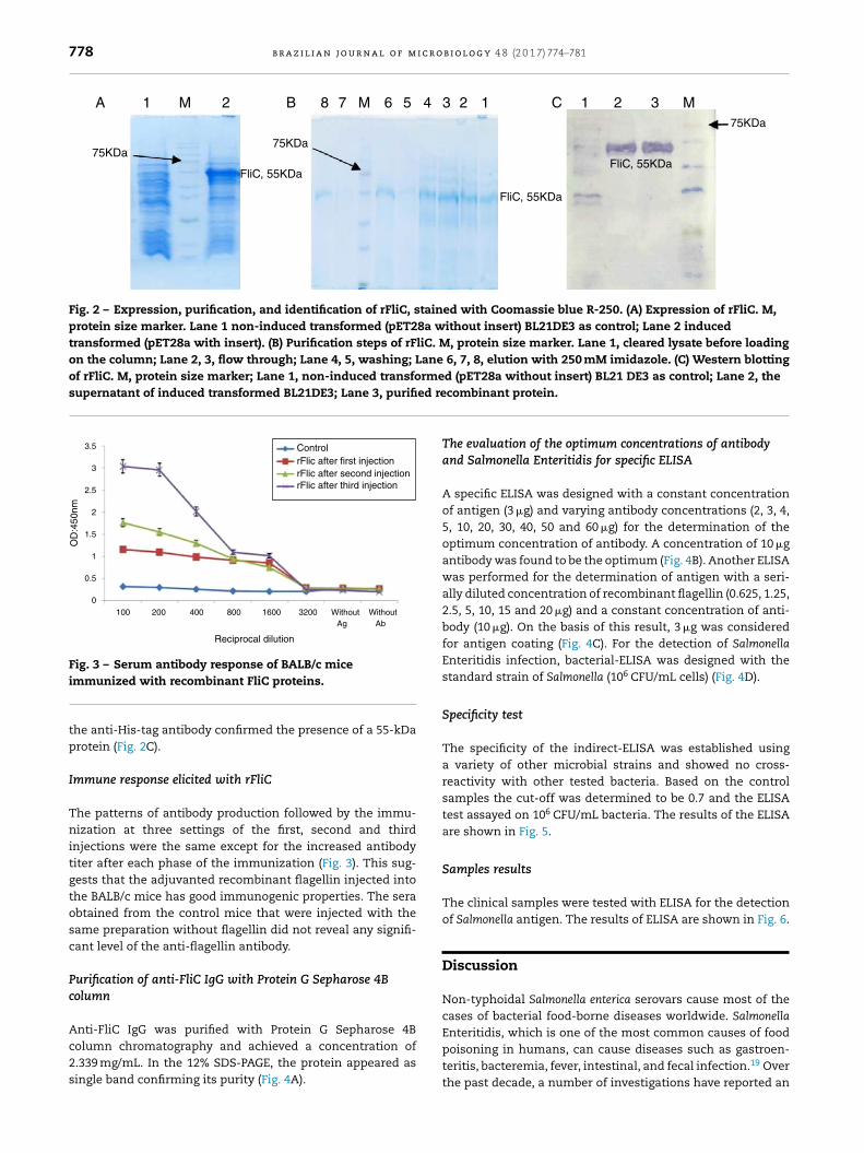
The recombinant FliC protein with N-terminal His-tag wasexpressed in E. coli BL21-DE3 cells (Fig. 2A) and purified byNi-NTA affinity chromatography. The SDS-PAGE of the puri-fied product is depicted in Fig. 2B. The Western blotting with
1 2 3 4 5 M
FliC
1518bp
C
1000bp
n analysis on an agarose gel. (A) Lane M, DNA size marker; fliC gene. (C) Lane M, DNA size marker; Lanes 1–5, digested
778 b r a z i l i a n j o u r n a l o f m i c r o b i o l o g y 4 8 (2 0 1 7) 774–781
1 M 8 7 M M6 5 4 3 32 21 12
75KDa75KDa
FliC, 55KDa
FliC, 55KDa
FliC, 55KDa
75KDa
A B C
Fig. 2 – Expression, purification, and identification of rFliC, stained with Coomassie blue R-250. (A) Expression of rFliC. M,protein size marker. Lane 1 non-induced transformed (pET28a without insert) BL21DE3 as control; Lane 2 inducedtransformed (pET28a with insert). (B) Purification steps of rFliC. M, protein size marker. Lane 1, cleared lysate before loadingon the column; Lane 2, 3, flow through; Lane 4, 5, washing; Lane 6, 7, 8, elution with 250 mM imidazole. (C) Western blottingof rFliC. M, protein size marker; Lane 1, non-induced transformed (pET28a without insert) BL21 DE3 as control; Lane 2, thesupernatant of induced transformed BL21DE3; Lane 3, purified re
3.5
3
2.5
2
1.5
1
0.5
0100 200 400 800
Reciprocal dilution
1600
OD
:450
nm
3200 Without WithoutAbAg
ControlrFlic after first injectionrFlic after second injectionrFlic after third injection
Fig. 3 – Serum antibody response of BALB/c mice
immunized with recombinant FliC proteins.the anti-His-tag antibody confirmed the presence of a 55-kDaprotein (Fig. 2C).
Immune response elicited with rFliC
The patterns of antibody production followed by the immu-nization at three settings of the first, second and thirdinjections were the same except for the increased antibodytiter after each phase of the immunization (Fig. 3). This sug-gests that the adjuvanted recombinant flagellin injected intothe BALB/c mice has good immunogenic properties. The seraobtained from the control mice that were injected with thesame preparation without flagellin did not reveal any signifi-cant level of the anti-flagellin antibody.
Purification of anti-FliC IgG with Protein G Sepharose 4Bcolumn
Anti-FliC IgG was purified with Protein G Sepharose 4Bcolumn chromatography and achieved a concentration of2.339 mg/mL. In the 12% SDS-PAGE, the protein appeared assingle band confirming its purity (Fig. 4A).
combinant protein.
The evaluation of the optimum concentrations of antibodyand Salmonella Enteritidis for specific ELISA
A specific ELISA was designed with a constant concentrationof antigen (3 �g) and varying antibody concentrations (2, 3, 4,5, 10, 20, 30, 40, 50 and 60 �g) for the determination of theoptimum concentration of antibody. A concentration of 10 �gantibody was found to be the optimum (Fig. 4B). Another ELISAwas performed for the determination of antigen with a seri-ally diluted concentration of recombinant flagellin (0.625, 1.25,2.5, 5, 10, 15 and 20 �g) and a constant concentration of anti-body (10 �g). On the basis of this result, 3 �g was consideredfor antigen coating (Fig. 4C). For the detection of SalmonellaEnteritidis infection, bacterial-ELISA was designed with thestandard strain of Salmonella (106 CFU/mL cells) (Fig. 4D).
Specificity test
The specificity of the indirect-ELISA was established usinga variety of other microbial strains and showed no cross-reactivity with other tested bacteria. Based on the controlsamples the cut-off was determined to be 0.7 and the ELISAtest assayed on 106 CFU/mL bacteria. The results of the ELISAare shown in Fig. 5.
Samples results
The clinical samples were tested with ELISA for the detectionof Salmonella antigen. The results of ELISA are shown in Fig. 6.
Discussion
Non-typhoidal Salmonella enterica serovars cause most of thecases of bacterial food-borne diseases worldwide. Salmonella
Enteritidis, which is one of the most common causes of foodpoisoning in humans, can cause diseases such as gastroen-teritis, bacteremia, fever, intestinal, and fecal infection.19 Overthe past decade, a number of investigations have reported an
b r a z i l i a n j o u r n a l o f m i c r o b i o l o g y 4 8 (2 0 1 7) 774–781 779
1 M 20.9
0.8
0.7
0.6
0.5
0.4
0.3
0.2
0.1
0
Control
Control Log 10 CFU/mL
Purified Anti-Flic antibody
Control10µg Anti-Flic antibody
Antigen concentration (µg)Bacterial count log 10
60 50 40 20 10 5 4 3 2
With
out A
g
With
out A
b
With
out A
g12345678
With
out A
b
Concentration (µg)
OD
:450
nm
OD
:450
nm
OD
:450
nm
A
C
B
20
3
2.5
2
1.5
1
0.5
0
3
2.5
2
1.5
1
0.5
0
–0.515 10 5 2.5 1.25 0.625 Without
Ag
Without
Ab
Fig. 4 – The purification of IgG antibody and establishing ELISA using the recombinant FliC antigen. (A) Purification of IgGantibody by G Sepharose 4B column. Lane 1, SDS-PAGE of purified IgG with 2ME; M, protein size marker. Lane 2, SDS-PAGEof purified IgG without 2ME. (B) Development of ELISA after IgG purification. (C) Development of antigen with thed
iTifdeidlsScfdspspmP(st
on 6XHis-tag using the Ni-NTA column. The purified rFliC wasused for the immunization of mice. The ELISA test resultsshowed that all immunized mice produced a high titer of
2.5
2
1.5
1
0.5
0
–0.5
Salmon
ella
Shigell
a
seud
omon
asE.co
li
Staph
Klebsie
lla
With
out A
g
With
out A
b
OD
:450
nm
etermination of purified antibody. (D) Bacterial ELISA.
ncreasing incidence of non-typhoid Salmonella bacteremia.20
herefore, a rapid and proper detection of this bacterium ismportant for food safety. The bacterial culture based methodsor the identification of Salmonella are time-consuming (5–7ays) and require enrichment of the culture.21 As such, sev-ral screening methods such as nucleic acid-based assays andmmunology-based assays have been examined for a rapidetection of Salmonella in the recent decades.20,22–24 In ear-
ier studies, lipopolysaccharides (LPS) present on the bacterialurface were used in the ELISA-assay for the detection ofalmonella, but the problem with lipopolysaccharides is theross-reactivity with other gram-negative bacteria.25–27 There-ore, flagellin (FliC) was sought as an alternative for rapidiagnosis of Salmonella and, during the last decade, severaltudies have used it for the detection of Salmonella. For theroduction of recombinant FliC (rFlic), in this study, a standardtrain of Salmonella enterica serovar Enteritidis was used thatroduces an antigen with high immunogenicity.28 A DNA frag-ent of the expected size (near 1518 bp) was amplified by
CR and expressed using chemically inducible T7 promoterpET28a). The best condition for the recombinant rFliC expres-ion was achieved at 1 mM IPTG concentration, the cultureemperature of 37 ◦C and with overnight induction. However,
the protein expression showed an increase with the incuba-tion time up to 18 h. rFliC was separated on SDS-PAGE and itsbiochemical nature was confirmed by immunoblotting usingmice anti-His-tag specific antibody. rFliC was purified based
PBacterial cells:106 CFU/mL
Fig. 5 – Cross-reactivity of mouse anti-S. enteritidis IgG withother microbial strains. Assays were conducted in triplicate.

780 b r a z i l i a n j o u r n a l o f m i c r o b i o l o g y 4 8 (2 0 1 7) 774–781
2.5
2
1.5
1
0.5
0
Bacterial cells: 106 CFU/mL
s1 s2 s3 s4 s5 s6 s7 s8 s9 s10
s11
s12
s13
s14
s15
s16
s17
s18
s19
s20
Contro
l
OD
:450
nm
Fig. 6 – ELISA of clinical samples for the detection of Salmonella Enteritidis. Negative controls are also indicated. Assaycut-off was calculated by replicate analysis of negative samples.
r
Enteritidis, Hadar, Heidelberg, and Typhimurium in poultry.Agriculture. 2013;3(3):381–397.
anti-FliC antibody upon injecting rFliC three times. Then anELISA system was designed for the detection of SalmonellaEnteritidis, which is an important pathogen of human andpoultry. Accurate and quick tests are essential to ensure foodsafety by the governments.29 The availability of sufficientquantities of recombinant antigens for the rapid detectionof Salmonella can be achieved in serum samples in a shorttime.30 Thus, new methods for quick detection of Salmonellahave been developed in the past decade. Among the molec-ular techniques, PCR is a common and accurate method andcan also be used for the detection of low levels of pathogenicbacteria.31 ELISA can be used to screen a variety of samplesand components in the presence of Salmonella spp.
The ELISA test, designed in this study, did not show anycross-reaction with other bacteria. Sensitivity analysis of thistest showed that it could detect 103 bacteria in clinical sam-ples, given that the infectious dose of Salmonella is about 106
approximately.Besides ours, the studies of Minicozzi (2013),5 Jindal (2012)13
and Bang (2012)21 have also reported the production of recom-binant flagellar antigen (r-FliC) and development of ELISA forthe detection of Salmonella. In previous works, the investiga-tions were carried out mainly with sera obtained from infectedflocks and birds but, in this study, we used mice for the pro-duction of antibody and Protein G Sepharose 4B column forthe purification. The flic gene of Salmonella Enteritidis wasamplified, cloned successfully using pET-28a(+) expressionvector and was introduced into E. coli BL21 (DE3) as a suit-able host. The recombinant flagellin was purified successfullyand demonstrated good immunogenic properties based on theimmunization of BALB/c mice and its ability to induce a sig-nificant serum flagellin-specific IgG immune response, whichwas measured by ELISA. We provide further evidence that therecombinant flagellin can be one of the excellent indicatorsfor the detection of Salmonella Enteritidis.
Conflicts of interest
The authors declare that they have no competing interest.
Acknowledgments
This article is a part a Ph.D. thesis being prepared by AliMirhosseini. The study was supported by the grants fromApplied Microbiology Research Center Baqiyatallah Universityof Medical Sciences.
e f e r e n c e s
1. Hyeon J-Y, Chon J-W, Choi I-S, Park C, Kim D-E, Seo K-H.Development of RNA aptamers for detection of SalmonellaEnteritidis. J Microbiol Methods. 2012;89(1):79–82.
2. Kilic A, Bedir O, Kocak N, et al. Analysis of an outbreak ofSalmonella enteritidis by repetitive-sequence-based PCR andpulsed-field gel electrophoresis. Intern Med. 2009;49(1):31–36.
3. Ranjbar R, Giammanco GM, Farshad S, Owlia P, Aleo A,Mammina C. Serotypes, antibiotic resistance, and class 1integrons in Salmonella isolates from pediatric cases ofenteritis in Tehran, Iran. Foodborne Pathog Dis.2011;8(4):547–553.
4. Chashni S, Hassanzadeh M, Fard M, Mirzaie S.Characterization of the Salmonella isolates from backyardchickens in north of Iran, by serotyping, multiplex PCR andantibiotic resistance analysis. Arch Razi Inst. 2009;64(2):77–83.
5. Nosraty S, Fallah F, Sabokbar A, Dezfoolyan M, Adabian S,Esmaeilnejad N. Detection of Salmonella enteritidis, typhiand typhimurium in foods by multiplex PCR in childrenhospital. BMC Infect Dis. 2012;12(suppl 1):P95.
6. Dallal MMS. Prevalence of Salmonella spp. in packed andunpacked red meat and chicken in south of Tehran.Jundishapur J Microbiol. 2014;7(4).
7. Fierer J, Guiney DG. Diverse virulence traits underlyingdifferent clinical outcomes of Salmonella infection. J ClinInvest. 2001;107(7):775–780.
8. Minicozzi J, Sanchez S, Lee MD, Holt PS, Hofacre CL, MaurerJJ. Development of recombinant flagellar antigens forserological detection of Salmonella enterica serotypes
9. Desin TS, Köster W, Potter AA. Salmonella vaccines in poultry:past, present and future. Expert Rev Vaccines. 2013;12(1):87–96.

r o b i
b r a z i l i a n j o u r n a l o f m i c10. Oracz G, Feleszko W, Golicka D, Maksymiuk J, Klonowska A,Szajewska H. Rapid diagnosis of acute Salmonellagastrointestinal infection. Clin Infect Dis. 2003;36(1):112–115.
11. Siregar TH, Elliman J, Owens L. Development of real timepolymerase chain reaction for detection of Salmonellatyphimurium and Salmonella enteritidis in fish. Squalen.2013;7(2):51–58.
12. Amani J, Ahmadpour A, Fooladi AAI, Nazarian S. Detection ofE. coli O157: H7 and Shigella dysenteriae toxins in clinicalsamples by PCR-ELISA. Braz J Infect Dis. 2015;19(3):278–284.
13. Jindal G, Tewari R, Gautam A, Pandey SK, Rishi P.Immunological characterization of recombinant Salmonellaenterica serovar Typhi FliC protein expressed in Escherichiacoli. AMB Express. 2012;2(1):1–9.
14. Bergman MA, Cummings LA, Barrett SLR, et al. CD4+ T cellsand toll-like receptors recognize Salmonella antigensexpressed in bacterial surface organelles. Infect Immun.2005;73(3):1350–1356.
15. Hajam IA, Dar PA, Sekar SC, et al. Expression, purification,and functional characterisation of flagellin, a TLR5-ligand.Vet Ital. 2013;49(2):181–186.
16. Cummings LA, Wilkerson WD, Bergsbaken T, Cookson BT. Invivo, fliC expression by Salmonella enterica serovarTyphimurium is heterogeneous, regulated by ClpX, andanatomically restricted. Mol Microbiol. 2006;61(3):795–809.
17. Haiko J, Westerlund-Wikström B. The role of the bacterialflagellum in adhesion and virulence. Biology.2013;2(4):1242–1267.
18. Bollag D, Michael D, Stuart J. Protein Methods. 2nd ed. NewYork: John Wiley and Sons, Inc Pub; 1996.
19. Dehghani B, Rasooli I, Gargari SLM, Nadooshan MRJ, Owlia P,Nazarian S. Immunogenicity of Salmonella enterica serovarEnteritidis virulence protein, InvH, and cross-reactivity of itsantisera with Salmonella strains. Microbiol Res.2013;168(2):84–90.
20. Kuhn KG, Falkenhorst G, Ceper TH, et al. Detectingnon-typhoid Salmonella in humans by ELISAs: a literature
review. J Med Microbiol. 2012;61(1):1–7.21. Bang J, Shukla S, Kim Y, Kim M. Development of indirectcompetitive ELISA for the detection of SalmonellaTyphimurium. Rom Biotechol Lett. 2012;17:7194–7204.
o l o g y 4 8 (2 0 1 7) 774–781 781
22. Rodpaia E, Moongkarndia P, Tungrugsasutb W,Phisannoradeja R, Kanaratc S. Comparison of multiplexpolymerase chain reaction and immunoassay to detectSalmonella spp., S. Typhimurium, and S. Enteritidis in Thaichicken meat. ScienceAsia. 2013;39(2):150–159.
23. Majeed LJ, Znad KH, Thamir MK, Baqir HI. Study of ELISA andantibiotic sensitivity test for Salmonella enteritidis asexperimental infection in mice. Um-Salama Sci J.2009;6(1):114–122.
24. Mandal J, Akhter MZ, Kabir MS, Ahsan S. Development of amultiplex polymerase chain reaction protocol for thesimultaneous detection of Salmonella enterica serovar Typhiand Class 1 integron. Asian Pac J Trop Dis. 2014;4:S808–S812.
25. Isomaki O, Vuento R, Granfors K. Serological diagnosis ofSalmonella infections by enzyme immunoassay. Lancet.1989;333(8652):1411–1414.
26. Dalby T, Strid MA, Beyer NH, Blom J, Mølbak K, Krogfelt KA.Rapid decay of Salmonella flagella antibodies during humangastroenteritis: a follow up study. J Microbiol Methods.2005;62(2):233–243.
27. Strid MA, Dalby T, Mølbak K, Krogfelt KA. Kinetics of thehuman antibody response against Salmonella entericaserovars Enteritidis and Typhimurium determined bylipopolysaccharide enzyme-linked immunosorbent assay.Clin Vaccine Immunol. 2007;14(6):741–747.
28. Crayford G, Coombes JL, Humphrey TJ, Wigley P. Monophasicexpression of FliC by Salmonella 4,[5], 12: i:-DT193 does notalter its pathogenicity during infection of porcine intestinalepithelial cells. Microbiology. 2014;160(Pt 11):2507–2516.
29. Lee K-M, Runyon M, Herrman TJ, Phillips R, Hsieh J. Review ofSalmonella detection and identification methods: aspects ofrapid emergency response and food safety. Food Control.2015;47:264–276.
30. Kremer C, O’Meara K, Layton S, Hargis B, Cole K. Evaluationof recombinant Salmonella expressing the flagellar proteinfliC for persistence and enhanced antibody response incommercial turkeys. Poult Sci. 2011;90(4):752–758.
31. Ricke S, Pillai S, Norton R, Maciorowski K, Jones F.Applicability of rapid methods for detection of Salmonellaspp. in poultry feeds: a review. J Rapid Methods AutomMicrobiol. 1998;6(4):239–258.