production of butyrate from lysine and the amadori...
TRANSCRIPT
ARTICLE
Received 16 Apr 2015 | Accepted 29 Oct 2015 | Published 1 Dec 2015
Production of butyrate from lysine and the Amadoriproduct fructoselysine by a human gut commensalThi Phuong Nam Bui1, Jarmo Ritari2, Sjef Boeren3, Pieter de Waard4, Caroline M. Plugge1 & Willem M. de Vos1,2,5
Human intestinal bacteria produce butyrate, which has signalling properties and can be used
as energy source by enterocytes thus influencing colonic health. However, the pathways
and the identity of bacteria involved in this process remain unclear. Here we describe the
isolation from the human intestine of Intestinimonas strain AF211, a bacterium that can convert
lysine stoichiometrically into butyrate and acetate when grown in a synthetic medium.
Intestinimonas AF211 also converts the Amadori product fructoselysine, which is abundantly
formed in heated foods via the Maillard reaction, into butyrate. The butyrogenic pathway
includes a specific CoA transferase that is overproduced during growth on lysine. Bacteria
related to Intestinimonas AF211 as well as the genetic coding capacity for fructoselysine
conversion are abundantly present in colonic samples from some healthy human subjects.
Our results indicate that protein can serve as a source of butyrate in the human colon, and its
conversion by Intestinimonas AF211 and related butyrogens may protect the host from the
undesired side effects of Amadori reaction products.
DOI: 10.1038/ncomms10062 OPEN
1 Laboratory of Microbiology, Wageningen University, Dreijenplein 10, 6703 HB Wageningen, The Netherlands. 2 Department of Basic Veterinary Medicine,University of Helsinki, Agnes Sjoberginkatu 2, FIN-00790 Helsinki, Finland. 3 Laboratory of Biochemistry, Wageningen University, Dreijenlaan 3, 6703 HAWageningen, The Netherlands. 4 Wageningen NMR Centre, Dreijenlaan 3, 6703 HA Wageningen, The Netherlands. 5 Department of Bacteriology andImmunology, Haartmaninkatu 3, University of Helsinki, FIN-0014 Helsinki, Finland. Correspondence and requests for materials should be addressed toW.M.d.V. (email: [email protected]).
NATURE COMMUNICATIONS | 6:10062 | DOI: 10.1038/ncomms10062 | www.nature.com/naturecommunications 1
Our intestinal tract contains trillions of microbes thatimpact our health1. The metabolic capacity of thesemicrobes is enormous2 and includes the production of
short-chain fatty acids, such as butyrate that serves as a majorsource of energy for the intestinal enterocytes and signals to thehost3. Butyrogenic gut bacteria belong to Lachnospiraceaeand Ruminococcaceae4, and recent studies showed an inversecorrelation between these butyrogenic Firmicutes andinflammatory bowel disease, type 2 diabetes and various otherdisorders5,6. Moreover, there is considerable interest in thesebutyrogens, since butyrate reduces proinflammatory signals3 andhas a protective role in maintaining a healthy colon7,8. Thus, it isof paramount importance to identify the microbes that arecapable of producing butyrate and understand the mechanismsunderlying its production.
Most butyrate produced in the human intestine is assumedto derive from carbohydrates, while some specializedbacteria have been found that convert lactate plus acetateinto butyrate9,10. All of these produce butyrate via theacetyl-CoA pathway11 (converting acetyl-CoA to butyrate)that involves a complex cascade of reactions, in whichbutyrate CoA transferase (But) or butyrate kinase (Buk) arekey enzymes. In a recent (meta)genome-based study, it waspredicted that the But-mediated route was 10-fold moreabundant than that mediated by Buk, while these two enzymesshowed high diversity among butyrogens12. Three other routesfor butyrate synthesis, the glutamate, succinate and lysinepathways, have been described. On the basis of the distributionof the genes in the intestinal metagenome libraries, the acetyl-CoA pathway was the most prevalent followed by the lysinepathway. Yet, no intestinal bacterium has been reportedthat contained all genes coding for butyrogenesis from thisamino acid13.
In addition to carbohydrates, proteins are crucial dietarycompounds that are receiving increased attention to their use inspecial diets. Proteolytic enzymes in the upper intestinal tractgenerate amino acids that are rapidly taken up by the host, butalso may reach the colon, partly depending on the intake.Moreover, protein recycling in the colon has been described,adding to the interest in colonic amino-acid fermentations.Amadori products (fructosamines), formed via non-enzymaticreactions between reducing sugars and free amino groups ofproteins during the food heating process, are of significantinterest as they have shown to be associated with the ageingprocess and several chronic diseases14. Fructoselysine is a keyintermediate in the formation of advanced glycationendproducts (AGEs)14 and not yet any organism has beenknown to convert fructoselysine into butyrate. Only a fewmicrobes have been reported to metabolize lysine into butyrate,and they derive from ecosystems other than the gut. A lysinedegradation pathway was described over 50 years ago inClostridium sticklandii isolated from black mud15. Morerecently, this pathway was detected in Clostridiumsubterminale SB4 isolated from sewage and Fusobacteriumnucleatum isolated from the oral cavity, and eight genesencoding a lysine fermentation pathway were identified fromthe genome of F. nucleatum16. Several enzymes involved inbutyrogenesis from lysine have been biochemicallycharacterized17–20, but so far no intestinal microorganism hasbeen found to contain the entire pathway in spite of thepredicted presence of the lysine fermentation pathway in thehuman intestine13,16–18. Here we describe the butyrogenicconversion of lysine and the Amadori product fructoselysineby a novel Intestinimonas strain from a healthy volunteer andcharacterize its butyrate synthesis pathway by using a combinedphysiological, biochemical and proteogenomic approach.
ResultsLysine degradation by Intestinimonas AF211. In a searchfor new butyrogens, strain AF211 was isolated from the stoolof a healthy subject using a mineral bicarbonate-buffered mediumwith lactate and acetate, under strict anoxic conditions (butyratewas found as the main end product). Strain AF211 containedtwo copies of the 16S rRNA gene that differed in a singlenucleotide, and subsequent phylogenetic analysis showed that thisstrain belonged to clostridial cluster IV (Lachnospiraceae) ofthe Firmicutes phylum. Further analysis showed that this 16SrRNA sequence was highly similar to that of the mouse intestinalisolate Intestinimonas butyriciproducens DSM26588T (ref. 21),indicating that the newly isolated strain AF211 belongs to thegenus Intestinimonas (Supplementary Fig. 1). However,strain Intestinimonas AF211 was a human isolate and showedphysiological differences with I. butyriciproducens from mouse(to be reported elsewhere). Butyrate was found to be a majormetabolite from all substrates where growth was observed(Supplementary Table 1). Intestinimonas AF211 hardly grew onglucose in mineral medium (generation time of 88 days), but itsgrowth rate tripled in the presence of acetate and yeast extract. Inmedia containing 20 mM glucose with 20 mM acetate and 2%yeast extract, Intestinimonas AF211 produced up to 4.4 mMbutyrate and trace amounts of lactate and ethanol (2-weekincubation). Remarkably, the strain grew much better (generationtime of 7.5 h) in L-lysine but not in any other natural amino acidsor D-lysine (Supplementary Table 1).
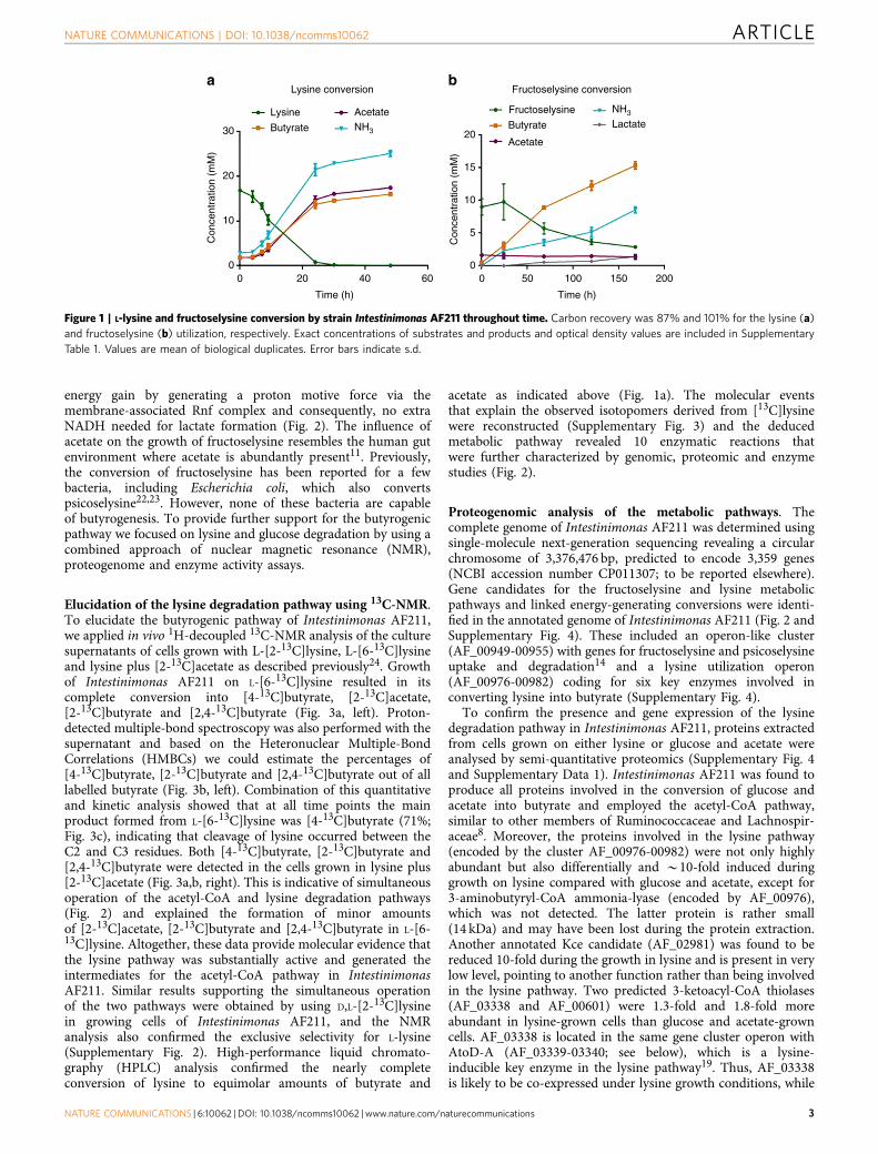
To study the capacity to convert lysine into butyrate,Intestinimonas AF211 was grown in bicarbonate-bufferedmedium containing L-lysine as the sole carbon and energysource. A total of 16.8±0.4 mM lysine was converted into14.2±0.6 mM butyrate, 15.6±0.7 mM acetate and 22.1±0.5 mMNH3 when the cells reached the stationary phase after 2 daysat 37 �C (Fig. 1a). This suggests that part of the releasedammonia is sequestered into proteins during anabolism ofIntestinimonas AF211. Hence, we propose the fermentationreaction as: C6H14O2N2þ 2H2O-C4H8O2þC2H4O2þ 2NH3.Clearly, Intestinimonas AF211 is capable of growing in definedmedia with L-lysine as sole carbon and energy source with amaximum growth rate of 0.1 h� 1.
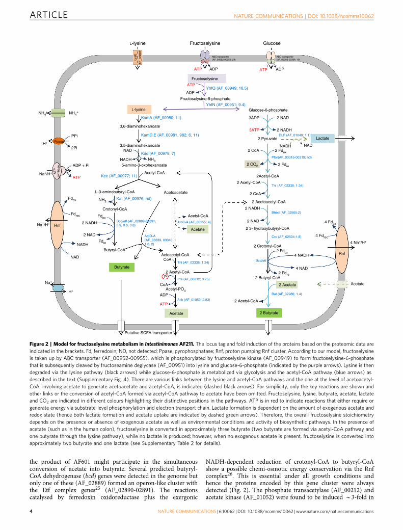
Growth of Intestinimonas AF211 on fructoselysine. As fructo-selysine is a key intermediate in the formation of AGEs, weaddressed its degradation by Intestinimonas AF211 and found itto be readily metabolized (Fig. 1b; generation time 24 h). Butyrate,NH3 and lactate were formed as the main products. Theconversion equation is proposed as: C12H24O7N2þ 2 H2O-2C4H8O2þ 2 NH3þ 1 CO2þ 1 C3H6O3. We anticipate that thefructoselysine pathway includes the simultaneous degradation ofboth lysine and the sugar moiety in two branches (termed lysinepathway and acetyl-CoA pathway; Fig. 2). Some deviations in thebutyrate/fructoselysine ratio were observed (SupplementaryTables 1 and 2). These may be attributed to the different envir-onmental conditions but also be caused by the intracellularbalance between the two branches of the fructoselysine pathway(Fig. 2). However, the initial concentration of acetate was pre-dicted to be a major factor affecting the butyrate/fructoselysineratio. We experimentally verified this and observed that in thepresence of extra acetate, fructoselysine was completely convertedinto butyrate and NH3 (Supplementary Table 2) according tothe equation: C12H24O7N2þC2H4O2þH2O-3 C4H8O2þ 2NH3þ 2 CO2. The growth rate in fructoselysine plus acetate wassignificantly higher than that in fructoselysine alone(Supplementary Table 2), which could be due to stimulation ofbutyrate production via external acetate, leading to an increased
ARTICLE NATURE COMMUNICATIONS | DOI: 10.1038/ncomms10062
2 NATURE COMMUNICATIONS | 6:10062 | DOI: 10.1038/ncomms10062 | www.nature.com/naturecommunications
energy gain by generating a proton motive force via themembrane-associated Rnf complex and consequently, no extraNADH needed for lactate formation (Fig. 2). The influence ofacetate on the growth of fructoselysine resembles the human gutenvironment where acetate is abundantly present11. Previously,the conversion of fructoselysine has been reported for a fewbacteria, including Escherichia coli, which also convertspsicoselysine22,23. However, none of these bacteria are capableof butyrogenesis. To provide further support for the butyrogenicpathway we focused on lysine and glucose degradation by using acombined approach of nuclear magnetic resonance (NMR),proteogenome and enzyme activity assays.
Elucidation of the lysine degradation pathway using 13C-NMR.To elucidate the butyrogenic pathway of Intestinimonas AF211,we applied in vivo 1H-decoupled 13C-NMR analysis of the culturesupernatants of cells grown with L-[2-13C]lysine, L-[6-13C]lysineand lysine plus [2-13C]acetate as described previously24. Growthof Intestinimonas AF211 on L-[6-13C]lysine resulted in itscomplete conversion into [4-13C]butyrate, [2-13C]acetate,[2-13C]butyrate and [2,4-13C]butyrate (Fig. 3a, left). Proton-detected multiple-bond spectroscopy was also performed with thesupernatant and based on the Heteronuclear Multiple-BondCorrelations (HMBCs) we could estimate the percentages of[4-13C]butyrate, [2-13C]butyrate and [2,4-13C]butyrate out of alllabelled butyrate (Fig. 3b, left). Combination of this quantitativeand kinetic analysis showed that at all time points the mainproduct formed from L-[6-13C]lysine was [4-13C]butyrate (71%;Fig. 3c), indicating that cleavage of lysine occurred between theC2 and C3 residues. Both [4-13C]butyrate, [2-13C]butyrate and[2,4-13C]butyrate were detected in the cells grown in lysine plus[2-13C]acetate (Fig. 3a,b, right). This is indicative of simultaneousoperation of the acetyl-CoA and lysine degradation pathways(Fig. 2) and explained the formation of minor amountsof [2-13C]acetate, [2-13C]butyrate and [2,4-13C]butyrate in L-[6-13C]lysine. Altogether, these data provide molecular evidence thatthe lysine pathway was substantially active and generated theintermediates for the acetyl-CoA pathway in IntestinimonasAF211. Similar results supporting the simultaneous operationof the two pathways were obtained by using D,L-[2-13C]lysinein growing cells of Intestinimonas AF211, and the NMRanalysis also confirmed the exclusive selectivity for L-lysine(Supplementary Fig. 2). High-performance liquid chromato-graphy (HPLC) analysis confirmed the nearly completeconversion of lysine to equimolar amounts of butyrate and
acetate as indicated above (Fig. 1a). The molecular eventsthat explain the observed isotopomers derived from [13C]lysinewere reconstructed (Supplementary Fig. 3) and the deducedmetabolic pathway revealed 10 enzymatic reactions thatwere further characterized by genomic, proteomic and enzymestudies (Fig. 2).
Proteogenomic analysis of the metabolic pathways. Thecomplete genome of Intestinimonas AF211 was determined usingsingle-molecule next-generation sequencing revealing a circularchromosome of 3,376,476 bp, predicted to encode 3,359 genes(NCBI accession number CP011307; to be reported elsewhere).Gene candidates for the fructoselysine and lysine metabolicpathways and linked energy-generating conversions were identi-fied in the annotated genome of Intestinimonas AF211 (Fig. 2 andSupplementary Fig. 4). These included an operon-like cluster(AF_00949-00955) with genes for fructoselysine and psicoselysineuptake and degradation14 and a lysine utilization operon(AF_00976-00982) coding for six key enzymes involved inconverting lysine into butyrate (Supplementary Fig. 4).
To confirm the presence and gene expression of the lysinedegradation pathway in Intestinimonas AF211, proteins extractedfrom cells grown on either lysine or glucose and acetate wereanalysed by semi-quantitative proteomics (Supplementary Fig. 4and Supplementary Data 1). Intestinimonas AF211 was found toproduce all proteins involved in the conversion of glucose andacetate into butyrate and employed the acetyl-CoA pathway,similar to other members of Ruminococcaceae and Lachnospir-aceae8. Moreover, the proteins involved in the lysine pathway(encoded by the cluster AF_00976-00982) were not only highlyabundant but also differentially and B10-fold induced duringgrowth on lysine compared with glucose and acetate, except for3-aminobutyryl-CoA ammonia-lyase (encoded by AF_00976),which was not detected. The latter protein is rather small(14 kDa) and may have been lost during the protein extraction.Another annotated Kce candidate (AF_02981) was found to bereduced 10-fold during the growth in lysine and is present in verylow level, pointing to another function rather than being involvedin the lysine pathway. Two predicted 3-ketoacyl-CoA thiolases(AF_03338 and AF_00601) were 1.3-fold and 1.8-fold moreabundant in lysine-grown cells than glucose and acetate-growncells. AF_03338 is located in the same gene cluster operon withAtoD-A (AF_03339-03340; see below), which is a lysine-inducible key enzyme in the lysine pathway19. Thus, AF_03338is likely to be co-expressed under lysine growth conditions, while
Lysine conversion
30
Lysine
Butyrate NH3
NH3Acetate
20
20
Fructoselysine
Fructoselysine conversion
Butyrate
Acetate
Lactate
10
10
15
5
00 0 50 100 150 200
020 40 60
Time (h) Time (h)
Con
cent
ratio
n (m
M)
Con
cent
ratio
n (m
M)
Figure 1 | L-lysine and fructoselysine conversion by strain Intestinimonas AF211 throughout time. Carbon recovery was 87% and 101% for the lysine (a)
and fructoselysine (b) utilization, respectively. Exact concentrations of substrates and products and optical density values are included in Supplementary
Table 1. Values are mean of biological duplicates. Error bars indicate s.d.
NATURE COMMUNICATIONS | DOI: 10.1038/ncomms10062 ARTICLE
NATURE COMMUNICATIONS | 6:10062 | DOI: 10.1038/ncomms10062 | www.nature.com/naturecommunications 3
the product of AF601 might participate in the simultaneousconversion of acetate into butyrate. Several predicted butyryl-CoA dehydrogenase (bcd) genes were detected in the genome butonly one of these (AF_02889) formed an operon-like cluster withthe Etf complex genes25 (AF_02890-02891). The reactionscatalysed by ferredoxin oxidoreductase plus the exergonic
NADH-dependent reduction of crotonyl-CoA to butyryl-CoAshow a possible chemi-osmotic energy conservation via the Rnfcomplex26. This is essential under all growth conditions andhence the proteins encoded by this gene cluster were alwaysdetected (Fig. 2). The phosphate transacetylase (AF_00212) andacetate kinase (AF_01052) were found to be induced B3-fold in
L-lysine Fructoselysine
Fructoselysine
YhfQ (AF_00949; 16.5)
YhfN (AF_00951; 9.4)
KamA (AF_00980; 11)
KamD,E (AF_00981, 982; 6, 11)
Kdd (AF_00979; 7)
Kce (AF_00977; 11)
Kal (AF_00976; nd)
Bcd/etf (AF_02889-02891;0.9, 0.6, 0.8)
AtoD-A(AF_03339, 03340;1.6, 2)
Thl (AF_03338; 1.34)
Pta (AF_00212; 3.25)
Ack (AF_01052; 2.83)
But (AF_02986; 1.4)
Cro (AF_02504;1.8)
Bcd/etf
Bhbd (AF_02505;2)
Thl (AF_03338; 1.34)
Pfor(AF_00315-00319; nd)
DLF (AF_01040; 1.1)
AtoC-A (AF_00155; 4)
Fructoselysine-6-phosphate
Glucose-6-phosphateL-lysine
3,6-diaminohexanoate
3,5-diaminohexanoateNAD
NADH5-amino-3-oxohexanoate
L-3-aminobutyryl-CoA
Crotonyl-CoA
Acetoacetate
Acetate
CoA
CoA
2 CoA
P
Acetyl-CoA
2 NADH
2 NADH
Lactate
RnfNa+/H+
Na+/H+
Rnf
4 Na+/H+
H+
H+
Na+
NH4+ NH4
+
2 NAD
2 NADH
2 Acetyl-CoA
2 3- hydroxybutyryl-CoA
2 Crotonyl-CoA
2 Butyryl-CoA
2 CoA
2 NAD
4 NADH
4 NAD
2 NAD
Butyryl-CoAActoacetyl-CoA
2 Acetoacetyl-CoA
2 Acetyl-CoA
2Acetyl-CoA
2 Pyruvate
Putative SCFA transporter
Acetate
Acetate2 Acetate
2 Butyrate
2 Acetyl-CoA
Butyrate
NADH
NADH
NAD
NAD
Acetyl-CoA
NH3
NH3
Fdox
2 Fdox
Fdre
2 CO2 2 Fdre
2 Fdox
4 Fdox
2 Fdre
4 Fdrec
Fdox
Fdrec
Glucose
ADPADP
ABC transporter(AF_00952-00955; 24)
ABC transporter(AF_02302-02304; 10)
ADP + Pi
2Pi
PPiPpase
AT
Psy
ntha
se
ADP
3ADP
ATPATP
ATP
ATP
3ATP
ADP
ATP
LysE
AF
_008
87
Acetyl-PO4
Figure 2 | Model for fructoselysine metabolism in Intestinimonas AF211. The locus tag and fold induction of the proteins based on the proteomic data are
indicated in the brackets. Fd, ferredoxin; ND, not detected; Ppase, pyrophosphatase; Rnf, proton pumping Rnf cluster. According to our model, fructoselysine
is taken up by ABC transporter (AF_00952-00955), which is phosphorylated by fructoselysine kinase (AF_00949) to form fructoselysine-6-phosphate
that is subsequently cleaved by fructoseamine deglycase (AF_00951) into lysine and glucose-6-phosphate (indicated by the purple arrows). Lysine is then
degraded via the lysine pathway (black arrows) while glucose-6-phosphate is metabolized via glycolysis and the acetyl-CoA pathway (blue arrows) as
described in the text (Supplementary Fig. 4). There are various links between the lysine and acetyl-CoA pathways and the one at the level of acetoacetyl-
CoA, involving acetate to generate acetoacetate and acetyl-CoA, is indicated (dashed black arrows). For simplicity, only the key reactions are shown and
other links or the conversion of acetyl-CoA formed via acetyl-CoA pathway to acetate have been omitted. Fructoselysine, lysine, butyrate, acetate, lactate
and CO2 are indicated in different colours highlighting their distinctive positions in the pathways. ATP is in red to indicate reactions that either require or
generate energy via substrate-level phosphorylation and electron transport chain. Lactate formation is dependent on the amount of exogenous acetate and
redox state (hence both lactate formation and acetate uptake are indicated by dashed green arrows). Therefore, the overall fructoselysine stoichiometry
depends on the presence or absence of exogenous acetate as well as environmental conditions and activity of biosynthetic pathways. In the presence of
acetate (such as in the human colon), fructoselysine is converted in approximately three butyrate (two butyrate are formed via acetyl-CoA pathway and
one butyrate through the lysine pathway), while no lactate is produced; however, when no exogenous acetate is present, fructoselysine is converted into
approximately two butyrate and one lactate (see Supplementary Table 2 for details).
ARTICLE NATURE COMMUNICATIONS | DOI: 10.1038/ncomms10062
4 NATURE COMMUNICATIONS | 6:10062 | DOI: 10.1038/ncomms10062 | www.nature.com/naturecommunications

lysine-grown cells compared with glucose and acetate-growncells, indicative of the involvement of these two enzymes in thelysine pathway. Remarkably, proteome analysis revealed thefructoselysine operon-like genes to be induced up to 25-foldduring growth on lysine (Supplementary Fig. 4 andSupplementary Data 1), most likely due to a high amount oflysine, which was also an intermediate in the fructoselysinepathway (Fig. 2).
Phylogeny of CoA transferases and enzyme activity. A crucialreaction involved in butyrate formation from lysine is the transferof a CoA moiety from one molecule to another, catalysed bymembers of the CoA transferase family. Remarkably, analysis ofthe genome of Intestinimonas AF211 predicted the presence of14 such enzymes, and a phylogenetic tree was generated basedon these and CoA transferases from intestinal bacteria andenvironmental isolates (Fig. 4a). These include the CoA
transferases AtoD-A, 4Hbt and But, for which experimentalevidence for their involvement in butyrate synthesis has beendescribed in several anaerobes, including Clostridium SB4,Roseburia spp., Faecalibacterium prausnitzii and Clostridumacetobutylicum8,19,27. Five well-separated clades of enzymes couldbe distinguished. One clade included the But enzymes from themain butyrate producers in the human intestine, belonging to theLachnospiraceae, and Ruminococcaceae4 that are all capable ofbutyrogenesis from glucose or lactate plus acetate8,28. TheIntestinimonas AF211 enzyme encoded by AF_02986 belongedto this But clade and was detected in the proteome underboth growth conditions, indicating that it participated in theacetyl-CoA pathway rather than 4-aminobutyrate or glutamatepathways. The second clade harboured the 4Hbt enzymes andincluded several predicted CoA transferases of IntestinimonasAF211 but none was detected at the protein level. The third andfourth clades held the a and b subunits of the AtoD-A enzymes,respectively, that are encoded by two juxtaposed genes in
[6-13C]lysine
L-[6-13C]lysine
L-[6-13C]lysine
[6-13C]lysine
[6-13C]lysine
[2-13C]butyrate
[2-13C]butyrate
22.5 6.4
39
71
2.0 1.5 1.0
50
40
30
20
0.5 F2[p.p.m.]
2.0 1.5 1.0 0.5 F2[p.p.m.]
F1
[p.p
.m.]
F1
[p.p
.m.]
50
40
30
20
6
SubstratesProducts (%)
55
[2,4-13C]butyrate
[2-13C]butyrate
[2-13C]butyrate
[2-13C]acetate
[2-13C]acetate
Lysine plus [2-13C]acetate
Lysine plus [2-13C]acetate
Lysine + [2-13C]acetate
[4-13C]butyrate[4-13C]butyrate
[4-13C]butyrate
72 h 40 h
24 h 20 h
0 h
Acetate H-2/C-2 Acetate H-2/C-2
H-3/C-2 H-3/C-2H-2/C-2 H-2/C-2H-4/C-2 H-4/C-2
H-4/C-4 H-4/C-4H-3/C-4 H-3/C-4H-2/C4 H-2/C4
0 h
p.p.
m.
p.p.
m.161820 2025303522242628303234363840 404244
Figure 3 | Elucidation of lysine pathway via 1H-decoupled 13C-NMR spectrum and 2D HMBC spectrum. (a) High-resolution 1H-decoupled 13C-NMR
spectra showing L-[6-13C]lysine 13C-labelled fermentation products. [2-13C]butyrate, [2-13C]acetate and [4-13C]butyrate had a chemical shift of 42.33,
25.99 and 15.95 p.p.m., respectively. (b) 2D HMBC spectrum for [6-13C]lysine is shown. (c) Percentages of labelled butyrate fractions (see Supplementary
Figs 2 and 3 for more details).
NATURE COMMUNICATIONS | DOI: 10.1038/ncomms10062 ARTICLE
NATURE COMMUNICATIONS | 6:10062 | DOI: 10.1038/ncomms10062 | www.nature.com/naturecommunications 5

the well-studied anaerobes: F. nucleatum, C. sticklandii andC. acetobutylicum29–33. Within these two AtoD-A subunit clades,three gene pairs of Intestinimonas AF211 were clustered.However, only one of these, encoded by AF_03339-03340, washighly abundant and induced two-fold under lysine growth
conditions, indicative of its involvement in butyrate formationvia the lysine pathway (Fig. 2). A fifth cluster of acetyl-CoA:acetoacetate CoA transferases (AtoC) contained enzymesnot only from Intestinimonas AF211 (indicated by AF_00155,AF_02540 and AF_01396) but also from Anaerostipes
I. AF211 (AF_03339)
C. stickfandii (CBH21243)
F. nucloatum ATCC25586 (AAL93956)
C. acetobutylicum (P33752)
I. AF211 (AF_02976)A. rhanosivorans 284608
I. AF211 (AF_03340)
C. acetobutylicum (P23673)
I. AF211 (AF_02977)
A. c
acce
a L1
-92
(DQ
1514
51)
A. r
hano
sivo
rans
574
146
C. k
luyv
eri (
P38
942)
C. a
min
obut
ylic
um (C
AB60
036)
I. A
F211
(AF_
0285
)
C. tet
ani (
NP7811
74)
I. AF211 (A
F_01888)
I. AF211 (A
F_02577)I. AF211 (AF_02537)
I. AF211 (AF_02986)
F. praunitzii A2-165 (DQ072259)
Roseburia sp.A2-183 (AY796317) 100
100
0.2
57
52
100
68
6687
99
44
43 60
100
100
100
100
77
62
98
97
77
90
90
98
93
938271
R. intestinalis L1-82 (C7GB37)
R. inulinivorans (D2WEY6)
E. rectale (D2WEY1)
E. hallii L2-7 (DQ072258)
B. fibrisolvens (D2WEY7)
A. caccae L1-92 (DQ
151450)A
. rhanosivorans 3260971
I. AF211 (AF_03131)
C. stickfandii (C
BH
21244)F. nucloatum
ATC
C25586(A
AL93955)
I. AF211 (A
F_02422)
I. AF21
1 (A
F_013
96)
I. A
F21
1 (A
F_0
2540
)
I.AF
211(
AF
_001
55)
A. r
hano
slvo
rans
302
9088
A. r
hano
slvo
rans
123
2588
A. rh
anos
lvor
ans
2056
991
Butyryl-CoA:acetoacetate CoA transferaseO O
O–
O–
O
CoA
CoA
Acetoacetyl-CoA
ButyrateButyryl-CoA
Acetoacetate
300
200
100
0
Lysine Glucose +acetate
Negativecontrol
Ato
CoA
tran
sfer
ase
activ
ity(U
mg–1
pro
tein
)
H3C
H3CH3C
H3C
O
O O
Figure 4 | Phylogeny of CoA transferase and enzyme activity. (a) Phylogenetic tree of predicted CoA transferases from Intestinimonas AF211 (bold) and
other representative anaerobes. The tree was based on sequences from butyryl-CoA:acetate CoA transferase (But, in blue), butyryl-CoA:4-hydroxybutyrate
CoA transferase (4Hbt, in purple), butyryl-CoA:acetoacetate CoA transferase (Ato) alpha subunit (AtoD, in orange), beta subunit (AtoA, in brown) and
acetyl-CoA:acetoacetate CoA transferase (AtoC, in green), respectively. Green dots indicate non-intestinal isolates. Intestinimonas AF211 proteins induced
during growth on lysine are indicated by the red arrows. (b) Butyryl-CoA:acetoacetate CoA transferase activity in crude cell extracts. Each measurement
was performed with biological duplicates and 4–6 replicate measurements. Values represent mean of replicates. Error bars indicate s.d.’s. Green and red
bars represent the enzyme activity of Intestinimonas AF211 grown in lysine and glucose plus acetate, respectively; blue bar is the negative control with A.
rhamnosivorans DSM26241 grown on glucose, which is not capable of lysine fermentation.
ARTICLE NATURE COMMUNICATIONS | DOI: 10.1038/ncomms10062
6 NATURE COMMUNICATIONS | 6:10062 | DOI: 10.1038/ncomms10062 | www.nature.com/naturecommunications
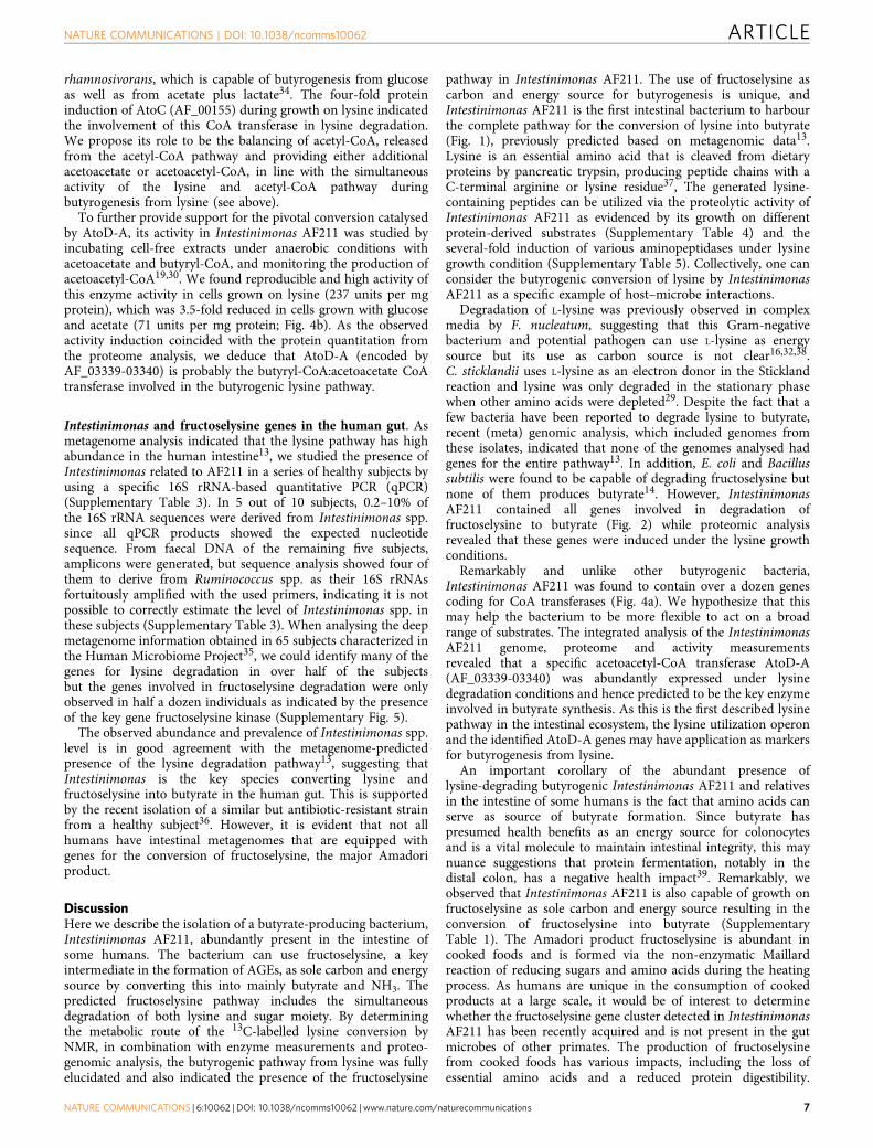
rhamnosivorans, which is capable of butyrogenesis from glucoseas well as from acetate plus lactate34. The four-fold proteininduction of AtoC (AF_00155) during growth on lysine indicatedthe involvement of this CoA transferase in lysine degradation.We propose its role to be the balancing of acetyl-CoA, releasedfrom the acetyl-CoA pathway and providing either additionalacetoacetate or acetoacetyl-CoA, in line with the simultaneousactivity of the lysine and acetyl-CoA pathway duringbutyrogenesis from lysine (see above).
To further provide support for the pivotal conversion catalysedby AtoD-A, its activity in Intestinimonas AF211 was studied byincubating cell-free extracts under anaerobic conditions withacetoacetate and butyryl-CoA, and monitoring the production ofacetoacetyl-CoA19,30. We found reproducible and high activity ofthis enzyme activity in cells grown on lysine (237 units per mgprotein), which was 3.5-fold reduced in cells grown with glucoseand acetate (71 units per mg protein; Fig. 4b). As the observedactivity induction coincided with the protein quantitation fromthe proteome analysis, we deduce that AtoD-A (encoded byAF_03339-03340) is probably the butyryl-CoA:acetoacetate CoAtransferase involved in the butyrogenic lysine pathway.
Intestinimonas and fructoselysine genes in the human gut. Asmetagenome analysis indicated that the lysine pathway has highabundance in the human intestine13, we studied the presence ofIntestinimonas related to AF211 in a series of healthy subjects byusing a specific 16S rRNA-based quantitative PCR (qPCR)(Supplementary Table 3). In 5 out of 10 subjects, 0.2–10% ofthe 16S rRNA sequences were derived from Intestinimonas spp.since all qPCR products showed the expected nucleotidesequence. From faecal DNA of the remaining five subjects,amplicons were generated, but sequence analysis showed four ofthem to derive from Ruminococcus spp. as their 16S rRNAsfortuitously amplified with the used primers, indicating it is notpossible to correctly estimate the level of Intestinimonas spp. inthese subjects (Supplementary Table 3). When analysing the deepmetagenome information obtained in 65 subjects characterized inthe Human Microbiome Project35, we could identify many of thegenes for lysine degradation in over half of the subjectsbut the genes involved in fructoselysine degradation were onlyobserved in half a dozen individuals as indicated by the presenceof the key gene fructoselysine kinase (Supplementary Fig. 5).
The observed abundance and prevalence of Intestinimonas spp.level is in good agreement with the metagenome-predictedpresence of the lysine degradation pathway13, suggesting thatIntestinimonas is the key species converting lysine andfructoselysine into butyrate in the human gut. This is supportedby the recent isolation of a similar but antibiotic-resistant strainfrom a healthy subject36. However, it is evident that not allhumans have intestinal metagenomes that are equipped withgenes for the conversion of fructoselysine, the major Amadoriproduct.
DiscussionHere we describe the isolation of a butyrate-producing bacterium,Intestinimonas AF211, abundantly present in the intestine ofsome humans. The bacterium can use fructoselysine, a keyintermediate in the formation of AGEs, as sole carbon and energysource by converting this into mainly butyrate and NH3. Thepredicted fructoselysine pathway includes the simultaneousdegradation of both lysine and sugar moiety. By determiningthe metabolic route of the 13C-labelled lysine conversion byNMR, in combination with enzyme measurements and proteo-genomic analysis, the butyrogenic pathway from lysine was fullyelucidated and also indicated the presence of the fructoselysine
pathway in Intestinimonas AF211. The use of fructoselysine ascarbon and energy source for butyrogenesis is unique, andIntestinimonas AF211 is the first intestinal bacterium to harbourthe complete pathway for the conversion of lysine into butyrate(Fig. 1), previously predicted based on metagenomic data13.Lysine is an essential amino acid that is cleaved from dietaryproteins by pancreatic trypsin, producing peptide chains with aC-terminal arginine or lysine residue37, The generated lysine-containing peptides can be utilized via the proteolytic activity ofIntestinimonas AF211 as evidenced by its growth on differentprotein-derived substrates (Supplementary Table 4) and theseveral-fold induction of various aminopeptidases under lysinegrowth condition (Supplementary Table 5). Collectively, one canconsider the butyrogenic conversion of lysine by IntestinimonasAF211 as a specific example of host–microbe interactions.
Degradation of L-lysine was previously observed in complexmedia by F. nucleatum, suggesting that this Gram-negativebacterium and potential pathogen can use L-lysine as energysource but its use as carbon source is not clear16,32,38.C. sticklandii uses L-lysine as an electron donor in the Sticklandreaction and lysine was only degraded in the stationary phasewhen other amino acids were depleted29. Despite the fact that afew bacteria have been reported to degrade lysine to butyrate,recent (meta) genomic analysis, which included genomes fromthese isolates, indicated that none of the genomes analysed hadgenes for the entire pathway13. In addition, E. coli and Bacillussubtilis were found to be capable of degrading fructoselysine butnone of them produces butyrate14. However, IntestinimonasAF211 contained all genes involved in degradation offructoselysine to butyrate (Fig. 2) while proteomic analysisrevealed that these genes were induced under the lysine growthconditions.
Remarkably and unlike other butyrogenic bacteria,Intestinimonas AF211 was found to contain over a dozen genescoding for CoA transferases (Fig. 4a). We hypothesize that thismay help the bacterium to be more flexible to act on a broadrange of substrates. The integrated analysis of the IntestinimonasAF211 genome, proteome and activity measurementsrevealed that a specific acetoacetyl-CoA transferase AtoD-A(AF_03339-03340) was abundantly expressed under lysinedegradation conditions and hence predicted to be the key enzymeinvolved in butyrate synthesis. As this is the first described lysinepathway in the intestinal ecosystem, the lysine utilization operonand the identified AtoD-A genes may have application as markersfor butyrogenesis from lysine.
An important corollary of the abundant presence oflysine-degrading butyrogenic Intestinimonas AF211 and relativesin the intestine of some humans is the fact that amino acids canserve as source of butyrate formation. Since butyrate haspresumed health benefits as an energy source for colonocytesand is a vital molecule to maintain intestinal integrity, this maynuance suggestions that protein fermentation, notably in thedistal colon, has a negative health impact39. Remarkably, weobserved that Intestinimonas AF211 is also capable of growth onfructoselysine as sole carbon and energy source resulting in theconversion of fructoselysine into butyrate (SupplementaryTable 1). The Amadori product fructoselysine is abundant incooked foods and is formed via the non-enzymatic Maillardreaction of reducing sugars and amino acids during the heatingprocess. As humans are unique in the consumption of cookedproducts at a large scale, it would be of interest to determinewhether the fructoselysine gene cluster detected in IntestinimonasAF211 has been recently acquired and is not present in the gutmicrobes of other primates. The production of fructoselysinefrom cooked foods has various impacts, including the loss ofessential amino acids and a reduced protein digestibility.
NATURE COMMUNICATIONS | DOI: 10.1038/ncomms10062 ARTICLE
NATURE COMMUNICATIONS | 6:10062 | DOI: 10.1038/ncomms10062 | www.nature.com/naturecommunications 7
Fructoselysine is key product leading to the formation of AGEs inthe human body that have been associated with chronic diseasesand development of diabetes complication40–42. Moreover, recentfood interventions in mice showed the deleterious effect of AGEson these and other diseases43. The butyrogenic conversion offructoselysine by Intestinimonas AF211 illustrates the importantrole of this anaerobe in the human intestinal tract, whereas theobservation that some but not all human carry genes forfructoselysine degradation indicates the potential for specificinterventions with the newly discovered strain. In conclusion, ourstudy underlines the need for cultivating novel microbes to get acomprehensive understanding of the intestinal metabolicprocesses and the beneficial effect on the human host. Inaddition, Intestinimonas AF211 and related bacteria may play animportant role in the intestinal tract by maintaining proteinbalance and gut homeostasis.
MethodsEnrichment, isolation and growth. Strain Intestinimonas AF211 was isolatedfrom the stool of a healthy adult. The faecal sample was enriched in an anaerobicbicarbonate-buffered mineral salt medium44 containing 40 mM lactate and 40 mMacetate as energy and carbon source. The head space was filled with CO2/N2 (1:4) at1.5 atm and incubation was at 37 �C. Subsequently, the enrichment culture wastransferred to reinforced clostridium medium (RCM, Difco) in serial dilutions andplated at least three times on RCM agar, which resulted in an axenic culture. Thepurity of the strain, designated as strain AF211, was confirmed by 16S rRNA genesequencing and microscopy. The strain was routinely maintained in RCM mediumat 37 �C. A phylogenetic tree of the 16S rRNA of Intestinimonas AF211 (GenBankaccession JX273469) and closely related strains was constructed as describedpreviously34 (Supplementary Fig. 1).
For physiological characterization, different amino acids and amino-acidderivatives were tested including 20 mM of L-lysine, fructoselysine (USBiological,USA), glutamate, glutamine, glycine, proline, arginine, aspartate and methionine,which were added into the bicarbonate-buffered medium from 1 M sterile anoxicstock solutions. For sugar utilization 20 mM glucose plus 20 mM acetate wereadded into the bicarbonate-buffered medium from 1 M stock solutions. The growthwas determined via product formation by HPLC34 and optical densitymeasurement by a spectrophotometer at a wavelength of 600 nm. Generation timeswere calculated employing Gompertz modelling45.
Analytical methods. Lysine was quantified on a HPLC using a Polaris C18-Acolumn (Agilent) running at 45 �C and an ultraviolet–visible detector at wavelengthof 436 nm. Flow rate was 0.5 ml min� 1. An eluent mobile phase consisted of24 mM acetic acid:8% acetonitrile (pH 6.6) as solvent A and acetonitrile:2-propanol(60:40) as solvent B. The gradient elution was used from 95% eluent A and 5%eluent B to 25% eluent A and 75% eluent B for first 15 min. The column wassubsequently washed with 100% eluent B for 7 min before the next sample injec-tion. Norleucine (4 mM) was used as internal standard. The volatile fatty acidformation was measured on a Thermo Scientific Spectra HPLC system equippedwith an Agilent Metacarb 67H 300� 6.5-mm column kept at 37 �C and runningwith 10 mM arabinose as an eluent. The detector was a refractive index detector.The eluent flow was 0.8 ml min� 1. Gas production was performed as previouslydescribed46. All analyses were performed in duplicate. Ammonium was determinedusing Spectroquant test kit according to the manufacture’s instruction.Fructoselysine was separated by ion-exchange chromatography and quantified bypost column reaction with ninhydrin using photometric detection at 570 nm(International Organization for Standardization (ISO) 13903).
Nuclear magnetic resonance. Strain AF211 was cultivated in a bicarbonate-buffered medium containing 20 mM of [2-13C]L-lysine or [6-13C]L-lysine or lysineplus [2-13C]acetate. The growth conditions were as described above. 13C-labelledlysine was purchased from Campro Scientific (Veenendaal, The Netherlands).Samples were taken from an overnight culture and centrifuged at 10,000g. D2O(50 ml; 99.9 atom%, Sigma Aldrich) was added to the supernatants (0.5 ml) andsubsequently transferred in NMR tubes (Campro Scientific). 1H-decoupled 13C-NMR spectra were recorded at a probe temperature of 300 K on a Bruker Avance-III-500 spectrometer located at the Wageningen NMR Centre (WNMRC),Wageningen, the Netherlands. Chemical shifts are expressed in p.p.m. relative tothe C-6 of added [6-13C]lysine at 41.75 p.p.m., added [2-13C]lysine at 57.19 p.p.m.,formed [2-13C]butyrate at 42.33 p.p.m., formed [4-13C]butyrate at 15.95 p.p.m. andformed [2-13C]acetate at 25.99 p.p.m. (Biological Magnetic Resonance Data Bank,http://www.bmrb.wisc.edu/metabolomics/metabolomics_standards). For theHBMC spectra 400 experiments of 8 scans were recorded resulting in a measuringtime of 50 min, using a standard Bruker pulse sequence. The products wereidentified based on chemical shifts as compared with database as mentioned above.
In the HMBC experiment no decoupling is used (Fig. 3b). Therefore, thesingle-bond couplings will result in double cross-peak split by the large single-bondcoupling. The active coupling between 5 and 10 Hz of two- and three-bondcouplings remains within the width of a cross-peak in a HMBC spectrum, resultingin single cross-peaks. When the carbon attached to the proton involved in athree-bond coupling is also enriched, this cross-peak will be split by the largesingle-bond coupling with this carbon. These splittings are visible as extra peaksnext to the single cross-peaks between H-2 and C-4 and between H-4 and C-2(arrows in the figures), indicating double enrichment of both C-2 and C-4. Thecalculation was shown in Fig. 3c. Both the split and non-split HMBC cross-peakswith C-4 at H-2 and with C-2 at H-4 were integrated. The integrals of the split andnon-split cross-peaks at a single proton are comparable, since they are based on thesame three-bond couplings and experience similar relaxation behaviour. The splitcross-peaks of H-2 with C-4 and of H-4 with C-2 show a small difference due tosensitivity of the HMBC experiment for difference in relaxation and differencein coupling values. Since these split cross-peaks refer to the same population of[2,4-13C]butyrate, the factor needed to equalize them can also be used to correctthe value of the non-split values, thus revealing the percentages of all three possiblefractions of labelled butyrate.
Proteomics. The protein abundances in cultures growing with different substrateswere investigated with liquid chromatography–mass spectrometry/mass spectro-metry. Strain AF211 was grown in triplicate in 500 ml bicarbonate-bufferedmedium containing with 20 mM lysine and 40 mM glucose plus 40 mM sodiumacetate as carbon and energy sources. Yeast extract was supplemented in theculture of glucose plus acetate (2 g l� 1). Cells were collected in the exponentialphase by centrifugation at 10,000g at 4 �C for 20 min. Cell pellets were washedtwice in 100 mM Tris-HCl (pH 7.5), 1 mM dithioerythreitol and suspended in 1 mlof SDT-lysis buffer, which contained 100 mM Tris-HCl (pH 7.5), 4% SDS and0.1 M dithiotreitol. Protein extractions, separation, tryptic digestion and analysiswere performed as described previously47. An Intestinimonas AF211 databasedownloaded from Uniprot (http://www.uniprot.org) was used together with acontaminant database that contains sequences of common contaminants; forinstance, trypsin, keratin, bovine serum albumin. The proteomics result containedpeptides and proteins with a false discovery rate of o1% and proteins with at leasttwo identified peptides of which should be unique and one should be unmodifiedwithout any reversed hits. The normal logarithm was taken from protein label-freequantitation (LFQ; normalized with respect to the total amount of protein and allof its identified peptides) intensities. Zero ‘Log LFQ’ values were replaced by avalue of 5.4 (just below the lowest value) to make sensible ratio calculationspossible. Relative protein quantitation of sample to control was done with Perseus1.3.0.4 by applying a two sample t-test using the ‘LFQ intensity’ columns obtainedwith false discovery rate set to 0.05 and S0 set to 1.
Preparation of Intestinimonas AF211 cell extract. Strain AF211 was grown in150 ml anaerobic bicarbonate-buffered medium containing 20 mM lysine or40 mM glucose plus 40 mM acetate as carbon and energy sources. Cells of strainA. rhamnosivorans DSM26241 (ref. 34) grown in 150 ml anaerobic bicarbonate-buffered medium containing 20 mM glucose were used as a negative control of theenzyme assay. Cells were collected in the exponential phase by centrifugation at10,000g at 4 �C for 20 min. Cell pellets were washed twice in an anaerobic buffercontaining 100 mM Tris-HCl (pH7.5), 1 mM dithioerythreitol and suspended in1 ml of the same buffer. Cells were disrupted by sonication 5 times� 30 s and thecell suspension was cooled on ice for 30 s in between. Finally, the suspension wascentrifuged for 10 min at 8,000g. The cell-free extract was transferred to serumbottles, flushed with N2 and either stored at � 20 �C or used directly for enzymeactivity assays. All steps were performed in an anaerobic chamber with a N2/H2
(96:4; v/v) atmosphere, circulated over a palladium catalyst to eliminate traces ofoxygen.
Detection of acetoacetyl-CoA transferase activity. CoA transferase activity wasdetermined using a spectrophotometric assay19 at 310 nm, 25 �C with 100 mMTris-Cl (pH 8.1), 20 mM MgCl2 and 50 mM butyryl-CoA plus 10 mM lithiumacetoacetate as substrates. For enzyme activity assay the formation of acetoacetyl-CoA (E310nm¼ 15.1 mM� 1 cm� 1) from butyryl-CoA and acetoacetate wasfollowed by measuring the increase in A310nm by means of an absorbance-recordingspectrophotometer. Total volume of the mixture was 1.0 ml. The activities wereexpressed as U mg� 1 of total proteins (U¼mmol min� 1). All assays were donewith biological duplicates and 4–6 replicate measurements were performed. Totalprotein was quantified using Qubit2.0 Fluorometer (Invitrogen) according to themanufacturer’s instructions.
Phylogenetic analysis of CoA transferase. For the construction of a CoAtransferase phylogenetic tree, all butyryl-CoA:acetate CoA transferases (Ato) andbutyryl-CoA:4-hydroxybutyrate CoA transferases (4Hbt) from known intestinalbutyrate-producing bacteria according to refs. 28,48 were retrieved from the NCBIdatabase (Fig. 4). Selected 4-hydroxybutyrate CoA transferases from Clostridiumkluyveri, C. acetobutyricum and C. tetani were also included28. Butyryl-CoA-acetoacetate CoA tranferases (Ato) from C. sticklandii DSM519, F. nucleatum
ARTICLE NATURE COMMUNICATIONS | DOI: 10.1038/ncomms10062
8 NATURE COMMUNICATIONS | 6:10062 | DOI: 10.1038/ncomms10062 | www.nature.com/naturecommunications
ATCC25586 and C. acetobutylicum ATCC824 were collected from theirgenomes29,33,49. These CoA transferases are catalysing either butyrate or butanolformation. All amino-acid sequences of 4Hbt and AtoA/C/D from strainIntestinimonas AF211 were aligned with retrieved sequences using theCLUSTAL_X programme. A phylogenetic tree was constructed using theneighbour-joining algorithm by the MEGA 5 with 1,000 bootstraps to obtainconfidence levels for the branches.
Quantification of Intestinimonas in the human colon. Stool samples of 10 Dutchhealthy volunteers at different age and gender were collected (SupplementaryTable 3). Genomic DNA was isolated via the bead-beating protocol describedpreviously50. The 16S rRNA sequences of phylogenetically related species wereretrieved from GenBank (www.ncbi.nlm.nih.gov) and used to perform multiplealignments by using CLUSTALW. Primers for qPCR were designed usingDNASTAR programme according to Walter et al.51 Primers were PFF590f:50-AAAACTATGGGCTCAACCCA-30 and PFF702r: 50-GTCAGTTAATGTCCAGCAGG-30 to quantify Intestinimonas AF211, which resulted in a 100-bpamplicon. Total bacteria were quantified using BAC1396F and PROK1492R primerpairs52. The 16S rRNA gene of strain AF211 was used for optimizing temperatureand primer concentration, and for making standard curves. The programme thatwas used to amplify the partial 16S rRNA gene of strain AF211 was as following:95 �C for 5 min and 35 cycles consisting of 95 �C for 30 s, 56.7 �C for 10 s and 72 �Cfor 30 s; 95 �C for 1 min and 60 �C for 1 min and total bacteria as followed: 95 �Cfor 10 min or 95 �C for 20 s, 56.3 �C for 30 s and 72 �C for 30 s; 95 �C for 1 minand 60 �C for 1 min. Both were followed by a melting curve analysis. DNAcopies were calculated from standard curves and subsequently used for thestrain quantities.
Metagenome analysis. The metagenomic protein sequence data from faecalsamples of 65 Human Microbiome Project subjects were obtained from MG-RAST(http://metagenomics.anl.gov/). The following samples were included: SRR063550,SRR063552, SRR063553, SRR063555, SRR063556, SRR063558, SRR063561,SRR063562, SRR063564, SRR063565, SRR061730, SRR063567, SRR063568,SRR063570, SRR063571, SRR063573, SRR063574, SRR063576, SRR063577,SRR063579, SRR063580, SRR063538, SRR063582, SRR063585, SRR063586,SRR063588, SRR063589, SRR063802, SRR063899, SRR063900, SRR063902,SRR063903, SRR063540, SRR063905, SRR063906, SRR063908, SRR063909,SRR063539, SRR063542, SRR063545, SRR063548, SRR063551, SRR063554,SRR063541, SRR063557, SRR063560, SRR063563, SRR063566, SRR063569,SRR063572, SRR063575, SRR063578, SRR063581, SRR063584, SRR063543,SRR063587, SRR063801, SRR063898, SRR063901, SRR063904, SRR063907,SRR063544, SRR063546, SRR063547 and SRR063549.
AF211 butyrogenic pathway protein homologues were searched from themetagenomic data by using Usearch v. 8.0.1517 with settings usearch_globaland id¼ 0.6. The results were analysed in R v. 3.1.1 software environment(http://www.R-project.org)53.
References1. Sommer, F. & Backhed, F. The gut microbiota-masters of host development
and physiology. Nat. Rev. Microbiol. 11, 227–238 (2013).2. Qin, J. et al. A human gut microbial gene catalogue established by metagenomic
sequencing. Nature 464, 59–65 (2010).3. Hamer, H. M. et al. Review article: the role of butyrate on colonic function.
Aliment. Pharmacol. Ther. 27, 104–119 (2008).4. Rajilic-Stojanovic, M. & de Vos, W. M. The first 1000 cultured species of the
human gastrointestinal microbiota. FEMS Microbiol. Rev. 38, 996–1047 (2014).5. Flint, H. J., Scott, K. P., Ducan, S., Louis, P. & Forano, E. Microbial degradation
of complex carbohydrates in the gut. Gut Microbes 3, 289–306 (2012).6. Qin, J. et al. A metagenome-wide association study of gut microbiota in type 2
diabetes. Nature 490, 55–60 (2012).7. Scheppach, W., Luehrs, H. & Menzel, T. Beneficial health effects of low-
digestible carbohydrate consumption. Br. J. Nutr. 85, S23–S30 (2001).8. Duncan, S. H., Barcenilla, A., Stewart, C. S., Pryde, S. E. & Flint, H. J. Acetate
utilization and butyryl coenzyme A (CoA):acetate-CoA transferase in butyrate-producing bacteria from the human large intestine. Appl. Environ. Microbiol.68, 5186–5190 (2002).
9. Belenguer, A. et al. Two routes of metabolic cross-feeding betweenBifidobacterium adolescentis and butyrate-producing anaerobes from thehuman gut. Appl. Environ. Microbiol. 72, 3593–3599 (2006).
10. Barcenilla, A. et al. Phylogenetic relationships of butyrate-producing bacteriafrom the human gut. Appl. Environ. Microbiol. 66, 1654–1661 (2000).
11. Louis, P., Hold, G. L. & Flint, H. J. The gut microbiota, bacterial metabolitesand colorectal cancer. Nat. Rev. Microbiol. 12, 661–672 (2014).
12. Louis, P. et al. Restricted distribution of the butyrate kinase pathway amongbutyrate-producing bacteria from the human colon. J. Bacteriol. 186,2099–2106 (2004).
13. Vital, M., Howe, A. C. & Tiedje, J. M. Revealing the bacterial butyrate synthesispathways by analyzing (meta)genomic data. MBio 5, e00889 (2014).
14. Deppe, V., Bongaerts, J., O’Connell, T., Maurer, K. H. & Meinhardt, F.Enzymatic deglycation of Amadori products in bacteria: mechanisms,occurrence and physiological functions. Appl. Microbiol. Biotechnol. 90,399–406 (2011).
15. Stadtman, T. C. & White, F. H. Tracer studies on ornithine, lysine, and formatemetabolism in an amino acid fermenting clostridium. J. Bacteriol. 67, 651–657(1954).
16. Kreimeyer, A. et al. Identification of the last unknown genes in thefermentation pathway of lysine. J. Biol. Chem. 282, 7191–7197 (2007).
17. Yorifuji, T., Jeng, I. M. & Barker, H. A. Purification and properties of 3-keto-5-aminohexanoate cleavage enzyme from a lysine-fermenting clostridium. J. Biol.Chem. 252, 20–31 (1977).
18. Baker, J. J., Jeng, I. & Barker, H. A. Purification and properties of l-erythro-3,5-diaminohexanoate dehydrogenase from a lysine-fermenting clostridium. J. Biol.Chem. 247, 7724–7734 (1972).
19. Barker, H. A. et al. Butyryl-CoA:acetoacetate CoA-transferase from a lysine-fermenting clostridium. J. Biol. Chem. 253, 1219–1225 (1978).
20. Chirpich, T. P., Zappia, V., Costilow, R. N. & Barker, H. A. Lysine 2,3-aminomutase: purification and properties of a pyridoxal phosphate andS-adenosylmethionine-activated enzyme. J. Biol. Chem. 245, 1778–1789 (1970).
21. Klaring, K. et al. Intestinimonas butyriciproducens gen. nov., sp. nov., abutyrate-producing bacterium from the mouse intestine. Int. J. Syst. Evol.Microbiol. 63, 4606–4612 (2013).
22. Wiame, E. & Van Schaftingen, E. Fructoselysine 3-epimerase, an enzymeinvolved in the metabolism of the unusual Amadori compound psicoselysine inEscherichia coli. Biochem. J. 378, 1047–1052 (2004).
23. Wiame, E., Delpierre, G., Collard, F. & Van Schaftingen, E. Identification of apathway for the utilization of the Amadori product fructoselysine in Escherichiacoli. J. Biol. Chem. 277, 42523–42529 (2002).
24. Plugge, C. M., van Leeuwen, J., Hummelen, T., Balk, M. & Stams, A. J. M.Elucidation of the pathways of catabolic glutamate conversion in threethermophilic anaerobic bacteria. Arch. Microbiol. 176, 29–36 (2001).
25. Herrmann, G., Jayamani, E., Mai, G. & Buckel, W. Energy conservation viaelectron-transferring flavoprotein in anaerobic bacteria. J. Bacteriol. 190,784–791 (2008).
26. Li, F. et al. Coupled ferredoxin and crotonyl coenzyme A (CoA) reduction withNADH catalyzed by the butyryl-CoA dehydrogenase/Etf complex fromClostridium kluyveri. J. Bacteriol. 190, 843–850 (2008).
27. Boynton, Z. L., Bennet, G. N. & Rudolph, F. B. Cloning, sequencing, andexpression of clustered genes encoding beta-hydroxybutyryl-coenzyme A(CoA) dehydrogenase, crotonase, and butyryl-CoA dehydrogenase fromClostridium acetobutylicum ATCC 824. J. Bacteriol. 178, 3015–3024 (1996).
28. Charrier, C. et al. A novel class of CoA-transferase involved in short-chain fattyacid metabolism in butyrate-producing human colonic bacteria. Microbiology152, 179–185 (2006).
29. Fonknechten, N. et al. Clostridium sticklandii, a specialist in amino aciddegradation:revisiting its metabolism through its genome sequence. BMCGenomics 11, 1–12 (2010).
30. Wiesenborn, D. P., Rudolph, F. B. & Papoutsakis, E. T. Coenzyme A transferasefrom Clostridium acetobutylicum ATCC 824 and its role in the uptake of acids.Appl. Environ. Microbiol. 55, 323–329 (1989).
31. Bennett, G. N. & Rudolph, F. B. The central metabolic pathway from acetyl-CoA to butyryl-CoA in Clostridium acetobutylicum. FEMS Microbiol. Rev. 17,241–249 (1995).
32. Barker, H. A., Kahn, J. M. & Hedrick, L. Pathway of lysine degradation inFusobacterium nucleatum. J. Bacteriol. 152, 201–207 (1982).
33. Kapatral, V. et al. Genome sequence and analysis of the oral bacteriumFusobacterium nucleatum strain ATCC 25586. J. Bacteriol. 184, 2005–2018(2002).
34. Bui, T. P. N., de Vos, W. M. & Plugge, C. M. Anaerostipes rhamnosivorans sp.nov., a human intestinal, butyrate-forming bacterium. Int. J. Syst. Evol.Microbiol. 64, 787–793 (2014).
35. The Human Microbiome Project Consortium. Structure, function and diversityof the healthy human microbiome. Nature 486, 207–214 (2012).
36. Rettedal, E. A., Gumpert, H. & Sommer, M. O. A. Cultivation-based multiplexphenotyping of human gut microbiota allows targeted recovery of previouslyuncultured bacteria. Nat. Commun. 5, 4714 (2014).
37. Leiros, H. K. et al. Trypsin specificity as elucidated by LIE calculations, X-raystructures, and association constant measurements. Protein Sci. 13, 1056–1070(2004).
38. Rogers, A. H., Chen, J., Zilm, P. S. & Gully, N. J. The behaviour ofFusobacterium nucleatum chemostat-grown in glucose- and amino acid-basedchemically defined media. Anaerobe 4, 111–116 (1998).
39. Windey, K. et al. Modulation of protein fermentation does not affect fecal watertoxicity: a randomized cross-over study in healthy subjects. PLoS ONE 7,e52387 (2012).
40. Brownlee, M. Glycation and diabetic complications. Diabetes 43, 836–841(1994).
NATURE COMMUNICATIONS | DOI: 10.1038/ncomms10062 ARTICLE
NATURE COMMUNICATIONS | 6:10062 | DOI: 10.1038/ncomms10062 | www.nature.com/naturecommunications 9

41. Baynes, J. W. & Thorpe, S. R. Glycoxidation and lipoxidation in atherogenesis.Free Radic. Biol. Med. 28, 1708–1716 (2000).
42. West, R. K. et al. Dietary advanced glycation end products are associated withdecline in memory in young elderly. Mech. Ageing Dev. 140, 10–12 (2014).
43. Cai, W. et al. Oral glycotoxins are a modifiable cause of dementia and themetabolic syndrome in mice and humans. Proc. Natl Acad. Sci. USA 111,4940–4945 (2014).
44. Stams, A. J. M., Van Dijk, J. B., Dijkema, C. & Plugge, C. M. Growth ofsyntrophic propionate-oxidizing bacteria with fumarate in the absence ofmethanogenic bacteria. Appl. Environ. Microbiol. 59, 1114–1119 (1993).
45. Zwietering, M. H., Jongenburger, I., Rombouts, F. M. & Van ’t Riet, K.Modeling of the bacterial growth curve. Appl. Environ. Microbiol. 56,1875–1881 (1990).
46. van Gelder, A. H., Aydin, R., Alves, M. M. & Stams, A. J. M. 1,3-Propanediolproduction from glycerol by a newly isolated Trichococcus strain. Microbiol.Biotechnol. 5, 573–578 (2012).
47. Oosterkamp, M. J., Boeren, S., Plugge, C. M., Schaap, P. J. & Stams, A. J. M.Metabolic response of Alicycliphilus denitrificans strain BC toward electronacceptor variation. Proteomics 13, 2886–2894 (2013).
48. Louis, P. & Flint, H. J. Diversity, metabolism and microbial ecology of butyrate-producing bacteria from the human large intestine. FEMS Microbiol. Lett. 294,1–8 (2009).
49. Gerischer, U. & Durre, P. Cloning, sequencing, and molecular analysis of theacetoacetate decarboxylase gene region from Clostridium acetobutylicum.J. Bacteriol. 172, 6907–6918 (1990).
50. Salonen, A. et al. Comparative analysis of fecal DNA extraction methods withphylogenetic microarray: Effective recovery of bacterial and archaeal DNAusing mechanical cell lysis. J. Microbiol. Methods 81, 127–134 (2010).
51. Walter, J. et al. Detection of Lactobacillus, Pediococcus, Leuconostoc, andWeissella species in human feces by using group-specific PCR primers anddenaturing gradient gel electrophoresis. Appl. Environ. Microbiol. 67,2578–2585 (2001).
52. Suzuki, M. T., Taylor, L. T. & DeLong, E. F. Quantitative analysis of small-subunit rRNA genes in mixed microbial populations via 50-nuclease assays.Appl. Environ. Microbiol. 66, 4605–4614 (2000).
53. Edgar, R. C. Search and clustering orders of magnitude faster than BLAST.Bioinformatics 26, 2460–2461 (2010).
AcknowledgementsThis research has partly been supported by grant 250172-Microbes Inside of theEuropean Research Council to W.M.d.V. and the Soehngen Institute of AnaerobicMicrobiology, funded by the Netherlands Organization for Scientific Research. We thankDr Erwin Zoetendal and Prof. Stephen O’Keefe for providing faecal samples used inthis study, Dr Elsa Wiame for the kind gift of fructoselysine, Prof. Fons Stams forhelpful discussions, Ton van Gelder for technical support and Mark Davids for sharingcomputational infrastructure.
Author contributionsW.M.d.V., C.M.P. and T.P.N.B. designed the research; T.P.N.B. performed theresearch; J.R. contributed to the (meta)genome analyses; S.B. performed the liquidchromatography–mass spectrometry/mass spectrometry and P.d.W. contributed tothe 13C-NMR and HBMC analysis; T.P.N.B., J.R., S.B., P.d.W., C.M.P. and W.M.d.V.analysed the data; and T.P.N.B., C.M.P. and W.M.d.V. wrote the paper.
Additional informationSupplementary Information accompanies this paper at http://www.nature.com/naturecommunications
Competing financial interests: The authors declare no competing financial interests.
Reprints and permission information is available online at http://npg.nature.com/reprintsandpermissions/
How to cite this article: Bui, T.P.N. et al. Production of butyrate from lysine and theAmadori product fructoselysine by a human gut commensal. Nat. Commun. 6:10062doi: 10.1038/ncomms10062 (2015).
This work is licensed under a Creative Commons Attribution 4.0International License. The images or other third party material in this
article are included in the article’s Creative Commons license, unless indicated otherwisein the credit line; if the material is not included under the Creative Commons license,users will need to obtain permission from the license holder to reproduce the material.To view a copy of this license, visit http://creativecommons.org/licenses/by/4.0/
ARTICLE NATURE COMMUNICATIONS | DOI: 10.1038/ncomms10062
10 NATURE COMMUNICATIONS | 6:10062 | DOI: 10.1038/ncomms10062 | www.nature.com/naturecommunications