production and characterization of monoclonal antibody
TRANSCRIPT
Copyright© Winter 2017, Iran J Allergy Asthma Immunol. All rights reserved. 60 Published by Tehran University of Medical Sciences (http://ijaai.tums.ac.ir)
ORIGINAL ARTICLE
Iran J Allergy Asthma Immunol
February 2017; 16(1):60-71.
Production and Characterization of Monoclonal Antibody
against Recombinant Virus Coat Protein CP42
Naeimeh Shibaei1, Jafar Majidi2,3, Khadijeh Razavi1, Ali Asghar Karkhane4,
Nemat Sokhandan-Bashir5, and Leili Aghebati-Maleki2
1 Department of Agricultural Biotechnology, National Institute of Genetic Engineering
and Biotechnology (NIGEB), Tehran, Iran 2 Department of Immunology, Faculty of Medicine, Tabriz University of Medical Sciences, Tabriz, Iran
3 Immunology Research Center (IRC), Tabriz University of Medical Sciences, Tabriz, Iran
4 Department of Industrial and Environmental Biotechnology, National Institute of Genetic
Engineering and Biotechnology (NIGEB), Tehran, Iran 5 Department of Plant Protection, University of Tabriz, Tabriz, Iran
Received: 9 March 2016; Received in revised form: 25 June 2016; Accepted: 5 July 2016
ABSTRACT
There are many studies related to the production of a ELISA kit for diagnosing virus
infections. However, production of most kits depends on purification of whole virus particles,
which involves the use of costly equipment and reagents. The purpose of this study was to check
out if the anti-CP42 antibodies could be used as a diagnostic assay for detection of Grapevine
fanleaf Virus (GFLV).
In this study, recombinant GFLV coat protein gene related to selected antigenic
determinants was inserted into pET-28a bacterial expression vector and the construct (pET-28a
CP42) was cloned into E. coli strain (DE3). Expressed protein was verified with western blotting
assay by the use of commercially available anti-GFLV antibody. The recombinant protein was
purified using nickel–nitrilotriacetic acid (Ni–NTA) resin. Balb/c mice were immunized with
purified protein and splenocytes of hyperimmunized mice were fused with murine myeloma
Sp2/0 cells. Positive hybridomas were selected by ELISA using CP42 as coating antigen.
The results showed that monoclonal antibody (MAb) specific to CP42 has been successfully
generated. Efficiency of produced antibody was analyzed by ELISA and western blotting assay
using some confirmed grapevine samples. The infection was confirmed previously based on
morphological features and ELISA assay, performed using commercial anti-GFLV antibody. The
monoclonal antibody reacted with antigen in ELISA and immunoblot method.
Our results demonstrated that anti recombinant CP42 monoclonal antibodies are able to
diagnose whole virus in infected grapevine sample using ELISA test.
Keywords: Capsid protein; Epitopes; Enzyme-linked immunosorbent assay; Grapevine
fanleaf virus (GFLV); Monoclonal antibodies
Corresponding Author: Jafar Majidi, PhD;
Department of Immunology, Faculty of Medicine, Tabriz University
of Medical Sciences, Tabriz, Iran. Tel/Fax: (+98 41) 3336 4665,
E-mail: [email protected]

3B7 Anti Chimeric Protein
Vol. 16, No. 1, February 2017 Iran J Allergy Asthma Immunol, Winter 2017 /61 Published by Tehran University of Medical Sciences (http://ijaai.tums.ac.ir)
INTRODUCTION
Monoclonal antibodies (MAb) can be generated
through some techniques, among which hybridoma
technique is the most common one. The production of
monoclonal antibodies allows the isolation of reagents
with a unique, selected specificity. This is due to
identical identity of antibodies related to descendent of
one hybridoma cell. Monoclonal antibodies are
vigorous reagents for development of diagnostic kits.
Hybridoma cell lines provide an unlimited supply of
antibodies and thereby, they are permanent sources of
kit production.1
Production of recombinant protein as an antigen has
several applications such as the preparation of
antibodies and candidate vaccine development.2,3
Expression of viral coat protein gene (CP) in bacteria
as antigen has been used commonly to prepare
antibodies against viruses. It has been strengthened by
the advent of recombinant DNA technology in recent
years.4 In this method, there is no need for virus
purification as a crucial step in conventional antibody
preparation, which is the significant advantage of the
study. In addition, the cloned CP gene can be
maintained safely in deep freezer for future uses.
Grapevine fanleaf Virus (GFLV) is one of the most
devastative viral diseases of grapevine, which
contributes to yield losses of up to 80% in the case of
susceptible cultivars.5 The icosahedral GFLV capsid is
organized by 60 copies of the CP arranged according to
a pseudo T=3 symmetry and has a predicted molecular
mass of approximately, 56 kDa. The GFLV exists in
vineyards of Iran.6-11
There are many different ways for
GFLV transmission and disease spreading, including
transmission by infected pollen, seeds, propagative
materials, and also through the longidorid nematode
Xiphinema index.12
It seems that the widespread occurrence of GFLV
in the vineyards of Iran originates from the absence
of an efficient certification program for virus free
grapes propagation materials. To detect GFLV in
grapevine biological index, serological diagnostic
techniques such as enzyme-linked immunosorbent
assay (ELISA), and gene diagnostic techniques such as
reverse transcription-polymerase chain reaction
(RT-PCR)11
and immunocapture reverse transcription-
PCR (IC-RT-PCR)13
have been developed. Biological
indexing is time-consuming and it is not a common
screening method of infected materials.14
Molecular
methods such as RT-PCR or IC-RT-PCR are generally
not suitable tests for indexing large numbers of samples
and also require specialized equipments and trained
personnel.15
Therefore, serological tests particularly
ELISA has been widely used for low technical
skill requirement, cost effectiveness, and the ability
of screening a large number of samples.16
Therefore, ELISA is a frequently preferred diagnostic
technique because of its speed, specificity, and
simplicity.17
Huss et al. in 1987 was the first scientist to produce
GFLV monoclonal antibody and used it in ELISA
distinguishing different GFLV variant from five
countries. After him, different monoclonal and
polyclonal antibodies were produced by other
scientists.18
In spite of all benefits of present
commercial antibodies, they are not able to detect all
infected grapevine in the area. Since Iranian GFLV
strain is in distinct cluster of parsimonious tree and
they have independent evolution11
, the inefficacy of
present commercial antibodies is predictable. Thus, the
limitation of current anti-GFLV MAbs in
immunodiagnostic test of GFLV was the main reason
for researchers in this study to perform further
investigation for development of another anti-GFLV
monoclonal antibody. However, the efficiency of
monoclonal antibody production is directly related to
the quality of immunogen. Regarding this, we have
previously designed and expressed the new chimeric
GFLV coat protein (CP42), which is composed of some
of antigenic determinants of whole protein (GenBank
KU640965). The new chimeric coat protein consists of
377 amino acids which are less than that of native
antigen. In this study, a new anti-GFLV monoclonal
antibody, 3B7 MAb, was developed and characterized
based on novel 42 KDa chimeric coat protein antigen
for diagnostic purposes.
MATERIALS AND METHODS
Selection of Antigenic Determinants
Continuous B cell epitopes were predicted using
BcePred (http://www.imtech.res.in/raghava/bcepred),19-21
ABCpred (http://www.imtech.res.in/raghava/abcpred/),22
BCPREDS server (http://ailab.ist.psu.edu/bcpred/predict.html)23
and protein structure prediction and annotation Protean
software (PROTEAN subroutine in the DNASTAR 5.0
software package from DNASTAR company, USA).24
Tertiary structure of different constructs was predicted by
N. Shibaei, et al.
62/ Iran J Allergy Asthma Immunol, Winter 2017 Vol. 16, No. 1, February 2017 Published by Tehran University of Medical Sciences (http://ijaai.tums.ac.ir)
two different modeling website such as SWISS-MODEL
Workspace (http://swissmodel.expasy.org/repository/),25
and I-TASSER (http://zhanglab.ccmb.med.umich.edu/ I-
TASSER/).26
For conformational B cell epitope prediction,
the predictive models were uploaded to Disco Tope 1.2
server (http://www.cbs.dtu.dk/services/DiscoTope/).27
Synthesis of Chimeric Construct and Expression of
Recombinant Protein in the Prokaryotic Host
A synthetic DNA coding for potential epitopes of
GFLV CP (GenBank KU640965) was chemically
synthesized by ShineGene Molecular Biotech
(Shanghai, China). The synthetic gene was sub-cloned
into pET-28a expression vector (Novagen, USA), and
the recombinant vector containing the antigenic
determinants named as CP42 was transformed into E.
coli strain BL21 (DE3). The overnight culture of
recombinant cells was used to inoculate 10 mL of
Luria–Bertani medium for recombinant protein
expression. The culture was grown at 37 ◦C up to an
OD600 of 0.9 (OD, optical density). The protein
expression was induced by adding 1 mM IPTG
(isopropyl-β -d-thiogalactoside) (Sigma, St. Louis, MO,
USA) and grow the culture for 7 h of induction.
Samples from the cultures were taken prior to the
induction and 7 h of the induction. Cells were harvested
by centrifugation at 3,000 g for 10 min at 4°C. Pellet
was re-suspended in 50 µL of homemade 1X SDS-
PAGE sample buffer and was analyzed by SDS-
PAGE.28
Purification of the Recombinant Protein and
Confirmation by Western Blot Analysis
The recombinant protein was purified using nickel-
nitrilotriacetic acid (Ni–NTA) (Qiagen, Valencia, CA,
USA) resin under denaturing conditions. The pellet of
200-mL induced culture was re suspended in 8 mL
lysis buffer (50 mM NaH2PO4 pH 8.0, 300 mM NaCl,
10 mM imidazole, and 0.2 mg/mL lysozyme). The
bacterial suspension was lysed at room temperature for
1 h, and the lysate was then centrifuged at 16,000g for
20 min. The supernatant was poured into the Ni–NTA
affinity column, and the low-through fractions were
collected. The column was washed with washing buffer
(100 mM NaH2PO4, 10 mM Tris-Cl, 8 M urea, pH
6.8), and the bound protein was eluted with 2 mL of
elution buffer (100 mM NaH2PO4, 10 mM Tris-Cl, 8
M urea, pH 4.5). Collected fractions were analyzed on
10% SDS-PAGE, and the fractions containing purified
protein were pooled and diluted (1:10) into refolding
buffer (100 mM Tris, 0.1 M NaCl, 5 mM 2-ME,
0.1glycine 2.5% Glycerol and 0.5% triron X-100, pH
8.5) on a gently rotating magnetic stirring apparatus.
Sample was dialyzed several times using buffers with
decreasing concentration of 100 mM Tris (pH 8.5), 0.1
M NaCl, and the last dialysis was performed against
phosphate-buffered saline (PBS) (137 mM NaCl, 2.7
mM KCl, and 4.3 mM Na2HPO4, 1.4 mM KH2PO4,
pH 7.3). The protein concentration was estimated by
Bradford assay.29
The purified protein was transferred
from SDS-PAGE gel to polyvinylidene fluoride
(PVDF) membrane (Biocom Semi Dry Blotters, UK)
using transfer buffer (192 mM glycine, 25 mM Tris
base and 20% methanol). The membrane was incubated
in the blocking buffer composed of 3% skim milk in
PBS for 2 h at room temperature. After three times of
washing, the membrane was incubated for 2 h at 37 °C
with the commercial anti-GFLV antibody (virus
research center, Turin, Italy). Again, after five times of
washing, it was incubated for 2 h at 37 °C with Goat
Anti- Rabbit IgG HRP conjugated antibody (1/2000
dilution). The membrane was washed and exposed to
ECL (Amersham Phamacia Biotech Inc, USA)
hyperfilm for 5 min.
Ethical Approval
All authors hereby declare that "Principles of
laboratory animal care" (NIH publication No. 85-23,
revised 1985) were followed, as well as specific
national laws where applicable. All experiments have
been examined and approved by the appropriate ethics
committee (IR.NIGEB.EC.1395.11.10.A).
Statistical Analysis
All results in this study are presented as
mean±standard deviation (SD). Statistical significance of
differences between groups was explored by One-way
ANOVA and two way ANOVA followed by Bonferroni
using GraphPad Prism 6.01 (software GraphPad
Software, Inc., USA, http://www.graphpad.com). A p
value less than 0.05 was considered significant.
Immunization Procedure and Screening of
Immunized Animals
Four female Balb/c (6-8 weeks old) mice were used
for immunization. Each mouse was immunized 4 times
with an interval of 2-3 weeks subcutaneously. The first
Immunization was performed using Freund's complete

3B7 Anti Chimeric Protein
Vol. 16, No. 1, February 2017 Iran J Allergy Asthma Immunol, Winter 2017 /63 Published by Tehran University of Medical Sciences (http://ijaai.tums.ac.ir)
adjuvant (Sigma-Aldrich Co. St. Louis, MO, USA).
Incomplete Freund's adjuvant (Sigma-Aldrich Co. St.
Louis, MO, USA) was used for subsequent
immunization (50 μg protein/Immunization/mouse).
One week after each immunization, blood samples
were collected by a vertical incision of the tail vein of
mice for determination of antibody titers by plate
trapped antibody-enzyme linked immuno sorbent assay
(PTA-ELISA) method using 96 wells plates coated
with 20 μg/mL of CP42 and HRP goat anti mouse
antibody as second antibody.30
The mouse with the
highest serum antibody titer was selected as the spleen
donor. The last injection of 50µg of antigen (without
any adjuvant) was performed intravenously three days
before the cell fusion. Sera collected from non-
immunized and immunized mice served as negative
and positive controls.
Cell Fusion and Hybridoma Production
Three days after final immunization, spleen of the
immunized mouse was aseptically removed and fused
with SP2/0 myeloma cell line at a ratio of 1:5 (1 SP2/0
and 5 spleen cells) by polyethyleneglycol (PEG , MW
1450, Sigma, UK) as fusogen. Selective hypoxanthine,
aminopterin, Thymidine (HAT) medium (Gibco,
Thermofisher, USA) was added to the fused cells and
cells were seeded into five 96-well microtitre plates
(Nunc, Thermofisher, USA) containing feeder layer.
The cells were incubated at 37 °C with 5% CO2 for 2-
3 days. Cell growth and colony formations were
examined daily. Colonies were appeared after 5-7 days.
Once the colony diameter reached to 1 mm the
presence of antibody against the immunized antigen
was determined by ELISA method31
. The reactivity of
hybridoma supernatants were determined using 96-
wells pre-coated ELISA plates with 20 µg mL−1 of
PBS dissolved CP42.
Cloning of Hybridoma Cells by Limiting Dilution
(LD) Assay
After screening, the clones with high absorbance
were selected for cloning by LD method. The cells
were diluted so that contained only one cell in each 10
μL. Twelve days after LD, the supernatants of
monoclones were screened for production of antibody.
Suitable monoclones possessing high absorbance were
selected for further characterization and were
considered for mass production.
Assessments of Monoclonal Supernatant for GFLV
Detection by ELISA and Western Blotting
In order to determine efficiency of the new
monoclonal antibody for detection of GFLV disease in
vineyard, virus infected symptomatic grape samples
were collected from vineyards of Maragheh, Bonab and
Urmia (all cities located in Iran). For more reliability,
sample infection confirmation was done using ELISA
test with commercial polyclonal antibody (virus
research center Turin, Italy). For this aim, Phloem
tissue was scraped from shoot and ground (1:7 ratio) in
Extraction buffer «General» containing 2.40 g Tris-
base, 8 g NaCl, 20g PVP K25 (MW 24000), 0.50%
Tween20, 0.20 g KCl and 0.20 g NaN3 (pH 7.4; for
1000 mL). Each step was incubated overnight at -4◦c
and washed 3 times with PBS containing 0.05% Tween
20 (PBS-T) for 5 min. Last step was carried out using a
1:2000 dilution of polyclonal rabbit anti-GFLV and
then its alkaline phosphatase conjugate (Loewe,
Germany). The absorbance values of the treated wells
were measured at 405 nm wavelength in STAT FAX
303+ ELISA plate reader. After serological diagnosis
of symptomatic GFLV infections in Grapevine, ELISA
assay was repeated on the same plants using
supernatants of suitable clones as monoclonal antibody
for antibody efficiency analyses.
Western blotting technique was used for
confirming the result of ELISA and to see pattern of
specificity of anti-GFLV monoclonal antibody. It was
performed as above mentioned method and using
supernatants of suitable clones as primary antibody and
rabbit anti-mouse IgG conjugate (1/2000 dilution) as
secondary one. The membrane was washed and
detected by ECL (Amersham Phamacia Biotech Inc,
USA) hyperfilm after exposure for 5 min.
Isotype Determination
The class and subclass of the 3B7 MAb were
determined by ELISA with a mouse monoclonal sub-
isotyping kit containing rabbit anti-mouse IgG1, IgG2a,
IgG2b, IgG3, IgM and IgA, following the procedure
provided by the manufacturer (Thermo, USA).
RSSULTS
Chimeric Construct Synthesis and Expression
A synthetic DNA coding for potential epitopes of
GFLV CP (GenBank KU640965) was chemically
synthesized by ShineGene Molecular Biotech

N. Shibaei, et al.
64/ Iran J Allergy Asthma Immunol, Winter 2017 Vol. 16, No. 1, February 2017 Published by Tehran University of Medical Sciences (http://ijaai.tums.ac.ir)
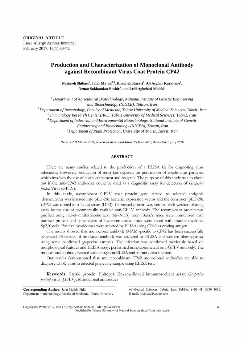
Figure 1. A diagram showing antigenic determinant in Grapevine fanleaf virus coat protein gene cloned into the expression
vector. RBS: Ribosome binding site; Kan R: Kanamycin resistance gene; HIS: Histidine tag.
(Shanghai, China). The synthetic gene was sub-cloned
into pET-28a expression vector (Novagen, USA)
between NcoI and XhoI restriction sites (Figure 1),
then the recombinant vector containing the antigenic
determinants was transformed into E. coli strain DH532
and the transformants were selected on kanamycin.
Recombinant Protein Expression, Purification, and
Characterization:
The synthetic gene was expressed in E. coli BL21
(DE3), with 6X-His-tag at 5׳ ends. The optimum
condition for expression was achieved after 7 h
induction by IPTG (1 mM), at 37 ◦C and OD 600
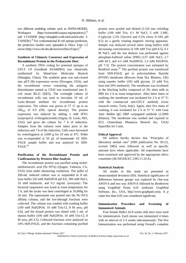
of 0.9. Purification of the recombinant protein was
carried out under denaturing conditions, and SDS-
PAGE analysis revealed as a major band (∼42 kDa) in
all the eluted fractions (Figure 2). The approximate
yield of purification was more than 15 mg/L culture.
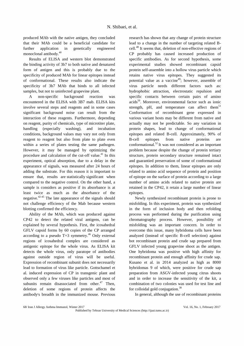
Western blotting analyzes using commercial anti-
GFLV antibody (virus research center Turin, Italy)
was confirmed CP42 as native virus coat protein
(Figure 3).
Figure 2. SDS-PAGE analysis of expression and refolding of CP42 recombinant protein. Lane 1, 2: un-induced bacterial
extract; Lane 3, 4, 5: induced bacterial extract. Lane 6, 7: refolded protein; Lane 8: ladder.

3B7 Anti Chimeric Protein
Vol. 16, No. 1, February 2017 Iran J Allergy Asthma Immunol, Winter 2017 /65 Published by Tehran University of Medical Sciences (http://ijaai.tums.ac.ir)
Figure 3. Western blot analysis of CP42 using commercial anti-GFLV antibody. Lane 1: induced Bacterial extract; Lane 2:
un-induced Bacterial extract. GFLV: Grapevine fanleaf virus
Immunization Procedure and Limiting Dilution
Assay
The titers of antibodies against determinants of
GFLV coat protein (synthetic peptides), in the sera of
immunized mice showed that all mice were immunized
against the antigen, but in two mice increase of
antibody titer was more than others and mice number 2
had higher anti-CP42 antibody (Figure 4). The serum
of the immune mouse at 1:32000 dilution, indicated the
highest absorbance in reaction with CP42 using ELISA
method (Figure 5). So the immune mouse was selected
for the fusion. The final result of the successful fusion
of the immune mouse spleen cells with myeloma SP2/0
cells were about 250 wells, of which, 12 wells
contained positive clones with high absorbance in
reaction with synthetic peptides and 1 clone had anOD
over than 3. Positive clone was named as 3b7 and was
selected for cloning by LD method. The yield of LD
was many clones with absorbance over than 3 and
about 2 at 0.01 dilutions.
Assessments of Monoclonal Supernatant for GFLV
Detection
The specificity of the anti-GFLV was assessed by
an ELISA and western blotting procedure. Grapevine
samples were collected based on symptomatic signs
such mosaic, open petiole leaf, vein banding, leaf
deformation, mottling, and fanleaf.
For ELISA confirmation assay, an uninfected and
four infected plants with typical symptoms were
chosen. ELISA was done using standard polyclonal
antibody in two repeats and one sample of purified
CP42 was used as positive control. The rate of
absorption in all infected samples and CP were greater
than 2.5 times of healthy ones. The one way ANOVA
test, also confirmed the result by a p value less than the
significance level of 0.0001. Then, another ELISA
assay was done using supernatant of monoclonal in two
repeats. The results showed that monoclonal
Figure 4. Enhancement of immune responses in mice by
2nd and 3rd injection (Inj.3 p value < 0.1) of CP42 in sera of
two Balb/c mice evaluated by ELISA assay using 1:2000
dilution of immune mice serum.
Figure 5. Titration of immunized mouse serum against
chimeric CP42 antigen (Two way ANOVA, p value <
0.0001). A serial dilution of serum were added to CP42
pre-coated 96-well plates and titration of antibody was
assayed by ELISA

N. Shibaei, et al.
66/ Iran J Allergy Asthma Immunol, Winter 2017 Vol. 16, No. 1, February 2017 Published by Tehran University of Medical Sciences (http://ijaai.tums.ac.ir)
antibody could isolate the contaminated and non-
contaminated samples efficiently (Table 1). The one
way ANOVA test, confirmed the result by a p value
less than the significance level of 0.001. Result of
western blotting shows that, harvested antibody against
CP42 could acceptably recognize presence of whole
GFLV in the infected samples (Figure 6).
Isotype Determination
Isotype of this MAb was identified as IgG1 and
Kappa light chain (Table 2); ince, their OD was more
than twice the other chains. The ANOVA test, also
confirmed the result by a p value less than the
significance level of 0.0001.
Table 1. Mean absorbance of supernatant of the 3B7 Mab (p value <0.001) and commercial antibody (p value <0.0001) in two
repeats. Optical density value of infected (8D, 13D, 14d, 20D) and non-infected (38D) Grapevine samples and recombinant
CP42 as control (P), at 405 nm.
Sample Commercial antibody Supernatant of the 3B7
Rep.1 Rep.2 Rep.1 Rep.2
8D 2.965 2.999 2.986 2.861
13D 2.973 2.987 2.954 2.961
14D 2.031 2.093 2.781 2.983
20D 2.849 2.741 2.286 2.449
P 1.598 1.622 2.949 2.955
38D 0.182 0.171 1.101 1.202
Figure 6. Western blot determination of the 3B7 MAb. Lanes 1, 2, and 3 are related to the infected grapevine samples. Lanes
4 and 5 are the uninfected grapevine samples. Lanes 6 and 7 are CP42.
Table 2. Determination of the isotype of MAb by ELISA. Classes and subclasses of 3B7 MAb. (p value < 0.0001)

3B7 Anti Chimeric Protein
Vol. 16, No. 1, February 2017 Iran J Allergy Asthma Immunol, Winter 2017 /67 Published by Tehran University of Medical Sciences (http://ijaai.tums.ac.ir)
DISCUSSION
This study has been conducted to evaluate
diagnostic potential of anti-recombinant GFLV-CP,
which is directed against a part of whole protein
antigenic epitopes, for detection of intact virus
particles. Sokhandan et al. (2011) reported cloning and
sequencing of Iranian GFLV isolate׳s coat protein
gene.11
Followed by their study, improvement of
detection system and control strategies for GFLV
infections in Iran, were the main aim of the current
study. However, in this study, we produced a
monoclonal antibody against antigenic determinant of
GFLV coat protein instead of its full length. This
antigen is a novel chimeric 377 amino acids containing
two parts of antigenic regions of whole protein which
are linked together by a linker peptide (GenBank
KU640965). In this study, it was confirmed by ELISA
that a high level of monoclonal antibody against
chimeric CP42 was produced. The chimeric CP42
antigen had appropriate immunogenic properties to
stimulate immune system for producing suitable level
of antibody. Immunoblotting assay also confirmed
these results and demonstrated that 3b7 MAb has been
able to specifically recognize both CP42 and also
native virus particle in the infected plant tissues.
In this study, mouse antiserum with titers of
1/32,000 was obtained, suggesting that CP42 is
relatively a good immunogen. Related to this study,
Etienne et al, obtained polyclonal antisera with titers of
1/20,000 using GFLV particles as antigen then they
concluded that the virus particle is immunogenic.33
In
other experiment, Sokhandan et al, prepared polyclonal
antibodies against GFLV coat protein expressed in E.
coli. A 1/1,000 dilution of the IgG purified from the
antiserum was efficient to detection of the virus in
infected plant.18
The same antibody at 1/500 dilution
detected GFLV in infected grapevine leaf by
immunocapture reverse transcription-polymerase chain
reaction (IC-RT-PCR).13
Therefore, CP42 is
significantly more immunogenic than above mentioned
proteins.
There are several ways to diagnose the GFLV
disease. Symptoms and ELISA assay are two basic
detection methods. Nevertheless, GFLV symptoms are
affected by both environmental conditions such as
symptoms attributed to herbicides and virus strain such
as other grapevine-infecting viruses or Grapevine
yellow speckle viroid (causing GFLV-like symptoms in
ELISA-negative grapevines).11
On the other hand,
ELISA testing alone cannot be used to definitive
diagnose of GFLV because other grapevine-infecting
viruses such as ArMV that serologically cross reacts
with anti-GFLV polyclonal IgG can cause false-
positive result, with the ELISA screen.34
Therefore, the
combination of both methods was used to detect viral
infection in the present study. In order to analyse3b7
MAb efficiency, firstly some of the infected and the
non-infected grapevine were confirmed based on
morphological symptoms and ELISA of commercial
anti-GFLV antibody. Verified samples were used to
evaluate the performance of 3B7 MAb.
In the present study, supernatant of 3b7 MAb
efficiently detect CP42 and infected grapevine in
ELISA and western blot., There are some similarities
and differences between our experiment and other
studies, who immunized mice with whole virus particle
for production of hybridoma.35,36
Huss et al., in 1987
reported the production and characterization of first
monoclonal antibody to GFLV and use of them for
diagnostic tests.37
These antibodies were also used in
attempts to differentiate between serologically closely
related GFLV isolates that differ in their geographical
origin and virulence.36
Relative to above study, Nolke
et al. using purified GFLV particles, produced GFLV-
specific monoclonal antibodies and used its ScFv
sequences for conferring GFLV and Arabis mosaic
virus (ArMV) resistance in Nicotiana benthamiana35
,
however, some scientists suggest the possibility of
using recombinant virus coat protein as antigen, instead
of whole virus. Kusano et al. produced mouse
monoclonal antibodies against recombinant viral coat
protein. They used two lines of antibody from different
paratopes as immunochromatic strip test for direct
detection of Apple stem grooving virus in citrus
samples.38
Fasihi-Ramandi et al., were other scientists
who produced monoclonal antibody against
recombinant protein and used it for ELISA and
agglutination test.39
In the present study, CP42 has been
used for both immunization of mice and screening of
hybridoma cells in order to production of monoclonal
antibodies. Our work is similar to those of other groups
of scientists who produced monoclonal antibody
against chimeric antigen that was designed using
selected part of protein instead of its full length. Based
on the high affinity and appropriate reactivity of
N. Shibaei, et al.
68/ Iran J Allergy Asthma Immunol, Winter 2017 Vol. 16, No. 1, February 2017 Published by Tehran University of Medical Sciences (http://ijaai.tums.ac.ir)
produced MAb with the native antigen, they concluded
that their MAb could be a beneficial candidate for
further application in genetically engineered
monoclonal antibody.40
Results of ELISA and western blot demonstrated
the binding activity of 3b7 to both native and denatured
form of antigen and this is probably due to the
specificity of produced MAb for linear epitopes instead
of conformational. These results also indicate the
specificity of 3b7 MAb that binds to all infected
samples, but not to uninfected grapevine plant.
A non-specific background reaction was
encountered in the ELISA with 3B7 mab. ELISA kits
involve several steps and reagents and in some cases
significant background noise can result from the
interaction of these reagents. Furthermore, depending
on reagent, purity of chemicals, type of microtiter plate,
handling (especially washing), and incubation
conditions, background values may vary not only from
reagent to reagent but also from plate to plate even
within a series of plates testing the same pathogen.
However, it may be managed by optimizing the
procedure and calculation of the cut-off value.41
In this
experiment, optical absorption, due to a delay in the
appearance of signals, was measured after 24 hours of
adding the substrate. For this reason it is important to
ensure that, results are statistically significant when
compared to the negative control. On the other hand, a
sample is considers as positive if its absorbance is at
least twice as much as the absorbance of the
negative.42-45
The late appearance of the signals should
not challenge efficiency of the Mab because western
blotting confirmed the efficiency.
Ability of the MAb, which was produced against
CP42 to detect the related viral antigens, can be
explained by several hypothesis. First, the icosahedral
GFLV capsid forms by 60 copies of the CP arranged
according to a pseudo T=3 symmetry.46
Only external
regions of icosahedral complex are considered as
antigenic epitope for the whole virus. As ELISA kit
detects the whole virus, only paratope of antibodies
against outside region of virus will be useful.
Expression of recombinant subunit does not necessarily
lead to formation of virus like particle. Gottschamel et
al. induced expression of CP in transgenic plant and
observed only a few viruses like particles and most of
subunits remain disassociated from other.47
Then,
deletion of some regions of protein affects the
antibody׳s breadth in the immunized mouse. Previous
research has shown that any change of protein structure
lead to a change in the number of targeting related B-
cell.48
It seems that, deletion of non-effective regions of
CP probably has caused increased production of
specific antibodies. As for second hypothesis, some
experimental studies showed recombinant capsid
protein self-assemble into a hollow virus particle which
retains native virus epitopes. They suggested its
potential value as a vaccine49
; however, assemble of
virus particle needs different factors such as:
hydrophobic attraction, electrostatic repulsion and
specific contacts between certain pairs of amino
acids50
. Moreover, environmental factor such as ionic
strength, pH, and temperature can affect them51
Conformation of recombinant gene expressed in
various variant hosts may be different from native and
actually may not be predictable. So any variation in
protein shapes, lead to change of conformational
epitopes and related B-cell. Approximately, 90% of
B-cell epitopes from native proteins are
conformational.52
It was not considered as an important
problem because despite the change of protein tertiary
structure, protein secondary structure remained intact
and guarantied preservation of some of conformational
epitopes. In addition to them, linear epitopes are only
related to amino acid sequence of protein and position
of epitope on the surface of protein according to a large
number of amino acids related to native protein are
retained in the CP42, it retain a large number of linear
epitopes.
Newly synthesized recombinant protein is prone to
misfolding. In this experiment, protein was synthesized
in the form of inclusion body and then refolding
process was performed during the purification using
chromatography process. However, possibility of
misfolding was an important concern. In order to
overcome this issue, many hybridoma cells have been
analyzed (instead of specific B-cell selection) against
bot recombinant protein and crude sap prepared from
GFLV infected young grapevine shoot as the antigen.
One hybridoma was positive with high affinity for
recombinant protein and enough affinity for crude sap.
Kusano et al. in 2014 analyzed as high as 8000
hybridomas 9 of which, were positive for crude sap
preparation from ASGV-infected young citrus shoots
and in order to increase the sensitivity of the kit, a
combination of two colonies was used for test line and
for colloidal gold conjugation.38
In general, although the use of recombinant proteins

3B7 Anti Chimeric Protein
Vol. 16, No. 1, February 2017 Iran J Allergy Asthma Immunol, Winter 2017 /69 Published by Tehran University of Medical Sciences (http://ijaai.tums.ac.ir)
as antigens is an efficient method in production of
monoclonal antibody, but it is not cost-effective due to
the necessity of simultaneous analyze for a large
number of colonies. Consequently obtaining a fast
result is not guaranteed. The limitation of our study
could be the lack of cross reactivity, specificity,
affinity, and avidity data. However, in this study, a new
anti-GFLV monoclonal antibody, 3B7, was developed
and characterized based on novel 42 KDa chimeric coat
protein antigen for diagnostic purposes.
Our results indicate that anti-CP42 antibodies
showed reactivity against whole GFLV particle in
ELISA and western blot performed in this study.
Utilization of recombinant proteins as antigen is a
helpful method to produce monoclonal antibody and
production of diagnostic kits. Mass usage of it, will
also be affordable. Based on the high affinity of 3b7
MAb and its efficacy in recognition of GFLV particle
together with its specific binding to CP42 antigen, it
could be concluded that 3b7 MAb is a suitable choice
for production of ELISA kits or applying in other
methods.
ACKNOWLEDGEMENTS
We would like to thank the National Institute of
Genetic Engineering and Biotechnology, the
Immunology Research Center (IRC), and Drug Applied
Research Center, for their kind assistance. The
manuscript was written based on a dataset of a PhD
thesis registered in National Institute of Genetic
Engineering and Biotechnology.
REFERENCES
1. Greenfield EA. Antibodies: A Laboratory Manual,
Second edition. Dana-Farber Cancer Institute: csh press;
2014.
2. Jafarzadeh A, Shokri F. TH1 and TH2 responses are
influenced by HLA antigens in healthy neonates
vaccinated with recombinant hepatitis B vaccine. IIran J
Allergy Asthma Immunol 2012;11(4):308-15.
3. Ghasemi A, Salari MH, Zarnani AH, Pourmand MR,
Ahmadi H, Shirazi MH, et al. Immunogenicity
assessment of Brucella mellitensis HSP and TF proteins
by immunized rabbit serum. Iran J Allergy Asthma
Immunol 2013; 12(2):192-4.
4. Ling KS, Zhu HY, Petrovic N, Gonsalves D. Serological
Detection of Grapevine leafroll virus 2 Using an
Antiserum Developed against the Recombinant Coat
Protein†. Journal of Phytopathology. 2007;155(2):65-9.
5. Andret-Link P, Schmitt-Keichinger C, Demangeat G,
Komar V, Fuchs M. The specific transmission of
Grapevine fanleaf virus by its nematode vector
Xiphinema index is solely determined by the viral coat
protein. Virology 2004; 320(1):12-22.
6. Sokhandan Bashir N, Pashaei A, Doulati-Baneh H.
Characterization of the Full Length Coat Protein Gene of
Iranian Grapevine fanleaf virus isolates, genetic variation
and phylogenetic analysis. Iran J Biotech 2011; 9(3):213-
21.
7. Pourrahim R, Farzadfar S, Golnaraghi AR, Ahoonmanesh
A. Incidence and distributions of Grapevine fanleaf virus
in north-east of Iran. Plant Pathology Journal 2007;
6:254-9.
8. Rakhshandehroo F, Pourrahim R, Zamani Zadeh H,
Rezaee S, Mohammadi M. Incidence and distribution of
viruses infecting Iranian vineyards. Journal of
Phytopathology. 2005;153:480-4.
9. Izadpanah K, Zaki-Aghl M, Zhang YP, Daubert SD,
Rowhani A. Bermuda grass as a potential reservoir host
for Grapevine fanleaf virus. Plant Disease 2003;
87(10):1179-82.
10. Zaki-Aghl M, Izadpanah K. Serological and molecular
identification and prevalence of Grapevine fanleaf virus
in Iran. Iranian jouran of Plant Pathology 2003; 39(3-
4):161-71.
11. Sokhandan-Bashir N, Pashaei A, Doulati-Baneh H.
Characterization of the full length coat protein gene of
Iranian Grapevine fanleaf virus isolates, genetic variation
and phylogenetic analysis. Iranian Journal of Biotechnolo
2011; 9(3):213-21.
12. Martelli GP, Walter B, Pink L. Grapevine Fanleaf Virus.
CMI/AAB Description of Plant Viruses No. 385.
Commonwealth Mycological Institute/Association of
Applied Biologists, Kew, Surrey, England. 2001.
13. Koolivand D, Sokhandan-Bashir N, Behjatnia SAA,
Jafari Joozani RA. Detection of Grapevine fanleaf virus
by immunocapture reverse transcription-polymerase
chain reaction (IC-RT-PCR) with recombinant antibody.
Archives of Phytopathology and Plant Protection 2014;
47(17):2070-7.
14. Nickel O, Targon MLPN, Fajardo TVM, Machado MA,
Trivilin AP. Polyclonal antibodies to the coat protein of
Apple stem grooving virus expressed in Escherichia coli:
production and use in immunodiagnosis. Fitopatologia
Brasileira. 2004;29(5):558-62.
15. Fajardo TVM, Barros DR, Nickel O, Kuhn GB, Zerbini
FM. Expression of Grapevine leafroll-associated virus 3
coat protein gene in Escherichia coli and production of

N. Shibaei, et al.
70/ Iran J Allergy Asthma Immunol, Winter 2017 Vol. 16, No. 1, February 2017 Published by Tehran University of Medical Sciences (http://ijaai.tums.ac.ir)
polyclonal antibodies. Fitopatologia Brasileira.
2007;32(6):496-500.
16. Ward E, Foster SJ, Fraaije BA, McCartney HA. Plant
pathogen diagnostics: immunological and nucleic acid-
based approaches. Annals of Applied Biology 2004;
145(1):1-16.
17. Rowhani A. Use of F(ab') 2 Antibody Fragment in ELISA
for Detection of Grapevine Viruses. American Journal of
Enology and Viticulture 1992; 43(1):38-40.
18. Sokhandan-Bashir N, Koolivand D, Behjatnia A.
preparation of polyclonal antibodies to Grapevine fanleaf
virus coat protein expressed in escherichia coli. Ansinet
biotechnology 2015; 14(4):173-80.
19. Parker JM, Guo D, Hodges RS. New hydrophilicity scale
derived from high-performance liquid chromatography
peptide retention data: correlation of predicted surface
residues with antigenicity and X-ray-derived accessible
sites. Biochemistry 1986; 25(19):5425-32.
20. Kolaskar AS, Tongaonkar PC. A semi-empirical method
for prediction of antigenic determinants on protein
antigens. FEBS Lett 1990; 276(1-2):172-4.
21. Ponnuswamy PK, Prabhakaran M, Manavalan P.
Hydrophobic packing and spatial arrangement of amino
acid residues in globular proteins. Biochim Biophys Acta
1980; 623(2):301-16.
22. Saha S, Raghava GP. Prediction of continuous B-cell
epitopes in an antigen using recurrent neural network.
Proteins 2006; 65(1):40-8.
23. El-Manzalawy Y, Dobbs D, Honavar V. Predicting linear
B-cell epitopes using string kernels. J Mol Recognit 2008;
21(4):243-55.
24. Darnell S, Riese M, inventors; Precise Predictions of
Linear B Cell Epitopes in Protean 3D2012.
25. Bordoli L, Kiefer F, Arnold K, Benkert P, Battey J,
Schwede T. Protein structure homology modeling using
SWISS-MODEL workspace. Nat Protocols 2008; 4(1):1-
13.
26. Zhang Y. I-TASSER server for protein 3D structure
prediction. BMC Bioinformatics 2008; 9(40):1471-2105.
27. Haste Andersen P, Nielsen M, Lund O. Prediction of
residues in discontinuous B-cell epitopes using protein
3D structures. Protein Sci 2006; 15(11):2558-67.
28. UK L. Cleavage of structural proteins during the
assembly of the head of bacteriophage T4. Nature 1970;
227(5259):680-5.
29. Bradford MM. A rapid and sensitive method for the
quantitation of microgram quantities of protein utilizing
the principle of protein-dye binding. Analytical
biochemistry. 1976;72(1-2):248-54.
30. Converse RH, Martin RR. ELISA Methods for Plant
Viruses. In: Hampton R, Ball E, DeBoer S, editors.
Serological Methods for Detection and Identification of
Viral and Bacterial Plant Pathogens: A Laboratory
Manual. USA: The American Phytopathological Society,
St. Paul, MN.; 1990. p. 179-96.
31. Majidi J, Zavaran Ilosseini A, Hassan Z, Alimohamadian
M. Production of monoclonal antibody against human
immunoglobulin Iranian Journal of Allergy, Asthma and
Immunology. 2000;1(2):81-7.
32. Chung CT, Niemela SL, Miller RH. One-step preparation
of competent Escherichia coli: transformation and storage
of bacterial cells in the same solution. Proc Natl Acad Sci
U S A 1989; 86(7):2172-5.
33. Etienne L, Clauzel JM, Fuchs M. Simultaneous Detection
of Several Nepoviruses Infecting Grapevine in a Single
DAS-ELISA Test Using Mixed Antisera. Journal of
Phytopathology 1991; 131(2):89-100.
34. Wetzel T, Jardak R, Meunier L, Ghorbel A, Reustle GM,
Krczal G. Simultaneous RT/PCR detection and
differentiation of arabis mosaic and grapevine fanleaf
nepoviruses in grapevines with a single pair of primers. J
Virol Methods 2002; 101(1-2):63-9.
35. Nölke G, Cobanov P, Uhde-Holzem K, Reustle G,
Fischer R, Schillberg S. Grapevine fanleaf virus (GFLV)-
specific antibodies confer GFLV and Arabis mosaic virus
(ArMV) resistance in Nicotiana benthamiana. Molecular
Plant Pathology 2009; 10(1):41-9.
36. Huss B, Muller S, Sommermeyer G, Walter B, Van
Regenmortel MHV. Grapevine Fanleaf Virus Monoclonal
Antibodies: their Use to Distinguish Different Isolates.
Journal of Phytopathology 1987; 119(4):358-70.
37. Huss B, Walter, B., Etienne, L. & Van Regenmortel,
M.H.V. . Grapevine fanleaf virusdetection in various
grapevineorgans using polyclonal and monoclonal
antibodies. Vitis 1986; 25:178-88.
38. Kusano N, Iwanami T, Narahara K, Tanaka M.
Production of monoclonal antibodies specific for the
recombinant viral coat protein of Apple stem grooving
virus-citrus isolate and their application for a simple,
rapid diagnosis by an immunochromatographic assay. J
Virol Methods 2014; 195:86-91.
39. Fasihi-Ramandi M, Nedjad-Moghaddam A, Arabsalmany
F, Asgari S, Ahmadi-Renani S. Production and
characterization of monoclonal and polyclonal antibody
against recombinant outer membrane protein. American
Journal of Immunology. 2014;10:56-62.
40. Fasihi-Ramandi M, Amani J, Salmanian A-H, Moazzeni
SM, Ahmadi K. Production and Characterization of New
Anti-Human CD20 Monoclonal Antibody. Iran J Allergy
Asthma Immunol 2015; 14(5):502-8.

3B7 Anti Chimeric Protein
Vol. 16, No. 1, February 2017 Iran J Allergy Asthma Immunol, Winter 2017 /71 Published by Tehran University of Medical Sciences (http://ijaai.tums.ac.ir)
41. Ridge SR, Vizard AL. Determination of the optimal
cutoff value for a serological assay : an example using the
Determination of the Optimal Cutoff Value for a
Serological Assay: an Example Using the Johne' s
Absorbed EIA. J Clin Microbiol 1993; 31(5):1256-61.
42. Eris FN, Akisu C, Aksoy U. Evaluation of Two ELISA
and Two Indirect Hemagglutination Tests for
Serodiagnosis of Pulmonary Hydatid Disease. Korean J
Parasitol 2009; 47(4):427-9.
43. Noel Masihi K, Lange W. Enzyme-linked immunosorbent
assay for the detection of influenza type-specific
antibodies. J Immunol Methods 1980; 36(2):173-9.
44. Silva JM, Carnelossi PR, Bijora T, Facco CU, Picoli
MHS, Souto ER, et al. Immunocapture-RT-PCR detection
of Cassava common mosaic virus in cassava obtained
from meristem-tip culture in Paraná state. Tropical Plant
Pathology 2011; 36:271-5.
45. Yang JG, Wang FL, Chen DX, Shen LL, Qian YM, Liang
ZY, et al. Development of a one-step immunocapture real-
time RT-PCR assay for detection of Tobacco mosaic virus
in soil. Sensors (Switzerland) 2012; 12(12):16685-94.
46. Schellenberger P, Sauter C, Lorber B, Bron P, Trapani S,
Bergdoll M, et al. Structural insights into viral
determinants of nematode mediated grapevine fanleaf
virus transmission. PLoS Pathogens 2011; 7(5):1-14.
47. Gottschamel J, Castellano M, Maghuly F, Laimer M.
Detection of Virus like particles (Vlps) by ISEM in
transgenic grapevines expressing different GFLV CP-
constructs. Biotechnology, VIBT- BOKU, 1190:
Bodenkultur, VIENNA, Austria 2008.
48. Rudolph R, Lilie H. In vitro folding of inclusion body
proteins. Faseb J 1996; 10(1):49-56.
49. Xing L, Kato K, Li T, Takeda N, Miyamura T, Hammar
L, et al. Recombinant Hepatitis E Capsid Protein Self-
Assembles into a Dual-Domain T = 1 Particle Presenting
Native Virus Epitopes. Virology 1999; 265(1):35-45.
50. Roos WH, Bruinsma R, Wuite GJL. Physical virology.
Nat Phys. 2010;6(10):733-43.
51. Zlotnick A, Aldrich R, Johnson JM, Ceres P, Young MJ.
Mechanism of capsid assembly for an icosahedral plant
virus. Virology 2000; 277(2):450-6.
52. Horsfall AC, Hay FC, Soltys AJ, Jones MG. Epitope
mapping. Immunology Today 1991; 12(7):211-3.