procyanidin b2 induces nrf2 translocation and glutathione s-transferase p1 expression via erks and...
TRANSCRIPT

ORIGINAL CONTRIBUTION
Procyanidin B2 induces Nrf2 translocation and glutathioneS-transferase P1 expression via ERKs and p38-MAPK pathwaysand protect human colonic cells against oxidative stress
Ildefonso Rodrıguez-Ramiro • Sonia Ramos •
Laura Bravo • Luis Goya • Maria Angeles Martın
Received: 26 July 2011 / Accepted: 19 October 2011 / Published online: 1 November 2011
� Springer-Verlag 2011
Abstract
Purpose Procyanidin B2 (PB2) is a naturally occurring
flavonoid widely found in cocoa, red wine and grape juice.
Recent studies have suggested that PB2 could protect
against oxidative stress- and chemical-induced injury in
colonic cells by modulating the endogenous cellular
defence. However, the precise mechanism for this protec-
tion is not fully understood. Herein, we examined the effect
of PB2 on the expression of one of the major antioxidant/
detoxificant enzymes related to intestinal protection, the
glutathione S-transferase P1 (GSTP1), and the molecular
mechanisms involved.
Methods Human colonic Caco-2 cells were treated with
PB2 at different times and enzymatic activity, and mRNA
and protein levels of GSTP1 were evaluated. The nuclear
translocation of the transcription factor NF-erythroid
2-related factor (Nrf2) and the phosphorylation states of
specific proteins central to intracellular signalling cascades
were also investigated.
Results PB2 induced the expression and activity of
GSTP1 and the nuclear translocation of Nrf2. Interestingly,
two important signalling proteins involved in Nrf2 trans-
location, the extracellular signal-regulated protein kinases
(ERKs) and the p38 mitogen-activated protein kinase
(MAPK) were also activated. Further experiments with
specific inhibitors of both pathways confirmed their critical
role in the beneficial effects induced by PB2.
Conclusions The present results show that PB2 protects
against oxidative injury in colonic cells and up-regulate the
expression of GSTP1 via a mechanism that involves ERK
and p38 MAPK activation and Nrf2 translocation. These
results provide a molecular basis for the potential contri-
bution of PB2 in the prevention of oxidative stress-related
intestinal injury and gut pathologies.
Keywords Cocoa flavonoids � Glutathione enzymes �Signalling pathways � Oxidative stress
Introduction
The gastrointestinal tract, especially the colon, is con-
stantly exposed to reactive oxygen species (ROS) gener-
ated during normal cellular metabolism and pathological
processes [1]. ROS play an important role in the develop-
ment and progression of many human diseases, including
cancer [2]; therefore, there is an increasing interest in the
potential effect of nutritional antioxidants on the preven-
tion of intestinal pathological states that are linked to
oxidative stress and on the mechanisms of their actions [3].
Among diet antioxidants, flavonoids, naturally phenolic
compounds extensively found in vegetables, fruits and
plant-derived beverages such as tea, cocoa and red wine are
the most abundant ones [4]. Flavonoids exert a potent
antioxidant activity, acting as reactive oxygen species
(ROS) scavengers, metal ions chelators and free radical
reaction terminators [5]. Additionally, they can also act
indirectly as antioxidants stimulating phase II detoxifying
and antioxidant defence enzymes to preserve cellular
integrity and tissue homeostasis [6].
One of the most important antioxidant/detoxifying
enzymes related to intestinal protection is the glutathione
I. Rodrıguez-Ramiro � S. Ramos � L. Bravo � L. Goya �M. A. Martın (&)
Department of Metabolism and Nutrition, Instituto de Ciencia y
Tecnologia de Alimentos y Nutricion (ICTAN-CSIC), Jose
Antonio Novais 10, Ciudad Universitaria, 28040 Madrid, Spain
e-mail: [email protected]
123
Eur J Nutr (2012) 51:881–892
DOI 10.1007/s00394-011-0269-1

S-transferases family (GSTs). GSTs catalyse the conjuga-
tion of a variety of electrophilic xenobiotics with gluta-
thione facilitating their excretion and providing cellular
protection against free radical and carcinogenic compounds
[7]. Recently, interest has grown in the physiologic prop-
erties of the p class of GST (GSTP1), not only because of
its function in drug detoxification but also because of its
possible roles in cell transformation and carcinogenesis [8].
GSTP1 expression is regulated by multiple factors
including the transcription factor nuclear factor-erythroid
2-related factor 2 (Nrf2) that binds to antioxidant response
elements (ARE), specific nucleotide sequences presented in
the promoter region of the gene encoding for GSTP1 [9].
Accumulating data indicate that various natural compounds
possess the capacity to differentially activate a number of
cellular kinases, including mitogen-activated protein kina-
ses (MAPKs) and phosphatidylinositol 3-kinase (PI3K),
which phosphorylate Nrf2 [10]. Nrf2 phosphorylation
facilitates the nuclear translocation of Nrf2 and thereby the
transcription of several genes encoding cytoprotective
enzymes, including GSTs [11]. Accordingly, modulation of
Nrf2 and antioxidant and detoxificant enzymes is consid-
ered an effective strategy of natural antioxidant compounds
for cytoprotection and chemoprevention.
At present, extensive attention has focused on identify-
ing natural phenolic compounds commonly consumed in
our daily diets as potential inducers of antioxidant/detox-
ificant enzymatic defence. Procyanidins, naturally occur-
ring flavonoids widely found in fresh and in processed
foodstuffs such as cocoa, red wine and grape juice, have
attracted great interest in nutrition and medicine due to
their potent antioxidant capacity and their potential pro-
tective effects on human health [12]. In addition, procy-
anidins are poorly absorbed in the intestine, and
consequently, its beneficial effects would be mainly
focused on the gastrointestinal tract where they may have
an important local function neutralizing oxidants [13]. One
of the main procyanidins, the dimer procyanidin B2 [epi-
catechin-(4b-8)-epicatechin] (PB2) (Fig. 1a), has been
shown to exert several physiological effects, such as anti-
oxidant activity [14] and antitumour effects [15]. More
recently, it has been described that PB2 could also modu-
late cellular redox status and the antioxidant enzyme
defence system in colonic cells protecting against oxidative
stress and xenobiotics [16, 17]. However, up to date, the
precise molecular mechanisms responsible for this protec-
tion are still not fully understood.
Considering this, in the present work, we have
investigated the mode of action of PB2 to protect against
oxidative stress in Caco-2 cells, a human cell line orig-
inating from the gastrointestinal tract that retains many of
the morphological and enzymatic features typical of
normal human enterocytes. To this end, we used the
potent pro-oxidant t-BOOH to induce an oxidative injury
in Caco-2 cells. Previous studies have demonstrated that
t-BOOH-induced oxidative stress is a useful model for
evaluating the cytoprotective effect of natural antioxi-
dants in vitro [18–20]. Our results show for the first time
that PB2 is able to attenuate oxidative injury in colonic
cells and up-regulate the activity and expression of the
cytoprotective enzyme GSTP1 via a mechanism that
involved ERK and p38 MAPKs activation and Nrf2
translocation.
ProcyanidinB2
(a)
(b)
0
2000
4000
6000
8000
10000
12000
14000
a a
c
b
(c)
Flu
ores
cenc
e un
its
% c
ellv
iabi
lity
0 0 1 5 10 20 PB2 (µM)
t-BOOH (400µM)
b
a
0
20
40
60
80
100
120
aa
c
b
0 0 1 5 10 20 PB2 (µM)
t-BOOH (400µM)
cc
Fig. 1 Protective effect of PB2 against oxidative stress and cell death
induced by t-BOOH. a Chemical structure of procyanidin B2. Caco-2
cells were treated with the noted doses of PB2 for 20 h and further
exposed to 400 lM t-BOOH for 2 and 6 h to evaluate ROS generation
and cell viability, respectively. b ROS generation expressed in
fluorescence units. c Percentage values of cell death relative to the
control condition. Data represent means ± SD. of ten different
samples per condition. Means sharing the same letter are not
significantly different from each other while means that have differentletter are significantly dissimilar from each other (P \ 0.05)
882 Eur J Nutr (2012) 51:881–892
123

Materials and methods
Materials and chemical
Gentamicin, penicillin G, streptomycin, SB203580
[4-(4-fluorophenyl)-2-(4-methylsulphonylphenyl)-5-(4-
pyridyl)-1H-imidazole], PD98059 [2-(2-amino-3-methoxy
phenyl)-4H-1-benzopyran-4-one], tert-butyl hydroperoxide
(t-BOOH) and 3,4-dihydroxy phenylacetic acid were
purchased from Sigma Chemical (Madrid, Spain). The
fluorescent probe 20,70-dichlorofluoriscin diacetate (DCFH-
DA) was from Molecular Probes (Eugene, OR). PB2 was
purchased from Extrasynthese (Genay, France). Anti-AKT
and antiphospho-AKT (p-AKT), anti-ERK1/2 and anti-
phospho-ERK1/2 (p-ERKs), anti-JNK and antiphospho-
JNK (p-JNKs) and antiphospho-p38 MAPK (p-p38) and
anti-b-actin were obtained from Cell Signaling Technology
(Izasa, Madrid, Spain). Anti-GSTP1 (sc-66000), anti-p38
(sc-535), anti-Nrf2 (sc-722), anti-poly (ADP-ribose) poly-
merase (PARP, sc-7150) and anti-growth factor receptor-
bound protein 2 (GRB2, sc-255) were purchased from
Santa Cruz (Quimigen, Madrid, Spain). Materials and
chemicals for electrophoresis and the Bradford reagent
were from BioRad (BioRad Laboratories S.A., Madrid,
Spain). RNA isolation kit was obtained from Qiagen (Izasa,
Madrid, Spain), primers for RT-PCR were obtained from
Isogen (Barcelona Spain), dNTPs and reverse transcriptase
were from Promega (Madrid, Spain), and Taq polymerase
was obtained from Roche (Barcelona, Spain). Cell culture
dishes were from Falcon (Cajal, Madrid, Spain) and cell
culture medium and foetal bovine serum from Lonza
(Madrid, Spain).
Cell culture and PB2 treatment
Human Caco-2 cells were grown in a humidified incubator
containing 5% CO2 and 95% air at 37 �C. They were
grown in DMEM F-12 medium, supplemented with 10%
foetal bovine serum (FBS) and 50 mg/L of gentamicin,
penicillin and streptomycin. All cells were changed to
serum-free conditions 20 h before harvesting time.
Throughout this time, some cells were treated with the
different concentrations of PB2 (1, 5, 10 and 20 lM) while
control cells remained untreated in serum-free medium. In
order to study the time-course effects of the flavanol, the
cells were also changed to serum-free conditions 20 h
before harvesting time and were treated with PB2 for the
last indicated hours before harvesting time. Then, control
and PB2-treated cells were all collected at the same time.
In the experiments with the pharmacological inhibitors,
cells were preincubated with 50 lM PD98059 (specific
inhibitor of ERK MAPK) or with 10 lM SB203580
(inhibitor of p38 MAPK) for 1 h prior to 6 or 20 h of PB2
treatment. To evaluate the protective effect of PB2 against
an oxidative insult, after PB2 treatment, the medium was
discarded and fresh medium containing 400 lM of
t-BOOH was added at different times.
Determination of ROS generation
Cellular ROS were quantified by the DCFH assay using a
microplate reader. For the assay, cells were plated in
24-well multiwells at a rate of 2 9 105 cells per well and
changed to FBS-free medium and the different treatments
the day before the assay. After that, 10 lM DCFH was
added to the wells for 30 min at 37 �C. Then, the cells were
washed twice with PBS, and 0.5 mL of serum-free medium
or serum-free medium with t-BOOH was added per well.
After being oxidized by intracellular oxidants, DCFH will
become dichorofluorescein (DCF) and emit fluorescence.
ROS generation was evaluated at different times in a
fluorescent microplate reader at an excitation wavelength
of 485 nm and an emission wavelength of 530 nm (Bio-
Tek, Winooski, VT, USA).
Cell viability
Cell viability was determined by using the crystal violet
assay. Although this method is not strictly a viability assay,
it is considered as a simple and reproducible analysis
widely used to detect and quantify cells. Caco-2 cells were
seeded at low density (2 9 105 cells per well) in 24-well
plates, grown for 20 h with the different treatments and
incubated with crystal violet (0.2% in ethanol) for 20 min.
Plates were rinsed with distilled water, allowed to dry, and
1% sodium dodecyl sulphate (SDS) added. The absorbance
of each well was measured using a microplate reader at
570 nm (Bio-Tek, Winooski, VT, USA).
RNA extraction and RT-PCR
The level of human GSTP1 expression was quantified by
semi-quantitative RT-PCR. Cellular RNA was extracted by
a Qiagen RNA isolation kit (RNeasy Mini Kit) as described
in the manufacturer0s manual. Two micrograms of total
RNA was submitted to reverse transcriptase, and the cDNA
products were amplified by PCR using the following cou-
ples of primers: 50-TCCGCTGCAAATACATCTCC-30 and
50-TGTTTCCCGTTGCCATTGAT-30 for amplification of
human GSTP1 and 50-ACCACAGTCCATGCCATCAC-30
and 50-TCCACCACCCTGTTGCTGTA-30 for glyceralde-
hyde 3-phosphate dehydrogenase (GADPH) as a house-
keeping gene. The samples were incubated in a Thermo
Cycler (PCR Express, Thermo Hybaid, Ashford, UK) using
the following parameters: 92 �C for 1 min, 55 �C 1 min
and 72 �C for 1 min (30 cycles) followed by a 10-min
Eur J Nutr (2012) 51:881–892 883
123

extension at 72 �C for GADPH amplification; and 92 �C
for 1 min, 55 �C for 1 min and 72 �C for 1 min (35 cycles)
followed by a 10-min extension at 72 �C for GST. The
PCR products were electrophoresed on 1.5% agarose gel
containing ethidium bromide. The gel was photographed
under ultraviolet transillumination, and the bands were
quantified by laser scanner (HP Scanjet G2710, HP,
Madrid, Spain) and the Scion Image software (Scion Cor-
poration, MD, USA). Band intensity was normalized to
values for GADPH that was used as an internal control.
Determination of GST activity
GST activity was determined by using a commercial GST
fluorimetric activity assay kit (Biovision Research Prod-
ucts, CA, USA). Treated cells were collected in PBS and
centrifuged at low speed (300 g) for 5 min to pellet cells.
Cell pellets were resuspended in sample buffer, sonicated
and centrifuged at 3,000 g for 15 min, and the enzyme
activity was measured in the supernatants. The assay uti-
lizes monochlorobimane (MCB) as an artificial substrate
and glutathione to determine total GST activity. GST cat-
alyzes the MCB-glutathione reactions, and the fluorescence
levels are proportional to the amounts of GST present in
the reaction. GST levels in samples were detected in a
fluorescent microplate reader at an excitation wavelength
of 380 nm and an emission wavelength of 460 nm (Bio-
Tek, Winooski, VT, USA). Protein was measured by the
Bradford reagent.
Preparation of total cell lysates for Western blotting
To detect GSTP1, total Nrf2, AKT, p-AKT, ERKs,
p-ERKs, JNKs, p-JNK, p38 MAPK and p-p38 MAPK cells
were lysed at 4 �C in a buffer containing 25 mM HEPES
(pH 7.5), 0.3 M NaCl, 1.5 mM MgCl2, 0.2 mM EDTA,
0.5 mM 1,4-dithiothreitol (DTT), 0.1% Triton X-100,
200 mM ß-glycerolphosphate, 0.1 mM Na3VO4, 2 lg/mL
leupeptin, and 1 mM phenylmethylsulphonyl fluoride
(PMSF). The supernatants were collected, assayed for
protein concentration using the Bradford reagent, aliquoted
and stored at -80 �C until used for Western blot analyses.
Preparation of nuclear and cytosolic extracts
To evaluate the cytosolic and nuclear Nrf2 content, the
cells were resuspended at 4 �C in 10 mM HEPES, pH 7.9,
1.5 mM MgCl2, 10 mM KCl, 0.5 mM DTT, 0.2 mM
PMSF (buffer A), allowed to swell on ice for 10 min and
then vortexed for 10 s. Samples were centrifuged at
10,000 g for 2 min, and the supernatant containing the
cytosolic fraction was stored at -80 �C. The pellet was
resuspended in cold buffer B (20 mM HEPES pH 7.9, 25%
glycerol, 420 mM NaCl, 1.5 mM MgCl2, 0.2 mM EDTA,
0.5 mM DTT, 0.2 mM PMSF, 2.5 lg/mL leupeptin and
2.5 lg/mL aprotinin) and incubated on ice for 20 min for
high salt extraction. Cellular debris was removed by cen-
trifugation at 13,000 g for 10 min at 4 �C, and the super-
natant fraction containing nuclear protein extract was
stored at -80 �C. Proteins were measured using the Bio-
Rad protein reagent.
Protein determination by Western blotting
Equal amounts of protein (100 lg) were separated by
SDS–PAGE and transferred to polyvinylidene difluoride
filters (PVDF) (Protein Sequencing Membrane, BioRad).
Membranes were probed with the corresponding primary
antibody followed by incubation with peroxide-conjugated
antirabbit Ig (GE Healthcare, Madrid, Spain). Blots were
developed with the ECL system (GE Healthcare). Anti-
growth factor receptor-bound protein 2 (GRB2) and anti-
poly (ADP-ribose) polymerase antibodies (PARP) were
used as markers for the cytosolic and nuclear extracts,
respectively. Normalization of Western blot was ensured
by ß-actin, and bands quantification was carried out with a
scanner (HP Scanjet G2710, HP, Madrid, Spain) and the
Scion Image software (Scion Corporation, MD, USA).
Statistics
Prior to statistical analysis, the data were tested for
homogeneity of variances by the test of Levene; for mul-
tiple comparisons, one-way ANOVA was followed by a
Bonferroni test when variances were homogeneous or by
the Tamhane test when variances were not homogeneous.
The level of significance was P \ 0.05. A SPSS version
19.0 program has been used.
Results
Procyanidin B2 protects against t-BOOH-induced ROS
generation and cell death in Caco-2 cells
In order to study whether PB2 was able to protect against
an oxidative insult, we used the potent pro-oxidant
t-BOOH to induce oxidative stress and cell death in Caco-2
cells. Caco-2 cells were treated for 20 h with different
concentrations of PB2 before being exposed to 400 lM
t-BOOH; then, generation of ROS and cell death were
evaluated at 2 and 6 h, respectively. Figure 1b and 1c
shows that treatment of control cells with t-BOOH pro-
voked a significant increase in the generation of ROS that
was accompanied by a decrease in cell viability (about
50%). However, PB2 pretreatment significantly suppressed
884 Eur J Nutr (2012) 51:881–892
123

the damage triggered by t-BOOH in human Caco-2 cells,
indicating that PB2 protects against t-BOOH-induced oxi-
dative stress and cell death in a dose-dependent manner.
Since 10 lM of PB2 is considered a realistic physiological
concentration [13] and was able to evoke a significant
protection in Caco-2 cells, all subsequent experiments were
performed with this concentration.
Procyanidin B2 induces GSTP1 expression
and enzymatic activity in Caco-2 cells
Since induction of antioxidant cellular defences is consid-
ered one of the crucial mechanisms to protect cells against
oxidative injuries, time-course experiments were then
carried out to evaluate the effect of PB2 on the mRNA and
protein levels and on the activity of GST. Accordingly,
Caco-2 cells were incubated with 10 lM of PB2 for 4, 8
and 20 h, and levels of mRNA and protein expression were
analysed by semiquantitative RT-PCR and Western blot,
respectively. As shown in Fig. 2a, PB2 significantly
increases the mRNA levels of GSTP1 in Caco-2 cells at 4,
8 and 20 h of incubation, and this enhancement was
accompanied by a significant increment in the levels of
protein expression at 8 at 20 h (Fig. 2b). Similarly, the
activity of GST was also induced in the presence of PB2
(Fig. 2c). These results indicate that PB2 is able to up-
regulate the mRNA and protein levels of GSTP1 and its
activity in Caco-2 cells.
Procyanidin B2 provokes the nuclear translocation
of Nrf2 in Caco-2 cells
Nrf2 has been described as the main transcription factor that
binds to the antioxidant response element (ARE) sequence in
the promoter region of the gene encoding for GST. Therefore,
we next examined whether PB2 was able to induce the total
levels and nuclear localization of Nrf2. To this end, Caco-2
cells were treated with 10 lM of PB2 for 3, 6 and 20 h, and
C 4 8 20
Time (hours)
0
50
100
150
200
250
0
50
100
150
200
250
0
50
100
150
200
250
Opt
ical
den
sity
(%
)
GST
act
ivit
y (%
)
GSTP1
ß-actin
GSTP1
GADPH
mR
NA
incr
ease
(%
)
a
b
a
b
a
c
b
c
(a)
(c)
a
bb
b
C 4 8 20
Time (hours)
C 4 8 20
Time (hours)
C 4 8 20
Time (hours) C 4 8 20
Time (hours)
(b)Fig. 2 Effect of PB2 on GST
mRNA, protein levels and
enzymatic activity. Caco-2 cells
were treated with 10 lM PB2
for 4, 8 and 20 h, and GSTP1
mRNA, protein levels and
enzymatic activity were
evaluated. a Representative
RT-PCR of five different
experiments and percentage
values of mRNA levels of
GSTP1 relative to control
condition (means ± SD).
b Representative Western blot
of five different experiments
and percentage values of
GSTP1 protein levels relative to
the control conditions (means ±
SD). c Percentage values of
GST activity relative to the
control conditions. Values are
means of 8–10 different samples
per condition. Means sharing
the same letter are not
significantly different from each
other, while means that have
different letter are significantly
dissimilar from each other
(P \ 0.05)
Eur J Nutr (2012) 51:881–892 885
123

Nrf2 proteins in total lysates and in the cell nuclear or cytosolic
compartment were measured by Western blot. As shown in
Fig. 3, treatment of Caco-2 cells with PB2 increased the
protein levels of Nrf2 in the nucleus at 3 h, peaked at 6 h and
continued elevated up to 20 h of treatment. Accordingly, the
increase in Nrf2 in the nuclear fraction was accompanied by a
parallel decrease in the protein in the cytosolic compartment
whereas Nrf2 in total cell lysates remained unchanged at any
time, indicating that PB2 induced the nuclear translocation of
Nrf2 but not its expression.
Procyanidin B2 increases the phosphorylation of ERKs
and p38 in Caco-2 cells
Nrf2 translocation seems to involve the activation of sev-
eral signalling cascades including the PI3K/AKT and
MAPKs pathways. To further elucidate the upstream sig-
nalling pathways involved in the translocation of Nrf2 by
PB2, we examined the phosphorylation states of AKT and
MAPKs subfamilies, ERKs, JNKs and p38 MAPK in the
presence of the procyanidin. Caco-2 cells were exposed to
10 lM PB2 during 1, 2, 4, 8 and 20 h, and then the
immunoblots were performed using phospo- and non-
phospho-antibodies against AKT, ERKs, JNKs and p-38.
Figure 4 shows that PB2-treated cells significantly
increased the phosphorylation levels of ERKs and p38
proteins after 1 and 4 h of treatment, respectively,
remaining enhanced up to 20 h. There was no difference in
the phosphorylated levels of AKT and JNKs and in the
total levels of JNKs, ERKs, p38 or AKT. These results
point out that PB2 treatment increased the phosphorylation/
activation of ERKs and p38.
0
50
100
150
0
50
100
150
0
100
200
300
400
(a)
(b)
Opt
ical
den
sity
(%
)
Opt
ical
den
sity
(%
)
CytosolicNrf2
Opt
ical
den
sity
(%
)
Nuclear Nrf2
C 3 6 20 hoursC 3 6 20 hours
C 3 6 20 hours
Total Nrf2
b
c
a
d
a
b bc
c
a a a a
CytosolicNrf2
Nuclear Nrf2
Total Nrf2
C 3 6 20 hours
ß-actin
GRB2
PARP
Fig. 3 Effect of PB2 on
nuclear, cytosolic and total Nrf2
levels. Caco-2 cells were
incubated with 10 lM PB2 for
3, 6 and 20 h and Nrf2 levels
were determined by Western
blot in total lysates and in the
cell nuclear or cytosolic
compartment. a Representative
bands of five different
experiments. b Percentagevalues of nuclear, cytosolic and
total Nrf2 levels relative to the
control condition (means ±
SD). Means sharing the sameletter are not significantly
different from each other, while
means that have different letterare significantly dissimilar from
each other (P \ 0.05)
886 Eur J Nutr (2012) 51:881–892
123

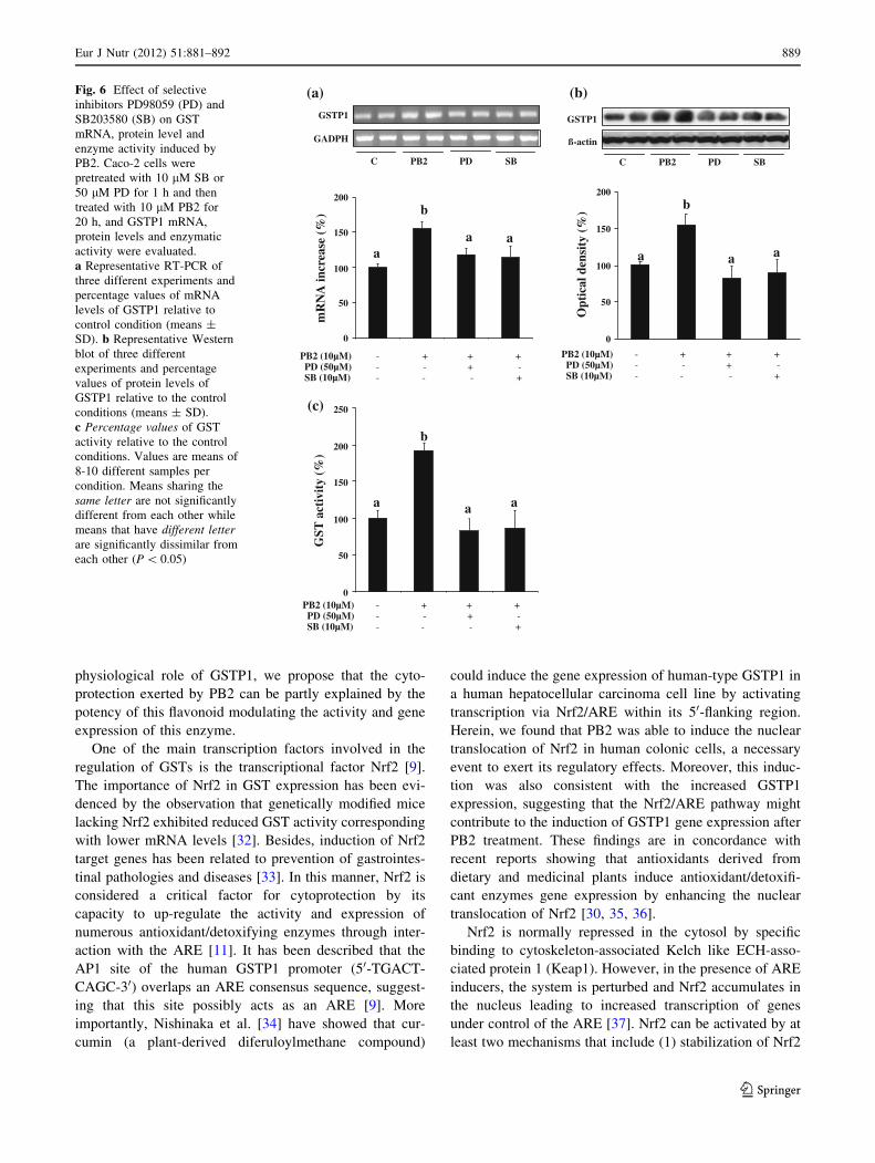
ERKs and p38 pathways are related to procyanidin
B2-induced Nrf-2 nuclear translocation and GSTP1
expression and enzymatic activity in Caco-2 cells
To confirm whether phosphorylation of ERKs and p38
pathways was essential for PB2-induced nuclear translo-
cation of Nrf2 and GSTP1 expression, we then examined
the effects of PD98059 (PD) and SB203580 (SB), specific
inhibitors for the ERKs and p38, respectively, on these
processes. Caco-2 cells were pretreated with PD and SB
during 1 h and then treated with 10 lM PB2 for 6 h.
Figure 5 shows that the inhibition of the two kinases sig-
nificantly reduced the nuclear translocation induced by
PB2. Similarly, treatment of Caco-2 cells with the ERKs
and p38 inhibitors completely suppresses the increase in
mRNA and protein levels and the activity of GSTP1
induced by PB2 (Fig. 6). Therefore, we can conclude that
phosphorylation of ERK and p38 seems to be essential for
PB2-induced Nrf2 translocation and GSTP1 expression and
enzymatic activity.
Role of ERKs and p38 pathways in cytoprotection
induced by procyanidin B2 in Caco-2 cells
Finally, we investigated whether the activation of ERK and
p38 induced by PB2 could be implicated in the cytopro-
tective effect of PB2 from t-BOOH-induced injury. Thus,
Caco-2 cells were pretreated with the specific inhibitors PD
and SB during 1 h and then treated with or without PB2 for
20 h. After that, the PB2-treated cells and the controls were
exposed to 400 lM t-BOOH, and the generation of ROS
and cell death were evaluated at 2 and 6 h, respectively. PD
or SB alone did not have any effect on ROS generation or
cell viability (data not shown). As shown in Fig. 7a,
(a)
C 1 2 4 8 20 hours
p-ERKs
ERKs
p-p38
p38
p-AKT
AKT
ß-actin
(b)
0
50
100
150
p-AKT/AKT
a a a
aaa
Opt
ical
den
sity
(%)
C 1 2 4 8 20 hours C 1 2 4 8 20 hours
C 1 2 4 8 20 hoursC 1 2 4 8 20 hours
p-JNKs
JNKs
0
50
100
150
p-JNKs/JNKs
Opt
ical
den
sity
(%)
a a aaaa
0
50
100
150
200
c
p-ERKs/ERKs
Opt
ical
den
sity
(%)
ab
c c c
0
50
100
150
200
250
p-p38/p38
Opt
ical
den
sity
(%
)
a
abab
b
cc
Fig. 4 Effect of PB2 on total
and phosphorylated levels of
AKT, ERKs, JNKs and p38.
Cells treated with 10 lM PB2
for 1, 2, 4, 8 and 20 h were
subjected to Western blot
analysis using phospho-specific
antibodies to AKT, ERKs, JNKs
and p38. The same cell lysates
were subjected to Western blot
analysis using the
corresponding non-phospho-
specific antibodies to detect
total AKT, ERKs, JNKs or p38.
a Bands are representative of
4-5 experiments. b Percentagevalues of the p-AKT/AKT,
p-ERKs/ERKs, p-JNKs/JNKs
and p-p38/p38 ratios relative to
the control condition (means ±
SD) are shown. Means sharing
the same letter are not
significantly different from each
other while means that have
different letter are significantly
dissimilar from each other
(P \ 0.05)
Eur J Nutr (2012) 51:881–892 887
123

pretreatment of cells with 10 lM PB2 for 20 h completely
blocked the formation of oxidative radicals caused by
t-BOOH, and this reduction was significantly abrogated by
the specific inhibitors of ERKs and p38. In the same line,
treatment of Caco-2 cells with PB2 significantly suppressed
the deleterious effect induced by t-BOOH on cell viability,
and the inhibition of both pathways blocked the cytopro-
tective effect of PB2 against cell death induced by the pro-
oxidant (Fig. 7b). Altogether, these findings suggest that
PB2 protects against oxidative injury by targeting the
ERKs and p38 signalling pathways.
Discussion
There is a substantial body of scientific literature that
supports a positive role of flavonoids on health [5]; nev-
ertheless, how specific flavonoids exert these benefits is
under intense investigation. Recent evidences suggest a
large number of potential mechanisms of action of phenolic
compounds in preventing disease, which may be beyond
their conventional antioxidant activities [21]. In particular,
it has been shown that they possess the ability to interact
with several cell signalling pathways and gene expression
factors to exert their biological activities [22]. The flavo-
noid PB2 has been identified to exhibit many clinically
relevant properties, such as anti-inflammatory and anti-
tumour activities in addition to its role as an antioxidant
[12, 23]. However, the mechanisms of its actions have not
been well defined. In this study, we show for the first time
that the protective effect of PB2 against oxidative injury in
Caco-2 cells was accompanied by the stimulation of ERKs
and p38 pathways, the activation of Nrf2 translocation and
the up-regulation of GSTP1 expression and enzymatic
activity.
Induction of cytoprotective enzymes possesses great
potential to effectively attenuate toxicity following expo-
sure to environmental toxicants [24]. The main physio-
logical role of the expression of the detoxification enzyme
GSTP1 is to protect cells against the adverse effects of
compounds such as toxins, carcinogens and free radical
oxidants [25]. The importance of GSTP1 in cytoprotection
is supported by numerous studies showing that increased
GSTP1 levels are critical for tissues vulnerable to oxidative
stress [20, 26]. It has been shown that mice deficient in
GSTP1 are more susceptible to cancer development
increasing the formation and multiplicity of adenomas in
lungs [27] and colon [28]. Altogether, GSTs have been
recognized as an important target for a number of che-
mopreventive and cytoprotective agents, including dietary
factors [29]. Consisting with this, in the present study, we
showed that long-term pretreatment of Caco-2 cells with
PB2 increased the activity of GSTP1 and effectively pre-
vented the augment in intracellular ROS and the cell death
induced by t-BOOH. Furthermore, the increased GST
activity was correlated with a parallel raise in its mRNA
and protein levels, indicating that PB2 can modulate
enzymes gene expression, as previously described with
other phenolic compounds [6, 30, 31]. Due to the
(a)
(b)
(c)
0
50
100
150
200
Opt
ical
den
sity
(%
)O
ptic
al d
ensi
ty (
%)
CytosolicNrf2
Nuclear Nrf2
0
50
100
150
200
250
300
350
PB2 (10µM)PD (50µM)SB (10µM)
PB2 (10µM)PD (50µM)SB (10µM)
bb
a
c
aab
b
c
- + + + - - + -- - - +
- + + + - - + -- - - +
C PB2 PD SB
CytosolicNrf2
GRB2
PARP
Nuclear Nrf2
Fig. 5 Effect of selective inhibitors PD98059 (PD) and SB203580
(SB) on nuclear and cytosolic Nrf2 levels in the presence of PB2.
Caco-2 cells were pretreated with 10 lM SB or 50 lM PD for 1 h and
then treated with 10 lM PB2 for 6 h. Nrf2 levels were determined by
Western blot in the cell nuclear or cytosolic compartment. a Bands
are representative of three different experiments. b Percentage valuesof nuclear and cytosolic Nrf2 levels relative to the control condition
(means ± SD). Means sharing the same letter are not significantly
different from each other while means that have different letter are
significantly dissimilar from each other (P \ 0.05)
888 Eur J Nutr (2012) 51:881–892
123
physiological role of GSTP1, we propose that the cyto-
protection exerted by PB2 can be partly explained by the
potency of this flavonoid modulating the activity and gene
expression of this enzyme.
One of the main transcription factors involved in the
regulation of GSTs is the transcriptional factor Nrf2 [9].
The importance of Nrf2 in GST expression has been evi-
denced by the observation that genetically modified mice
lacking Nrf2 exhibited reduced GST activity corresponding
with lower mRNA levels [32]. Besides, induction of Nrf2
target genes has been related to prevention of gastrointes-
tinal pathologies and diseases [33]. In this manner, Nrf2 is
considered a critical factor for cytoprotection by its
capacity to up-regulate the activity and expression of
numerous antioxidant/detoxifying enzymes through inter-
action with the ARE [11]. It has been described that the
AP1 site of the human GSTP1 promoter (50-TGACT-
CAGC-30) overlaps an ARE consensus sequence, suggest-
ing that this site possibly acts as an ARE [9]. More
importantly, Nishinaka et al. [34] have showed that cur-
cumin (a plant-derived diferuloylmethane compound)
could induce the gene expression of human-type GSTP1 in
a human hepatocellular carcinoma cell line by activating
transcription via Nrf2/ARE within its 50-flanking region.
Herein, we found that PB2 was able to induce the nuclear
translocation of Nrf2 in human colonic cells, a necessary
event to exert its regulatory effects. Moreover, this induc-
tion was also consistent with the increased GSTP1
expression, suggesting that the Nrf2/ARE pathway might
contribute to the induction of GSTP1 gene expression after
PB2 treatment. These findings are in concordance with
recent reports showing that antioxidants derived from
dietary and medicinal plants induce antioxidant/detoxifi-
cant enzymes gene expression by enhancing the nuclear
translocation of Nrf2 [30, 35, 36].
Nrf2 is normally repressed in the cytosol by specific
binding to cytoskeleton-associated Kelch like ECH-asso-
ciated protein 1 (Keap1). However, in the presence of ARE
inducers, the system is perturbed and Nrf2 accumulates in
the nucleus leading to increased transcription of genes
under control of the ARE [37]. Nrf2 can be activated by at
least two mechanisms that include (1) stabilization of Nrf2
0
50
100
150
200
250
0
50
100
150
200
0
50
100
150
200
PB2 (10µM) PD (50µM) SB (10µM)
a a a
b
ß-actin
GSTP1
GADPH
mR
NA
incr
ease
(%
)
Opt
ical
den
sity
(%
)
GST
act
ivit
y (%
)
(a)
(c)
PB2 (10µM) PD (50µM) SB (10µM)
PB2 (10µM) PD (50µM) SB (10µM)
- + + + - - + -- - - +
C PB2 PD SB C PB2 PD SB
aa a
b
a a a
b
- + + + - - + -- - - +
- + + + - - + -- - - +
GSTP1
(b)Fig. 6 Effect of selective
inhibitors PD98059 (PD) and
SB203580 (SB) on GST
mRNA, protein level and
enzyme activity induced by
PB2. Caco-2 cells were
pretreated with 10 lM SB or
50 lM PD for 1 h and then
treated with 10 lM PB2 for
20 h, and GSTP1 mRNA,
protein levels and enzymatic
activity were evaluated.
a Representative RT-PCR of
three different experiments and
percentage values of mRNA
levels of GSTP1 relative to
control condition (means ±
SD). b Representative Western
blot of three different
experiments and percentage
values of protein levels of
GSTP1 relative to the control
conditions (means ± SD).
c Percentage values of GST
activity relative to the control
conditions. Values are means of
8-10 different samples per
condition. Means sharing the
same letter are not significantly
different from each other while
means that have different letterare significantly dissimilar from
each other (P \ 0.05)
Eur J Nutr (2012) 51:881–892 889
123

via Keap1 cysteine thiol modification and (2) phosphory-
lation of Nrf2 by upstream kinases [38]. While Nrf2 sta-
bilization has been proposed as a major plausible
mechanism underlying activation of Nrf2, phosphorylation
of Nrf2 at specific serine and/or tyrosine residues also
represents another important event in cytoprotective gene
induction, which is modulated by many dietary chemo-
preventives [38]. Accordingly, in the present work, PB2
has no obvious influence on Nrf2 expression, suggesting
that Nrf2 translocation from the cytosol into the nucleus
could be caused by the dissociation of Nrf2 from Keap 1
repression through Nrf2 phosphorylation. Supporting this,
we found that pretreatment of Caco 2 cells with PB2
facilitated the phosphorylation of ERKs and p-38 MAPKs,
whereas AKT and JNKs were not activated. Furthermore,
up-regulation of GSTP-1 as well as induction of Nrf2
nuclear translocation by PB2 was remarkably inhibited by
selective inhibitors of ERKs and p-38 MAPK pathways,
indicating that the activation of both pathways by PB2
could be directly implicated in Nrf2 nuclear translocation
and GSTP1 expression. Similarly, other natural compounds
have been reported to induce cytoprotective enzymes in
different cell types via the activation of PI3K/AKT and/or
the MAPKs pathways [38]. Altogether, our results suggest
that PB2 could induce the expression levels and the activity
of GSTP1 through a mechanism involving nuclear trans-
location of Nrf2 and the activation of ERKs and p-38
MAPK pathways.
It should be pointed out that flavonoids acting as mild
pro-oxidants also modulate Nrf2 translocation. In this
case, stimulation of NO or ROS production by these
phenolic compounds could lead to Nrf2/ARE/EpRE-
mediated defence gene expression [39, 40]. However, we
have recently demonstrated that treatment of Caco-2 cells
with different concentrations of PB2 in the micromolar
range (1–20 lM) did not induce intracellular ROS pro-
duction and/or GSH depletion [16]. Thus, we can exclude
any involvement of oxidative stress in Nrf2 nuclear
translocation by PB2. As recently shown [22], flavonoids
can directly bind to ATP-binding sites, activation loops as
well as allosteric sites of protein kinases to regulate
multiple cell signalling pathways. Therefore, one of the
possible mechanisms of PB2 action could involve the
procyanidins directly binding to these protein kinases to
alter their phosphorylation state. Nevertheless, further
investigations are still required to fully elucidate the
detailed molecular mechanism underlying the activation
of Nrf2–ARE signalling pathway induced by natural
compounds [41].
To further demonstrate the important role of up-regu-
lation of GSTP1 in the protection exerted by PB2, we
then investigated the effect of specific inhibitors of ERKs
and p38 MAPK in ROS generation and cell death induced
by t-BOOH. We found that PB2-mediated cytoprotection
was significantly reduced in the presence of ERKs and
p38 MAPK inhibitors, indicating that the activation of
both pathways and the subsequent increase in GST
expression are involved in the protective mechanism
exerted by PB2. Our results added further convincing
evidence about the ability of PB2 to modulate signalling
pathways and gene expression to exert its biological
actions. For example, it has been recently shown that PB2
can attenuate the 4-hydroxynonenal-induced apoptosis in
0
2000
4000
6000
8000
10000
aa
b
c
b
(a)
(b)
Flu
ores
cen
ceu
nits
0
20
40
60
80
100
120
t-BOOH (400µM)PB2 (10µM)PD (50µM)SB (10µM)
% c
ell
viab
ilit
y
aa
b
c
d
- + + + + - - + + + - - - + -- - - - +
- + + + + - - + + + - - - + -- - - - +
t-BOOH (400µM)PB2 (10µM)PD (50µM)SB (10µM)
Fig. 7 Effect of PB2 and selective inhibitors PD98059 (PD) and
SB203580 (SB) on intracellular ROS generation and cell death
induced by t-BOOH. Caco-2 cells were pretreated with 10 lM SB or
50 lM PD for 1 h and then treated with 10 lM PB2 for 20 h. After
that, cells were further exposed to 400 lM t-BOOH for 2 and 6 h to
evaluate ROS generation and cell viability, respectively. a ROS
generation expressed in fluorescence units. b Percentage values of
cell death relative to the control condition. Data represent means ±
SD of ten different samples per condition. Means sharing the sameletter are not significantly different from each other while means that
have different letter are significantly dissimilar from each other
(P \ 0.05)
890 Eur J Nutr (2012) 51:881–892
123

PC12 cells by inhibiting the activation of the mitogen-
activated protein kinase kinase 4 [42]. Similarly, PB2
suppressed neoplastic transformation and COX-2 expres-
sion by modulating upstream signalling kinases impli-
cated in the activation of the transcription factors AP-1
and NF-kB in an epidermal cell line [15]. These findings
support our results showing that the beneficial effect of
PB2 against oxidative stress seems to be due not only to
its antioxidant properties but also to its ability to modu-
late specific proteins central to intracellular signalling
cascades. The induction of phase II enzymes, especially
GSTs, which detoxifies xenobiotics and potential carcin-
ogens results in protection against oxidative damage,
toxicity and chemical carcinogenesis, especially during
the initiation phase. Supporting this, it has been recently
shown that expression of GSTT2 is up-regulated in
colonic HT29 cells after pretreatment with an apple pol-
yphenol [43] and that this increase is related with pro-
tection from genotoxic stress [44]. Hence, we can assume
that the induction of GSTP1 may be one of the mecha-
nisms underlying the multiple actions of PB2 and could
explain, at least in part, the strong antioxidant and che-
mopreventive properties of this flavanol.
It is interesting to note that the in vitro effects exerted by
flavonoids cannot be extrapolated to in vivo function since
their bioavailability and metabolism must be taken into
account. However, recent studies have shown that PB2 is
stable during gastric transit, conserving their biological
activity inside the body [13, 45]. Therefore, the stability of
PB2 in the stomach and its very limited intestinal absorp-
tion suggest that it may have an important local function in
the gut, neutralizing the deleterious effects of oxidants and
carcinogenic compounds.
In summary, our study demonstrates for the first time
that PB2 improved the intestinal endogenous antioxidant
potential through the induction of the main detoxification
enzyme GSTP1 by a mechanism in which activation of
ERKs and p38MAPKs plays an essential role. In addition,
PB2 was identified as a potent inducer of Nrf2 transloca-
tion providing an argument for the involvement of this
transcription factor in the induction of GSTP1 by the
procyanidin. The results of the present study add further
evidence of the molecular mechanisms that allow PB2 to
exert protective effects and reaffirm its potential role as a
therapeutic agent in the treatment of oxidative stress-rela-
ted intestinal pathologies.
Acknowledgments This work was supported by the grant
AGL2007-64042/ALI and project CSD 2007-00063 from Programa
Consolider-Ingenio 2010 from the Spanish Ministry of Science and
Innovation (CICYT). I. Rodriguez-Ramiro is a predoctoral fellow of
the Consejo Superior de Investigaciones Cientıficas. M.A. Martın is
affiliated to CIBER de Diabetes y Enfermedades Metabolicas Aso-
ciadas (CIBERDEM), ISCIII, Madrid, Spain.
References
1. Aw TY (1999) Molecular and cellular responses to oxidative
stress and changes in oxidation-reduction imbalance in the
intestine. Am J Clin Nutr 70:557–565
2. Reuter S, Gupta SC, Chaturvedi MM, Aggarwal BB (2010)
Oxidative stress, inflammation, and cancer: how are they linked?
Free Radic Biol Med 49:1603–1616
3. Calder PC, Albers R, Antoine JM, Blum S, Bourdet-Sicard R,
Ferns GA, Folkerts G, Friedmann PS, Frost GS, Guarner F, Lovik
M, Macfarlane S, Meyer PD, M’ Rabet L, Serafini M, van Eden
W, van Loo J, Vas Dias W, Vidry S, Winklhofer-Roob BM, Zhao
J (2009) Inflammatory disease processes and interactions with
nutrition. Br J Nutr 101:1–45
4. Aron PM, Kennedy JA (2008) Flavan-3-ols: nature, occurrence
and biological activity. Mol Nutr Food Res 52:79–104
5. Crozier A, Jaganath IB, Clifford MN (2009) Dietary phenolics:
chemistry, bioavailability and effects on health. Nat Prod Rep
26:1001–1043
6. Masella R, Di Benedetto R, Varı R, Filesi C, Giovannini C (2005)
Novel mechanisms of natural antioxidant compounds in biolog-
ical systems: involvement of glutathione and glutathione-related
enzymes. J Nutr Biochem 16:577–586
7. Frova C (2006) Glutathione transferases in the genomics era: new
insights and perspectives. Biomol Engin 23:149–169
8. Tsuchida S, Sato K (1992) Glutathione transferases and cancer.
Crit Rev Biochem Mol Biol 27:337–384
9. Xia C, Hu J, Ketterer B, Taylor JB (1996) The organization of the
human GSTP1–1 gene promoter and its response to retinoic acid
and cellular redox status. Biochem J 313:155–161
10. Eggler AL, Gay KA, Mesecar AD (2008) Molecular mechanisms
of natural products in chemoprevention: Induction of cytopro-
tective enzymes by Nrf2. Mol Nutr Food Res 52:S84–S94
11. Nguyen T, Yang CS, Pickett CB (2004) The pathways and
molecular mechanisms regulating Nrf2 activation in response to
chemical stress. Free Radic Biol Med 37:433–441
12. Ramiro-Puig E, Castell M (2009) Cocoa: antioxidant and
immunomodulator. Br J Nutr 101:931–940
13. Serra A, Macia A, Romero MP, Valls J, Blade C, Arola L,
Motilva MJ (2010) Bioavailability of procyanidin dimers and
trimers and matrix food effects in vitro and in vivo models. Br J
Nutr 103:944–952
14. Visioli F, Bernaert H, Corti R (2009) Chocolate, lifestyle, and
health. Crit Rev Food Sci Nutr 49:299–312
15. Kang NJ, Lee KW, Lee DE, Rogozin EA, Bode AM, Lee HJ,
Dong Z (2008) Cocoa procyanidins suppress transformation by
inhibiting mitogen-activated protein kinase kinase. J Biol Chem
283:20664–20673
16. Rodrıguez-Ramiro I, Martın MA, Ramos S, Bravo L, Goya L
(2011) Comparative effects of dietary flavanols on antioxidant
defences and their response to oxidant-induced stress on Caco2
cells. Eur J Nutr. doi:10.1007/s00394-010-0139-2
17. Rodrıguez-Ramiro I, Ramos S, Bravo L, Goya L, Martın MA
(2011) Procyanidin B2 and a cocoa polyphenolic extract inhibit
acrylamide-induced apoptosis in human Caco-2 cells by pre-
venting oxidative stress and activation of JNK pathway. J Nutr
Biochem. doi:10.1016/j.jnutbio.2010.10.005
18. Goya L, Mateos R, Bravo L (2007) Effect of the olive oil phenol
hydroxytyrosol on human hepatoma HepG2 cells. protection
against oxidative stress induced by tert-butylhydroperoxide. Eur J
Nutr 46:70–78
19. Lima CF, Valentao PC, Andrade PB, Seabra RM, Fernandes-
Ferreira M, Pereira-Wilson C (2007) Phenolic compounds protect
HepG2 cells from oxidative damage: relevance of glutathione
levels. Chem Biol Interact 167:107–115
Eur J Nutr (2012) 51:881–892 891
123
20. Martın MA, Serrano AB, Ramos S, Pulido MI, Bravo L, Goya L
(2010) Cocoa flavonoids up-regulate antioxidant enzyme activity
via the ERK1/2 pathway to protect against oxidative stress-
induced apoptosis in HepG2 cells. J Nutr Biochem 21:196–205
21. Scalbert A, Johnson IT, Saltmarsh M (2005) Polyphenols: anti-
oxidants and beyond. Am J Clin Nutr 81:215S–217S
22. Hou DX, Kumamoto T (2010) Flavonoids as protein kinase
inhibitors for cancer chemoprevention: direct binding and
molecular modeling. Antioxid Redox Signal 13:691–719
23. Chou SC, Kaur M, Thompson JA, Agarwal R, Agarwal C (2010)
Influence of gallate esterification on the activity of procyanidin
B2 in androgen-dependent human prostate carcinoma LNCaP
cells. Pharm Res 27:619–627
24. Dinkova-Kostova AT, Talalay P (2008) Direct and indirect
antioxidant properties of inducers of cytoprotective proteins. Mol
Nutr Food Res 52:S128–S138
25. Lo HW, Ali-Osman F (2007) Genetic polymorphism and function
of glutathione S-transferases in tumor drug resistance. Curr Opin
Pharmacol 7:367–374
26. Reszka E, Wasowicz W, Gromadzinska J (2006) Genetic poly-
morphism of xenobiotic metabolising enzymes, diet and cancer
susceptibility. Br J Nutr 96:609–619
27. Ritchie KJ, Henderson CJ, Wang XJ, Vassieva O, Carrie D,
Farmer PB, Gaskell M, Park K, Wolf CR (2007) Glutathione
transferase pi plays a critical role in the development of lung
carcinogenesis following exposure to tobacco-related carcinogens
and urethane. Cancer Res 67:9248–9257
28. Ritchie KJ, Walsh S, Sansom OJ, Henderson CJ, Wolf CR (2009)
Markedly enhanced colon tumorigenesis in Apc(Min) mice
lacking glutathione S-transferase Pi. Proc Natl Acad Sci USA
106:20859–20864
29. Lii CK, Tsai CW, Wu CC (2006) Garlic allyl sulfides display
differential modulation of rat cytochrome P450 2B1 and the
placental form glutathione S-transferase in various organs.
J Agric Food Chem 54:5191–5196
30. Martın MA, Ramos S, Granado-Serrano AB, Rodrıguez-Ramiro
I, Trujillo M, Bravo L, Goya L (2010) Hydroxytyrosol induces
antioxidant/detoxificant enzymes and Nrf2 translocation via
extracellular regulated kinases and phosphatidylinositol-3-kinase/
protein kinase B pathways in HepG2 cells. Mol Nutr Food Res
54:956–966
31. Soyalan B, Minn J, Schmitz HJ, Schrenk D, Will F, Dietrich H,
Baum M, Eisenbrand G, Janzowski C (2011) Apple juice inter-
vention modulates expression of ARE-dependent genes in rat
colon and liver. Eur J Nutr 50:135–143
32. Chanas SA, Jiang Q, McMahon M, McWalter GK, McLellan LI,
Elcombe CR, Henderson CJ, Wolf CR, Moffat GJ, Itoh K,
Yamamoto M, Hayes JD (2002) Loss of the Nrf2 transcription
factor causes a marked reduction in constitutive and inducible
expression of the glutathione S-transferase Gsta1, Gsta2, Gstm1,
Gstm2, Gstm3 and Gstm4 genes in the livers of male and female
mice. Biochem J 365:405–416
33. Aleksunes LM, Manautou JE (2007) Emerging role of Nrf2 in
protecting against hepatic and gastrointestinal disease. Toxicol
Pathology 35:459–473
34. Nishinaka T, Ichijo I, Ito M, Kimura M, Katsuyama M, Iwata K,
Miura T, Terada T, Ch Yabe-Nishimura (2007) Toxicol Lett
170:238–247
35. Farombi EO, Shrotriya S, Na H-K, Kim SH, Surh Y-J (2008)
Curcumin attenuates dimethylnitrosamine-induced liver injury in
rats through Nrf2-mediated induction of heme oxygenase-1. Food
Chem Toxicol 46:1279–1287
36. Boettler U, Volz N, Pahlke G, Teller N, Kotyczka C, Somoza V,
Stiebitz H, Bytof G, Lantz I, Lang R, Hofmann T, Marko D
(2011) Coffees rich in chlorogenic acid or N-methylpyridinium
induce chemopreventive phase II-enzymes via the Nrf2/ARE
pathway in vitro and in vivo. Mol Nutr Food Res 55:798–802
37. Eggler AL, Gay KA, Mesecar AD (2009) Molecular mechanisms
of natural products in chemoprevention: Induction of cytopro-
tective enzymes by Nrf2. Mol Nutr Food Res 52:S84–S94
38. Surh Y-J, Kundu JK, Na H-K (2008) Nrf2 as a master redox
switch in turning on the cellular signaling involved in the
induction of cytoprotective genes by some chemopreventive
phytochemicals. Planta Med 74:1526–1539
39. Mann GE, Bonacasa B, Ishii T, Siow RCM (2009) Targeting the
redox sensitive Nrf2–Keap1 defense pathway in cardiovascular
disease: protection afforded by dietary isoflavones. Curr Opin
Pharmacol 9:139–145
40. Lima CF, Pereira-Wilson C, Rattan SIS (2011) Curcumin induces
heme oxygenase-1 in normal human skin fibroblasts through
redox signaling: relevance for anti-aging intervention. Mol Nutr
Food Res 55:430–442
41. Zhao CR, Gao ZH, Qua XJ (2010) Nrf2–ARE signaling pathway
and natural products for cancer chemoprevention. Cancer
Epidemiol 34:523–533
42. Cho ES, Jang YJ, Kang NJ, Hwang MK, Kim YT, Lee KW,
Lee HJ (2009) Cocoa procyanidins attenuate 4-hydroxynonenal-
induced apoptosis of PC12 cells by directly inhibiting mitogen-
activated protein kinase kinase 4 activity. Free Rad Biol Med
46:1319–1327
43. Veeriah S, Miene C, Habermann N, Hofmann T, Klenow S, Sauer J,
Bohmer F, Wolf S, Pool-Zobel BL (2008) Apple polyphenols
modulate expression of selected genes related to toxicological
defence and stress response in human colon adenoma cells. Int J
Cancer 122:2647–2655
44. Petermann A, Miene C, Schulz-Raffelt G, Palige K, Holzer J,
Glei M, Bohmer F-D (2009) GSTT2, a phase II gene induced by
apple polyphenols, protects colon epithelial cells against geno-
toxic damage. Mol Nutr Food Res 53:1245–1253
45. Appeldoorn MM, Vincken J-P, Gruppen H, Hollman PCH (2009)
Procyanidin dimers A1, A2, and B2 are absorbed without con-
jugation or methylation from the small intestine of rats. J Nutr
139:1469–1473
892 Eur J Nutr (2012) 51:881–892
123