proc. nati. acad. sci. - pnas · proc. nati. acad. sci. usa vol. 91, pp ... the outstanding feature...
TRANSCRIPT

Proc. Nati. Acad. Sci. USAVol. 91, pp. 10039-10043, October 1994Microbiology
Site-specific integration by adeno-associated virus is directed by acellular DNA sequenceCATHERINE GIRAUD, ERNEST WINOCOUR*, AND KENNETH I. BERNStDepartment of Microbiology, Hearst Microbiology Research Center, Cornell University Medical College, 1300 York Avenue, New York, NY 10021
Communicated by Daniel Nathans, June 9, 1994 (received for review April 8, 1994)
ABSTRACT Different regions of an 8.2-kb cloned DNAsegment containing the target for adeno-associated virus(AAV) integration in human chromosome 19ql3.3-qter(AAVSI locus) were subdoned in an Epstein-Barr virus-basedshuttle vector and propagated as episomes in a derivative ofthe293 human embryonic kidney cell line. Preferential recombi-nation with an infecting AAV genome was assessed by mea-suring the frequency ofrecombinants among the shuttle vectorsrecovered in Escherichia coli. The signals which direct recom-bination with theAAV genome were localized to a 510-nt regionat the 5' end of the 8.2-kb AAVSI DNA. Hence, the resultsindicate that site-specific integration of AAV is directed by aspecific DNA sequence on human chromosome 19. An unusualdegree of DNA heterogeneity in the recovered vector was alsoassociated with the 510 nt at the 5' end of AAVSI DNA,suggesting that theAAV chromosomal integration locus may beinvolved in genomic instability.
The outstanding feature of the life cycle of the humanparvovirus adeno-associated virus 2 (AAV) is the establish-ment of latent infection by integration into the cellulargenome. The significance of latent infection in the life cycleis perhaps best illustrated by the fact that replication of thevirus in cell culture normally requires coinfection with anunrelated helper virus, either adenovirus or herpesvirus (1).More recently it has been demonstrated that some lines ofcells can be made permissive for productive infection byAAV alone if the cells are exposed to a variety of genotoxicagents (2, 3). The conditions which render a cell permissivefor AAV replication, either infection by a cytolytic nuclearDNA virus or exposure to genotoxic agents, have led to amodel for the regulation of the viral life cycle (1). The modelhypothesizes that if the virus infects a healthy cell, a latentinfection will be established. The viral genome will remainrelatively quiescent in the integrated state until the cell isseverely stressed (e.g., by exposure to a genotoxic agent orvirus). Such stress will not only activate cellular repair genesbut also activate AAV gene expression, leading to rescuefrom the integrated state and viral replication.
Integration of the AAV genome occurs at a specific locus,19ql3.3-qter, in a majority of clones when several continu-ous lines of human cells are latently infected at a highmultiplicity of infection (4-7). The preintegration site onchromosome 19q has been cloned and a 4-kb region has beensequenced (6). The sequence, which shows no homology tothat of the AAV genome, has several notable features,including an overall G+C content of 65%. The 900 bases atthe 5' end have a G+C content of 82% and are close to thesite of recombination with viral sequences in a number ofclones. The same region also contains binding sites forseveral transcriptional transactivators. The 900 bases at the5' end and the 1500 bases at the 3' end have a larger thanexpected number of direct repeats, while the middle region
contains several open reading frames corresponding to anisolated cDNA from a A phage library of human foreskinfibroblasts (6). Most interestingly, the 5'-most 500 basescontain a dodecamer, (GCTC)3, also present in the invertedterminal repeat of the viral DNA, which has been identifiedas a binding site for the AAV Rep68/78 protein, the primaryviral regulatory protein (8, 9). The Rep68/78 protein is botha site-specific DNA nickase and a helicase and may beinvolved in the integration process (10-12). Indeed, AAVmodified to serve as a gene vector in a manner which removesthe rep gene no longer displays the same degree of sitespecificity of integration (R. J. Samulski, personal commu-nication; W. G. Kearns, personal communication).From the above, it appears that the sequence of the
preintegration site is likely to be a determining factor in thesite specificity ofAAV integration. However, the question ofwhether the sequence of the preintegration site is sufficient,in terms of cellular parameters, to determine site specificityhas not been answered. In this paper we describe experi-ments which directly address this issue by using an Epstein-Barr virus (EBV)-based shuttle vector which has been re-ported to be highly stable during propagation in mammaliancells (13). DNA containing theAAVSJ preintegration site wasplaced in the EBV-based shuttle vector and the ability ofthatDNA, when no longer in the normal context of the long (q)arm of chromosome 19, to direct AAV integration wasassessed. We find thatAAV integration is still directed by thepreintegration DNA even when it is in an episome. ThecellularDNA sequence directing integration was localized toa 510-nt region at the 5' end of the AAVSJ locus. Thepresence of this 510-nt sequence generates instability of theEBV episome in mammalian cells.
MATERIALS AND METHODSSubcdoning of AAVS1 in p220.2. The 8.2-kb AAVSJ DNA
containing the chromosome 19q preintegration locus wasderived by EcoRI digestion ofpRI-A (6) and subcloned in theSal I site of the EBV-based shuttle vector p220.2 (A. Beatonand K.I.B., unpublished data). p220.2 was a gift of B. Sugden(14, 15). The resulting construct, p220.2/AAVS1(kb 0-8.2),contains the AAVS1 sequence in the B orientation and wasused as starting material to generate the following set ofAAVS1 deletion constructs (Fig. 1): p220.2/AAVS1(kb0-0.51), p220.2/AAVS1(kb 0-1.6), p220.2/AAVS1(kb0-3.5), and p220.2/AAVS1(kb 0-4.4) were derived by diges-tion of p220.2/AAVS1(kb 0-8.2) with, respectively, Xba I(site in the p220.2 polylinker)/Pvu II, BamHI, Xba I/Kpn I,and Xba I/Pvu II (partial digest) and religation of the diges-tion products. The 5'-end deletion mutants p220.2/AAVS1(kb 0.51-1.6), p220.2/AAVS1(kb 0.51-3.5), andp220.2/AAVS1(kb 0.51-4.4) were derived, respectively,
Abbreviations: AAV, adeno-associated virus; EBV, Epstein-Barrvirus; EBNA-1, EBV-encoded nuclear antigen 1; SV40, simian virus40.*Permanent address: Department of Molecular Genetics and Virol-ogy, Weizmann Institute of Science, Rehovot, Israel.tTo whom reprint requests should be addressed.
10039
The publication costs of this article were defrayed in part by page chargepayment. This article must therefore be hereby marked "advertisement"in accordance with 18 U.S.C. §1734 solely to indicate this fact.

Proc. Natl. Acad. Sci. USA 91 (1994)
from p220.2/AAVSI(kb 0-1.6), p220.2/AAVS1(kb 0-3.5),and p220.2/AAVS1 (kb 0-4.4) digested with Pvu II/HindIII(a site in the p220.2 polylinker) and religation of the products.p220.2/AAVS1(kb 0-6) was derived by reinsertion of the6-kb HindIII fragment from p220.2/AAVS1(kb 0-8.2) intothe Xba I polylinker site of p220.2. p220.2/AAVS1 (kb5.2-8.2) was obtained by digestion of p220.2/AAVS1(kb0-8.2) with EcoRV plus Xba I and reinsertion of the resulting3-kb fragment into the Xba I polylinker site of p220.2. Thecontrol plasmids p220.2/RS-2 kb and p220.2/RS-6 kb con-tain, respectively, 2-kb and 6-kb DNAs that were selected atrandom from a human WI-38 (embryonic lung fibroblasts)genomic library by Xba I digestion and subcloned in the XbaI site of the p220.2 polylinker (R. M. Linden and K.I.B.,unpublished data). Neither of these control segments ofhuman DNA hybridize with AAVS] DNA.Propagation of EBV p220.2-Based Shuttle Vectors in C17
Cells. The C17 cell line (16), an EBNA-l-expressing deriva-tive of the 293 cell line, was kindly provided by R. F.Margolskee. C17 cells were grown in Dulbecco's modifiedEagle's medium (DMEM) with 10% fetal bovine serum andG418 at 600 Mg/ml (to maintain the EBNA-1 gene, which hadbeen cotransfected with pSV2neo). Each vectorDNA (10,ug)was transfected into C17 cells by calcium phosphate copre-cipitation (17) or by the cationic lipid N-[1-(2,3-dioleoyloxy)-propyl]-N, N, N-trimethylammonium methyl sulfate(DOTAP) (18, 19) according to the manufacturer's recom-mendations (Boehringer Mannheim). Three days later, thecells were split 1:10 into DMEM/10% fetal bovine serum withG418 (600 pg/ml) and hygromycin B (200 ,ug/ml). For eachconstruct, 10-30 hygromycin-resistant cell clones arisingfrom a single transfection experiment were pooled and pas-saged every 4 or 5 days at a split ratio of 1:10 to 1:20 in theconstant presence of both G418 and hygromycin. By passagelevel 2, all of the control mock-transfected C17 cells had beenkilled by hygromycin. Cells at passage levels 3-45 were usedfor AAV infection experiments.
Virus Infection. Subconfluent cells (5 x 10⁶) in 10-cm-diameter dishes were infected with a single cesium chloride-purified stock of AAV2 (2) at an input multiplicity of 20infectious units per cell (400 virus particles per cell). Controlcultures were either mock-infected or infected with simianvirus 40 (SV40) at 20 plaque-forming units per cell. Theinfected and control cells were incubated for 48 hr in G418/hygromycin medium.
Isolation of Extrachromosomal DNA. Forty-eight hoursafter infection, extrachromosomal DNA was isolated by thestandard Hirt procedure (20) except that the supernatantfraction, obtained after centrifugation at 48,000 x g for 30 minat 4°C, was digested with proteinase K (100 Mg/ml) for 4 hrat 42°C and for 24 hr at room temperature. After furtherdeproteinization by extractions with phenol and with chlo-roform containing 2% isoamyl alcohol, the nucleic acids werepurified by three successive ethanol (100%) precipitations inthe presence of 0.2 M NaCl. The extrachromosomal nucleicacids from a single plate of 107 cells were resuspended in 20,l of 10 mM Tris HCl/1 mM EDTA, pH 8.0.Recovery of Vectors in Escherichia coi and Colony Hybrid-
ization. One-seventh of the extrachromosomal DNA ex-tracted from 107 cells was transfected into the SURE (Strate-gene) strain of E. coli by electroporation (Bio-Rad E. coliPulser) at an applied voltage of 1.9 kV, using a 0.2-cm-gapcuvette (time constant, 4.0-4.2 msec). The electroporatedbacteria (40 ,l) were diluted in 1 ml of SOC medium (Strat-agene) and incubated at 37°C for 1 hr. As measured withpUC18 plasmid DNA, the efficiency of transfection was 5 x10⁹ ampicillin-resistant (AmpR) colonies per pg ofDNA. TheE. coli SURE strain (21) minimizes DNA rearrangements,due to mutations in the recB, uvrC, umuC, and sbcC genes.High-efficiency electroporation of the recombination-
deficient SURE strain ofE. coli was an important componentof the experimental protocol. One-tenth (100 sud) of theelectroporated bacteria were spread on LB/Amp agar platesto determine the number of AmpR colonies. A sample of theremaining fraction, adjusted to produce 500-1000 colonies,was diluted with 10 ml of LB medium and plated undersuction on a 8.2-cm-diameter BA85 nitrocellulose filter(Schleicher & Schuell) prewashed with LB medium. Thefilter was then incubated, bacteria-side-up, on top of LB/Amp agar until colonies 1-2 mm in diameter appeared.Imprints ("replicas") made from the "master" filters bystandard procedures were incubated on LB/Amp agar untilcolonies 0.5-1.0 mm in diameter developed. Colony hybrid-ization of the replica filters with a nonradioactive single-strandedAAVDNA probe (prepared from purified virus) wascarried out using the Genius system (Boehringer Mannheim).Hybridization was at 68TC for 18-20 hr.
A
22i2
7,
B
AAVS 1.%8 'J
5* .---,- J-
p220. AAV
;o .o. ib: c0,.5 .6:klD0.51 1351
, 5,2V m 2:
FIG. 1. Subcloning of the AAV preintegration locus AAVS1 inthe EBV-based vector p220.2. (A) The 8.9-kb p220.2 plasmid,containing the herpes simplex virus (HSV) thymidine kinase (tk)promoter and polyadenylylation signal (poly a) flanking a hygromy-cin-resistance gene for selection in mammalian cells. EBNA-1,EBV-encoded nuclear antigen 1. (B) Schematic representation ofthe8.2-kb AAVSI DNA sequence and the p220.2 vectors containingvarious segments of AAVS1. The AAVSJ sequence (top bold hori-zontal line) shows a CpG island (nt 1-900; stippled box), a regioncorresponding to a partial cDNA clone (nt 1620-2318; hatched box),a minisatellite repetitive DNA sequence (nt 3660-4021; open bar),crossover sites associated with AAV integration (arrows), and abinding site for the AAV rep gene products within nt 353-468(asterisk) (8). The restriction sites on the AAVSI sequence and on thepolylinker used for the construction of the p220.2 vectors are shownon the lower scale (thin and dotted horizontal line). p220.2/RS-2 kband p220.2/RS-6 kb contain random fragments of human DNA froma WI-38 library, inserted in the polylinker.
10040 Microbiology: Giraud et al.
0
-...IA

Proc. Natl. Acad. Sci. USA 91 (1994) 10041
Table 1. Recombination between AAV and the p220.2 shuttlevector carrying the 8.2-kb insert of chromosome 19qpreintegration DNA (AAVSI)
Passage Hours Recombinationno. of after AAV-hybridizing
moi cell line infection colonies, no./total %
20 4 48 16/500 3.28 48 13/500 2.6
17 48 8/540 1.545 48 22/1000 2.2
50 11 10 1/1305 0.0823 41/2142 1.946 65/2943 2.2
C17 cells carrying the vector p220.2/AAVS1(kb 0-8.2) wereinfected with AAV (at 20 or 50 infectious units per cell, as indicatedby the multiplicity of infection, moi) at the passage shown (no. of cellpassages after the start of hygromycin selection). At the designatedtimes after infection, extrachromosomal DNA was isolated andelectroporated into E. coli. Recombination is given by the percentageof AmpR colonies which hybridized with an AAV DNA probe.
RESULTSRecombination Hotspot Within the AAVS] Preintegration
Locus. To assess preferential recombination between theAAV genome and its integration site on the q arm of humanchromosome 19, the following experiment was devised.Defined portions of a cloned 8.2-kb DNA segment containingthe preintegration locus AAVSI were inserted into the EBV-based shuttle vector p220.2 and propagated as extrachromo-somal elements in C17 cells (Fig. 1). p220.2 contains the EBVlatent origin of replication oriP, the EBNA-1 gene, whoseproduct transregulates oriP, a hygromycin-resistance genefor the selection of C17 cells propagating this shuttle vector,and the pBR322 replication origin and ampicillin-resistancegene for selection in E. coli (14, 15). The C17 derivative of theadenovirus 5-transformed human 293 cell line expressesEBNA-1 constitutively, which may facilitate the propagationof p220.2 as an extrachromosomal nuclear episome (16, 22).C17 cells carrying p220.2/AAVS1 episomes were infectedwith AAV at various cell passage levels. Forty-eight hourslater, recombination between the infecting viral genome andthe resident p220.2/AAVS1 episomes was measured byelectroporating the extrachromosomal DNA fraction into therecombination-deficient SURE strain ofE. coli and by colonyhybridization with a single-stranded AAV virion DNA probe.The number of AAV-positive AmpR colonies expressed as apercentage of the total number of AmpR colonies was takenas the measure of the frequency of recombination. Theresults are presented in Tables 1 and 2.
p220.2 containing the entire 8.2-kb AAVS] DNA [p220.2/AAVS1(kb 0-8.2)], when propagated as an episome in C17cells, recombined with the infecting AAV genome at everycell passage tested (Table 1). The recombination values, inthe range 1.5-3.2%, did not decrease with increasing cell-passage levels, indicating that the recombination targets onp220.2/AAVS1 were conserved during continuous passageof the cells. As a control, we examined the recombinogeniccapacity of p220.2 vectors containing 2-kb and 6-kb randomsegments of human DNA. No evidence for recombinationbetween those vectors propagated in C17 cells and theinfecting AAV genome was obtained (>4000 AmpR colonieswere screened). Similarly, no recombination between theparental p220.2 vector and the infecting AAV genome wasdetected. The time course of accumulation of recombinantsshowed that near-maximum yields were obtained by 23 hrpostinfection (Table 1). Only a rare recombinant was de-tected at 10 hr postinfection; this observation supports ourassumption that the primary recombination events occurredin C17 cells and not in the recombination-deficient E. coli
Table 2. Recombination between AAV and the p220.2 shuttlevector containing different portions of AAVS1 DNA
AAVS] insert Passage Recombinationin p220.2 no. of AAV-hybridizingvector, kb cell line colonies, no./total %
0-6 11 3/240 1.20-4.4 14 1/504 0.20-1.6 9 8/957 0.80-1.6 12 5/288 1.70-0.51 11 17/2140 0.80-0.51 12 2/459 0.4
0.51-4.4 12 0/1000 <0.10.51-4.4 14 0/1640 <0.060.51-3.5 14 0/2740 <0.040.51-1.6 9 0/3800 <0.030.51-1.6 12 0/648 <0.155.2-8.2 11 0/4000 <0.03
See Fig. 1 for the derivation of the various AAVSI inserts.
SURE bacteria. A time interval of 48 hr postinfection waschosen, however, for the routine isolation of the shuttlevectors from the infected cells.We next investigated the recombinogenic potential of
different regions of the 8.2-kb AAVS] DNA inserted into thep220.2 vector (Table 2). p220.2 vectors carrying the AAVS]segments spanning kb 0-6.0, 0-4.4, 0-1.6, and 0-0.51 allrecombined with AAV. However, deletion ofthe 510 nt at the5' end of AAVSJ completely abrogated the capacity of theAAVSJ DNA segments (kb 0.51-4.4, 0.51-3.5, 0.51-1.6) torecombine with the AAV genome. In these cases, not a singlecolony out of -9800 AmpR colonies screened reacted withtheAAV DNA probe. A 3-kb segment at the 3' end ofAAVS](kb 5.2-8.2) did not recombine with the AAV genome (=z4000AmpR colonies were screened). Together, >14,000 AmpRcolonies screened gave a negative hybridization result, high-lighting the specificity of the positive reactions betweenvectors carrying the 5'-most 510 nt of AAVSJ and AAVDNA. These data clearly localized the recombinogenic sig-nals to 510 nt at the 5' end of AAVSL.To determine whether the AAVSI hotspot for AAV re-
combination is specific for that virus, we infected the C17lines propagating p220.2 vectors containingAAVS1 DNA (kb0-0.51, 0-1.6, and 0-8.2) with SV40 and screened, by colonyhybridization, for p220.2/AAVS1/SV40 recombinantsamong the vectors recovered in E. coli. No recombinantswere detected out of the 928 colonies screened. The 293parent line of C17 is permissive for SV40 infection, asdetermined by replication of the viral DNA (23). It appears,therefore, that the AAVS] hotspot for AAV recombination isnot a comparable hotspot for SV40 recombination.Episomes Containiog the 5' End of AAVS1 Are Unstable.
During the course ofthe above experiments, it was noted thatthe recovery of p220.2 vectors in E. coli, measured by thenumber ofAmpR colonies resulting from the electroporationof the extrachromosomal DNA fraction of 107 C17 cells,varied widely, depending upon the segment ofAAVS] DNApresent in the vector (Table 3). When the insert in p220.2 wasderived from the 3' end ofAAVS] (kb 5.2-8.2), the recoveryin E. coli (e.g., 120,000 AmpR colonies per 107 C17 cells;Table 3, Exp. 1) was comparable to that of p220.2 vectorscontaining a 2-kb random segment of human DNA (140,000AmpR colonies per 107 C17 cells) or no DNA insert (184,000colonies per 107 C17 cells). In contrast, when the insertscontained the 5' end of AAVSI (kb 0-8.2, 0-4.4, 0-1.6,0-0.51) the efficiency of recovery in E. coli was reduced bya factor of 50-100. Deletion of the 510 nt at the 5' end (kb0.51-1.6, 0.51-3.5, or 0.51-4.4) showed an increase of the
Microbiology: Giraud et al.

Proc. Natl. Acad. Sci. USA 91 (1994)
Table 3. Recovery of p220.2 vectors in E. coli depends upon theDNA of their inserts
AmpR colonies perExp. Insert in p220.2 vector Infection extract of 107 cells
1 AAVSIkb 0-8.2 AAV 2,345kbO-8.2 SV40 2,200kb 0-1.6 AAV 2,211kb 0-0.51 AAV 4,958kb 0.51-1.6 AAV 13,400kb 5.2-8.2 AAV 120,000
Random human DNA, 2 kb SV40 140,000None AAV 184,000
2 AAVS]kb 0-8.2 AAV 3,015kb 0-1.6 AAV 2,144kb 0-0.51 AAV 3,417kb 0-0.51 SV40 3,484kb 0-0.51 None 1,600kb 0.51-1.6 AAV 4,824kb 0.51-1.6 None 5,829kb 0.51-4.4 AAV 26,800kb 0.51-4.4 None 26,000kb 5.2-8.2 None 268,000
3 AAVS]kb 0-4.4 AAV 1,698kb 0-4.4 None 1,447kb 0-1.6 None 1,206kb 0.51-4.4 AAV 19,025kb0.51-3.5 AAV 31,356kb 0.51-3.5 None 31,658
efficiency of recovery by a factor 5-10. These data show thatthe recovery of the shuttle vectors in E. coli decreases in thepresence of sequences from the first 4.4 kb at the 5' end ofAAVS1. This negative influence on the recovery of thevectors in E. coli was observed irrespective of the infectingvirus (AAV or SV40), as well as in mock-infected cell lines.
Restriction digests of p220.2/AAVS1 vectors recoveredfrom mock-infected C17 cells by electroporation of the ex-trachromosomal DNA fraction into the recombination-deficient SURE strain of E. coli are shown in Fig. 2. In eachcase, five independent AmpR colonies were isolated and theplasmids were analyzed by restriction analysis. When the5'-most 510 nt of AAVS1 were present in the vector, prop-agation as an episome in C17 cells generated an unusualdegree of heterogeneity, as judged by the restriction digestsof the recovered vectors compared with those of the inputp220.2/AAVS1 DNAs transfected into C17 cells (Fig. 2 a-c).In striking contrast, when the 510 nt at the 5' end ofAAVS]were deleted from the insert in p220.2, the digests of therecovered vectors were identical to those of the parentalDNAs transfected into C17 cells (Fig. 2 d and e), as well aswith p220.2 plasmids carrying random human sequences(Fig. 2]). Interestingly, the altered pattern, compared withthat of the parental plasmid, of episomes rescued from thep220.2/AAVS1(kb 0-1.6) and -(kb 0-0.51) cell lines wasidentical for several of the clones (Fig. 2 b and c).
DISCUSSIONThe site-specific integration of AAV into the q arm ofchromosome 19 in established lines of human cells implies aspecific recognition signal, possibly encoded in the chromo-somal DNA sequence or possibly acting indirectly via theconformation or some unique activity of the integrationlocus. A major conclusion of the results reported here is thatAAV integration is directed by signals encoded in the primaryDNA sequence of chromosome 19. These signals were lo-calized to a 510-bp DNA segment at the 5' end of the AAVS]
aA1 1 ) 1> Id :)'
bNA11 9) 0'q z>
C
d eMl 2 c'I C1 5
FIG. 2. Restriction pattern of p220.2 vectors rescued from mock-infected C17 cell lines. (a and c) Cla I/HindIII digest offive plasmidsrecovered from p220.2/AAVS1(kb 0-8.2) and p220.2/AAVS1(kb0-0.51) cell lines, respectively. (b, e, and f) Pst I digest of fiveplasmids recovered from p220.2/AAVS1(kb 0-1.6), p220.2/AAVS1(kb 0.51-1.6), and p220.2/RS-6 kb cell lines, respectively. (d)BamHI/Sal I digest of five plasmids recovered from a p220.2/AAVS1(kb 5.2-8.2) cell line. Lane C, the parental plasmid DNA(before transfection in C17 cells) digested by the same enzymes; laneM, DNA molecular size marker (MIII or MVII of BoehringerMannheim).
preintegration locus. Virus-cell recombinantjunctions in oneclone of latently infected human cells have been mapped toAAVS1 nt 1026-1030 and 1144-1146 (6); in several otherlatently infected human cell lines, the recombinant junctionswere mapped to the same region, plus or minus 100 nt (7).Hence, the data suggest that the signals directing AAVintegration are to the 5' side of the actual recombinationcrossover points.A recent discovery of particular interest is that the 5' end
ofAAVS] DNA contains a binding site for the AAV rep geneproducts, Rep 68 and Rep 78; this binding site is locatedwithin aDNA segment bordered by nt 353-468 (8) within the510-bp region that directs recombination withAAV (Fig. 3A).Rep68/78 has been shown to bind specifically to AAV
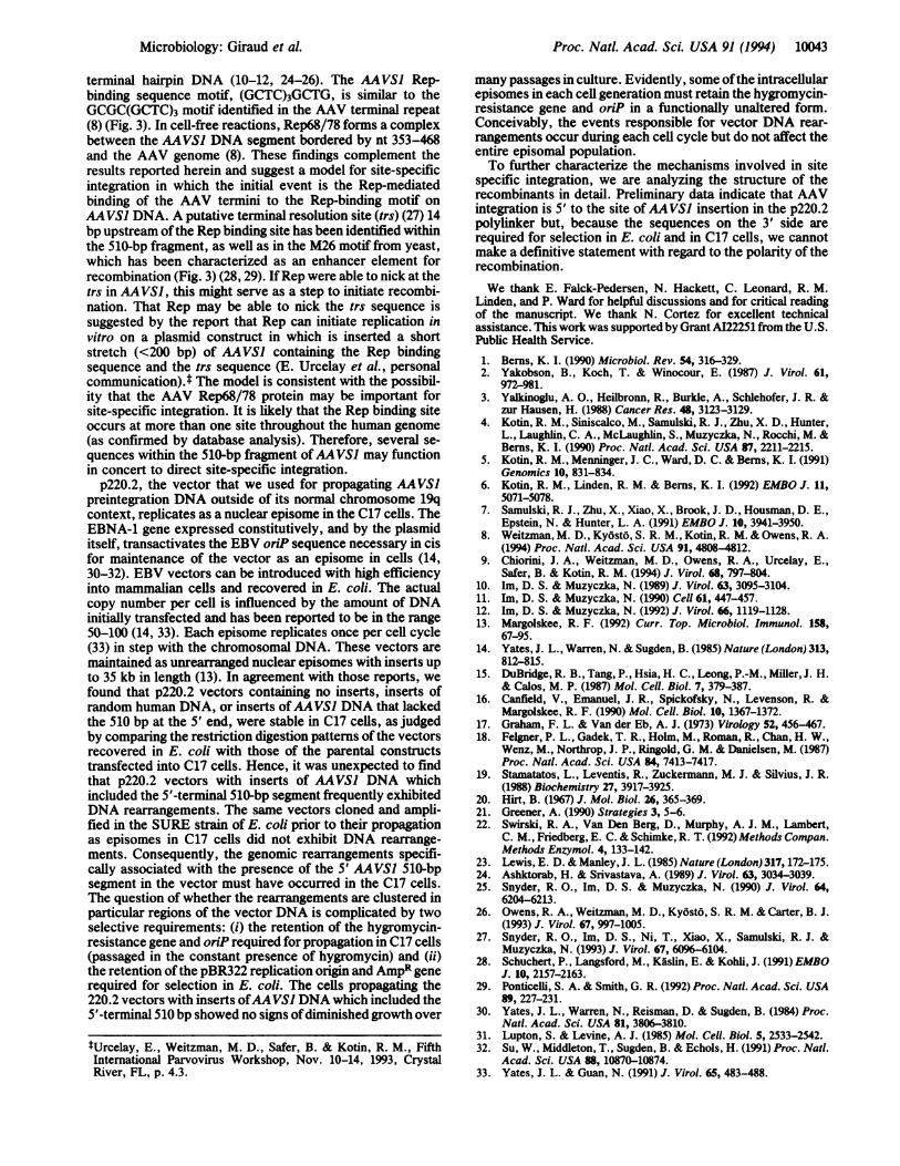
a AAvs
5' vM 26 motif putative trs Rep binding site
?VJ , ya'
M 26 motif putative trs Rep binding site 3,CC C=GGCGG1&GGGGCTCCGGGCGCGTC GCTCIGCTCIGCTC GCTGI GG275 381 405 422
b AAv5' M 26 motif tiS Rep binding site 3'cOAT ACGTAATC=TGGAGTTGGCCACTCCCTCTCT IGCGCGCTCIGCTCIGGOCT AC184 130 85
FIG. 3. (a) Characteristics of the 510 nt at the 5' end of AAVSIpreintegration sequence. The 510 nt are part of a CpG island (nt1-900) and a region with high frequencies of short repeats (6). Bindingelements for upstream binding factor 1 (UBF1) or the reversecomplement (CGGCC, GGCCG) and the Spi binding sites areindicated by U and S, respectively (6). The cAMP-responsiveelement (TGACGTCA) is designated by CRE (6). The enlarged partof the sequence shows the Rep binding site (8), a putative terminalresolution site (trs), and the M26 motif from yeast (28). (b) AAV2DNA sequence; the nucleotide numbers refer to the complementarystrand of the AAV sequence. trs is indicated by a vertical arrow (27).
10042 Microbiology: Giraud et al.
Proc. Natl. Acad. Sci. USA 91 (1994) 10043
terminal hairpin DNA (10-12, 24-26). The AAVS] Rep-binding sequence motif, (GCTC)3GCTG, is similar to theGCGC(GCTC)3 motif identified in the AAV terminal repeat(8) (Fig. 3). In cell-free reactions, Rep68/78 forms a complexbetween the AAVS1 DNA segment bordered by nt 353-468and the AAV genome (8). These findings complement theresults reported herein and suggest a model for site-specificintegration in which the initial event is the Rep-mediatedbinding of the AAV termini to the Rep-binding motif onAAVS] DNA. A putative terminal resolution site (trs) (27) 14bp upstream of the Rep binding site has been identified withinthe 510-bp fragment, as well as in the M26 motif from yeast,which has been characterized as an enhancer element forrecombination (Fig. 3) (28, 29). IfRep were able to nick at thetrs in AAVS], this might serve as a step to initiate recombi-nation. That Rep may be able to nick the trs sequence issuggested by the report that Rep can initiate replication invitro on a plasmid construct in which is inserted a shortstretch (<200 bp) of AAVS1 containing the Rep bindingsequence and the trs sequence (E. Urcelay et al., personalcommunication).t The model is consistent with the possibil-ity that the AAV Rep68/78 protein may be important forsite-specific integration. It is likely that the Rep binding siteoccurs at more than one site throughout the human genome(as confirmed by database analysis). Therefore, several se-quences within the 510-bp fragment of AAVS1 may functionin concert to direct site-specific integration.
p220.2, the vector that we used for propagating AAVS1preintegration DNA outside of its normal chromosome 19qcontext, replicates as a nuclear episome in the C17 cells. TheEBNA-1 gene expressed constitutively, and by the plasmiditself, transactivates the EBV oriP sequence necessary in cisfor maintenance of the vector as an episome in cells (14,30-32). EBV vectors can be introduced with high efficiencyinto mammalian cells and recovered in E. coli. The actualcopy number per cell is influenced by the amount of DNAinitially transfected and has been reported to be in the range50-100 (14, 33). Each episome replicates once per cell cycle(33) in step with the chromosomal DNA. These vectors aremaintained as unrearranged nuclear episomes with inserts upto 35 kb in length (13). In agreement with those reports, wefound that p220.2 vectors containing no inserts, inserts ofrandom human DNA, or inserts ofAAVS] DNA that lackedthe 510 bp at the 5' end, were stable in C17 cells, as judgedby comparing the restriction digestion patterns of the vectorsrecovered in E. coli with those of the parental constructstransfected into C17 cells. Hence, it was unexpected to findthat p220.2 vectors with inserts of AAVS1 DNA whichincluded the 5'-terminal 510-bp segment frequently exhibitedDNA rearrangements. The same vectors cloned and ampli-fied in the SURE strain of E. coli prior to their propagationas episomes in C17 cells did not exhibit DNA rearrange-ments. Consequently, the genomic rearrangements specifi-cally associated with the presence of the 5' AAVS1 510-bpsegment in the vector must have occurred in the C17 cells.The question of whether the rearrangements are clustered inparticular regions of the vector DNA is complicated by twoselective requirements: (i) the retention of the hygromycin-resistance gene and oriP required for propagation in C17 cells(passaged in the constant presence of hygromycin) and (ii)the retention of the pBR322 replication origin and AmpR generequired for selection in E. coli. The cells propagating the220.2 vectors with inserts ofAAVS] DNA which included the5'-terminal 510 bp showed no signs ofdiminished growth over
many passages in culture. Evidently, some ofthe intracellularepisomes in each cell generation must retain the hygromycin-resistance gene and oriP in a functionally unaltered form.Conceivably, the events responsible for vector DNA rear-rangements occur during each cell cycle but do not affect theentire episomal population.To further characterize the mechanisms involved in site
specific integration, we are analyzing the structure of therecombinants in detail. Preliminary data indicate that AAVintegration is 5' to the site ofAAVSI insertion in the p220.2polylinker but, because the sequences on the 3' side arerequired for selection in E. coli and in C17 cells, we cannotmake a definitive statement with regard to the polarity of therecombination.We thank E. Falck-Pedersen, N. Hackett, C. Leonard, R. M.
Linden, and P. Ward for helpful discussions and for critical readingof the manuscript. We thank N. Cortez for excellent technicalassistance. This work was supported by Grant AI22251 from the U.S.Public Health Service.1. Berns, K. I. (1990) Microbiol. Rev. 54, 316-329.2. Yakobson, B., Koch, T. & Winocour, E. (1987) J. Virol. 61,
972-981.3. Yalkinoglu, A. O., Heilbronn, R., Burkle, A., Schlehofer, J. R. &
zur Hausen, H. (1988) Cancer Res. 48, 3123-3129.4. Kotin, R. M., Siniscalco, M., Samulski, R. J., Zhu, X. D., Hunter,
L., Laughlin, C. A., McLaughlin, S., Muzyczka, N., Rocchi, M. &Berns, K. I. (1990) Proc. NatI. Acad. Sci. USA 87, 2211-2215.
5. Kotin, R. M., Menninger, J. C., Ward, D. C. & Berns, K. I. (1991)Genomics 10, 831-834.
6. Kotin, R. M., Linden, R. M. & Berns, K. I. (1992) EMBO J. 11,5071-5078.
7. Samulski, R. J., Zhu, X., Xiao, X., Brook, J. D., Housman, D. E.,Epstein, N. & Hunter, L. A. (1991) EMBO J. 10, 3941-3950.
8. Weitzman, M. D., Kyosto, S. R. M., Kotin, R. M. & Owens, R. A.(1994) Proc. Natl. Acad. Sci. USA 91, 4808-4812.
9. Chiorini, J. A., Weitzman, M. D., Owens, R. A., Urcelay, E.,Safer, B. & Kotin, R. M. (1994) J. Virol. 68, 797-804.
10. Im, D. S. & Muzyczka, N. (1989) J. Virol. 63, 3095-3104.11. Im, D. S. & Muzyczka, N. (1990) Cell 61, 447-457.12. Im, D. S. & Muzyczka, N. (1992) J. Virol. 66, 1119-1128.13. Margolskee, R. F. (1992) Curr. Top. Microbiol. Immunol. 158,
67-95.14. Yates, J. L., Warren, N. & Sugden, B. (1985) Nature (London) 313,
812-815.15. DuBridge, R. B., Tang, P., Hsia, H. C., Leong, P.-M., Miller, J. H.
& Calos, M. P. (1987) Mol. Cell. Biol. 7, 379-387.16. Canfield, V., Emanuel, J. R., Spickofsky, N., Levenson, R. &
Margolskee, R. F. (1990) Mol. Cell. Biol. 10, 1367-1372.17. Graham, F. L. & Van der Eb, A. J. (1973) Virology 52, 456-467.18. Feigner, P. L., Gadek, T. R., Holm, M., Roman, R., Chan, H. W.,
Wenz, M., Northrop, J. P., Ringold, G. M. & Danielsen, M. (1987)Proc. Natl. Acad. Sci. USA 84, 7413-7417.
19. Stamatatos, L., Leventis, R., Zuckermann, M. J. & Silvius, J. R.(1988) Biochemistry 27, 3917-3925.
20. Hirt, B. (1967) J. Mol. Biol. 26, 365-369.21. Greener, A. (1990) Strategies 3, 5-6.22. Swirski, R. A., Van Den Berg, D., Murphy, A. J. M., Lambert,
C. M., Friedberg, E. C. & Schimke, R. T. (1992) Methods Compan.Methods Enzymol. 4, 133-142.
23. Lewis, E. D. & Manley, J. L. (1985) Nature (London) 317,172-175.24. Ashktorab, H. & Srivastava, A. (1989) J. Virol. 63, 3034-3039.25. Snyder, R. O., Im, D. S. & Muzyczka, N. (1990) J. Virol. 64,
6204-6213.26. Owens, R. A., Weitzman, M. D., Kyost6, S. R. M. & Carter, B. J.
(1993) J. Virol. 67, 997-1005.27. Snyder, R. O., Im, D. S., Ni, T., Xiao, X., Samulski, R. J. &
Muzyczka, N. (1993) J. Virol. 67, 6096-6104.28. Schuchert, P., Langsford, M., Kaslin, E. & Kohli, J. (1991) EMBO
J. 10, 2157-2163.29. Ponticelli, S. A. & Smith, G. R. (1992) Proc. Natl. Acad. Sci. USA
89, 227-231.30. Yates, J. L., Warren, N., Reisman, D. & Sugden, B. (1984) Proc.
Natl. Acad. Sci. USA 81, 3806-3810.31. Lupton, S. & Levine, A. J. (1985) Mol. Cell. Biol. 5, 2533-2542.32. Su, W., Middleton, T., Sugden, B. & Echols, H. (1991) Proc. Natl.
Acad. Sci. USA 88, 10870-10874.33. Yates, J. L. & Guan, N. (1991) J. Virol. 65, 483-488.
tUrcelay, E., Weitzman, M. D., Safer, B. & Kotin, R. M., FifthInternational Parvovirus Workshop, Nov. 10-14, 1993, CrystalRiver, FL, p. 4.3.
Microbiology: Giraud et A