principles of pressure management - iswp · pdf fileprinciples of pressure management douglas...
TRANSCRIPT
Principles of Pressure Management
Douglas A. Hobson, Ph.D.
RERC on Wheeled Mobility*Department of Rehabilitation Science and Technology
University of Pittsburgh
*support by: the National Institute on Disability and Rehabilitation Research
This presentation is intended to provide an overview of the basic principles of pressure sorecausation and the procedures used for clinical management, as viewed from the assistivetechnology perspective.
Hobson, D.A. September 1999
Pressure Sore Incidence and Costs
• 40% occur in hospital- (lying ulcers-sacrum,heels, elbows)
• 30% occur post discharge (sitting ulcers -sacrum, ischium)
• Cause of mortality in about 4% of SCI people• Repair costs-$15-60k/ incident• Social/psychological costs immeasurable!!
Pressure sores or decubitus ulcers is still a costly, unresolved problem.

Hobson, D.A. September 1999
Populations at Risk
• Spinal Cord Injured• Immobile Elderly• Diabetics• ALS, MS, MD• Immobility is greatest risk factor• Risk assessment scales have been developed
(e.g., Braden scale, Norton scale)
It still can not be predicted with a high degree of certainty which individuals are most likely todevelop pressure sores.

Hobson, D.A. September 1999
The Etiology of Pressure Ulcers(prime factors)
• Local pressure above capillary (32 mmHg)reduces or occludes blood flow and oxygen totissues (ischemia).
• Higher pressures usually occur over bonyareas: ischial tuberosities, trochanters, andsacrum.
• If pressure sustained over pressure/timethreshold, normal recovery will not occur.

Pressure Tolerance Guidelines
Reswick and Rogers - (1976)Reswick and Rogers - (1976)
Reswick and Rogers - (1976)
Rewick and Rogers demonstrated that there seemed to be a relationship between the maximumpressure being experienced by the supporting tissues and the time over which that maximumpressure was applied. If the pressure/time index fell above the curve, subjects exhibited pressuresore histories. If it fell in the acceptable zone, they did not shoe pressure problems. This classicstudy has served as the basis for clinical management practices until this day.

Hobson, D.A. September 1999
The Etiology of Pressure Ulcers(prime factors)
• If repeated assault occurs, the breakdownprocess will begin.
• Deep tissue distortion due to shear forcesalso major contributing factor.
• Surface sores can result from repeatedsurface friction/abrasion.
The exact effects of shear forces and resulting distortion of deeper tissues remains largelyunknown and subject of current research studies.

Hobson, D.A. September 1999
Other Contributing Factors• Immobility- close correlation between onset
and frequency of movement.• Loss of sensation-evidence that denervated
tissue has reduced nutrition and oxygenperfusion.
• Body Type- does not seem to be anyrelationship between body-type and deepulcers. Surface friction may increase withheavier people.
Hobson, D.A. September 1999
Other Contributing Factors
• Nutrition-inadequate dietary intake can resultin muscle atrophy, anemia, low protein levelsand vitamin C deficiency– reducing skinintegrity.
• Infection-increased metabolic rate, higheroxygen demand endangers ischemic tissue.
Hobson, D.A. September 1999
Other Contributing Factors
• Age-normal loss of elasticity, muscle atrophy,& reduced tone increases vulnerability.
• Posture-pelvic/spinal deformity will causelocalized pressure increases.
• Microclimate- at the skin/seat interface• temperature, moisture, incontinence
• Impact injuries/abrasions.

Typical Superficial Ulcer Development
Moistureand Heat
Friction andShear Forces
SuperficialPressure Ulcer
Poor Nutrition
Infection
Extrinsic
Intrinsic
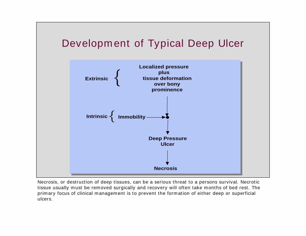
Development of Typical Deep Ulcer
tissue deformation over bony
prominence
Immobility
Deep Pressure Ulcer
Extrinsic
Intrinsic
Localized pressureplus
Necrosis
Necrosis, or destruction of deep tissues, can be a serious threat to a persons survival. Necrotictissue usually must be removed surgically and recovery will often take months of bed rest. Theprimary focus of clinical management is to prevent the formation of either deep or superficialulcers.

Five Clinical Stages ofPressure Sore Development
• Stage I - blanching of reddened areaexperiencing reactive hyperemia– circulation intact, redness disappears in reasonable
time (1-2 hrs)
• Stage II - blanching does not occur, rednessremains
• Stage III - ulceration progresses beyond thedermis to subcutaneous tissue– redness remains around edges, hardening of tissue
occurs

Hobson, D.A. September 1999
Five Clinical Stages ofPressure Development (cont’d)
• Stage IV - ulceration progresses to fat layerand muscle becomes swollen.
• Stage V - necrosis (dying tissue) penetratesdeep fascia, muscle and sometimes bone.Surgical repair necessary, requires 2-6months of inactivity.

Hobson, D.A. September 1999
Pressure Ulcer Risk Factors
Extrinsic Factors Intrinsic Factors
Excessive Uniaxial Pressure
Friction and Shear Forces
Heat
Moisture
Immobility
Poor Nutrition
Age
Sensory LossPressure
Ulcer
Incontinence
Disease
Body Type
Infection
Impact Injury
Posture
This diagram presents a summary of the multiple factors that are thought to contribute topressure ulcer formation.

Principles ofClinical Pressure Management
• Maximize the surface area: decrease thepressure on any one location (peak pressures)
• Redistribute body weight: use support surfaceshape and materials with required properties.
• Minimize asymmetries: i.e., unequal loading ofpelvic structures and tissues.
• Training for pressure relief: i.e., regularweight relieving movements
The first three above are goals that can be achieved by the selection or design of an appropriatebody support surface (wheelchair seat cushion).

Hobson, D.A. September 1999
Principles of Clinical PressureManagement (cont’d)
• Dietary instruction: critical to both goodhealth and skin integrity
• Instruction for lifting/ transferring: wheneverpossible person should instruct care-givers
• Personal hygiene and skin care: veryimportant!
Clearly the person themselves must be prepared to play a leading role in the prevention ofpressure ulcers through strict adherence to several basics of self-care.

Skin Viability Measurement Techniques
• Skin blanching• Measurement of interface pressure (single
site and mapping)• Thermography• Oxygenation of blood flow• Measurement of tissue deformation - Brienza• Measurement tissue stiffness - Brienza
Multiple approaches to assessing skin viability have been studied. Observance of skin coloration(blanching) and measurement of seat interface pressures have proven to be the most clinicallyviable to date. Research studies are being conducted currently on several techniques, especiallyon the last three listed above.

Hobson, D.A. September 1999
Pressure Measurement Technologies
• Electropneumatic bladder
• Pneumatic bladder
• Fluid filled bladder
• Thick film resist. element• Capacitive element
ValveHand Pump
Pressure Gage
Air Cell w/ internal contacts
Air Pump
Pressure Gage
Flow Meter
Air Cell
Fluid filled CellPressure Transducer
Patterned Conductor layer
Semi-conductive polymer
Spacer
Ohmmeter
These are the electro-mechanical technologies that have and are being used to measure thepressure between the person and their support surface, usually a wheelchair cushion.

Hobson, D.A. September 1999
Single Cell vs. Mapping Devices
• Single bladder type devices are:–more accurate and repeatable–more difficult to use–provide limited information–allow single or continuous measurements
One approach is to have the measurement device built into a single cell or measuring sensor. Theother approach to to combine many single cells into measuring mat, called a pressure mappingdevice.
Continuous measurements means that the device will take sequential measurements and keeptrack of each one. Continuous measures allow one to see what happens, for example, when theperson changes position

Single Cell vs. Mapping Devices(cont’d)
• Mapping devices:–provide posture and relative pressure
information–allow graphical displays–are quicker to use and provide much more
information–are more expensive–allow single or continuous measurements

Hobson, D.A. September 1999
Clinical Applications (Lipka, 1997)
• Objective representation of peak pressure• Differential comparison of support surfaces• Effectiveness of weight shifting interventions• Wheelchair configuration set-up• Clinical validation
Hobson, D.A. September 1999
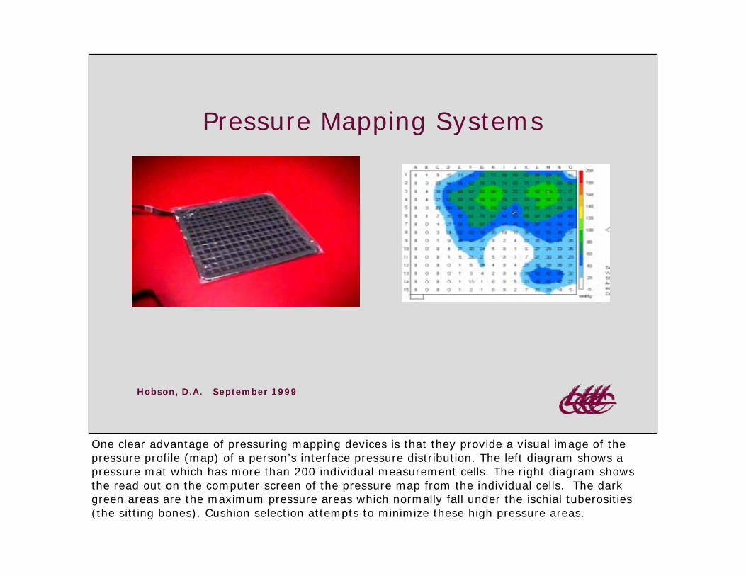
Pressure Mapping Systems
One clear advantage of pressuring mapping devices is that they provide a visual image of thepressure profile (map) of a person’s interface pressure distribution. The left diagram shows apressure mat which has more than 200 individual measurement cells. The right diagram showsthe read out on the computer screen of the pressure map from the individual cells. The darkgreen areas are the maximum pressure areas which normally fall under the ischial tuberosities(the sitting bones). Cushion selection attempts to minimize these high pressure areas.
Hobson, D.A. September 1999
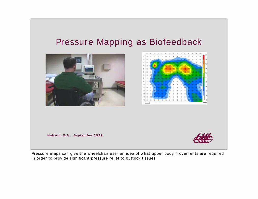
Pressure Mapping as Biofeedback
Pressure maps can give the wheelchair user an idea of what upper body movements are requiredin order to provide significant pressure relief to buttock tissues.

Hobson, D.A. September 1999
Shifting to One Side
This example shows the change in the support pressure pattern as the person shifts their upperbody mass to their right side.
Hobson, D.A. September 1999
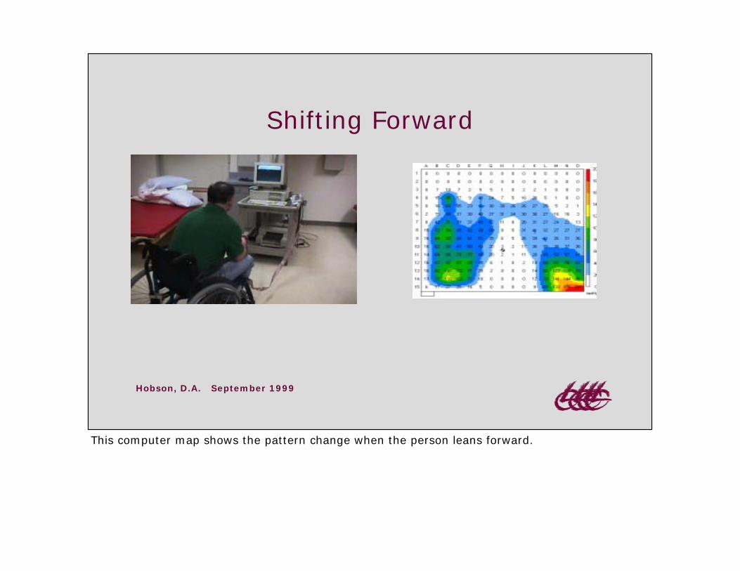
Shifting Forward
This computer map shows the pattern change when the person leans forward.
Hobson, D.A. September 1999
Comparative StudyFerguson-Pell & Cardi (1992)
• Three computer-based devices.• Four different cushion types.• Results, comparisons of readings between
different cushions, same subject, mayproduce errors as much as 10mmHg.
This important study provides an indication of the accuracy that can be expected from mappingdevices.
Hobson, D.A. September 1999
Interpretation of Pressure Data:A Few Precautions
• Comparison of absolute pressure valuescan be misleading.
• Measurement device may be altering thedistribution of surface forces (e.g,hammocking).
• Relative pressure comparisons areprobably most useful.
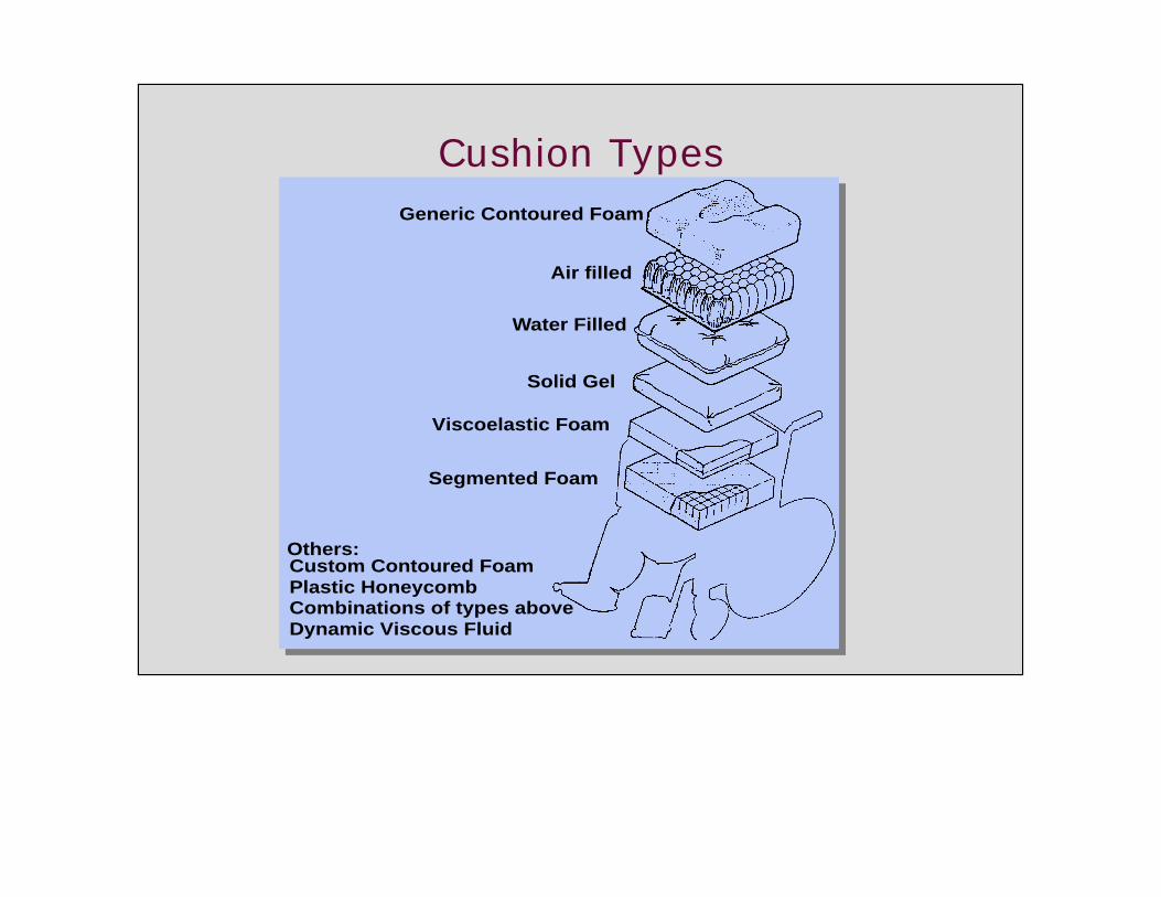
Cushion TypesGeneric Contoured Foam
Air filled
Water Filled
Solid Gel
Viscoelastic Foam
Segmented Foam
Others:Custom Contoured Foam Plastic Honeycomb Combinations of types above Dynamic Viscous Fluid
Hobson, D.A. September 1999
Key Properties of Cushion Materials(Sprigle, 1992)
• Density - weight/volume ratio.• Stiffness - measure of softness.• Resilience - ability to recover shape.• Dampening - absorb impact loads.• Envelopment - surface area covered.
Hobson, D.A. September 1999
Positioning for Pressure andPostural Management
• planer vs. sling seat surface• provide appropriate pressure relieving
cushion• incline seat (5-10°)• firm contoured back, reclined 10-20°• match backrest height to user needs.

Hobson, D.A. September 1999
Positioning for Pressure and PosturalManagement (cont’d)
• add lumbar pad (optional).• adjust arm and foot rests for optimal weight
distribution.• provide weight relief accessories to wheelchair
(recline/tilt) as necessary.• provide training on weight relief and use of
seating and/or wheelchair system.
Hobson, D.A. September 1999
References:
• Ferguson-Pell, M. & Cardi. (1992). Pressure Mapping, XXX,Oct., 30-35
• Ferguson-Pell, M. & Cardi. (1993). Development andcomparative evaluation of W/C pressure mapping. AssistiveTechnology. 5:78-91
• Sprigle, S. (1993). Using Seat Contour during SeatingEvaluations of Individuals with SCI, Assistive Technology. 5:24-35.
• Brienza, D.; Karg, P. & Brubaker, C. (1996). Seat cushiondesign for elderly wheelchair users based on minimization ofsoft tissue deformation using stiffness and pressuremeasurements. IEEE Transactions on RehabilitationEngineering, 4(4)320-328.