principles of inheritance and variation...principles of inheritance and variation (a) (b) figure...
TRANSCRIPT

81
PRINCIPLES OF INHERITANCE AND VARIATION
communication was not easy (as it is now) in those days and his work
could not be widely publicised. Secondly, his concept of genes (or
factors, in Mendel’s words) as stable and discrete units that controlled
the expression of traits and, of the pair of alleles which did not ‘blend’
with each other, was not accepted by his contemporaries as an
explanation for the apparently continuous variation seen in nature.
Thirdly, Mendel’s approach of using mathematics to explain biological
phenomena was totally new and unacceptable to many of the
biologists of his time. Finally, though Mendel’s work suggested that
factors (genes) were discrete units, he could not provide any physical
proof for the existence of factors or say what they were made of.
In 1900, three Scientists (de Vries, Correns and von Tschermak)
independently rediscovered Mendel’s results on the inheritance of
characters. Also, by this time due to advancements in microscopy that
were taking place, scientists were able to carefully observe cell division.
This led to the discovery of structures in the nucleus that appeared to
double and divide just before each cell division. These were called
chromosomes (colored bodies, as they were visualised by staining). By
1902, the chromosome movement during meiosis had been worked out.
Walter Sutton and Theodore Boveri noted that the behaviour of
chromosomes was parallel to the behaviour of genes and used
chromosome movement (Figure 5.8) to explain Mendel’s laws (Table 5.3).
Recall that you have studied the behaviour of chromosomes during mitosis
(equational division) and during meiosis (reduction division). The
important things to remember are that chromosomes as well as genes
occur in pairs. The two alleles of a gene pair are located on homologous
sites on homologous chromosomes.
Figure 5.8 Meiosis and germ cell formation in a cell with four chromosomes.Can you see how chromosomes segregate when germ cellsare formed?
2019-2020

82
BIOLOGY
Possibility I Possibility IIOne long orange and short green One long orange and short redchromosome and long yellow and chromosome and long yellow and short red chromosome at the short green chromosome at the
same pole same pole
Figure 5.9 Independent assortment of chromosomes
Can you tell which of these columns A or B represent the chromosome
and which represents the gene? How did you decide?
During Anaphase of meiosis I, the two chromosome pairs can align at
the metaphase plate independently of each other (Figure 5.9). To
understand this, compare the chromosomes of four different colour in
the left and right columns. In the left column (Possibility I) orange and
green is segregating together. But in the right hand column (Possibility
II) the orange chromosome is segregating with the red chromosomes.
Table 5.3: A Comparison between the Behaviour of Chromosomes
and Genes
A
Occur in pairs
Segregate at the time of gamete
formation such that only one of each
pair is transmitted to a gamete
Independent pairs segregate
independently of each other
B
Occur in pairs
Segregate at gamete formation and only
one of each pair is transmitted to a
gamete
One pair segregates independently of
another pair
2019-2020
83
PRINCIPLES OF INHERITANCE AND VARIATION
(a) (b)
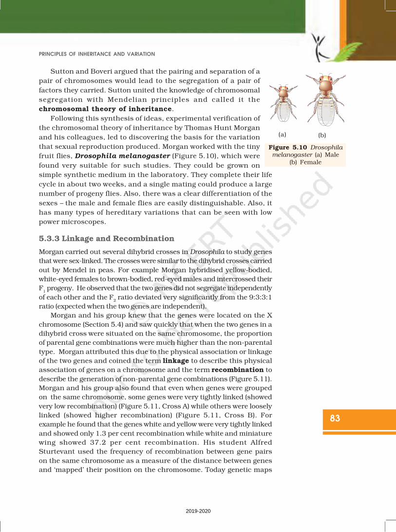
Figure 5.10 Drosophila
melanogaster (a) Male(b) Female
Sutton and Boveri argued that the pairing and separation of a
pair of chromosomes would lead to the segregation of a pair of
factors they carried. Sutton united the knowledge of chromosomal
segregation with Mendelian principles and called it the
chromosomal theory of inheritance.
Following this synthesis of ideas, experimental verification of
the chromosomal theory of inheritance by Thomas Hunt Morgan
and his colleagues, led to discovering the basis for the variation
that sexual reproduction produced. Morgan worked with the tiny
fruit flies, Drosophila melanogaster (Figure 5.10), which were
found very suitable for such studies. They could be grown on
simple synthetic medium in the laboratory. They complete their life
cycle in about two weeks, and a single mating could produce a large
number of progeny flies. Also, there was a clear differentiation of the
sexes – the male and female flies are easily distinguishable. Also, it
has many types of hereditary variations that can be seen with low
power microscopes.
5.3.3 Linkage and Recombination
Morgan carried out several dihybrid crosses in Drosophila to study genes
that were sex-linked. The crosses were similar to the dihybrid crosses carried
out by Mendel in peas. For example Morgan hybridised yellow-bodied,
white-eyed females to brown-bodied, red-eyed males and intercrossed their
F1 progeny. He observed that the two genes did not segregate independently
of each other and the F2 ratio deviated very significantly from the 9:3:3:1
ratio (expected when the two genes are independent).
Morgan and his group knew that the genes were located on the X
chromosome (Section 5.4) and saw quickly that when the two genes in a
dihybrid cross were situated on the same chromosome, the proportion
of parental gene combinations were much higher than the non-parental
type. Morgan attributed this due to the physical association or linkage
of the two genes and coined the term linkage to describe this physical
association of genes on a chromosome and the term recombination to
describe the generation of non-parental gene combinations (Figure 5.11).
Morgan and his group also found that even when genes were grouped
on the same chromosome, some genes were very tightly linked (showed
very low recombination) (Figure 5.11, Cross A) while others were loosely
linked (showed higher recombination) (Figure 5.11, Cross B). For
example he found that the genes white and yellow were very tightly linked
and showed only 1.3 per cent recombination while white and miniature
wing showed 37.2 per cent recombination. His student Alfred
Sturtevant used the frequency of recombination between gene pairs
on the same chromosome as a measure of the distance between genes
and ‘mapped’ their position on the chromosome. Today genetic maps
2019-2020

84
BIOLOGY
Figure 5.11 Linkage: Results of two dihybrid crosses conducted by Morgan. Cross A showscrossing between gene y and w; Cross B shows crossing between genes w and m.Here dominant wild type alleles are represented with (+) sign in superscript
Note: The strength of linkage between y and w is higher than w and m.
are extensively used as a starting point in the sequencing of whole
genomes as was done in the case of the Human Genome Sequencing
Project, described later.
84
2019-2020
85
PRINCIPLES OF INHERITANCE AND VARIATION
5.4 POLYGENIC INHERITANCE
Mendel’s studies mainly described those traits that have distinct alternateforms such as flower colour which are either purple or white. But if you
look around you will find that there are many traits which are not sodistinct in their occurrence and are spread across a gradient. For example,in humans we don’t just have tall or short people as two distinct
alternatives but a whole range of possible heights. Such traits are generallycontrolled by three or more genes and are thus called as polygenic traits.Besides the involvement of multiple genes polygenic inheritance also takes
into account the influence of environment. Human skin colour is anotherclassic example for this. In a polygenic trait the phenotype reflects thecontribution of each allele, i.e., the effect of each allele is additive. To
understand this better let us assume that three genes A, B, C control skincolour in human with the dominant forms A, B and C responsible fordark skin colour and the recessive forms a, b and c for light skin colour.
The genotype with all the dominant alleles (AABBCC) will have the darkestskin colour and that with all the recessive alleles (aabbcc) will have thelightest skin colour. As expected the genotype with three dominant alleles
and three recessive alleles will have an intermediate skin colour. In thismanner the number of each type of alleles in the genotype would determinethe darkness or lightness of the skin in an individual.
5.5 PLEIOTROPY
We have so far seen the effect of a gene on a single phenotype or trait.
There are however instances where a single gene can exhibit multiplephenotypic expression. Such a gene is called a pleiotropic gene. Theunderlying mechanism of pleiotropy in most cases is the effect of a gene
on metabolic pathways which contribute towards different phenotypes.An example of this is the disease phenylketonuria, which occurs inhumans. The disease is caused by mutation in the gene that codes for the
enzyme phenyl alanine hydroxylase (single gene mutation). This manifestsitself through phenotypic expression characterised by mentalretardation and a reduction in hair and skin pigmentation.
5.6 SEX DETERMINATION
The mechanism of sex determination has always been a puzzle before thegeneticists. The initial clue about the genetic/chromosomal mechanismof sex determination can be traced back to some of the experiments carried
out in insects. In fact, the cytological observations made in a number ofinsects led to the development of the concept of genetic/chromosomalbasis of sex-determination. Henking (1891) could trace a specific nuclear
structure all through spermatogenesis in a few insects, and it was alsoobserved by him that 50 per cent of the sperm received this structureafter spermatogenesis, whereas the other 50 per cent sperm did not receive
it. Henking gave a name to this structure as the X body but he could notexplain its significance. Further investigations by other scientists led tothe conclusion that the ‘X body’ of Henking was in fact a chromosome
2019-2020
86
BIOLOGY
and that is why it was given the nameX-chromosome. It was also observed that in
a large number of insects the mechanism ofsex determination is of the XO type, i.e., alleggs bear an additional X-chromosome
besides the other chromosomes(autosomes). On the other hand, some of thesperms bear the X-chromosome whereas
some do not. Eggs fertilised by sperm havingan X-chromosome become females and,those fertilised by sperms that do not have
an X-chromosome become males. Do you
think the number of chromosomes in the
male and female are equal? Due to the
involvement of the X-chromosome in thedetermination of sex, it was designated tobe the sex chromosome, and the rest of the
chromosomes were named asautosomes.Grasshopper is an example ofXO type of sex determination in which the
males have only one X-chromosome besidesthe autosomes, whereas females have a pairof X-chromosomes.
These observations led to the
investigation of a number of species to
understand the mechanism of sex
determination. In a number of other insects
and mammals including man, XY type of sex
determination is seen where both male and
female have same number of chromosomes.
Among the males an X-chromosome is
present but its counter part is distinctly
smaller and called the Y-chromosome.
Females, however, have a pair of X-
chromosomes. Both males and females bear
same number of autosomes. Hence, the males have autosomes plus XY,
while female have autosomes plus XX. In human beings and in
Drosophila the males have one X and one Y chromosome, whereas females
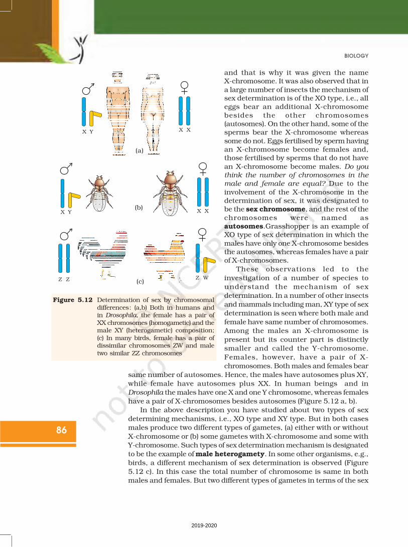
have a pair of X-chromosomes besides autosomes (Figure 5.12 a, b).
In the above description you have studied about two types of sex
determining mechanisms, i.e., XO type and XY type. But in both cases
males produce two different types of gametes, (a) either with or without
X-chromosome or (b) some gametes with X-chromosome and some with
Y-chromosome. Such types of sex determination mechanism is designated
to be the example of male heterogamety. In some other organisms, e.g.,
birds, a different mechanism of sex determination is observed (Figure
5.12 c). In this case the total number of chromosome is same in both
males and females. But two different types of gametes in terms of the sex
(a)
(b)
(c)
Figure 5.12 Determination of sex by chromosomaldifferences: (a,b) Both in humans andin Drosophila, the female has a pair ofXX chromosomes (homogametic) and themale XY (heterogametic) composition;(c) In many birds, female has a pair ofdissimilar chromosomes ZW and maletwo similar ZZ chromosomes
2019-2020
87
PRINCIPLES OF INHERITANCE AND VARIATION
chromosomes, are produced by females, i.e., female heterogamety. In
order to have a distinction with the mechanism of sex determination
described earlier, the two different sex chromosomes of a female bird has
been designated to be the Z and W chromosomes. In these organisms the
females have one Z and one W chromosome, whereas males have a pair of
Z-chromosomes besides the autosomes.
5.6.1 Sex Determination in Humans
It has already been mentioned that the sex determining mechanism in
case of humans is XY type. Out of 23 pairs of chromosomes present,
22 pairs are exactly same in both males and females; these are the
autosomes. A pair of X-chromosomes are present in the female, whereas
the presence of an X and Y chromosome are determinant of the male
characteristic. During spermatogenesis among males, two types of
gametes are produced. 50 per cent of the total sperm produced carry
the X-chromosome and the rest 50 per cent has Y-chromosome besides
the autosomes. Females, however, produce only one type of ovum with
an X-chromosome. There is an equal probability of fertilisation of the
ovum with the sperm carrying either X or Y chromosome. In case the
ovum fertilises with a sperm carrying X-chromosome the zygote develops
into a female (XX) and the fertilisation of ovum with Y-chromosome
carrying sperm results into a male offspring. Thus, it is evident that it
is the genetic makeup of the sperm that determines the sex of the child.
It is also evident that in each pregnancy there is always 50 per cent
probability of either a male or a female child. It is unfortunate that in
our society women are blamed for giving birth to female children and
have been ostracised and ill-treated because of this false notion.
5.6.2 Sex Determination in Honey Bee
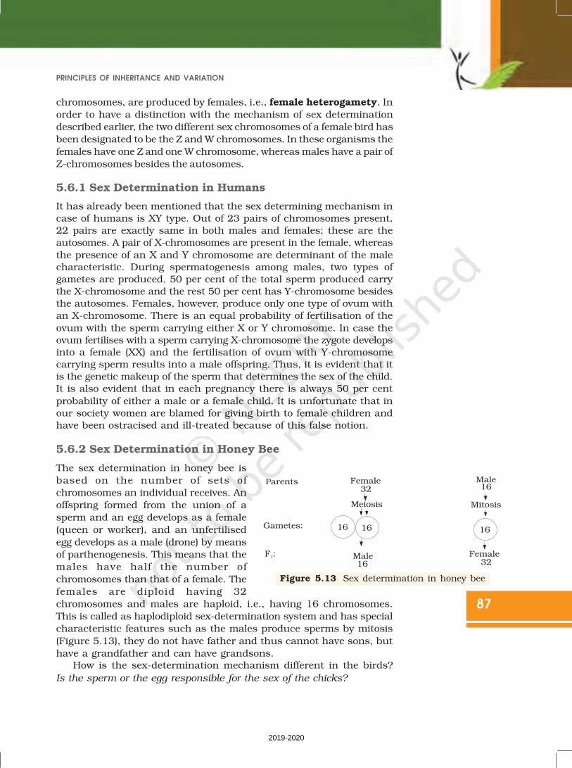
The sex determination in honey bee is
based on the number of sets of
chromosomes an individual receives. An
offspring formed from the union of a
sperm and an egg develops as a female
(queen or worker), and an unfertilised
egg develops as a male (drone) by means
of parthenogenesis. This means that the
males have half the number of
chromosomes than that of a female. The
females are diploid having 32
chromosomes and males are haploid, i.e., having 16 chromosomes.
This is called as haplodiploid sex-determination system and has special
characteristic features such as the males produce sperms by mitosis
(Figure 5.13), they do not have father and thus cannot have sons, but
have a grandfather and can have grandsons.How is the sex-determination mechanism different in the birds?
Is the sperm or the egg responsible for the sex of the chicks?
Figure 5.13 Sex determination in honey bee
2019-2020