principles of biochemistry fourth edition chapter 22 protein synthesis copyright © 2006 pearson...
TRANSCRIPT
Principles of BiochemistryFourth Edition
Chapter 22Protein Synthesis
Copyright © 2006 Pearson Prentice Hall, Inc.
Horton • Moran • Scrimgeour • Perry • Rawn

Chapter 22 - Protein Synthesis
The ribosome, a complex of RNA and protein, is the site where genetic information is translated into protein

22.1 The Genetic Code
• Codons - three letter genetic code (nonoverlapping)
• tRNA - adapters between mRNA and proteins
• Reading frame - each potential starting point for interpreting the 3 letter code

Fig 22.1 Overlapping vs nonoverlapping reading of the three-letter code

Fig 22.2 Three reading frames of mRNA
• Translation of the correct message requires selection of the correct reading frame

Fig 22.3 Standard genetic code

Features of the Genetic Code
1. The genetic code is unambiguous. In any organism each codon corresponds to only one amino acid.
2. There are multiple codons for most amino acids(code is degenerate), and synonymous codons specify the same amino acid
3. The first two nucleotides of a codon are often enough to specify a given amino acid

Features of the Code (continued)
4. Codons with similar sequences specify similar amino acids
5. Only 61 of the 64 codons specify amino acids
• Termination (stop codons): UAA, UGA, UAG
• Initiation codon - Methionine codon (AUG) also specifies initiation site for protein synthesis

A. Three-Dimensional Structure of tRNA– Transfer RNA molecules are the interpreters
of the genetic code– Every cell must contain at least 20 tRNA (one
for every amino acid)– Each tRNA must recognize at least one
codon– tRNAs have a “cloverleaf” type secondary
structure with several loops or arms
22.2 Transfer RNA
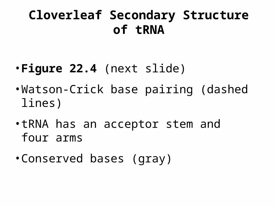
Cloverleaf Secondary Structure of tRNA
• Figure 22.4 (next slide)
• Watson-Crick base pairing (dashed lines)
• tRNA has an acceptor stem and four arms
• Conserved bases (gray)

Fig. 22.4
• Cloverleaf structure of tRNA

Fig 22.5 Tertiary structure of tRNA

tRNA Arms
• Acceptor stem - amino acid becomes covalently attached to tRNA at the 3’ end of this stem
• Anticodon arm - contains the anticodon, a three-base sequence that binds to a complementary codon in mRNA
(continued next slide)

tRNA Arms (continued)
• TC arm - contains thymidylate (T) and pseudouridylate () followed by C
• D arm - contains dihydrouridylate (D)
• Variable arm - ranges from 3-21 nucleotides

Fig 22.6
• Structure of tRNAPhe from yeast
AnimationsAnimations
• Structure of tRNA
B. tRNA Anticodons Base-Pair with mRNA Codons
• tRNA molecules are named for the amino acid that they carry (e.g. tRNAPhe)
• Base pairing between codon and anticodon is governed by rules of Watson-Crick (A-U, G-C)
• However, the 5’ anticodon position has some flexibility in base pairing (the “wobble” position)

Table 22.1

Fig 22.7 Inosinate Base Pairs• Inosinate (I) base pairs• Inosinate often found at 5’ wobble position• I can form H bonds with A, C, or U• Anticodon with I can recognize more than one
synonymous codon

Codon-Anticodon Recognition
• Wobble allows some tRNA molecules to recognize more than one codon
• Isoacceptor tRNA molecules - different tRNA molecules that bind the same amino acids
• Isoacceptor tRNAs identified by Roman numerals or codons: tRNAI
Ala, tRNAIIAla or tRNAGCG
Ala
• Bacteria have 30-60 different tRNAs, eukaryotes have up to 80 different tRNAs

Fig 22.8 Base pairing at the wobble position
22.3 Aminoacyl-tRNA Synthetases
• Aminoacyl-tRNA - amino acids are covalently attached to the 3’ end of each tRNA molecule (named as: alanyl-tRNAAla)
• Aminoacyl-tRNA synthetases catalyze reactions
• Most species have at least 20 different aminoacyl-tRNA synthetases (1 per amino acid)
• Each synthetase specific for a particular amino acid, but may recognize isoacceptor tRNAs

A. The Aminoacyl-tRNA Synthetase Reaction
• Aminoacyl-tRNAs are high-energy molecules (the amino acid has been “activated”)
• The activation of an amino acid by aminoacyl-tRNA synthetase requires ATP
Amino acid + tRNA + ATP
Aminoacyl-tRNA + AMP + PPi

Fig 22.9

Fig 22.9 (cont)

B. Specificity of Aminoacyl-tRNA Synthetase
• Attachment of the correct amino acid to the corresponding tRNA is a critical step
• Synthetase binds ATP and the correct amino acid (based on size, charge, hydrophobicity)
• Synthetase then selectively binds specific tRNA molecule based on structural features
• Synthetase may recognize the anticodon as well as the acceptor stem

Fig 22.10 Structure of E. coli tRNAGln bound to the synthetase

C. Proofreading Activity of Aminoacyl-tRNA Synthetases
• Some aa-tRNA synthetases can proofread
• Isoleucyl-tRNA synthetase may bind valine instead of isoleucine and incorporate it into valyl-adenylate
• The valyl-adenylate is usually then hydrolyzed to valine and AMP so that valyl-tRNAIle does not form

Fig 22.11 Model of substrate-binding site in isoleucyl-tRNA synthetase
• Ile-tRNA binds to Ile about 100x better than Val even though they have similar size and charge

22.4 Ribosomes
• Protein synthesis is carried out by a complex composed of the ribosome, accessory protein factors, mRNA and charged tRNA molecules
• Initiation complex assembles at first mRNA codon, and disassembles at termination step
• Ribosome complex moves 5’ 3’ along template mRNA
• Polypeptide is synthesized in N C direction
A. Ribosomes Are Composed of Both rRNA and Protein
• All ribosomes contain two subunits of unequal size
• E. coli: 70S composed of a 30S and a 50S
• Eukaryotes: 80S composed of a 40S and a 60S

Fig 22.12 Comparison of prokaryotic and eukaryotic ribosomes

Fig 22.13
• Assembly of the 30S ribosomal subunit and maturation of the 16S rRNA (E. coli)
• Ribosomal proteins (6-7) bind to 16S rRNA as it is being transcribed forming a 21S particle
• Processing and binding of other ribosomal proteins completes the mature 30S subunit

Fig 22.14 Structure of the 30S ribosomal subunit (T. thermophilus)

B. Ribosomes Contain Two Aminoacyl-tRNA Binding Sites
• Ribosome must align two charged tRNA molecules so that anticodons interact with correct codons of mRNA
• Aminoacylated ends of the tRNAs are positioned at the site of peptide bond formation
• Ribosome must hold both mRNA and growing polypeptide chain

Fig 22.15 Sites for tRNA binding in ribosomes

22.5 Initiation of Translation
• The translation complex is assembled at the beginning of the mRNA coding sequence
• Complex consists of: Ribosomal subunitsmRNA template to be translatedInitiator tRNA moleculeProtein initiation factors

Fig 22.3 Standard genetic code
A. Initiator tRNA
• First codon translated is usually AUG
• Each cell contains at least two methionyl-tRNAMet
molecules which recognize AUG
• The initiator tRNA recognizes initiation codons
• Second tRNAMet recognizes only internal AUG
• Bacteria: N-formylmethionyl-tRNAfMet
• Eukaryotes: methionyl-tRNAiMet

Fig 22.16 Structure of fMet-tRNAfMet

B. Initiation Complexes Assemble Only at Initiation Codons
• Ribosome must recognize protein synthesis start
• In prokaryotes, the 30S ribosome binds to a region of the mRNA (Shine-Dalgarno sequence) upstream of the initiation sequence
• S-D sequence also binds to a complementary base sequence at the 3’ end of the 16S rRNA
• Double-stranded RNA structure binds mRNA to the ribosome

Fig 22.17 (a) Shine-Dalgarno sequences in E. coli mRNA
• Ribosome-binding sites at the 5’ end of mRNA for several E. coli proteins
• S-D sequences (red) occur immediately upstream of initiation codons (blue)

Fig 22.17 (b)
• Complementary base pairing of S-D sequence

C. Initiation Factors Help Form Initiation Complex
• Initiation factors are required to form a complex
• Prokaryote factors: IF-1, IF-2, IF-3
• Eukaryote factors: eIFs (8 or more factors)
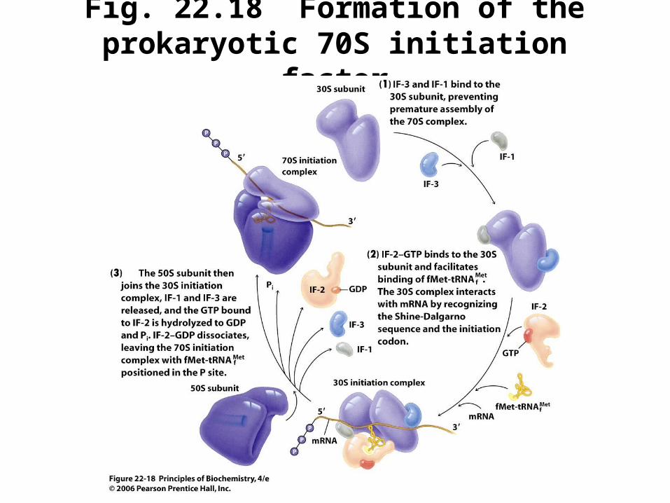
Fig. 22.18 Formation of the prokaryotic 70S initiation factor
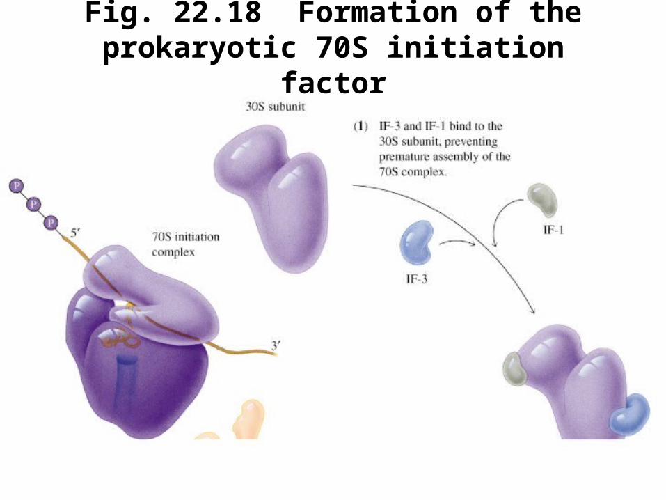
Fig. 22.18 Formation of the prokaryotic 70S initiation factor
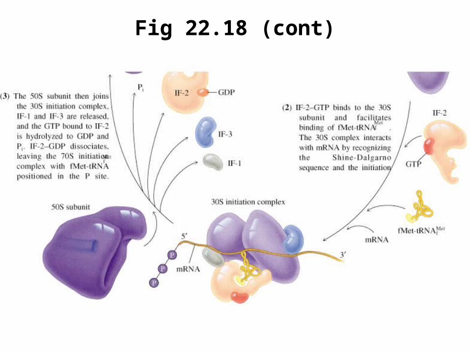
Fig 22.18 (cont)

D. Translation Initiation in Eukaryotes
• Eukaryotic initiation factor 4 (eIF-4), (or cap binding protein, CBP) binds to the (5’ end) 7-methylguanylate cap of eukaryotic mRNA
• A preinitiation complex forms (40S ribosome, aminoacylated initiator tRNA, other factors) and searches the mRNA 5’ 3’ for an initiator codon
• The Met-tRNAiMet binds to AUG, and the 60S
ribosomal subunit binds to complete the complex
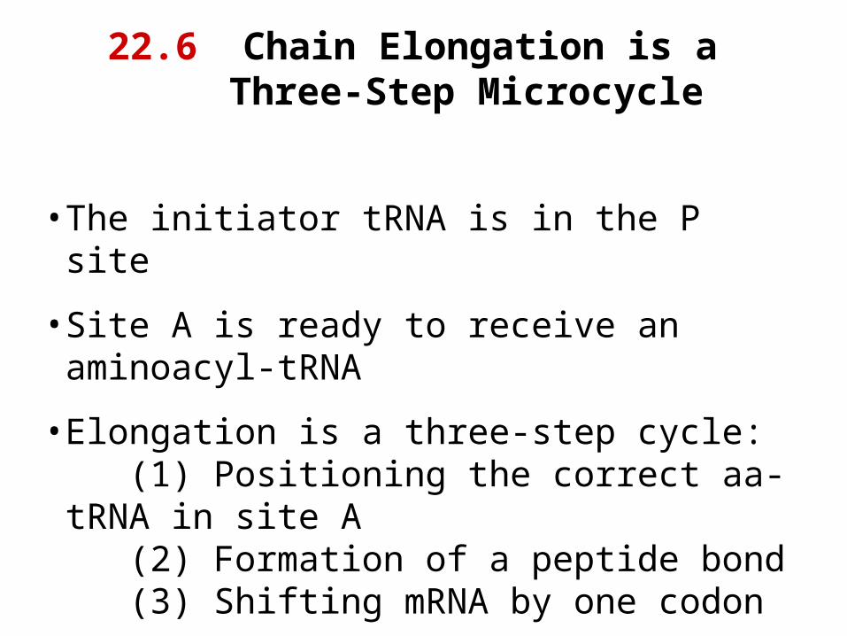
22.6 Chain Elongation is a Three-Step Microcycle
• The initiator tRNA is in the P site
• Site A is ready to receive an aminoacyl-tRNA
• Elongation is a three-step cycle: (1) Positioning the correct aa-tRNA in site A
(2) Formation of a peptide bond (3) Shifting mRNA by one codon

Fig 22.19 Coupled transcription and translation in bacteria
• Gene is being transcribed left to right
• Ribosomes bind to 5’ end of mRNA

A. Elongation Factors Dock an Aminoacyl-tRNA in the A Site
• Bacterial elongation factor EF-Tu helps the correct aa-tRNA insert into site A
• An EF-Tu-GTP complex binds to all aa-tRNA molecules except fMet-tRNAf
Met (initiator)
• A ternary complex of EF-Tu-GTP-aa-tRNA binds in the ribosomal A site
• If the anticodon of the aa-tRNA correctly base pairs with the mRNA codon, complex is stabilized

Fig 22.20 EF-Tu binds tRNAs
• EF-Tu binds to acceptor end of aminoacylated tRNA (Phe-tRNAPhe)
• Phe residue (green)
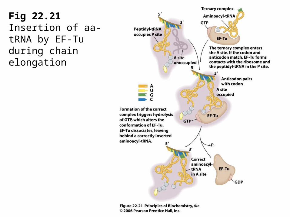
Fig 22.21 Insertion of aa-tRNA by EF-Tu during chain elongation

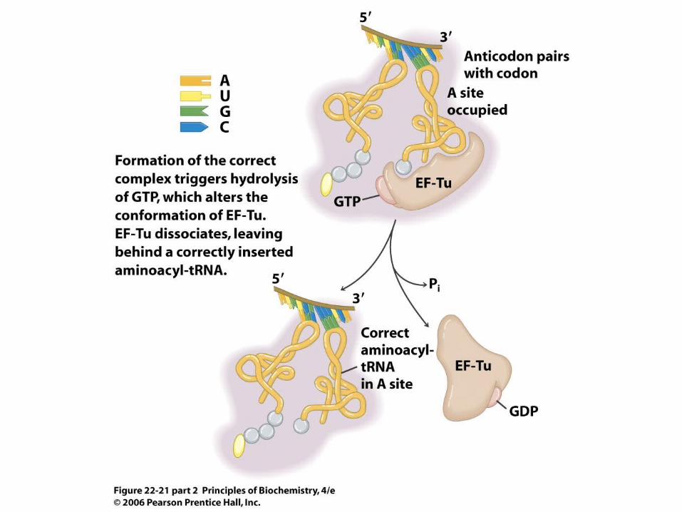

Fig 22.22 Cycling of EF-Tu-GTP
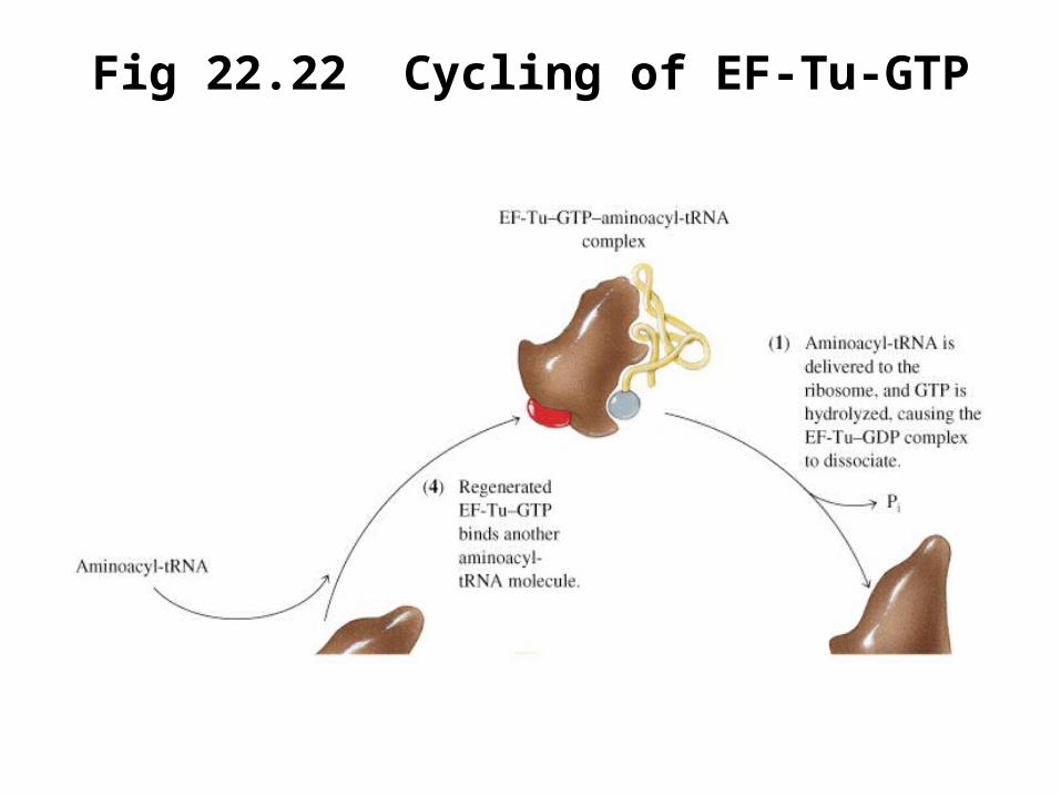
Fig 22.22 Cycling of EF-Tu-GTP

Fig 22.22 (cont)
B. Peptidyl Transferase Catalyzes Peptide Bond Formation
• Peptidyl transferase activity is contained within the large ribosomal subunit
• Substrate binding site in 23S rRNA and 50S ribosomal proteins
• Catalytic activity from 23S rRNA (an RNA-catalyzed reaction)

Fig. 22.23 Formation of a peptide bond

C. Translocation Moves the Ribosome by One Codon
• Translocation step: the new peptidyl-tRNA is moved from the A site to the P site, while the mRNA shifts by one codon
• The deaminoacylated tRNA has shifted from the P site to the E site (exit site)
• Binding of EF-G-GTP to the ribosome completes translocation of peptidyl-tRNA

Fig 22.24
• Translocation during protein synthesis in prokaryotes
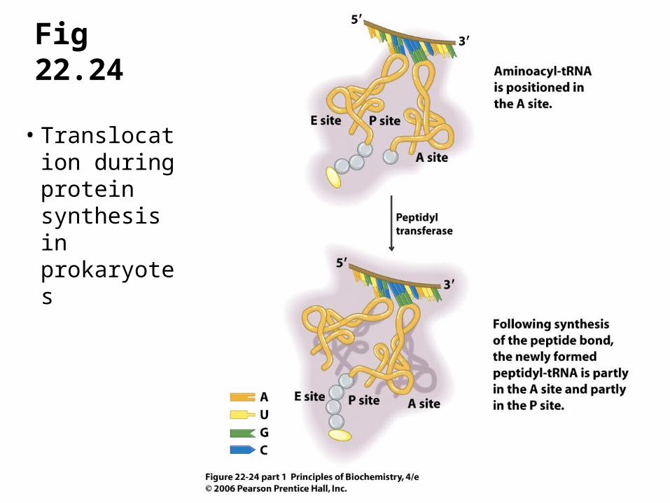
Fig 22.24
• Translocation during protein synthesis in prokaryotes

Fig 22.24 (cont)

Formation of the Peptide Chain
• Growing peptide chain extends from the peptidyl-tRNA (P site) through a tunnel in the 50S subunit
• Newly synthesized polypeptide does not begin to fold until it emerges from the tunnel
• Elongation in eukaryotes is similar to E. coli: EF-1- docks the aa-tRNA into A site EF-1- recycles EF-1 EF-2 - carries out translocation

AnimationsAnimations
• tRNA binding ribosomes
22.7 Termination of Translation
• E. coli release factors: RF-1, RF-2, RF-3
• Translocation positions one of three termination codons in A site: UGA, UAG, UAA
• No tRNA molecules recognize these codons and protein synthesis stalls
• One of the release factors binds and causes hydrolysis of the peptidyl-tRNA to release the polypeptide chain

AnimationsAnimations
• Protein synthesis

22.8 Protein Synthesis is Energetically Expensive
• Four phosphoanhydride bonds are cleaved for each amino acid added to a polypeptide chain
Amino acid activation: Two ~P bonds
ATP AMP + 2 Pi
Chain elongation: Two ~P bonds
2 GTP 2 GDP + 2 Pi

Box 22.1 Some Antibiotics Inhibit Protein Synthesis
• Some antibiotics prevent bacterial growth by inhibiting the formation of peptide bonds
• Puromycin (next slide) resembles the 3’ end of an aminoacyl-tRNA, and can enter the A site of a ribosome
• The peptidyl-puromycin formed is bound weakly in the A site and dissociates terminating protein synthesis


A. Ribosomal Protein Synthesis Is Coupled to Ribosome Assembly in E. coli
– Synthesis of ribosomal proteins is tightly regulated at the level of translation
– Ribosomal protein genes encode one ribosomal protein that inhibits translation of its own polycistrionic mRNA by binding near the initiation codon of the mRNA
22.9 Regulation of Protein Synthesis

Fig 22.25 Comparison of proposed secondary structures of S7-binding sites
(a) S7 site on 16S rRNA
(b) S7 site on the str mRNA
S7 protein inhibits translation by binding to the str mRNA molecule
B. Globin Synthesis Depends on Heme Availability
• Hemoglobin synthesis requires globin chains and heme in stoichiometric amounts
• Globin synthesis is controlled by regulation of translation initiation
• Heme-controlled inhibitor (HCI) phosphorylates factor eIF-2 which then cannot participate in translation initiation
• High heme levels interfere with HCI so that globin synthesis proceeds

Fig 22.26
• Inhibition of protein synthesis by phosphoryl-ation of eIF-2
HCI: heme-controlled inhibitorGEF: guaninenucleotide exchange factor

C. The E. coli trp Operon Is Regulated by Repression and Attenuation
• The trp operon in E. coli encodes the proteins necessary for tryptophan biosynthesis
• Because tryptophan is a negative regulator of its own biosynthesis, synthesis can be repressed when exogenous Trp is available
• Tryptophan is a corepressor of the trpO operator (Figure 22.27, next slide)
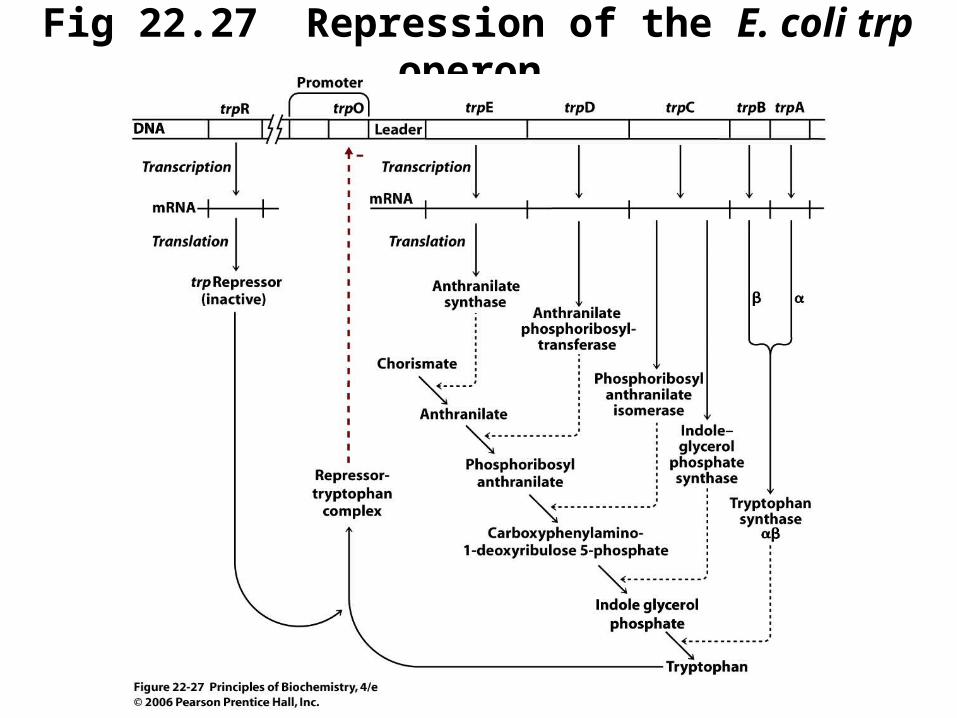
Fig 22.27 Repression of the E. coli trp operon
(continued next slide)

Fig 22.27 Repression of the E. coli trp operon
(continued next slide)

Fig 22.27 (continued)

Attenuation in E. coli
• A second mechanism for regulation of the E. coli trp operon depends on translation
• Determines whether transcription of the operon proceeds or terminates prematurely
• GC-rich regions in the mRNA trp leader region can base pair to form two alternative hairpin structures which affect transcription
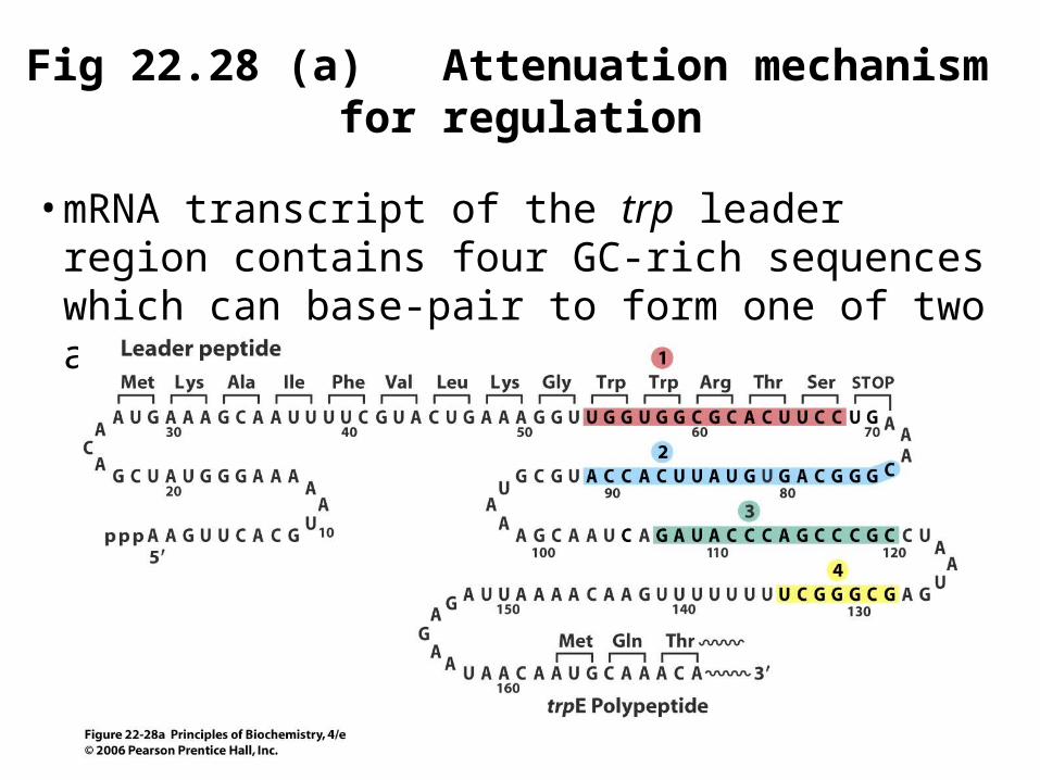
Fig 22.28 (a) Attenuation mechanism for regulation
• mRNA transcript of the trp leader region contains four GC-rich sequences which can base-pair to form one of two alternative structures
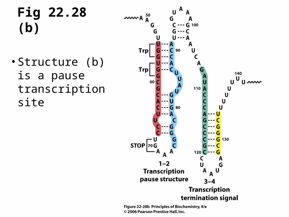
Fig 22.28 (b)
• Structure (b) is a pause transcription site

Fig 22.28 (c)
• Structure (c) is a more stable hairpin than (b)

Transcriptional Attenuation in the trp Operon

Transcriptional Attenuation in the trp Operon

22.10 Posttranslational Processing
• Posttranslational modifications can occur either before the polypeptide chain is complete (cotranslational) or after (posttranslational)
─ deformylation of N-terminal residue (prok)─ removal of N-terminal methionine residue─ formation of disulfide bonds─ cleavage by proteinases─ phosphorylation or acetylation

Fig 22.29
• Secretory pathway in eukaryotic cells
• Proteins synthesized in the cytosol are transported into the lumen of the endoplasmic reticulum
• After further modification in the Golgi, the proteins are secreted
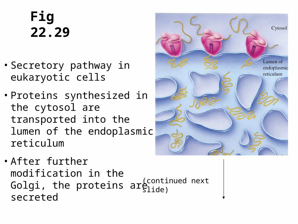
Fig 22.29
• Secretory pathway in eukaryotic cells
• Proteins synthesized in the cytosol are transported into the lumen of the endoplasmic reticulum
• After further modification in the Golgi, the proteins are secreted
(continued next slide)

Fig 22.29 (cont)

Fig 22.30 Secretory Vesicles in a maize rootcap cell

A. The Signal Hypothesis
• Secreted proteins are synthesized by ribosomes on the surface of the endoplasmic reticulum
• A signal peptide is present on the N-terminus that signals the protein to cross a membrane
• Signal peptides are 16-30 residues long, and include 4-15 hydrophobic residues
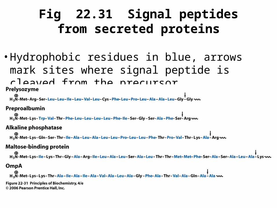
Fig 22.31 Signal peptides from secreted proteins
• Hydrophobic residues in blue, arrows mark sites where signal peptide is cleaved from the precursor

Fig 22.32
• Translocation of eukaryotic proteins into the lumen of the endoplasmic reticulum
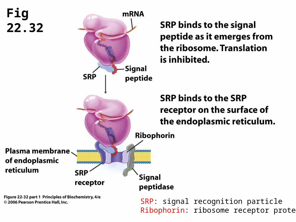
Fig 22.32
SRP: signal recognition particleRibophorin: ribosome receptor protein

Fig 22.32 (cont)
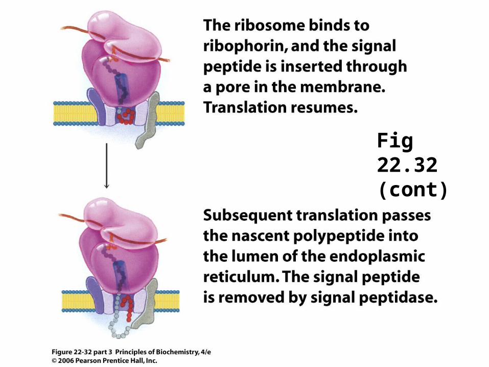
Fig 22.32 (cont)

B. Glycosylation of proteins
• Many integral membrane and secretory proteins contain covalently bound oligosaccharide chains
• Carbohydrate may be from 1% to 80% of the mass of the glycoprotein
• A common glycosylation reaction is the covalent attachment of a complex oligosaccharide to the side chain of an asparagine residue
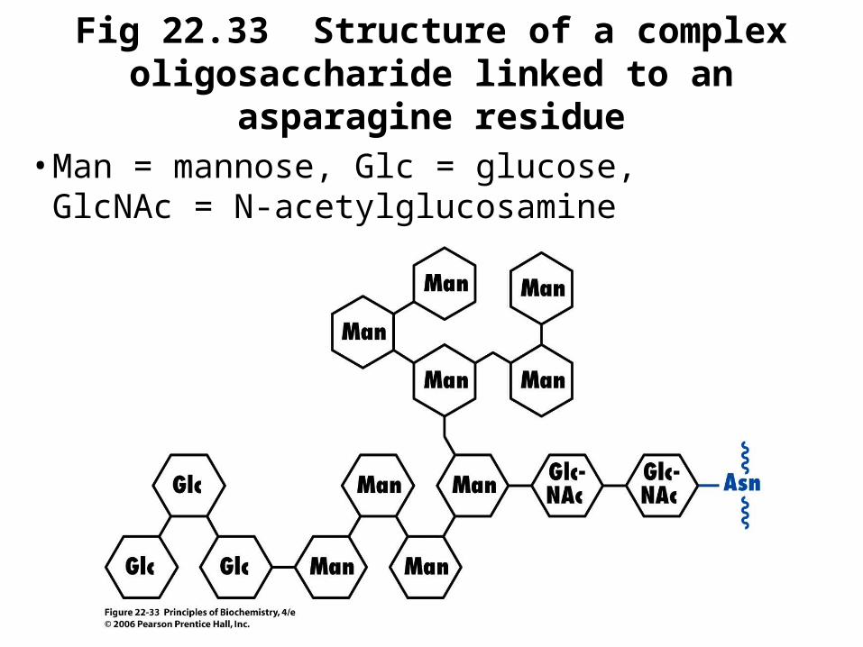
Fig 22.33 Structure of a complex oligosaccharide linked to an asparagine residue
• Man = mannose, Glc = glucose, GlcNAc = N-acetylglucosamine