principles and progress in ultrafast multidimensional ... · solution state at individual, site ......
TRANSCRIPT

ANRV373-PC60-21 ARI 25 February 2009 18:3
Principles and Progress inUltrafast MultidimensionalNuclear Magnetic ResonanceMor Mishkovsky and Lucio FrydmanDepartment of Chemical Physics, Weizmann Institute of Science, 76100 Rehovot, Israel;email: [email protected]
Annu. Rev. Phys. Chem. 2009. 60:429–48
First published online as a Review in Advance onDecember 1, 2008
The Annual Review of Physical Chemistry is online atphyschem.annualreviews.org
This article’s doi:10.1146/annurev.physchem.040808.090420
Copyright c© 2009 by Annual Reviews.All rights reserved
0066-426X/09/0505-0429$20.00
Key Words
NMR spectroscopy, multidimensional acquisitions, ultrafast methods,spatial encoding, hyperpolarized samples
AbstractMultidimensional acquisitions play a central role in the progress and applica-tions of nuclear magnetic resonance (NMR) spectroscopy. Such experimentshave been collected traditionally as an array of one-dimensional scans, withsuitably incremented delay parameters that encode along independent tem-poral domains the nD spectral distribution being sought. During the past fewyears, an ultrafast approach to nD NMR has been introduced that is capableof delivering any type of multidimensional spectrum in a single transient.This method operates by departing from the canonical nD NMR schemeand by replacing its temporal encoding with a series of spatial manipulationsderived from magnetic resonance imaging. The present survey introducesthe main principles of this subsecond approach to spectroscopy, focusingon the applications that have hitherto been demonstrated for single-scantwo-dimensional NMR in different areas of chemistry.
429
Ann
u. R
ev. P
hys.
Che
m. 2
009.
60:4
29-4
48. D
ownl
oade
d fr
om a
rjou
rnal
s.an
nual
revi
ews.
org
by W
eizm
ann
Inst
itute
of
Scie
nce
on 0
6/22
/09.
For
per
sona
l use
onl
y.

ANRV373-PC60-21 ARI 25 February 2009 18:3
MRI: magneticresonance imaging
Fourier transform(FT): themathematicalprocedure that enablesone to discern whichfrequencycontributions composea time-dependentsignal responsefunction
Free inductiondecay: traditionalterm used to denotetime-domain signals ofthe kind described inEquation 1 and arisingupon exciting a spinensemble in an NMRexperiment
T2: spin-spinrelaxation time,describing thedecoherence lifetimesof the spins’ signalfollowing theirexcitation into atransverse evolutionplane
1. INTRODUCTION
Nuclear magnetic resonance (NMR) is a spectroscopic technique based on monitoring the pre-cession of spin-endowed atomic nuclei when placed in a strong external magnetic field. With itsorigin in curiosity-driven investigations about the nature of quantum mechanical phenomena,NMR has over the years transformed into an indispensable applied tool that impacts a remarkablywide range of scientific disciplines (1). High-resolution NMR of dissolved molecules, for example,serves as the eyes of chemists involved in organic, pharmaceutical, and natural-products research(2). When employed in combination with high-resolution solid-state techniques, NMR provides aunique window to study the chemical structure of challenging heterogeneous materials, includingpolymorphic mixtures, polymers, glasses, and catalysts (3). It is one of the few methods avail-able for determining the structure and the dynamics of proteins and nucleic acids in their nativesolution state at individual, site-resolved levels (4). Moreover, even if concealed under differentnames [e.g., magnetic resonance spectroscopy or magnetic resonance imaging (MRI)], NMR hasevolved into a widely used in vivo tool capable of diagnosing and imaging malignancies, evaluatingmetabolic status, angiographing noninvasively, and revealing the activation of human brains evento the smallest of stimuli (5, 6).
Despite this outstandingly wide scope of applications, broadly speaking, one common mea-surement protocol underlies all these different uses of the quantum-mechanic spin-precessionphenomenon: the pulsed Fourier transform (FT) NMR method, whose purpose is to measureinductively the different Bohr evolution frequencies allowed to the spin ensemble under obser-vation within its quantized energy manifold (1–7). These in turn are governed by isotropic shiftor J-coupling interactions in liquid-state experiments, by these couplings plus a variety of spinanisotropies when dealing with solids, and by a combination of the above plus external ad hocfields in the case of NMR imaging. Regardless of which parameters or interactions are actuallysought, NMR extracts the frequency distributions originated by these couplings by monitoringthe responses that they impart on the spins’ time evolution. The result arising in these measure-ments upon subjecting spins to an excitation impulse is the so-called free induction decay, a signal(voltage) given by a weighted sum of oscillating functions,
S(t) =∑
acting�
I (�) exp(i�t) exp(−t/T2), (1)
defined by the allowed single-quantum spectral distribution of allowed transitions I(�), as well asby a T2 relaxation decay. S(t) can then clearly provide the I(�) spectrum sought by FT versus thesingle variable t, defining the time domain that supports it.
Although pulsed NMR experiments initially involved only this kind of data collection, as afunction of a single time axis (8), it was soon realized that significant benefits could result bycorrelating the spins’ evolution along multiple time domains (9, 10). These nD NMR experimentswould then not just measure but also correlate and separate different contributions to the overallspin-precession frequencies; this could improve the resolution of the experiment, as well as extractinformation that would be simply unavailable in the single-quantum one-dimensional (1D) trace.The canonical scheme for such experiments follows Jeener and Ernst’s seminal 2D NMR proposal,which laid the foundations for multidimensional spectroscopy based on the four blocks (9, 10)
Preparation − Evolution (t1) − Mixing − Acquisition (t2). (2)
By suitable incrementation of the t1 and t2 time variables, an NMR experiment of this kind canthen yield a 2D time-domain signal,
s (t1, t2) =∫
all�2
d�2
[∫all�1
d�1 I (�1, �2)e i�1t1 e−t1/T2
]e i�2t2 e−t2/T2 , (3)
430 Mishkovsky · Frydman
Ann
u. R
ev. P
hys.
Che
m. 2
009.
60:4
29-4
48. D
ownl
oade
d fr
om a
rjou
rnal
s.an
nual
revi
ews.
org
by W
eizm
ann
Inst
itute
of
Scie
nce
on 0
6/22
/09.
For
per
sona
l use
onl
y.

ANRV373-PC60-21 ARI 25 February 2009 18:3
from which correlations between so-called indirect- and direct-domain NMR frequencies �1 and�2 can be extracted by 2D FT analysis:
I (ν1, ν2) ∝∫
all t2dt2
[∫all t1
dt1S(t1, t2)e−iν1t1
]e−iν2t2 . (4)
At first glance, this approach to the retrieval of I(�1, �2) appears to be a simple extension of the1D time-domain NMR experiment to two dimensions. The multiple times involved in these ac-quisitions, however, actually possess different origins. t2 is a physical acquisition time along whichthe signal is directly digitized, akin to the t time involved in the 1D NMR free induction decayof Equation 1; the spins’ behavior along this direct-domain axis therefore can be characterizedby FT of data digitized throughout a single-scan experiment. The remaining time axes of theexperiment, however, cannot be sampled in the same fashion. This is resolved in the Jeener-Ernstparadigm by monitoring these domains indirectly, i.e., by carrying out Ni discrete incrementa-tions of certain time delays ti within the sequence throughout a series of independent experiments.Herein lies the brilliance, yet also a potential weakness, of this kind of acquisition. Indeed, re-gardless of sensitivity considerations, nested encoding schemes such as the one represented byEquation 2 require monitoring tens or hundreds of independent increments along each of then – 1 indirect-domain time axes to properly characterize its internal evolution frequencies. More-over, because each point along these n – 1 indirect time domains is associated with an independentsignal acquisition, this results in an exponential increase of the minimum experimental acquisitiontime with dimensionality n. The unambiguous gains resulting from expanding NMR from a 1Dto an nD experiment may therefore come at a price.
Driven by this reality, and stimulated by an increasing reliance of all the above-mentionedcontemporary NMR applications on high-dimensional experiments, a growing number of alter-natives that depart from the traditional sampling principles embodied by Equation 2 have emergedduring the past few years (11, 21). These include routes that process the acquired data by non-Fourier methods (13–16), acquisitions that incorporate frequency-based manipulations (17), andaccordioned derivations (18) whereby multiple indirect-domain time delays are incremented si-multaneously (19–21). Among these new proposals is also an ultrafast approach, departing bothfrom the traditional temporal encoding and from the alternatives just mentioned, that enables theacquisition of arbitrary multidimensional data sets within a single scan without requiring any a pri-ori information (22–24). At the heart of this proposal is a departure from the classical Jeener-Ernstserial incrementation scheme, which is replaced by a parallel encoding of the indirect-domain timeinformation along a spatial dimension. This review discusses the basic features and potential ap-plications of this ultrafast NMR method, particularly as it is used within the context of rapid 2DNMR acquisitions. We summarize first the physical principles underlying ultrafast 2D NMR,continue with an overview of some potential applications of this method, discuss its combinationwith a variety of schemes enabling its extension to higher dimensions, and conclude by review-ing additional applications that have been recently demonstrated based on the spatial-encodingconcepts on which ultrafast NMR spectroscopy relies.
2. SPATIAL ENCODING AND THE SINGLE-SCAN ACQUISITIONOF 2D NUCLEAR MAGNETIC RESONANCE SPECTRA
Ultrafast 2D NMR methods depart from traditional schemes in that, instead of triggering theindirect-domain evolutions homogenously, for all sites and positions within the analyzed sampleat once, they encode the evolution frequencies being sought in a spatially heterogeneous fashion.Several alternatives have been proposed for achieving such heterogeneous evolution (22, 25–30),
www.annualreviews.org • Ultrafast Multidimensional NMR 431
Ann
u. R
ev. P
hys.
Che
m. 2
009.
60:4
29-4
48. D
ownl
oade
d fr
om a
rjou
rnal
s.an
nual
revi
ews.
org
by W
eizm
ann
Inst
itute
of
Scie
nce
on 0
6/22
/09.
For
per
sona
l use
onl
y.
ANRV373-PC60-21 ARI 25 February 2009 18:3
a majority of which impart different evolution durations prior to the mixing period on spinspositioned at different coordinates within the sample. All these schemes share the application ofa train of either continuous or discrete frequency-swept radiofrequency (RF) pulses, acting incombination with suitably echoed magnetic field gradients. These procedures are tuned so as toimpart indirect-domain evolution times proportional to the spins’ coordinate along a particulardirection z. In other words, they rely on a spatial encoding of the spin interactions, wherebythe indirect-domain evolution time of a 2D NMR experiment is made t1 ≈ Cz, where C is aspatio-temporal constant under the experimentalist’s control. (This C parameter is usually givenby tmax
1L , the maximum t1 evolution time imparted, divided by the overall sample length L.) The
evolution coupling parameters �1 being sought thus impart over these periods an effective spatiallydependent precession phase φ(z) ≈ �1t1 = C�1z. It follows that within the framework of a Blochspace in which the spins’ magnetizations precess within a common rotating frame (Figure 1)every chemical site within the sample subtends a shift-induced helical pattern along the z spatialcoordinate:
Mx(z) + i My (z) ∝ I (�1)L
exp(iC�1z) exp(−Cz/T2). (5)
The helical winding of magnetizations represented by this equation is characterized by a pitchdepending on the �1 parameter one is attempting to measure but in general does not lead to anynet observable signal as these helices are characterized by a destructive interference among theirconstituent spin packets when considered over a macroscopic sample. Nevertheless, their encodedinformation can be preserved throughout the various coherent mixing processes involved in a 2DNMR pulse sequence and, at a final acquisition stage, can be read out by applying a z-dependentgradient acting in combination with the data sampling. Indeed, field gradients have the ability towind and unwind spin-magnetization patterns of their own according to
Mxγ Gz·t−−−→ Mx cos(γ Gzt) + My sin(γ Gzt)
= Mx cos (kz) + My sin (kz) . (6)
Under suitable conditions, therefore, these succeed to successively unwind the various helicesthat were subtended by the individual chemical sites, leading to observable echoes arising fromconstructive interference phenomena among spins positioned throughout the sample L (Figure 1).Moreover, as the timing of such echoes depends on the strengths of the �1 internal interactions thatcreated each site’s winding, this allows one to map the indirect-domain spectral information beingsought by monitoring the positions of the resulting echo peaks. Mathematically, these spectralpeak positions are characterized by the values taken by the wave numbers k = γa
∫ t0 Ga (t′)dt′,
representing the action of the unwinding acquisition gradient Ga. Assuming that this MRI-likeacquisition process begins in a spatially encoded magnetization pattern of the kind described byEquation 5, the integral of the observable signal over the sample length is then
S[k(t)] ≈∑�1
I (�1)∫
Lexp[iC�1z] exp[−Cz/T2] exp[ik(t)z]dz
≈∑�1
I (�1)δ[C�1 + k]. (7)
This latter spectral sum includes well-behaved δ-functions such as sinc-, Lorentzian- or Gaussianlineshapes, leading to peaks whenever –k/C matches an existing precession frequency. This ratiotherefore becomes the equivalent, in this kind of experiment, to the indirect-domain frequencyscale ν1 in a conventional 2D acquisition.
A procedure such as the one summarized in Figure 1 allows one to read out an NMR spectrumwith the aid of a gradient-driven action—without the use of a numerical FT. A special feature of
432 Mishkovsky · Frydman
Ann
u. R
ev. P
hys.
Che
m. 2
009.
60:4
29-4
48. D
ownl
oade
d fr
om a
rjou
rnal
s.an
nual
revi
ews.
org
by W
eizm
ann
Inst
itute
of
Scie
nce
on 0
6/22
/09.
For
per
sona
l use
onl
y.

ANRV373-PC60-21 ARI 25 February 2009 18:3
Ω1
Ω2
Ga
k/ν1
z
Spin 1
Shift Ω1
Spin 2
Shift Ω2
k1,2
= –C(Ω1,2
)
RF
a b c
Gz
z
Spatialencoding
z
Homogeneoussequence
+Ge
–Ge
Gradient-drivenreadout
Init
ial
sta
tes
in (M
x, My, M
z) B
loch
sp
ace
Sp
ati
all
y e
nco
de
d s
tate
s in
(M
x, My, M
z)
MixingMixingMixing
Figure 1Imparting and reading out NMR spectral data by gradient-driven processes. (Top panel ) A summary of thepulses and gradients applied to the spins. (Bottom panel ) The idealized behavior imparted by thesemanipulations on two chemically inequivalent sites, each represented by an array of magnetization vectors asa function of their spatial z coordinates throughout the sample. (a) Spatial-encoding stage incorporatingfrequency-swept pulses and suitably refocused magnetic field gradients, Ge. The radiofrequency (RF)achieves a sequential excitation of spins along the direction of the gradient; because these are applied in asign-alternating fashion, no phase related to the spatial position of spins is retained. The result is a shift-driven winding of the magnetizations along the gradient’s direction (bottom panel ). (b) This can be followed byconventional, homogenous sequences, for instance, those involved in arbitrary mixing processes. (c) Finally,data are collected while in the presence of an acquisition gradient, Ga, capable of unwinding the shift-inducedmagnetization spirals encoded during the excitation. The sharp echoes that are then generated unveil anarray of peaks, delivering the NMR spectrum that acted on the spins during the stage shown in panel a.
such gradient-driven readout is that it can be implemented over a very short time, on the order ofTa ≈ tmax
12π SW1γa Ga L , where SW1 denotes the window of spectral frequencies one is seeking to charac-
terize. These gradient effects can then be reversed immediately, simply by reversing the currentsflowing through the gradient’s amplifier, and can therefore be repeated multiple times over thecourse of a t2 direct-domain acquisition time. This constitutes the second principal ingredientenabling the completion of the 2D experiment within a single scan: By rapidly alternating thesign of the decoding acquisition gradients, one can observe the I(�1) spectrum repetitively and
www.annualreviews.org • Ultrafast Multidimensional NMR 433
Ann
u. R
ev. P
hys.
Che
m. 2
009.
60:4
29-4
48. D
ownl
oade
d fr
om a
rjou
rnal
s.an
nual
revi
ews.
org
by W
eizm
ann
Inst
itute
of
Scie
nce
on 0
6/22
/09.
For
per
sona
l use
onl
y.

ANRV373-PC60-21 ARI 25 February 2009 18:3
a Oscillating-gradient acquisition
…
Time t
+Ga
Δt2 = T
a
+Ga
–Ga
–Ga
N2
t2
k/ν1
k/ν1
FID(t) = S(k/ν1,t
2)
FT along t2
ν2
b Rearrangement and processing
FID(t)
1 4
3
2
1
2
3 4
Figure 2The extension of the single gradient–driven refocusing process illustrated in Figure 1 to a multi-echoprocess capable of yielding full 2D NMR spectra within a single scan. (a) By performing multiple, rapid± Ga oscillations, one can read out the spatially encoded indirect-domain spectrum I(�1) many (N2) timesseparated by relatively short intervals �t2 = Ta. The phase modulation then affecting the different echoes(continuous lines) is given by their respective direct-domain evolution frequencies �2. (b) A full 2D NMRspectrum can therefore be obtained by rearranging the single-scan interferogram I(k/ν1, t2) represented bythis free induction decay (FID) into its proper position within a 2D place, followed by its 1D Fouriertransform (FT) as a function of t = t2.
thereby monitor the phase modulation of the indirect-domain frequency peaks arising as a func-tion of a detection time t2. The time-domain signal S(t) arising during the course of such anoscillating-gradient procedure thereby constitutes a 2D interferogram in the space subtended bythe ν1 = –k/C indirect-domain frequency axis and the t2 = t direct-domain acquisition time.Signals collected throughout this process and rearranged into their proper positions within suchmixed frequency-/time-domain space can therefore lead to the desired 2D NMR spectrum if theseecho signals are subjected to a final 1D FT process along the direct domain (Figure 2), all of thiswithin a single scan.
3. POTENTIAL APPLICATIONS OF ULTRAFAST 2D NUCLEARMAGNETIC RESONANCE
Further details underlying ultrafast NMR’s spatial encoding and ways to implement such processesin contemporary NMR commercial hardware have been summarized recently in a comprehensivearticle (31). Therefore, we turn to a discussion of some potential applications and extensions thathave been demonstrated based on this new approach to the single-transient collection of 2D NMRspectra.
3.1. Real-Time Ultrafast 2D Nuclear Magnetic Resonanceof Samples Subject to Constant Flow
The capability of gaining structural information using 2D NMR transforms the single-scan ap-proach described above into a potential candidate for identifying compounds and tracking chemicalseparations in real time. Indeed the past decade has witnessed a growing number of applicationsthat use NMR spectroscopy to monitor compounds passing through its main observation coil,both as a metabonomics diagnostic tool (32) and in combination with liquid-chromatographyprocedures (33). When considering this kind of NMR observation, two main options arise: One
434 Mishkovsky · Frydman
Ann
u. R
ev. P
hys.
Che
m. 2
009.
60:4
29-4
48. D
ownl
oade
d fr
om a
rjou
rnal
s.an
nual
revi
ews.
org
by W
eizm
ann
Inst
itute
of
Scie
nce
on 0
6/22
/09.
For
per
sona
l use
onl
y.
ANRV373-PC60-21 ARI 25 February 2009 18:3
7 8 9
7
8
9
7 8 9
7
8
9
7 8 9
7
8
9
7 8 9
7
8
91.85 min 3.7 min 9.25 min
1H
sh
ift
(pp
m)
Real time 2D TOCSY NMRacquisitions on a mixture
subject to continuouschromatographic elution
1H shift (ppm) 1H shift (ppm) 1H shift (ppm) 1H shift (ppm)
O O
0.62 min
Background Compound #1 Compound #2 Compound #3
Br
Br Br
NO2
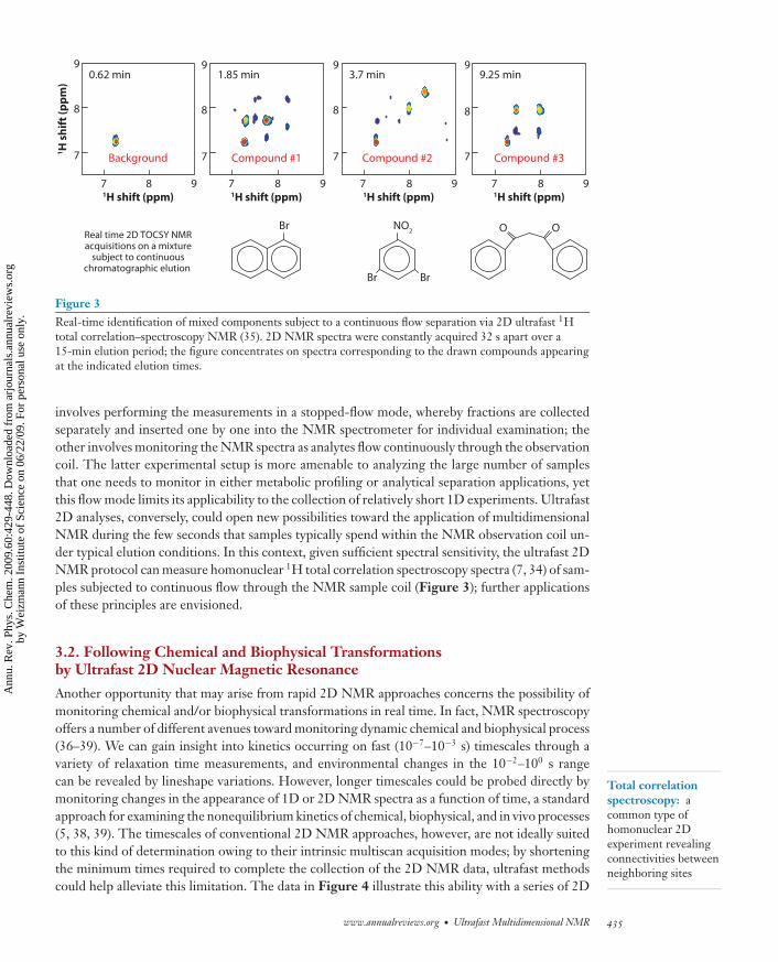
Figure 3Real-time identification of mixed components subject to a continuous flow separation via 2D ultrafast 1Htotal correlation–spectroscopy NMR (35). 2D NMR spectra were constantly acquired 32 s apart over a15-min elution period; the figure concentrates on spectra corresponding to the drawn compounds appearingat the indicated elution times.
Total correlationspectroscopy: acommon type ofhomonuclear 2Dexperiment revealingconnectivities betweenneighboring sites
involves performing the measurements in a stopped-flow mode, whereby fractions are collectedseparately and inserted one by one into the NMR spectrometer for individual examination; theother involves monitoring the NMR spectra as analytes flow continuously through the observationcoil. The latter experimental setup is more amenable to analyzing the large number of samplesthat one needs to monitor in either metabolic profiling or analytical separation applications, yetthis flow mode limits its applicability to the collection of relatively short 1D experiments. Ultrafast2D analyses, conversely, could open new possibilities toward the application of multidimensionalNMR during the few seconds that samples typically spend within the NMR observation coil un-der typical elution conditions. In this context, given sufficient spectral sensitivity, the ultrafast 2DNMR protocol can measure homonuclear 1H total correlation spectroscopy spectra (7, 34) of sam-ples subjected to continuous flow through the NMR sample coil (Figure 3); further applicationsof these principles are envisioned.
3.2. Following Chemical and Biophysical Transformationsby Ultrafast 2D Nuclear Magnetic Resonance
Another opportunity that may arise from rapid 2D NMR approaches concerns the possibility ofmonitoring chemical and/or biophysical transformations in real time. In fact, NMR spectroscopyoffers a number of different avenues toward monitoring dynamic chemical and biophysical process(36–39). We can gain insight into kinetics occurring on fast (10−7–10−3 s) timescales through avariety of relaxation time measurements, and environmental changes in the 10−2–100 s rangecan be revealed by lineshape variations. However, longer timescales could be probed directly bymonitoring changes in the appearance of 1D or 2D NMR spectra as a function of time, a standardapproach for examining the nonequilibrium kinetics of chemical, biophysical, and in vivo processes(5, 38, 39). The timescales of conventional 2D NMR approaches, however, are not ideally suitedto this kind of determination owing to their intrinsic multiscan acquisition modes; by shorteningthe minimum times required to complete the collection of the 2D NMR data, ultrafast methodscould help alleviate this limitation. The data in Figure 4 illustrate this ability with a series of 2D
www.annualreviews.org • Ultrafast Multidimensional NMR 435
Ann
u. R
ev. P
hys.
Che
m. 2
009.
60:4
29-4
48. D
ownl
oade
d fr
om a
rjou
rnal
s.an
nual
revi
ews.
org
by W
eizm
ann
Inst
itute
of
Scie
nce
on 0
6/22
/09.
For
per
sona
l use
onl
y.

ANRV373-PC60-21 ARI 25 February 2009 18:3
678
6
7
8
678
6
7
8
41.4 s
678
6
7
8
11.5 s4.6 s
NO2
O2N
CN
+ MeO– +
i
ii
i
ii ii
NO2
H OMe
O2N
HH
CN
NO2
H
OMe
O2N
H
CN
H
1H shift (ppm)1H shift (ppm)1H
sh
ift
(pp
m)
1H shift (ppm)
(i) (ii)
Figure 4Real-time monitoring of a chemical reaction using ultrafast 2D total correlation spectroscopy NMR (40),illustrating the appearance of compounds (bottom) at the indicated times since triggering the reaction insidethe NMR magnet. Both reactants were prepolarized prior to the reaction: One reactant was loaded into the5-mm NMR tube, whereas the other was held within a capillary prior to its sudden injection.
Heteronuclearmultiple-quantumcorrelation: acommon 2D NMRapproach fordetermining theconnectivity betweenheteronuclear pairs(e.g., 1H and 15N)
NMR snapshots collected while a chemical reaction was triggered in situ within the NMR tube.Data acquisition in this kind of experiment began slightly before triggering the chemical process,and a series of 2D total correlation spectroscopy NMR spectra recorded every 2.3 s could reflecteven transient stages of the ensuing Messenhimer complexation reaction (40).
If and when sensitivity suffices, biophysical transformations such as H/D exchange processesor folding events could also become amenable to this kind of measurement. In these biomolecularcases (in which sensitivity is always a challenge), it is often convenient to couple the ultrafast2D protocol with other acquisition methods capable of minimizing the experiment’s recycle de-lay and thereby maximize the number of 2D NMR frames available per unit acquisition time.One such recent proposal is the band-selective, optimized flip-angle, short-transient, heteronu-clear multiple-quantum correlation NMR experiment (41, 42), which enables the reduction ofthe interscan repetition delay down to ≤100 ms while preserving high sensitivity by relying onan accelerated spin-lattice relaxation imparted from a selective excitation in combination withoptimized flip angles to enhance the steady-state signal arising from the excited spins (43–45).This in return allows the recording of conventionally sampled 2D 1H-15N or 1H-13C correlationspectra within minimal experimental times of just several seconds. Moreover, if combined withultrafast 2D NMR, this methodology can yield spectra at very high frame rates, compatible withthe continuous following of biophysical processes. Figure 5 illustrates an example of these spec-tra (46), with an H/D exchange process followed under experimental conditions that combinethe very short interscan delay afforded by the optimized flip-angle protocol, with the single-scancapabilities of ultrafast NMR. This method is capable of collecting 2D NMR correlation spectraat ≈1-mM protein concentrations with frame rates of approximately 1 Hz; this could become animportant aid in studying subsecond processes in which peak positions or intensities change owingto protein or nucleic acid folding, binding, or dynamics.
436 Mishkovsky · Frydman
Ann
u. R
ev. P
hys.
Che
m. 2
009.
60:4
29-4
48. D
ownl
oade
d fr
om a
rjou
rnal
s.an
nual
revi
ews.
org
by W
eizm
ann
Inst
itute
of
Scie
nce
on 0
6/22
/09.
For
per
sona
l use
onl
y.

ANRV373-PC60-21 ARI 25 February 2009 18:3
2.4 s 4.8 s
9.6 s 14.4 s
77.588.59
110
115
120
125
77.588.59
110
115
120
125
77.588.59
110
115
120
125
77.588.59
110
115
120
125
19.2 s 24 s
31.2 s 40.8 s
77.588.59
110
115
120
125
77.588.59
110
115
120
125
77.588.59
110
115
120
125
77.588.59
110
115
120
125
S65
H68
I61I13
1H shift (ppm)
15N
sh
ift
(pp
m)
15N
sh
ift
(pp
m)
15N
sh
ift
(pp
m)
15N
sh
ift
(pp
m)
1H shift (ppm)
Figure 5Representative series of real-time 2D ultrafast heteronuclear multiple-quantum correlation NMR spectrarecorded on an ubiquitin solution, using the band-selective, optimized-flip-angle, short-transient protocol,following the dissolution of an initially fully protonated lyophilized powder onto a D2O-based buffer as afunction of the time delay elapsed since the dissolution (46). The repetition time between full recording was∼2.4 s, and the data were monitored over a 20-min interval. Red circles indicate selected NH resonancesthat rapidly disappear owing to the H → D exchange.
3.3. Ultrafast 2D Nuclear Magnetic Resonance on Prepolarized Samples
In recent years, we have witnessed the emergence of a variety of NMR prepolarization methodscapable of building up nuclear polarizations that exceed their thermal counterparts by severalorders of magnitude (47–50). In many instances, these methodologies are constrained to extracttheir superspectra within a single or at most a few transients, making them poor starting pointsfor conventional 2D NMR experiments requiring the collection of a large set of scans. Sucha scenario presents another instance in which new possibilities could arise through the use of
www.annualreviews.org • Ultrafast Multidimensional NMR 437
Ann
u. R
ev. P
hys.
Che
m. 2
009.
60:4
29-4
48. D
ownl
oade
d fr
om a
rjou
rnal
s.an
nual
revi
ews.
org
by W
eizm
ann
Inst
itute
of
Scie
nce
on 0
6/22
/09.
For
per
sona
l use
onl
y.
ANRV373-PC60-21 ARI 25 February 2009 18:3
a Single-scan: dark b Single-scan: light
c-Alad-Ala-Tyr-Tyr-Glu-Glu-Alad-Ala] = 0.5 mM
Hβ
H3/H5
1
1
2
3
4
5
6
7
2 3 4 5 6 71H shift (ppm)
1 2 3 4 5 6 71H shift (ppm)
1H
sh
ift
(pp
m)
Tyrosine residuesTyrosine residuesTyrosine residues
Figure 6Potential benefits resulting from combining chemically induced dynamic nuclear polarization (CIDNP)prepolarization and ultrafast 2D methods. Both panels illustrate single-scan 2D total correlationspectroscopy 1H NMR spectra recorded on a cyclic octapeptide dissolved in D2O at a 0.5-mMconcentration. (a) Spectrum under standard conditions. (b) Spectrum resulting from pre-irradiating thesample for 0.5 s using a 480-nm light source (2W), generating a CIDNP enhancement of tyrosine’s aromaticprotons (52).
Dynamic nuclearpolarization (DNP):a generic method toenhance nuclear spinpolarizations bysaturating theresonances of nearbyelectrons
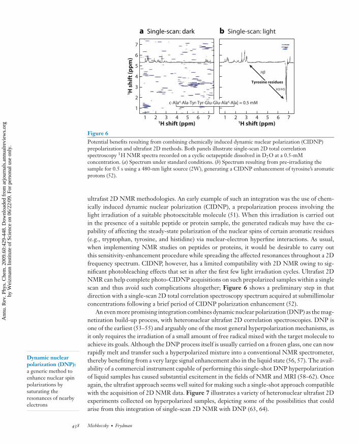
ultrafast 2D NMR methodologies. An early example of such an integration was the use of chem-ically induced dynamic nuclear polarization (CIDNP), a prepolarization process involving thelight irradiation of a suitable photoexcitable molecule (51). When this irradiation is carried outin the presence of a suitable peptide or protein sample, the generated radicals may have the ca-pability of affecting the steady-state polarization of the nuclear spins of certain aromatic residues(e.g., tryptophan, tyrosine, and histidine) via nuclear-electron hyperfine interactions. As usual,when implementing NMR studies on peptides or proteins, it would be desirable to carry outthis sensitivity-enhancement procedure while spreading the affected resonances throughout a 2Dfrequency spectrum. CIDNP, however, has a limited compatibility with 2D NMR owing to sig-nificant photobleaching effects that set in after the first few light irradiation cycles. Ultrafast 2DNMR can help complete photo-CIDNP acquisitions on such prepolarized samples within a singlescan and thus avoid such complications altogether; Figure 6 shows a preliminary step in thatdirection with a single-scan 2D total correlation spectroscopy spectrum acquired at submillimolarconcentrations following a brief period of CIDNP polarization enhancement (52).
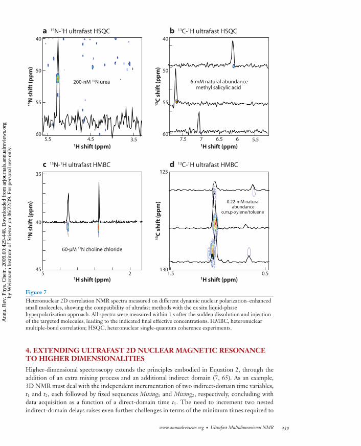
An even more promising integration combines dynamic nuclear polarization (DNP) as the mag-netization build-up process, with heteronuclear ultrafast 2D correlation spectroscopies. DNP isone of the earliest (53–55) and arguably one of the most general hyperpolarization mechanisms, asit only requires the irradiation of a small amount of free radical mixed with the target molecule toachieve its goals. Although the DNP process itself is usually carried on a frozen glass, one can nowrapidly melt and transfer such a hyperpolarized mixture into a conventional NMR spectrometer,thereby benefiting from a very large signal enhancement also in the liquid state (56, 57). The avail-ability of a commercial instrument capable of performing this single-shot DNP hyperpolarizationof liquid samples has caused substantial excitement in the fields of NMR and MRI (58–62). Onceagain, the ultrafast approach seems well suited for making such a single-shot approach compatiblewith the acquisition of 2D NMR data. Figure 7 illustrates a variety of heteronuclear ultrafast 2Dexperiments collected on hyperpolarized samples, depicting some of the possibilities that couldarise from this integration of single-scan 2D NMR with DNP (63, 64).
438 Mishkovsky · Frydman
Ann
u. R
ev. P
hys.
Che
m. 2
009.
60:4
29-4
48. D
ownl
oade
d fr
om a
rjou
rnal
s.an
nual
revi
ews.
org
by W
eizm
ann
Inst
itute
of
Scie
nce
on 0
6/22
/09.
For
per
sona
l use
onl
y.
ANRV373-PC60-21 ARI 25 February 2009 18:3
a 15N-1H ultrafast HSQC
6-mM natural abundancemethyl salicylic acid
0.51.55 2
125
130
60-μM 15N choline chloride
0.22-mM naturalabundance
o,m,p-xylene/toluene
200-nM 15N urea
5.5 4.5 3.560
55
40
50
45
40
35
7.5 7 66.5 5.560
55
40
50
b 13C-1H ultrafast HSQC
c 15N-1H ultrafast HMBC d 13C-1H ultrafast HMBC
1H shift (ppm)1H shift (ppm)
1H shift (ppm)
13C
sh
ift
(pp
m)
15N
sh
ift
(pp
m)
15N
sh
ift
(pp
m)
1H shift (ppm)
13C
sh
ift
(pp
m)
Figure 7Heteronuclear 2D correlation NMR spectra measured on different dynamic nuclear polarization–enhancedsmall molecules, showing the compatibility of ultrafast methods with the ex situ liquid-phasehyperpolarization approach. All spectra were measured within 1 s after the sudden dissolution and injectionof the targeted molecules, leading to the indicated final effective concentrations. HMBC, heteronuclearmultiple-bond correlation; HSQC, heteronuclear single-quantum coherence experiments.
4. EXTENDING ULTRAFAST 2D NUCLEAR MAGNETIC RESONANCETO HIGHER DIMENSIONALITIES
Higher-dimensional spectroscopy extends the principles embodied in Equation 2, through theaddition of an extra mixing process and an additional indirect domain (7, 65). As an example,3D NMR must deal with the independent incrementation of two indirect-domain time variables,t1 and t2, each followed by fixed sequences Mixing1 and Mixing2, respectively, concluding withdata acquisition as a function of a direct-domain time t3. The need to increment two nestedindirect-domain delays raises even further challenges in terms of the minimum times required to
www.annualreviews.org • Ultrafast Multidimensional NMR 439
Ann
u. R
ev. P
hys.
Che
m. 2
009.
60:4
29-4
48. D
ownl
oade
d fr
om a
rjou
rnal
s.an
nual
revi
ews.
org
by W
eizm
ann
Inst
itute
of
Scie
nce
on 0
6/22
/09.
For
per
sona
l use
onl
y.
ANRV373-PC60-21 ARI 25 February 2009 18:3
1H
15N
13C
Gz
F1spatial
encoding
F2temporalencoding
F3signal
digitization
F 2/13C
(±10
00 H
z)(±
1000
Hz)
F 2/13C
(±10
00 H
z)
a Hybrid 3D pulse sequence b 3D HNCO 2-mM U-13C/15N LAF
2
τ τ τ τ
Τ Τ Τ Τπ/2π/2 (ii)(ii)
(i)(i)
(ii)
(i)
Cα+Ge
–Ge
+Ga
–Ga
N1
t2
2t2
t1maxt1
max t1max2Ta
Acquisition time = 85 sAcquisition time = 85 sAcquisition time = 85 s
F3/1H (±1000 Hz)
(±1000 Hz)F1 / 15N (±1187 Hz)
(±1187 Hz) F3/1 H (±1000 Hz)
F1 / 15N (±1187 Hz)
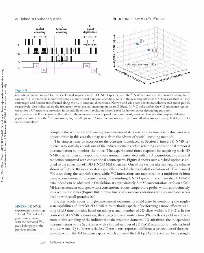
Figure 8(a) Pulse sequence assayed for the accelerated acquisition of 3D HNCO spectra, with the 15N dimension spatially encoded along the zaxis and 13C interactions monitored using a conventional temporal encoding. Data in the resulting ultrafast 2D planes are then suitablyrearranged and Fourier transformed along the t2, t3 temporal dimensions. Narrow and wide bars denote nonselective π/2 and π pulses,respectively; also indicated are the frequency-swept spatial-encoding pulses (π/2 labels). All 13C pulses affect the CO resonance regionexcept for a Cα-specific π inversion in the middle of the t2 evolution (shaped pulse) for homonuclear decoupling purposes.(b) Experimental 3D spectrum collected with the sequence shown in panel a on a uniformly enriched leucine-alanine-phenylalaninepeptide solution. For the 13C dimension, �t2 = 500 μs and 16 time increments were used; overall, 64 scans with a recycle delay of 1.3 swere accumulated.
HNCO: 3D NMRexperiment correlating1H and 15N peaks of agiven amide groupwith the carbonyl 13Cpeak belonging to theprevious residue
complete the acquisition of these higher-dimensional data sets; this section briefly discusses newopportunities in this area that may arise from the advent of spatial-encoding methods.
The simplest way to incorporate the concepts introduced in Section 2 into a 3D NMR se-quence is to spatially encode one of the indirect domains, while retaining a conventional temporalincrementation to monitor the other. The experimental times required for acquiring such 3DNMR data set then correspond to those normally associated with a 2D acquisition, a substantialreduction compared with conventional counterparts. Figure 8 shows such a hybrid option as ap-plied to the collection of a 3D HNCO NMR data set. Out of the various alternatives, the schemeshown in Figure 8a incorporates a spatially encoded chemical-shift evolution of 1H-enhanced15N sites along the sample’s z axis, while 13C interactions are monitored in a multiscan fashionusing a conventional t2 incrementation. The resulting HNCO spectrum confirms that 3D NMRdata indeed can be obtained in this fashion at approximately 2-mM concentration levels on a 500-MHz spectrometer equipped with a conventional room-temperature probe, within approximately90-s acquisition times (Figure 8b). Similar timescales and concentrations are also amenable whendealing with small proteins (66).
Further accelerations of high-dimensional experiments could arise by combining the single-scan capabilities of ultrafast 2D NMR with methods capable of performing a more efficient scan-ning of nD time domains based on taking a small number of 2D slices within it (19–21). In thecontext of 3D NMR acquisition, these projection-reconstruction (PR) methods yield an efficientroute to the sampling of the indirect-domain evolution domains. PR substitutes the independentincrementation of the (t1,t2) times with a limited number of 2D NMR acquisitions involving fixedratios α = tan−1( t2
t1) of these variables. These in turn represent different α-projections of the spec-
tral data within the 3D frequency space, which can yield the full F1F2F3 3D spectrum being sought
440 Mishkovsky · Frydman
Ann
u. R
ev. P
hys.
Che
m. 2
009.
60:4
29-4
48. D
ownl
oade
d fr
om a
rjou
rnal
s.an
nual
revi
ews.
org
by W
eizm
ann
Inst
itute
of
Scie
nce
on 0
6/22
/09.
For
per
sona
l use
onl
y.

ANRV373-PC60-21 ARI 25 February 2009 18:3
TOCSY-based 3D UF PR
n-butyl chloride
a HSQC-based 3D UF PR15N FMOC-Ala/15N FMOC-Val
b
1
0
–1
4
3
2
7
6
99 8 7 6 5 4
F2 (H1, ppm)
F 3 (H
-1, p
pm)F 1
(H
-1, p
pm
)
F2 (N-15, ppm)
F 1 (
H-1
, pp
m)
3 2 1 0 –1
8
3.75
2.25
0.755
4.0
5.5
5.0
4.5
7.0
6.5
116 112 108F 3
(H-1
, ppm
)
104 100 98 92
7.5
5.2
5.0
4.86.0
Figure 9Examples of ultrafast (UF)/projection-reconstruction (PR) 3D NMR acquisitions based on (a) homonuclearand (b) heteronuclear correlations. Each data set was reconstructed from three single-scan ultrafast 2Dprojections, leading to a total experimental time of just 5 s (67). HSQC, heteronuclear single-quantumcoherence; TOCSY, total correlation spectroscopy.
if inverse-radon-transformed for each discrete direct-domain F3 frequency. As the individual pro-jected planes needed by this protocol involve the collection of 2D time-domain signals, these inturn can be greatly accelerated by translating the PR principles into the spatio-temporal termsinvolved in ultrafast NMR. Assuming that the full sample length L is employed when applyingthese joint indirect-domain (t1,t2) encodings, the original PR conditions can be cast into theseterms by demanding that the spatio-temporal constants {Ci}i = 1,2 associated with each indirectdomain be incremented according to α = tan−1( C2
C1) (67). Then, 3D NMR spectra can be recorded
by combining a small number of 2D projections, each of which in turn is collected within a singlescan. Figure 9 illustrates this integration with 3D correlation data reconstructed using only threesingle-scan 2D NMR projections.
Section 2 describes how frequency-swept RF pulses applied in combination with field gradientscould be used to spatially encode and subsequently read out the indirect domain of a 2D NMRexperiment. Provided that linearly independent gradient geometries are used, these argumentscan be extended further to include an arbitrary number of indirect dimensions (24) and thereby toacquire arbitrarily high nD NMR data within a single transient. For example, Figure 10a incorpo-rates two separate gradients arranged along linearly independent z and y geometries to implementthe consecutive encodings of the spin evolution that would be needed for a 3D NMR acquisition.The first of these processes induces an �1t1-dependent winding of the spin packets along the zdirection, whereas for each z coordinate, a second gradient generates an �2t2-dependent encodingalong the y axis. Because of the ensuing double-winding of spin packets, the overall bulk magne-tization is reduced again to zero, and an acquisition process implemented on the resulting sampleis associated with a null initial signal. Moreover, only the simultaneous application of suitable Gz
a ,Gy
a acquisition gradients can succeed in aligning the spin packets throughout the sample’s volume,leading to a definition of the peak’s indirect-domain frequencies as a function of two independentvariables: kz/Cz ∝ ∫
Gza (t)dt(Lz/tmax
1 ) and ky/Cy ∝ ∫Gy
a (t)dt(Ly/tmax2 ). Oscillation of these two
wave numbers as a function of a direct-domain acquisition time t3 yields a rasterization of the3D F1F2t3 mixed domain, and 1D FT as a function of t3 can thereby provide the full set of peak
www.annualreviews.org • Ultrafast Multidimensional NMR 441
Ann
u. R
ev. P
hys.
Che
m. 2
009.
60:4
29-4
48. D
ownl
oade
d fr
om a
rjou
rnal
s.an
nual
revi
ews.
org
by W
eizm
ann
Inst
itute
of
Scie
nce
on 0
6/22
/09.
For
per
sona
l use
onl
y.
ANRV373-PC60-21 ARI 25 February 2009 18:3
Τ Τ ΤΤ
ττττ
ππ
ππ
1H
15N
13C
Gz
+Gez
–Gez
+Gey
–Gey
–Gay –G
ay
+Gay+G
ay
N1
N1 N
2
+Gaz
–Gaz
+Gaz
–Gaz
Gy
Gx
a 3D HNCO with full spatial encoding
t1
max t2
max 2Ta
2Ta
F 3/1H
(±
29
0 H
z)(±
29
0 H
z)
F1/1515N (±650 Hz)
(±650 Hz)
F2 / 1313C (±770 Hz)
(±770 Hz)
F 3/1
H (
±2
90
Hz)
b 3D HNCO 2-mM U-13C/15N LAF
(ii)(ii)
(i)(i)
(ii)
(i)
Acquisition time = 85 sAcquisition time = 85 sAcquisition time = 2 s
F1/15 N (±650 Hz)
F2 / 13C (±770 Hz)
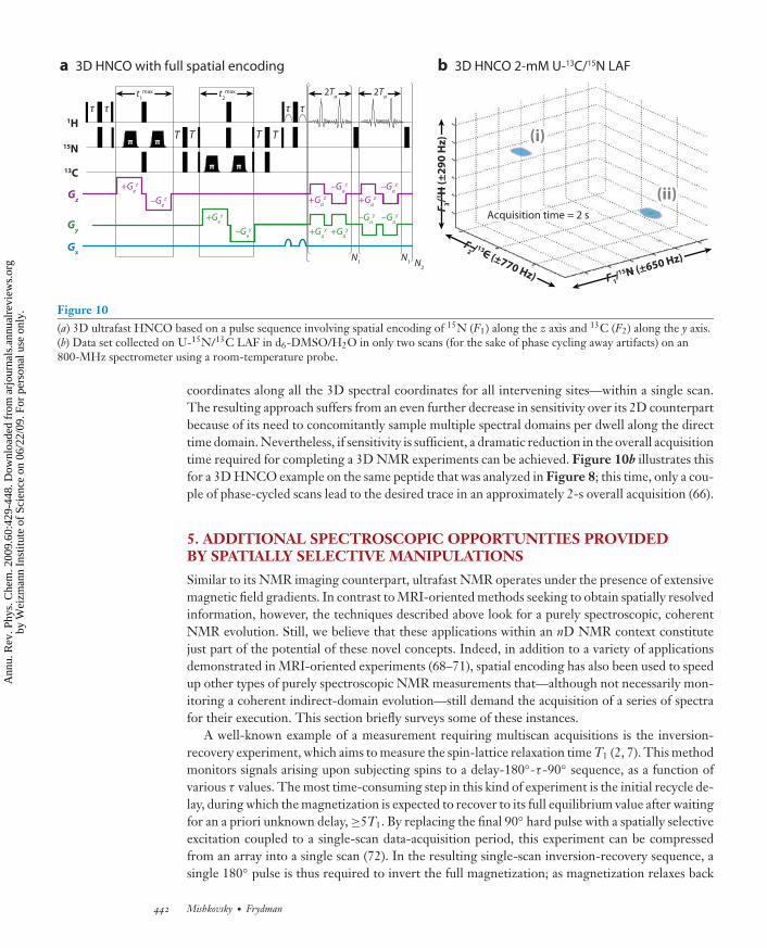
Figure 10(a) 3D ultrafast HNCO based on a pulse sequence involving spatial encoding of 15N (F1) along the z axis and 13C (F2) along the y axis.(b) Data set collected on U-15N/13C LAF in d6-DMSO/H2O in only two scans (for the sake of phase cycling away artifacts) on an800-MHz spectrometer using a room-temperature probe.
coordinates along all the 3D spectral coordinates for all intervening sites—within a single scan.The resulting approach suffers from an even further decrease in sensitivity over its 2D counterpartbecause of its need to concomitantly sample multiple spectral domains per dwell along the directtime domain. Nevertheless, if sensitivity is sufficient, a dramatic reduction in the overall acquisitiontime required for completing a 3D NMR experiments can be achieved. Figure 10b illustrates thisfor a 3D HNCO example on the same peptide that was analyzed in Figure 8; this time, only a cou-ple of phase-cycled scans lead to the desired trace in an approximately 2-s overall acquisition (66).
5. ADDITIONAL SPECTROSCOPIC OPPORTUNITIES PROVIDEDBY SPATIALLY SELECTIVE MANIPULATIONS
Similar to its NMR imaging counterpart, ultrafast NMR operates under the presence of extensivemagnetic field gradients. In contrast to MRI-oriented methods seeking to obtain spatially resolvedinformation, however, the techniques described above look for a purely spectroscopic, coherentNMR evolution. Still, we believe that these applications within an nD NMR context constitutejust part of the potential of these novel concepts. Indeed, in addition to a variety of applicationsdemonstrated in MRI-oriented experiments (68–71), spatial encoding has also been used to speedup other types of purely spectroscopic NMR measurements that—although not necessarily mon-itoring a coherent indirect-domain evolution—still demand the acquisition of a series of spectrafor their execution. This section briefly surveys some of these instances.
A well-known example of a measurement requiring multiscan acquisitions is the inversion-recovery experiment, which aims to measure the spin-lattice relaxation time T1 (2, 7). This methodmonitors signals arising upon subjecting spins to a delay-180◦-τ -90◦ sequence, as a function ofvarious τ values. The most time-consuming step in this kind of experiment is the initial recycle de-lay, during which the magnetization is expected to recover to its full equilibrium value after waitingfor an a priori unknown delay, ≥5T1. By replacing the final 90◦ hard pulse with a spatially selectiveexcitation coupled to a single-scan data-acquisition period, this experiment can be compressedfrom an array into a single scan (72). In the resulting single-scan inversion-recovery sequence, asingle 180◦ pulse is thus required to invert the full magnetization; as magnetization relaxes back
442 Mishkovsky · Frydman
Ann
u. R
ev. P
hys.
Che
m. 2
009.
60:4
29-4
48. D
ownl
oade
d fr
om a
rjou
rnal
s.an
nual
revi
ews.
org
by W
eizm
ann
Inst
itute
of
Scie
nce
on 0
6/22
/09.
For
per
sona
l use
onl
y.

ANRV373-PC60-21 ARI 25 February 2009 18:3
2 3
2
3 10 ms
2 3
2
3 450 ms
2 3
2
3 1.25 s
H3C H
3C
C
O
CH3
NCH
3
C
O
CH3
NCH
3
2 3
2
3 850 ms
1H shift (ppm)1H shift (ppm)
1H
sh
ift
(pp
m)
1H shift (ppm)1H shift (ppm)
1
1
2
2
3 1
3
2
3
Figure 11Array of single-scan 2D exchange NMR spectra collected on a dimethylacetamide/D2O solution as afunction of the indicated mixing periods. All these 2D spectra rely on a common initial spatial-encodingprocess along the sample’s z axis; each spectrum was then recalled and measured following different exchangedelays, using 90◦ final excitation pulses and selecting different planes along the sample’s x direction. Thetime required to collect each of the 2D NMR experiments was 66 ms plus the indicated duration of themixing times; the acquisition of the full series of 2D NMR spectra required only 1.499 s, including all thenested mixing delays (73).
to equilibrium, different slices of the sample are then successively excited and probed as a functionof different recovery durations τ—thus completing the measurement of the full array in a singletransient. A somewhat similar idea, incorporating an array of a 2D set of exchange experiments asa function of varying mixing periods (73), has also been demonstrated in this fashion. Figure 11illustrates how, in very short acquisition times, the effects of chemical exchange can be clearlyfollowed by the growth of the ensuing off-diagonal exchange-derived cross peaks.
Another spatial manipulation principle that—although it does not include the encoding of spincoherences—also compresses an arrayed acquisition into a single scan has been discussed recentlyin connection with the 2D diffusion ordered spectroscopy experiment (74, 75). This shift-resolvedcharacterization of molecular diffusion is used widely, among other applications, for separatingNMR peaks according to the individual chemical components partaking of a complex mixture andfor extracting the approximate hydrodynamic radii of molecules in solution (76–78). A suitableapplication of swept pulses and refocused gradients also manages, in this case, to impart along asample’s axis an array of diffusion-dependent q-encoding values of the kind needed to extract afull 2D diffusion ordered spectroscopy spectral set. Figure 12 summarizes the type of results thatcan become available by this single-scan methodology.
An additional example of how spatial-encoding strategies can be exploited to compress whatusually entails a series of different scans into a single transient has been demonstrated recentlywithin the context of the phase cycling of RF pulses for coherence selection and/or artifact suppres-sion purposes. Phase cycling is particularly onerous scan-wise within the context of conventional2D acquisitions, given the large number of pulses involved, the numerous coherence transferpathways thus potentially created, and the need to couple their suitable filtration to the samplingof numerous incremented indirect-domain evolution delays (2, 7). It is consequently particularlyattractive to employ spatially selective strategies to compress complex cycling schemes effectivelyinto a single scan. The feasibility of this approach was demonstrated by the implementation of thevarious RF manipulations that would normally be executed throughout the independent step of thecycling scheme, at once and throughout different positions along the sample. The joint detectionof the signals from all slices and their suitable combination could then yield the desired coher-ence pathway contributions to the final observable signal (79). In another development relatedto artifact suppression in high-resolution 2D NMR, spatially selective manipulations have beenapplied to eliminate zero-quantum coherence (ZQC) contributions often arising upon executing
www.annualreviews.org • Ultrafast Multidimensional NMR 443
Ann
u. R
ev. P
hys.
Che
m. 2
009.
60:4
29-4
48. D
ownl
oade
d fr
om a
rjou
rnal
s.an
nual
revi
ews.
org
by W
eizm
ann
Inst
itute
of
Scie
nce
on 0
6/22
/09.
For
per
sona
l use
onl
y.

ANRV373-PC60-21 ARI 25 February 2009 18:3
781H NMR shift dimension (ppm)
Single-scan 2D DOSY experiment
D d
om
ain
(*1
06 m
m2 m
s–1)
9100
1
2
3
Figure 12Single-scan 2D diffusion ordered spectroscopy (DOSY) behavior observed for a mixture of tetraphenyl-porphine, benzaldehyde, and diphenylether (≈50 mM each) in CDCl3 at 25◦C. The horizontal axiscorresponds to the high-resolution trace afforded by this sequence along the 1H dimension; the vertical axismeasures the diffusion coefficients extracted for each peak in the spectrum as a function of chemical shift.Notice the clear separation between the slower diffusivity shown by the porphyrin molecule peaks(D ≈ 1.2 × 10⁻⁶ mm2 ms−1) and the remaining, smaller aromatic molecules (75).
2D nuclear Overhauser enhancement spectroscopy–type NMR sequences. These experiments arechallenged by difficulties in differentiating longitudinal Mz-type magnetizations and/or spin-orderstates from ZQCs—given that both behave identically when subject to conventional phase-cyclingor gradient-purging procedures. Conversely, the action of a frequency-swept 180◦ pulse in thepresence of a magnetic field gradient enables one to eliminate ZQC-derived artifacts while re-taining the remaining longitudinal components because of the destructive interference betweenZQC contributions that then derive from different parts in the sample (80, 81).
6. CONCLUDING REMARKS
This review presents the basic principles of and some opportunities that could arise from recentlyproposed NMR spatial-encoding procedures, particularly for compressing what would normallyrequire the collection of multiple 1D NMR spectra, into a single scan. One main feature of thisMRI-derived approach to spectroscopy is that, inasmuch as nD NMR acquisitions are concerned,it is general: Therefore, provided that sufficient sensitivity is available, it can be applied to acquirea variety of homo- and heteronuclear 2D NMR spectra within a single scan. The availability ofa protocol capable of delivering such rich information within short timescales could thus benefitcertain chemical and biological studies that have hitherto been impractical with the aid of 2Dspectroscopy. Potential applications discussed within this context include the use of 2D NMR asa real-time tool for following chemical and biophysical processes, for monitoring the fingerprintsof samples undergoing continuous flow, and for exploiting the sensitivity that can be providedby single-shot nuclear hyperpolarization methods. A number of spectroscopic extensions of theseideas are also discussed, including their use in accelerating higher-dimensional (≥3D) NMR ex-periments; in speeding up diffusion, exchange, and relaxation measurements; and in acceleratingand perfecting the elimination of spectral artifacts.
Despite the exiting new opportunities that arise when considering the various applications ofthese concepts, we stress that these benefits can only materialize in practice if there is sufficientspectral sensitivity. Indeed, whereas spatial encoding enables the compression of a wide variety
444 Mishkovsky · Frydman
Ann
u. R
ev. P
hys.
Che
m. 2
009.
60:4
29-4
48. D
ownl
oade
d fr
om a
rjou
rnal
s.an
nual
revi
ews.
org
by W
eizm
ann
Inst
itute
of
Scie
nce
on 0
6/22
/09.
For
per
sona
l use
onl
y.

ANRV373-PC60-21 ARI 25 February 2009 18:3
of arrayed NMR experiments into a single scan, useful results can only be extracted from suchacquisitions if there is sufficient sensitivity to observe the desired information. In this respect, ona per-scan basis, ultrafast nD NMR methods prove less sensitive than conventional counterpartsbecause of their need to rapidly sample multiple dimensions at once. This in turn brings about aneed to further expand the NMR’s receiver bandwidth, thereby causing an increase in the incomingspectral noise. All this highlights the importance of enhancing sensitivity as a major goal to focuson, if intending to bring out the full potential of these accelerated acquisition methods.
SUMMARY POINTS
1. Imaging-derived concepts can be used to encode the time evolution normally undergoneby spins over the course of an NMR experiment, along a spatial domain.
2. This spatial-encoding approach enables the compression of arrayed NMR acquisitions,particularly those involved in multidimensional NMR experiments, into a single scan.
3. This compression enables the reduction of the overall acquisition times of arbitraryhomo- or heteronuclear 2D NMR experiments by several orders of magnitude, resultingin an ultrafast approach to multidimensional spectroscopy that may enable new applica-tions in chemical, biophysical, and in vivo NMR.
4. Among the possible applications for this new acquisition mode are real-time 2D NMRmeasurements of analytes subject to continuous flow, the following of transient chemi-cal and biophysical rearrangements, 2D measurements on hyperpolarized samples, andextensions to higher-dimensional experiments.
5. The success of these new potential applications hinges on the ability of achieving sufficientsensitivity to observe the desired multidimensional NMR signals within a single, or atmost a few, transients.
DISCLOSURE STATEMENT
The authors are not aware of any biases that might be perceived as affecting the objectivity of thisreview.
ACKNOWLEDGMENTS
We are grateful to Boaz Shapira, Yoav Shrot, and Maayan Gal for the insight they providedthroughout the research work hereby described. This research was supported by the U.S.-IsraelBinational Science Foundation (BSF 2004298), the Israel Science Foundation (ISF 1206/05), theEuropean Commission (EU-NMR contract no. 026145), and the generosity of the Perlman FamilyFoundation.
LITERATURE CITED
1. Grant DM, Harris RK, eds. 1996. Encyclopedia of NMR. Chichester, NY: Wiley & Sons2. Derome AE. 1987. Modern NMR Techniques for Chemistry Research. Oxford: Pergamon3. Frydman L. 2001. Perspectives in solid state NMR: spin-1/2 and beyond. Annu. Rev. Phys. Chem. 52:463–984. Cavanagh J, Fairbrother WJ, Palmer AG, Skelton NJ. 1996. Protein NMR Spectroscopy: Principles and
Practice. San Diego: Academic
www.annualreviews.org • Ultrafast Multidimensional NMR 445
Ann
u. R
ev. P
hys.
Che
m. 2
009.
60:4
29-4
48. D
ownl
oade
d fr
om a
rjou
rnal
s.an
nual
revi
ews.
org
by W
eizm
ann
Inst
itute
of
Scie
nce
on 0
6/22
/09.
For
per
sona
l use
onl
y.

ANRV373-PC60-21 ARI 25 February 2009 18:3
5. deGraaf R. 2007. In Vivo NMR Spectroscopy: Principles and Techniques. Chichester, NY: Wiley & Sons6. Buxton RB. 2001. An Introduction to Functional MRI: Principles and Techniques. Cambridge: Cambridge
Univ. Press7. Ernst RR, Boudenhausen G, Wokaun A. 1987. Principles of Nuclear Magnetic Resonance in One and Two
Dimensions. Oxford: Clarendon8. Ernst RR, Anderson WA. 1966. Application of Fourier transform spectroscopy to magnetic resonance.
Rev. Sci. Instrum. 37:93–1029. Jeener J. 1994 (1971). Ampere Summer School lecture notes. In NMR and More in Honor of Anatole
Abragam, ed. M Goldman, M Porneuf, pp. 1–379. Les Ulis, France: Ed. Phys.10. Aue WP, Bartholdi E, Ernst RR. 1976. Two dimensional spectroscopy: application to nuclear magnetic
resonance. J. Chem. Phys. 64:2229–4611. Kupce E, Nishida T, Freeman R. 2003. Hadamard NMR spectroscopy. Prog. Nucl. Magn. Reson. Spectrosc.
42:95–12212. Atreya HS, Szyperski T. 2005. Rapid NMR data collection. Methods Enzymol. 394:78–10813. Mandelshtam VA. 2000. The multidimensional filter diagonalization method I. Theory and numerical
implementation. J. Magn. Reson. 144:343–5614. Hoch JC, Stern AS. 2001. Maximum entropy reconstruction, spectrum analysis and deconvolution in
multidimensional nuclear magnetic resonance. Methods Enzymol. 338:159–7815. Orekhov Y, Ibraghimov I, Billeter M. 2003. Optimizing resolution in multidimensional NMR by three-
way decomposition. J. Biomol. NMR 27:165–7316. Bruschweiler R, Zhang F. 2004. Covariance nuclear magnetic resonance spectroscopy. J. Chem. Phys.
120:5253–6017. Kupce E, Freeman R. 2003. Frequency-domain Hadamard spectroscopy. J. Magn. Reson. 162:158–6518. Bodenhausen G, Ernst RR. 1981. The accordion experiment, a simple approach to three-dimensional
NMR spectroscopy. J. Magn. Reson. 45:367–7319. Szyperski T, Wider G, Bushweller JH, Wuthrich K. 1993. Reduced dimensionality in triple-resonance
NMR experiments. J. Am. Chem. Soc. 115:9307–820. Kim S, Szyperski T. 2003. GFT NMR, a new approach to rapidly obtain precise high-dimensional NMR
spectral information. J. Am. Chem. Soc. 125:1385–9321. Kupce E, Freeman R. 2004. Projection-reconstruction technique for speeding up multidimensional NMR
spectroscopy. J. Am. Chem. Soc. 126:6429–4022. Frydman L, Scherf T, Lupulescu A. 2002. The acquisition of multidimensional NMR spectra within a
single scan. Proc. Natl. Acad. Sci. USA 99:15858–6223. Frydman L, Scherf T, Lupulescu A. 2003. Principles and features of single-scan two-dimensional NMR
spectroscopy. J. Am. Chem. Soc. 125:9204–1724. Shrot Y, Frydman L. 2003. Single-scan NMR spectroscopy at arbitrary dimensions. J. Am. Chem. Soc.
125:11385–9625. Pelupessy P. 2003. Adiabatic single scan two-dimensional NMR spectroscopy. J. Am. Chem. Soc.
125:12345–5026. Shrot Y, Shapira B, Frydman L. 2004. Ultrafast 2D NMR spectroscopy using continuous spatial encoding
of the spin interactions. J. Magn. Reson. 171:163–7027. Tal A, Shapira B, Frydman L. 2005. A continuous phase-modulated approach to spatial encoding in
ultrafast 2D NMR spectroscopy. J. Magn. Reson. 176:107–1428. Andersen NS, Kockenberger W. 2005. A simple approach for phase-modulated single-scan 2D NMR
spectroscopy. Magn. Reson. Chem. 43:795–9729. Shapira B, Shrot Y, Frydman L. 2006. Symmetric spatial encoding in ultrafast 2D NMR spectroscopy.
J. Magn. Reson. 178:33–4130. Giraudeau P, Akoka S. 2007. A new detection scheme for ultrafast 2D J-resolved spectroscopy. J. Magn.
Reson. 186:352–5731. Shrot Y, Frydman L. 2008. Spatial encoding strategies for ultrafast multidimentional nuclear magnetic
resonance. J. Chem. Phys. 128:05220932. Lindon JC, Holmes E, Nicholson JK. 2004. Toxicological applications of magnetic resonance. Progr. Nucl.
Magn. Reson. Spectrosc. 45:109–43
446 Mishkovsky · Frydman
Ann
u. R
ev. P
hys.
Che
m. 2
009.
60:4
29-4
48. D
ownl
oade
d fr
om a
rjou
rnal
s.an
nual
revi
ews.
org
by W
eizm
ann
Inst
itute
of
Scie
nce
on 0
6/22
/09.
For
per
sona
l use
onl
y.

ANRV373-PC60-21 ARI 25 February 2009 18:3
33. Albert K, ed. 2002. On-Line LC-NMR and Related Techniques. Chichester, NY: Wiley & Sons34. Braunschweiler L, Ernst RR. 1983. Coherence transfer by isotropic mixing: applications to proton cor-
relation spectroscopy. J. Magn. Reson. 53:521–2835. Shapira B, Karton A, Aronzon D, Frydman L. 2004. Real-time 2D NMR identification of analytes un-
dergoing continuous chromatographic separation. J. Am. Chem. Soc. 126:1262–6536. Dayie KT, Wagner G, Lefevre J-F. 1996. Theory and practice of nuclear spin relaxation in proteins.
Annu. Rev. Phys. Chem. 47:242–8337. Bain AD. 2003. Chemical exchange in NMR. Prog. Nucl. Magn. Reson. Spectrosc. 43:63–10338. Dobson CM, Hore PJ. 1998. Kinetic studies of protein folding using NMR spectroscopy. Nat. Struct.
Biol. 5:504–739. Ishima R, Torchia DA. 2000. Protein dynamics from NMR. Nat. Struct. Biol. 7:740–4340. Gal M, Mishkovsky M, Frydman L. 2006. Real-time monitoring of chemical transformation by ultrafast
2D NMR spectroscopy. J. Am. Chem. Soc. 128:951–5641. Muller L. 1979. Sensitivity enhanced detection of weak nuclei using heteronuclear multiple quantum
coherence. J. Am. Chem. Soc. 101:4481–8442. Schanda P, Brutscher B. 2005. Very fast two-dimentional NMR spectroscopy for real-time investigation
of dynamic events in proteins on the time scale of seconds. J. Am. Chem. Soc. 127:8014–1543. Pervushin K, Voegeli B, Eletsky A. 2002. Longitudinal 1H relaxation optimization in TROSY NMR
spectroscopy. J. Am. Chem. Soc. 124:12898–90244. Freeman R, Hill HDW. 1971. Phase and intensity abnormalities in Fourier transform NMR. J. Magn.
Reson. 4:366–8345. Ross A, Salzmann M, Senn H. 1997. Fast-HMQC using Ernst angle pulses: an efficient tool for screening
of ligand binding to target protein. J. Biomol. NMR 10:289–9646. Gal M, Schanda P, Brutsher B, Frydman L. 2007. UltraSOFAST HMQC NMR and the repetitive
acquisition of 2D protein spectra at Hz rates. J. Am. Chem. Soc. 129:1372–7747. Bowers CR, Weitekamp DP. 1986. Transformation of symetrization order in nuclear magnetic resonance.
Phys. Rev. Lett. 57:2645–4848. Eisenschmid TC, Kirss RU, Deutsch PP, Hommeltoft SI, Eisenberg R, et al. 1987. Parahydrogen induced
polarization in hydrogenation reaction. J. Am. Chem. Soc. 109:8089–9149. Albert MS, Cates GD, Driehuys B, Happer W, Saam B, et al. 1994. Biological magnetic resonance imaging
using laser polarized 129Xe. Nature 370:199–20150. Navon G, Song YQ, Room T, Appelt S, Taylor RE, Pines A. 1996. Enhancement of solution NMR and
MRI with laser polarized xenon. Science 271:1848–5151. Muus LT, Atkins PW, McLauchlen KA, Pedersen JB. 1977. Chemically Induced Magnetic Polzarization.
Dordrecht: D. Reidel52. Shapira B, Morris E, Muszkat KA, Frydman L. 2004. Sub-second 2D NMR spectroscopy at submilimolar
consentrations. J. Am. Chem. Soc. 126:11756–5753. Carver TR, Slichter CP. 1953. Polarization of nuclear spin in metals. Phys. Rev. 92:212–1354. Hausser KH, Stehilk D. 1968. Dynamic nuclear polarization in liquids. Adv. Magn. Reson. 3:79–13955. Abragam A, Goldman M. 1982. Nuclear Magnetism: Order and Disorder. Oxford: Oxford Univ. Press56. Ardenkjaer-Larsen JH, Fridlund B, Gram A, Hansson G, Hansson L, et al. 2003. Increase in signal-to-
noise ratio of >10,000 times in liquid-state NMR. Proc. Natl. Acad. Sci. USA 100:10158–6357. Joo CG, Hu KN, Bryant JA, Griffin RG. 2006. In situ temperature jump high-frequency dynamic nu-
clear polarization experiments: enhanced sensitivity in liquid-state NMR spectroscopy. J. Am. Chem. Soc.128:9428–32
58. Golman K, Ardenkjær-Larsen JH, Petersson JS, Mansson S, Leunbach I. 2003. Molecular imaging withendogenous substances. Proc. Natl. Acad. Sci. USA 100:10435–39
59. Golman K, Zandt R, Thaning M. 2006. Real time metabolic imaging. Proc. Natl. Acad. Sci. USA 103:11270–75
60. Chen AP, Albers MJ, Cunningham CH, Kohler SJ, Yen YF, et al. 2007. Hyperpolarized C-13 spectroscopicimaging of the TRAMP mouse at 3T: initial experience. Magn. Reson. Med. 58:1099–106
61. Day SE, Kettunen MI, Gallagher FA, Hu DE, Lerche M, et al. 2007. Detecting tumor response totreatment using hyperpolarized 13C magnetic resonance imaging. Nat. Med. 13:1382–87
www.annualreviews.org • Ultrafast Multidimensional NMR 447
Ann
u. R
ev. P
hys.
Che
m. 2
009.
60:4
29-4
48. D
ownl
oade
d fr
om a
rjou
rnal
s.an
nual
revi
ews.
org
by W
eizm
ann
Inst
itute
of
Scie
nce
on 0
6/22
/09.
For
per
sona
l use
onl
y.

ANRV373-PC60-21 ARI 25 February 2009 18:3
62. Gallagher FA, Kettunen MI, Day SE, Hu DE, Ardenkjaer-Larsen JH, et al. 2008. Magnetic resonanceimaging of pH in vivo using hyperpolarized 13C-labelled bicarbonate. Nature 457:940–43
63. Frydman L, Blazina D. 2007. Ultrafast two-dimensional nuclear magnetic resonance spectroscopy ofhyperpolarized solutions. Nat. Phys. 3:415–19
64. Mishkovsky M, Frydman L. 2008. Progress in hyperpolarized ultrafast 2D nuclear magnetic resonance.Chem. Phys. Chem. 16:2340–48
65. Sattler M, Schleucher J, Griesinger C. 1999. Heteronuclear multidimensional NMR experiments for thestructure determination of proteins in solution employing pulsed field gradients. Progr. Nucl. Magn. Reson.Spectrosc. 34:93–158
66. Mishkovsky M, Gal M, Frydman L. 2007. Spatially-encoded strategies in the execution of biomolecular-oriented 3D NMR experiments. J. Biomol. NMR 39:291–301
67. Mishkovsky M, Kupce E, Frydman L. 2007. Ultrafast based projection-reconstruction three-dimensionalnuclear magnetic resonance spectroscopy. J. Chem. Phys. 127:034507
68. Shrot Y, Frydman L. 2005. Spatially-encoded NMR and the acquisition of 2D magnetic resonance imageswithin a single scan. J. Magn. Reson. 172:179–90
69. Tal A, Frydman L. 2006. Spatial encoding and the acquisition of high definition MR images in inhomo-geneous magnetic fields. J. Magn. Reson. 181:179–94
70. Chamberlain R, Park JY, Corum C, Yacoub E, Ugurbil K, et al. 2007. RASER: a new ultrafast magneticresonance imaging method. Magn. Reson. Med. 58:794–99
71. Tal A, Frydman L. 2007. Spectroscopic imaging from spatially-encoded single-scan multidimensionalMRI data. J. Magn. Reson. 189:46–58
72. Bhattacharyya R, Kumar A. 2003. A fast method for the measurement of long-spin lattice relaxation timesby single scan inversion recovery experiment. Chem. Phys. Lett. 383:99–103
73. Shapira B, Frydman L. 2003. Arrayed acquisition of 2D exchange NMR spectra within a single experiment.J. Magn. Reson. 165:320–24
74. Thrippleton MJ, Loening NM, Keeler J. 2003. A fast method for the measurement of diffusion coefficients:one-dimensional DOSY. Magn. Reson. Chem. 41:441–47
75. Shrot Y, Frydman L. 2008. Single scan 2D DOSY NMR. J. Magn. Reson. 195:226–3176. Morris KF, Johnson CS. 1993. Resolution of discrete and continuous molecular-size distributions by
means of diffusion-ordered 2D NMR spectroscopy. J. Am. Chem. Soc. 115:4291–9977. Barjat H, Morris GA, Smart S, Swanson AG, Williams SCR. 1995. High-resolution diffusion-ordered 2D
spectroscopy (HR-DOSY): a new tool for the analysis of complex mixtures. J. Magn. Reson. B 108:170–7278. Johnson CS. 1999. Diffusion-ordered NMR spectroscopy: principles and applications. Prog. NMR Spec-
trosc. 34:203–5579. Parish DN, Szyperski T. 2008. Simultaneously cycled NMR spectroscopy. J. Am. Chem. Soc. 130:4925–3380. Thrippleton JM, Keeler J. 2003. Elimination of zero quantum interference in two dimensional NMR
spectra. Angew. Chem. Int. Ed. Engl. 42:3938–4181. Cano E, Thrippleton JM, Keeler J, Shaka AJ. 2004. Cascaded z-filters for efficient single-scan suppression
of zero-quantum coherence. J. Magn. Reson. 167:291–97
448 Mishkovsky · Frydman
Ann
u. R
ev. P
hys.
Che
m. 2
009.
60:4
29-4
48. D
ownl
oade
d fr
om a
rjou
rnal
s.an
nual
revi
ews.
org
by W
eizm
ann
Inst
itute
of
Scie
nce
on 0
6/22
/09.
For
per
sona
l use
onl
y.

AR373-FM ARI 25 February 2009 17:55
Annual Review ofPhysical Chemistry
Volume 60, 2009 Contents
Frontispiece � � � � � � � � � � � � � � � � � � � � � � � � � � � � � � � � � � � � � � � � � � � � � � � � � � � � � � � � � � � � � � � � � � � � � � � � � � � � � � � � � � � � � xiv
Sixty Years of Nuclear MomentsJohn S. Waugh � � � � � � � � � � � � � � � � � � � � � � � � � � � � � � � � � � � � � � � � � � � � � � � � � � � � � � � � � � � � � � � � � � � � � � � � � � � � � � � � � 1
Dynamics of Liquids, Molecules, and Proteins Measured with Ultrafast2D IR Vibrational Echo Chemical Exchange SpectroscopyM.D. Fayer � � � � � � � � � � � � � � � � � � � � � � � � � � � � � � � � � � � � � � � � � � � � � � � � � � � � � � � � � � � � � � � � � � � � � � � � � � � � � � � � � � � �21
Photofragment Spectroscopy and Predissociation Dynamics of WeaklyBound MoleculesHanna Reisler � � � � � � � � � � � � � � � � � � � � � � � � � � � � � � � � � � � � � � � � � � � � � � � � � � � � � � � � � � � � � � � � � � � � � � � � � � � � � � � � � �39
Second Harmonic Generation, Sum Frequency Generation, and χ (3):Dissecting Environmental Interfaces with a Nonlinear Optical SwissArmy KnifeFranz M. Geiger � � � � � � � � � � � � � � � � � � � � � � � � � � � � � � � � � � � � � � � � � � � � � � � � � � � � � � � � � � � � � � � � � � � � � � � � � � � � � �61
Dewetting and Hydrophobic Interaction in Physical and BiologicalSystemsBruce J. Berne, John D. Weeks, and Ruhong Zhou � � � � � � � � � � � � � � � � � � � � � � � � � � � � � � � � � � � � � � � � �85
Photoelectron Spectroscopy of Multiply Charged AnionsXue-Bin Wang and Lai-Sheng Wang � � � � � � � � � � � � � � � � � � � � � � � � � � � � � � � � � � � � � � � � � � � � � � � � � � � � � � 105
Intrinsic Particle Properties from Vibrational Spectra of AerosolsOmar F. Sigurbjörnsson, George Firanescu, and Ruth Signorell � � � � � � � � � � � � � � � � � � � � � � � � � 127
Nanofabrication of Plasmonic StructuresJoel Henzie, Jeunghoon Lee, Min Hyung Lee, Warefta Hasan, and Teri W. Odom � � � � 147
Chemical Synthesis of Novel Plasmonic NanoparticlesXianmao Lu, Matthew Rycenga, Sara E. Skrabalak, Benjamin Wiley,and Younan Xia � � � � � � � � � � � � � � � � � � � � � � � � � � � � � � � � � � � � � � � � � � � � � � � � � � � � � � � � � � � � � � � � � � � � � � � � � � � � � � 167
Atomic-Scale Templates Patterned by Ultrahigh Vacuum ScanningTunneling Microscopy on SiliconMichael A. Walsh and Mark C. Hersam � � � � � � � � � � � � � � � � � � � � � � � � � � � � � � � � � � � � � � � � � � � � � � � � � � 193
DNA Excited-State Dynamics: From Single Bases to the Double HelixChris T. Middleton, Kimberly de La Harpe, Charlene Su, Yu Kay Law,Carlos E. Crespo-Hernández, and Bern Kohler � � � � � � � � � � � � � � � � � � � � � � � � � � � � � � � � � � � � � � � � � � � � 217
viii
Ann
u. R
ev. P
hys.
Che
m. 2
009.
60:4
29-4
48. D
ownl
oade
d fr
om a
rjou
rnal
s.an
nual
revi
ews.
org
by W
eizm
ann
Inst
itute
of
Scie
nce
on 0
6/22
/09.
For
per
sona
l use
onl
y.

AR373-FM ARI 25 February 2009 17:55
Dynamics of Light Harvesting in PhotosynthesisYuan-Chung Cheng and Graham R. Fleming � � � � � � � � � � � � � � � � � � � � � � � � � � � � � � � � � � � � � � � � � � � � � 241
High-Resolution Infrared Spectroscopy of the Formic Acid DimerOzgür Birer and Martina Havenith � � � � � � � � � � � � � � � � � � � � � � � � � � � � � � � � � � � � � � � � � � � � � � � � � � � � � � � 263
Quantum Coherent Control for Nonlinear Spectroscopyand MicroscopyYaron Silberberg � � � � � � � � � � � � � � � � � � � � � � � � � � � � � � � � � � � � � � � � � � � � � � � � � � � � � � � � � � � � � � � � � � � � � � � � � � � � � 277
Coherent Control of Quantum Dynamics with Sequences of UnitaryPhase-Kick PulsesLuis G.C. Rego, Lea F. Santos, and Victor S. Batista � � � � � � � � � � � � � � � � � � � � � � � � � � � � � � � � � � � � � 293
Equation-Free Multiscale Computation: Algorithms and ApplicationsIoannis G. Kevrekidis and Giovanni Samaey � � � � � � � � � � � � � � � � � � � � � � � � � � � � � � � � � � � � � � � � � � � � � � 321
Chirality in Nonlinear OpticsLevi M. Haupert and Garth J. Simpson � � � � � � � � � � � � � � � � � � � � � � � � � � � � � � � � � � � � � � � � � � � � � � � � � � � 345
Physical Chemistry of DNA VirusesCharles M. Knobler and William M. Gelbart � � � � � � � � � � � � � � � � � � � � � � � � � � � � � � � � � � � � � � � � � � � � � 367
Ultrafast Dynamics in Reverse MicellesNancy E. Levinger and Laura A. Swafford � � � � � � � � � � � � � � � � � � � � � � � � � � � � � � � � � � � � � � � � � � � � � � � � 385
Light Switching of Molecules on SurfacesWesley R. Browne and Ben L. Feringa � � � � � � � � � � � � � � � � � � � � � � � � � � � � � � � � � � � � � � � � � � � � � � � � � � � � � 407
Principles and Progress in Ultrafast Multidimensional NuclearMagnetic ResonanceMor Mishkovsky and Lucio Frydman � � � � � � � � � � � � � � � � � � � � � � � � � � � � � � � � � � � � � � � � � � � � � � � � � � � � � � 429
Controlling Chemistry by Geometry in Nanoscale SystemsL. Lizana, Z. Konkoli, B. Bauer, A. Jesorka, and O. Orwar � � � � � � � � � � � � � � � � � � � � � � � � � � � � � 449
Active Biological MaterialsDaniel A. Fletcher and Phillip L. Geissler � � � � � � � � � � � � � � � � � � � � � � � � � � � � � � � � � � � � � � � � � � � � � � � � � 469
Wave-Packet and Coherent Control DynamicsKenji Ohmori � � � � � � � � � � � � � � � � � � � � � � � � � � � � � � � � � � � � � � � � � � � � � � � � � � � � � � � � � � � � � � � � � � � � � � � � � � � � � � � � 487
Indexes
Cumulative Index of Contributing Authors, Volumes 56–60 � � � � � � � � � � � � � � � � � � � � � � � � � � � 513
Cumulative Index of Chapter Titles, Volumes 56–60 � � � � � � � � � � � � � � � � � � � � � � � � � � � � � � � � � � � � 516
Errata
An online log of corrections to Annual Review of Physical Chemistry articles may befound at http://physchem.annualreviews.org/errata.shtml
Contents ix
Ann
u. R
ev. P
hys.
Che
m. 2
009.
60:4
29-4
48. D
ownl
oade
d fr
om a
rjou
rnal
s.an
nual
revi
ews.
org
by W
eizm
ann
Inst
itute
of
Scie
nce
on 0
6/22
/09.
For
per
sona
l use
onl
y.