primer 01 microbiology 101 - gwumc.edu easily decolorized (appear red) and some gram-negative...
TRANSCRIPT
Primer 01Microbiology 101
Patrick R. Murray, Ph.D.Worldwide Director, Scientific Affairs
Becton Dickinson DiagnosticsAdjunct Professor, Dept. of Pathology
University of MarylandBaltimore, Maryland
(c) 2013 Infectious Disease Board Review 1

DISCLOSURESFinancial Relationships with Relevant Commercial Interests
• Becton Dickinson Diagnostics – Employee and stockholder/ownership interest
(c) 2013 Infectious Disease Board Review 2

• Because most organisms are colorless and transparent, various dyes (stains) are used to see the individual cells.
• A variety of different types of stains are used in the microbiology lab including –– Contrast stains (e.g., methylene blue, lactophenol
cotton blue, India ink, iodine)– Differential stains (e.g., Gram stain, acid-fast
stains, spore stains, Giemsa stain, silver stains, Trichrome stain)
– Fluorescent stains (e.g., acridine orange, auramine-rhodamine, calcofluor white, antibody-conjugated fluorescent stains)
Overview – Microbiological Stains
(c) 2013 Infectious Disease Board Review 3

• Contrast stains are nonspecific stains used to detect the normally transparent organisms such as bacteria and fungi.
• The methylene blue stain was one of the first ones developed (used to stain C. diphtheriae in this example).
• This stain has now been replaced for the most part with other contrast stains.
Contrast Stain: Methylene Blue Stain
(c) 2013 Infectious Disease Board Review 4

Contrast Stain: Lactophenol CottonBlue (LCB) Stain
• LCB is a commonly used contrast stain used primarily for observing the morphology of fungal molds.
• This is an example of Aspergillus fumigatus.
(c) 2013 Infectious Disease Board Review 5

Differential Stain: Gram Stain• The Gram stain is the most
commonly used differential stain for bacteria.
• It is used to distinguish gram-positive (purple; top figure) from gram-negative (red; bottom figure) bacteria.
• Some gram-positive bacteria can be easily decolorized (appear red) and some gram-negative bacteria can retain the crystal violet and appear somewhat gram-positive.
• The shape and spatial arrangement of cells is as important as the color of cells for interpreting this stain.
(c) 2013 Infectious Disease Board Review 6

Differential Stains: Acid-Fast Stains• One of the most common acid-fast
stains is the Kinyoun (cold) stain.
• This is a variation of the older Ziehl-Neelsen (hot) acid-fast stain where the slides had to be heated during staining.
• Mycobacteria (top) stain with both stains.
• “Modified” acid-fast stain – if a weak decolorizing solution is used to remove the primary stain, then partially or weakly acid-fastorganisms can be stained. Partially acid-fast organisms include:– Nocardia (bottom figure)– Rhodococcus– Tsukamurella– Gordonia
(c) 2013 Infectious Disease Board Review 7

Differential Stains: Trichrome stain• The trichrome stain is used
for the detection of intestinal protozoa.
• The parasite’s cytoplasm stains blue-green and the internal structure stain red or purplish red (arrow).
• Background debris stain blue-green so care must be taken to distinguish between the protozoa and the background.
(c) 2013 Infectious Disease Board Review 8

Fluorescent Stain: Auramine Rhodamine Stain
• The AR stain is a fluorochrome stain used to detect acid-fast bacteria.
• This is essentially the same as a Kinyoun stain except the carbol fuchsin dye is replaced with fluorescent dyes (auramine and rhodamine) and the stained smears are examined under UV illumination using a fluorescent microscope.
• Because of the high contrast between the fluorescing rods and the black background, this stain is more sensitive than the Kinyoun stain.
(c) 2013 Infectious Disease Board Review 9
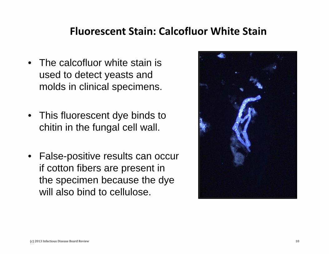
Fluorescent Stain: Calcofluor White Stain
• The calcofluor white stain is used to detect yeasts and molds in clinical specimens.
• This fluorescent dye binds to chitin in the fungal cell wall.
• False-positive results can occur if cotton fibers are present in the specimen because the dye will also bind to cellulose.
(c) 2013 Infectious Disease Board Review 10

Antibody‐Conjugated Fluorescent Stains
• Fluorescent antibody stains are specific stains where antibodies are attached to a fluorochrome (such as fluorescein).
• The antibody-antigen binding is detected by the fluorescence.
• Two examples of this test are illustrated here – Pneumocystis (top figure) and tissue culture cells infected with Varicella-Zoster virus (bottom figure) and then stained with fluorescein-tagged antibodies.
(c) 2013 Infectious Disease Board Review 11

Bacteriology
(c) 2013 Infectious Disease Board Review 12

Gram-Positive Cocci
StreptococcusStaphylococcus
Enterococcus(c) 2013 Infectious Disease Board Review 13
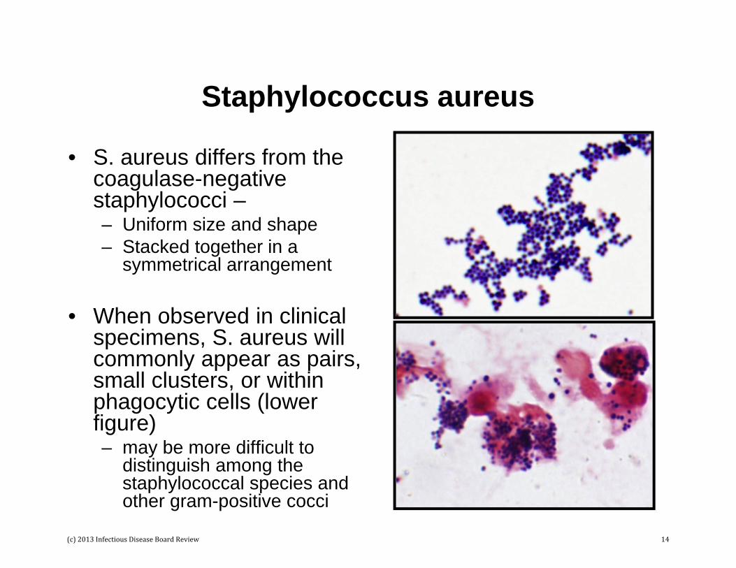
Staphylococcus aureus
• S. aureus differs from the coagulase-negative staphylococci –– Uniform size and shape– Stacked together in a
symmetrical arrangement
• When observed in clinical specimens, S. aureus will commonly appear as pairs, small clusters, or within phagocytic cells (lower figure) – may be more difficult to
distinguish among the staphylococcal species and other gram-positive cocci
(c) 2013 Infectious Disease Board Review 14

Staphylococcus aureus and Candida albicans
• This photo illustrates the size difference between S. aureus (black arrow) and yeasts, in this case Candida albicans (red arrow).
• Yeast can appear as gram-positive although they tend to decolorize readily.
(c) 2013 Infectious Disease Board Review 15

Streptococcus pyogenes
• Most group A streptococci are Streptococcus pyogenes
• Group A streptococci form long chains or round cells, described as a “string of pearls”
• Streptococcus pyogenes is the only Streptococcus species that is PYR positive.
(c) 2013 Infectious Disease Board Review 16
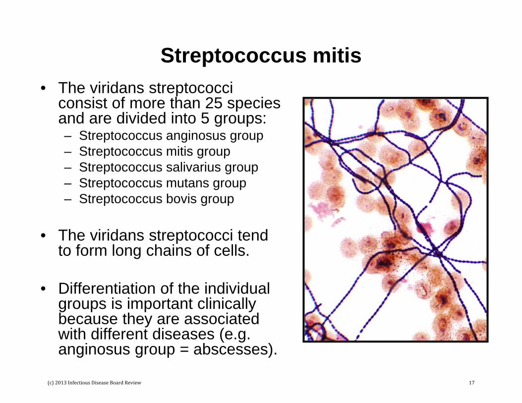
Streptococcus mitis• The viridans streptococci
consist of more than 25 species and are divided into 5 groups:– Streptococcus anginosus group– Streptococcus mitis group– Streptococcus salivarius group– Streptococcus mutans group– Streptococcus bovis group
• The viridans streptococci tend to form long chains of cells.
• Differentiation of the individual groups is important clinically because they are associated with different diseases (e.g. anginosus group = abscesses).
(c) 2013 Infectious Disease Board Review 17
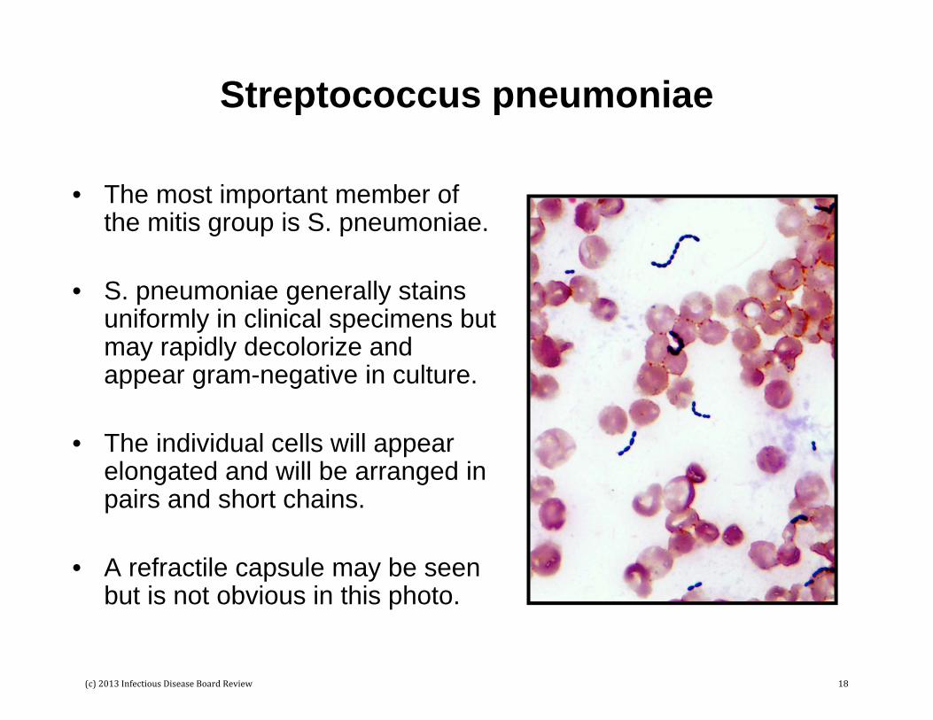
Streptococcus pneumoniae
• The most important member of the mitis group is S. pneumoniae.
• S. pneumoniae generally stains uniformly in clinical specimens but may rapidly decolorize and appear gram-negative in culture.
• The individual cells will appear elongated and will be arranged in pairs and short chains.
• A refractile capsule may be seen but is not obvious in this photo.
(c) 2013 Infectious Disease Board Review 18

Enterococcus
• Enterococci will appear as elongated gram-positive cocci arranged in pairs and short chains.
• These bacteria cannot be distinguished from S. pneumoniae by Gram stain.
• S. pneumoniae can be rapidly identified by exposing the cell to bile (or another detergent) that will dissolve the bacteria (“bile soluble”).
• Enterococci can be identified by the PYR test (positive test for the enzyme pyrrolidonyl arylamidase).
• The most important enterococci are E. faecium, E. faecalis, E. gallinarum, and E. casseliflavus.
(c) 2013 Infectious Disease Board Review 19
Gram-Positive Rods
The most common or important gram-positive rods are: • Spore-forming rods
– Aerobes – Bacillus species (e.g., B. cereus, B. anthracis)– Anaerobes – Clostridium species (e.g., C. perfringens,
C. septicum, C. difficile)
• Non-sporeforming rods– Uniform shape – Listeria, Lactobacillus– Irregular (coryneform) shape – Corynebacterium,
Propionibacterium
• Acid-fast rods– Acid-fast – Mycobacterium– Weakly (partially) acid-fast – Nocardia, Rhodococcus
(c) 2013 Infectious Disease Board Review 20

Spore-Forming Rods
• Two genera of bacteria commonly isolated in the lab form spores: Bacillus (aerobe) and Clostridium (anaerobe).
• These images are Gram stains of B. cereus from a culture plate.
• Spores are not stained with the Gram stain and will appear as clear areas in the cell (red arrow).
(c) 2013 Infectious Disease Board Review 21

Bacillus anthracis
• This is a Gram stain of B. anthracis in the blood culture from a bacteremic patient.
• Note that spores are not seen and the bacteria form long chains. This is characteristic of this pathogen.
(c) 2013 Infectious Disease Board Review 22

Clostridium perfringens
• C. perfringens (arrows) in a mixed culture with E. coli and K. pneumoniae.
• This is one of the most common species of Clostridium isolated from clinical specimens.
• C. perfringens spores are almost never seen; rods are described as “boxcar shaped” or rectangular and are generally larger than most bacteria.
(c) 2013 Infectious Disease Board Review 23
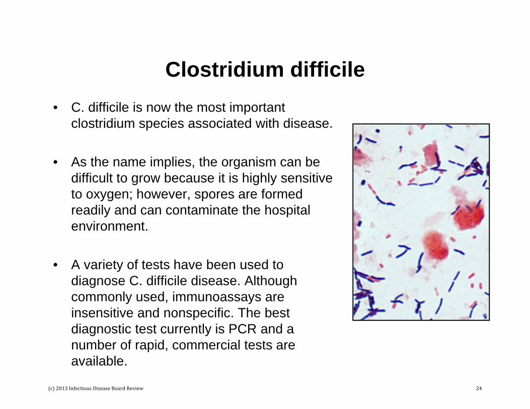
Clostridium difficile• C. difficile is now the most important
clostridium species associated with disease.
• As the name implies, the organism can be difficult to grow because it is highly sensitive to oxygen; however, spores are formed readily and can contaminate the hospital environment.
• A variety of tests have been used to diagnose C. difficile disease. Although commonly used, immunoassays are insensitive and nonspecific. The best diagnostic test currently is PCR and a number of rapid, commercial tests are available.
(c) 2013 Infectious Disease Board Review 24
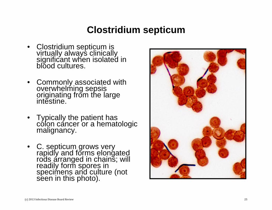
Clostridium septicum• Clostridium septicum is
virtually always clinically significant when isolated in blood cultures.
• Commonly associated with overwhelming sepsis originating from the large intestine.
• Typically the patient has colon cancer or a hematologic malignancy.
• C. septicum grows very rapidly and forms elongated rods arranged in chains; will readily form spores in specimens and culture (not seen in this photo).
(c) 2013 Infectious Disease Board Review 25
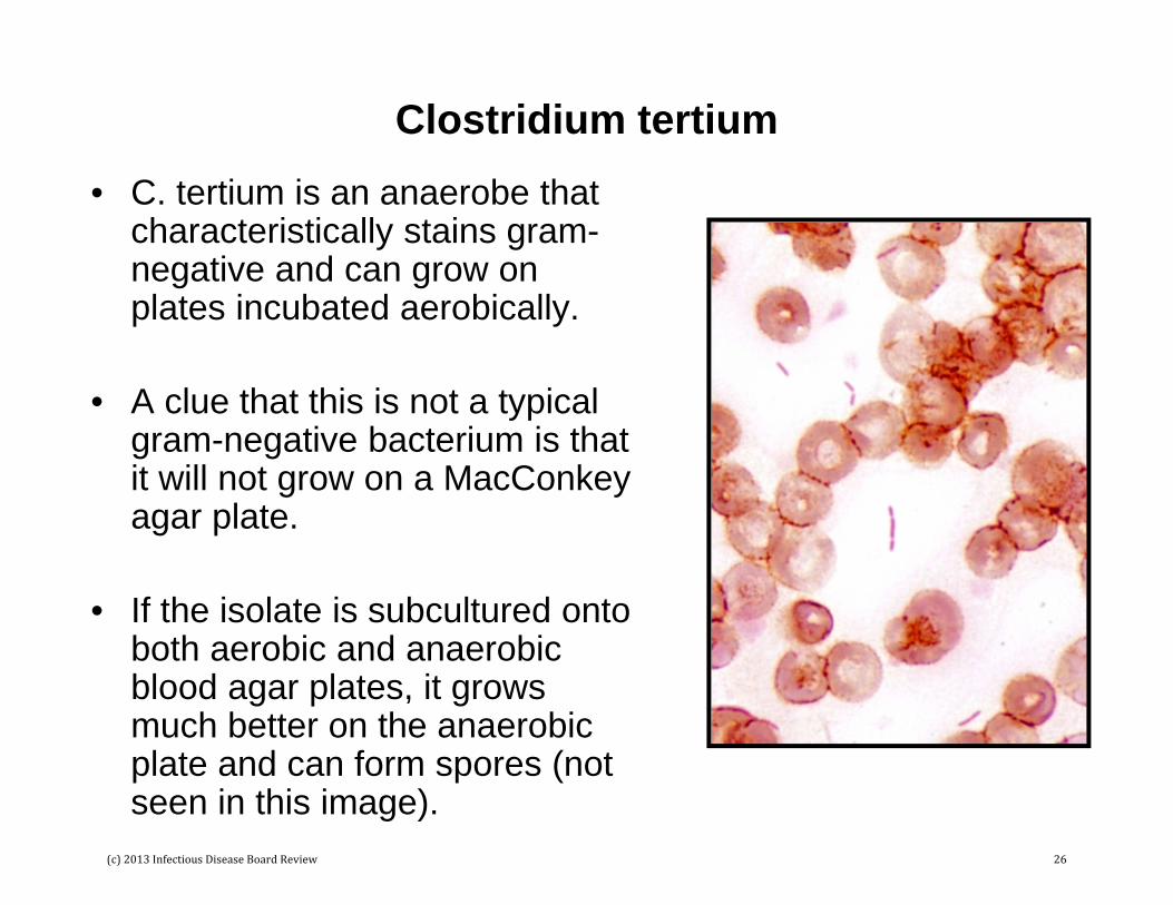
Clostridium tertium• C. tertium is an anaerobe that
characteristically stains gram-negative and can grow on plates incubated aerobically.
• A clue that this is not a typical gram-negative bacterium is that it will not grow on a MacConkeyagar plate.
• If the isolate is subcultured onto both aerobic and anaerobic blood agar plates, it grows much better on the anaerobic plate and can form spores (not seen in this image).
(c) 2013 Infectious Disease Board Review 26
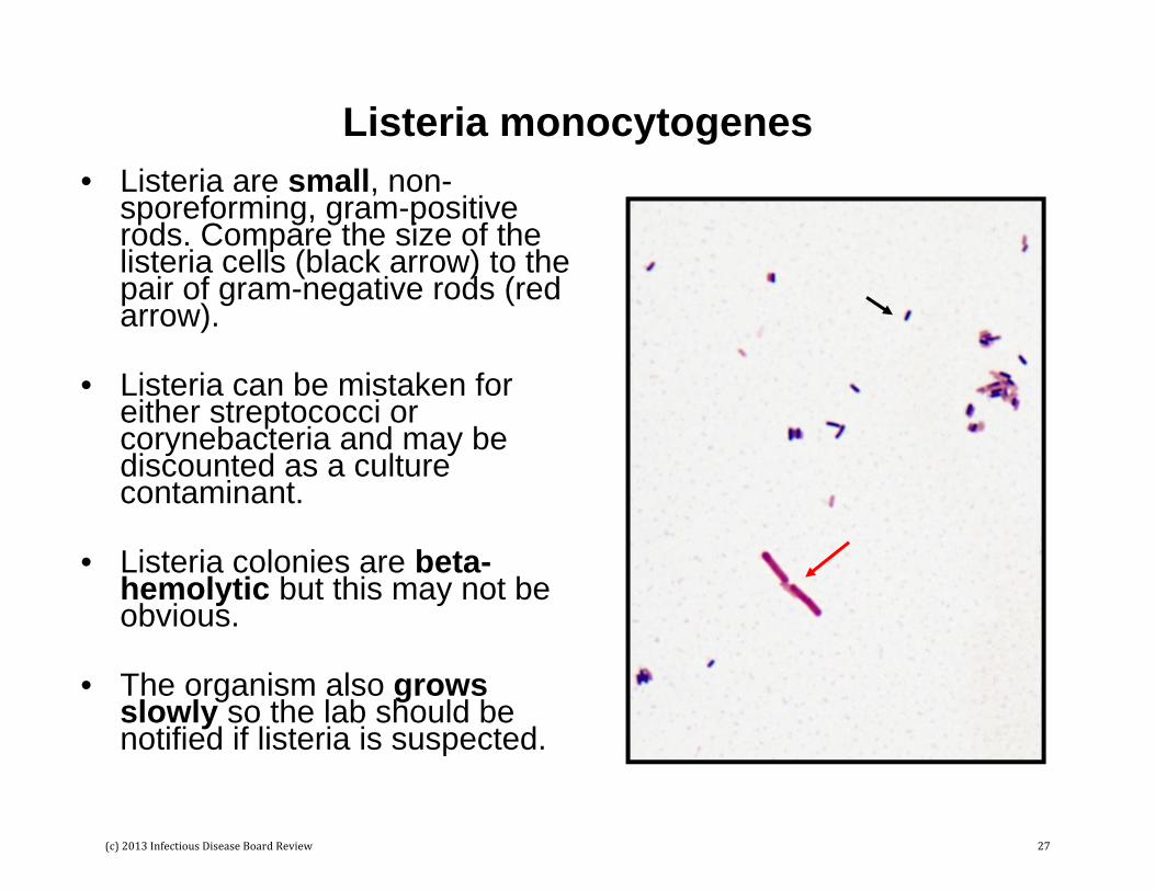
Listeria monocytogenes• Listeria are small, non-
sporeforming, gram-positive rods. Compare the size of the listeria cells (black arrow) to the pair of gram-negative rods (red arrow).
• Listeria can be mistaken for either streptococci or corynebacteria and may be discounted as a culture contaminant.
• Listeria colonies are beta-hemolytic but this may not be obvious.
• The organism also grows slowly so the lab should be notified if listeria is suspected.
(c) 2013 Infectious Disease Board Review 27

Corynebacterium Species
• Corynebacteria are commonly isolated as contaminants in blood cultures.
• The organisms tend to clump together and have an irregular shape (“coryneform” shaped; top figure).
• The organism in the bottom figure is C. jeikeium. This species is important clinically, causing disease in hospitalized patients and is frequently resistant to most commonly used antibiotics except vancomycin.
(c) 2013 Infectious Disease Board Review 28
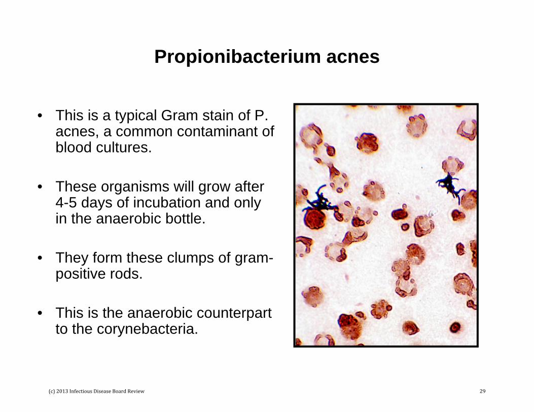
Propionibacterium acnes
• This is a typical Gram stain of P. acnes, a common contaminant of blood cultures.
• These organisms will grow after 4-5 days of incubation and only in the anaerobic bottle.
• They form these clumps of gram-positive rods.
• This is the anaerobic counterpart to the corynebacteria.
(c) 2013 Infectious Disease Board Review 29

Gram-Negative Cocci, Coccobacilli, and Rods
Gram-negative bacteria are among the most commonly isolated bacteria. Examples include:
• Cocci– Neisseria– Moraxella catarrhalis
• Coccobacilli– Moraxella, other species– Acinetobacter– Haemophilus
• Rods– Enterobacteriaceae (e.g., Escherichia, Klebsiella)– Pseudomonas, Stenotrophomonas, Burkholderia– Miscellaneous (e.g., Bacteroides, Fusobacterium)
(c) 2013 Infectious Disease Board Review 30
Neisseria
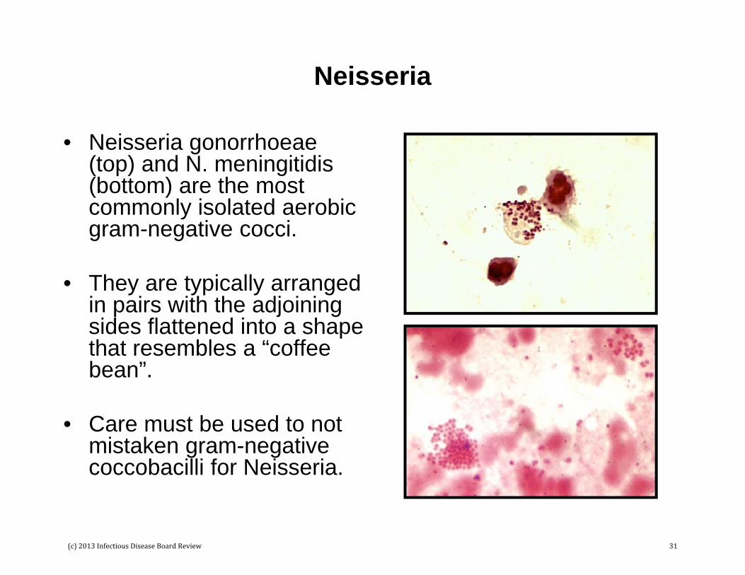
• Neisseria gonorrhoeae (top) and N. meningitidis (bottom) are the most commonly isolated aerobic gram-negative cocci.
• They are typically arranged in pairs with the adjoining sides flattened into a shape that resembles a “coffee bean”.
• Care must be used to not mistaken gram-negative coccobacilli for Neisseria.
(c) 2013 Infectious Disease Board Review 31

Moraxella catarrhalis
• Moraxella species are gram-negative rods. However, M. catarrhalis was originally classified as a Neisseria because the morphology of these bacteria closely resembles Neisseria.
• This photo is M. catarrhalisin a sputum from a patient with pneumonia. Note the inflammatory cells and large number of bacteria – this is typical of respiratory infections with this organism.
(c) 2013 Infectious Disease Board Review 32

Acinetobacter
• Acinetobacter are gram-negative coccobacilli that can retain the crystal violet and resemble gram-positive cocciin pairs (black arrow).
• The other bacteria in this figure is Pseudomonas (red arrow), clearly gram-negative rods arranged in a chain (arranged in pairs is more common).
• Acinetobacter are larger than Neisseria and the adjoining sides are not flattened.
(c) 2013 Infectious Disease Board Review 33

Haemophilus
• Haemophilus are very smallgram-negative rods that could also be mistaken for a gram-negative cocci (top figure).
• Long pleomorphic forms (bottom figure) can be seen in patients receiving antibiotics. This is a Gram stain of CSF from a child with Haemophilus meningitis.
(c) 2013 Infectious Disease Board Review 34
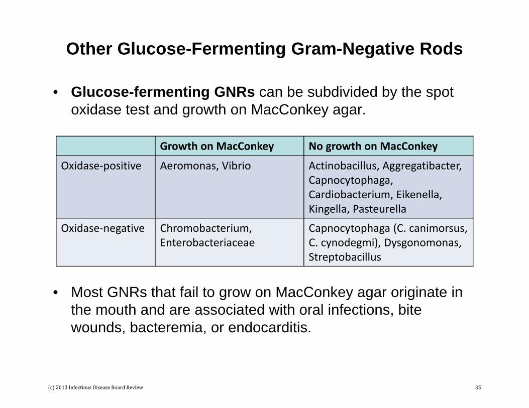
Other Glucose-Fermenting Gram-Negative Rods
• Glucose-fermenting GNRs can be subdivided by the spot oxidase test and growth on MacConkey agar.
• Most GNRs that fail to grow on MacConkey agar originate in the mouth and are associated with oral infections, bite wounds, bacteremia, or endocarditis.
Growth on MacConkey No growth on MacConkey
Oxidase‐positive Aeromonas, Vibrio Actinobacillus, Aggregatibacter, Capnocytophaga, Cardiobacterium, Eikenella, Kingella, Pasteurella
Oxidase‐negative Chromobacterium, Enterobacteriaceae
Capnocytophaga (C. canimorsus, C. cynodegmi), Dysgonomonas, Streptobacillus
(c) 2013 Infectious Disease Board Review 35

Enterobacteriaceae• The Enterobacteriaceae is a large family
of more than 100 genera of glucose-fermenting bacteria.
• With one exception (Plesiomonas), all Enterobacteriaceae are oxidase-negative; all grow on MacConkey agar.
• Escherichia, Klebsiella (figure), Enterobacter, Proteus, Salmonella, and Shigella are some common members of the family.
• All members of this family are relatively large gram-negative rods that tend to stain more intensely at their ends (“bipolar” staining).
(c) 2013 Infectious Disease Board Review 36

Other Glucose-Oxidizing Gram-Negative Rods
Glucose-oxidizing GNRs can also be subdivided by their oxidase reaction and ability to grow on MacConkey agar.
Growth on MacConkey No growth on MacConkey
Oxidase‐positive Achromobacter, Burkholderia, Elizabethkingia, Ochrobactrum, Oligella, Pseudomonas, Roseomonas, Sphingomonas
Brucella, Methylobacterium, Moraxella (M. catarrhalis)
Oxidase‐negative Acinetobacter, Pseudomonas (P. oryzihabitans, P. luteola), Stenotrophomonas
Acidovorax, Bartonella, Francisella
(c) 2013 Infectious Disease Board Review 37

Glucose-Oxidizing Gram-Negative Rods• “Pseudomonads” (e.g.,
Pseudomonas, Burkholderia, Stenotrophomonas) are small gram-negative rods typically arranged singly or in pairs.
• The Gram stain morphology is characteristic of this group of organisms but individual genera cannot be reliably differentiated by Gram stain.
• Pseudomonas aeruginosa, the most commonly isolated pseudomonad, may be surrounded by a thick mucus capsule (bottom figure).
(c) 2013 Infectious Disease Board Review 38

Other Gram-Negative Rods• Bacteroides fragilis is a anaerobic gram-
negative rod that is pleomorphic – short and long rods can be seen in the top image. One way to determine that these are the same organisms is to note that the diameter of the cells is the same even though the length is variable.
• Some gram-negative rods are thin and long. Species of Fusobacterium (middle figure) and Capnocytophaga (bottom figure) are the most common, although other bacteria may look like this if the patient is receiving antibiotics (refer to the Haemophilis photo previously shown).
(c) 2013 Infectious Disease Board Review 39

Acid-Fast and Partially Acid-Fast Bacteria
• Very few bacteria stain with acid-fast stains.
• Members of the genus Mycobacterium are acid-fast and members of the other genera listed here are “weakly or partially acid-fast”.
• Some of the rapidly growing mycobacteria are also weakly acid-fast.
• Acid-fast and partially acid-fast bacteria include:– Mycobacterium
• Rapid-growers• Slow-growers
– Nocardia– Rhodococcus– Gordonia– Tsukamurella
(c) 2013 Infectious Disease Board Review 40

Acid-Fast Stains
• The Ziehl-Neelsen is the original stain and requires heating the slide after basic fuchsin is added so the stain penetrates into the bacteria.
• The Kinyoun stain and modified Kinyoun stain are referred to as a cold acid-fast stains. Heating is not needed because the concentration of basic fuchsin is increased as well as the concentration of phenol.
• The modified Kinyoun stain differs from the Kinyoun stain by using a weak acid solution in alcohol. Nocardia, Rhodococcus, Gordonia, and Tsukamurella will retain some of the basic fuchsin stain when this weak solution is used but not when the higher concentration of acid is used.
• The fluorochrome stain replaces basic fuchsin with two fluorescent dyes, auramine and rhodamine. The fluorochrome is a weak acid-fast stain so all acid-fast organisms will stain.
(c) 2013 Infectious Disease Board Review 41
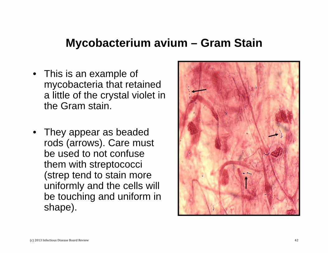
Mycobacterium avium – Gram Stain
• This is an example of mycobacteria that retained a little of the crystal violet in the Gram stain.
• They appear as beaded rods (arrows). Care must be used to not confuse them with streptococci (strep tend to stain more uniformly and the cells will be touching and uniform in shape).
(c) 2013 Infectious Disease Board Review 42

Mycobacterium
• Top figure: Kinyoun (carbol-fuchsin) stain of mycobacteria. Note the beaded appearance.
• Bottom figure: Fluorochrome (auramine-rhodamine) stain of mycobacteria.
• Acid-fast bacteria are much easier to detect using the fluorochrome stain because of the contrast with the dark background.
(c) 2013 Infectious Disease Board Review 43

Nocardia – Gram Stain• Nocardia is the second most
commonly isolated acid-fast organism.
• This Gram stain illustrates the thin, filamentous, branching forms that stain irregularly with the Gram stain (note the blue and red sections of the structures).
• No other acid-fast organism forms long, branching structures.
• Important ways to differentiate this organism from streptococci include: (1) true branching of the filaments, and (2) the observation that the “beads” (arrows) do not touch and are irregularly distributed.
(c) 2013 Infectious Disease Board Review 44

Nocardia – Modified Acid-Fast Stain
• Modified acid-fast stain of Nocardia in a sputum specimen.
• Note the branching forms and the fact the organism does not stain uniformly.
• If a regular Kinyoun stain was used, most of the basic fuchsinstain would be removed by the strong acid-alcohol solution and the organism would stain very weakly.
(c) 2013 Infectious Disease Board Review 45

Rhodococcus – Gram Stain4 hour Broth Culture
• Rhodococcus was originally classified as a Corynebacterium.
• These organisms retain the crystal violet dye more uniformly than either mycobacteria or nocardia.
• After growth for a few hours, they stain well with the Gram stain and appear rod-like.
(c) 2013 Infectious Disease Board Review 46
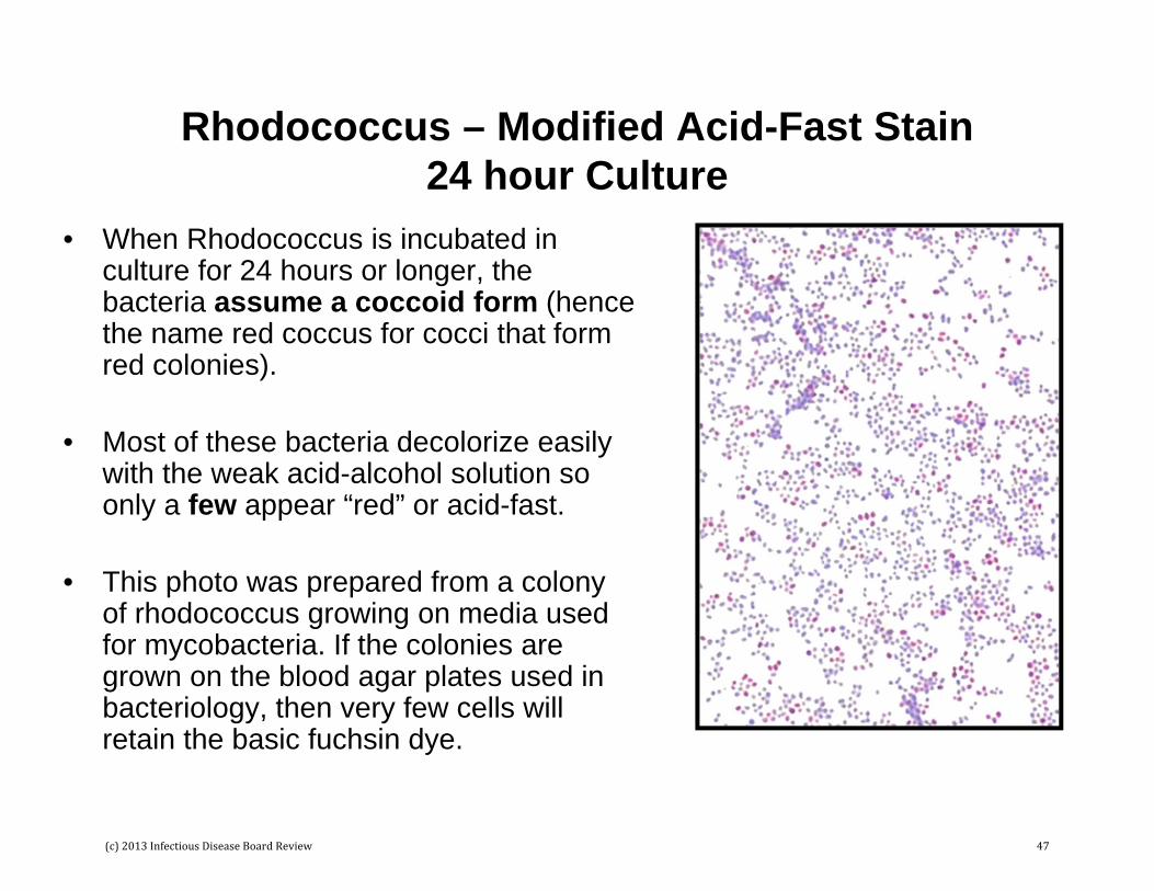
Rhodococcus – Modified Acid-Fast Stain24 hour Culture
• When Rhodococcus is incubated in culture for 24 hours or longer, the bacteria assume a coccoid form (hence the name red coccus for cocci that form red colonies).
• Most of these bacteria decolorize easily with the weak acid-alcohol solution so only a few appear “red” or acid-fast.
• This photo was prepared from a colony of rhodococcus growing on media used for mycobacteria. If the colonies are grown on the blood agar plates used in bacteriology, then very few cells will retain the basic fuchsin dye.
(c) 2013 Infectious Disease Board Review 47

Mycology
(c) 2013 Infectious Disease Board Review 48

Yeasts and Molds• Fungi can be subdivided into yeasts (single cell
organisms) and molds (multicell organisms). A few important fungi (dimorphic fungi) can exist in both forms (e.g., Histoplasma, Blastomyces, Sporothrix)
• The most important genera of yeast are:– Candida (e.g., C. albicans, C. glabrata, C. tropicalis, C.
parapsilosis, C. krusei)– Cryptococcus (e.g., C. neoformans)– Trichosporon (e.g., T. asahii, T. mucoides; the former species
T. beigelii has been subdivided into 6 species and the old name is not currently accepted)
– Malassezia (e.g., M. furfur)– Pneumocystis (maybe more appropriately called a nonmold
rather than a yeast cell)
(c) 2013 Infectious Disease Board Review 49

Candida Species
• Top figure: Candida albicansisolated in blood culture; note the yeast cells and pseudohyphaeor pseudo branched forms.
• Bottom figure: Candida glabrata; these are smaller than other yeasts; they also do not form pseudohyphae; common cause of urinary tract infections and second most common cause of fungemia
(c) 2013 Infectious Disease Board Review 50

Germ Tube Test
• This is a rapid test for the identification of C. albicans. Only one other Candida species (C. dubliniensis) is germ tube positive.
• Yeast cells inoculated in serum will form “germ tubes” within 2 hours.
• Note the “tube is a continuous extension from the yeast cell and no septum exists. This distinguishes germ tubes from pseudohyphae.
(c) 2013 Infectious Disease Board Review 51
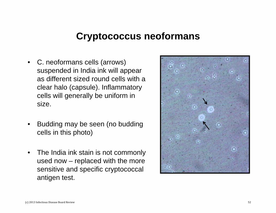
Cryptococcus neoformans
• C. neoformans cells (arrows) suspended in India ink will appear as different sized round cells with a clear halo (capsule). Inflammatory cells will generally be uniform in size.
• Budding may be seen (no budding cells in this photo)
• The India ink stain is not commonly used now – replaced with the more sensitive and specific cryptococcalantigen test.
(c) 2013 Infectious Disease Board Review 52

Malassezia
• Malassezia species grow on the skin surface and can cause pityriasisversicolor and rarely systemic infections.
• The small yeast cells have a prominent “collarette” (arrow) that forms where the daughter cells bud.
• Short hyphal elements may also be seen in skin scrapings (referred to as “spaghetti and meatballs”).
• Culture of the most important species in this genus require supplementation of media with lipids (e.g., olive oil).
(c) 2013 Infectious Disease Board Review 53

Pneumocystis
• The taxonomy of this group of organisms has changed. – Previously classified as a parasite, it is now accepted as a
fungus.– Previously a single genus and species was recognized,
Pneumocystis carinii. It is now recognized that there are multiple species, each with a specific mammalian host. P. jirovecii is the human pathogen
• The two developmental forms that are observed in human tissues are the trophozoite and the cyst(terminology is a hold over from when these organisms were classified as parasites).
(c) 2013 Infectious Disease Board Review 54

Pneumocystis – Methenamine Silver Stains
• Stain performed in surgical pathology
• Cysts stain brown to black; trophozoites are unstained
• Nonspecific staining in the background may make interpretation difficult if only a few cysts are present
(c) 2013 Infectious Disease Board Review 55
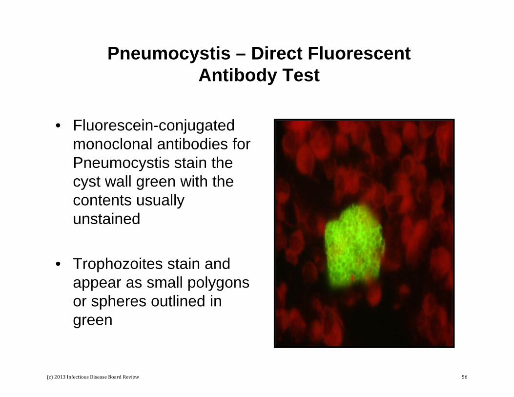
Pneumocystis – Direct Fluorescent Antibody Test
• Fluorescein-conjugated monoclonal antibodies for Pneumocystis stain the cyst wall green with the contents usually unstained
• Trophozoites stain and appear as small polygons or spheres outlined in green
(c) 2013 Infectious Disease Board Review 56

Dimorphic Fungi
• Dimorphic fungi exists in two forms – typically as yeasts at body temperature and molds at room temperature.
• The most commonly isolated dimorphic molds in the US are:– Histoplasma capsulatum– Blastomyces dermatitidis– Coccidioides immitis– Sporothrix schenckii
(c) 2013 Infectious Disease Board Review 57
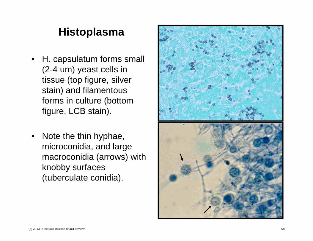
Histoplasma
• H. capsulatum forms small (2-4 um) yeast cells in tissue (top figure, silver stain) and filamentous forms in culture (bottom figure, LCB stain).
• Note the thin hyphae, microconidia, and large macroconidia (arrows) with knobby surfaces (tuberculate conidia).
(c) 2013 Infectious Disease Board Review 58

Blastomyces• B. dermatitidis forms large
(8-15 um) yeast cells in tissue and hyphal forms in culture at ambient temperatures.
• The yeasts have thick wall and form a broad base where the daughter cell buds.
• The mold form has thin hyphae with numerous small microconidia attached to the hyphae by thin branches (resembles lollypops).
(c) 2013 Infectious Disease Board Review 59
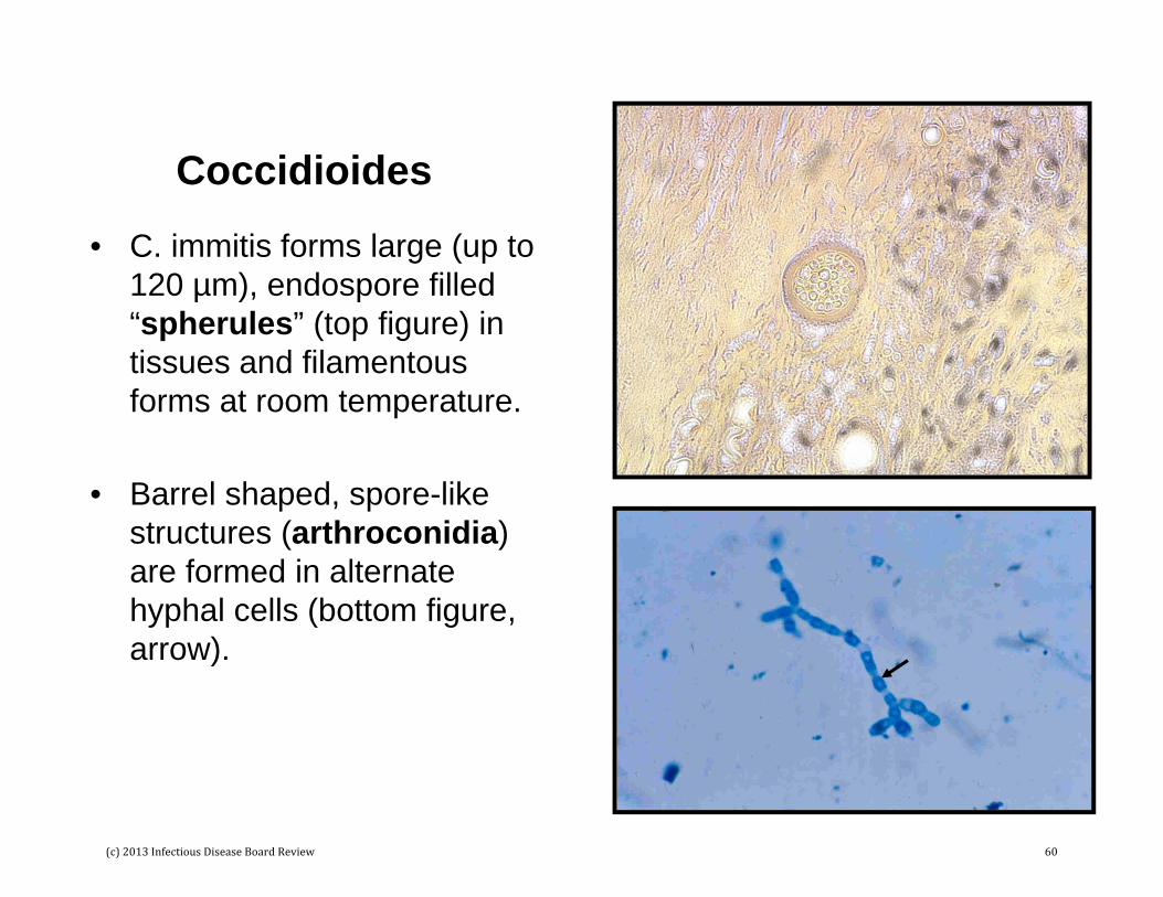
Coccidioides
• C. immitis forms large (up to 120 µm), endospore filled “spherules” (top figure) in tissues and filamentous forms at room temperature.
• Barrel shaped, spore-like structures (arthroconidia) are formed in alternate hyphal cells (bottom figure, arrow).
(c) 2013 Infectious Disease Board Review 60

Sporothrix
• S. schenckii forms narrow based yeast cells in tissue (top figure) and delicate hyphae with a cluster (“flowerette”) of conidia (spores) at the end of a narrow stalk (bottom figure).
• The yeasts and conidia can be darkly pigmented so this fungus is classified as a dematiaceous (dark) mold.
(c) 2013 Infectious Disease Board Review 61
Filamentous Fungi - Molds• The taxonomic classification of molds is complex and generally
confusing for nonmycologists. Traditionally these fungi are classified by morphologic features and some clinical properties:– Nonseptated molds (e.g., Rhizopus, Mucor, Rhizomucor, Absidia)– Lightly colored or hyaline (moniliaceous), septated molds
• Opportunistic fungi (e.g., Aspergillus, Fusarium, Paecilomyces, Scopulariopsis, Penicillium)
• Dermatophytes (e.g., Trichophyton, Epidermophyton, Microsporum)– Darkly pigmented (dematiaceous), septated molds (e.g., Alternaria,
Bipolaris, Curvularia, Exophiala)
• Current work is underway to classify these organisms by gene sequencing and mass spectrometry (MALDI). It is anticipated that these approaches will be more rapid and objective.
• It is impractical to give a comprehensive summary of all molds here; rather, I will illustrate the diversity of morphologic forms with selected photographs.
(c) 2013 Infectious Disease Board Review 62

Mucor
• Mucor is an example of a zygomyces.
• These fungi are characterized by a lack of septae (divisions) within the hyphae.
• In tissue (top figure; silver stain) the hyphae appear ribbon-like.
• The bottom figure is the mold in culture.
(c) 2013 Infectious Disease Board Review 63
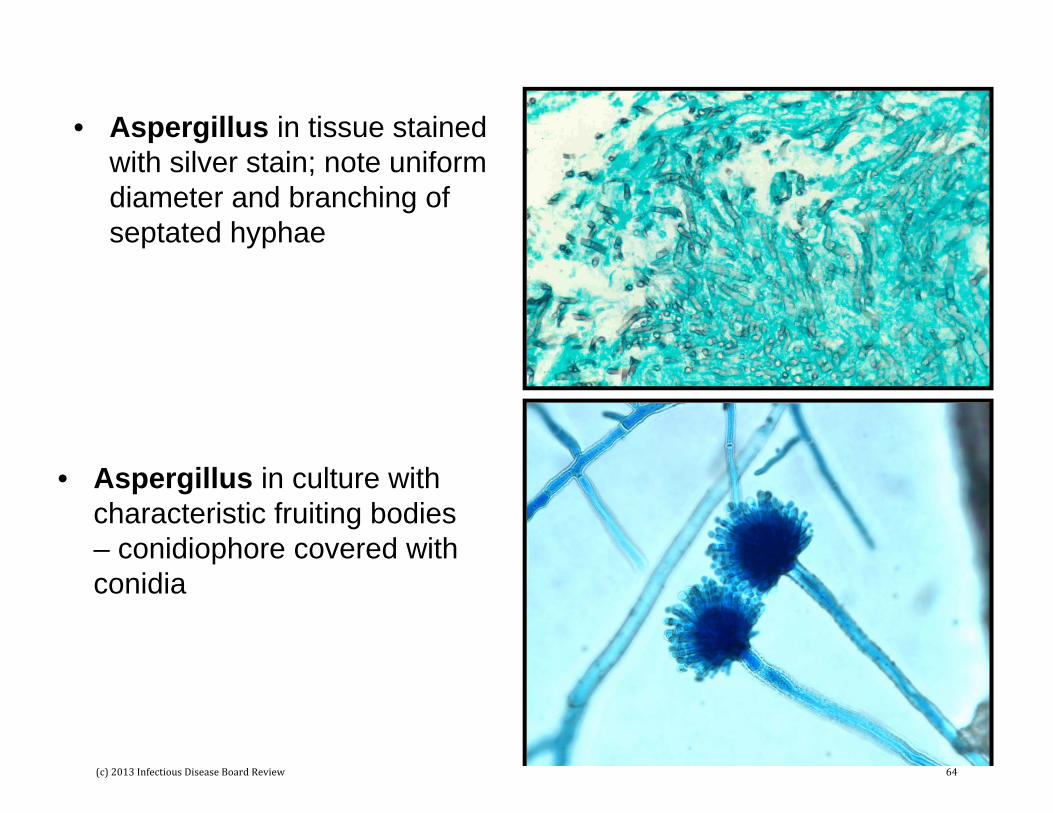
• Aspergillus in tissue stained with silver stain; note uniform diameter and branching of septated hyphae
• Aspergillus in culture with characteristic fruiting bodies – conidiophore covered with conidia
(c) 2013 Infectious Disease Board Review 64

• Fusiform or sickle shaped multicelled macroconidia of Fusarium.
• Fruiting structures and thin hyphae of Penicillium in culture.
(c) 2013 Infectious Disease Board Review 65
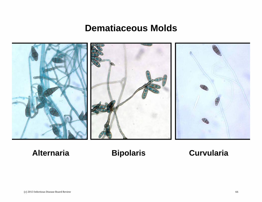
Dematiaceous Molds
Alternaria Bipolaris Curvularia
(c) 2013 Infectious Disease Board Review 66
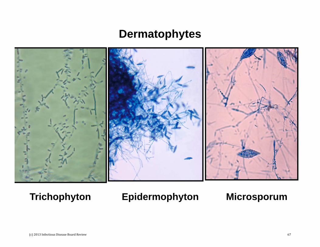
Dermatophytes
Epidermophyton MicrosporumTrichophyton
(c) 2013 Infectious Disease Board Review 67

Parasites
• Parasites can be subdivided into protozoa and helminths.
• Protozoa are subdivided into:– Amoeba (including intestinal ameba and free-living
ameba)– Flagellates and ciliates– Coccidia and Microsporidia– Plasmodium and Babesia– Leishmania and Trypanosomes
• Helminths (worms) are subdivided into:– Nematodes or roundworms– Trematodes or flatworms (flukes)– Cestodes or tapeworms
(c) 2013 Infectious Disease Board Review 68
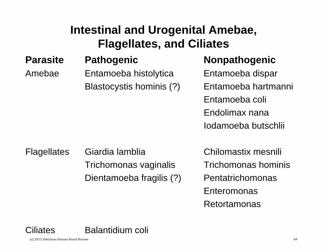
Intestinal and Urogenital Amebae, Flagellates, and Ciliates
Parasite Pathogenic NonpathogenicAmebae Entamoeba histolytica Entamoeba dispar
Blastocystis hominis (?) Entamoeba hartmanniEntamoeba coliEndolimax nanaIodamoeba butschlii
Flagellates Giardia lamblia Chilomastix mesniliTrichomonas vaginalis Trichomonas hominisDientamoeba fragilis (?) Pentatrichomonas
EnteromonasRetortamonas
Ciliates Balantidium coli(c) 2013 Infectious Disease Board Review 69

Amebae
• One intestinal ameba is a clear human pathogen, Entamoebahistolytica, and must be differentiated from 3 other nonpathogenic species: E. dispar, E. hartmanni, and E. coli.
• Blastocystis hominis is a common protozoa that has occasionally been associated with human disease.
• Two stages exists for most amebae: actively replicating trophozoite and dormant, stable cyst. The cyst stage is infectious because it is not destroyed by gastric acids when ingested.
• Detection and identification of most amebae is by recognition of the cyst or trophozoite forms in stool specimens. The exception is E. histolytica where antigen detection tests have also been developed (most commonly enzyme immunoassays, EIAs) as well as PCR molecular tests.
(c) 2013 Infectious Disease Board Review 70

E. histolytica
E. hartmanni
E. coli
Trophozoites Cysts
(c) 2013 Infectious Disease Board Review 71
Flagellates
• Two flagellates are well-recognized human pathogens: Giardia lamblia and Trichomonas vaginalis
• A third flagellate, Dientamoeba fragilis, has been implicated as an “occasional” human pathogen; however, most isolates of this organism represent insignificant colonization
• If a urogenital specimen is contaminated with fecal matter, then nonpathogenic flagellates (e.g., Pentatrichomonas, Enteromonas, and Retortamonas) may be confused with T. vaginalis.
• Chilomastix mesnili is another nonpathogenic flagellate that is occasional found in fecal specimens.
(c) 2013 Infectious Disease Board Review 72
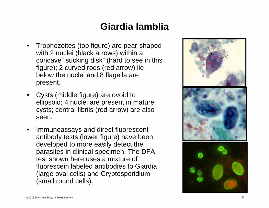
Giardia lamblia
• Trophozoites (top figure) are pear-shaped with 2 nuclei (black arrows) within a concave “sucking disk” (hard to see in this figure); 2 curved rods (red arrow) lie below the nuclei and 8 flagella are present.
• Cysts (middle figure) are ovoid to ellipsoid; 4 nuclei are present in mature cysts; central fibrils (red arrow) are also seen.
• Immunoassays and direct fluorescent antibody tests (lower figure) have been developed to more easily detect the parasites in clinical specimen. The DFA test shown here uses a mixture of fluorescein labeled antibodies to Giardia (large oval cells) and Cryptosporidium (small round cells).
(c) 2013 Infectious Disease Board Review 73

Trichomonas vaginalis
• The figure shows two T. vaginalistrophozoites; no cyst stage exists
• The trophozoites are pear-shaped
• The nucleus contains many chromatin granules and a small karyosome
• An undulating membrane (black arrows) extends half way down the parasite (this membrane extends the length of P. hominis – key differential characteristic)
• Flagella (red arrows) extend beyond the bottom of the parasite.
• Diagnosis can be made by microscopy or culture; however, the most sensitive and specific test is PCR.
(c) 2013 Infectious Disease Board Review 74

Ciliates: Balantidium coli
• One ciliated ameba causes human disease: B. coli.
• Trophozoite and cyst stages exist
• This parasite can be recognized by its very large size and the presence of cilia (arrow) that coat the surface
• A macronucleus and small micronucleus may also be observed, as well as cytoplasmic vacuoles.
(c) 2013 Infectious Disease Board Review 75
Coccidia and Microsporidia• Four groups of parasites stain with acid-fast stains:
– Microsporidia (1-4 um)– Cryptosporidium (4-6 um)– Cyclospora (5-10 um)– Isospora (14 x 25 um)
• These parasites do not stain uniformly acid-fast (some stain weakly, and some cells do not stain)
• These parasites are most easily differentiated by their size: Microsporidia are the smallest, Isospora the largest.
• Most of these parasites do not stain with the traditional stains used for Ova and Parasite (O&P) examinations (e.g., Trichrome stain).
• Two parasites (Cyclospora and Isospora) will autofluorescewhen examined under a UV light.
(c) 2013 Infectious Disease Board Review 76

Panels A and B – Cryptosporidia; Panel C – Cyclospora; panel D - Isospora
(c) 2013 Infectious Disease Board Review 77
Blood and Tissue Protozoa
• The most common blood and tissue protozoa are:– Plasmodium: P. falciparum, P. vivax, P. ovale, P. malariae– Babesia: B. microti– Leishmania: L. donovani, L. tropica, L. major (and many
others)– Trypanosoma: T. cruzi, T. brucei rhodensiense, T. brucei
gambiense
• These parasites are most commonly detected by Giemsa staining of blood and hematoxylin and eosin (H&E) staining of tissues.
• Representative examples will be shown on the next few slides.
(c) 2013 Infectious Disease Board Review 78

P. falciparum ring forms (left) and gametocyte (middle); B. microti (right). Care must be taken to differentiate these two protozoa
(c) 2013 Infectious Disease Board Review 79

P. vivax P. malariae
(c) 2013 Infectious Disease Board Review 80

Leishmania amastigotes in tissue (left); Leishmania promastigote in culture (middle); Tryponosoma typomastigote in blood (right)
(c) 2013 Infectious Disease Board Review 81
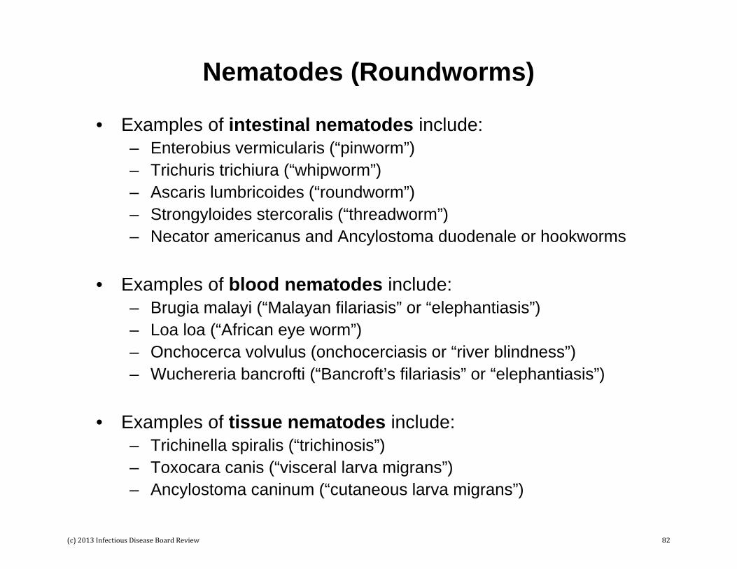
Nematodes (Roundworms)
• Examples of intestinal nematodes include: – Enterobius vermicularis (“pinworm”)– Trichuris trichiura (“whipworm”)– Ascaris lumbricoides (“roundworm”)– Strongyloides stercoralis (“threadworm”)– Necator americanus and Ancylostoma duodenale or hookworms
• Examples of blood nematodes include:– Brugia malayi (“Malayan filariasis” or “elephantiasis”)– Loa loa (“African eye worm”)– Onchocerca volvulus (onchocerciasis or “river blindness”)– Wuchereria bancrofti (“Bancroft’s filariasis” or “elephantiasis”)
• Examples of tissue nematodes include:– Trichinella spiralis (“trichinosis”)– Toxocara canis (“visceral larva migrans”)– Ancylostoma caninum (“cutaneous larva migrans”)
(c) 2013 Infectious Disease Board Review 82
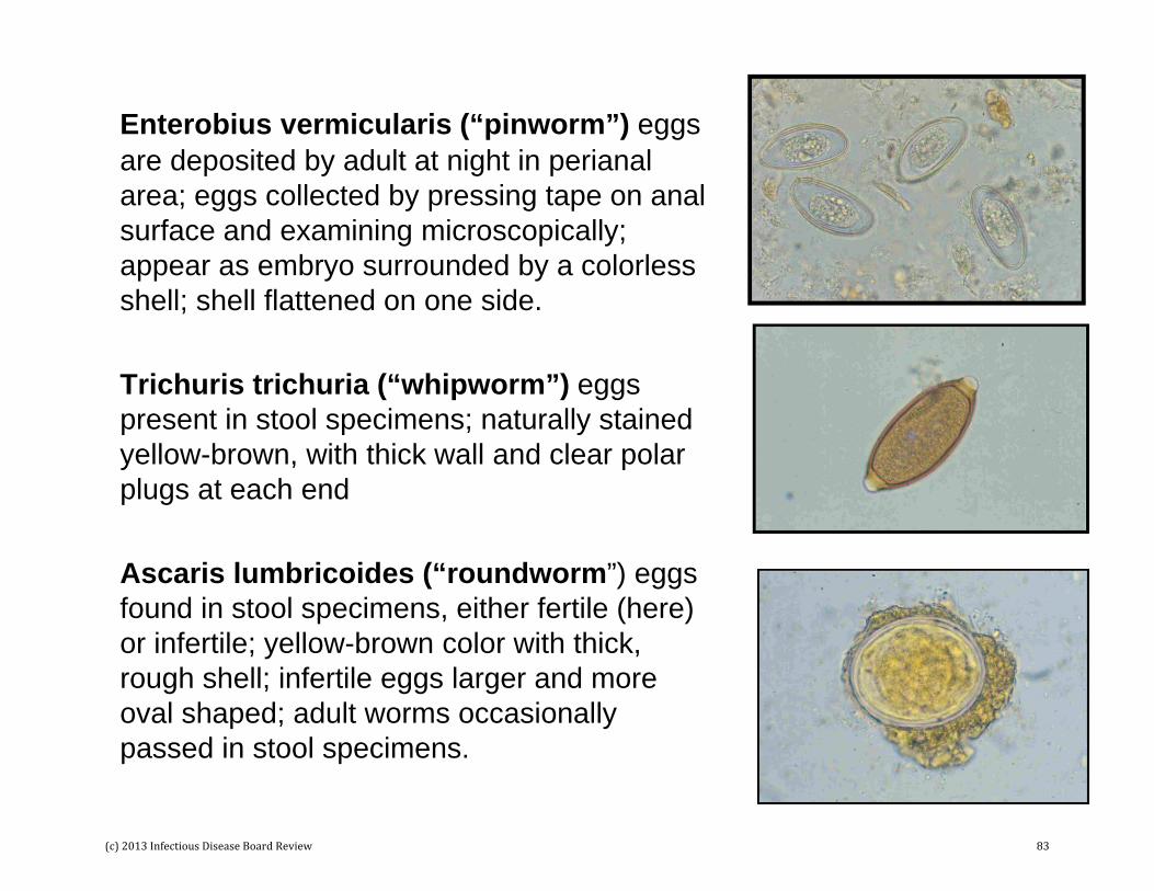
Enterobius vermicularis (“pinworm”) eggs are deposited by adult at night in perianal area; eggs collected by pressing tape on anal surface and examining microscopically; appear as embryo surrounded by a colorless shell; shell flattened on one side.
Trichuris trichuria (“whipworm”) eggs present in stool specimens; naturally stained yellow-brown, with thick wall and clear polar plugs at each end
Ascaris lumbricoides (“roundworm”) eggs found in stool specimens, either fertile (here) or infertile; yellow-brown color with thick, rough shell; infertile eggs larger and more oval shaped; adult worms occasionally passed in stool specimens.
(c) 2013 Infectious Disease Board Review 83
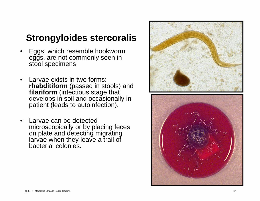
Strongyloides stercoralis• Eggs, which resemble hookworm
eggs, are not commonly seen in stool specimens
• Larvae exists in two forms: rhabditiform (passed in stools) and filariform (infectious stage that develops in soil and occasionally in patient (leads to autoinfection).
• Larvae can be detected microscopically or by placing feces on plate and detecting migrating larvae when they leave a trail of bacterial colonies.
(c) 2013 Infectious Disease Board Review 84

Ancylostoma (“Old World”) and Necator (“New World”) Hookworms
• Hookworm eggs have a thin, colorless shell surrounding the developing larva.
• If stool specimens are left at room temperature, the larvae can hatch and will resemble Strongyloides larvae.
(c) 2013 Infectious Disease Board Review 85

Blood and Tissue Nematodes: General Comments
• Infections caused by these roundworms are uncommon in the US; most infections are in residents of endemic areas or international travelers
• In contrast with other roundworm infections, disease is most commonly diagnosed by detection of larval forms in blood or tissues
• Diagnosis is also aided by knowledge of the patient’s travel history because these parasites exist in restricted geographic area (in large part determined by the distribution of the vectors)
(c) 2013 Infectious Disease Board Review 86

Wuchereria bancrofti
Large microfilariae covered with a transparent sheath (black arrow) and nuclei extending to tip of tail (red arrow); transmitted by mosquito bite
(c) 2013 Infectious Disease Board Review 87
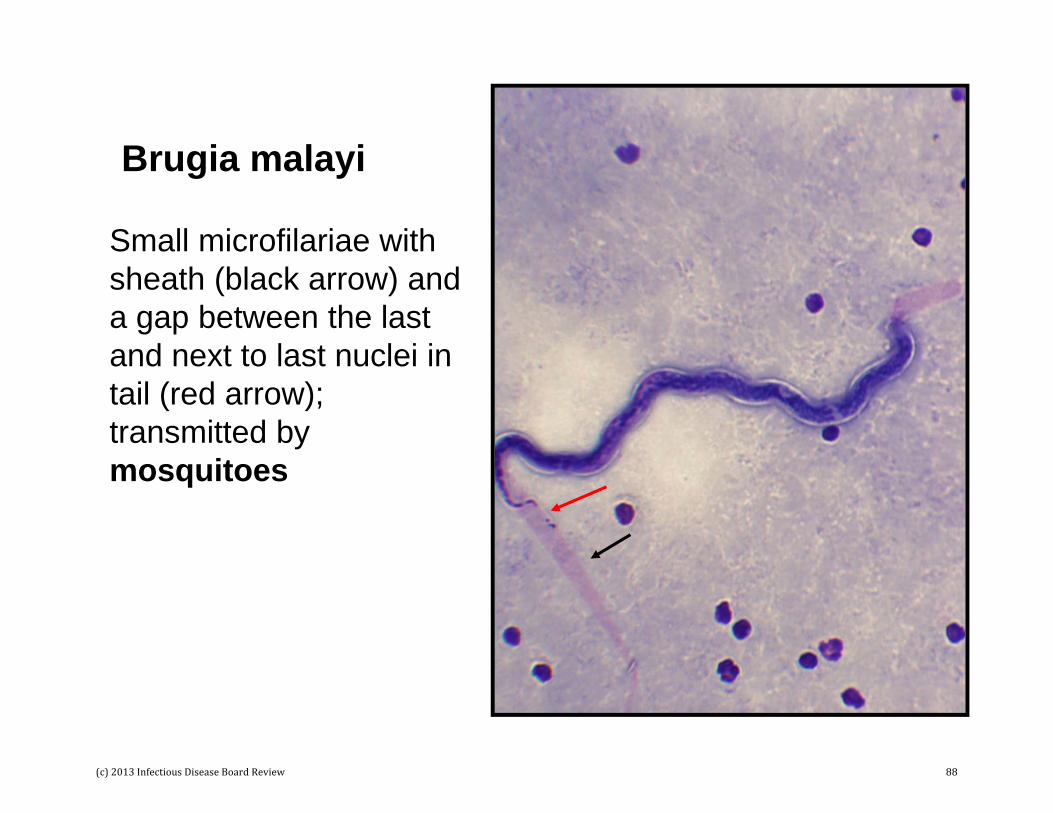
Brugia malayi
Small microfilariae with sheath (black arrow) and a gap between the last and next to last nuclei in tail (red arrow); transmitted by mosquitoes
(c) 2013 Infectious Disease Board Review 88

Loa loa
Small microfilariae covered with a sheath (black arrow); nuclei extend to the tip of the tail (red arrow); transmission by bite of tabanid flies(Chrysops)
(c) 2013 Infectious Disease Board Review 89

Onchocerca volvulus
Adults mature in subcutaneous nodules and microfilariae are found in skin; microfilaria do not have a sheath and the nuclei do not extend to tip of tail; transmission by bite of black fly (Simulium)
(c) 2013 Infectious Disease Board Review 90
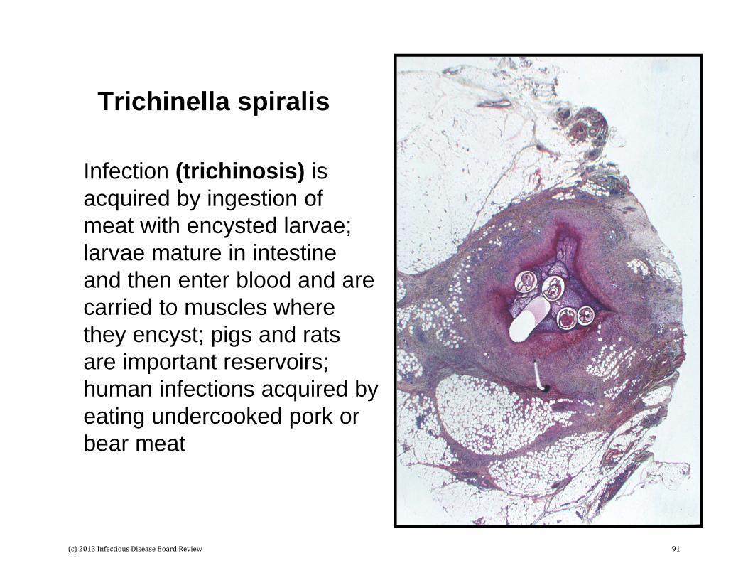
Trichinella spiralis
Infection (trichinosis) is acquired by ingestion of meat with encysted larvae; larvae mature in intestine and then enter blood and are carried to muscles where they encyst; pigs and rats are important reservoirs; human infections acquired by eating undercooked pork or bear meat
(c) 2013 Infectious Disease Board Review 91

Toxocara canis
Visceral larval migrans –Dogs are normal host with adult worms in small intestine; eggs (figure) released in feces; eggs large (75 x 80 um) and covered with thick, pitted shell; humans accidental hosts – ingest eggs, larvae released in intestines and migrate extensively before encapsulation in various organs.
(c) 2013 Infectious Disease Board Review 92

Trematodes or Flatworms• Trematode infections are relatively uncommon in
the US; most infections in residents of endemic areas who migrate to the US.
• Example of intestinal trematodes include:– Fasciolopsis buski (“porcine liver fluke”)
• Examples of tissue trematodes include:– Fasciola hepatica (“sheep liver fluke”)– Clonorchis sinensis (“Chinese liver fluke”)– Paragonimus westernmani (“oriental lung fluke”)
• Examples of blood trematodes include:– Schistosoma mansoni (“intestinal bilharziasis”)– Schistosoma japonica (“oriental blood fluke”)– Schistosoma haematobium (“urinary bilharziasis”)
(c) 2013 Infectious Disease Board Review 93

Fasciolopsis buski
• The “liver fluke” is the largest and most pathogenic human intestinal fluke.
• Pigs and humans are the primary hosts; infection acquired by ingestion of metacercariae encysted on aquatic vegetation (e.g., water chestnuts)
• Large eggs (130-140 um x 80-85 um) with inconspicuous operculum
(c) 2013 Infectious Disease Board Review 94

Fasciola hepatica
• Infection with the “sheep liver fluke” is acquired by ingestion of aquatic vegetation (e.g., watercress) where the metacercariae have encysted.
• Eggs are large (130-150 um x 63-90 um) with inconspicuous operculum
(c) 2013 Infectious Disease Board Review 95

Clonorchis sinensis
• Infection with the “Chinese liver fluke” is acquired by ingestion of metacercariae in the flesh of fish.
• Eggs 17-30 um x 13-18 um), with operculum (black arrow) at small end and knob (red arrow) at opposite end.
(c) 2013 Infectious Disease Board Review 96

Paragonimus westermani
• Infection with the “lung fluke” is acquired by eating raw or inadequately cooked crabs and crayfish
• Eggs 80-120 um x 45-70 um; prominent operculum (arrow)
(c) 2013 Infectious Disease Board Review 97

Schistosoma japonicumand Schistosoma mansoni
• Infection with schistosomes is acquired when cercariae released into water from infected snails penetrate through the skin of humans.
• Large, thin-shelled eggs without operculum; lateral spine on S. mansoni (top); inconspicuous spine on S. japonicum (bottom); terminal spine on S. haemotobium (not shown).
(c) 2013 Infectious Disease Board Review 98
Cestodes or Tapeworms
• Cestode infections are relatively uncommon in the US.
• Examples of intestinal cestodes include:– Diphyllobothrium latum (“fish tapeworm”)– Taenia solium (“pork tapeworm”)– Taenia saginata (“beef tapeworm”)– Hymenolepis nana (“dwarf tapeworm”)– Hymenolepis diminuta (“rat tapeworm”)– Dipylidium caninum (“dog tapeworm”)
• Examples of tissue cestodes include:– Echinococcus granulosus “unilocular hydatidosis”
or “hydatid disease”)– Taenia solium (“cysticercosis”)
(c) 2013 Infectious Disease Board Review 99
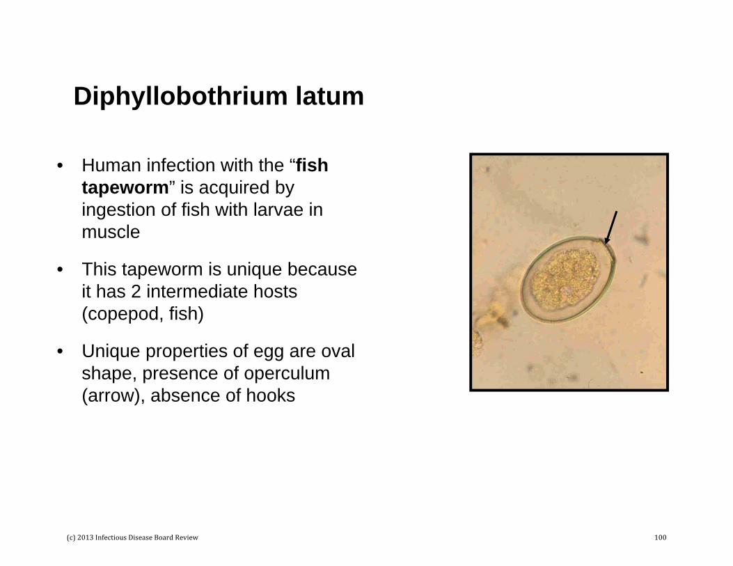
Diphyllobothrium latum
• Human infection with the “fish tapeworm” is acquired by ingestion of fish with larvae in muscle
• This tapeworm is unique because it has 2 intermediate hosts (copepod, fish)
• Unique properties of egg are oval shape, presence of operculum (arrow), absence of hooks
(c) 2013 Infectious Disease Board Review 100
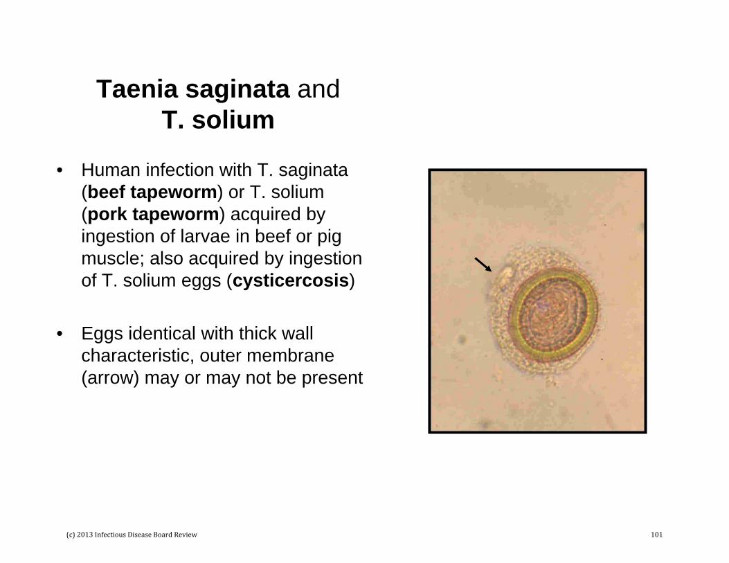
Taenia saginata and T. solium
• Human infection with T. saginata(beef tapeworm) or T. solium(pork tapeworm) acquired by ingestion of larvae in beef or pig muscle; also acquired by ingestion of T. solium eggs (cysticercosis)
• Eggs identical with thick wall characteristic, outer membrane (arrow) may or may not be present
(c) 2013 Infectious Disease Board Review 101
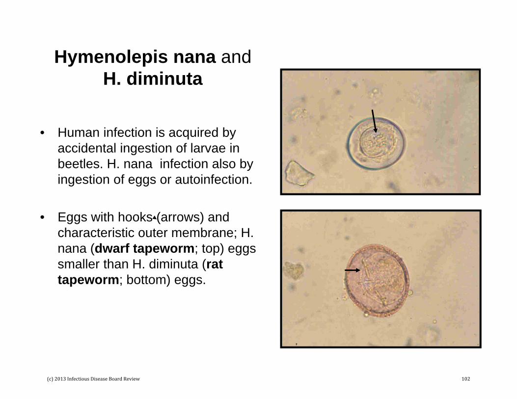
Hymenolepis nana and H. diminuta
• Human infection is acquired by accidental ingestion of larvae in beetles. H. nana infection also by ingestion of eggs or autoinfection.
• Eggs with hooks (arrows) and characteristic outer membrane; H. nana (dwarf tapeworm; top) eggs smaller than H. diminuta (rat tapeworm; bottom) eggs.
(c) 2013 Infectious Disease Board Review 102