primary open angle glaucoma (poag)
TRANSCRIPT

MODERATOR – DR. O. P. GUPTA
PRIMARY OPEN ANGLE
GLAUCOMA (POAG)

GLAUCOMA
Chronic, progressive optic neuropathy caused by a group of ocular conditions which lead to damage of the optic nerve with loss of visual function
IOP is the major risk factor
Normal tension glaucoma

POAG
• K/a Chronic simple glaucoma
• Most prevalent of all glaucoma
• Affects both sexes equally

POAG• An IOP >21 mmHg
• Glaucomatous optic nerve damage
• An open anterior chamber angle
• Characteristic visual field loss
• Absence of signs of secondary glaucoma or a non-glaucomatous cause for the optic neuropathy

RISK FACTORS
• IOP
• Age
• Race
• Family history
• Diabetes Mellitus
• Myopia
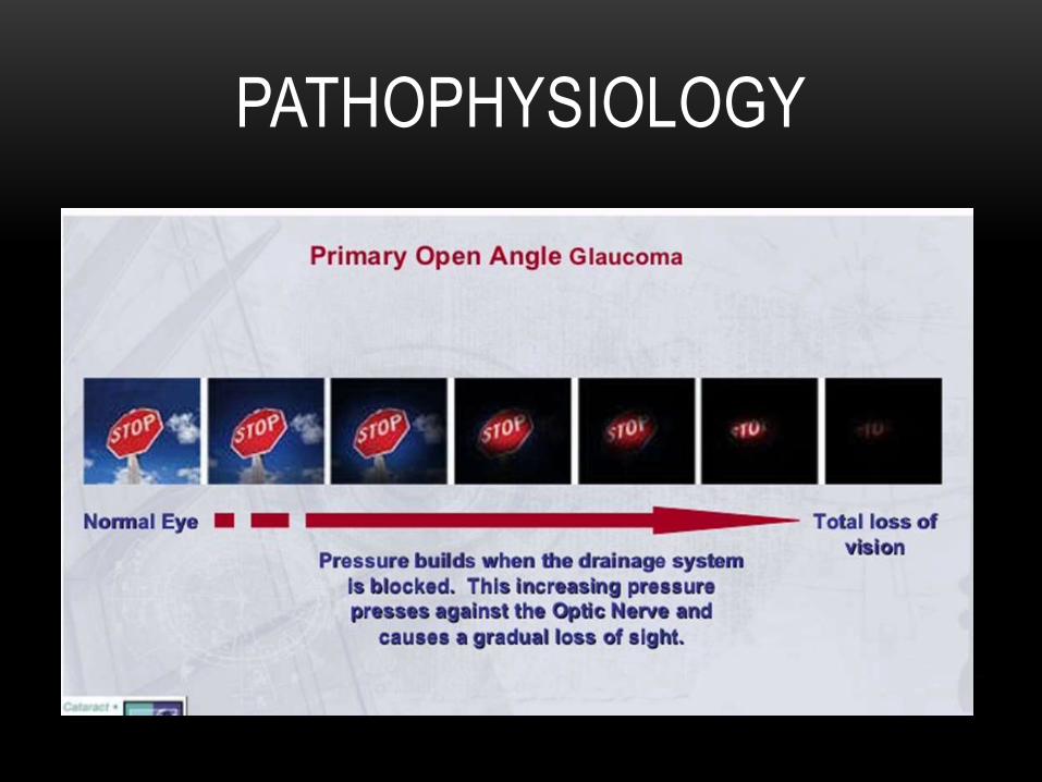
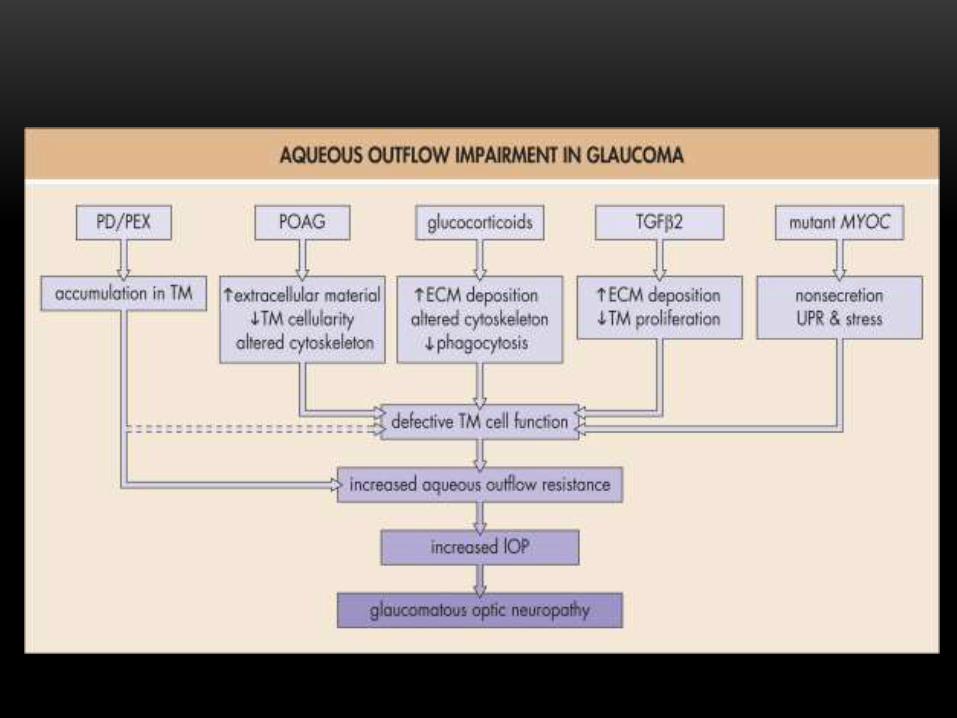
PATHOPHYSIOLOGY
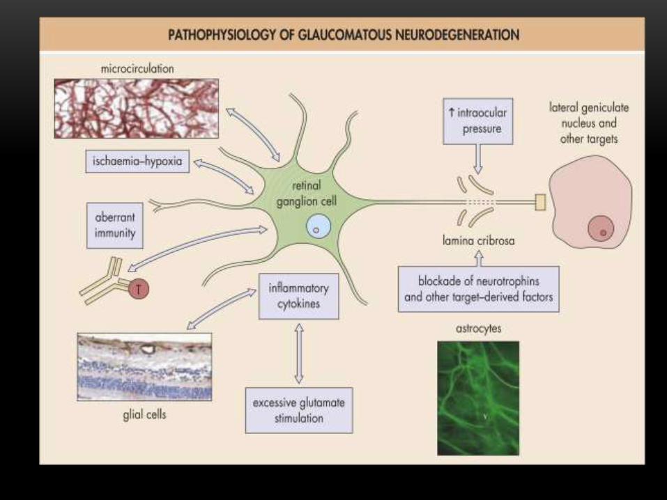

PATHOGENESIS OF GLAUCOMATOUS OPTIC
NEUROPATHY
1. Ischaemic theory
Suggests that poor blood perfusion of ONH
causes ischaemia and resultant loss of optic nerve
fibre
2. Mechanical theory
Suggests that weakness of supporting tissues of
optic nerve head makes it susceptible to mechanical
deformation by IOP with resultant nerve fibre damage
3. Immune theory
Increased incidence of paraproteinemia and auto
antibodies and antiglutathione S-transferace
antibodies
Cause retinal ganglion cell apoptosis
4. Apoptotic theory
Genetically programmed destruction of retinal
ganglion cells may play a part in the pathogenesis
CLINICAL FEATURES
• Usually asymptomatic until a significant visual field loss has occurred
• Eye ache, headache, haloes
• Delayed dark adaptation
• Frequent changes of presbyopic glasses
• Raised IOP & fluctuations in IOP
CHANGES IN IOP
• IOP >21 mm Hg on more than one occasion
• Circardian variation of IOP >8 mm Hg
• Asymmetry of IOP >5 mm Hg between two eyes

BASE LINE INFORMATION
• History: Ocular, Systemic, Family history,
History of medication
• Pupillary reaction
• Slit lamp biomicroscopy:
Anterior segment to r/o 2° causes- shallow anterior
chamber, pxf, inflammation
Fundus evaluation to rule out lesions which can
cause visual field defects
AT, DVT

• CCT > 555µm: false high IOP
< 540µm: false low IOP
• Gonioscopy
• Perimetry: Automatic static threshold perimetry
• Provocative Tests: Water drinking test

OPTIC NERVE HEAD CHANGES
• Asymmetry of CDR >0.2
• A localized notch or thinning of NRR.
• Enlarged CDR >0.5 in vertical axis
• Superficial disc hemorrhages
• Shift of vessels to nasal side
• Bayonetting
• Parapapillary atrophy
• Lamellar-dot sign

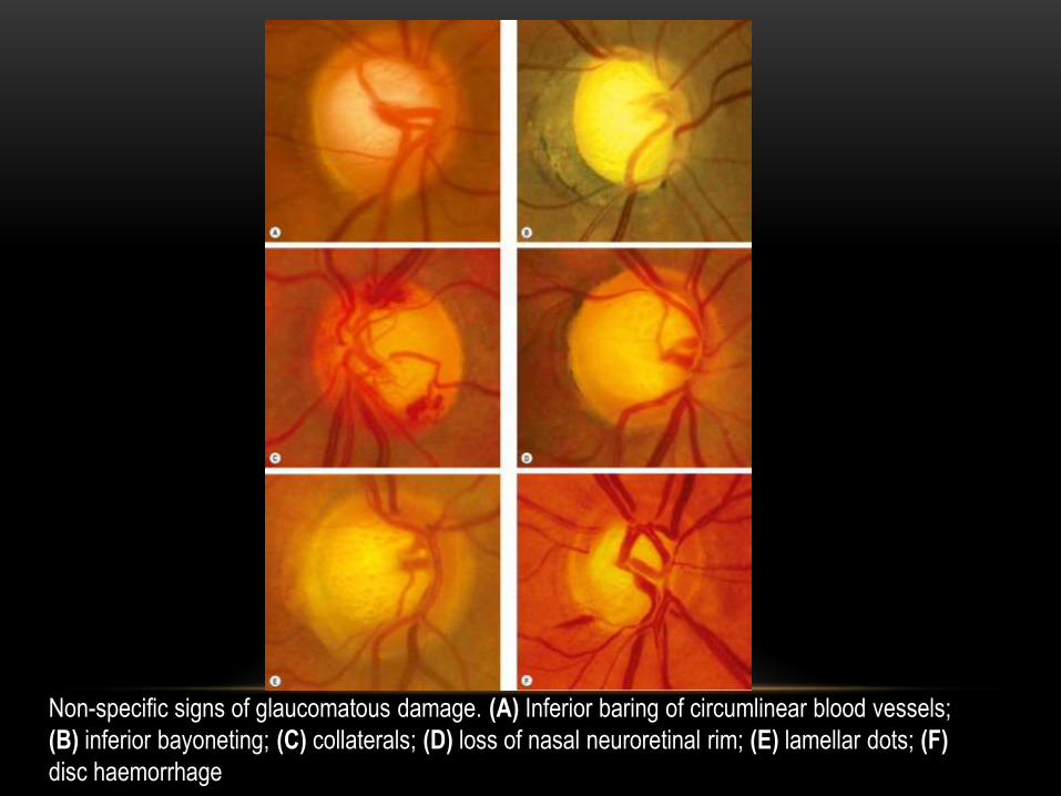
Non-specific signs of glaucomatous damage. (A) Inferior baring of circumlinear blood vessels;
(B) inferior bayoneting; (C) collaterals; (D) loss of nasal neuroretinal rim; (E) lamellar dots; (F)
disc haemorrhage

ANDERSON’S CRITERIA
On static perimetry, glaucomatous field loss is considered
significant if:
1. Analysis of glaucoma hemi-field test is abnormal in 2
consecutive occasion
2. 3 contiguous non-edge points on the pattern deviation
plot within Bjerrum area have a probability of < 5% of
being in normal population, one of which have a
probability of < 1%
3. Pattern standard deviation (PSD) should have a
probablity of < 5% confirmed on two consecutive tests

VISUAL FIELD ABNORMALITIES
• Initially observed in Bjerrum area, 10- 25° from fixation
• Correlate with abnormalities seen on optic nerve head
• Field defects:
1. Paracentral scotomas
2. Nasal step
3. Siedel scotoma
4. Arcuate scotoma
5. Double arcuate or ring scotoma
6. End-stage or near total defect with only a residual temporal island of vision


GRADING OF GLAUCOMATOUS DAMAGE
• MILD DAMAGE
Minimal cupping
Nasal step / paracentral
step
MD < -6dB
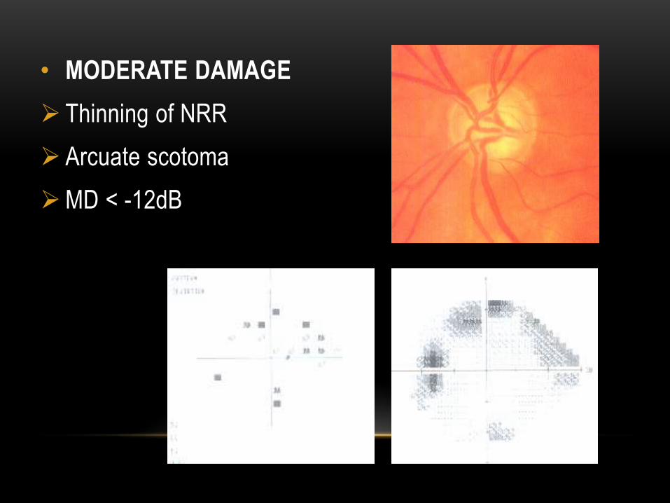
• MODERATE DAMAGE
Thinning of NRR
Arcuate scotoma
MD < -12dB

• SEVERE DAMAGE
Marked cupping
Extensive visual field loss
including defects within
central 5 degree
MD > -12dB
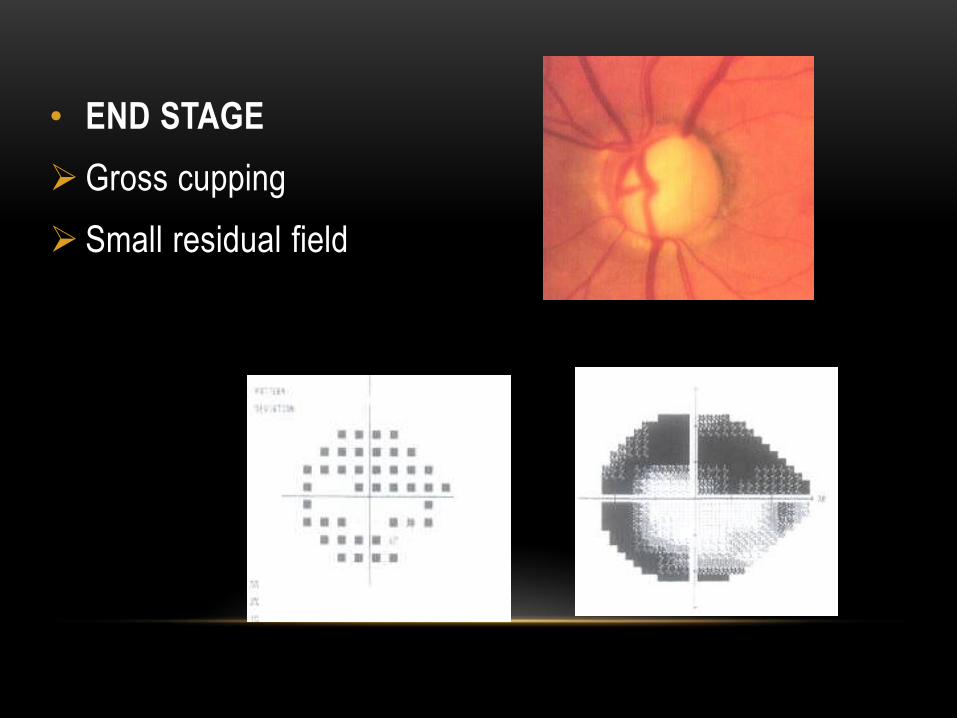
• END STAGE
Gross cupping
Small residual field
TREATMENT
MEDICAL MANAGEMENT
LASER
SURGICAL

PRINCIPLE OF TREATMENT
• Usually start with MEDICAL THERAPY.
• Before starting the treatment - Assess each eye individually, inform patients
• Start treatment in worse eye first
• Set TARGET PRESSURE

TARGET IOP DEPENDS UPON
• IOP at which damage has occurred
• Severity of Visual Field damage
• Rate of progression of damage
• Age and Life Expectancy

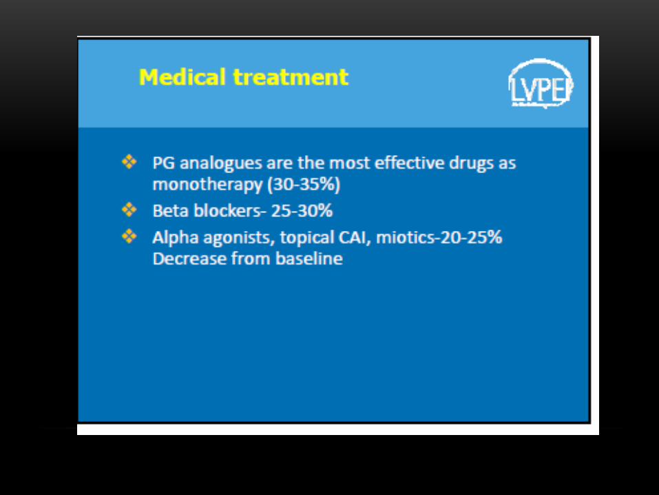
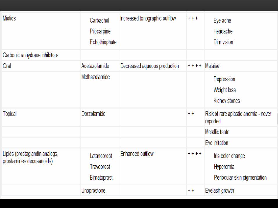
CLASSIFICATION• Drugs decreasing AQUEOUS PRODUCTION
Beta-blockers
Alpha-2-agonists
CAI
• Drugs increasing TRABECULAR OUTFLOW
Parasympathomimetics
Non selective agonists
Prostamides
• Drugs increasing UVEOSCLERAL OUTFLOW
Alpha-2-agonists
PG & PM


I Line II Line III Line


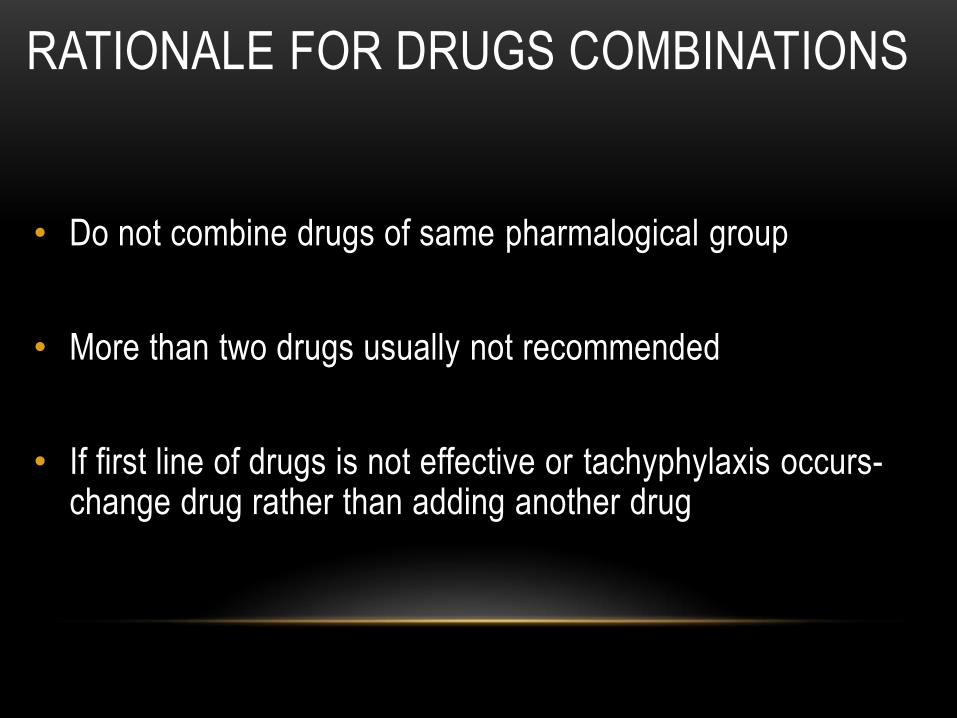
RATIONALE FOR DRUGS COMBINATIONS
• Do not combine drugs of same pharmalogical group
• More than two drugs usually not recommended
• If first line of drugs is not effective or tachyphylaxis occurs-change drug rather than adding another drug

LASERS IN POAG
• Outflow Enhancement
• Laser Trabeculoplasty
• Inflow reduction
• Cyclophotocoagulation (in end stage disease)

LASER TRABECULOPLASTY
• Uncontrolled glaucoma despite maximal tolerated medical therapy particularly in elderly
• Avoidance of polypharmacy
• Avoidance of surgery
• Poor compliance

SURGERY IN POAG
Indications:
• Failure of medical therapy
• Anticipated progressive damage or intolerably high IOP
• Combined with cataract procedure (phacotrabeculectomy)
• Primary therapy

• Penetrating filteration surgeries
• TRABECULECTOMY
• Nonpenetrating filteration surgery(NPFS)
• Deep Sclerectomy
• Viscocanalostomy
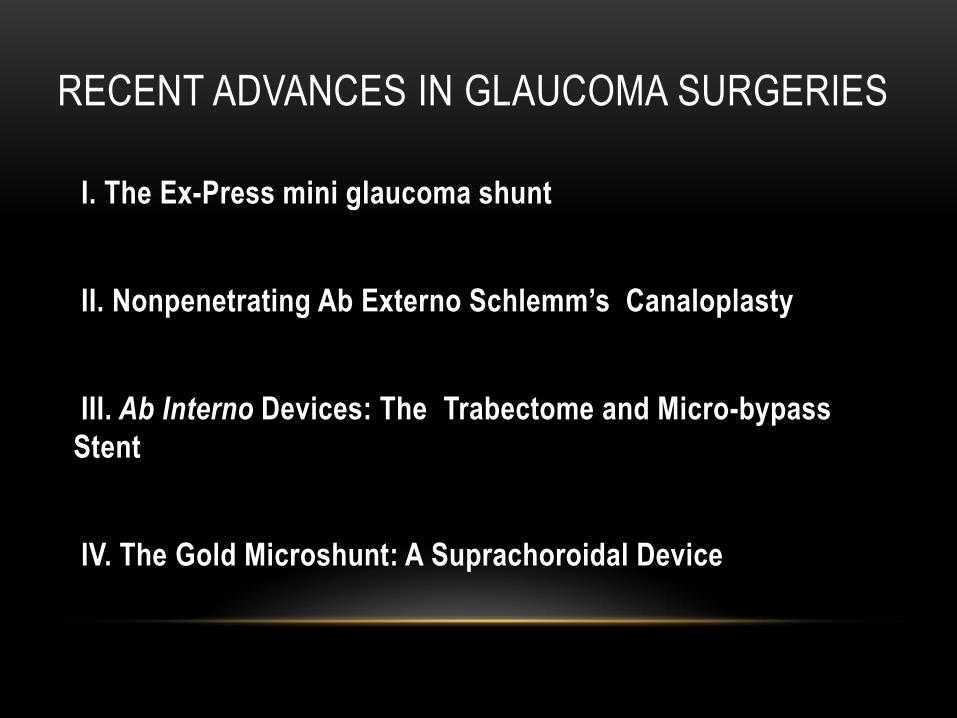
RECENT ADVANCES IN GLAUCOMA SURGERIES
I. The Ex-Press mini glaucoma shunt
II. Nonpenetrating Ab Externo Schlemm’s Canaloplasty
III. Ab Interno Devices: The Trabectome and Micro-bypass
Stent
IV. The Gold Microshunt: A Suprachoroidal Device