prevalence of unexpected red cell antibodies in …
TRANSCRIPT

PREVALENCE OF UNEXPECTED RED
CELL ANTIBODIES IN HEALTHY DONOR
POPULATION IN A TERTIARY CARE
CENTER IN SOUTH KERALA
Dissertation submitted to
SREE CHITRA TIRUNAL INSTITUTE FOR MEDICAL
SCIENCES AND TECHNOLOGY, TRIVANDRUM
In the partial fulfilment of the requirement for the degree for
M.D. in Transfusion Medicine
By
Dr. Gayathri A M
Under the Guidance of
Dr. Debasish Gupta
2017 – 2019

DECLARATION BY THE CANDIDATE
I hereby declare that this dissertation titled “Prevalence of
Unexpected Red Cell Antibodies in Healthy Donor Population
in a Tertiary Care Center in South Kerala” is a bonafide and
genuine research work carried out by me under the guidance of
Dr. Debasish Gupta, Professor and Head, Department of
Transfusion Medicine, Sree Chitra Tirunal Institute for Medical
Sciences and Technology (SCTIMST), Trivandrum.
Dr. Gayathri A M
Place: Trivandrum
Date: 30th June 2019

CERTIFICATE BY THE GUIDE
This is to certify that the dissertation titled “Prevalence of
Unexpected Red Cell Antibodies in Healthy Donor Population
in a Tertiary Care Center in South Kerala”, is a bonafide
research work done by Dr. Gayathri A M in partial fulfilment of
the requirement of the degree of MD Transfusion Medicine under
my guidance and supervision.
Dr. Debasish Gupta
Professor and Head
Department of Transfusion Medicine, SCTIMST, Trivandrum

ACKNOWLEDGEMENT
Thanks to Almighty for giving me strength and ability to understand, learn
and completion of my ventures. With boundless love and appreciation, I
would like to extend my heartfelt gratitude to the people who helped me to
bring this study into reality.
I consider myself fortunate indeed to have had the opportunity to pursue
my post-graduation and completion of MD thesis under the guidance of one
of the most prestigious personalities in the field of Indian Transfusion
Medicine, Prof. Debasish Gupta, Professor and Head, Department of
Transfusion Medicine, SCTIMST, Trivandrum. His dedication and keen
interest above all his overwhelming attitude to help his students had been
solely and mainly responsible for completing my work. His timely advice,
meticulous scrutiny, scholarly advice and scientific approach have helped
me to a very great extent to accomplish this task.
I owe a deep sense of gratitude to Dr. P V Sulochana and
Dr. S Sathyabhama, Department of Transfusion Medicine, SCTIMST,
Trivandrum for their keen interest on me at every stage of my research.
Their prompt inspirations, timely suggestions with kindness, enthusiasm
and dynamism have enabled me to complete my thesis. I am deeply
indebted to Dr. Jissa V T, Scientist C, Achutha Menon Centre for Health
Science Studies, Trivandrum, who helped me with the statistical analysis
of the data.
I would like to express my gratitude to Dr. Raj Bharath R and
Dr. Amita R, Associate and Assistant Professors, Department of
Transfusion Medicine, SCTIMST, for their valuable suggestions and
tremendous support.

I extend my respect to my seniors’ Dr. Revathy Nair and Dr. Vinu
Rajendran for their valuable advice and unconditional support as well as
my affection to my juniors’ Dr. Anila Mani and Dr. Sreethu Chand for
their constant help and support for completing my thesis work. I am deeply
indebted towards Mrs. Sheela Devi K S and Mrs. Sindhu P N, Scientific
Officers, Department of Transfusion Medicine, SCTIMST and to all other
Technical Assistants, Unit helpers and Diploma students of the Department
of Transfusion Medicine for their unconditional support for completion of
my thesis.
My family is my circle of strength, faith and ultimate heaven guarded by
my mother Smt. Ajithakumari V M, my father Sri. Mohanan Nair C,
my sister Ms. Gopika A M, my grandmother Smt. Madhavi Amma, my
better half Adv. Abhilash R and my little prince Advaith Krishnan.
Without their support I would not have achieved my academic well-being.
Last but not the least, I express my heartfelt gratitude to the study
participants who voluntarily agreed to be part of my study and without them
this thesis would have been infeasible.
Dr. Gayathri A M

CONTENTS
TOPICS PAGE NUMBER
Introduction 1-4
Aims & Objectives 5-6
Review of Literature 7-27
Materials & Methods 28-51
Results 52-71
Discussion 72-78
Conclusion 79-80
Limitations of the study 81-82
Summary 83-84
Bibliography 85-94
Annexure 95-110

ABBREVIATIONS
AABB American Association of Blood Banks
AHG Anti-Human Globulin
AHTR Acute Hemolytic Transfusion Reaction
AIHA Autoimmune Hemolytic Anemia
ALI Acute Lung Injury
APAb Anti-Phospholipid Antibody
BCA Benign Cold Agglutinins
C3d Complement Component 3
CA Cold Agglutinins
CAT Column Agglutination Technology
CTT Conventional Tube Technique
DAT Direct Antiglobulin Test
DGHS Directorate General of Health Services
DHTR Delayed Hemolytic Transfusion Reaction
DNA Deoxy-ribo nucleic acid
DTT Dithiothreitol
HDFN Hemolytic Disease of Fetus and Newborn
HLA Human Leucocyte Antigen

IAT Indirect Antiglobulin Test
Ig Immunoglobulin
IRL Immunohematology Reference
Laboratory
ISBT International Society of Blood
Transfusion
IVIG Intravenous Immunoglobulin
MHLBI National Heart, Lung, and Blood Institute
NACO National AIDS control Organisation
PBS Phosphate-buffered saline
PMN Poly morpho nuclear cells
PRBC Packed red blood cells
RBC Red blood cell
REDS III Recipient Epidemiology and Donor
Evaluation Study-III
RNA Ribo-nucleic acid
rpm Rotations per minute
SC Screening cells
SPRCA Solid Phase Red Cell Adherence
TRALI Transfusion Related Acute Lung Injury
TTI Transfusion Transmitted Infections

1
INTRODUCTION

2
Naturally occurring anti-A and anti-B are the only red cell antibodies that
are commonly found in human serum or plasma. All other antibodies are
called “unexpected red cell antibodies [1]. There are two types of
unexpected red cell antibodies: alloantibodies and auto-antibodies.
Alloimmunization occurs because of red cells antigenic differences
between donor and recipient in previous transfusions or between mother
and fetus. Auto-antibodies are those produced against one’s own antigens.
Immune humoral response in the presence of autoantibodies against
intracellular antigens characteristically occurs in a majority of connective
tissue diseases namely systemic lupus erythematous, systemic sclerosis,
Sjögren syndrome, mixed connective tissue disease, polymyositis, and
dermatomyositis [2]. Presence of these antibodies, alone or in combination,
makes difficulties with compatibility testing, thereby delaying in issue of a
compatible blood unit or may reduce post transfusion RBC life span [3].
The compatibility test comprises ABO/Rh determination, antibody screen
and cross-match. Type & cross-match technique/Immediate spin cross
match is routinely practised now which only detects ABO
incompatibility between donor RBCs and recipient serum/plasma. Type &
screen method is performed only when the recipient has unexpected
alloantibodies to detect additional RBC incompatibility [4]. Because of the
presence of auto-antibodies, all crossmatches become incompatible.
Studies conducted based on these unexpected antibodies have largely
concentrated on multiply transfused patient populations or antenatal
women. Alloimmunization in these groups has a reported incidence up to
60 percent, with an up to fourfold increased risk of multiple antibodies
compared to the risk of single antibodies [5]. However, such studies related
to healthy donor population are not done extensively. The incidence of RBC
alloimmunization depends on the demography and characteristics of the

3
population being studied. The specificity, Ig class, thermal range and
concentration of the antibody can predict its clinical significance as well as
patient’s individual immune response is also significant factors. The
balance between sensitivity and specificity can be influenced by the
methods and technologies selected. It is not possible to detect all potentially
clinically significant antibodies, or to avoid detecting all clinically
insignificant antibodies [6].
The Direct Antiglobulin test (DAT) is a simple test used to determine if red
cells have been coated in vivo with immunoglobulin (Ig), complement or
both. It is used primarily for the investigation of hemolytic transfusion
reactions, haemolytic disease of the fetus and newborn (HDFN),
autoimmune hemolytic anemia (AIHA), and drug-induced immune
hemolysis. An indirect antiglobulin test (IAT) is used to detect and identify
unexpected antibodies in the serum of blood donors, prospective
transfusion recipients, and prenatal patients [7]. IAT detect in
vitro antibody-antigen reactions and detect very low concentrations of
antibodies present in an individual’s plasma/serum.
When unexpected antibodies are present, as indicated by positive screening
tests, they must be identified. At a minimum, this involves testing the
patient’s serum against a panel of fully phenotyped reagent red cell samples
as well as the patient’s own cells [6]. A recent study suggests that a positive
DAT result in a healthy blood donor may be a marker of risk of future
development of malignancy [8]. All these point towards the need of Type
& Screening system to be followed routinely in transfusion practices rather
than Type & Matching system. Antibody screening is mandatory as laid
down by Drug and Cosmetic Act 1940 and Directorate General of Health
Services (DGHS) guidelines. Hence through this thesis work, we are

4
implementing routine antibody screening along with DAT testing in every
donated units in our Institute.
Our donor pool consists of 100% voluntary regular donors who are
considered to be absolutely safe and almost free of any infections and
highly motivated. This study also helps to find the prevalence of irregular
antibodies in such healthy donors there by aiding best transfusion practices
to be followed in the institution since there is scarce data available on
prevalence and type of irregular antibodies in Indian donor population.

5
AIMS AND OBJECTIVES

6
To study the prevalence of Unexpected Red Cell Antibodies in Healthy
Donor Population in a Tertiary Care Centre in South Kerala with the
following objectives:
1. To establish unexpected red cell antibody screening in
voluntary blood donors as a routine mandatory practice in the
Institute
2. To ensure blood components issued to patients are safe and
less likely to cause any red cell antibody mediated transfusion
reactions in the recipient
3. To add information about the prevalence of unexpected red
cell antibodies from south India to the national database

7
REVIEW OF LITERATURE

8
Just as genetic information determines the colour of one’s eyes, it also plays
an important role in determining the blood groups expressed. The term
blood group is usually restricted to blood cell surface antigens, and
generally to red blood cell (RBC) surface antigens. Blood groups are
inherited in Mendelian fashion. Each parent contributes half of the
inheritance and genetic information is carried on double strands of
deoxyribonucleic acid (DNA) known as chromosomes. Human cells have
23 pairs of chromosomes in the cell nucleus and out of these, 22 pairs are
autosomes and 1 pair of sex chromosomes. The units that code for various
expressions of inherited genetic information are known as genes which are
found in specific places along the chromosomes termed loci. For each
locus, there may be several different forms of a gene, which are known as
alleles. When both the inherited alleles are identical, the person is
homozygous and if different, the person is heterozygous. Sometimes,
homozygous inheritance produces a stronger expression of the gene than
would be seen in a heterozygous individual. This stronger expression is
known as dosage and is important because some antibodies react more
strongly with RBCs homozygous for a particular blood group inheritance
than with those with heterozygous inheritance.
The concept of population genetics of blood group antigens plays an
important role in blood banking. The term genotype is the set of genes in
our DNA which is responsible for a particular trait (detectable products)
and phenotype is the physical expression, or characteristics, of that trait.
Based on mode of inheritance, whether dominant or recessive in autosomes
or sex chromosomes decide the expression of antigens on the cell surface
of the individual. Phenotype frequencies in a population are determined by
randomly testing RBCs from a large number of individuals of that particular
population. The percentage of those positive (or negative) for a particular

9
trait is then calculated and indicates the frequency of a particular phenotype
(percentage or decimal). Once the individual frequencies of traits are
known, it is possible to calculate the frequency of multiple traits by
multiplying the frequencies of each trait. When two alleles are involved, it
is possible to use the Hardy–Weinberg equation to determine the frequency
of each allele. This equation states the following: p2 + 2pq + q2 = 1. The
frequency of an allele in the population is represented by the sum of the
frequency of the allele in the homozygous state plus the frequency of the
allele in the heterozygous state (9).
Frequency of p= p2 + 2pq
An exception to Mendelian law known as linkage, if two genes are linked,
they do not assort independently. Linked genes are located close to each
other on the same chromosome, and there is a 50% or greater chance that
they will remain together and be passed to the same gamete. Several blood
group genes have been found to be linked to other genes. The linkage of the
locus for the Lutheran blood group trait to the locus for the secretor trait
was the first known example of autosomal linkage in humans, reported by
Mohr in 1951. Because the genes are not inherited independently of each
other, they are not in equilibrium and hence known as linkage
disequilibrium.
An antigen is a substance that is capable of reacting with the product of an
immune response. Antigen combines with antibody (i.e., “antigen–antibody
reaction”. Introduction of nonself immunogens present on human RBCs,
white blood cells, and platelets may elicit antibody production. On a
biochemical level, an immunogen is a substance with a molecular weight
of 10,000 D or more. Substances with a molecular weight of less than 5,000
D seldom cause antibody formation. If coupled with larger carrier

10
molecules, however, these substances, known as haptens, can become
immunogenic (able to stimulate an immune response). Once an immune
response has been initiated by the hapten–carrier complex, the hapten alone
can react with the product of the immune response (i.e., antibody). Complex
biochemical compounds, especially those containing proteins or
polypeptide– carbohydrate combinations, are highly immunogenic. The
more diverse the molecule is, the more immunogenic it becomes. As
previously indicated, the structure must be recognized by the cells of the
recipient’s immune system to be non-self.
Biochemical analysis of blood group antigens has shown that they fall into
two main types: (i) protein determinants, which represent the primary
products of blood group genes; and (ii) carbohydrate determinants on
glycoproteins and glycolipids, in which the products of the genes
controlling antigen expression are glycosyltransferase enzymes (9). Some
antigens are defined by the amino acid sequence of a glycoprotein, but are
dependent on the presence of carbohydrate for their recognition
serologically. In recent years, molecular genetical techniques have been
introduced into the study of human blood groups and now most of the genes
governing blood group systems have been cloned and sequenced. Many
serological complexities of blood groups are now explained at the gene
level by a variety of mechanisms, including point mutation, unequal
crossing-over, gene conversion, and alternative ribonucleic acid (RNA)
splicing. Currently, there are 346 recognized blood group antigens
identified, out of which 308 are clustered within 36 blood groups systems.
These blood group antigens have the ability to mount immune challenge
and result in antibody production.

11
INCIDENCES OF IRREGULAR ANTIBODIES
The detection of antibodies directed against red cell antigens is critical in
pretransfusion compatibility testing methods such as Conventional Tube
Technique (CTT), Column Agglutination Technique (CAT) and Solid
Phase Red Cell Adherence (SPRCA) [10]. It is one of the principle tools
for investigating potential hemolytic transfusion reactions and immune
hemolytic anemias. In addition, it aids in detecting and monitoring patients
who are at risk of delivering infants with HDFN. The focus of antibody
detection methods is on “irregular” or “unexpected” antibodies, as opposed
to the “expected” antibodies of the ABO system. There are many different
and important characteristics of blood group antibodies, such as whether
they are polyclonal or monoclonal, naturally occurring or immune, and
alloantibodies or autoantibodies [9]. An antigen usually consists of
numerous epitopes, and it is the epitopes and not the entire antigen that a B
cell is stimulated to produce antibody against. Hence different epitopes of
same antigen produce individual antibodies resulting in polyclonal
antibody production. Monoclonal antibodies are prepared in controlled
environment through hybridoma technology. The unexpected antibodies of
primary importance are the immune alloantibodies, which are produced in
response to RBC stimulation through transfusion, transplantation, or
pregnancy. Other unexpected antibodies may be “naturally occurring” (i.e.,
produced without RBC stimulation). Naturally occurring antibodies may
form as a result of exposure to environmental sources, such as pollen,
fungus, and bacteria, which have structures similar to some RBC antigens.
Antibodies produced in one individual and then transmitted to another via
plasma-containing blood components or derivatives such as intravenous
immunoglobulin (IVIG) are known as passively acquired antibodies, a third
category of unexpected antibody. The presence of naturally occurring and

12
passive antibodies may complicate the detection and identification of
immune, clinically significant antibodies.
A clinically significant antibody can be defined as one capable of causing
accelerated destruction of a significant proportion of transfused cells, or one
capable of crossing the placenta and causing haemolytic disease of the fetus
and newborn. When the alloantibodies combine with the corresponding
RBC antigens, it can cause acute haemolytic transfusion reaction (AHTR)
or delayed haemolytic transfusion reaction (DHTR) [9]. The specificity, Ig
class, thermal range and concentration of the antibody can predict clinical
significance; however, the clinical situation and patient’s individual
immune response are also significant factors [9]. The aim is to detect
antibodies that are likely to cause problems in the clinical setting for which
the antibody screening is designed, for example patient pretransfusion,
antenatal or donor.
All immunoglobulins can be significant for transfusion medicine; however,
IgG, IgM, and IgA have the most significance. Most clinically significant
antibodies that react at body temperature (37°C) are IgG isotype and are
capable of destroying transfused antigen-positive RBCs, causing anemia
and transfusion reactions of various severities. IgM antibodies are most
commonly encountered as naturally occurring antibodies in the ABO
system and are believed to be produced in response to commonly occurring
antigens like intestinal flora and pollen grains. Other blood groups such as
Lewis, Ii, P, and MNS may also produce IgM antibodies, which usually
react best at ambient temperature (22°C to 24°C). The primary testing
problem encountered with IgM antibodies is that they can interfere with the
detection of clinically significant IgG antibodies by masking their
reactivity. Unlike IgG, IgM exists in both monomeric and polymeric forms
(as pentamers) containing a J (joining) chain [9].

13
IgG antibodies are significant in transfusion medicine, because they are the
class of immunoglobulins that are made in response to transfusion with
nonself antigens on blood products. IgG antibodies are important in HDFN,
because antibodies can be formed in response against allo-antigens on fetal
RBCs that enter the mother’s circulation, usually during delivery. Antibody
screening, Rh typing, and passive anti-D antibody have prevented HDFN
from developing in D-negative mothers who give birth to D-positive babies
[10]. Fetal anemia in anti-Kell HDFN is associated with suppression of
erythropoiesis due to destruction of erythroid precursor cells, which can be
additional to destruction of circulating antigen-positive RBCs, as seen in
anti-D HDFN. Serum IgA is found in both monomeric and polymeric
forms; however, secretory IgA is usually found in the mucosal tissues of
the body. Its polymer form acquires a glycoprotein secretory component as
it passes through epithelial cell walls of mucosal tissues and appears in
nearly all body fluids. Around 30% of anti-A and anti-B antibodies are of
the IgA class and also, anti-IgA antibodies can cause severe anaphylaxis if
IgA are transfused in plasma products to patients who are deficient in IgA.
Another reason for the importance of IgA is that it can increase the effect
of IgG induced RBC hemolysis [9].
Karl Landsteiner, the father of transfusion medicine discovered the first
human blood group system, ABO in 1901. This marked the beginning of
the concept of individual uniqueness defined by the RBC antigens present
on the RBC membrane. For the next 45 years, only those antibodies that
directly agglutinate red cells could be studied. With the development of the
antiglobulin test by Coombs, Mourant, and Race in 1945, non-agglutinating
antibodies could be detected and the science of blood group serology
blossomed. Between 1946 and 1947 with the use of the indirect antiglobulin
test the Kell (K), Duffy (Fy), Kidd (Jk), Diego (Di), Cartwright (Yt), Xg,

14
Dombrock (Do) and Colton (Co) blood group systems were discovered.
The International Society of Blood Transfusion (ISBT) Working Party on
Terminology for Red Cell Surface Antigens was set up in 1980 to establish
a uniform nomenclature for all blood group antigens. Now 346 blood group
antigens are identified and out of them, 308 are clustered within 36 blood
groups systems. Remaining are accommodated in the Collections and
Series: 700 Series of Low Incidence Antigens & 901 Series of High
Incidence Antigens.
The AHG test/Coombs’ test, is based on the principle that antihuman
globulins obtained from immunized non-human species bind to human
globulins such as IgG or complement, either free in serum or attached to
antigens on red blood cells. Because of their large pentamer structure, IgM
antibodies bind to corresponding antigen and directly agglutinate RBCs
suspended in saline. Some IgG antibodies are termed non-agglutinating, or
incomplete antibodies, because their monomer structure is too small to
directly agglutinate sensitized RBCs. Adding AHG that contains anti-IgG
to RBCs sensitized with IgG antibodies allows for hemagglutination of
these sensitized cells. We use an anti-antibody to observe the formation of
an Ag/Ab complex that would otherwise go undetected. Some blood group
antibodies have the ability to bind complement to the RBC membrane. In
such cases, an anti-complement component can be added to the AHG
reagent, rendering it polyspecific. Antiglobulin tests detect IgG or
complement-sensitized RBCs [9].
Apart from AHTR and DHTR along with HDFN, transfusion-related acute
lung injury (TRALI) is the most severe adverse outcome from transfusion,
with estimates of prevalence ranging from 0.02% to 0.16% of patients
receiving transfusions [12,13] which is also an antibody mediated adverse
reaction. TRALI is the clinical syndrome of new acute lung injury (ALI)

15
that develops with a clear temporal relationship to transfusion, in patients
with or without alternate risk factors for ALI [14]. TRALI manifests as
fever, hypoxemia, and pulmonary oedema occurring acutely (within 6
hours) following transfusion of blood components. The exact
pathophysiology of TRALI is unclear, though TRALI is strongly associated
with the presence of allotypic antibodies present in donor blood products.
Antibodies against HLA class I and class II molecules have been found in
50% to 89% of products associated with TRALI. Additionally, antibodies
to HNA-1a, HNA-1b, HNA-2a, and HNA-3a, allo-antigens found
specifically on neutrophils, have been described in as many as 72% of blood
products associated with TRALI [15,16]. The current model of TRALI
suggests that these allotypic antibodies activate neutrophils in the lung,
which release cytokine and chemokine mediators that cause pulmonary
oedema [17]. The current models of the pathogenesis of TRALI secondary
to the infusion of donor antibodies directed against recipient HLA class II
antigens are based on associative data and require proinflammatory
activation of the pulmonary vascular endothelium and circulating
monocytes, resulting in polymorphonuclear neutrophil (PMN)-mediated
endothelial damage, capillary leak, and ALI [18].
Detection of ‘insignificant’ antibodies creates additional work and leads to
potential delay in providing compatible blood for transfusion, or
unnecessary repeat testing/interventions in pregnancy. The balance
between sensitivity and specificity can be influenced by the methods and
technologies selected. It is not possible to detect all potentially clinically
significant antibodies, or to avoid detecting all clinically insignificant
antibodies, that is ‘noise’ [19]. Antibody detection (screening) can be
optimized by designing protocols appropriate to the population to be
screened, utilizing knowledge of the characteristics of red cell antibodies,

16
understanding of the principles, strengths and limitations of the
methods/technologies validated for use, and ongoing quality assurance of
testing. All alloantibodies detected in screening should be investigated for
specificity, and the results interpreted in clinical context. Automation is
happening in every arena of Transfusion Medicine and now we also have
an automated system (software) to interpret specificity of allo-antibody.
Such software uses an algorithm based on behaviour and characteristics of
antibodies. Antibody identification software can support the technicians in
identifying antibodies and can also guide to carry out supplementary tests
to rule out other antibodies. It may be used as a supplement or may
eventually replace manual method of antibody identification [20].
INCIDENCE OF UNEXPECTED ANTIBODIES IN
DIFFERENT POPULATIONS
According to a study from New England Research Institute, only 0.3–1.0%
of general population have unexpected antibodies and the incidence is
higher in women due to the pregnancy and patients receiving multiple
transfusions [21]. But generally, the prevalence of clinically significant
alloantibodies has been reported from less than 0.3% to up to 60% of
samples depending on the study populations and the test method sensitivity
[22,23]. Not uncommonly, autoantibodies can also be found along with
alloantibodies which have been reported to be as high as 28% [24].
A study from Middle east Kuwaiti population states that prevalence of
alloimmunization among blood recipients and donors (without the
inclusion of pregnant women) was 0.49%. In general, the five most
frequently identified alloantibodies were anti-D (27.3%), anti-E (18.5%),
anti-K (15.6%), anti-Lea (8.7%) and anti-Leb (6.6%) [25]. A study on

17
prevalence and specificities of red blood cell alloantibodies in transfused
Ugandans with different diseases [26] states that out of 113 transfused
patients, 13 developed RBC alloantibodies in which 11 patients produced a
total of 12 RBC alloantibodies of known specificity and remaining two
patients possessed pan-reactive antibodies. The specificities of the
alloantibodies identified were: 6 patients developed anti-E, 3 got anti- S and
1 each of anti-D, -K and -Lea. In one patient, two alloantibodies, anti-E plus
anti-K, presented as a combination; the rest of the alloantibodies were as
single specificities. In an Indian study, incidence of RBC alloimmunization
in transfused patients was 3·4% (18/531), with anti-c being the most
common specificity (38·8%), followed by anti-E (22·2%), anti-M (11·1%),
anti-Lea (11·1%), anti-D (5·6%), anti-Jka (5·6%) and anti-Leb(5·6%) [27].
In Chinese population of Guangxi, a study on RBC alloimmunisation rates
among 20283 hospitalised patients, 166 alloantibodies were identified in
150 (0·74%) patients. Of the 150 patients with alloantibodies, 134
(89·33%) had a single alloantibody, whereas the remaining 16 (10·67%)
had multiple alloantibodies. Out of these antibodies, the five most
frequently identified alloantibodies were anti-E (39·76%), anti-Mia /Mur
(11·45%), anti-c (10·84%), anti-Lea (8·43%) and anti-M (6·63%) [28].
From a Northern Indian study, the cumulative incidence of irregular
antibodies amongst the patients and donors were found as 0.12% and
0.009%, respectively. The incidence (0.09%) of Rh antibodies was found
to be highest among the clinically significant antibodies. Anti-D antibody
was the commonest antibody in Rh blood group system and the second most
common blood group associated with the relatively higher rate of antibody
incidence was MNS blood group system. The preponderance of Rh
antibodies is also because of absence of universal RhD prophylaxis in
pregnant women in the country as a whole. However, amongst the blood

18
donors the commonest blood group system associated with highest
incidence of clinically significant antibody was MNS [29]. The incidence
of DAT amongst the healthy blood donors in this was 0.04%. In western
literature, similar studies in 1977 & 1989 reported as 0.3% and 38%
respectively [30,31]. The pioneer study report on incidence of irregular
antibodies and DAT in northern Indian population is 0.009% & 0.41%
respectively in blood donors. It does add a new perspective that the
probability of an irregular antibody is around 13 times (0.009% vs. 0.12%)
lower in a healthy donor as compared to a patient [29]. This also brings
forth a paradigm that 4 out of 10,000 (0.04%) healthy donors can have DAT
positive without any obvious clinical signs and symptoms of auto-immune
hemolysis. It is this category of donor units which show up as
‘incompatible’ even when patient’s type and screen is negative. Therefore,
as an institutional policy, these units are discarded (because these units
cause problems in the compatibility testing). There is also a need to do an
elaborate long-term follow-up on these donors since there is an evidence of
a significantly increased risk of cancer, especially hematologic
malignancies, among blood donors with a positive DAT [29,32]. Another
Indian study on prevalence of unexpected red cell antibodies in blood donor
population revealed that 0.27% (227/82,153) were positive for antibody
screening using pooled O cells [33].
In western literature, Recipient Epidemiology and Donor Evaluation Study-
III (REDS-III) by National Heart, Lung, and Blood Institute (NHLBI,
Bethesda), indicates that of the 2,045,204 donations, 10,450 (0.5%) had a
positive RBC allo-antibody screen. Of the 632,378 unique donors,
4,861(0.77%) had a positive antibody screen on one or more of their
donations over the study interval. The distribution of antibody specificities
varied by sex, with anti-D being more prevalent in females and anti-K being

19
more prevalent in males [34]. Another Colombian study on blood donors’
states that during the period 2007–2009, there were 60,309 whole-blood
donations, and 438 alloantibodies were found (0.73%), 66.7% of them in
female blood donors and 33.3% in male donors. The clinically significant
were 140 (31.96%) and non-clinically significant were 300 (68.5%). The
descending order of prevalence were anti-Lea, antibodies against low
prevalence antigens, cold antibodies, non-specific IgG, anti-Leb, anti-D,
anti-E, anti-K and anti-M [35].
DAT POSITIVITY AMONG HEALTHY DONORS
The incidence of positive DATs amongst healthy blood donors with a
normal haemoglobin level varies from 1 per 9,000 to 1 per 14,000 [36,37].
In the majority of case, long term follow-up of these donors reveals no
major clinical sequelae and no apparent cause for DAT positivity.
Consequently, the reason for the positive DATs cannot be explained
satisfactorily [37].
Healthy individuals can have 5–90 IgG molecules/red cell [38] and 5–40
C3d molecules/red cell [39]; these levels are usually below the threshold of
detection in routine testing. Depending on the technique and reagents
used, the DAT can detect 100–500 molecules of IgG/red cell and 400–1100
molecules of C3d/ red cell. Positive DATs are reported in 1:1000 up to
1:14,000 blood donors and 1–15% of hospitalised patients [40]. These large
differences in incidence probably relate to the different techniques used for
performing the test. Based on Glasgow and West of Scotland Blood
Transfusion Service studies that serves a population of approximately three
million people, the incidence of positive DAT among healthy blood donors
20
in the west of Scotland is approximately 1 per 7,000 donations [41]. But in
some populations, positive DATs are related to intercurrent viral infections
or existing immune disorders [42,43]. Positive DATs due to
antiphospholipid antibodies (APAbs) have now been well described, both
in the antiphospholipid syndrome with systemic lupus erythematosus and
the primary antiphospholipid syndrome, the incidence being 16% in the
former and 4.3% in the latter [44]. APAbs may occur in association with a
variety of auto-immune disorders including systemic lupus erythematosus,
malignancy or infection and, very occasionally in the apparently healthy
[42]. In Glasgow and West Scotland study, they found that sixty-six units
from 42 donors were found to be DAT positive and eight blood donors with
positive DATs also had false positive VDRL tests out of 474,545 donations
over 3 years [41]. Out of these eight donors, 3 had elevated APAbs also.
The well-documented association of APAbs in the antiphospholipid
syndrome with positive DATs led them to believe that the positive DATs
in these 3 healthy blood donors (3 out of 42) was due to cross-reacting
APAbs adsorbing non-specifically on to the erythrocyte membrane and
binding to phospholipid epitopes. Therefore, APAbs may be an incidental
cause of positive DATs amongst healthy blood donors.
However, a small number of donors, in particular, those with persistence of
IgG on the RBCs, may be in the early stages of an autoimmune illness and
may be developing a serum protein abnormality that could presage a more
serious illness such as a malignancy or a hematologic disorder. As the
opportunity exists for early diagnosis of a clinically significant and
potentially treatable illness, the follow-up of blood donors with a positive
DAT and IgG on the RBCs is recommended by most authors. If a donor is
deferred based on a positive DAT, it is appropriate that they be referred to
a physician for periodic assessment and follow-up [45,46]. Legal and

21
ethical standards recognize that a blood donor has the right to be provided
with any information that may be significant for his or her health and that
these are also important considerations in determining policies for
management of blood donors in this situation.
CLINICALLY SIGNIFICANT RED CELL ANTIBODIES
Clinically significant red cell antibodies are usually IgG antibodies active
at 370C and/or by the indirect antiglobulin test. They cause HDFN and
HTRs by destroying the red cells and thereby reducing its lifespan and
associated consequences. AHTR consists of acute hemolysis with
accompanying presenting symptoms within 24 hours of transfusion which
may be immune or non-immune origin. Previously formed IgM or IgG
antibodies in the recipient recognize the corresponding donor red cell
antigens, immune complexes are formed, the complement cascade is
activated, vasoactive amines and inflammatory mediators are released into
the plasma, and the coagulation cascade is activated [47]. The activated
lytic arm (membrane attack complex) of the complement cascade causes
hemoglobinemia and hemoglobinuria, the hallmarks of intravascular
hemolysis. DHTR is classically caused by a secondary immune reaction
where alloantibodies undetectable at the time of transfusion rebound
following exposure to the corresponding RBC antigens, leading to the RBC
destruction [48]. DHTR occurs between 3 and 22 days after RBC
transfusion. The Immunohematology Reference Laboratory (IRL, formerly
the Red Cell Reference Laboratory) has served the Pacific Northwest as a
regional reference laboratory for over 30 years. IRL classifies red cell
antibodies into Group I to Group IV. Group I is clinically significant
antibodies which includes ABO, Rh (D, C, E, c, e), Kell, Duffy, Kidd, S

22
and s. Group II being benign antibodies mainly Chido/Rodgers (Cha/Rga),
Xga, Bg, Csa, Kna, McCa, Yka and JMH. Group III consists of clinically
insignificant if not reactive at 37°C; possibly significant when reacting at
37°C (Lewis, MN, P, Lutheran and A1) and Group IV have antibodies that
are sometimes clinically significant (Yta, Vel, Ge, Gya, Hy, Sda). Only a
few antibody specificities are commonly seen: M, P1, and I antibodies react
at room temperature and are considered clinically insignificant; K, S, s, Fya,
Fyb, Jka, and Jkb antibodies react in the antiglobulin phase and are clinically
significant.
American Association of Blood Banking (AABB) states that if an antibody
screen is positive, an attempt should be made to identify the antibody and
cross match for the same along with ABO and Rh. If the antibody screen is
negative, they recommend only ABO/Rh compatibility testing to be done
before transfusion. However, there is great paucity of literature regarding
the scenario where the antibody screen is positive but the antibody
identification is inconclusive, to see if this has any clinical implications. As
the literature states antibodies against Rh system especially Rh (D, C, E, c,
e) is very common among general population because of absence of
universal RhD prophylaxis in pregnant women in almost all developing and
underdeveloped countries as a whole.
Most of the published literature refers to antibodies of Lewis blood group
system to be insignificant, whereas antibodies to M and N blood groups are
associated with variable clinical significance. Antibodies against MN and
Lewis blood group antigens with their thermal amplitudes in the range of
22-30°C gain special importance in certain conditions of induced
hypothermia. These antibodies with a higher thermal range, which would
otherwise be termed clinically insignificant, will induce in vivo hemolysis
in patients with lowered core body temperature, which is now a common

23
practice in various surgeries such as neuro surgeries, cardiac surgeries etc
[49]. Therefore, the thermal amplitude of the antibody must always be
determined and if judged to be clinically significant, corresponding antigen
negative blood must be provided. Many examples of anti-M are naturally
occurring saline agglutinins that react below 37°C. the antibody can have
IgM and IgG components. They do not bind complement, regardless of
their immunoglobulin class, and they do not react with enzyme-treated
RBCs. Anti-M appears to be more common in children than in adults and
is particularly common in patients with bacterial infections [50]. When anti-
M reacts at 37°C, it is sufficient to provide units that are crossmatch-
compatible at 37°C and at the antiglobulin phase without typing for M
antigen. Anti-M rarely causes HTRs, decreased red cell survival, or HDFN.
Lewis antibodies are often naturally occurring and made by Le (a– b–)
persons; that is, they occur without any known RBC stimulus. They are
generally IgM and do not cross the placenta. Because of this and because
the Lewis antigens are not well developed on fetal RBCs, the antibodies do
not cause HDFN. Lewis antibodies occur quite frequently in the sera of
pregnant women who transiently exhibit the Le (a– b–) phenotype. Anti-
Lea is the most commonly encountered of the Lewis antibodies and is often
detected in room temperature tests, but it sometimes reacts at 37°C and in
the indirect antiglobulin test. Rare hemolytic transfusion reactions have
been reported in patients with anti-Lea who were transfused with Le(a+)
RBCs, so anti-Lea that are reactive at 37°C, particularly those that cause in
vitro hemolysis, should not be ignored [51].
In the twentieth century, Landsteiner [52] first described blood
agglutination at cold temperatures and Schubothe named the disorder ‘‘cold
agglutinin disease’’ [53] ‘Physiological’ cold agglutinins (CA) or benign
cold agglutinins (BCAs) develop because of change in expression of red

24
cell antigens that occurs after birth. These antibodies react best at about 40C
and are present in the serum of almost all individuals as low titre (<1:10)
auto-antibodies [54]. Moderately, high titre CAs with higher thermal
amplitude was associated with certain infections. For example, anti-I with
Mycoplasma, Listeria, Leishmaniasis; anti-i with infectious
mononucleosis, and anti-Pr with viral infections like rubella or Varicella
[55]. Pathological cold agglutinins are maximally reactive at around 28–
310C and tend to occur at very low titres [56]. In 90 % of cases, cold
agglutinins are IgM by nature. Rarely, monoclonal IgG, IgA, or k light
chain restriction, are responsible. CAs can interfere with blood grouping
and cross-matching. It is necessary to use O reagent cells in the reverse
grouping and also to carefully note the strength of the agglutination. In
cases of severe disease, samples have to be collected and maintained at
370C until the plasma and red cells are separated or pre-warmed plasma
(plasma kept at 370C for 10–15 min) may be used as was done in this case.
For the DAT, the patient’s red cells were washed with warm saline to
remove residual plasma containing cold antibodies. Warming reduces the
adsorption of the cold antibodies onto the red cells. Minimizing adsorption
during sample processing yields more accurate titres of cold antibodies, and
avoids false-positive results [57]. Spontaneous cold auto-agglutination
phenomenon observed in blood units stored under blood bank conditions
appears to be due to a presence of CA. As these auto-antibodies are strictly
reactive at low temperatures, they do not give rise to in vivo hemolysis. As
the antibody specificities identified are known to be associated with certain
infections, their presence in healthy blood donors could be attributed to
exposure to such infections in recent past or present in its subclinical phase
at the time of blood donation [55].

25
Anti-I is a common autoantibody that can be found in virtually all sera,
although testing at 4°C and/or against enzyme treated RBCs may be
required to detect the reactivity [51]. Consistently strong agglutination with
adult RBCs and weak or no agglutination with cord or adult i RBCs define
its classic activity. Auto-anti-I, found in the serum of many normal healthy
individuals, is benign—that is, not associated with in vivo RBC destruction.
It is usually a weak, naturally occurring, saline-reactive IgM agglutinin
with a titre less than 64 at 4°C. Stronger examples agglutinate test cells at
room temperature and bind complement, which can be detected in the
antiglobulin test if polyspecific reagents are used. Occasionally, benign
cold auto-anti-I can cause problems in pretransfusion testing. Usually,
avoiding room temperature testing and using anti-IgG instead of a
polyspecific antihuman globulin help to eliminate detection of cold reactive
antibodies that may bind complement at lower temperatures. Cold auto-
adsorption to remove the autoantibody from the serum may be necessary
for stronger examples; cold auto-adsorbed plasma or serum can also be used
in ABO typing. Pathogenic auto-anti-I (e.g., the type associated with cold
agglutinin syndrome) typically consists of strong IgM agglutinins with
higher titres and a broad thermal range of activity, reacting up to 30° or
32°C. When peripheral circulation cools in response to low ambient
temperatures, these antibodies attach in vivo and cause autoagglutination
and peripheral vascular occlusion (acrocyanosis) or hemolytic anemia. The
production of auto-anti-I may be stimulated by microorganisms carrying, I-
like antigen on their surface e.g. M pneumoniae [9]. Many other I-related
antibodies have been described: anti- IA, -IB, -IAB, -IH, -iH, -IP1, -ITP1,
-IHLeb, and -iHLeb due to close relationship of I to the biochemical
structures of ABH, Lewis, and P antigens. These specificities are not
mixtures of separable antibodies; rather, both antigens must be present on

26
the RBCs for the antibody to react. Anti-IH is commonly encountered in
the serum of group A1 individuals. Anti-IH reacts stronger with group O
and group A2 RBCs than with group A1 RBCs. Anti- IH should be
suspected when serum from a group A individual directly agglutinates all
group O RBCs but is compatible with most group A donor units [9].
IMPORTANCE OF TYPE AND SCREEN
Several studies have proven that there is high incidence of irregular
antibodies was in the patients following transfusion. In western literature as
well as Indian studies the irregular antibody incidence range from 0.75% to
38% among these most of them were multi-transfused [23,27,58]. The least
incidences indicate most of them followed Type and Screen strategy
[59,60] than Type and Matching System which is routinely practiced in
many places. There are two Indian studies [59,60] which have compared
type and screen with conventional AHG cross-match. They have slightly
different results. While the first study [60] had 45,373 patients receiving a
total of 61,668 units of packed RBCs (PRBCs) which were cross-matched
in the AHG phase (irrespective of the result of the antibody screen) with
simultaneous antibody screen carried out in all the patients. They did not
find a single case where the antibody screen was negative and AHG cross-
match showed incompatibility. The study concluded that type and screen
policy can be safe, efficient, cost-effective, and beneficial to the transfusion
service in India. In the second study [59] on 2026 patients, one sample gave
a negative antibody screen while conventional crossmatch was
incompatible. The authors said that this may be due to a rare antibody in
the patient sera against which the corresponding antigen was not present on
the screening cells but present on the donor red cell. They said that a

27
transfusion reaction would have occurred in this case since it was reacting
at 370C in AHG. The authors opined that since, the screening panel used in
the present study was not from Indian sub-continent (panels are from
Caucasian donors); therefore, it would be advisable to prepare screening
cell panels that include RBC antigens which are prevalent within the
local/regional population e.g., In, Mia etc.

28
MATERIALS AND
METHODS

29
Using a cross sectional study design, randomly chosen 7000 voluntary
donors coming for blood donation during a period of 1 year from Dec 2017
to Dec 2018 were screened for presence of unexpected red cell antibodies.
Donors were screened according to departmental as well as DGHS
guidelines and were given prescribed questionnaires to fill up. Later they
underwent counselling and a medical examination as a part of blood donor
assessment. Eligible donors underwent phlebotomy and after blood
donation, blood samples were collected for routine immunohematological
investigations, Transfusion Transmitted Infections (TTI) screening and
antibody screening as per prescribed DGHS guidelines. For this study, an
additional 2ml of blood was collected in EDTA test tube for performing
DAT, along with pilot test tubes.
All donors were screened for red cell antibodies by Direct Antiglobulin
Test and Indirect Antiglobulin Test primarily along with routine ‘O’
papanisation during reverse grouping. In DAT, donor cells are screened and
in IAT donor serum is used for antibody detection. DAT positivity indicates
in vivo sensitisation as well as IAT indicates in vitro sensitisation of the
donor with an antibody. DAT and IAT after incubation at 370C positive
blood unit will be discarded as hospital blood transfusion policy. Auto-
control using donor cells and serum is required for checking the validity of
the test as well as confirmation of auto-agglutinins after incubation of the
same at different temperatures (40C, 22 0C & 370C).
If Direct Antiglobulin Test is found positive, then further antibody
determination is done by different types of Elution methods namely heat
elution and ether elution techniques. If IAT is positive, then antibody
screening and identification is done using red cell panels. Antibody titration
also follows identification procedures. Secretor status was also performed

30
in 3 donors with anti-IH and anti-Lewis antibodies. These antiglobulin test
positive donors were called back for notification and another fresh sample
were taken for retesting and given proper counselling as well as follow up
of the donor was also performed. My thesis concentrated on healthy
voluntary donor population and later on similar study can be extended out
to patient population to get a better prevalence among general population.
TESTING FOR ABNORMAL RED CELL ANTIBODIES APART
FROM TESTING PAPAINISED O POOLED CELLS ALONG
WITH REVERSE GROUPING
(Adopted from AABB Technical Manual 18th edition; USA 2014)
METHOD 1: PREPARING A 3-5% RED CELL SUSPENSION
Principle: A 3% red cell suspension is a common reagent in many serologic
procedures. This achieves the appropriate serum-to-cell ratio for most test
procedures and for an adequate number of red cells so one can read and
grade the reactions.
Materials
1. EDTA whole blood sample
2. Test tubes
3. Glass Pauster pipettes (5-mL serologic)
4. 0.9% Normal saline (NS)
5. Table top centrifuge (3000 rpm or equivalent)
6. Laboratory prepared 3% pooled O red cell suspension

31
Procedure:
Step Action
1 Transfer at least 1 mL of whole blood to a 10-mL tube
2 Wash the red cells in saline, centrifuging for 5 minutes to pellet
the cells. Repeat two or three times. The final supernatant should
be clear and should be completely removed by aspiration
3 Transfer 0.3 mL of the washed red cells to a tube with 9.7 mL
of saline and thoroughly mix the red cells and saline by gently
inverting the tube several times
4 Label the tube as 3% donor cell suspension with donor ID
number
METHOD 2: SALINE INDIRECT ANTIGLOBULIN TEST
PROCEDURE
Principle: An indirect antiglobulin test demonstrates in-vitro reactions
between red cells and antibodies, and is used in antibody detection,
antibody identification, crossmatching, and blood group phenotyping. The
saline method is performed when red cells are washed to remove unbound
globulins.
Specimen: Serum is used from clotted pilot tube

32
Reagents:
1. Normal saline
2. Polyspecific and monospecific Antihuman globulin (AHG)
reagent
3. 3-5% pooled group O cell suspension
4. A 3-5% suspension of donor red cells in saline after 3 times
washing in normal saline
5. IgG-coated red cells
Procedure:
Step Action
1 Add 1 drops of donor serum to properly labelled tubes
2 Add 1 drop of 3-5% saline-suspended reagent group O red
cells and 3-5% donor red cell saline suspension (auto-control)
to each tube and mix
3 Centrifuge and observe for hemolysis and agglutination.
Grade and record the results
4 Incubate at 370C for 60 minutes
5 Centrifuge and observe for hemolysis and agglutination.
Grade and record the results

33
6 Wash the red cells three times with saline, and completely
decant the final wash
7 Add 1 drop polyclonal AHG to the dry red cell button.
Mix well and then centrifuge immediately at 1000 rpm for 1
minute.
8 Observe for agglutination. Grade and record the results.
Confirm the validity of negative results by adding IgG-coated
red cells to the supernatant.
Interpretation:
1. The presence of agglutination/hemolysis after incubation at 370C
constitutes a positive test result.
2. The presence of agglutination after addition of polyclonal AHG
constitutes a positive test result. Test with monoclonal AHG for
specificity.
3. Antiglobulin test results are negative when no agglutination is
observed after initial centrifugation followed by agglutination with
the addition of IgG-coated red cells and centrifugation. If the IgG-
coated red cells are not agglutinated, the negative result is invalid and
the test must be repeated.

34
METHOD 3: PERFORMING A DIRECT ANTIGLOBULIN TEST
Principle: The direct antiglobulin test (DAT) can determine if red cells
have been coated in vivo with immunoglobulin, complement, or both. It is
used primarily for the investigation of hemolytic transfusion reactions,
hemolytic disease of the fetus and newborn, autoimmune hemolytic
anemia, and drug-induced immune hemolysis.
Specimen: Red cells from an EDTA-anticoagulated blood sample
Reagents:
1. Antihuman globulin (AHG) reagent: polyspecific antiglobulin reagent,
anti-IgG, anticomplement antisera
2. A control reagent (e.g., saline or 6% albumin) is required when all
antisera tested give a positive result
3. IgG-coated red cells.
Procedure:
Steps Action
1 Dispense 1 drop of 3- 5% suspension of donor red cells into a
tube for each antiglobulin reagent or control to be tested
2 Immediately add polyspecific AHG and mix followed by
centrifugation at 1000 rpm for 1 minute
3 Examine the cells for agglutination. Grade and record the
reaction

35
4 Confirm the specificity by testing with monoclonal AHG
5 Add IgG-coated red cells to test supernatant containing anti-IgG
to validate the negative result
Interpretation:
1. The DAT is positive when agglutination is observed either after
immediate centrifugation or after the centrifugation that followed
room-temperature incubation. IgG-coated red cells usually give
immediate reactions, whereas complement coating may be more
easily demonstrable after incubation. Monospecific AHG reagents
are needed to confirm which globulins are present.
2. The DAT is negative when no agglutination is observed at either test
phase and the IgG-coated cells are agglutinated. If the IgG-coated
cells are not agglutinated, the negative DAT result is considered
invalid, and the test must be repeated. A negative DAT does not
necessarily mean that the red cells have no attached globulin
molecules. Polyspecific and anti-IgG reagents detect 150 to 500
molecules of IgG per cell, but patients may still experience
autoimmune hemolytic anemia when IgG coating is below this level.
3. No interpretation can be made if the control reagent is reactive. This
may indicate the presence of a strong cold auto-agglutinin or
spontaneous agglutination due to warm reactive IgM or IgG
antibodies. Warming the red cells to 370C and/or washing with warm
(370C) saline should resolve reactivity due to cold auto-agglutinins.

36
METHOD 4: AUTO-CONTOL AND SERUM WITH POOLED O
CELLS INCUBATION AT LOWER TEMPERATURES
FOR WEAK AGGLUTININS
Principle: Prolonged incubation at low temperatures can enhance antibody
binding and detection of IgM antibodies in the donor serum.
Specimen:
1) washed 3-5% donor red cell suspension
2) Donor serum
Reagents:
1) Kahn tubes
2) Washed 3-5% pooled O cells
Procedure:
Steps Action
1 Add one drop of donor serum to 4 test tubes labelled A, B,
C & D
2 Now add one drop of donor cells to each of test tubes A &
B and add one drop of pooled O cells to tubes C & D
3 Now incubate tubes A & C at room temperature and tubes
B & D at 40C for 30 minutes
4 Centrifuge tubes at 1000rpm and gently resuspend cell
buttons and examine for agglutination

37
Interpretation:
1. Tubes A and B are auto-controls at 220C and 40C respectively &
positivity indicates presence of auto-agglutinins or benign cold
agglutinins (IgM) which are enhanced by cold temperatures.
2. Tubes C and D with pooled O cells at 220C and 40C respectively give
positive reaction if IgM auto/allo-agglutinins or benign cold agglutinins
that are present in the donor serum.
3. Normally benign cold antibodies present no serologic problem because
routine antibody detection tests are not performed at this temperature.
The typical cold agglutinin has a relatively low titre (less than 64 at 4°C).
Occasionally, these benign cold autoantibodies may agglutinate cells at
room temperature (20°C to 24°C); however, even in this situation,
strongest reactivity is found at 4°C
METHOD 5: ANTIBODY SCREENING USING 3 CELL PANEL
Principle: The RBC reagents used in the antibody screen come from group
‘O’ individuals who have been typed for the most common, and the most
significant, RBC antigens. Group O cells are used so that anti-A and anti-
B will not interfere in the detection of antibodies to other blood group
systems.
Specimens: 1) donor serum
Reagents:
1. 3% RBC reagent screening cells (SC) with 3 cell profile
2. Polyclonal AHG reagent
3. Saline

38
Procedure:
Steps Action
1 Label 3 test tubes with proper labelling and add 1 drop of donor
serum to each to the test tubes 1, 2 & 3
2 Add one drop of SC I to test tube 1, SC II to test tube 2 and SC
III to test tube 3
3 Spin immediately at 1000rpm for 1 minute and look for
agglutination by gentle resuspension of cell button
4 Now incubate the test tubes at 370C for 1 hour
5 Centrifuge and observe for hemolysis and agglutination. Grade
and record the results
6 Wash the red cells three times with saline, and completely decant
the final wash
7 Add 1 drop polyclonal AHG to the dry red cell button.
Mix well and then centrifuge immediately at 1000 rpm for 1 minute.
8 Observe for agglutination. Grade and record the results. Confirm
the validity of negative results by adding IgG-coated red cells to
the supernatant.
Interpretation:
1. The presence of agglutination/hemolysis after incubation at
370C constitutes a positive test result.
2. The presence of agglutination after addition of anti-IgG
constitutes a positive test result.

39
3. Antiglobulin test results are negative when no agglutination is
observed after initial centrifugation followed by agglutination with
the addition of IgG-coated red cells and centrifugation. If the IgG-
coated red cells are not agglutinated, the negative result is invalid and
the test must be repeated.
4. The antigram is used to conclude the possible most clinically
significant antibodies.
Note:
1. The screen cells are packaged in sets of two or three cell suspensions,
each having a unique combination of antigens. Within the set, there
should be one cell that is positive for each of the following antigens:
D, C, c, E, e, K, k, Fya, Fyb, Jka, Jkb, Lea, Leb, P1, M, N, S, and s.
2. Each set of screen cells will be accompanied by an antigen profile
sheet,
detailing which antigens are present in each vial of cells. These
profiles are lot-specific and should not be interchanged.
3. There will be homozygous expression of many of the antigens within
the screen cell set, allowing for detection of antibodies that show
dosage (e.g., Kidd system), making it more reliable in detecting
weakly reacting antibodies.

40
METHOD 6: ANTIBODY IDENTIFICATION USING 11 RED CELL
REAGENT PANEL
Principle: An antibody identification panel is a collection of 11 to 20 group
O RBCs with various antigen expression. The pattern of antigen expression
should be diverse so that it will be possible to distinguish one antibody from
another and should include cells with homozygous expression of Rh, Duffy,
Kidd, and MNSs antigens.
Specimen: 1) Donor serum
Reagents:
1) 3% RBC reagent cells with 11 cell profile
2) Polyclonal AHG reagent
3) Saline
Procedure:
Steps Action
1 Label 3 test tubes with proper labelling and add 1 drop of donor
serum to each to the test tubes 1, 2 & 3
2 Add one drop of SC I to test tube 1, SC II to test tube 2 and SC
III to test tube 3
3 Spin immediately at 1000rpm for 1 minute and look for
agglutination by gentle resuspension of cell button
4 Now incubate the test tubes at 370C for 1 hour
5 Centrifuge and observe for hemolysis and agglutination. Grade
and record the results
6 Wash the red cells three times with saline, and completely
decant the final wash

41
7 Add 1 drop polyclonal AHG to the dry red cell button.
Mix well and then centrifuge immediately at 1000 rpm for 1
minute.
8 Observe for agglutination. Grade and record the results.
Confirm the validity of negative results by adding IgG-coated
red cells to the supernatant.
Interpretation:
1. The presence of agglutination/hemolysis after incubation at 370C
constitutes a positive test result.
2. The presence of agglutination after addition of anti-IgG
constitutes a positive test result.
3. Antiglobulin test results are negative when no agglutination is
observed after initial centrifugation followed by agglutination
with the addition of IgG-coated red cells and centrifugation. If the
IgG-coated red cells are not agglutinated, the negative result is
invalid and the test must be repeated.
4. The antigram is used to solve the antibodies under question by
application of different principles related to ruling in and ruling
out possible antibodies
Note:
1. The pattern of antigen expression in the 11cell panel should be
diverse so that it will be possible to distinguish one antibody from
another and should include cells with homozygous expression of
Rh, Duffy, Kidd, and MNSs antigens.

42
2. As with the screen cells, the profile sheet is lot-specific and
should not be interchanged with that of another panel. The profile
sheet will indicate the presence of rare cells, which are positive
for low-prevalence antigens or negative for high-prevalence
antigens.
METHOD 7: USING SULFHYDRYL REAGENTS TO
DISTINGUISH IgM FROM IgG ANTIBODIES
Principle: Treating IgM antibodies with sulfhydryl reagents abolishes both
agglutinating and complement-binding activities. Observations of antibody
activity before and after sulfhydryl treatment are useful in determining
immunoglobulin class. Sulfhydryl treatment can also be used to abolish
IgM antibody activity to permit detection of coexisting IgG antibodies.
Specimen: 2 mL of serum to be treated
Reagents:
1. Phosphate-buffered saline (PBS) at pH 7.3.
2. 0.01 M dithiothreitol (DTT) prepared by dissolving 0.154 g of
DTT in 100 mL of pH 7.3 PBS. Store at –180C or lower.
Procedure:
Steps Action
1 Dispense 1 mL of serum or plasma into each of two test tubes
2 To one tube (labeled dilution control), add 1 mL of pH 7.3 PBS
3 To the other tube (labeled test), add 1 mL of 0.01 M DTT.
43
4 Mix and incubate at 370C for 30 to 60 minutes
5 Test the DTT-treated and dilution control samples in standard
procedures
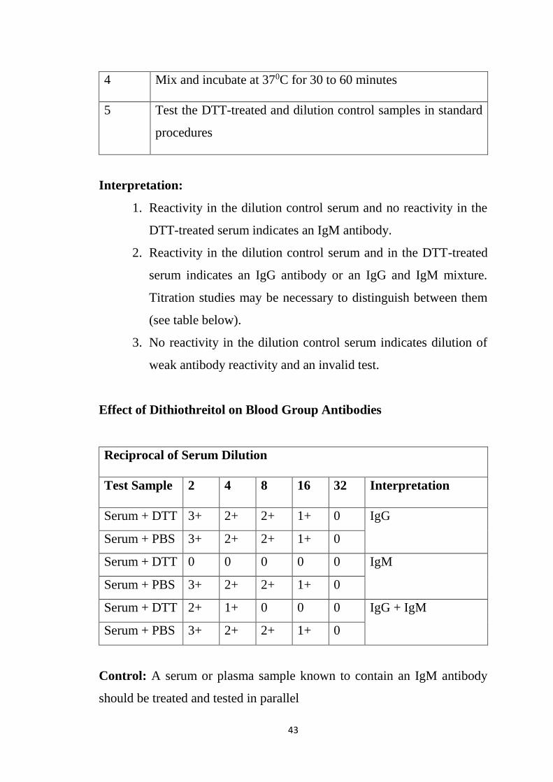
Interpretation:
1. Reactivity in the dilution control serum and no reactivity in the
DTT-treated serum indicates an IgM antibody.
2. Reactivity in the dilution control serum and in the DTT-treated
serum indicates an IgG antibody or an IgG and IgM mixture.
Titration studies may be necessary to distinguish between them
(see table below).
3. No reactivity in the dilution control serum indicates dilution of
weak antibody reactivity and an invalid test.
Effect of Dithiothreitol on Blood Group Antibodies
Reciprocal of Serum Dilution
Test Sample 2 4 8 16 32 Interpretation
Serum + DTT 3+ 2+ 2+ 1+ 0 IgG
Serum + PBS 3+ 2+ 2+ 1+ 0
Serum + DTT 0 0 0 0 0 IgM
Serum + PBS 3+ 2+ 2+ 1+ 0
Serum + DTT 2+ 1+ 0 0 0 IgG + IgM
Serum + PBS 3+ 2+ 2+ 1+ 0
Control: A serum or plasma sample known to contain an IgM antibody
should be treated and tested in parallel

44
Notes:
1. 2-mercaptoethanol can also be used for this purpose.
2. Sulfhydryl reagents used at low concentration may weaken
antigens of the Kell system. For investigation of antibodies in the
Kell system, it may be necessary to use other methods.
3. Gelling of a serum or plasma sample may be observed during
treatment with DTT. This gelling can occur if the DTT has been
prepared incorrectly and has a concentration above 0.01 M.
Gelling may also occur if serum and DTT are incubated too long.
An aliquot of the sample undergoing treatment can be tested after
30 minutes of incubation; if the activity thought to be caused by
IgM has disappeared, there is no need to incubate further. Gelled
samples cannot be tested for antibody activity because
overtreatment with DTT causes the denaturation of all serum
proteins.
METHOD 8: ANTIBODY TITRATION PROCEDURE
Principle: Titration is a semiquantitative method used to
determine the concentration of antibody in a serum sample or to
compare the strength of antigen expression on different red cell
samples. The usual applications of titration studies are as follows:
1) estimating antibody activity in allo-immunized pregnant women
to determine whether and when to perform more complex
invasive investigation of the fetal condition
2) elucidating autoantibody specificity
3) characterizing antibodies as having high titre and low avidity,
traits common in antibodies to antigens of the Knops and

45
Chido/Rodgers systems, Csa, and JMH; and 4) observing the
effect of sulfhydryl reagents on antibody behaviour, to determine
immunoglobulin class (IgG or IgM).
Specimen: Donor serum with abnormal antibody
Reagents:
1. Red cells that express the antigen(s) corresponding to the
antibody specificity(ies), in a 2% to 5% saline suspension.
Uniformity of red cell suspensions is very important to ensure
comparability of results
2. Saline for dilutions
Procedure:
Steps Action
The master dilution technique for titration studies is as follows:
1 Label 10 test tubes according to the serum dilution (e.g., 1:1, 1:2,
etc). A 1:1 dilution means one volume of serum undiluted; a 1:2
dilution means one volume of serum in a final volume of two, or
a 50% solution of serum in the diluent.
2 Deliver one volume of saline to all test tubes except the first
(undiluted, 1:1) tube.
3 Add an equal volume of serum to each of the first two tubes
(undiluted and 1:2).

46
4 Using a clean pipette, mix the contents of the 1:2 dilution several
times, and transfer one volume into the next tube (the 1:4
dilution).
5 Continue the same process for all dilutions, using a clean pipette
to mix and transfer each dilution. Remove one volume of diluted
serum from the final tube, and save it for use if further dilutions
are required.
6 Label 10 tubes for the appropriate dilutions.
7 Using separate pipettes for each dilution, transfer 2 drops of each
diluted serum into the appropriately labelled tubes, and add 2
drops of a 2% red cell suspension. Alternatively, for
convenience, add 1 drop of a 3% to 4% suspension of red cells
as supplied by the reagent manufacturer, although this method is
less precise.
8 Mix well and test by a serologic technique appropriate to the
antibody
9 Examine test results macroscopically; grade and record the
reactions. The prozone phenomenon may cause reactions to be
weaker in the more concentrated serum preparations than in
higher dilutions. If one is to avoid misinterpretation of results, it
may be preferable to examine first the tube containing the most
dilute serum and then to proceed through the more concentrated
samples to the undiluted specimen.

47
Interpretation:
1. Observe the highest dilution that produces 1+ macroscopic
agglutination. The titre is reported as the reciprocal of the dilution
level (e.g., 32—not 1 in 32 or 1:32). If there is agglutination in the
tube containing the most dilute serum, the endpoint has not been
reached, and additional dilutions should be prepared and tested.
2. In comparative studies, a significant difference in titre is three or
more dilutions. Variations in technique and inherent biologic
variability can cause duplicate tests to give results that differ by one
dilution in either direction. Serum containing antibody at a true titre
of 32 may show, on replicate tests, the endpoint in the 1:32 tube, the
1:64 tube, or the 1:16 tube.
3. Titre values alone can be misleading if the strength of agglutination
is not also evaluated. The observed strength of agglutination can be
assigned a number, and the sum of these numbers for all tubes in a
titration study represents the score, another semiquantitative
measurement of antibody reactivity. The arbitrarily assigned
threshold for significance in comparing scores is a difference of 10
or more between different test samples.
4. Antibodies with high-titre and low-avidity characteristics generally
have a titre greater than 64, with most tubes showing consistently
weak reactivity.
5. The table below shows the results obtained with three sera, each of
which shows no more agglutination after 1:256 dilution. The
differences in score, however, indicate considerable variation in
strength of reactivity.

48
Examples of Antibody Titres, Endpoints, and Scores
Reciprocal of Serum Dilution
1 2 4 8 16 32 64 128 256 512 Titre Score
Sample
1
Strength 3+ 3+ 3+ 2+ 2+ 2+ 1+ + + 0 64
(256)
Score 10 10 10 8 8 8 5 3 2 0 64
Sample
2
Strength 4+ 4+ 4+ 3+ 3+ 2+ 2+ 1+ + 0 128
(256)
Score 12 12 12 10 10 8 8 5 3 0 80
Sample
3
Strength 1+ 1+ 1+ 1+ + + + + + 0 8
(256)
Score 5 5 5 5 3 3 3 2 2 0 33
METHOD 9: DETERMINING THE SPECIFICITY OF COLD-
REACTIVE AUTOAGGLUTININS
Principle: Cold-reactive auto-agglutinins are usually IgM, which binds to
red cells in the lower temperature of the peripheral circulation and causes
complement components to attach to the red cells. As the red cells circulate
to warmer areas, the IgM dissociates but the complement remains.
Specimen:
1. Serum separated at 370C from a blood sample maintained and/or
allowed to clot at 370C, or plasma, separated from an anticoagulated
sample after periodic inversion at 370C for approximately 15
minutes.
2. Autologous red cells.

49
Reagents:
Test red cells of the following phenotypes:
1. A pool of two or more examples of adult group O I adult red
cells; these can be the reagent cells routinely used for
alloantibody detection
2. Group O i cord red cells
3. The patient’s own (autologous) red cells, washed at least three
times with 370C saline
4. Red cells of the same ABO group as the patient, if the patient
is not group O. If the patient is group A or AB, use both A1
and A2 cells
5. Saline or phosphate-buffered saline (PBS), pH 7.3.
Procedure:
Steps Action
1 Prepare serial two-fold dilutions of the serum or plasma in saline
or PBS. The dilution range should be from 1 in 2 to 1 in 4096
(12 tubes), and the volumes prepared should be more than the
total volume needed to test all of the desired red
cells.
2 Label a set of 12 tubes with the dilution (e.g., 2, 4, 8, etc) for
each kind of red cells to be tested (e.g., adult, cord, autologous).
3 Dispense 2 drops of each dilution into the appropriate tubes.
4 Add 1 drop of a 3% to 5% saline suspension of each red cell
sample to the appropriate set of tubes.
5 Mix and incubate at room temperature for 30 to 60 minutes.

50
6 Centrifuge for 15 to 20 seconds at 1000 rpm. Examine the tubes
one by one macroscopically for agglutination, starting with the
set of tubes at the highest dilution for each cell tested (i.e., read
all the tubes for each dilution as a set). Grade and record the
results.
7 Incubate the tubes at 40C for 1 to 2 hours
8 Centrifuge for 15 to 20 seconds at 1000 rpm. Immediately place
the tubes in a rack in an ice water-bath. Examine the tubes as in
step 6. Grade and record the results.
Interpretation:
Typical Relative Reactivity Patterns of Cold Autoantibodies
Antibody Specificity
Red cell Anti-I Anti-i Anti-IT Anti-IH Anti-Pr
O I adult + 0/↓ 0/↓ + +
O i cord 0/↓ + + ↓ +
A1 I adult 0/↓ + 0/↓ ↓ +
A1 I cord 0/↓ + + ↓ +
O I enzyme
treated
+ 0/↓ 0/↓ ↓ +
Autologous ↑ ↑ ↑ ↑ 0
+: reactive; 0: nonreactive; ↓: weaker reaction; ↑: stronger reaction
51

52
RESULTS

53
The sample size chosen for this study was 7000 healthy non-remunerated
voluntary donors who were randomly chosen over a period between 26th
November 2017 and 15th February 2019. Among the total subjects 5046
(72.08%) were males and 1954 (27.2%) were females (Figure 1). The age-
wise distribution based on gender is shown in Graph 1. The males are more
among all age groups and the number of blood donors are more in 18-30
years category and minimal donors below 50 years of age.
Among the 7000 samples analysed only 7 cases were positive for clinically
significant irregular red cell antibodies. Hence the prevalence of
alloimmunisation in healthy voluntary donor population in current study is
1 per 1000. There was no case of DAT positive in this study.
Figure 1
72%
28%
Gender Distribution
Males
Females

54
Graph 1
Out of the 7 positive cases of abnormal red cell antibodies, 2 cases were anti-
M (28.57%), 2 cases were anti-Lewis (28.57%), 1 case of anti-D, 1 case of
anti-C, 1 case of anti-IH. The cross-fabulation or testing the prevalence with
respect to age group or sex is not possible because of this very low
prevalence value. The details of the 7 cases that showed unexpected reactions
are as follows:
16.04 11.130.7 0.04
29.5628.01
14.10.41
45.6
39.14
14.8 0.46
0
10
20
30
40
50
60
70
80
90
100
18-30y 31-40y 41-50y >50
GENDER BASED DISTRIBUTION
AMONG DIFFERENT AGE
GROUPS
Female Male Total

55
Case 1: A 19-year-old male, first time donor, A negative blood group, with
no previous history of previous history of transfusion, found to be having
reaction with O cells and papanised O cells on reverse grouping. He had 2+
agglutination at 370C a with a negative DAT and auto-control at 370C and
220C. There was 4+ reaction with A1 lectin and no reaction with anti-H
lectin. He had a positive auto-control at 40C. Pan-reactivity observed on 3
cell screening panel and 11 cell panel at 370C. There was no reaction
observed with washed A positive and negative cells and washed A positive
and negative cord cells.
Cells O+ O- A+ A- O+ cord O- cord A+ cord A- cord
370C +2 +2 0 0 +wk +wk 0 0
220C +2 +2 0 0 0 0 0 0
170024
370C 22OC
+
+
+
+
+
+

56
Titre at 220C: 64
Titre at 370C: 32
After DTT treatment of serum, IgG titre: 2.
+
+
+
+
+
+
+
+
+
+
+
370C 220C
+
+
+
+
+
+
+
+
+
+
+
170024

57
Case 2: A 34-year-old first time male donor, A negative blood group with
no history of blood transfusion. The O cells and O papanised cells were
having positive reaction during reverse grouping and IAT was positive with
a negative DAT. Screening cell (I, II, III) panel: +2, +2, 0 and 11 cell panel
indicated the presence of anti-Leb with a wide thermal range indicating that
it had both IgM and IgG components. Auto-control at 220C and 370C were
negative and 40C was positive. Titre of the antibody at 40C was found to be
64, at 220C was 32 and at 370C after DTT treatment was 8. Secretor status of
the donor was found to be non- secretor. On phenotyping, the donor was
homozygous for Lea antigen.
+
+
0
+
+
0
170183
370C 22OC

58
17
01
83
+ 0 0 0 + + + 0 + + +
+ 0 0 0 + + + 0 + + +
AH
G
2
20 C

59
Case 3: A 35-year-old first time female AB negative donor, with no
previous history of transfusion but has 2 pregnancies with Rh positive
offsprings. She had reaction with papanised O cells in reverse grouping,
IAT positive, DAT negative and on Screening cells (I, II, III): reactions
were +2, +2, 0. Inhouse 6 cell panel revealed the antibody to be anti-D.
The IgG component titre was found to be 32.
Sl no
Rh-hr D C c E e K k M N S s P1 Lea Leb Fya Fyb Jka Jkb Lua Lub Cell
No.
AHG
1 R1R1 + + 0 0 + 0 + + + + + + 0 + 0 + 0 + 0 + 1 +
2 R1R1 + + 0 0 + + + 0 + 0 + 0 + 0 + + 0 + 0 + 2 +
3 R2R2 + 0 + + 0 0 + + 0 + 0 + 0 + + 0 0 + 0 + 3 +
4 rr 0 0 + 0 + + + + + + 0 + 0 + + + + + 0 + 4 0
5 rr 0 0 + 0 + 0 + + 0 0 + + + 0 0 + + 0 0 + 5 0
6 rr 0 0 + 0 + 0 + + + + + + + + 0 + 0 + 0 + 6 0
A/c rr 0 0 + 0 + 0 + 0 + + + 0 0 + + 0 0 + 0 + A/c 0
AHG
+ + 0
172226 821011
31/07/2018
2-80C

60
Case 4: A 34-year-old repeat male donor with blood group B positive, with
no history of blood transfusion in the past. He had a blood group
discrepancy in reverse grouping. 2+ agglutination with O cell at 370C and
negative DAT. On Screening cells (I, II, III), there was reaction at 370C
saline phase: 2+, 2+, 0.
Reagent Anti-A Anti-B Anti-A1B Anti-A1 Anti-H Anti-D
(IgM+IgG)
Anti-D
(IgM)
Reaction 0 4+ 4+ 0 2+ 3+ 3+
Reagent A1 cells B cells O cells Pap O cells
Reaction 4+ 1+ 2+ 0
Auto-control at 220C and 370C was negative but saline phase at 40C was
positive. On inhouse 6 cell panel, the antibody was identified as anti-M
which had a wide thermal range. IgM titre was 64 and IgG titre was 16. On
phenotyping: homozygous for N antigen.

61
Sl
no
Rh-hr D C c E e K k M N S s P1 Lea Leb Fya Fyb Jka Jkb Lua Lub Cell
No.
37 C
22. C
4C
1 R1R1 + + 0 0 + 0 + + + + + + 0 + 0 + 0 + 0 + 1 + + +
2 R1R1 + + 0 0 + + + 0 + 0 + 0 + 0 + + 0 + 0 + 2 0 0 +
3 R2R2 + 0 + + 0 0 + + 0 + 0 + 0 + + 0 0 + 0 + 3 + + +
4 rr 0 0 + 0 + + + + + + 0 + 0 + + + + + 0 + 4 + + +
5 rr 0 0 + 0 + 0 + + 0 0 + + + 0 0 + + 0 0 + 5 + + +
6 rr 0 0 + 0 + 0 + + + + + 0 + + 0 + 0 + 0 + 6 + + +
A/c rr 0 0 + 0 + 0 + 0 + + + 0 0 + + 0 0 + 0 + A/c 0 0 +
370C
+
+
0
170272
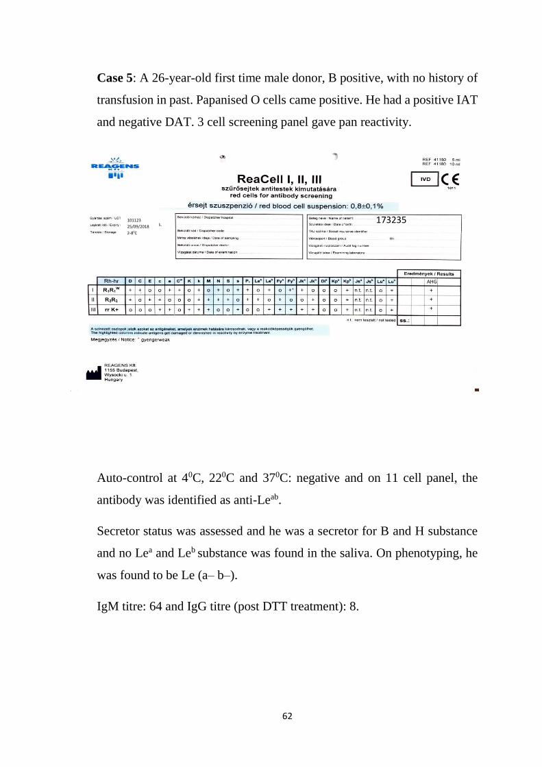
62
Case 5: A 26-year-old first time male donor, B positive, with no history of
transfusion in past. Papanised O cells came positive. He had a positive IAT
and negative DAT. 3 cell screening panel gave pan reactivity.
Auto-control at 40C, 220C and 370C: negative and on 11 cell panel, the
antibody was identified as anti-Leab.
Secretor status was assessed and he was a secretor for B and H substance
and no Lea and Leb substance was found in the saliva. On phenotyping, he
was found to be Le (a– b–).
IgM titre: 64 and IgG titre (post DTT treatment): 8.
AHG
+
+
+
173235 101123
25/09/2018
2-80C

63
+ 0 + 0 + + + + + + +
AH
G
22
C
+ 0 + 0 + + + + + + +
17
22
35

64
Case 6: A 32-year-old male, repeat donor, O positive had a blood group
discrepancy on reverse grouping. DAT negative, IAT positive and auto-
control at 220C and 370C: negative.
Reagent Anti-A Anti-B Anti-A1B Anti-A1 Anti-H Anti-D
(IgM+IgG)
Anti-D
(IgM)
Reaction 0 0 0 0 4+ 3+ 3+
Reagent A1 cells B cells O cells Pap O cells
Reaction 4+ 4+ 2+ 0
Auto-control at 40C, 220C and 370C: negative. Antibody identified as anti-
M and 11 cell panel reactions enhanced at 40C. On phenotyping: M(–) N(+).
IgM titre: 32 and IgG after DTT treatment: 16.
721901
2018.10.28
2-80C
0
+
+
AHG
173878
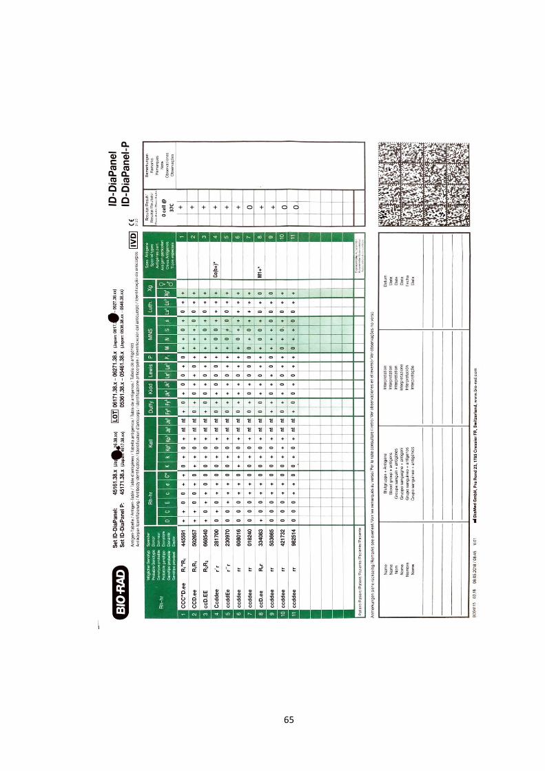
65
0 ce
ll @
37C
+ + + + + + 0 + + 0 0

66
Case 7: A 29-year-old female, repeat donor, B negative blood group, with
a past history of pregnancy (2 years back) and no history of transfusion.
She received anti-D prophylaxis after her delivery. The IAT was positive
and DAT came negative. Auto-control negative at 40C, 220C and 370C. On
papanised O cells reaction enhanced, 3 cell screening panel gave 2+, 0, 0
reaction. On 11 cell panel, antibody identified as anti-C with IgG titre of 8.
721901
2019.02.28
2-80C
+
0
0
AHG
174954

67
+ + 0 + 0 0 0 0 0 0 0
A
HG

68
Benign Cold Agglutinins
Cold autoantibodies are often detected in healthy individuals, with the
majority being benign and IgM in nature. These antibodies react best at 40C.
The prevalence of benign cold agglutinins (BCA) at 40C for auto-control as
well as with pooled O cells were 56.40% and 65.24% respectively and
31.16% of donors showed reaction in both auto-control and pooled O cells.
The chi-square test using R & C table in OpenEpi (online software) [61]
was used to find out the significance among the gender and age-wise
distribution of occurrence of BCA. It was found that there was statistically
significant difference observed in gender distribution (p value <0.001) and
age-wise distribution (p value <0.001) stating that the male blood donors
had more prevalence of BCA than female blood donor and also the age
group of 18-30 years had more BCAs.
AUTOCONTOL AT 4C
O CELL AT 4C
BOTH
3114
3660
1807
834
907
374
Gender based distribution of Benign Cold
Agglutinins
Males Females
3948
2181
4567

69
The prevalence of IgM antibodies reacting at 220C were 0.46% for auto-
control and 0.69% for pooled O cells respectively. Here also, the prevalence
was more with males compared to females.
0.00% 10.00% 20.00% 30.00% 40.00% 50.00% 60.00% 70.00%
Autocontrol at 4C
O cell at 4C
Both
Age-wise distribution of Benign Cold
Agglutinins
18-30 31-40 41-60
14.89% 11.06% 5.21%
33.81% 23.86% 7.57%
29.27% 20.27% 6.68%
years
0.46%0.34%
0.11%
0.69%
0.47%
0.21%
TOTAL CASE MALES FEMALES
Cold Agglutinins at 22 degree C
Autocontol at 22C O cell at 22C
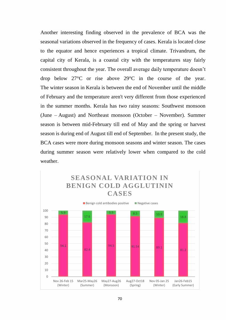
70
Another interesting finding observed in the prevalence of BCA was the
seasonal variations observed in the frequency of cases. Kerala is located close
to the equator and hence experiences a tropical climate. Trivandrum, the
capital city of Kerala, is a coastal city with the temperatures stay fairly
consistent throughout the year. The overall average daily temperature doesn’t
drop below 27°C or rise above 29°C in the course of the year.
The winter season in Kerala is between the end of November until the middle
of February and the temperature aren't very different from those experienced
in the summer months. Kerala has two rainy seasons: Southwest monsoon
(June – August) and Northeast monsoon (October – November). Summer
season is between mid-February till end of May and the spring or harvest
season is during end of August till end of September. In the present study, the
BCA cases were more during monsoon seasons and winter season. The cases
during summer season were relatively lower when compared to the cold
weather.
94.182.4
94.5 91.54 89.181.2
5.917.6
5.5 8.5 10.918.8
0
10
20
30
40
50
60
70
80
90
100
Nov 26-Feb 15(Winter)
Mar25-May26(Summer)
May27-Aug26(Monsoon)
Aug27-Oct18(Spring)
Nov 05-Jan 25(Winter)
Jan26-Feb15(Early Summer)
SEASONAL VARIATION IN
BENIGN COLD AGGLUTININ
CASES
Benign cold antibodies positive Negative cases

71
During the month of August to September 2018, Kerala was hit by heavy
rainfall and floods and hence the cases of BCA were more during this time
interval which was otherwise the period of spring season in Kerala with a
warm weather.

72
DISCUSSION

73
Various worldwide literature calls out the importance of donor unexpected
antibody screening and also a topic of debate whether to make this
mandatory for the recipient safety. The guidelines for the screening of
donor blood to identify iso-immune erythrocyte antibodies have been laid
down in National Blood Policy (62), 2007 (National Aids Control
Organization/NACO, Ministry of Health & Family Welfare). DGHS,
Ministry of Health & Family Welfare (Government of India) (63) also
warrants screening of donated blood for irregular erythrocyte antibodies.
Red cell allo-immunisation rates
The incidence of RBC alloimmunization depends largely upon the
demography of the population studied (64). Few international studies states
that the prevalence of red cell allo- and autoantibodies has been reported in
several study populations including hospital based patients, transfusion
dependent patients with chronic haematological disorders, pregnant
females and blood donors, and the incidence of alloantibodies detected
worldwide is 0.2%-0.9% in healthy blood donors, 2%-9% in patients with
a history of blood transfusion, 9%-30% among chronic transfusion
dependant patients, 0.5%-1.9% among antenatal women and 0.5%-1% of
red cell autoantibodies among transfused patients (3, 65-71).
Global literature states that the reported incidence of erythrocyte allo-
antibodies in the donor population varies from 0.32 to 2.4% (3,4,6,8-14).
REDS III study (Recipient Epidemiology and Donor Evaluation Study)
gives inference of a positive antibody screen noted overall in 0.5% of
donations and 0.77% of blood donors at the four centres in US included in
the study over the 4.5-year study period, utilizing methodologies ranging
from solid phase to gel card to tube testing (34). Comparative evaluation of

74
allo-immunization rates is difficult as different studies have used different
serological techniques. In the present study we followed tube technique
which is less sensitive than column agglutination technology/solid phase
haematological procedures. From the current study on healthy voluntary
donors, the prevalence of allo-antibodies is 0.1%. This may be because our
population of blood donors are 100% voluntary non-remunerated healthy
people who are highly motivated and having good awareness on blood
donation. Moreover, we follow a stringent selection of blood donors
attending our camp and inhouse blood donations.
Nature of allo-antibodies
A study on the Shaoguan area describes that frequency of irregular
antibodies in female was higher than that in male and Rh blood group
antibodies (47.6%) such as anti-D, anti-E, and anti-c were common
(65). Another US study on male military veterans, the prevalence of red cell
antibodies was in the increasing order of K, E, D, Lea, Fya, c, C, P1, Jka, and
Leb (72). Another study from Minnesota, the most frequent allo-antibodies
were E, Lea, K, D, Leb, M, P1, Fya, C & c in the order (67). A study on
frequency of allo-immunisation in Kuwaiti population describes that five
most frequently identified alloantibodies were anti-D, anti-E, anti-K, anti-
Lea & anti-Leb in the descending order (25). Malaysian study on prevalence
and specificities of red cell alloantibodies among blood recipients shows
that anti-E antibody comprised the most common alloantibody followed by
the anti-Lea antibodies & the anti-M antibody (69). A study done in
Southern Thai population showed following antibodies frequency wise as
most common – anti-Mia, anti-E, anti-Lea, anti-c and anti-Leb (70).

75
Several Indian studies mostly in Northern population are available in the
literature. 0.9% prevalence of irregular red cell antibodies was noted among
healthy donors in Delhi population (73). The most common alloantibodies
were of MNS system (39.13%) followed by anti-D (13%) and anti-Lewis
(10.8%). Another similar study involving blood donors in northern India
showed the prevalence of 0.05% alloantibodies was observed (64). A
tertiary care centre in northern India reported positive screening cells and
pooled O cells in 0.27% of donors, 66.08% of these donors had
autoantibodies, 1 had auto-antibodies with underlying alloantibody anti-Jka
(0.001%), and 0.09% had alloantibodies alone in their plasma. Anti-M was
the most common antibody (18.94%) identified, followed by anti-D
(9.25%). Another study from State of Odisha, India done on patient
population shows that weak/missing antibody, weak antigen expression,
rouleaux, cold autoantibodies, cold alloantibodies, Bombay phenotype with
the frequency of 13.51%, 2.70%, 2.70%, 54.06%, 8.11%, 18.92%
respectively (74). In Indian literature on red cell allo-antibodies, males have
higher incidence when compared to females (mostly with a history of
pregnancy). In Indian scenario, female blood donations are fewer when
compared to the male blood donations and hence this may be a reason that
the female population with positive red cell alloimmunisation may be a tip
of iceberg phenomenon.
In our study, the prevalence of irregular antibodies was much lower than
other national and international studies (0.01%). Moreover, the most
common alloimmunisation identified were anti-M and anti-Lewis
antibodies (0.03% each), followed by Rh antibodies (anti-C and anti-D,
0.01% each) and anti-IH (0.01%) which is comparable with other Indian
studies. Here, the males were having higher incidence of red cell

76
alloimmunisation and all of them were devoid of a prior sensitisation via
blood transfusion indicating that all the 5 cases were naturally occurring
allo-antibodies. Whereas females with irregular antibody belong to Rh
antibodies (anti-D and anti-C) and results from a previous history of
pregnancy.
First time donor Vs Repeat donors
Myhre et al reported that an average of 0.14% of the total units of blood
yearly contain unexpected red cell antibodies and this group of donors
includes mainly first time donors (84%), but contains in addition a few
repeat donors who had not given blood since their screening program began
or who had been recently sensitized (71). Since 25% of the total donor
population in their study composed of first-time donors, the percentage of
antibodies found in first time donors is 0.56%. Similar finding was
observed in our study with most of the irregular antibodies were seen
among the first-time donors and the repeat donors who came positive were
recently sensitized.
Gender distribution of allo-antibodies
In our study, we found that alloimmunisation was common among males
than females, but males had naturally occurring antibodies and females had
allo-antibodies as a result of past pregnancy. Male to female ratio of
alloantibodies was found to be 8.25:1 in Garg N et al study (73) and 4.84:1
in Makroo et al study (33). These studies also pointed out the female allo-
immunisation resulted out from past pregnancy whereas males had mostly
naturally occurring allo-antibodies followed by past history of blood
transfusion.

77
DAT positivity among study population
In present study, there was no case of auto-immunisation or DAT positivity
in total 7000 healthy blood donors studied. Issitt and Anstee (51) reported
that, of blood donors with a positive DAT and IgG coating the RBCs, 5–
10% will develop AIHA, 20–25% will become DAT negative over time,
and 60–70% will remain DAT positive but hematologically normal. The
incidence of strongly positive DATs in healthy individuals was 1:3300 in a
Canadian study (75). Another French study in 1980, the incidence was
found to be 1: 13000 (76). Incidence of a positive DAT amongst the healthy
blood donors was 0.04% in an Indian study done in northern Indian
population by Tiwari et al (29). Makroo et al (33) states that in their study,
the prevalence of DAT positivity among healthy donors was 0.18% and in
Kaur et al study, it was 0.05% (64).
Benign cold agglutinin distribution
Cold autoantibodies are often detected in healthy individuals, with the
majority being benign which react more strongly at 0–40C than at warmer
temperatures. Pathological cold auto-antibodies are characterized by wide
thermal amplitude and/or high antibody titres (56). Joshi SR et al states that
cold auto-antibodies observed in their study were found among blood
donors with no apparent health problems and may be due to an exposure to
infectious agents as antibody stimulant in past was open as the population
in the city of Surat was exposed to vector-borne infections such as malaria,
gastroenteritis, and infective hepatitis during the contemporary period of
this study (55). In the current study, we found prevalence of benign cold
agglutinins at 40C for auto-control as well as with pooled O cells were
56.40% and 65.24% respectively and 31.16% of donors showed reaction in

78
both auto-control and O cells. Since Kerala is a tropical country with near
perennial rainfall and vector borne diseases like dengue, etc. as well as
frequent viral upper respiratory infections, the occurrence of cold
agglutinins may be associated with the above. Cold agglutinins active at
220C are of very low prevalence (<1%) from the current study.

79
CONCLUSION

80
Out of 7000 healthy voluntary blood donor population, the prevalence of
irregular red cell antibodies was found to be 1in 1000. Male blood
donations were more when compared to female and blood donors among
18-30 years of age were greater in number in the study population. There
were no cases of DAT positivity encountered in this study. Male: Female
ratio is 2.5:1 and males had naturally occurring allo-antibodies whereas
females had a previous history of pregnancy. Anti-M and anti-Lewis
antibodies were commonest allo-antibodies followed by anti-Rh (D and C)
antibodies and anti-IH in this current study. Benign cold agglutinins were
found predominately in the younger male population with a significant
seasonal variation noted in the prevalence of these antibodies.
81
LIMITATIONS OF THE
STUDY
82
The study concentrated only on voluntary healthy blood donors and hence
do not represent the general South Kerala population. The efficient pre-
donation counselling and medical examination filtered out people suffering
from major clinical conditions especially autoimmune diseases.
Conventional spin tube method was used in the current study which is
inferior technology when compared with gel card method. Gel card
technique is well known for its simplicity, stability of results, dispensation
of controls, absence of wash phase with comparable sensitivity and
specificity as well as appropriate for antibody screening and identification.

83
SUMMARY

84
The aim of this study was to determine the prevalence of irregular red cell
antibodies in healthy voluntary donors attending our camp and inhouse
blood donations. Institute policy is 100% voluntary blood donations and the
donor pool consisted mainly of comprehensive, altruistic and motivated
people. Since the literature states a very low prevalence of irregular red cell
antibody positive cases among healthy blood donors, a sample size of 7000
was adapted after consulting with the institute statistician. The study was
conducted over a period of 1 year and 3 months.
The prevalence of irregular red cell antibodies was found to be 1 in 1000.
There were no DAT positive cases obtained during the study period. The
seven cases reported were 5 males with no past history of blood transfusion
and 2 females (Rh negative) with a history of past pregnancy. Anti-M and
anti-Lewis antibodies were commonest followed by anti-Rh (C and D) and
anti-IH antibodies.
Another interesting finding was the prevalence of benign cold agglutinins
at 40C (auto-control as well as with pooled O cells) and was found to be
were 56.40% and 65.24% respectively and 31.16% of donors showed
reaction in both auto-control and pooled O cells. These BCA positive cases
were seen more during winter season and rainy seasons when compared to
hot climatic conditions.
The importance of antibody screening is highlighted through this study
because all antibodies identified are notorious to cause immune hemolytic
transfusion reactions in the recipients. Moreover, the DGHS guidelines
states that the antibody screening is mandatory alongside the routine blood
grouping and Rh typing of blood donors.

85
BIBLIOGRAPHY

86
1. Fung M K, Grossman B J, Hillyer C D & Westhoff C M, AABB
Technical Manual18th ed., USA 2014
2. Fernandez S A V, Lobo A Z C, et al. Prevalence of Antinuclear
Autoantibodies in the Serum of Normal Blood Donors. Rev. Hosp.
Clín.Fac. Med. S. Paulo 2003;58(6):315-19
3. Zaman S, Chaurasia R, et al. Prevalence and Specificity of RBC
Alloantibodies in Indian Patients Attending a Tertiary Care
Hospital. Hindawi Hematology, 2014;749218
4. Garratty G. Screening for RBC antibodies: what should we expect
from antibody detection RBCs. Immunehematol 2002; 18:71-6
5. Schonewille H, VanDeWatering L M G, and Brand A. Additional
red blood cell alloantibodies after blood transfusions in a non-
hematologic alloimmunized patient cohort: is it time to take
precautionary measures? Transfusion 2006; 46:630–35
6. Simon T L, Snyder E L, Solheim B G, Stowell C P et al. Rossi’s
Principle of Transfusion Medicine 4th ed.,USA 2009
7. Hillyer C D, Silberstein, Ness, Anderson & Roback, Blood banking
and transfusion medicine 2nd edition, USA 2007
8. National AIDS Control Organisation. An Action Plan for Blood
Safety: Introduction, India 2007
9. Harmening DM, FA Davis. Modern blood banking & transfusion
practices,6th ed.; USA 2012
10. Bhagwat S N, Sharma J H, Jose J, Modi C J. Comparison between
conventional and automated techniques for blood grouping and
crossmatching: Experience from a tertiary care centre. J Lab
Physicians.2015; 7(2): 96–102

87
11. White J, Qureshi H, Massey E et al. BCSH guidelines for blood
grouping and red cell antibody testing in pregnancy, Transfusion
Medicine 2016;26:246–263
12. Popovski, MA, and Moore, SB: Diagnostic and pathogenic
considerations in transfusion-related acute lung injury. Transfusion
1985; 25:573.
13. Sillimann, CC, et al: Transfusion-related acute lung injury:
Epidemiology and a prospective analysis of etiologic factors. Blood
2003; 101:454.
14. Toy, P, et al: Transfusion-related acute lung injury: Definition and
review. Crit Care Med 2005; 33:721.
15. Popovski, MA, et al: Transfusion-related acute lung injury: A
neglected, serious complication of hemotherapy. Transfusion 1992;
32:589.
16. Kopko, PM, et al: HLA class II antibodies in transfusion-related
acute lung injury. Transfusion 2001; 41:1244.
17. Eder, AF, et al: Transfusion-related acute lung injury surveillance
(2003–2005) and the potential impact of the selective use of plasma
from male donors in the American Red Cross. Transfusion 2007;
47:599.
18. Kehler R M, Banerjee A, Gamboni F, et al. Antibodies to major
histocompatibility complex class II antigens directly prime
neutrophils and cause acute lung injury in a two-event in vivo rat
model. Transfusion 2016;56;3004-11
19. White J: Red cell antibodies – clinical significance or just noise?
ISBT Science Series 2017; 12:19–24

88
20. Tiwari A K, Dara R C, Aurora D, Aggarwal G, et al. Allo-antibody
identification: a software approach! Transfusion and Apheresis
Science 2014; 51:197–202
21. Rosse WF, Gallagher D, Kinney TR, et al. Transfusion and
alloimmunization in sickle cell disease. The cooperative study of
sickle cell disease. Blood 1990; 76:1431–7.
22. Schonewille H, Van De Watering L M G, and Brand A: Additional
red blood cell alloantibodies after blood transfusions in a non-
hematologic alloimmunized patient cohort: is it time to take
precautionary measures?” Transfusion 2006; 46(4):630–35.
23. Walker R H, Lin D T and Hartrick M B: Alloimmunization
following blood transfusion. Archives of Pathology and Laboratory
Medicine 1989;113(3):254–61.
24. Ahrens N, Pruss A, K¨ahne A, Kiesewetter H, and Salama A.
Coexistence of autoantibodies and alloantibodies to red blood cells
due to blood transfusion. Transfusion 2007;47(5):813–816
25. Ameen R, Al-Eyaadi O, Al-Shemmari S, et al. Frequency of Red
Blood Cell Alloantibody in Kuwaiti Population.Med Princ Pract
2005;14:230-234
26. Natukunda B, Schonewille H, van de Watering L &. Brand A.
Prevalence and specificities of red blood cell alloantibodies in
transfused Ugandans with different diseases. Vox Sang 2010;98:
167–71
27. Thakral B, Saluja K, Sharma R R, Marwaha N. Red cell allo-
immunization in a transfused patient population: a study from a
tertiary care hospital in north India. Hematology 2008;13(5):313-17

89
28. Mo Z, Li H, Huang L & Jiao W. Prevalence and specificity of RBC
alloantibodies in the general hospitalised population in Guangxi.
Transfusion Medicine 2015; 25:313–19
29. Tiwari A K, Pandey P, Sharma J, Shailja K, et al. Incidence of
clinically significant antibodies in patients and healthy blood
donors: A prospective cross-sectional study from a tertiary
healthcare center in India. Transfusion and Apheresis Science 2014;
50:230–34
30. Giblett ER. Blood group alloantibodies: an assessment of some
laboratory practices. Transfusion 1977; 17:299–308.
31. Walker RH, Lin DT, Hatrick MB. Alloimmunization following
blood transfusion. Arch Pathol Lab Med 1989; 113:254–61.
32. Rottenberg Y, Yahalom V, Shinar E, Barchana M, et al. Blood
donors with positive direct antiglobulin tests are at increased risk
for cancer. Transfusion 2009;49(5):838–42.
33. Makroo R N, Rajput S, Agarwal S, et al. Prevalence of irregular red
cell antibodies in healthy blood donors attending a tertiary care
hospital in North India. AJTS 2018;12(1):17-20
34. Karafin M S, Tan S, Tormey C A, Spencer B R, et al. Prevalence
and risk factors for RBC alloantibodies in blood donors in the
Recipient Epidemiology and Donor Evaluation Study-III(REDS-
III). Transfusion 2019;59;217–225
35. Garcia M A, Bautista L, Palomino F. Should blood donors be
routinely screened for irregular antibodies? Immunohematology
2012; 28:60-6
36. Weiner W: ‘Coombs positive’ ‘normal’ people. Bibl Haematol,
Basel, Karger, 1965, No.23, p 35.

90
37. Gorst DW, Rawlinson VI, Merry AH, Stratton F: Positive direct
antiglobulin test in normal individuals. Vox Sang 1980; 38:99–105.
38. Garratty G. The significance of IgG on the red cell surface. Transfus
Med Rev 1987; 1:47–57.
39. Freedman J. The significance of complement on the red cell surface.
Transfus Med Rev 1987; 1:58–70.
40. Petz LD, Garratty G. Immune hemolytic anemias. 2nd ed.
Philadelphia: Churchill-Livingstone; 2004.
41. Win N, Islam S I A M, Peterkin M A, et al: Positive Direct
Antiglobulin Test due to Antiphospholipid Antibodies in Normal
Healthy Blood Donors. Vox Sang 1997; 72:182–84
42. Solal-Celigny P, Vazeux R, Vroclans M, et al: Positive Coombs test
in acute leukaemia. Br J Haematol 1984; 57:563–69.
43. Rondeau E, Solal-Celigny P, Dhermy D, Vroclans M, et al: Immune
disorders in agnogenic myeloid metaplasia: Relations to
myelofibrosis. Br J Haematol 1983; 33:451–58.
44. Vianna JL, Ordi-Ros J, Lopez-Soto A, Franz J, et al: Comparison
of the primary and secondary antiphospholipid syndrome: A
European Multicenter study of 114 patients. Am J Med 1994; 96:1–
9.
45. Creagh MD, Greaves M: Lupus anticoagulant. Blood Rev 1991;
5:162–167.
46. Hannon J L: Blood donors and donations from DAT-positive
individuals. Transfusion Medicine Reviews, Vol 26, No 2 (April),
2012: pp 142-152
47. Mazzei, CA, Popovsky, MA, and Kopko, PM: Noninfectious
complications of blood transfusion. In Roback, JD (ed): Technical
Manual, 16th ed. AABB Press, Bethesda, MD, 2009;715–49.
91
48. Merle N S, Boudhabhay I, Leon J, et al. Complement activation
during intravascular hemolysis: Implication for sickle cell disease
and hemolytic transfusion reactions. Transfusion Clinique et
Biologique 2019 in press
49. Atkinson V P, Soeding P, Horne G, Tatoulis J. Cold agglutinins in
cardiac surgery: Management of myocardial protection and cardio-
pulmonary bypass. Ann Thorac Surg. 2008; 85:310–1.
50. Daniels, G: The Human Blood Groups, 2nd ed. Blackwell Science,
Oxford, 2002.
51. Issitt, P D, and Anstee, D J: Applied Blood Group Serology, 4th ed.
Montgomery Scientific, Durham, NC, 1998.
52. Landsteiner K (1903) Ueber Beziehungen zwischen dem Blutserum
und den Korperzellen. Munch Med Wochenschr 50:1812–1814
53. Schubothe H (1966) The cold hemagglutinin disease. Semin
Hematol 3(1):27–47
54. Finland M, Peterson OL, Allen HE, Samper BA, Barnes MW, Stone
MB. Cold agglutinins. I. Occurrence of cold iso-hemagglutinins in
various conditions. J Clin Invest.1945;24:451-7.
55. Joshi SR, Naik RA, Gupte SC. Unusual spontaneous cold auto-
hemagglutination phenomenon in blood units stored under blood
bank condition: A retrospective analysis. Asian J Transfus Sci
2015;9:141-144
56. Javed R, Datta SS, Basu S, Chakrapani A. Resolution of Serologic
Problems Due to Cold Agglutinins in Chronic Lymphocytic
Leukemia. Indian Journal of Hematology and Blood Transfusion.
2016 Jun 1;32(1):290-3.
57. Roback JD, Grossman BJ, Harris T, Hillyer CD (2014) Technical
manual, 18th edn. AABB

92
58. Giblett ER. Blood group alloantibodies: an assessment of some
laboratory practices. Transfusion 1977; 17:299–308.
59. Chaudhary R, Agarwal N. Safety of type and screen method
compared to conventional antiglobulin crossmatch procedures for
compatibility testing in Indian setting. Asian J Transfus Sci
2011;5(2):157.
60. Pathak S, Chandrashekhar M, Wankhede GR. Type and screen
policy in the blood bank: is AHG cross-match still required? A
study at a multispecialty corporate hospital in India. Asian J
Transfus Sci 2011; 5:153–6.
61. Dean AG, Sullivan KM, Soe MM. OpenEpi: Open Source
Epidemiologic Statistics for Public Health, Version.3.01
62. National Blood Policy, 2007 (National Aids Control Organization,
NACO), Ministry of Health & Family Welfare.
63. Directorate General of Health Services (DGHS), Ministry of Health
& Family Welfare (Government of India), Screening and
identification of antibodies. Transfusion medicine, Technical
manual, 2nd ed; 2003.
64. Kaur D, Bains L, Kandwal M, Parmar I. Erythrocyte
alloimmunization and autoimmunization among blood donors and
recipients visiting a tertiary care hospital. JCDR. 2017
Mar;11(3):EC12
65. Zhu JY, Lan JC, Luo HQ. Screening analysis of irregular antibodies
from random donor population in Shaoguan area. J Exp Haematol.
2007;15(3):630-31
66. Pahuja S, Gupta AK, Pujani M, Jain M. The prevalence of irregular
erythrocyte antibodies [9] among antenatal women. Blood Transfus.
2011;9(4):388-93.

93
67. Winters JL, Pinedo AA, Gorden LD, Bryant SC, Melton LJ,
Vamvakas EC, et al. RBC alloantibody specificity and antigen
potency in Olmsted County, Minnesota. Transfusion. 2001;41:
1413-20.
68. Pandey HC, Das SS, Chaudhary R. Red cell alloimmunisation in
transfused patients: A [11] silent epidemic revisited. Asian J Transf
Sci. 2014;8(2):75-77
69. Al-Joudi F, Ali AB, Ramli MB, [12] Ahmed S, Ismail M.
Prevalence and specificities of red cell alloantibodies among blood
recipients in the Malaysian state of Kelantan. Asian J Transf Sci.
2011;5(1): 42–45.
70. Promwong C, Siammai S, Hassarin S, Buakaew J, Yeela T,
Soisangwan P, et al. Frequencies and specificities of red cell
alloantibodies in the Southern Thai population. Asian J Transfus
Sci. 2013; 7:16-20.
71. Myhre BA, Greenwalt TJ, Gajewski M. Incidence of irregular
antibodies occurring in healthy donor sera. Transfusion. 1965;
5:350–4.
72. Tormey CA, Fisk J, Stack G. RBC alloantibody frequency,
specificity and properties in a population of male military veterans.
Transfusion. 2008; 48:2069–76.
73. Garg N, Sharma T, Singh B. Prevalence of irregular red blood cell
antibodies among healthy blood donors in Delhi population.
Transfusion and Apheresis Science. 2014 50(3):415-7
74. Mishra D, Parida P, Mahapatra S, et al. Resolving blood group
discrepancy in patients of tertiary care centre in Odisha, India. Int J
Res Med Sci. 2018;6(7):2348-53

94
75. Hannon JL. Management of blood donors and blood donations from
individuals found to have a positive direct antiglobulin test.
Transfusion medicine reviews. 2012; 26(2):142-52.
76. Habibi B, Muller A, Lelong F, et al. Auto-immunisation
erythrocytaire dans la population “normale”. Nouv Presse Méd
1980; 9:3253-7.

95
ANNEXURE

96
97

98
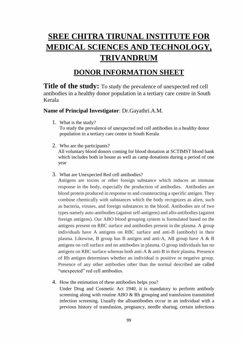
99
SREE CHITRA TIRUNAL INSTITUTE FOR
MEDICAL SCIENCES AND TECHNOLOGY,
TRIVANDRUM
DONOR INFORMATION SHEET
Title of the study: To study the prevalence of unexpected red cell
antibodies in a healthy donor population in a tertiary care centre in South
Kerala
Name of Principal Investigator: Dr.Gayathri.A.M.
1. What is the study?
To study the prevalence of unexpected red cell antibodies in a healthy donor
population in a tertiary care centre in South Kerala
2. Who are the participants?
All voluntary blood donors coming for blood donation at SCTIMST blood bank
which includes both in house as well as camp donations during a period of one
year
3. What are Unexpected Red cell antibodies?
Antigens are toxins or other foreign substance which induces an immune
response in the body, especially the production of antibodies. Antibodies are
blood protein produced in response to and counteracting a specific antigen. They
combine chemically with substances which the body recognizes as alien, such
as bacteria, viruses, and foreign substances in the blood. Antibodies are of two
types namely auto-antibodies (against self-antigens) and allo-antibodies (against
foreign antigens). Our ABO blood grouping system is formulated based on the
antigens present on RBC surface and antibodies present in the plasma. A group
individuals have A antigens on RBC surface and anti-B (antibody) in their
plasma. Likewise, B group has B antigen and anti-A, AB group have A & B
antigens on cell surface and no antibodies in plasma. O group individuals has no
antigens on RBC surface whereas both anti-A & anti-B in their plasma. Presence
of Rh antigen determines whether an individual is positive or negative group.
Presence of any other antibodies other than the normal described are called
“unexpected” red cell antibodies.
4. How the estimation of these antibodies helps you?
Under Drug and Cosmetic Act 1940, it is mandatory to perform antibody
screening along with routine ABO & Rh grouping and transfusion transmitted
infection screening. Usually the alloantibodies occur in an individual with a
previous history of transfusion, pregnancy, needle sharing, certain infections

100
and idiopathic (unknown reason). Estimation of allo-antibodies helps these
individuals in helping them for finding antigen negative blood groups for their
future blood requirements.
Auto-antibodies are found in donors with any autoimmune conditions or other
disease conditions. Estimation of these antibodies may aid in early diagnosis of
the above mentioned conditions. Also the prevalence of these antibodies in the
target population can be evaluated from this study. An added point is that these
antibodies may not cause any harm to the donor itself.
5. What is the procedure being done?
An addition 2ml of blood sample will be collected from the tubings of the blood
bag along with other pilot tubes taken for grouping, compatibility testing and
infection screening. No other additional pricking or any other invasive
procedure is needed. From this additional sample antibody screening (Direct
Antiglobulin Test & Indirect Antiglobulin Test) is done.
6. What is the benefit for you from the study?
As I mentioned above, for alloantigen estimation will help you in finding
antigen negative blood in future blood requirements & autoantibody estimation
may help you in early diagnosis of certain unidentified disease conditions.
There is no monetary burden from your side. The individuals with positive auto-
antibodies will be further evaluated by a physician.
7. Any risk related to this study?
There are no inherent risks envisaged in this study.
8. How does this study help the common public?
This study is performed on voluntary healthy population and detection of
unexpected antibodies may help the common public in obtaining appropriate
blood units if any future blood requirements arise and also in identifying occult
disease conditions in the early stage itself. This study also helps in aiding best
transfusion practices to be followed in the institution since there is scarce data
available on prevalence and type of irregular antibodies in Indian donor
population
9. Can you withdraw from this study after it starts?
Your participation in this study is entirely voluntary and you are also free to
withdraw from the data collection part of the study at any point of time, without
giving any reason, without your medical care or legal rights being affected
10. Will your personal details be kept confidential?
This research work if approved will be published as Thesis or in any
Transfusion Medicine Journal. But none of the personal data will be mentioned

101
anywhere in it. However, your medical notes may be reviewed by the ethical
committee members and regulatory authorities associated with the study,
without your additional permission.
*For any queries, please contact myself Dr. Gayathri.A.M. (mob: 9446208632),or Dr.
Debasish Gupta (tel: 04712524177)
email: [email protected] /[email protected]
Name of the Principal Investigator (PI): Dr. Gayathri.A.M.
Address and Contact Details: Junior Resident, Department of Transfusion Medicine,
SCTIMST, Trivandrum-695011, Contact-04712524636/9446208632
Signature of the PI
Date:
For any clarifications regarding the study’s ethics clearance you may contact Dr. Mala
Ramanathan (Member Secretary of the Ethics Committee- SCTIMST). The phone
number is 0471-2524234 and the email id is [email protected].
102
INFORMED CONSENT
STUDY TITLE: Prevalence of Unexpected Red Cell Antibodies in
Healthy Donor Population in a Tertiary Care Centre in South Kerala.
STUDY NUMBER:
PARTICIPANT’S INITIAL:
DATE OF BIRTH:
DATE:
I confirm that I have read and understood the information sheet dated
_________ for the above study and have had the opportunity to ask
questions. I understand that my participation in the study is voluntary and
that I am free to withdraw at any time, without giving any reason, without
my medical care or legal rights being affected. I understand that the Ethics
Committee and the regulatory authorities will not need my permission to
look at my health records both in respect of the current study and any further
research that may be conducted in relation to it, even if I withdraw from the
study. I agree to this access. However, I understand that my identity will
not be revealed in any information released to third parties or published. I
agree not to restrict the use of any data or results that arise from this study
provided such a use is only for scientific purpose. I agree to take part in the
above study.
Signature (or Thumb impression) of the participant/ Signature of the Investigator
Legally Acceptable Representative: Date:
Date: Study Investigator’s Name:
Signatory’s Name:

103

104

105

106
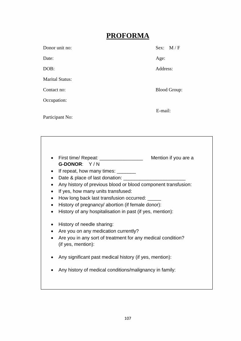
107
PROFORMA
Donor unit no: Sex: M / F
Date: Age:
DOB: Address:
Marital Status:
Contact no: Blood Group:
Occupation:
E-mail:
Participant No:
• First time/ Repeat: ________________ Mention if you are a
G-DONOR: Y / N
• If repeat, how many times: _______
• Date & place of last donation: _______________________
• Any history of previous blood or blood component transfusion:
• If yes, how many units transfused:
• How long back last transfusion occurred: _____
• History of pregnancy/ abortion (if female donor):
• History of any hospitalisation in past (if yes, mention):
• History of needle sharing:
• Are you on any medication currently?
• Are you in any sort of treatment for any medical condition?
(if yes, mention):
• Any significant past medical history (if yes, mention):
• Any history of medical conditions/malignancy in family:

108
Laboratory Reports
➢ Blood group:
➢ DAT results: Positive / Negative
➢ Antibody detection (Donor Serum + pooled O cell 3-5%
suspension):
• At 40C:
• 220C:
• 370C:
• IAT:
➢ Auto-control (Donor Serum + Donor cell 3-
5%suspension):
• At At 40C:
• 220C:
• 370C:
• IAT:
• Enzymes:
➢ Antibody Identification:
➢ Titre of antibody: IgM ________ IgG ___________

109
MASTER SHEET

110
T
itre
IgG
2
8
16
32
8
16
8
IgM
64
32
64
-
64
32
-
T
yp
e o
f
An
tib
od
y
IgM
+Ig
G
IgM
+Ig
G
IgM
+Ig
G
I
gG
IgM
+Ig
G
IgM
+Ig
G
I
gG
An
tib
od
y
Iden
tifi
ed
An
ti-I
H
An
ti-L
eb
An
ti-M
An
ti-D
An
ti-L
eab
An
ti-M
An
ti-C
Pa
p
O c
ells
+
+
0
+
+
0
+
O C
ells
37
0
(IA
T)
+
+
+
+
+
+
+
22
0
+
+
+
0
+
+
0
40
+
+
+
0
+
0
0
Au
to-c
on
trol 37
0
(AH
G)
0
0
0
0
0
0
0
22
0
0
0
0
0
0
0
0
40
+
+
0
0
+
0
0
DA
T
0
0
0
0
0
0
0
Ty
pe
of
Do
no
r
Fir
st
Tim
e
Fir
st
tim
e
Rep
eat
Fir
st
Tim
e
Fir
st
Tim
e
Rep
eat
Rep
eat
Blo
od
Gro
up
A n
eg
A n
eg
B +
AB
neg
B +
0 +
B n
eg
Sex
M
M
M
F
M
M
F
Ag
e
19
34
34
35
26
32
29
Do
no
r
ID
17
002
4
17
018
3
17
027
2
17
222
6
17
323
5
17
387
8
17
495
4
Da
te
12
/02
/201
8
02
/03
/201
8
24
/05
/201
8
25
/07
/201
8
17
/09
/201
8
28
/10
/201
8
31
/01
/201
8
Sl
no
.
1
2
3
4
5
6
7