prevalence, antimicrobial susceptibility patterns, and
TRANSCRIPT
Research ArticlePrevalence, Antimicrobial Susceptibility Patterns, and RiskFactors Associated with Enterococci among Pediatric Patients atDessie Referral Hospital, Northeastern Ethiopia
Admasu Abera,1 Mihret Tilahun,2 Saba Gebremichael Tekele ,2
and Melaku Ashagrie Belete 2
1Department of Medical Laboratory Science, Debre Berhan Health Science College, Debre Berhan, Ethiopia2Department of Medical Laboratory Science, College of Medicine and Health Sciences, Wollo University, Dessie, Ethiopia
Correspondence should be addressed to Melaku Ashagrie Belete; [email protected]
Received 9 February 2021; Revised 1 April 2021; Accepted 8 April 2021; Published 16 April 2021
Academic Editor: Mohamed Salah Abbassi
Copyright © 2021 Admasu Abera et al. This is an open access article distributed under the Creative Commons Attribution License,which permits unrestricted use, distribution, and reproduction in any medium, provided the original work is properly cited.
Background. Enterococcus species, which is previously considered as medically not important, now becomes one of the leadingcauses of nosocomial infections. Even though it becomes the most public health concern and emerging multidrug-resistantpathogen, there is no enough data in the study area. Objective. To determine the prevalence, antimicrobial resistance pattern,and associated risk factors of enterococci infection in pediatric patients. Methods. A hospital-based cross-sectional study wasconducted from February to May 2019 at Dessie Referral Hospital, Northeastern Ethiopia. A total of 403 pediatric patients wereincluded in the study, and a pretested structured questionnaire was used to collect sociodemographic and risk factor-relateddata. Clinical samples such as urine, blood, wound swabs, discharges, and other body fluids were collected aseptically andinoculated on to Bile Esculin Azide Agar, and colony characteristics, Gram stain, catalase, salt, and temperature tolerance testswere employed for bacterial identification. Antimicrobial sensitivity tests were performed using the modified Kirby-Bauer diskdiffusion method. Data was entered into SPSS software version 25 and descriptive statistics; bivariate and multivariate logisticregression analyses were performed. In all cases, a P value ≤ 0.05 with corresponding 95% confidence interval were consideredas statistically significant. Result. The overall prevalence of enterococci was 2.7% (11/403). Of which, the highest number ofenterococci infection was recovered from urine sample (54.5%) followed by blood (27.3%), wound swab (9.1%), and other bodyfluids (9%). The overall multidrug resistance rate was 54.5%. Higher drug resistance pattern was observed against tetracycline,chloramphenicol, and amoxicillin/clavulanate. Having history of invasive procedure (P < 0:001), chronic illness (P < 0:001) andprevious admission history of the children (P < 0:001) were statistically significant associated risk factors for pediatricsenterococci infection. Conclusion. The prevalence of enterococci from pediatric patients in this study was relatively lowcompared to other studies. Significant rates of MDR and VRE were identified, and the risk of infection became high whenchildren had a history of different chronic illnesses and history of admission and underwent invasive treatment procedures.Therefore, efforts should be made to prevent enterococci infections and spread of multidrug-resistant enterococci.
1. Introduction
Enterococci are members of Gram-positive Enterococcaceaefamily and are nonspore forming, facultatively anaerobic, oxi-dase, and catalase negative bacteria that occur singly, in pairsor short chains [1]. Moreover, enterococci are known to sur-vive in a range of hostile environments, including growth in
the presence of 6.5% NaCl, temperature of 5 to 65°C,pH4.5 to 10, and hydrolyze esculin in the presence of 40%bile [1, 2]. Enterococci are common indigenous flora of thegastrointestinal tracts and can also normally present in theoral cavity, vagina, oropharynx, and urethra of humans [3].
In fact, enterococci species have low virulence factors.However, under conditions in which the host encountered
HindawiBioMed Research InternationalVolume 2021, Article ID 5549847, 9 pageshttps://doi.org/10.1155/2021/5549847
immune suppression or the integrity of the gastrointestinalor genitourinary tract has been disrupted, they spread to nor-mally sterile sites and cause various infections. These infec-tions include urinary tract infections, wound infection,sepsis, endocarditis, intra-abdominal abscesses, and biliarytract infections [4].
According to Centers for Disease Control and Prevention(CDC) nationwide report, enterococci are one of the leadingcauses of nosocomial infections worldwide, with healthcare-associated infections occurring more frequently in resource-limited settings than in developed countries [5]. Moreover,the pathogen distribution and antimicrobial resistance pat-terns in healthcare-associated infections (HAIs) from pediat-ric groups also indicated 8% of enterococci species [6].
Different risk factors have been reported to be associatedwith the spread of enterococci infections including concurrentinfections, history antibiotic use, surgery, catheterization, lon-ger duration of hospitalization, and underlying immunosup-pressing diseases such as HIV, diabetics, and cancer [7].
The emergence and spread of antimicrobial resistanceamong enterococcus species pose enormous challenges forclinicians, especially in the management of severe infections.The increased prevalence and dissemination of multidrug-resistant enterococcus has narrowed the therapeutic optionsglobally, as the majority of enterococcus isolates exhibit ahigh level of resistance to ampicillin, penicillin, and vanco-mycin, which are indeed the most historically useful anti-enterococci antibiotics [8].
In 2018, the national survey for antimicrobial resistancein Europe report indicated that vancomycin-resistantenterococcus species increased significantly from 10.4% in2014 to 14.9% in 2017 in different countries [9]. Moreover,according to worldwide surveillance summary of drugpotency of Gram-positive pathogens, the overall prevalenceof vancomycin-resistant enterococci (VRE) in Europe, Asiaand Pacific, and Latin and North America ranges between1% and 9.8% [10].
The prevalence and drug resistance figures are signifi-cantly high in developing countries. In Africa and Ethiopia,some reports indicated the overall prevalence of enterococciand antibiotic resistance ranged from 2.2% to 76% and6.3% to 95.5%, respectively [11–16]. Therefore, efforts shouldbe made to prevent enterococci infections and emergence ofmultidrug-resistant enterococci. Hence, studying the preva-lence, dispersion, and correlation of possible associated riskfactors and examining the drug resistance pattern of entero-coccus species, particularly in vulnerable target groups suchas pediatrics, are important for effective prevention, control,and management of infections. Therefore, the study wasaimed at determining the prevalence, antimicrobial suscepti-bility patterns, and associated risk factors of enterococciamong pediatric patients in Dessie Referral Hospital(DRH), Northeastern Ethiopia.
2. Method and Materials
2.1. Study Design, Area, and Period. A hospital-based cross-sectional study was conducted from February 2019 to May2019 at Dessie Referral Hospital, South Wollo Zone of
Amhara regional state, Northeast Ethiopia. Dessie ReferralHospital is located in Dessie city, 400 km from the capital,Addis Ababa, and 471 km far from Bahir Dar, the capital ofthe Amhara regional state. The city has one referral hospital,one general hospital, three private general hospitals, fivehealth centers, eight private higher clinics, and one PublicHealth Research Institute. Dessie Referral Hospital providesemergency, antiretroviral therapy (ART) services, chroniccare, surgical, dental, medical, pediatric, gynecologic, obstet-ric, and other services. The hospital serves patients from allparts of the region which comprises a population of morethan 4 million people. Dessie Referral Hospital pediatric ser-vices consist of inpatient and outpatient departments. Theoutpatient pediatric department consists of 15 beds and fiverooms, while the inpatient pediatric ward consists of 13rooms and 49 beds, with an average of about 7 childrenadmitted per day.
2.2. Inclusion and Exclusion Criteria. All children youngerthan 15 years old, attending DRH and who were requestedfor laboratory investigation during the study period, wereincluded in this study. On the other hand, pediatric patientswho received antibiotics within the past 2 weeks wereexcluded. Moreover, as enterococci are normal flora of somespecific sites including respiratory, genital, and gastrointesti-nal tracts, samples such as sputum, throat swab, stool, andvaginal swabs were excluded from the study.
2.3. Sample Size Determination and Sampling Technique. Thesample size was determined using a single population pro-portion formula considering 50% prevalence, marginal errorof 5%, and 95%confidence interval = 1:96 by using the fol-lowing sample size determination formula:
n = z2p 1 − pð Þd2
, ð1Þ
where n is the minimum sample size required, Za/2 is the sig-nificant value for 95% confidence interval, P is the expectedprevalence of enterococci infection, and d is the margin oferror.By including 5% nonresponse rate, a total of 403 chil-dren were included in this study using systematic randomsampling.
2.4. Data and Specimen Collection. Data were collected usinga short interview guided by pretested structured question-naire consisting of the client’s sociodemographic, clinical,and risk factor data. Clinical samples were collected fromeach study participant aseptically. About 5ml of the bloodsample was collected from children and dispensed into bloodculture bottle prepared with 25ml of Tryptic Soya Broth (FLMedical, Italy) aseptically. Ten ml of freshly voided mid-stream urine specimen was collected using wide mouth,leak-proof, sterile, plastic container under the supervisionof the principal investigator and processed within 2 hoursof collection. Approximately 5ml of cerebrospinal fluid(CSF) sample was collected aseptically into sterile tube bylumbar or ventricular puncture performed by a physicianand processed within one hour of collection. Wound swab,
2 BioMed Research International
pus, eye, and ear discharges were obtained using sterile cot-ton tip applicator stick aseptically. The blood sample wastransported with the blood culture broth while all swabs weretransported within BHI broth (HiMedia™). The collectedspecimens were then stored in a cold box and transportedto the Department of Microbiology Laboratory, AmharaPublic Health Institute (APHI), Dessie Branch. Immediateinoculation had been performed for all specimens on arrivalto the laboratory.
2.5. Isolation and Identification of Enterococci. Specimenswere inoculated on appropriate culture media to isolate theenterococci bacteria. The blood culture bottles were incu-bated at 37°C and observed after 24 hrs daily for consecutive5 days for the presence of turbidity, hemolysis, gas formation,or color changes which are evidence of microbial growth. Ifthe culture bottle does not show any growth within 7 days,it was reported as negative. Whenever visible growth appears,the bottle was opened aseptically, a small amount of brothwas taken with a sterile loop and subcultured on Bile EsculinAzide agar (BEAA) (Oxoid Ltd., UK). Urine samples wereinoculated on BEAA media with a 1μl calibrated loop andincubated at 37°C for 24hr. Presence of ≥104 colony formingunit (CFU) per ml of urine with the black colored colony wasconsidered as significant enterococci in urine specimen.Other clinical samples were directly subcultured on BEAAand incubated at 37°C for 24 hr and were checked for growthof very small colony with blackening media. Colony charac-teristics, Gram staining reaction, catalase, salt tolerance,and temperature tolerance tests were used for identificationof enterococci [17].
2.6. Antimicrobial Susceptibility Testing. The antimicrobialsusceptibility testing of enterococci isolates was performedusing the Kirby-Bauer disk diffusion technique as modifiedby the Clinical and Laboratory Standard Institute (CLSI) in2019 [18]. From a pure culture, 3-5 selected colonies of bac-teria were taken and transferred to a tube containing 5mlsterile normal saline and mixed gently to make homogenoussuspension and the turbidity of the suspension was adjustedcomparably to a McFarland 0.5 turbidity standard. A sterilecotton swab was used to streak the plates, and the excess sus-pension was removed by gentle pressing and rotation of theswab against the inside wall surface of the tube. The swabwas then used to distribute the bacteria evenly over the entiresurface of Mueller Hinton agar (MHA) (Conda Ltd., USA).The inoculated plates were then left at room temperature todry for 3-5 minutes, and a set of 6 antibiotic discs wereapplied on the MHA. Based on availability, CLSI recommen-dation, and prescription practice of DRH, the test was carriedout for the following drugs: erythromycin (E, 15μg), chlor-amphenicol (C, 30μg), tetracycline (TE, 30μg), ampicillin(AMP, 10μg), nitrofurantoin (NIT, 30μg), ciprofloxacin(CIP, 5μg), penicillin (P, 10 IU) and vancomycin (VA,30μg), and amoxicillin-clavulanic acid (AMC, 20/10μg).All antibiotic discs were from Oxoid Ltd., UK. The plateswere then incubated at 37°C for 24 hours. Diameters of thezone of inhibition around the discs were measured using adigital caliper. The interpretation of the results of the antimi-
crobial susceptibility tests was based on the standardizedtable supplied by CLSI [18] criteria as sensitive, intermediate,and resistant.
2.7. Quality Assurance. Data quality was ensured by usingstandardized data collection materials. The questionnairewas pretested on 5% of the sample size in the nearby BoruMeda Hospital before the actual study commenced to makesure whether the questionnaire is appropriate and under-standable. All the questions in structured questionnaire wereprepared in a clear and precise way and translated into locallanguage (Amharic). The collected data were checked dailyfor completeness. Moreover, all laboratory analyses were per-formed by maintaining quality control procedures. Standardoperating procedures (SOPs) were strictly followed verifyingthat media meet expiration date and quality control parame-ters per CLSI guideline. All culture media were prepared fol-lowing the manufacturers’ instructions. Batch of preparedmedia was checked for pH, performance, and sterility testby incubating samples of the plate at 37°C for 24 hrs, andreagents for Gram stain and biochemical tests were checkedusing known standard strains of Enterococcus faecalis ATCC29212, Escherichia coli ATCC 25922, Streptococcus pyogenesATCC 19615, and Staphylococcus aureus ATCC 25923, andcotrimoxazole antibiotic drug was used to check quality ofMHA. All reference strains were obtained fromMicrobiologyLaboratory of APHI, Dessie Branch.
2.8. Statistical Analysis. Data was entered and analyzed usingStatistical Package for Social Sciences (SPSS) version 25.0.(IBM, USA), and descriptive statistics, binary, and multivar-iate logistic regression were computed. The bivariate analysisusing maximum likelihood estimates of the categorical vari-ables was used to determine the association of each variablewith the dependent variable. Furthermore, variables with P< 0:2 in the bivariate analysis were subjected to multivariatelogistic regression to identify the independent predictors ofenterococci infections. P value < 0.05 with 95% confidenceinterval was considered statistically significant.
3. Result
3.1. Sociodemographic Characteristics of Study Participants.Atotal of 403 study participants were included during the studyperiod. Of these, 188 (46.7%) were males, while 215 (53.3%)were females. The age of the study participants ranged from15 days to 14 years. The age distribution of participants indi-cated that majority 159 (39.5%) of the study participantswere in the age group of 5 to 9 years followed by 124(30.8%) belonging to the age group ≤4 years. Slightly major-ity of the study participants 214 (53.1%) were rural dwellers.Moreover, the clinical data showed that outpatient was thepredominant group among the study participants, with aproportion of 67.2%, while the remaining 32.3% were frominpatient (Table 1).
3.2. Prevalence of Enterococci Isolates among PediatricPatients. A total of 11 (2.7%) enterococci isolates were iso-lated from all clinical samples. Seven out of 11 isolates wereidentified from outpatients. Relatively higher frequency of
3BioMed Research International
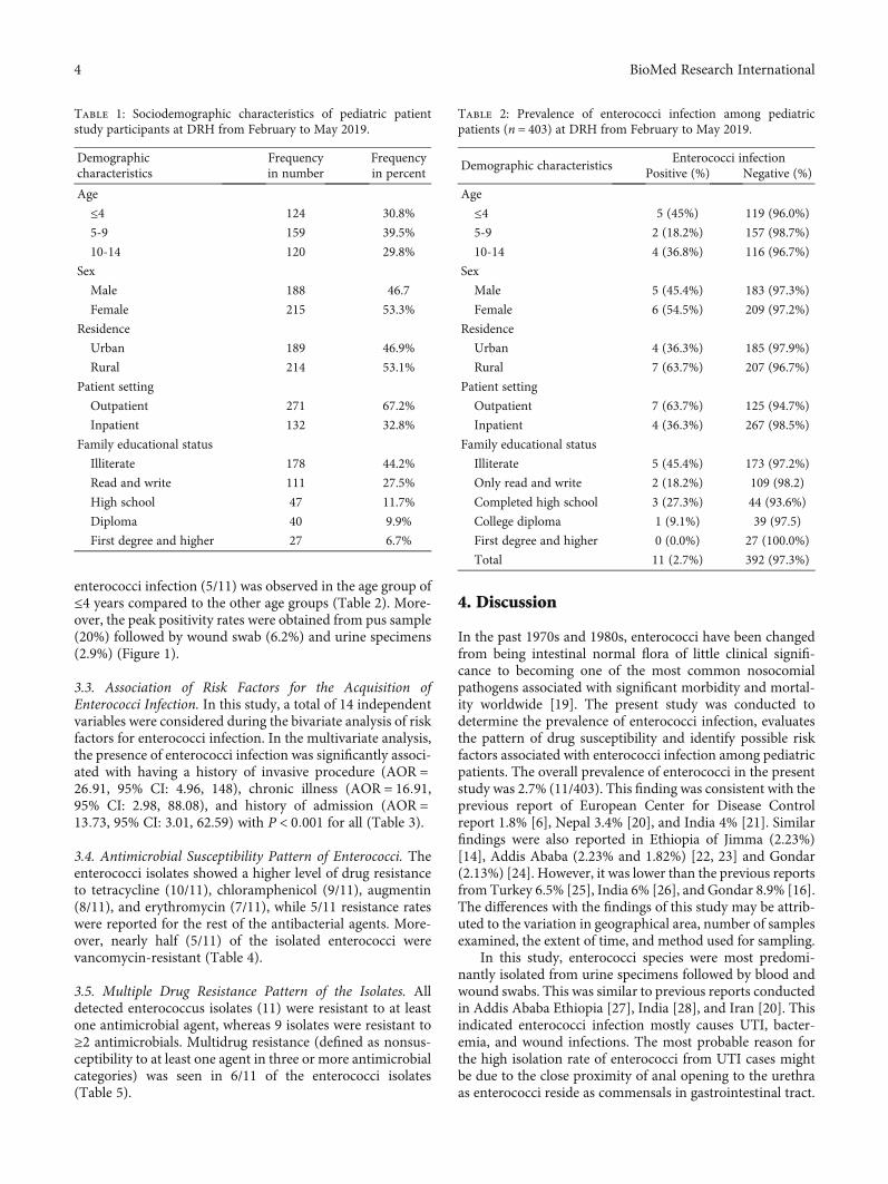
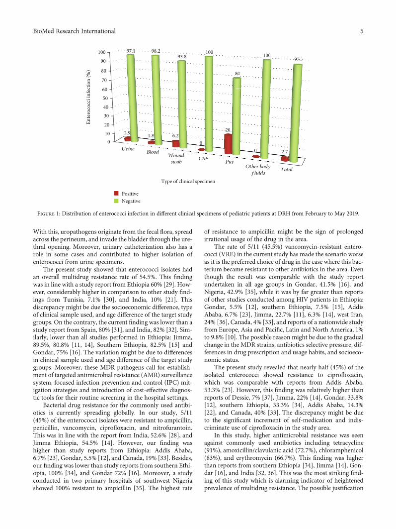
enterococci infection (5/11) was observed in the age group of≤4 years compared to the other age groups (Table 2). More-over, the peak positivity rates were obtained from pus sample(20%) followed by wound swab (6.2%) and urine specimens(2.9%) (Figure 1).
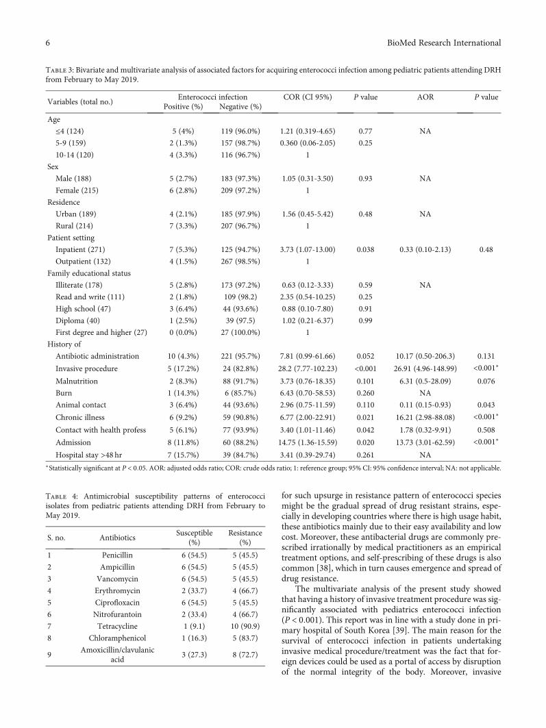
3.3. Association of Risk Factors for the Acquisition ofEnterococci Infection. In this study, a total of 14 independentvariables were considered during the bivariate analysis of riskfactors for enterococci infection. In the multivariate analysis,the presence of enterococci infection was significantly associ-ated with having a history of invasive procedure (AOR =26:91, 95% CI: 4.96, 148), chronic illness (AOR = 16:91,95% CI: 2.98, 88.08), and history of admission (AOR =13:73, 95% CI: 3.01, 62.59) with P < 0:001 for all (Table 3).
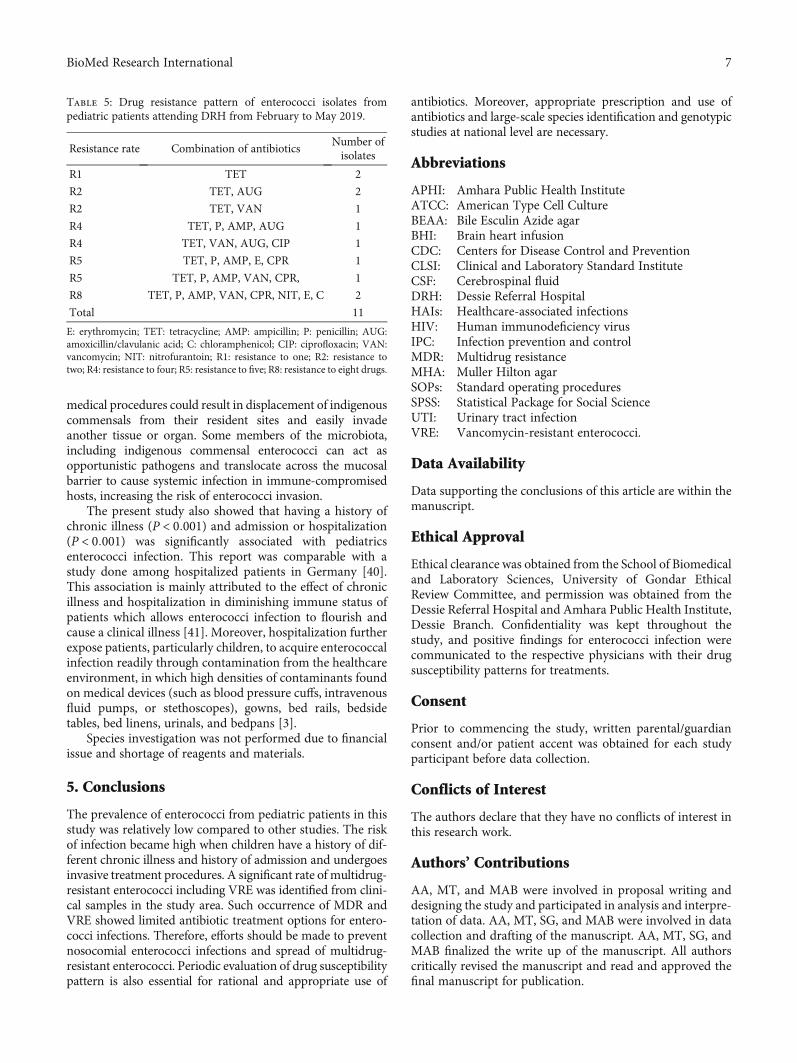
3.4. Antimicrobial Susceptibility Pattern of Enterococci. Theenterococci isolates showed a higher level of drug resistanceto tetracycline (10/11), chloramphenicol (9/11), augmentin(8/11), and erythromycin (7/11), while 5/11 resistance rateswere reported for the rest of the antibacterial agents. More-over, nearly half (5/11) of the isolated enterococci werevancomycin-resistant (Table 4).
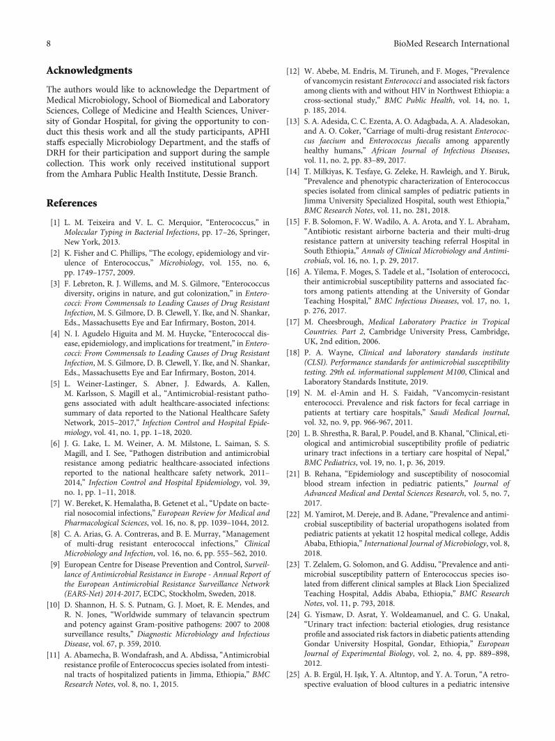
3.5. Multiple Drug Resistance Pattern of the Isolates. Alldetected enterococcus isolates (11) were resistant to at leastone antimicrobial agent, whereas 9 isolates were resistant to≥2 antimicrobials. Multidrug resistance (defined as nonsus-ceptibility to at least one agent in three or more antimicrobialcategories) was seen in 6/11 of the enterococci isolates(Table 5).
4. Discussion
In the past 1970s and 1980s, enterococci have been changedfrom being intestinal normal flora of little clinical signifi-cance to becoming one of the most common nosocomialpathogens associated with significant morbidity and mortal-ity worldwide [19]. The present study was conducted todetermine the prevalence of enterococci infection, evaluatesthe pattern of drug susceptibility and identify possible riskfactors associated with enterococci infection among pediatricpatients. The overall prevalence of enterococci in the presentstudy was 2.7% (11/403). This finding was consistent with theprevious report of European Center for Disease Controlreport 1.8% [6], Nepal 3.4% [20], and India 4% [21]. Similarfindings were also reported in Ethiopia of Jimma (2.23%)[14], Addis Ababa (2.23% and 1.82%) [22, 23] and Gondar(2.13%) [24]. However, it was lower than the previous reportsfrom Turkey 6.5% [25], India 6% [26], and Gondar 8.9% [16].The differences with the findings of this study may be attrib-uted to the variation in geographical area, number of samplesexamined, the extent of time, and method used for sampling.
In this study, enterococci species were most predomi-nantly isolated from urine specimens followed by blood andwound swabs. This was similar to previous reports conductedin Addis Ababa Ethiopia [27], India [28], and Iran [20]. Thisindicated enterococci infection mostly causes UTI, bacter-emia, and wound infections. The most probable reason forthe high isolation rate of enterococci from UTI cases mightbe due to the close proximity of anal opening to the urethraas enterococci reside as commensals in gastrointestinal tract.
Table 1: Sociodemographic characteristics of pediatric patientstudy participants at DRH from February to May 2019.
Demographiccharacteristics
Frequencyin number
Frequencyin percent
Age
≤4 124 30.8%
5-9 159 39.5%
10-14 120 29.8%
Sex
Male 188 46.7
Female 215 53.3%
Residence
Urban 189 46.9%
Rural 214 53.1%
Patient setting
Outpatient 271 67.2%
Inpatient 132 32.8%
Family educational status
Illiterate 178 44.2%
Read and write 111 27.5%
High school 47 11.7%
Diploma 40 9.9%
First degree and higher 27 6.7%
Table 2: Prevalence of enterococci infection among pediatricpatients (n = 403) at DRH from February to May 2019.
Demographic characteristicsEnterococci infection
Positive (%) Negative (%)
Age
≤4 5 (45%) 119 (96.0%)
5-9 2 (18.2%) 157 (98.7%)
10-14 4 (36.8%) 116 (96.7%)
Sex
Male 5 (45.4%) 183 (97.3%)
Female 6 (54.5%) 209 (97.2%)
Residence
Urban 4 (36.3%) 185 (97.9%)
Rural 7 (63.7%) 207 (96.7%)
Patient setting
Outpatient 7 (63.7%) 125 (94.7%)
Inpatient 4 (36.3%) 267 (98.5%)
Family educational status
Illiterate 5 (45.4%) 173 (97.2%)
Only read and write 2 (18.2%) 109 (98.2)
Completed high school 3 (27.3%) 44 (93.6%)
College diploma 1 (9.1%) 39 (97.5)
First degree and higher 0 (0.0%) 27 (100.0%)
Total 11 (2.7%) 392 (97.3%)
4 BioMed Research International
With this, uropathogens originate from the fecal flora, spreadacross the perineum, and invade the bladder through the ure-thral opening. Moreover, urinary catheterization also has arole in some cases and contributed to higher isolation ofenterococci from urine specimens.
The present study showed that enterococci isolates hadan overall multidrug resistance rate of 54.5%. This findingwas in line with a study report from Ethiopia 60% [29]. How-ever, considerably higher in comparison to other study find-ings from Tunisia, 7.1% [30], and India, 10% [21]. Thisdiscrepancy might be due the socioeconomic difference, typeof clinical sample used, and age difference of the target studygroups. On the contrary, the current finding was lower than astudy report from Spain, 80% [31], and India, 82% [32]. Sim-ilarly, lower than all studies performed in Ethiopia: Jimma,89.5%, 80.8% [11, 14], Southern Ethiopia, 82.5% [15] andGondar, 75% [16]. The variation might be due to differencesin clinical sample used and age difference of the target studygroups. Moreover, these MDR pathogens call for establish-ment of targeted antimicrobial resistance (AMR) surveillancesystem, focused infection prevention and control (IPC) mit-igation strategies and introduction of cost-effective diagnos-tic tools for their routine screening in the hospital settings.
Bacterial drug resistance for the commonly used antibi-otics is currently spreading globally. In our study, 5/11(45%) of the enterococci isolates were resistant to ampicillin,penicillin, vancomycin, ciprofloxacin, and nitrofurantoin.This was in line with the report from India, 52.6% [28], andJimma Ethiopia, 54.5% [14]. However, our finding washigher than study reports from Ethiopia: Addis Ababa,6.7% [23], Gondar, 5.5% [12], and Canada, 19% [33]. Besides,our finding was lower than study reports from southern Ethi-opia, 100% [34], and Gondar 72% [16]. Moreover, a studyconducted in two primary hospitals of southwest Nigeriashowed 100% resistant to ampicillin [35]. The highest rate
of resistance to ampicillin might be the sign of prolongedirrational usage of the drug in the area.
The rate of 5/11 (45.5%) vancomycin-resistant entero-cocci (VRE) in the current study has made the scenario worseas it is the preferred choice of drug in the case where this bac-terium became resistant to other antibiotics in the area. Eventhough the result was comparable with the study reportundertaken in all age groups in Gondar, 41.5% [16], andNigeria, 42.9% [35], while it was by far greater than reportsof other studies conducted among HIV patients in Ethiopia:Gondar, 5.5% [12], southern Ethiopia, 7.5% [15], AddisAbaba, 6.7% [23], Jimma, 22.7% [11], 6.3% [14], west Iran,24% [36], Canada, 4% [33], and reports of a nationwide studyfrom Europe, Asia and Pacific, Latin and North America, 1%to 9.8% [10]. The possible reason might be due to the gradualchange in the MDR strains, antibiotics selective pressure, dif-ferences in drug prescription and usage habits, and socioeco-nomic status.
The present study revealed that nearly half (45%) of theisolated enterococci showed resistance to ciprofloxacin,which was comparable with reports from Addis Ababa,53.3% [23]. However, this finding was relatively higher thanreports of Dessie, 7% [37], Jimma, 22% [14], Gondar, 33.8%[12], southern Ethiopia, 33.3% [34], Addis Ababa, 14.3%[22], and Canada, 40% [33]. The discrepancy might be dueto the significant increment of self-medication and indis-criminate use of ciprofloxacin in the study area.
In this study, higher antimicrobial resistance was seenagainst commonly used antibiotics including tetracycline(91%), amoxicillin/clavulanic acid (72.7%), chloramphenicol(83%), and erythromycin (66.7%). This finding was higherthan reports from southern Ethiopia [34], Jimma [14], Gon-dar [16], and India [32, 36]. This was the most striking find-ing of this study which is alarming indicator of heightenedprevalence of multidrug resistance. The possible justification
01020
30405060
70
80
90100
UrineBlood
Wound
swabCSF
Pus
Other bodyfluids
Total
2.9 1.8 6.20
20
0 2.7
97.1 98.293.8
100
80
10097.3
Ente
roco
cci i
nfec
tion
(%)
Type of clinical specimen
PositiveNegative
UrineBlood
WoundWW
swabCSF
Pus
2.9 1.8 6.220
200
00 2.7
93.8
80
10097.397 3
Figure 1: Distribution of enterococci infection in different clinical specimens of pediatric patients at DRH from February to May 2019.
5BioMed Research International
for such upsurge in resistance pattern of enterococci speciesmight be the gradual spread of drug resistant strains, espe-cially in developing countries where there is high usage habit,these antibiotics mainly due to their easy availability and lowcost. Moreover, these antibacterial drugs are commonly pre-scribed irrationally by medical practitioners as an empiricaltreatment options, and self-prescribing of these drugs is alsocommon [38], which in turn causes emergence and spread ofdrug resistance.
The multivariate analysis of the present study showedthat having a history of invasive treatment procedure was sig-nificantly associated with pediatrics enterococci infection(P < 0:001). This report was in line with a study done in pri-mary hospital of South Korea [39]. The main reason for thesurvival of enterococci infection in patients undertakinginvasive medical procedure/treatment was the fact that for-eign devices could be used as a portal of access by disruptionof the normal integrity of the body. Moreover, invasive
Table 3: Bivariate and multivariate analysis of associated factors for acquiring enterococci infection among pediatric patients attending DRHfrom February to May 2019.
Variables (total no.)Enterococci infection COR (CI 95%) P value AOR P value
Positive (%) Negative (%)
Age
≤4 (124) 5 (4%) 119 (96.0%) 1.21 (0.319-4.65) 0.77 NA
5-9 (159) 2 (1.3%) 157 (98.7%) 0.360 (0.06-2.05) 0.25
10-14 (120) 4 (3.3%) 116 (96.7%) 1
Sex
Male (188) 5 (2.7%) 183 (97.3%) 1.05 (0.31-3.50) 0.93 NA
Female (215) 6 (2.8%) 209 (97.2%) 1
Residence
Urban (189) 4 (2.1%) 185 (97.9%) 1.56 (0.45-5.42) 0.48 NA
Rural (214) 7 (3.3%) 207 (96.7%) 1
Patient setting
Inpatient (271) 7 (5.3%) 125 (94.7%) 3.73 (1.07-13.00) 0.038 0.33 (0.10-2.13) 0.48
Outpatient (132) 4 (1.5%) 267 (98.5%) 1
Family educational status
Illiterate (178) 5 (2.8%) 173 (97.2%) 0.63 (0.12-3.33) 0.59 NA
Read and write (111) 2 (1.8%) 109 (98.2) 2.35 (0.54-10.25) 0.25
High school (47) 3 (6.4%) 44 (93.6%) 0.88 (0.10-7.80) 0.91
Diploma (40) 1 (2.5%) 39 (97.5) 1.02 (0.21-6.37) 0.99
First degree and higher (27) 0 (0.0%) 27 (100.0%) 1
History of
Antibiotic administration 10 (4.3%) 221 (95.7%) 7.81 (0.99-61.66) 0.052 10.17 (0.50-206.3) 0.131
Invasive procedure 5 (17.2%) 24 (82.8%) 28.2 (7.77-102.23) <0.001 26.91 (4.96-148.99) <0.001∗
Malnutrition 2 (8.3%) 88 (91.7%) 3.73 (0.76-18.35) 0.101 6.31 (0.5-28.09) 0.076
Burn 1 (14.3%) 6 (85.7%) 6.43 (0.70-58.53) 0.260 NA
Animal contact 3 (6.4%) 44 (93.6%) 2.96 (0.75-11.59) 0.110 0.11 (0.15-0.93) 0.043
Chronic illness 6 (9.2%) 59 (90.8%) 6.77 (2.00-22.91) 0.021 16.21 (2.98-88.08) <0.001∗
Contact with health profess 5 (6.1%) 77 (93.9%) 3.40 (1.01-11.46) 0.042 1.78 (0.32-9.91) 0.508
Admission 8 (11.8%) 60 (88.2%) 14.75 (1.36-15.59) 0.020 13.73 (3.01-62.59) <0.001∗
Hospital stay >48 hr 7 (15.7%) 39 (84.7%) 3.41 (0.39-29.74) 0.261 NA∗Statistically significant at P < 0:05. AOR: adjusted odds ratio; COR: crude odds ratio; 1: reference group; 95% CI: 95% confidence interval; NA: not applicable.
Table 4: Antimicrobial susceptibility patterns of enterococciisolates from pediatric patients attending DRH from February toMay 2019.
S. no. AntibioticsSusceptible
(%)Resistance
(%)
1 Penicillin 6 (54.5) 5 (45.5)
2 Ampicillin 6 (54.5) 5 (45.5)
3 Vancomycin 6 (54.5) 5 (45.5)
4 Erythromycin 2 (33.7) 4 (66.7)
5 Ciprofloxacin 6 (54.5) 5 (45.5)
6 Nitrofurantoin 2 (33.4) 4 (66.7)
7 Tetracycline 1 (9.1) 10 (90.9)
8 Chloramphenicol 1 (16.3) 5 (83.7)
9Amoxicillin/clavulanic
acid3 (27.3) 8 (72.7)
6 BioMed Research International
medical procedures could result in displacement of indigenouscommensals from their resident sites and easily invadeanother tissue or organ. Some members of the microbiota,including indigenous commensal enterococci can act asopportunistic pathogens and translocate across the mucosalbarrier to cause systemic infection in immune-compromisedhosts, increasing the risk of enterococci invasion.
The present study also showed that having a history ofchronic illness (P < 0:001) and admission or hospitalization(P < 0:001) was significantly associated with pediatricsenterococci infection. This report was comparable with astudy done among hospitalized patients in Germany [40].This association is mainly attributed to the effect of chronicillness and hospitalization in diminishing immune status ofpatients which allows enterococci infection to flourish andcause a clinical illness [41]. Moreover, hospitalization furtherexpose patients, particularly children, to acquire enterococcalinfection readily through contamination from the healthcareenvironment, in which high densities of contaminants foundon medical devices (such as blood pressure cuffs, intravenousfluid pumps, or stethoscopes), gowns, bed rails, bedsidetables, bed linens, urinals, and bedpans [3].
Species investigation was not performed due to financialissue and shortage of reagents and materials.
5. Conclusions
The prevalence of enterococci from pediatric patients in thisstudy was relatively low compared to other studies. The riskof infection became high when children have a history of dif-ferent chronic illness and history of admission and undergoesinvasive treatment procedures. A significant rate of multidrug-resistant enterococci including VRE was identified from clini-cal samples in the study area. Such occurrence of MDR andVRE showed limited antibiotic treatment options for entero-cocci infections. Therefore, efforts should be made to preventnosocomial enterococci infections and spread of multidrug-resistant enterococci. Periodic evaluation of drug susceptibilitypattern is also essential for rational and appropriate use of
antibiotics. Moreover, appropriate prescription and use ofantibiotics and large-scale species identification and genotypicstudies at national level are necessary.
Abbreviations
APHI: Amhara Public Health InstituteATCC: American Type Cell CultureBEAA: Bile Esculin Azide agarBHI: Brain heart infusionCDC: Centers for Disease Control and PreventionCLSI: Clinical and Laboratory Standard InstituteCSF: Cerebrospinal fluidDRH: Dessie Referral HospitalHAIs: Healthcare-associated infectionsHIV: Human immunodeficiency virusIPC: Infection prevention and controlMDR: Multidrug resistanceMHA: Muller Hilton agarSOPs: Standard operating proceduresSPSS: Statistical Package for Social ScienceUTI: Urinary tract infectionVRE: Vancomycin-resistant enterococci.
Data Availability
Data supporting the conclusions of this article are within themanuscript.
Ethical Approval
Ethical clearance was obtained from the School of Biomedicaland Laboratory Sciences, University of Gondar EthicalReview Committee, and permission was obtained from theDessie Referral Hospital and Amhara Public Health Institute,Dessie Branch. Confidentiality was kept throughout thestudy, and positive findings for enterococci infection werecommunicated to the respective physicians with their drugsusceptibility patterns for treatments.
Consent
Prior to commencing the study, written parental/guardianconsent and/or patient accent was obtained for each studyparticipant before data collection.
Conflicts of Interest
The authors declare that they have no conflicts of interest inthis research work.
Authors’ Contributions
AA, MT, and MAB were involved in proposal writing anddesigning the study and participated in analysis and interpre-tation of data. AA, MT, SG, and MAB were involved in datacollection and drafting of the manuscript. AA, MT, SG, andMAB finalized the write up of the manuscript. All authorscritically revised the manuscript and read and approved thefinal manuscript for publication.
Table 5: Drug resistance pattern of enterococci isolates frompediatric patients attending DRH from February to May 2019.
Resistance rate Combination of antibioticsNumber ofisolates
R1 TET 2
R2 TET, AUG 2
R2 TET, VAN 1
R4 TET, P, AMP, AUG 1
R4 TET, VAN, AUG, CIP 1
R5 TET, P, AMP, E, CPR 1
R5 TET, P, AMP, VAN, CPR, 1
R8 TET, P, AMP, VAN, CPR, NIT, E, C 2
Total 11
E: erythromycin; TET: tetracycline; AMP: ampicillin; P: penicillin; AUG:amoxicillin/clavulanic acid; C: chloramphenicol; CIP: ciprofloxacin; VAN:vancomycin; NIT: nitrofurantoin; R1: resistance to one; R2: resistance totwo; R4: resistance to four; R5: resistance to five; R8: resistance to eight drugs.
7BioMed Research International
Acknowledgments
The authors would like to acknowledge the Department ofMedical Microbiology, School of Biomedical and LaboratorySciences, College of Medicine and Health Sciences, Univer-sity of Gondar Hospital, for giving the opportunity to con-duct this thesis work and all the study participants, APHIstaffs especially Microbiology Department, and the staffs ofDRH for their participation and support during the samplecollection. This work only received institutional supportfrom the Amhara Public Health Institute, Dessie Branch.
References
[1] L. M. Teixeira and V. L. C. Merquior, “Enterococcus,” inMolecular Typing in Bacterial Infections, pp. 17–26, Springer,New York, 2013.
[2] K. Fisher and C. Phillips, “The ecology, epidemiology and vir-ulence of Enterococcus,” Microbiology, vol. 155, no. 6,pp. 1749–1757, 2009.
[3] F. Lebreton, R. J. Willems, and M. S. Gilmore, “Enterococcusdiversity, origins in nature, and gut colonization,” in Entero-cocci: From Commensals to Leading Causes of Drug ResistantInfection, M. S. Gilmore, D. B. Clewell, Y. Ike, and N. Shankar,Eds., Massachusetts Eye and Ear Infirmary, Boston, 2014.
[4] N. I. Agudelo Higuita and M. M. Huycke, “Enterococcal dis-ease, epidemiology, and implications for treatment,” in Entero-cocci: From Commensals to Leading Causes of Drug ResistantInfection, M. S. Gilmore, D. B. Clewell, Y. Ike, and N. Shankar,Eds., Massachusetts Eye and Ear Infirmary, Boston, 2014.
[5] L. Weiner-Lastinger, S. Abner, J. Edwards, A. Kallen,M. Karlsson, S. Magill et al., “Antimicrobial-resistant patho-gens associated with adult healthcare-associated infections:summary of data reported to the National Healthcare SafetyNetwork, 2015–2017,” Infection Control and Hospital Epide-miology, vol. 41, no. 1, pp. 1–18, 2020.
[6] J. G. Lake, L. M. Weiner, A. M. Milstone, L. Saiman, S. S.Magill, and I. See, “Pathogen distribution and antimicrobialresistance among pediatric healthcare-associated infectionsreported to the national healthcare safety network, 2011–2014,” Infection Control and Hospital Epidemiology, vol. 39,no. 1, pp. 1–11, 2018.
[7] W. Bereket, K. Hemalatha, B. Getenet et al., “Update on bacte-rial nosocomial infections,” European Review for Medical andPharmacological Sciences, vol. 16, no. 8, pp. 1039–1044, 2012.
[8] C. A. Arias, G. A. Contreras, and B. E. Murray, “Managementof multi-drug resistant enterococcal infections,” ClinicalMicrobiology and Infection, vol. 16, no. 6, pp. 555–562, 2010.
[9] European Centre for Disease Prevention and Control, Surveil-lance of Antimicrobial Resistance in Europe - Annual Report ofthe European Antimicrobial Resistance Surveillance Network(EARS-Net) 2014-2017, ECDC, Stockholm, Sweden, 2018.
[10] D. Shannon, H. S. S. Putnam, G. J. Moet, R. E. Mendes, andR. N. Jones, “Worldwide summary of telavancin spectrumand potency against Gram-positive pathogens: 2007 to 2008surveillance results,” Diagnostic Microbiology and InfectiousDisease, vol. 67, p. 359, 2010.
[11] A. Abamecha, B. Wondafrash, and A. Abdissa, “Antimicrobialresistance profile of Enterococcus species isolated from intesti-nal tracts of hospitalized patients in Jimma, Ethiopia,” BMCResearch Notes, vol. 8, no. 1, 2015.
[12] W. Abebe, M. Endris, M. Tiruneh, and F. Moges, “Prevalenceof vancomycin resistant Enterococci and associated risk factorsamong clients with and without HIV in Northwest Ethiopia: across-sectional study,” BMC Public Health, vol. 14, no. 1,p. 185, 2014.
[13] S. A. Adesida, C. C. Ezenta, A. O. Adagbada, A. A. Aladesokan,and A. O. Coker, “Carriage of multi-drug resistant Enterococ-cus faecium and Enterococcus faecalis among apparentlyhealthy humans,” African Journal of Infectious Diseases,vol. 11, no. 2, pp. 83–89, 2017.
[14] T. Milkiyas, K. Tesfaye, G. Zeleke, H. Rawleigh, and Y. Biruk,“Prevalence and phenotypic characterization of Enterococcusspecies isolated from clinical samples of pediatric patients inJimma University Specialized Hospital, south west Ethiopia,”BMC Research Notes, vol. 11, no. 281, 2018.
[15] F. B. Solomon, F. W. Wadilo, A. A. Arota, and Y. L. Abraham,“Antibiotic resistant airborne bacteria and their multi-drugresistance pattern at university teaching referral Hospital inSouth Ethiopia,” Annals of Clinical Microbiology and Antimi-crobials, vol. 16, no. 1, p. 29, 2017.
[16] A. Yilema, F. Moges, S. Tadele et al., “Isolation of enterococci,their antimicrobial susceptibility patterns and associated fac-tors among patients attending at the University of GondarTeaching Hospital,” BMC Infectious Diseases, vol. 17, no. 1,p. 276, 2017.
[17] M. Cheesbrough, Medical Laboratory Practice in TropicalCountries. Part 2, Cambridge University Press, Cambridge,UK, 2nd edition, 2006.
[18] P. A. Wayne, Clinical and laboratory standards institute(CLSI). Performance standards for antimicrobial susceptibilitytesting. 29th ed. informational supplement M100, Clinical andLaboratory Standards Institute, 2019.
[19] N. M. el-Amin and H. S. Faidah, “Vancomycin-resistantenterococci. Prevalence and risk factors for fecal carriage inpatients at tertiary care hospitals,” Saudi Medical Journal,vol. 32, no. 9, pp. 966-967, 2011.
[20] L. B. Shrestha, R. Baral, P. Poudel, and B. Khanal, “Clinical, eti-ological and antimicrobial susceptibility profile of pediatricurinary tract infections in a tertiary care hospital of Nepal,”BMC Pediatrics, vol. 19, no. 1, p. 36, 2019.
[21] B. Rehana, “Epidemiology and susceptibility of nosocomialblood stream infection in pediatric patients,” Journal ofAdvanced Medical and Dental Sciences Research, vol. 5, no. 7,2017.
[22] M. Yamirot, M. Dereje, and B. Adane, “Prevalence and antimi-crobial susceptibility of bacterial uropathogens isolated frompediatric patients at yekatit 12 hospital medical college, AddisAbaba, Ethiopia,” International Journal of Microbiology, vol. 8,2018.
[23] T. Zelalem, G. Solomon, and G. Addisu, “Prevalence and anti-microbial susceptibility pattern of Enterococcus species iso-lated from different clinical samples at Black Lion SpecializedTeaching Hospital, Addis Ababa, Ethiopia,” BMC ResearchNotes, vol. 11, p. 793, 2018.
[24] G. Yismaw, D. Asrat, Y. Woldeamanuel, and C. G. Unakal,“Urinary tract infection: bacterial etiologies, drug resistanceprofile and associated risk factors in diabetic patients attendingGondar University Hospital, Gondar, Ethiopia,” EuropeanJournal of Experimental Biology, vol. 2, no. 4, pp. 889–898,2012.
[25] A. B. Ergül, H. Işık, Y. A. Altıntop, and Y. A. Torun, “A retro-spective evaluation of blood cultures in a pediatric intensive
8 BioMed Research International
care unit: a three-year evaluation,” Turkish Pediatrics Archive/-Turk Pediatri Arsivi, vol. 52, no. 3, 2017.
[26] J. Parameswarappa, V. P. Basavaraj, and C. M. Basavaraj, “Iso-lation, identification, and antibiogram of enterococci isolatedfrom patients with urinary tract infection,” Annals of AfricanMedicine, vol. 12, no. 3, pp. 176–181, 2013.
[27] A. A. Negash, D. Asrat, W. Abebe et al., BacteremicCommunity-Acquired Pneumonia in Ethiopian Children: Etiol-ogy, Antibiotic Resistance, Risk Factors, and Clinical Outcome.Open Forum Infectious Diseases, vol. 6, Oxford UniversityPress US, 2019.
[28] S. Sreeja, P. Sreenivasa Babu, and A. Prathab, “The prevalenceand the characterization of the enterococcus species from var-ious clinical samples in a tertiary care hospital,” Journal ofClinical and Diagnostic Research: JCDR, vol. 6, no. 9,pp. 1486–1488, 2012.
[29] A. Melese, C. Genet, and T. Andualem, “Prevalence of vanco-mycin resistant enterococci (VRE) in Ethiopia: a systematicreview and meta-analysis,” BMC Infectious Diseases, vol. 20,no. 1, p. 124, 2020.
[30] M. Tfifha, A. Ferjani, M. Mallouli, N. Mlika, S. Abroug, andJ. Boukadida, “Carriage of multidrug-resistant bacteria amongpediatric patients before and during their hospitalization in atertiary pediatric unit in Tunisia,” Libyan Journal of Medicine.,vol. 13, p. 1, 2018.
[31] A. P. Tedim, P. Ruiz-Garbajosa, J. Corander et al., “Populationbiology of intestinal Enterococcus isolates from hospitalizedand non-hospitalized individuals in different age groups,”Applied and Environmental Microbiology, vol. 81, no. 5,pp. 1820–1831, 2015.
[32] L. Kapoor, V. Randhawa, and M. Deb, “Antimicrobial resis-tance of enterococcal blood isolates at a pediatric care hospitalin India,” The Journal of Infectious Diseases, vol. 58, 2005.
[33] E. O. Billington, S. H. Phang, D. B. Gregson et al., “2 incidence,risk factors, and outcomes of enterococcus spp. blood stream 3infections: a population-based study,” International Journal ofInfectious Diseases, vol. 30, pp. e1–e7, 2014.
[34] A. Amsalu, Z. Geto, D. Asegu, and S. Eshetie, “Antimicrobialresistance pattern of bacterial isolates from different clinicalspecimens in southern Ethiopia: a three-year retrospectivestudy,” African Journal of Bacteriology Research., vol. 9, no. 1,pp. 1–8, 2017.
[35] K. O. Olawale, S. O. Fadiora, and S. S. Taiwo, “Prevalence ofhospital acquired enterococci infections in two primary-carehospitals in Osogbo, Southwestern Nigeria,” African Journalof Infectious Diseases, vol. 5, no. 2, pp. 40–46, 2011.
[36] M. R. Arabestani, M. Nasaj, and S. M. Mousavi, “Correlationbetween infective factors and antibiotic resistance in entero-cocci clinical isolates in west of Iran,” Chonnam Medical Jour-nal, vol. 53, no. 1, pp. 56–63, 2017.
[37] S. Ali, M. Alemayehu, M. Dagnew, and T. Gebrecherkos,“Vancomycin-Resistant Enterococci and Its Associated RiskFactors among HIV- Positive and -Negative Clients AttendingDessie Referral Hospital, Northeast Ethiopia,” InternationalJournal of Microbiology, vol. 2018, Article ID 4753460, 9 pages,2018.
[38] G. S. Bbosa and N. Mwebaza, Global irrational antibiotics/an-tibacterial drugs use: a current and future health and environ-mental consequences, Microbial pathogens and strategies forcombating them, Science, technology and education Formatex,Badajoz, 2013.
[39] Y. J. Kim, S. I. Kim, Y. R. Kim, J. Y. Lee, Y. J. Park, and M. W.Kang, “Risk factors for vancomycin-resistant enterococciinfection and mortality in colonized patients on intensive careunit admission,” American Journal of Infection Control,vol. 40, no. 10, pp. 1018-1019, 2012.
[40] M. Papadimitriou-Olivgeris, E. Drougka, F. Fligou et al., “Riskfactors for enterococcal infection and colonization byvancomycin-resistant enterococci in critically ill patients,”Infection, vol. 42, no. 6, pp. 1013–1022, 2014.
[41] I. M. Zacharioudakis, F. N. Zervou, P. D. Ziakas, L. B. Rice, andE. Mylonakis, “Vancomycin-resistant enterococci colonizationamong dialysis patients: a meta-analysis of prevalence, riskfactors, and significance,” American Journal of Kidney Dis-eases, vol. 65, no. 1, pp. 88–97, 2015.
9BioMed Research International