pressure support ventilation – a new triggered ventilation ... · pdf filepressure...
TRANSCRIPT
D-3
7-20
11
Pressure Support Ventilation –A New Triggered Ventilation Mode for Neonates
Jean Christophe Rozé Thomas Krüger
Important Notice: Medical knowledge changes constantly as a result of new research and clinical experience. The authors of this introductory guide has made every effort to ensure that the information given is completely up to date, particularly as regards applications and mode of operation. However, responsibility for all clinical measures must remain with the reader.
Written by:Prof. Jean Christophe Rozé, MDNeonatal intensive care unitHôpital Mère EnfantUniversity hospitalNantes, France 44035
Thoms KrügerDräger Medical GmbHMoislinger Allee 53/5523542 Lübeck
All rights, in particular those of duplication and distribution, are reserved by Dräger Medizintechnik GmbH. No part of this work may be reproduced or stored in any form using mechanical, electronic or photographic means, without the written permission of Dräger Medizintechnik GmbH.
ISBN 3-926762-41-1
Pressure Support Ventilation – a New Triggered Ventilation Mode for Neonates
Jean Christophe RozéThomas Krüger
CONTENTS
1.0 Introduction 6
2.0 PressureSupportVentilation 8
2.1 Definition 8
2.2 Advantages of Pressure Support Ventilation in Adults 10
2.3 Pressure Support Ventilation in Neonates 11
3.0 TriggeredVentilationinNeonates 12
3.1 Consequences of Asynchrony 12
3.2 Preventing Asynchrony 12
4.0 TriggerSignals 14
4.1 Principles of Triggering 14
4.1.1 Thoracic Impedance 14
4.1.2 Abdominal Movement 15
4.1.3 Airway Pressure Changes 15
4.1.4 Airway Flow Changes 16
4.1.5 Esophageal Pressure Changes 17
4.2 Specific Problems with Different Trigger Signals 18
4.2.1 Lack of Response 18
4.2.2 Autotriggering 18
4.2.3 Artefact 18
4.2.4 Antiphasic Trigger 19
4.2.5 Delayed Response Time 19
4.3 Technical Comparison of Different Trigger Signals 20
4.4 Clinical Comparison of Different Trigger Signals 21
5.0 DifferentVentilationModes 22
5.1 Untriggered Modes 22
5.2 Triggered Modes 24
5.3 Pressure Support Ventilation 26
5.3.1 Definition 26
5.3.2 Automatic Leak Adaptation 28
5.3.3 Backup Ventilation 30
5.3.4 Limitations and Contra-Indications 31
5.4 Clinical Studies Comparing Ventilation Modes 34
04|05
6.0 BenefitsofPressureSupportVentilation 34
6.1 Weaning Newborn Infants from the Ventilator 34
6.1.1 Easy Weaning 34
6.1.2 Difficult Weaning 34
6.2 Weaning Strategies 36
6.2.1 Selection of Weaning Type 36
6.2.2 Physiological Studies 36
6.2.3 Clinical Studies 38
6.2.4 PSV is better than A/C! 39
6.2.5 PSV with Volume Guarantee 40
7.0 PressureSupportVentilationinPractice 42
7.1 Ventilator Settings in PSV 42
7.1.1 Selecting the PSV Mode 42
7.1.2 Adjusting Trigger Threshold 43
7.1.3 Adjusting Inspiratory Flow 44
7.1.4 Adjusting Inspiratory Time (Backup TI) 45
7.1.5 Adjusting Initial Pressure Support Level 46
7.1.6 Setting the Backup Rate 46
7.2 Weaning by Pressure Support Ventilation 47
7.3 Monitoring Pressure Support Ventilation 49
7.3.1 Physiological Background 49
7.3.1.1 Chemical Control 49
7.3.1.2 The Respiratory Pump 51
7.3.1.3 Oxygen Consumption, Carbon Dioxide Production and Work of Breathing 51
7.3.1.4 Pulmonary Reflexes 51
7.3.1.5 Pattern of Respiration in Neonates with RDS 52
7.3.2 Monitoring in Practice 53
8.0 Conclusion 56
9.0 Appendix 58
9.1 Case Reports 58
9.2 Abbreviations 62
10.0References 64

PRESSURE SUPPORT VENTILATION | INTRODUCTION
Pressure Support Ventilation (PSV), a well known and widely accepted mode of respiratory support in adults has numerous publications, which describe application and benefits in this field of ventilation.Pressure Support Ventilation although available in a few neonatal/pediatric ventilators is seldom used due to technical limitations despite the wide use of triggered ventilation modes such as SIMV or A/C in neonatology.A specifically adapted neonatal Pressure Support Ventilation is now available with the Babylog 8000plus. This booklet sets out recommendations and descriptions, which refer to the Babylog 8000plus with software 5.0. The offered unique benefits for the use of PSV in neonates facilitate the application and improve the effectiveness of this new respiratory support.Nevertheless the first part of this booklet discusses theory of triggered ventilation in general and describes all the different ventilation modes. As well, an overview of the numerous publications in the field of triggered ventilation is given. The second part of this booklet focuses then on Pressure Support Ventilation as a further step in the evolution of neonatal triggered ventilation modes. PSV can be used during the acute phase of respiratory distress syndrome as well as during weaning, preferably in neonates who show high oxygen cost of breathing. The benefits, indications, limitations, ventilation strategies and control are described to help clinicians better understand and apply this new respiratory support. Moreover, the use of Pressure Support Ventilation in combination with the new mode Volume Guarantee is discussed.
1.0 Introduction
06|07
The outlined strategies are based on publications and the first hand personal experience gained with this new mode. Nevertheless, due to the constant change in medical knowledge some of the descriptions may require revision in the future.We hope that this booklet will help promote the use of Pressure Support Ventilation, based on the evidence so far there is the potential for many promising advances in the management of respiratory support of the critically ill neonate.
2.1 DEFINITION
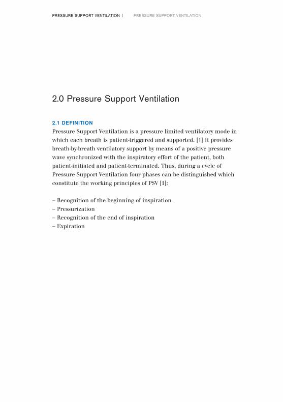
Pressure Support Ventilation is a pressure limited ventilatory mode in which each breath is patient-triggered and supported. [1] It provides breath-by-breath ventilatory support by means of a positive pressure wave synchronized with the inspiratory effort of the patient, both patient-initiated and patient-terminated. Thus, during a cycle of Pressure Support Ventilation four phases can be distinguished which constitute the working principles of PSV [1]:
– Recognition of the beginning of inspiration– Pressurization– Recognition of the end of inspiration– Expiration
PRESSURE SUPPORT VENTILATION | PRESSURE SUPPORT VENTILATION
2.0 Pressure Support Ventilation
08|09
Peak flow
Onset of inspirationphase 1
End of inspirationphase 3
15% of peak flow
pressurisationphase 2
expirationphase 4
PEEP
Pinsp
Flow
Paw
t
t
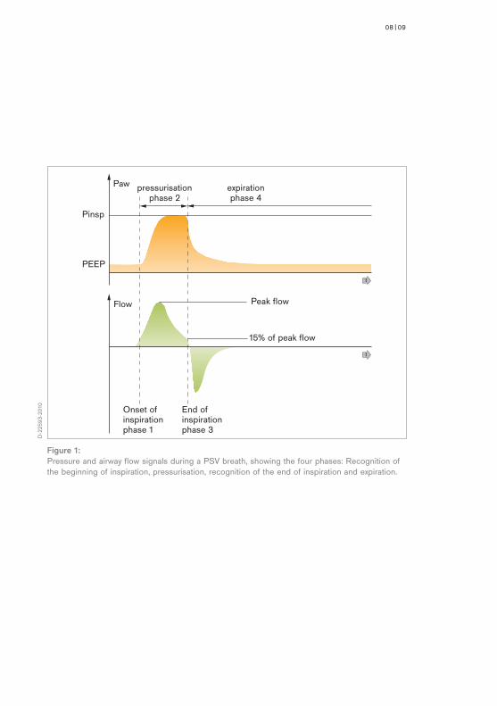
Figure 1: Pressure and airway flow signals during a PSV breath, showing the four phases: Recognition of the beginning of inspiration, pressurisation, recognition of the end of inspiration and expiration.
D-2
2593
-201
0
2.2 ADVANTAGES OF PRESSURE SUPPORT VENTILATION IN ADULTS [1]
In adult ventilation Pressure Support Ventilation is world wide the mostly used ventilation mode for weaning patients off the ventilator. Many studies have been performed to evaluate Pressure Support Ventilation in adult critical care. The main advantages [1] observed during these studies were:
– Better synchrony between patient and ventilator– Increased patient comfort– Reduced need for sedation– Decrease in work of breathing– Decrease in oxygen cost of breathing– Shorter duration of weaning process (observed only in few studies) [2]– Endurance oriented training of respiratory muscles [47]– Deepening of weak shallow spontaneous breathing
2.3 PRESSURE SUPPORT VENTILATION IN NEONATES
During conventional ventilation neonates are ventilated with continuous flow, pressure limited, time cycled ventilators. The introduction of triggered
PRESSURE SUPPORT VENTILATION | PRESSURE SUPPORT VENTILATION
10|11
ventilation has been a marked improvement in neonatal ventilation. Various triggered ventilation modes have been developed for neonates: Synchronous Intermittent Mandatory Ventilation (SIMV), Assist/Control Ventilation (A/C), and more recently Pressure Support Ventilation (PSV). Among these, PSV gives the patient optimum liberty during ventilation. The patient decides over start of inspiration and start of expiration and therefore controls inspiration time, breathing frequency and minute volume. Pressure Support Ventilation supports spontaneous breathing in a unique and harmonious way and is thus predestined to become the ventilation mode best suited to weaning patients off the ventilator also in neonatal respiratory care.
Before focusing on PSV, we first have to look at all the other triggered ventilation modes and their characteristics.
3.1 CONSEQUENCES OF ASYNCHRONY
Asynchrony between spontaneous ventilation and mechanical breaths can be problematic. [3] Asynchrony causing active expiration against ventilator inflation may occur irregularly or continuously, depending on the ventilator setting. [4,5] Consequences of active expiration may be a decrease in tidal volume and minute volume, an increase in oxygen consumption, an increase in intrathoracic pressure, a decrease in cardiac output and an increase in venous pressure. [3] Therefore during asynchrony, agitation of patient, inadequate gas exchange, increased risk of pneumothorax [6], and intraventricular hemorrhage [7] have been observed. However, when positive pressure inflation and spontaneous inspiration coincided, oxygenation improvement was found. [8] In babies paralysed by Pancuronium to avoid asynchrony, the risk of pneumothorax6 and intraventricular hemorrhage [9] was reduced.
3.2 PREVENTING ASYNCHRONY
Asynchrony may be prevented by adapting ventilator settings to the spontaneous breathing pattern, by using triggered ventilation modes, or by using heavy sedation or paralysis.Most neonatologists do not adapt the practice of regular paralysis in infants with active expiration, because paralysis has some disadvantages: in small infants, it has been associated with increases in oxygen requirements [10] as well as failure of skeletal muscle growth11 and delay in weaning process. [12]Sedation is more often used but also shows some disadvantages such as hypotension and modification of EEG.
3.0 Triggered Ventilation in Neonates
PRESSURE SUPPORT VENTILATION | TRIGGERED VENTILATION IN NEONATES

12|13
Flow 0
exp.
insp.
Pressure
Active expiration
0
Synchrony can sometimes be achieved by shortening inspiration time and/or increasing ventilator rate so as to match ventilator settings with the spontaneous respiratory rate of the infant. [8,13] But these modifications are not always successful and are not widely accepted in clinical practice because of the high intermittent mandatory rates required. [14]
An alternative approach is to detect the infant’s inspiratory effort and use it to trigger positive pressure inflation (triggered ventilation).
D-2
2594
-201
0
Figure 2: Tracing of pressure and flow during active expiration
4.1 PRINCIPLES OF TRIGGERING
4.0 Trigger Signals
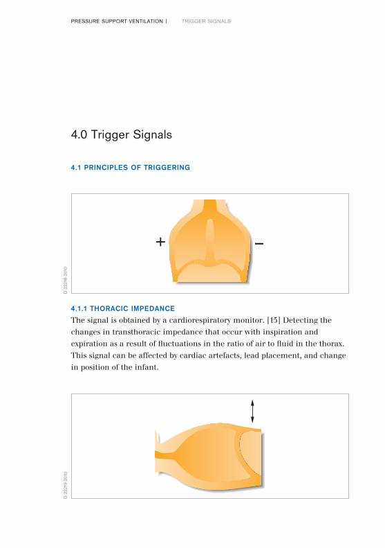
4.1.1 THORACIC IMPEDANCE
The signal is obtained by a cardiorespiratory monitor. [15] Detecting the changes in transthoracic impedance that occur with inspiration and expiration as a result of fluctuations in the ratio of air to fluid in the thorax. This signal can be affected by cardiac artefacts, lead placement, and change in position of the infant.
+ –
D-2
2218
-201
0D
-222
19-2
010
PRESSURE SUPPORT VENTILATION | TRIGGER SIGNALS
14|15
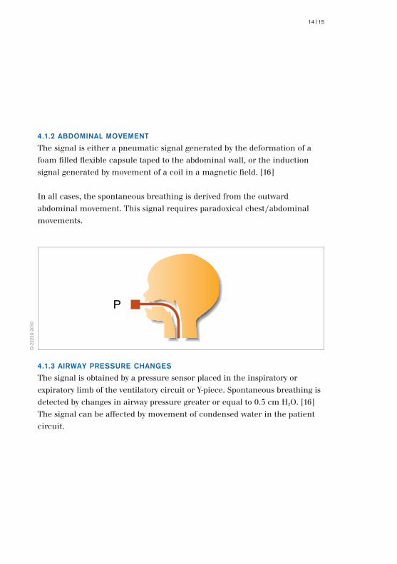
4.1.2 ABDOMINAL MOVEMENT
The signal is either a pneumatic signal generated by the deformation of a foam filled flexible capsule taped to the abdominal wall, or the induction signal generated by movement of a coil in a magnetic field. [16]
In all cases, the spontaneous breathing is derived from the outward abdominal movement. This signal requires paradoxical chest/abdominal movements.
P
D-2
2220
-201
0
4.1.3 AIRWAY PRESSURE CHANGES
The signal is obtained by a pressure sensor placed in the inspiratory or expiratory limb of the ventilatory circuit or Y-piece. Spontaneous breathing is detected by changes in airway pressure greater or equal to 0.5 cm H2O. [16] The signal can be affected by movement of condensed water in the patient circuit.
*
D-2
2221
-201
0
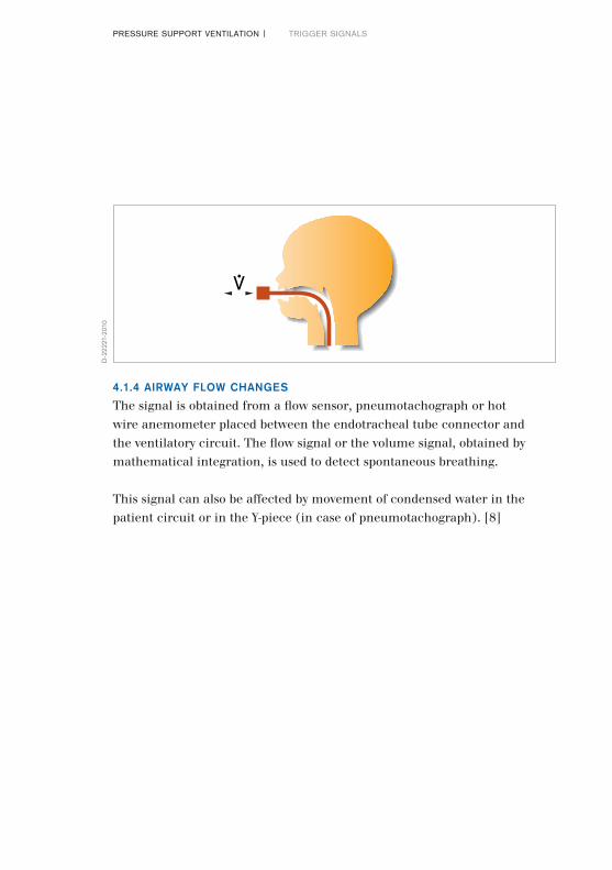
4.1.4 AIRWAY FLOW CHANGES
The signal is obtained from a flow sensor, pneumotachograph or hot wire anemometer placed between the endotracheal tube connector and the ventilatory circuit. The flow signal or the volume signal, obtained by mathematical integration, is used to detect spontaneous breathing.
This signal can also be affected by movement of condensed water in the patient circuit or in the Y-piece (in case of pneumotachograph). [8]
PRESSURE SUPPORT VENTILATION | TRIGGER SIGNALS
16|17
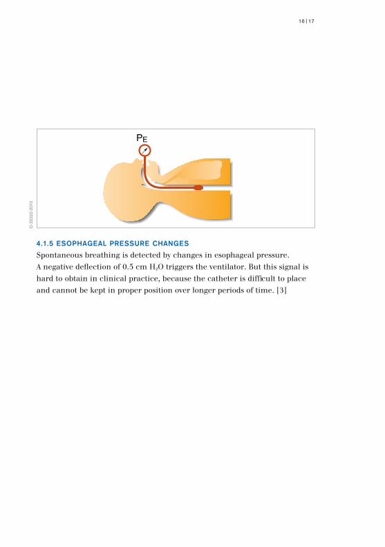
4.1.5 ESOPHAGEAL PRESSURE CHANGES
Spontaneous breathing is detected by changes in esophageal pressure. A negative deflection of 0.5 cm H2O triggers the ventilator. But this signal is hard to obtain in clinical practice, because the catheter is difficult to place and cannot be kept in proper position over longer periods of time. [3]
PE
D-2
2222
-201
0
4.2 SPECIFIC PROBLEMS WITH DIFFERENT TRIGGER SIGNALS
The possible problems with triggered ventilation in general are lack of response, autotriggering, artefacts, delayed response, and handling difficulties during clinical use, such as correct placement of sensors. [14,18]
4.2.1 LACK OF RESPONSE
Sometimes the spontaneous breathing is not detected by the trigger device. Then the trigger threshold may be too high, or the sensitivity of the trigger device too low. As a result, triggering may fail altogether or require unnecessarily high efforts (high work of breathing).
4.2.2 AUTOTRIGGERING
The ventilator is automatically triggered even without spontaneous breathing. Moving artefacts or an ET-tube leakage can cause autotriggering. Sometimes it is difficult to decide whether the patient is triggering the ventilator or the ventilator is autotriggering.
4.2.3 ARTEFACT
In general any type of artefact can disturb the detection of spontaneous breathing. Examples are abdominal movement in case of abdominal capsule sensor, peristalsis in case of esophageal pressure sensor (hiccup) or movement of water in the circuit in case of airway pressure sensor.
PRESSURE SUPPORT VENTILATION | TRIGGER SIGNALS
18|19
Flow
Paw
lack of response autotriggering artefacts antiphasic trigger delayed response
t
t
D-2
2595
-201
0
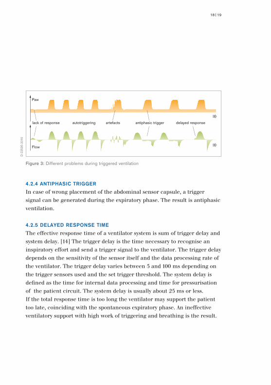
Figure 3: Different problems during triggered ventilation
4.2.4 ANTIPHASIC TRIGGER
In case of wrong placement of the abdominal sensor capsule, a trigger signal can be generated during the expiratory phase. The result is antiphasic ventilation.
4.2.5 DELAYED RESPONSE TIME
The effective response time of a ventilator system is sum of trigger delay and system delay. [14] The trigger delay is the time necessary to recognise an inspiratory effort and send a trigger signal to the ventilator. The trigger delay depends on the sensitivity of the sensor itself and the data processing rate of the ventilator. The trigger delay varies between 5 and 100 ms depending on the trigger sensors used and the set trigger threshold. The system delay is defined as the time for internal data processing and time for pressurisation of the patient circuit. The system delay is usually about 25 ms or less.If the total response time is too long the ventilator may support the patient too late, coinciding with the spontaneous expiratory phase. An ineffective ventilatory support with high work of triggering and breathing is the result.
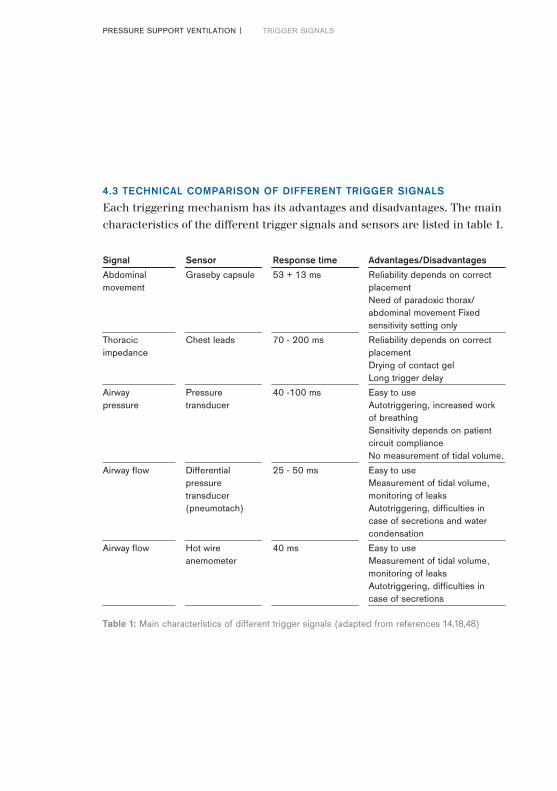
Table 1: Main characteristics of different trigger signals (adapted from references 14,18,48)
4.3 TECHNICAL COMPARISON OF DIFFERENT TRIGGER SIGNALS
Each triggering mechanism has its advantages and disadvantages. The main characteristics of the different trigger signals and sensors are listed in table 1.
PRESSURE SUPPORT VENTILATION | TRIGGER SIGNALS
Signal Sensor Responsetime Advantages/Disadvantages
Abdominal Graseby capsule 53 + 13 ms Reliability depends on correctmovement placement Need of paradoxic thorax/ abdominal movement Fixed sensitivity setting only
Thoracic Chest leads 70 - 200 ms Reliability depends on correct impedance placement Drying of contact gel Long trigger delay
Airway Pressure 40 -100 ms Easy to use pressure transducer Autotriggering, increased work
of breathing Sensitivity depends on patient circuit compliance No measurement of tidal volume.
Airway flow Differential 25 - 50 ms Easy to use pressure Measurement of tidal volume, transducer monitoring of leaks (pneumotach) Autotriggering, difficulties in case of secretions and water condensation
Airway flow Hot wire 40 ms Easy to use anemometer Measurement of tidal volume,
monitoring of leaks Autotriggering, difficulties in case of secretions
20|21
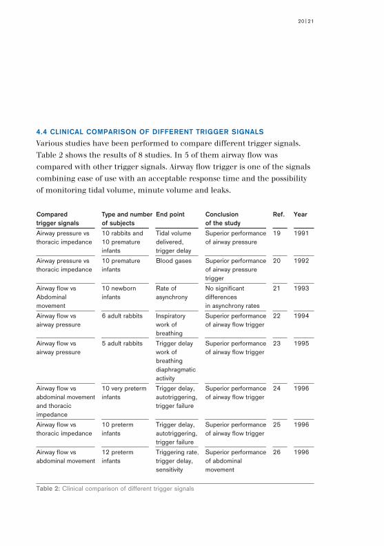
Compared Typeandnumber Endpoint Conclusion Ref. Yeartriggersignals ofsubjects ofthestudy
Airway pressure vs 10 rabbits and Tidal volume Superior performance 19 1991thoracic impedance 10 premature delivered, of airway pressure infants trigger delay
Airway pressure vs 10 premature Blood gases Superior performance 20 1992thoracic impedance infants of airway pressure trigger
Airway flow vs 10 newborn Rate of No significant 21 1993Abdominal infants asynchrony differences movement in asynchrony rates
Airway flow vs 6 adult rabbits Inspiratory Superior performance 22 1994airway pressure work of of airway flow trigger breathing
Airway flow vs 5 adult rabbits Trigger delay Superior performance 23 1995airway pressure work of of airway flow trigger breathing diaphragmatic activity
Airway flow vs 10 very preterm Trigger delay, Superior performance 24 1996abdominal movement infants autotriggering, of airway flow trigger and thoracic trigger failure impedance
Airway flow vs 10 preterm Trigger delay, Superior performance 25 1996thoracic impedance infants autotriggering, of airway flow trigger trigger failure
Airway flow vs 12 preterm Triggering rate, Superior performance 26 1996abdominal movement infants trigger delay, of abdominal sensitivity movement
Table 2: Clinical comparison of different trigger signals
4.4 CLINICAL COMPARISON OF DIFFERENT TRIGGER SIGNALS
Various studies have been performed to compare different trigger signals. Table 2 shows the results of 8 studies. In 5 of them airway flow was compared with other trigger signals. Airway flow trigger is one of the signals combining ease of use with an acceptable response time and the possibility of monitoring tidal volume, minute volume and leaks.
PRESSURE SUPPORT VENTILATION | DIFFERENT VENTILATION MODES
5.1 UNTRIGGERED MODES
During untriggered ventilation a ventilatory cycle occurs periodically at fixed intervals. It is strictly time cycled. There are two pressure controlled untriggered ventilation modes: CMV and IMV. The difference between Continuous Mandatory Ventilation (CMV = IPPV) and Intermittent Mandatory Ventilation (IMV) is only the difference in the set respiration rate of the ventilator:– During CMV the respiration rate of the ventilator is set faster than the
spontaneous respiratory rate (usually between 50 and 80 breaths per minute).
– During IMV the respiration rate of the ventilator is lower (less than 30 breaths per minute), thus between two controlled breaths, the baby can breathe spontaneously.
5.2 TRIGGERED MODES
Synchronized Intermittent Mandatory Ventilation (SIMV), Assist/Control (A/C) and Pressure Support Ventilation (PSV) are the three triggered ventilation modes used in neonatal ventilation. Assist/Control (A/C), Patient Triggered Ventilation (PTV) and Synchronized Intermittent Positive Pressure Ventilation (SIPPV) are different names for the same ventilatory mode. In the following sections of this booklet the term Assist/Control (A/C) is used. At least SIMV and A/C are available with modern neonatal ventilators.
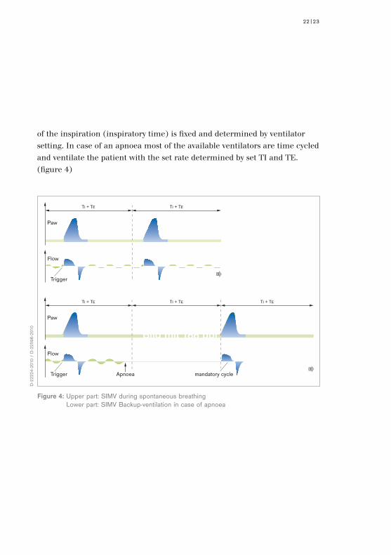
SIMV: The respiration rate of the ventilator is fixed to the set rate. Between the ventilator inflations the baby can breathe spontaneously on the positive end-expiratory pressure level. The set ventilator inflations per minute are synchronized with the spontaneous respiration of the baby. The duration
5. Different Ventilation Modes
TI + TE TI + TE TI + TE
mandatory cycleApnoea
Flow
Paw
Triggert
22|23
TI + TE TI + TE
Flow
Paw
Triggert
Figure 4: Upper part: SIMV during spontaneous breathing Lower part: SIMV Backup-ventilation in case of apnoea
D-2
2224
-201
0 /
D-2
2596
-201
0
Bild mit 188 ppi
of the inspiration (inspiratory time) is fixed and determined by ventilator setting. In case of an apnoea most of the available ventilators are time cycled and ventilate the patient with the set rate determined by set TI and TE. (figure 4)

Figure 5: Upper part: A/C during spontaneous breathing Lower part: A/C Backup ventilation in case of apnoea
TI + TE TI + TE TI + TE
Flow
Paw
Triggert
TI + TE TI + TE TI + TE
Apnoea mandatory cycle
Flow
Paw
Triggert
D-2
2226
-201
0 /
D-2
2597
-201
0
A/C: Each breath is assisted by the ventilator with a predefined level of pressure. The duration of the inspiration (inspiratory time) is fixed and determined by ventilator setting. The respiration rate can be controlled by the patient. In case of an apnoea most of the ventilators are time cycled and ventilate the patient with the set rate. (figure 5)
PRESSURE SUPPORT VENTILATION | DIFFERENT VENTILATION MODES

24|25
PSV: Each breath is assisted by the ventilator with a predefined level of pressure. The duration of the inspiration (inspiratory time) is automatically adjusted to the patient’s inspiratory time. Therefore the patient can control the respiration rate and the inspiratory time. (see also next chapter)
PSV + Volume Guarantee: This combination of Pressure Support Ventilation and Volume Guarantee (VG) allows the patient to control the rate and inspiratory time; at the same time a set target tidal volume is delivered by automatic adaptation of the pressure support level. The option Volume Guarantee is new type of volume controlled ventilation in neonates and is available with the Babylog 8000plus (for more details a separate booklet about Volume Guarantee is available).
Thus, during SIMV and A/C, inspiratory time is fixed and determined by ventilator setting. Despite an inspiratory trigger active expiration may occur in case of inadequately set inspiratory time TI.
UntriggeredModes TriggeredModes
CMV (IPPV) A/C (SIPPV, PTV)
IMV SIMV
PSV
PSV-VG
5.3 PRESSURE SUPPORT VENTILATION
5.3.1 DEFINITION
A ventilatory cycle during Pressure Support Ventilation consists of 4 phases.
1. phase: Recognition of the beginning of inspiration (trigger)2. phase: Pressurisation for the time of spontaneous inspiration3. phase: Recognition of the end of inspiration
(expiratory trigger or termination) and start of expiration4. phase: Expiration
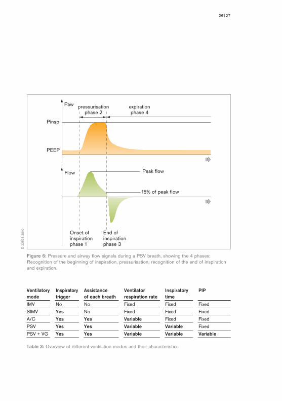
In the Babylog 8000plus the end of inspiration is determined by the diminution of inspiratory flow below 15 % of the peak inspiratory flow of the same cycle (figure 6). It is not necessary to adjust the termination criteria manually for adaptation to leak flow since the Babylog 8000plus is doing this automatically depending on the measured leakage (see also 5.3.2 Automatic Leak Adaptation).
During Pressure Support Ventilation each spontaneous breath is supported by the ventilator. Therefore respiration rate and duration of inspiration are controlled by the patient.
PRESSURE SUPPORT VENTILATION | DIFFERENT VENTILATION MODES
26|27
Peak flow
Onset of inspirationphase 1
End of inspirationphase 3
15% of peak flow
pressurisationphase 2
expirationphase 4
PEEP
Pinsp
Flow
Paw
t
t
D-2
2593
-201
0
Figure 6: Pressure and airway flow signals during a PSV breath, showing the 4 phases: Recognition of the beginning of inspiration, pressurisation, recognition of the end of inspiration and expiration.
Ventilatory Inspiratory Assistance Ventilator Inspiratory PIPmode trigger ofeachbreath respirationrate time
IMV No No Fixed Fixed Fixed
SIMV Yes No Fixed Fixed Fixed
A/C Yes Yes Variable Fixed Fixed
PSV Yes Yes Variable Variable Fixed
PSV + VG Yes Yes Variable Variable Variable
Table 3: Overview of different ventilation modes and their characteristics
5.3.2 AUTOMATIC LEAK ADAPTATION
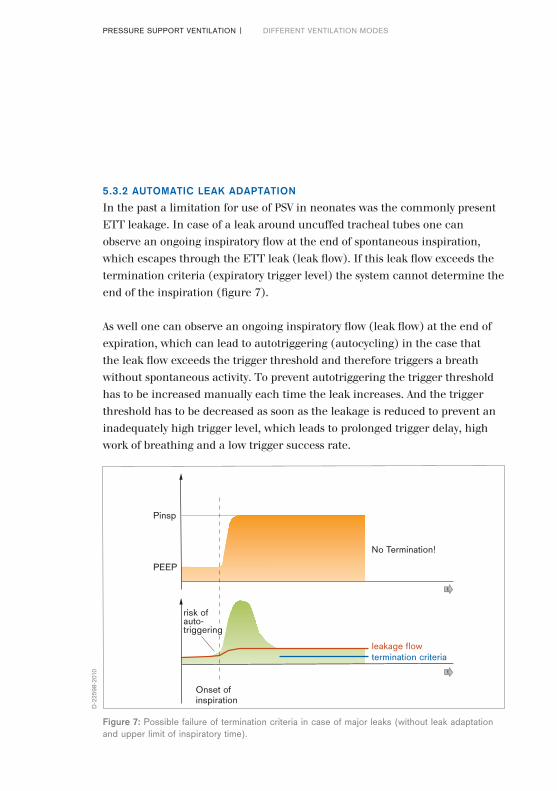
In the past a limitation for use of PSV in neonates was the commonly present ETT leakage. In case of a leak around uncuffed tracheal tubes one can observe an ongoing inspiratory flow at the end of spontaneous inspiration, which escapes through the ETT leak (leak flow). If this leak flow exceeds the termination criteria (expiratory trigger level) the system cannot determine the end of the inspiration (figure 7).
As well one can observe an ongoing inspiratory flow (leak flow) at the end of expiration, which can lead to autotriggering (autocycling) in the case that the leak flow exceeds the trigger threshold and therefore triggers a breath without spontaneous activity. To prevent autotriggering the trigger threshold has to be increased manually each time the leak increases. And the trigger threshold has to be decreased as soon as the leakage is reduced to prevent an inadequately high trigger level, which leads to prolonged trigger delay, high work of breathing and a low trigger success rate.
PRESSURE SUPPORT VENTILATION | DIFFERENT VENTILATION MODES
leakage flow
Onset of inspiration
PEEP
Pinsp
No Termination!
termination criteria
risk of auto-triggering
t
t
D-2
2598
-201
0
Figure 7: Possible failure of termination criteria in case of major leaks (without leak adaptation and upper limit of inspiratory time).
28|29
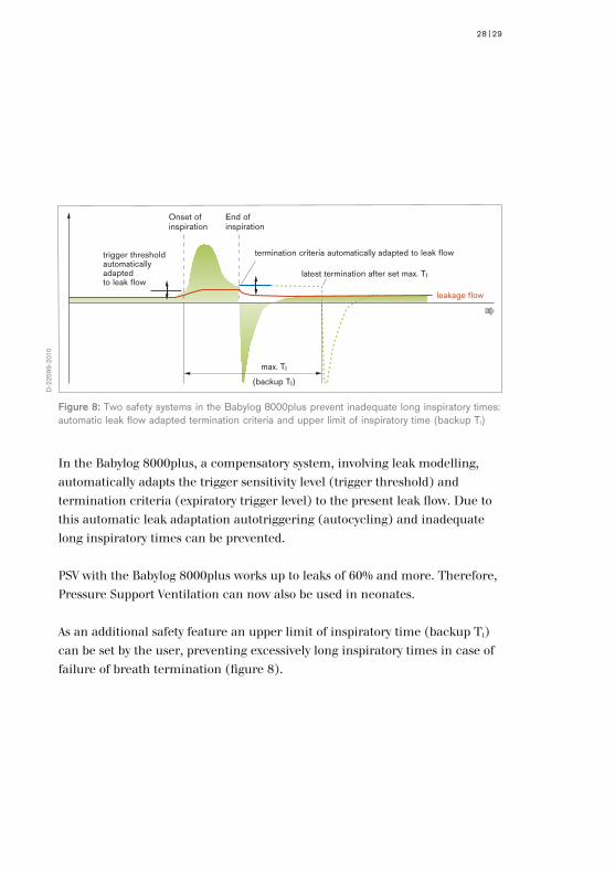
In the Babylog 8000plus, a compensatory system, involving leak modelling, automatically adapts the trigger sensitivity level (trigger threshold) and termination criteria (expiratory trigger level) to the present leak flow. Due to this automatic leak adaptation autotriggering (autocycling) and inadequate long inspiratory times can be prevented.
PSV with the Babylog 8000plus works up to leaks of 60% and more. Therefore, Pressure Support Ventilation can now also be used in neonates.
As an additional safety feature an upper limit of inspiratory time (backup TI) can be set by the user, preventing excessively long inspiratory times in case of failure of breath termination (figure 8).
Figure 8: Two safety systems in the Babylog 8000plus prevent inadequate long inspiratory times: automatic leak flow adapted termination criteria and upper limit of inspiratory time (backup TI)
max. TI
(backup TI)
Onset of inspiration
trigger threshold automatically adapted to leak flow
End of inspiration
termination criteria automatically adapted to leak flow
latest termination after set max. TI
t
leakage flow
D-2
2599
-201
0
5.3.3 BACKUP VENTILATION
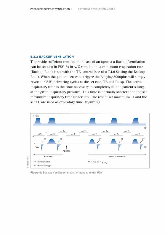
To provide sufficient ventilation in case of an apnoea a Backup Ventilation can be set also in PSV. As in A/C ventilation, a minimum respiration rate (Backup Rate) is set with the TE control (see also 7.1.6 Setting the Backup Rate). When the patient ceases to trigger the Babylog 8000plus will simply revert to CMV, delivering cycles at the set rate, TE and Pinsp. The active inspiratory time is the time necessary to completely fill the patient’s lung at the given inspiratory pressure. This time is normally shorter than the set maximum inspiratory time under PSV. The rest of set maximum TI and the set TE are used as expiratory time. (figure 9)
PRESSURE SUPPORT VENTILATION | DIFFERENT VENTILATION MODES
Flow
Paw
set TI set TI
set TE set TE
set TI set TIset TI
Spont. Resp.
f = patient controlled
ET = Expiratory Trigger
f = backup rate
Mandatory Ventilation
Apnoea
ET ET ET ET ET
set TE set TE
set TI
TI + TE=
1
t
t
D-2
2600
-201
0
Figure 9: Backup Ventilation in case of apnoea under PSV
30|31
5.3.4 LIMITATIONS AND CONTRA-INDICATIONS
Each individual patient requires his/her own optimal ventilation mode and parameters. Also Pressure Support Ventilation has its limitations and contra-indications. Bronchospasm and lack of spontaneous respiratory drive are two contra-indications for PSV.
In patients with low respiratory drive, Pressure Support Ventilation only has the advantage of automatic TI compared with Controlled Mandatory Ventilation (CMV) (see also 5.3.3 Backup Ventilation).
In case of bronchospasm, peak flow is decreased and inspiratory flow comes back to baseline very fast. Thus, spontaneous inspiration time is very short and the pressure support very brief while the newborn needs a higher level of support. This is a limitation for all ventilatory modes, that use the flow signal to adapt the ventilatory support (i.e. Proportional Assist Ventilation). [49]
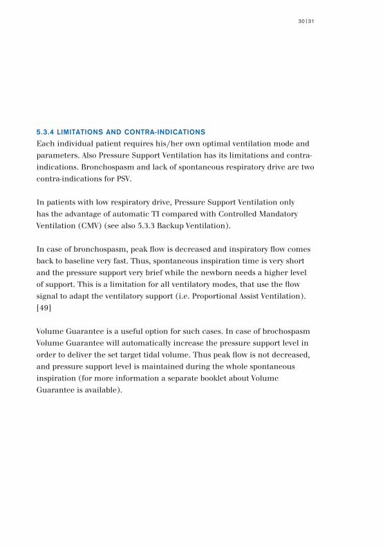
Volume Guarantee is a useful option for such cases. In case of brochospasm Volume Guarantee will automatically increase the pressure support level in order to deliver the set target tidal volume. Thus peak flow is not decreased, and pressure support level is maintained during the whole spontaneous inspiration (for more information a separate booklet about Volume Guarantee is available).
5.4 CLINICAL STUDIES COMPARING VENTILATION MODES
Many studies have been performed to compare different ventilatory modes. The layout and results of the main studies are represented in table 4.
PRESSURE SUPPORT VENTILATION | DIFFERENT VENTILATION MODES
D-2
2231
-201
0
Figure 10: Working principle of Volume Guarantee. According to a set tidal volume inspiratory pressure is automatically regulated by the ventilator.

32|33
Compared venti- Type and number End point Conclusion Ref. Yearlatory modes of subjects of the study
IMV vs CV neonates Duration of IMV > CV 27 1977 mechanical ventilation
SIMV vs CV 7 neonates Oxygenation, SIMV > CV 28 1994 Tidal volume, Work of breathing
SIMV vs IMV 30 neonates Consistancy of SIMV > IMV 29 1994 tidal volume
SIMV vs IMV neonates Oxygenation SIMV > IMV 30 1995
SIMV vs IMV 327 neonates Duration of SIMV > IMV 31 1996 mechanical in birth weight ventilation specific groups
SIMV vs IMV 77 neonates Duration of SIMV > IMV 32 1997 mechanical in premature ventilation, BPD neonates
A/C vs CV 14 neonates Oxygenation and A/C>CV 33 1993 blood pressure variations
A/C vs IMV 6 preterm infants Work of breathing A/C > IMV 34 1996
A/C vs IMV/CV 30 neonates 30 neonates A/C > IMV 57 1998 Adrenaline concentration
A/C vs IMV 40 preterm infants Duration of mechani- A/C > IMV 35 1993 cal ventilation
A/C vs SIMV 40 preterm infants Duration of weaning, A/C = SIMV 36 1994 Failure of weaning
A/C vs SIMV 2×40 preterm Duration of weaning A/C > SIMV 51 1995 infants at low rate (5/min), but not at 30/min
A/C vs SIMV 16 neonates Oxygen cost A/C > SIMV 37 1997 of breathing
PSV vs CV 15 neonates Cardiac output PSV > CV 38 1996
PSV vs CV+IMV 30 preterm infants Duration of mechani- PSV>CV+IMV 39 1994 cal ventilation
PSV vs IMV rabbit model Diaphragmatic activity PSV > IMV 40 1994 of neonate “>” means “superior to”
Table 4: Main clinical studies comparing different ventilatory modes in neonates.
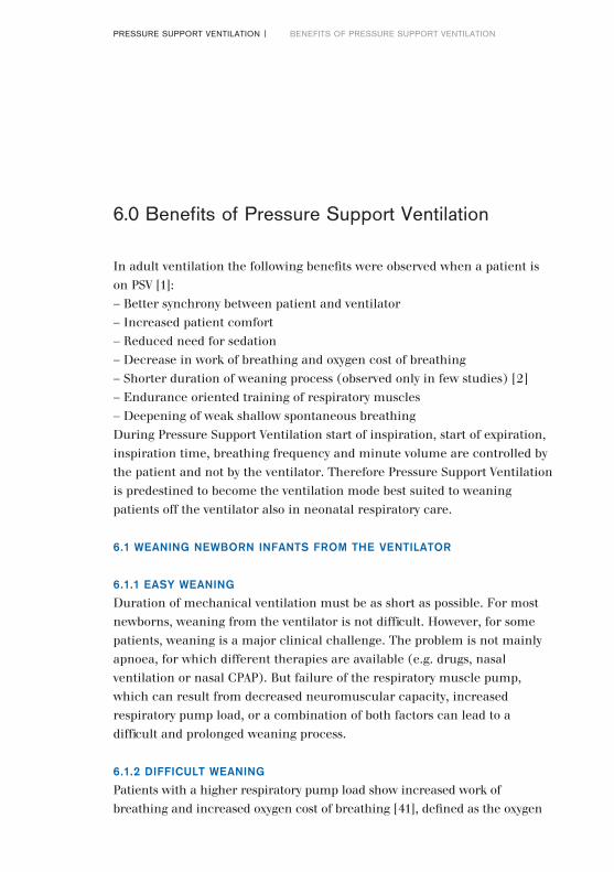
In adult ventilation the following benefits were observed when a patient is on PSV [1]:– Better synchrony between patient and ventilator– Increased patient comfort– Reduced need for sedation– Decrease in work of breathing and oxygen cost of breathing– Shorter duration of weaning process (observed only in few studies) [2]– Endurance oriented training of respiratory muscles– Deepening of weak shallow spontaneous breathingDuring Pressure Support Ventilation start of inspiration, start of expiration, inspiration time, breathing frequency and minute volume are controlled by the patient and not by the ventilator. Therefore Pressure Support Ventilation is predestined to become the ventilation mode best suited to weaning patients off the ventilator also in neonatal respiratory care.
6.1 WEANING NEWBORN INFANTS FROM THE VENTILATOR
6.1.1 EASY WEANING
Duration of mechanical ventilation must be as short as possible. For most newborns, weaning from the ventilator is not difficult. However, for some patients, weaning is a major clinical challenge. The problem is not mainly apnoea, for which different therapies are available (e.g. drugs, nasal ventilation or nasal CPAP). But failure of the respiratory muscle pump, which can result from decreased neuromuscular capacity, increased respiratory pump load, or a combination of both factors can lead to a difficult and prolonged weaning process.
6.1.2 DIFFICULT WEANING
Patients with a higher respiratory pump load show increased work of breathing and increased oxygen cost of breathing [41], defined as the oxygen
6.0 Benefits of Pressure Support Ventilation
PRESSURE SUPPORT VENTILATION | BENEFITS OF PRESSURE SUPPORT VENTILATION
34|35
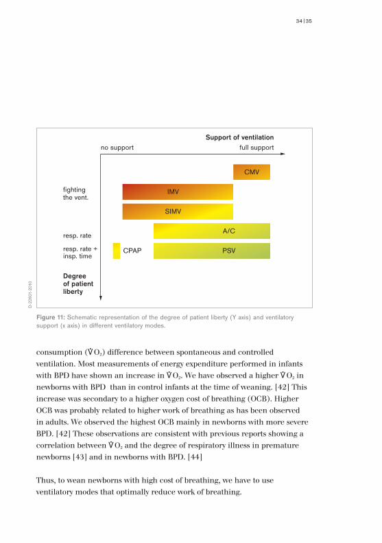
consumption (*O2) difference between spontaneous and controlled ventilation. Most measurements of energy expenditure performed in infants with BPD have shown an increase in *O2. We have observed a higher *O2 in newborns with BPD than in control infants at the time of weaning. [42] This increase was secondary to a higher oxygen cost of breathing (OCB). Higher OCB was probably related to higher work of breathing as has been observed in adults. We observed the highest OCB mainly in newborns with more severe BPD. [42] These observations are consistent with previous reports showing a correlation between *O2 and the degree of respiratory illness in premature newborns [43] and in newborns with BPD. [44]
Thus, to wean newborns with high cost of breathing, we have to use ventilatory modes that optimally reduce work of breathing.
fighting the vent.
no support
IMV
CMV
SIMV
A/C
PSVCPAP
full supportSupport of ventilation
resp. rate
resp. rate + insp. time
Degree of patient liberty
D-2
2601
-201
0
Figure 11: Schematic representation of the degree of patient liberty (Y axis) and ventilatory support (x axis) in different ventilatory modes.
6.2 WEANING STRATEGIES
6.2.1 SELECTION OF WEANING TYPE
There are two different types of weaning modes: Using IMV or SIMV the ventilatory respiration rate and the inspiratory pressure are reduced during the weaning process. Using A/C or PSV only the inspiratory pressure is reduced during weaning process. Some physiological and clinical studies can help to choose the right weaning strategy.
6.2.2 PHYSIOLOGICAL STUDIES
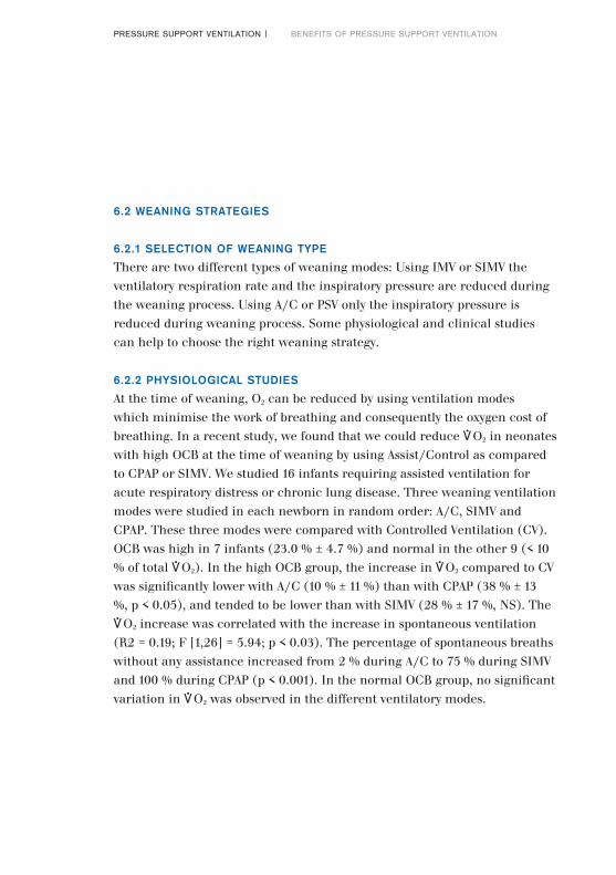
At the time of weaning, O2 can be reduced by using ventilation modes which minimise the work of breathing and consequently the oxygen cost of breathing. In a recent study, we found that we could reduce *O2 in neonates with high OCB at the time of weaning by using Assist/Control as compared to CPAP or SIMV. We studied 16 infants requiring assisted ventilation for acute respiratory distress or chronic lung disease. Three weaning ventilation modes were studied in each newborn in random order: A/C, SIMV and CPAP. These three modes were compared with Controlled Ventilation (CV). OCB was high in 7 infants (23.0 % ± 4.7 %) and normal in the other 9 (< 10 % of total *O2). In the high OCB group, the increase in *O2 compared to CV was significantly lower with A/C (10 % ± 11 %) than with CPAP (38 % ± 13 %, p < 0.05), and tended to be lower than with SIMV (28 % ± 17 %, NS). The *O2 increase was correlated with the increase in spontaneous ventilation (R2 = 0.19; F [1,26] = 5.94; p < 0.03). The percentage of spontaneous breaths without any assistance increased from 2 % during A/C to 75 % during SIMV and 100 % during CPAP (p < 0.001). In the normal OCB group, no significant variation in *O2 was observed in the different ventilatory modes.
PRESSURE SUPPORT VENTILATION | BENEFITS OF PRESSURE SUPPORT VENTILATION
36|37
In this study, we showed that the A/C mode can significantly reduce the increase in *O2 observed in some infants at the time of weaning as compared to SIMV or CPAP. A/C significantly reduced *O2 by 20 % in newborn infants with high OCB. [37] This decrease is probably related in part to reduced work of breathing as observed in adults [45] or children [46,50] during Pressure Support Ventilation. Besides, Jarreau et al showed that A/C reduces the work of breathing in premature infants. [34] Gullberg et al found PSV to increase cardiac output in neonates and infants in comparison to controlled ventilation. [38]Thus, A/C or rather PSV effect an endurance training of respiratory muscles (low pressure/ high volume work of breathing, consistent tidal volume). [47]
infants with high OCBinfants with normal OCB
Ventilation modes
*O
2 [m
l x m
in-1 x
kg-1]
0.0
5.0
10.0
15.0
20.0
CV PTV SIMV CPAP
D-2
2602
-201
0
Figure 12: Oxygen consumption for infants with high and normal oxygen cost of breathing in different ventilation modes. [37]
6.2.3 CLINICAL STUDIES
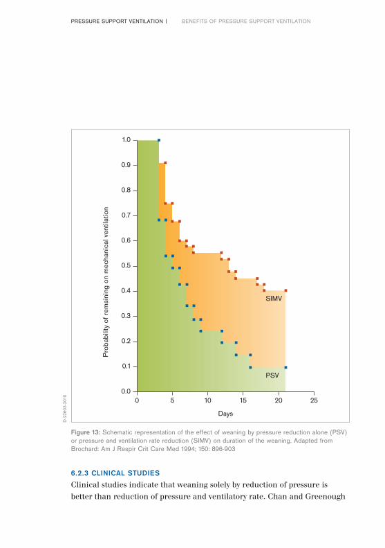
Clinical studies indicate that weaning solely by reduction of pressure is better than reduction of pressure and ventilatory rate. Chan and Greenough
PRESSURE SUPPORT VENTILATION | BENEFITS OF PRESSURE SUPPORT VENTILATION
Days
Pro
babi
lity
of r
emai
ning
on
mec
hani
cal v
entil
atio
n
0.0
0.1
0.2
0.3
0.4
0.5
0.6
0.7
0.8
0.9
1.0
0 5 10
SIMV
PSV
15 20 25
D-2
2603
-201
0
Figure 13: Schematic representation of the effect of weaning by pressure reduction alone (PSV) or pressure and ventilation rate reduction (SIMV) on duration of the weaning. Adapted from Brochard: Am J Respir Crit Care Med 1994; 150: 896-903

38|39
showed a reduction in weaning time using PTV instead of IMV. [35] Dimitriou et al found a shorter time of weaning using A/C in comparison to SIMV at low rates. [51] Jarreau et al demonstrated that PTV modifies the pattern of breathing by reduction of respiratory rate and decreases the WOB. [34] Donn et al compared IMV to PSV in a study of 30 preterm newborns (weight 1280 + 100 g, gest. age 29.5+1 wk.). They showed that patients treated with PSV were weaned more rapidly and had a significantly shorter mean time to extubation than those treated with conventional IMV. [39]
6.2.4 PSV IS BETTER THAN A/C!
Assist/Control is not Pressure Support Ventilation (PSV). During A/C, pressure is controlled, and each respiratory effort is assisted as in PSV. However, during A/C, inspiratory time is fixed by ventilator setting. During PSV, inspiratory time is adapted to the spontaneous inspiration of the patient. This prevents air trapping and inversion of the inspiratory /
0
10
20
30
40
50
0 0.1 0.2 0.3
36 w, 2.6 kg
0.4
0
10
20
30
40
50
0 0.1 0.2 0.3
26 w, 1.1 kg
0.4
0
10
20
30
40
50
0 0.1 0.2 0.3
35 w, 2.2 kg
0.4
0
10
20
30
40
50%
0 0.1 0.2 0.3
29 w, 0.8 kg
0.4
%%
%
sec
sec sec
sec
D-2
2604
-201
0
Bild 14: Häufigkeitsverteilung der Inspirationszeit bei 4 Frühgeborenen, die im PSV-Modus beatmet wurden. Der orangefarbene Balken zeigt die maximale Inspirationszeit, vorgegeben durch die Einstellung am Beatmungsgerät.
expiratory ratio when infants are breathing rapidly. We have observed a large variability (standard deviation x 100/mean) in the spontaneous inspiratory time of some newborns during PSV at the time of weaning, ranging from 19.3 to 24.2 % in 4 newborns during PSV (see figure 13). Thus PSV mode is probably more accurate than A/C because it adapts the pressure support to the patient’s spontaneous inspiratory time.
Due to technical limitations PSV for neonates was not available in the past. Now, a leak adapted PSV is available as an option for Babylog 8000plus.
6.2.5 PSV WITH VOLUME GUARANTEE
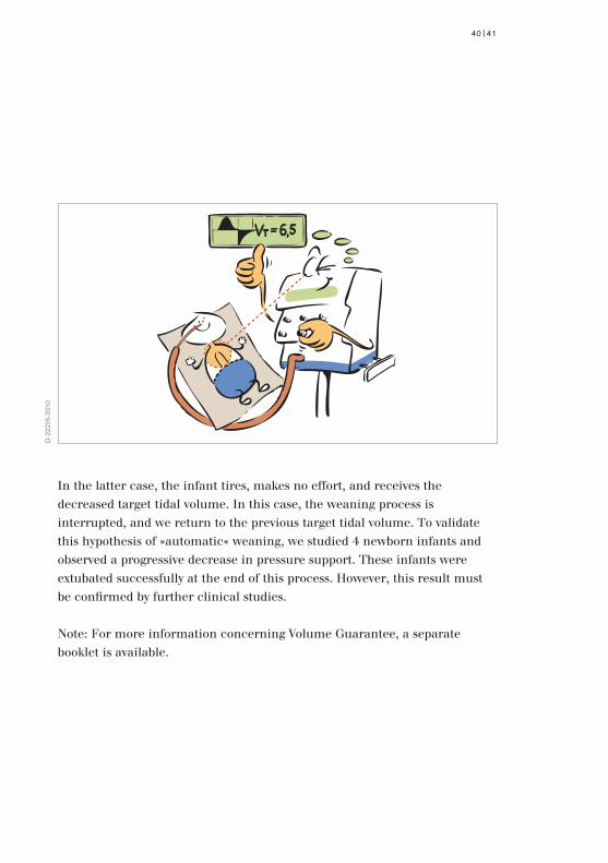
PSV with Volume Guarantee is an option available with the Babylog 8000plus. To wean patients with PSV, pressure support must be reduced, for example, from 20 to 4 mbar in 3-5 mbar steps. If PSV is used with Volume Guarantee, pressure will be regulated to deliver a target tidal volume defined by the ventilator setting. If we reduce target tidal volume, for example by 10 %, to begin the weaning process, there are two possibilities:
either the patient is ready to be weaned and will make an effort himself to maintain the delivered tidal volume at the initial value while the respirator gradually and automatically decreases the pressure support; or the patient is not yet ready to wean.
PRESSURE SUPPORT VENTILATION | BENEFITS OF PRESSURE SUPPORT VENTILATION
40|41
In the latter case, the infant tires, makes no effort, and receives the decreased target tidal volume. In this case, the weaning process is interrupted, and we return to the previous target tidal volume. To validate this hypothesis of »automatic« weaning, we studied 4 newborn infants and observed a progressive decrease in pressure support. These infants were extubated successfully at the end of this process. However, this result must be confirmed by further clinical studies.
Note: For more information concerning Volume Guarantee, a separate booklet is available.
D-2
2215
-201
0
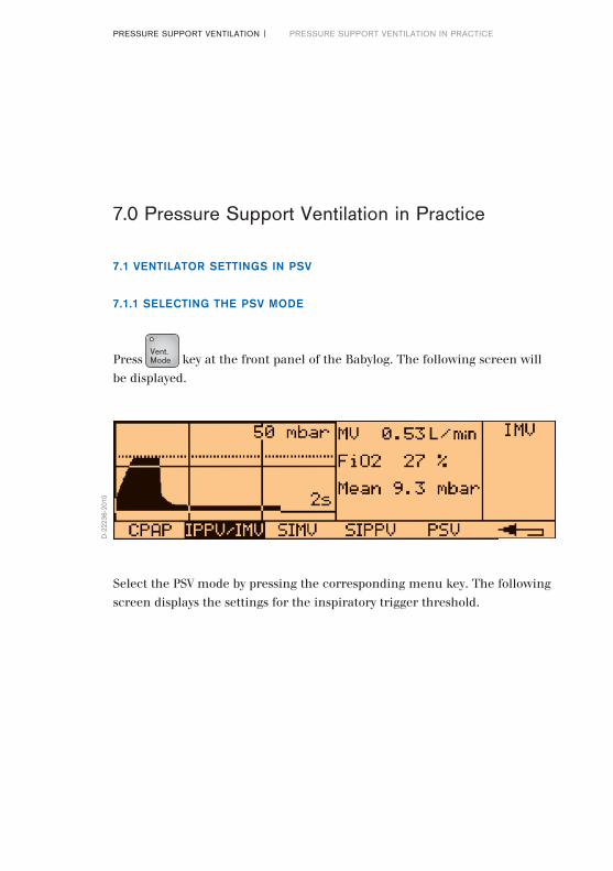
Select the PSV mode by pressing the corresponding menu key. The following screen displays the settings for the inspiratory trigger threshold.
7.1 VENTILATOR SETTINGS IN PSV
7.1.1 SELECTING THE PSV MODE
Press key at the front panel of the Babylog. The following screen will be displayed.
PRESSURE SUPPORT VENTILATION | PRESSURE SUPPORT VENTILATION IN PRACTICE
7.0 Pressure Support Ventilation in Practice
D-2
2236
-201
0
Vent. Mode
42|43
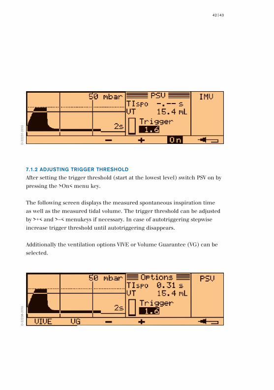
7.1.2 ADJUSTING TRIGGER THRESHOLD
After setting the trigger threshold (start at the lowest level) switch PSV on by pressing the >On< menu key.
The following screen displays the measured spontaneous inspiration time as well as the measured tidal volume. The trigger threshold can be adjusted by >+< and >–< menukeys if necessary. In case of autotriggering stepwise increase trigger threshold until autotriggering disappears.
Additionally the ventilation options VIVE or Volume Guarantee (VG) can be selected.
D-2
2237
-201
0D
-222
38-2
010
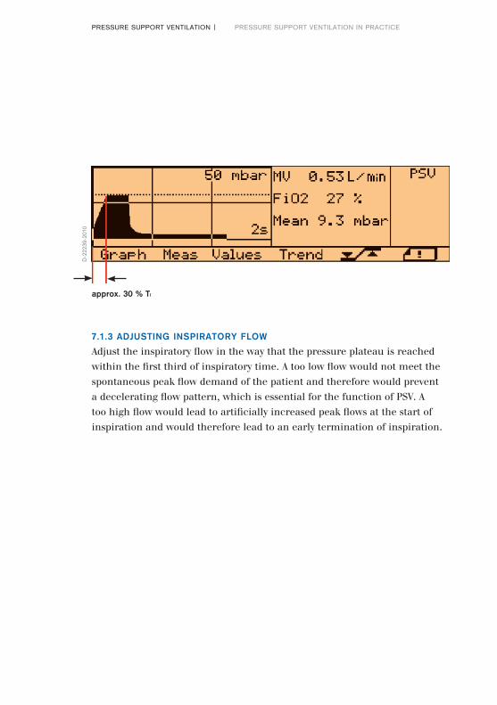
7.1.3 ADJUSTING INSPIRATORY FLOW
Adjust the inspiratory flow in the way that the pressure plateau is reached within the first third of inspiratory time. A too low flow would not meet the spontaneous peak flow demand of the patient and therefore would prevent a decelerating flow pattern, which is essential for the function of PSV. A too high flow would lead to artificially increased peak flows at the start of inspiration and would therefore lead to an early termination of inspiration.
PRESSURE SUPPORT VENTILATION | PRESSURE SUPPORT VENTILATION IN PRACTICE
D-2
2239
-201
0
approx. 30 % TI
44|45
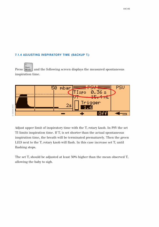
7.1.4 ADJUSTING INSPIRATORY TIME (BACKUP TI)
Press and the following screen displays the measured spontaneous inspiration time.
D-2
2240
-201
0
Adjust upper limit of inspiratory time with the TI rotary knob. In PSV the set TI limits inspiration time. If TI is set shorter than the actual spontaneous inspiration time, the breath will be terminated prematurely. Then the green LED next to the TI rotary knob will flash. In this case increase set TI until flashing stops.
The set TI should be adjusted at least 50% higher than the mean observed TI allowing the baby to sigh.
Vent. Mode

7.1.5 ADJUSTING INITIAL PRESSURE SUPPORT LEVEL
The pressure support level is adjusted by the Pinsp rotary knob. The initial pressure support level should be set to ensure a tidal volume of 4-6 ml/kg body weight.
D-2
2241
-201
0
PRESSURE SUPPORT VENTILATION | PRESSURE SUPPORT VENTILATION IN PRACTICE
7.1.6 SETTING THE BACKUP RATE
Starting at the main menu press the menu key >Values<. The following screen displays all the settings including frequency (rate). With the TE knob this Backup Rate can be set. In case of an apnoea controlled ventilation with the set Backup Rate starts (see also chapter 5.3.3).
D-2
2242
-201
0
46|47
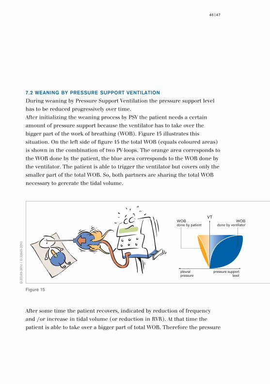
7.2 WEANING BY PRESSURE SUPPORT VENTILATION
During weaning by Pressure Support Ventilation the pressure support level has to be reduced progressively over time.After initializing the weaning process by PSV the patient needs a certain amount of pressure support because the ventilator has to take over the bigger part of the work of breathing (WOB). Figure 15 illustrates this situation. On the left side of figure 15 the total WOB (equals coloured areas) is shown in the combination of two PV-loops. The orange area corresponds to the WOB done by the patient, the blue area corresponds to the WOB done by the ventilator. The patient is able to trigger the ventilator but covers only the smaller part of the total WOB. So, both partners are sharing the total WOB necessary to gererate the tidal volume.
After some time the patient recovers, indicated by reduction of frequency and /or increase in tidal volume (or reduction in RVR). At that time the patient is able to take over a bigger part of total WOB. Therefore the pressure
D-2
2243
-201
0 /
D-2
2605
-201
0
Figure 15
WOBdone by patient
VTWOB
done by ventilator
pleuralpressure
pressure supportlevel
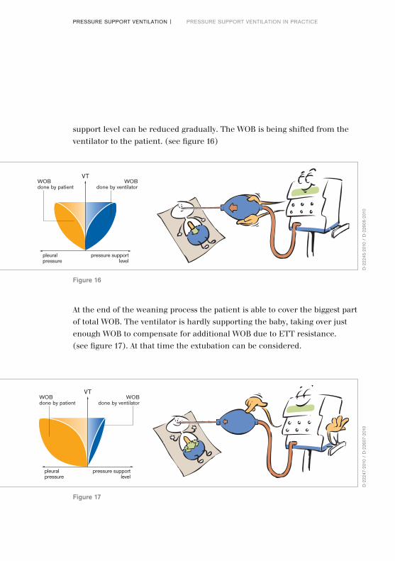
At the end of the weaning process the patient is able to cover the biggest part of total WOB. The ventilator is hardly supporting the baby, taking over just enough WOB to compensate for additional WOB due to ETT resistance. (see figure 17). At that time the extubation can be considered.
support level can be reduced gradually. The WOB is being shifted from the ventilator to the patient. (see figure 16)
PRESSURE SUPPORT VENTILATION | PRESSURE SUPPORT VENTILATION IN PRACTICE
D-2
2245
-201
0 /
D-2
2606
-201
0
Figure 16
VTWOBdone by patient
WOBdone by ventilator
pleuralpressure
pressure supportlevel
D-2
2247
-201
0 /
D-2
2607
-201
0
Figure 17
VTWOBdone by patient
WOBdone by ventilator
pleuralpressure
pressure supportlevel
48|49
7.3 MONITORING PRESSURE SUPPORT VENTILATION
Before we consider monitoring of Pressure Support Ventilation, we first have to remember the physiological background of respiratory control of the neonate.
7.3.1 PHYSIOLOGICAL BACKGROUND
7.3.1.1 CHEMICAL CONTROL
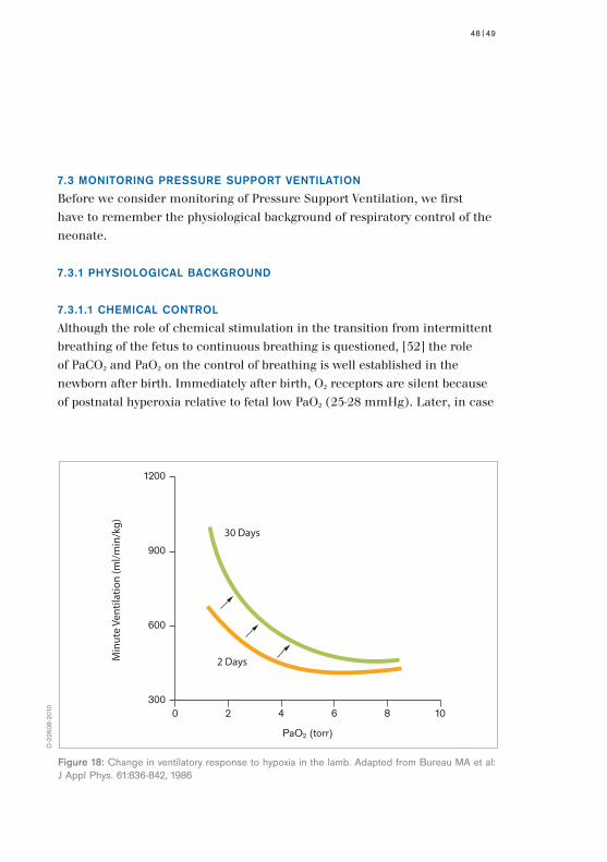
Although the role of chemical stimulation in the transition from intermittent breathing of the fetus to continuous breathing is questioned, [52] the role of PaCO2 and PaO2 on the control of breathing is well established in the newborn after birth. Immediately after birth, O2 receptors are silent because of postnatal hyperoxia relative to fetal low PaO2 (25-28 mmHg). Later, in case
PaO2 (torr)
Min
ute
Vent
ilatio
n (m
l/min
/kg)
300
1200
900
600
0 2 4 6
30 Days
2 Days
8 10
D-2
2608
-201
0
Figure 18: Change in ventilatory response to hypoxia in the lamb. Adapted from Bureau MA et al: J Appl Phys. 61:836-842, 1986
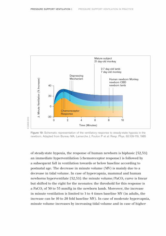
of steady-state hypoxia, the response of human newborn is biphasic [52,53]: an immediate hyperventilation (chemoreceptor response) is followed by a subsequent fall in ventilation towards or below baseline according to postnatal age. The decrease in minute volume (MV) is mainly due to a decrease in tidal volume. In case of hypercapnia, mammal and human newborns hyperventilate [52,53]: the minute volume/PaCO2 curve is linear but shifted to the right for the neonates: the threshold for this response is a PaCO2 of 50 to 55 mmHg in the newborn lamb. Moreover, the increase in minute ventilation is limited to 3 to 4 times baseline MV (in adults, the increase can be 10 to 20 fold baseline MV). In case of moderate hypercapnia, minute volume increases by increasing tidal volume and in case of higher
PRESSURE SUPPORT VENTILATION | PRESSURE SUPPORT VENTILATION IN PRACTICE
Time (Minutes)
∆ M
inut
e Ve
ntila
tion
(% In
crea
se)
-20
0
0 2
ChemoreceptorResponse
DepressingMechanism
Mature subject21 day-old monkey
2-7 day-old lamb7 day-old monkey
Human newborn Monkeynewborn CBDnewborn lamb
4 6 8 10
20
40
D-2
2609
-201
0
Figure 19: Schematic representation of the ventilatory response to steady-state hypoxia in the newborn. Adapted from Bureau MA, Lamarche J, Foulon P et al: Resp. Phys. 60:109-119, 1985

50|51
hypercapnia, tidal volume and frequency rate increase. However, increase of tidal volume can be limited by Hering Breuer reflex.
7.3.1.2 THE RESPIRATORY PUMP
The convection of gas into and out of the lung is secondary to the movement of rib cage and the excursion of the diaphragm with each contraction and relaxation phase. Chest wall muscles are critical for ventilation in the newborn with a pliable chest wall. Thus the main effector is the diaphragm. But neonatal diaphragm is different to adults by anatomical and histological aspects. Thus, the diaphragm of the neonate is prone to fatigue. [53,54]
7.3.1.3 OXYGEN CONSUMPTION, CARBON DIOXIDE PRODUCTION AND
WORK OF BREATHING
Running the respiratory pump has an energy cost. In case of increased work of breathing, the cost of oxygen consumption and carbon dioxide production increases. The consequence of that is an increase in minute ventilation and increase in work of breathing. This vicious circle must be cut by medical intervention giving adequate respiratory support.
7.3.1.4 PULMONARY REFLEXES
The most important reflex, particularly during neonatal period is the Hering-Breuer reflex. This reflex is initiated by lung distension and terminates inspiration and prolongs expiration time. Thus, increases in lung volume can cause apnoea. This reflex is much more pronounced in newborns, especially in newborn with low lung compliance. It is believed to be important as a protective mechanism against respiratory fatigue due to ineffective muscle work [53,55] and probably also against volutrauma.
7.3.1.5 PATTERN OF RESPIRATION IN NEONATES WITH RDS
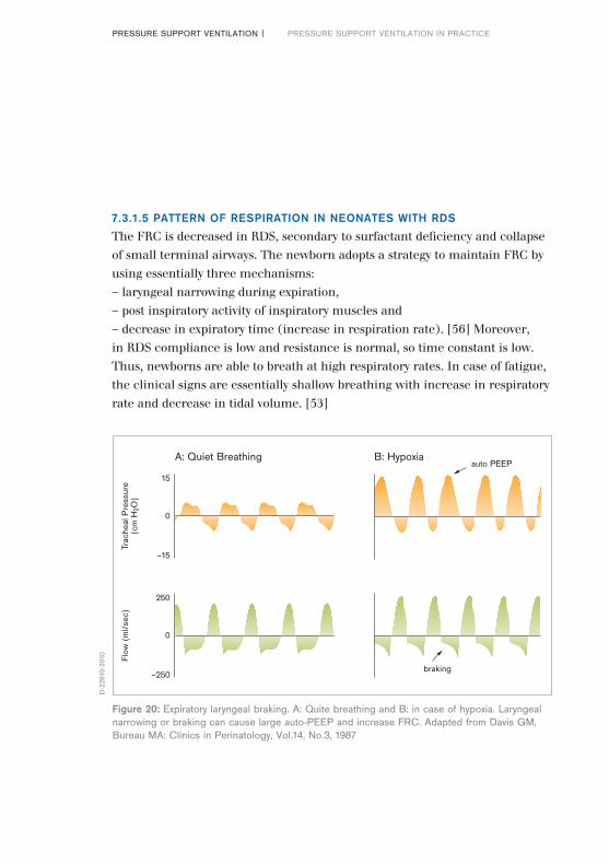
The FRC is decreased in RDS, secondary to surfactant deficiency and collapse of small terminal airways. The newborn adopts a strategy to maintain FRC by using essentially three mechanisms:– laryngeal narrowing during expiration,– post inspiratory activity of inspiratory muscles and– decrease in expiratory time (increase in respiration rate). [56] Moreover, in RDS compliance is low and resistance is normal, so time constant is low. Thus, newborns are able to breath at high respiratory rates. In case of fatigue, the clinical signs are essentially shallow breathing with increase in respiratory rate and decrease in tidal volume. [53]
PRESSURE SUPPORT VENTILATION | PRESSURE SUPPORT VENTILATION IN PRACTICE
15
Trac
heal
Pre
ssur
e(c
m H
2O)
0
–15
250
Flow
(m
l/se
c)
0
–250braking
A: Quiet Breathing B: Hypoxiaauto PEEP
D-2
2610
-201
0
Figure 20: Expiratory laryngeal braking. A: Quite breathing and B: in case of hypoxia. Laryngeal narrowing or braking can cause large auto-PEEP and increase FRC. Adapted from Davis GM, Bureau MA: Clinics in Perinatology, Vol.14, No.3, 1987
52|53
Hypercapnia
increase pressure difference
Pinsp (↑) PEEP (↓)
Ventilation (↑)Patients WOB (↓)
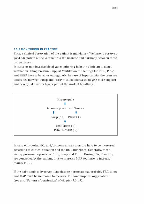
7.3.2 MONITORING IN PRACTICE
First, a clinical observation of the patient is mandatory. We have to observe a good adaptation of the ventilator to the neonate and harmony between these two partners.Invasive or non-invasive blood gas monitoring help the clinician to adapt ventilation. Using Pressure Support Ventilation the settings for FiO2, Pinsp and PEEP have to be adjusted regularly. In case of hypercapnia, the pressure difference between Pinsp and PEEP must be increased to give more support and hereby take over a bigger part of the work of breathing.
In case of hypoxia, FiO2 and/or mean airway pressure have to be increased according to clinical situation and the unit guidelines. Generally, mean airway pressure depends on TI, TE, Pinsp and PEEP. During PSV, TI and TE are controlled by the patient, thus to increase MAP you have to increase mainly PEEP.
If the baby tends to hyperventilate despite normocapnia, probably FRC is low and MAP must be increased to increase FRC and improve oxygenation. (see also ‘Pattern of respiration’ of chapter 7.3.1.5).

RVR (↑)
too much WOB for the patient
Pinsp (↑)
Moreover, during weaning, we can look at the respiratory rate / tidal volume ratio (Rate Volume Ratio = RVR). As with fatigue, newborns decrease tidal volume and increase respiratory rate and therefore increase the RVR. This ratio is a weaning criteria with high sensitivity and high specificity to predict successful extubation in adults (area under the ROC curve is 0.89). [41] As yet, there are no data for neonates available, but the rate volume ratio (RVR) can be used very well also for monitoring the weaning process in neonates. Based on the neonatal physiology, a gradual increase in RVR can indicate a beginning fatigue of the patient. In this case an increase of pressure support level (Pinsp) might be necessary.
PRESSURE SUPPORT VENTILATION | PRESSURE SUPPORT VENTILATION IN PRACTICE
Hypoxia
FiO2 (↑) MAP (↑)
PEEP (↑)
FRC (↑)
Oxygenation (↑)
54|55
D-2
2252
-201
0
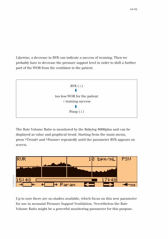
Likewise, a decrease in RVR can indicate a success of weaning. Then we probably have to decrease the pressure support level in order to shift a further part of the WOB from the ventilator to the patient.
The Rate Volume Ratio is monitored by the Babylog 8000plus and can be displayed as value and graphical trend. Starting from the main menu, press >Trend< and >Param< repeatedly until the parameter RVR appears on screen.
RVR (↓)
too less WOB for the patient= training success
Pinsp (↓)
Up to now there are no studies available, which focus on this new parameter for use in neonatal Pressure Support Ventilation. Nevertheless the Rate Volume Ratio might be a powerful monitoring parameter for this purpose.
Demonstrated AdvantagesPressure Support Ventilation is a pressure controlled ventilation mode now available for neonatology. This mode gives the patient optimum control during ventilation. The patient decides over start of inspiration and start of expiration and therefore controls inspiration time, breathing frequency and minute volume.
Technology behindPressure Support Ventilation has been extensively studied in adults. Many advantages have been observed: better synchrony between patient and ventilator, increased comfort of the patient, endurance oriented training of respiratory muscles and shorter duration of weaning in some clinical studies. The neonatal Pressure Support Ventilation of the Babylog 8000plus was specially designed for use in neonates and covers the specific problems of this patient population.
Set up operationPressure Support Ventilation is available at a fingertip and can be easily set up. After adjusting trigger sensitivity and maximum inspiration time an initial pressure support level can be set according to patient weight. Respiration rate, tidal volume and the Rate Volume Ratio (RVR) together with blood gases can be used to monitor PSV and further adapt ventilation parameters.
PRESSURE SUPPORT VENTILATION | CONCLUSION
8.0 Conclusion
56|57
Benefits for the neonateIn newborns, physiological and clinical studies have shown the benefits of triggered ventilation, and the superiority of Assist/Control mode over Synchronous Intermittent Mandatory Ventilation during weaning. Pressure Support Ventilation supports spontaneous breathing in a unique and harmonious way and is thus predestined to become the ventilation mode best suited to weaning patients off the ventilator. During weaning, Pressure Support Ventilation shows promising advance in reducing work of breathing in patients with high oxygen cost of breathing.
The first clinical trials show encouraging results. Nevertheless further clinical studies have to confirm these first experiences, despite the difficulties and the great number of patients to be included.

PRESSURE SUPPORT VENTILATION | APPENDIX
9.0 Appendix
9.1 CASE REPORTS
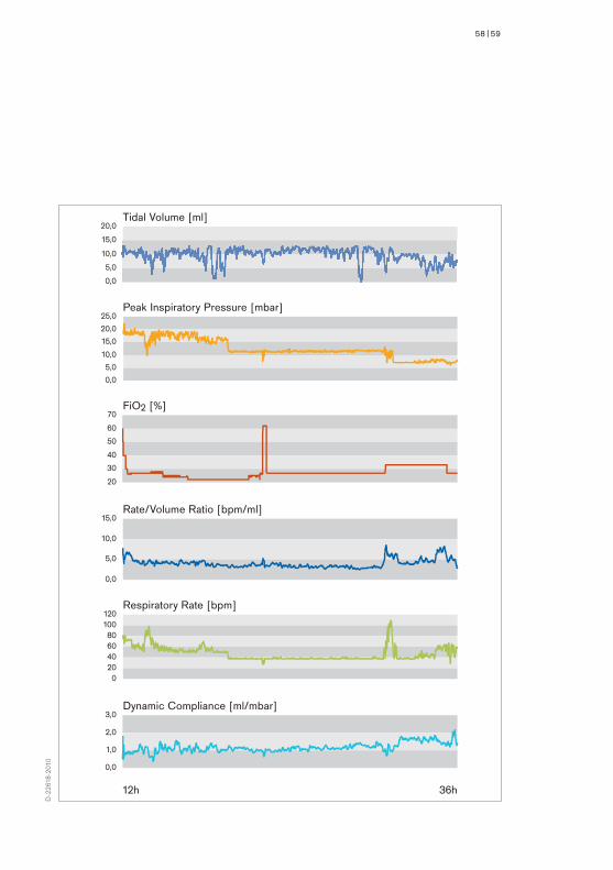
Case 1:Infant female GA 36 weeks, Birthweight 2060 g, with moderate respiratory distress syndrome. Weaning process was performed by use of PSV mode and gradual reduction of the pressure support level only.
Low FiO2 and the Rate/Volume Ratio were stable during the whole weaning process. The baby was weaned off the ventilator with success at the end of this process. The duration of mechanical ventilation was 1 day. The figures show the trend of peak inspiratory pressure, FiO2, RVR, tidal volume, respiratory rate and compliance during the last 24 hours.
58|59
c
Tidal Volume [ml]
Peak Inspiratory Pressure [mbar]
FiO2 [%]
Rate/Volume Ratio [bpm/ml]
Respiratory Rate [bpm]
Dynamic Compliance [ml/mbar]
12h 36h
0,0
5,0
10,0
15,0
20,0
0,0
5,0
10,0
15,0
20,0
25,0
20
30
40
50
60
70
0,0
5,0
10,0
15,0
020406080
100120
0,0
1,0
2,0
3,0
D-2
2618
-201
0
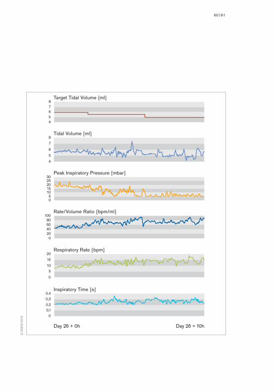
Case 2:Infant, male, GA 26 weeks, Birthweight 860 g with severe respiratory distress syndrome and later bronchopulmonary dysplasia. Weaning process was performed at day 26 in PSV mode with Volume Guarantee option. Target tidal volume was initially set to 6 ml and was later gradually decreased to 5 ml.
During the weaning period the peak inspiratory pressure was automatically reduced from 20 mbar to 5 mbar. Parallely inspiratory time as well as respiratory rate increased. Rate/Volume Ratio increased, probably initiated by reduction of pressure support level and therefore an increase in respiratory rate and in fatigue of this tiny baby. Nevertheless the RVR stabilised at a higher level. The tidal volume was relatively stable over the whole weaning period. As the baby could maintain a good minute ventilation at a very low pressure support level, we tried to extubate the baby after 10 hours of weaning. This trial was successful. The baby received supplementary oxygen for 5 days more.
PRESSURE SUPPORT VENTILATION | APPENDIX
60|61
Tidal Volume [ml]
Peak Inspiratory Pressure [mbar]
Rate/Volume Ratio [bpm/ml]
Respiratory Rate [bpm]
Inspiratory Time [s]
Target Tidal Volume [ml]
45678
4
5
6
7
8
05
1015202530
020406080
100
0
5
10
15
20
0
0,10,2
0,3
0,4
Day 26 + 0h Day 26 + 10h
D-2
2619
-201
0

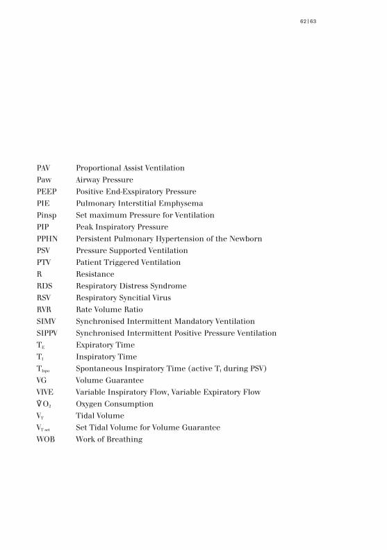
9.2 ABBREVIATIONS
A/C Assist Control VentilationBPD Bronchopulmonary DysplasiaC ComplianceCDH Congenital Diaphramatic HerniaCLD Chronic Lung DiseaseCMV Controlled Mandatory VentilationCPAP Continuous Positive Airway PressureCV Controlled VentilationEEG Electro EncephalogramETT Endotracheal Tubef Ventilation FrequencyFiO2 Fraction of Inspiratory O2 ConcentrationFRC Functional Residual CapacityICH Intracranial HaemorrhageI:E Inspiratory-to-Exspiratory RatioIMV Intermittent Mandatory VentilationIPPV Intermittent Positive Pressure Ventilationkg Kilogram bodyweightLED Light Emitting DiodesMAP Mean Airway PressureMV Minute VolumeNS Not SignificantOCB Oxygen Cost of Breathing
PRESSURE SUPPORT VENTILATION | APPENDIX
62|63
PAV Proportional Assist VentilationPaw Airway PressurePEEP Positive End-Exspiratory PressurePIE Pulmonary Interstitial EmphysemaPinsp Set maximum Pressure for VentilationPIP Peak Inspiratory PressurePPHN Persistent Pulmonary Hypertension of the NewbornPSV Pressure Supported VentilationPTV Patient Triggered VentilationR ResistanceRDS Respiratory Distress SyndromeRSV Respiratory Syncitial VirusRVR Rate Volume RatioSIMV Synchronised Intermittent Mandatory VentilationSIPPV Synchronised Intermittent Positive Pressure VentilationTE Expiratory TimeTI Inspiratory TimeTIspo Spontaneous Inspiratory Time (active TI during PSV)VG Volume GuaranteeVIVE Variable Inspiratory Flow, Variable Expiratory Flow*O2 Oxygen ConsumptionVT Tidal VolumeVT set Set Tidal Volume for Volume GuaranteeWOB Work of Breathing

[1] Brochard L. Pressure Support Ventilation. In Tobin eds, Principles and practice of mechanical ventilation, McGraw-Hill, New York 1994. p 239-253.
[2] Bernstein G. Patient triggered ventilation using cutaneous sensors. Semin Neonatol 1997; 2: 89-97
[3] Mancebo J. Weaning from mechanical ventilation. Eur Respir J 1996; 9: 1923-31.
[4] Field D, Milner AD, Hopkin IE. Inspiratory time and tidal volume during intermittent positive pressure ventilation. Arch Dis child 1985; 60: 259-61.
[5] Bernstein G, Heldt GP, Mannino FL. Increased and more consistent tidal volumes during synchronized intermittent mandatory ventilation in newborn infants. Am J Respir Crit Care Med 1994; 150: 1444-8.
[6] Greenough A., Morley C J, Davis J A. Pancuronium prevents pneumotorax in ventilated babies who actively expire against positive pressure inflation. Lancet 1984 ; i, 1-3.
[7] Perlman JM, McMenamin JB, Volpe JJ. Fluctuating cerebral blood flow velocity in respiratory distress syndrome. N Engl J Med 1983; 309: 204-9.
[8] Greenough A, Milner A. Patient triggered ventilation using flow or pressure sensors. Semin Neonatol 1997; 2: 99-104
10.0 References
PRESSURE SUPPORT VENTILATION | REFERENCES
64|65
[9] Perlman JM, Goodman S, Kreusser KL, Volpe JJ. Reduction in intraventricular hemorrhage by elimination of fluctuating cerebral blood flow velocity in respiratory distress syndrome. N Engl J Med 1985; 312: 1353-7.
[10] Runkle B, Bancalari E. Acute cardiopulmonary effects of pancuronium bromide in mechanically ventilated newborn infants. J Pediatr 1984; 104: 614-7.
[11. Ruteldge ML, Hawkins EP, Langston C. Skeletal muscle growth failure induced in premature newborn infants by prolonged pancuronium treatment. J Pediatr 1986: 883-6.
[12. Miller J, Law AB, Parker RA, Sundell H, Silberberg AR, Cotton RB. Effects of morphine and pancuronium on lung volume and oxygenation in premature infants with hyaline membrane disease. Pediatrics 1994; 125: 97-103
[13] Field D, Milner AD, Hopkin IE. Manipulation of ventilator settings to reduce expiration against positive pressure inflation. Arch DisChild 1985; 60: 1036-40
[14] Heldt G., Berstein G. Patient initiated mechanical ventilation. In New therapies for neonatal respiratory failure. Ed. B.R.Boynton, W.A. Carlo, A.H. Jobe. Cambridge university press. Cambridge 1994. 152-170.
[15] Visveshwara N, Freeman B, Peck M, Caliwag W, Shook S, Rajani KB. Patient-triggered synchronized assisted ventilation of newborns. Report of a preliminary study and three years‘ experience J Perinatol 1991; 4: 347-54.
[16] Nikischin W, Gerhardt T, Everett R, Gonzalez A, Hummler H, Bancalari E. Patient triggered ventilation: a comparison of tidal volume and chestwall and abdominal motion as trigger signals. Pediatr Pulmonol 1996; 22: 28-34.
[17] Greenough A, Hird MF, Chan V. Airway pressure triggered ventilation for preterm neonates. J Perinat Med 1991; 19: 471-6
[18] Donn SM, Sinha SK. Controversies in patient triggered ventilation. Clinics Perinat 1998; 25: 49-61.
[19] Hird MF, Greenough A. Patient triggered ventilation using a flow triggered system. Arch Dis Child 1991, 66: 1140-1142.
[20] Chan V, Greenough A. Evaluation of triggering systems for patient triggered ventilation for neonates ventilator-dependent beyond 10 days of age. Eur J Pediatr 1992; 151: 842-5
[21] Bernstein G, Cleary JP, Heldt GP, Rosas JF, Schellenberg LD, Mannino FL. Response time and reliability of three neonatal patient-triggered ventilators . Am Rev Respir Dis 1993; 148: 358-64.
[22] Nishimura M, Imanaka H, Yoshiya I, Kacmarek RM. Comparison of inspiratory work of breathing between flow-triggered and pressure-triggered demand flow systems in rabbits. Crit Care Med 1994; 22: 1002-9.
[23] Uchiyama A, Imanaka H, Taenaka N, Nakano S, Fujino Y, Yoshiya I. A comparative evaluation of pressure-triggering and flow-triggering in pressure support ventilation (PSV) for neonates using an animal model. Anaesth Intensive Care 1995; 23: 302-6.
PRESSURE SUPPORT VENTILATION | REFERENCES
66|67
[24] Brochard. Comparison of three methods of gradual withdrawl from mechanical ventilation. Am J Respir. Crit Care Med 1994; 150; 896-903.
[25] Hummler HD, Gerhardt T, Gonzalez A, Bolivar J, Claure N, Everett R, Bancalari E . Patient-triggered ventilation in neonates: comparison of a flow-and an impedance-triggered system Am J Respir Crit Care Med 1996; 154: 1049-54.
[26] Laubscher B, Greenough A, Kavadia V. Comparison of body surface and airway triggered ventilation in extremely premature infants. Acta Paediatr 1997 Jan; 86: 102-4
[27] Kirby RR. Intermittent mandatory ventilation in the neonate. Crit Care Med 1977; 5: 18-22.
[28] Mizuno K, Takeuchi T, Itabashi K, Okuyama K. Efficacy of synchronized IMV on weaning neonates from the ventilator. Acta Paediatr Jpn 1994; 36: 162-6
[29] Quinn MW, De Boer RC, Ansari N, Baumer JH. Stress response and mode of ventilation in preterm infants. Arch Dis Child 1998, 78: F195-8.
[30] Cleary JP, Bernstein G, Mannimo FL, Heldt GP. Improved oxygenation during synchronized intermittent mandatory ventilation in neonates with respiratory distress syndrome: a randomized crossover study. J Pediatr 1995: 126: 407-11.
[31] Bernstein G, Mannimo FL, Heldt GP, Callahan JD, Bull DH, Sola A, Ariagno RL, Hoffman GL, et al. Randomized multicenter trial comparing synchronized and conventional intermittent mandatory ventilation in neonates. J pediatr 1996; 128: 453-63.
[32] Chen JY, Ling UP, Chen JH. Comparison of synchronized and conventionan intermittent mandatory ventilation in neonates. Acta Paediatr Jpn 1997; 39: 578-83.
[33] Amitary M, Etches PC, Finner NN, Maidens JM. Synchronous mechanical ventilation of the neonates with respiratory disease. Crit Care Med 1993; 21: 118-24.
[34] Jarreau PH, Moriette G, Mussat P, Mariette C, Mohanna A, Harf A, Lorino H. Patient-triggered ventilation decreases the work of breathing in neonates. Am J Respir Crit Care Med 1996; 153: 1176-1181.
[35] Chan V, Greenough A. Randomised controlled trial of weaning by patient triggered ventilation or conventional ventilation. Eur J Pediatr 1993 Jan; 152(1): 51-4
[36] Chan V, Greenough A. Comparison of weaning by patient triggered ventilation or synchronous intermittent mandatory ventilation in preterm infants. Acta Paediatr 1994; 83: 335-7.
[37] Roze JC, Liet JM, Gournay V, Debillon T, Gaultier C. Oxygen cost of breathing and weaning process in newborn infants. Eur Respir J 1997;10: 2583-5
[38] Gullberg N, Winberg P, Sellden H. Pressure Support Ventilation increases cardiac output in neonates and infants. Paediatr Anaesth 1996; 6: 311-5
[39] Donn SM, Nicks JJ, Becker MA. Flow-synchronized ventilation of preterm infants with respiratory distress syndrome. J Perinatol 1994; 14: 90-4.
PRESSURE SUPPORT VENTILATION | REFERENCES

68|69
[40] Uchiyama A, Imanaka H, Taenaka N, Nakano S, Fujino Y, Yoshiya I. Comparative evaluation of diaphragmatic activity during pressure support ventilation and intermittent mandatory ventilation in animal model. Am J Respir Crit Care Med 1994; 150: 1564-8.
[41] Tobin MJ, Alex CG. Discontinuation of mechanical ventilation. In Tobin eds, Principles and practice of mechanical ventilation, McGraw-Hill, New York 1994 p 1177-1205.
[42] Rozé JC, Chambille B, Fleury MA, Debillon T, Gaultier C. Oxygen cost of breathing in newborn infants with long term ventilatory support. J Pediatr. 1995; 127:984-987.
[43] Wahlig TM, Gatto CW, Boros SJ, Mammel MC, Mills MM, Georgieff MK. Metabolic response of preterm infants to variable degrees of respiratory illness. J Pediatr. 1994; 124: 283-288.
[44] Abman SH, Groothius JR. Pathophysiology and treatment of bronchopulmonary dysplasia. Clin Pediatr. 1994; 41: 277-315.
[45] Brochard L, Harf A, Lorino H, Lemaire F. Inspiratory pressure support prevents diaphragmatic fatigue during weaning from mechanical ventilation. Am Rev Respir Dis 1989;139: 513-21.
[46] El-Khatib MF, Chatburn RL, Potts DL, Blumer JL, Smith PG. Mechanical ventilators optimized for pediatric use decrease work of breathing and oxygen consumption during pressure-support ventilation. Crit Care Med 1994; 22: 1942-48.
[47] MacIntyre NR. Respiratory function during pressure support ventilation Chest 1986; 89: 677-83
[48] Liubsys A, Norsted T, Jonzon A, Sedin G. Trigger delay in infant ventilators. Ups J Med Sci 1997;102:109-9.
[49] Schulze A, Schaller P. Assisted mechanical ventilation using resistive and elestic unloading. Semin Neonatal 1997; 2: 105-14.
[50] Tokioka H, Kinjo M, Hirakawa M. The effectiveness of pressure support ventilation for mechanical ventilatory support in children. Anesthesiology 1993; 78: 880-5.
[51] Dimitriou G, Greenough A, Griffin F, Chan V. Synchronous intermittent mandatory ventilation modes compared with patient triggered ventilation during weaning. Arch Dis Child Fetal Neonatal Ed 1995; 72: F188-90.
[52] Rigatto H. Control of breathing in fetal life and onset and control of breathing in the neonate. In Polin and Fox, Eds, Fetal and neonatal physiology, second edition, Philadelphia 1998, p11118-29.
[53] Davis GM, Bureau MA. Pulmonary and chest wall mechanics in the control of respiration in the newborn. Clinics in perinatalogy 1987; 14: 551-79.
[54] Mortola JP, Mechanics of breathing. In Polin and Fox Eds, Fetal and neonatal physiology, second edition, Philadelphia 1998, p 1118-29.
[55] Mortola JP, Fisher JT, Smith B, Fox G, Weeks SS. Dynamics of breathing in infants. J Appl Physiol 1982; 52: 1209-15.
[56] Martin RJ, Okken A, Katona PG, Klaus MH. Effect of lung volume on expiratory time in the newborn infant. J Appl Physiol 1978; 45 : 18-23
PRESSURE SUPPORT VENTILATION | REFERENCES

70|71
HEADQUARTERSDrägerwerk AG & Co. KGaAMoislinger Allee 53–5523558 Lübeck, Germany
www.draeger.com
REGION EUROPE CENTRAL AND EUROPE NORTHDrägerwerk AG & Co. KGaA Moislinger Allee 53–5523558 Lübeck, GermanyTel +49 451 882 0Fax +49 451 882 [email protected]
REGION EUROPE SOUTHDräger Médical S.A.S. Parc de Haute Technologie d’Antony 225, rue Georges Besse92182 Antony Cedex, FranceTel +33 1 46 11 56 00Fax +33 1 40 96 97 [email protected]
REGION MIDDLE EAST, AFRICA, CENTRAL AND SOUTH AMERICADrägerwerk AG & Co. KGaABranch Office DubaiDubai Healthcare City P.O. Box 505108Dubai, United Arab EmiratesTel + 971 436 24 762Fax + 971 436 24 [email protected]
REGION ASIA / PACIFICDraeger Medical South East Asia Pte Ltd25 International Business Park#04-27/29 German CentreSingapore 609916, SingaporeTel +65 6572 4388Fax +65 6572 4399 [email protected]
REGION NORTH AMERICADraeger Medical, Inc.3135 Quarry Road Telford, PA 18969-1042, USATel +1 215 721 5400Toll-free +1 800 437 2437Fax +1 215 723 [email protected]
90 9
7 49
9 |
15.0
7-2
| C
omm
unic
atio
ns &
Sal
es M
arke
ting
| P
P |
LE
| P
rinte
d in
Ger
man
y |
Sub
ject
to m
odifi
catio
ns |
© 2
015
Drä
gerw
erk
AG &
Co.
KG
aA