pressure is on 2 hr sd handout
TRANSCRIPT

4/13/21
1
The Pressure is On:Managing Challenging Cases in Glaucoma
Mark T. Dunbar, OD, FAAOBascom Palmer Eye Institute
Miami, FL
1
Mark Dunbar OD: Financial Disclosure
• Optometry Consultant– Carl Zeiss– Allergan– Regeneration– Genentech
• Advisory Board for:– Allergan – Carl Zeiss – Regeneron– Genentech
Mark Dunbar does not own stock in any of the above companies
2
Full Disclosure
• Nothing I say in this lecture will have been an original thought• I have shamelessly copied and pillaged almost every thought or
concept discussed in this talk
3
General Principles in Glaucoma Management
• The higher the IOP, the greater the risk of acquiring glaucomatous damage and the greater the risk for progression
• There are factors other then IOP that contribute to ON damage and determine an individual susceptibility to harm from IOP
• At the moment, there is no effective treatment for glaucoma other then lowering IOP
4
What other factors besides IOP contribute to glaucomatous ON damage?
5
80 yo White Female• Presented for “annual eye exam”
6

4/13/21
2
80 yo White Female
Oct 2017
7 8
9 10
So now what?
Is this low/normal tension glaucoma?
Would you begin treatment today?
11
Would you begin treating on this visit?
1. Yes2. No3. I would refer to ophthalmologist
12

4/13/21
3
This is what I did…
13
A Few Thoughts on Disc Heme
14
In the OHTS Study what percentage of disc hemorrhages were MISSED by the doctor?
1. 24%2. 44%3. 64%4. 84%
15
• Disc hemorrhages detected in 128 eyes of 123 participants
• 21 cases detected by both doctor and photos• 107 cases (84%) were detected only by a
review of photography
Ophthalmology Dec 2006
16
Perhaps the Bigger Question?
• How is it that a patient can continue to “progress” or develop a disc hemorrhage with a pressure ~ 12?
• What are the factors that result in progression?
17
• Blood pressure– High vs Low blood pressure (BP)
• Reduced ocular blood flow– Lower ocular perfusion pressure (OPP0
• Intracranial pressure (ICP)
What other factors besides IOP contribute to glaucomatous ON damage?
18

4/13/21
4
Ischemia vs. Mechanical• Ischemia: elevated IOP reduces blood flow to
the optic nerve, thus creating chronic ischemia within the neural tissue– Local ischemia-hypoxia develops ? dysfunction of
blood flow autoregulation
• Mechanical: damage due to the mechanical affects of the elevated IOP – “Pressure” on the ON/Lamina – Ganglion cell axons undergo deformation and
mechanical stress
FORCE (IOP)
19
Where Does Blood Pressure Fit In?
20
Where Does Blood Pressure Fit In?
• 1990’s: Hayreh, Drance, and others 1st raised the important issues of systemic hypotension and nocturnal blood pressure dips in the progression of glaucoma
• The problem: difficult to measure systemic BP during sleeping hours
21
When is the highest IOP during the 24-hour cycle?
• IOP is a dynamic physiological parameter that doesn't remain constant over the course of 24 hours
• Trough IOP levels tend to occur at the end of the waking period
• Peak IOP is usually recorded at the end of the nocturnal sleep period
Liu JH, Kripke DF, Twa M D, et al. Twenty-four hour pattern of intraocular pressure in the aging population. Invest Ophthalmol Vis Sci 1999;40:2912-2917.
22
Risk Factors For Progression
• BP is lowest at night• IOP is highest during the night time– Highest prior to waking
• Combination of é IOP and ê BP may result in a critical ê ocular perfusion pressure (OPP) in susceptible people– Patients with with faulty autoregulation
Mosaed S, Liu JH, Weinreb RN. Correlation between office and peak nocturnal intraocular pressures in healthy subjects and glaucoma patients. Am J Ophthalmol 2005;139:2:320-4.
23
Ocular Perfusion Pressure (OPP)
• OPP is the relative pressure at which blood enters the eye• Defined as the ocular arterial pressure minus the IOP• OPP is a delicate balance between IOP and blood pressure• Low ophthalmic perfusion pressure (OPP) is a risk factor for
progression– Low BP and/or high IOP
MOPP = 2/3 X [DBP + 1/3 X (SBP – DBP)] – IOPSimple: Diastolic BP – IOP = OPP
24

4/13/21
5
Low OPP and Glaucoma
1. Tielsch JM, et al. Arch Ophthalmol. 1995;113(2):216-221. 2. Bonomi L, et al. Ophthalmology. 2000;107(7):1287-1293. 3. Quigley HA, et al. Arch Ophthalmol. 2001;119(12):1819-1826. 4. Memarzadeh F, et al. Invest Ophthalmol Vis Sci. 2010;51(6):2872-2877. 5. Leske MC, et al. Arch Ophthalmol. 2002;120(7):954-959.
* Glaucoma risk from low DPP vs high DPP.
E p id e m io lo g ic S tu d ie s L in k in g D ia s to l ic P e r fu s io n P r e s s u r e a n d G la u c o m a
S tu d y D e s ig n P a r t ic ip a n tsGlaucoma Risk From Low DPP vs Normal DPP
B a l t im o r e E y e S u r v e y 1 C ro s s -s e c t io n a l N o n -H is p a n ic W h i te s a n d A f r ic a n A m e r ic a n s 6-fo ld
E g n a -N e u m a r k t S tu d y 2 C ro s s -s e c t io n a l N o n -H is p a n ic W h i te s 2.5-fo ld*
P r o y e c to V E R 3 C ro s s -s e c t io n a l H is p a n ic s 4-fo ld
L o s A n g e le s L a t in o E y eS tu d y 4 C ro s s -s e c t io n a l H is p a n ic s 1.9-fo ld
B a r b a d o s E y e S tu d y 5 L o n g i tu d in a l A f ro -C a r ib b e a n s3.2-fo ld
(4 years)
Abbreviation: DPP, diastolic perfusion pressure.
25
Low OPP and Glaucoma
26
27
Vascular Supply to the ON• COMPLEX arterial supply and an even more complex
venous drainage system• Which vascular network is most critical for development
of glaucoma?
28
OPP and Glaucoma – The Reality• Perfusion pressure is difficult to accurately measure• There is currently no widely accepted consensus
regarding which techniques should be used to evaluate blood flow or how the results should be interpreted
• None of the methods used to estimate blood flow have been standardized or externally validated for humans
• Ocular blood flow measurements are not currently used in the diagnosis or management of patients with glaucoma
29
CSFp and Glaucoma
30

4/13/21
6
CSF (ICP) causes more displacement of the lamina then IOP
31
2008
32
Mayo Clinic Study: CSF and Glaucoma
• Retrospective review of 31,786 patients that had lumbar punctures over a 11-year period
• Determined # who had complete eye exams • 28 met inclusion criteria of POAG, 49 controls• ICP was significantly lower in patients with POAG compared to
the non-glaucoma control
Berdahl JP, et al. Ophthalmology. 2008;115(5):763-768.
33
Berdahl 2nd Mayo Clinic Study: CSF and GlaucomaPOAG vs. NTG vs OHT
• Retrospective review of 62,468 patients that had lumbar punctures over a 20-year period
• 189 met inclusion criteria of complete eye exam• ICP was significantly lower in patients with POAG and NTG
and significantly higher in OHT
Berdahl JP, Fautsch MP, Stinnett SS, et al Intracranial pressure in primary open angle glaucoma, normal tension glaucoma, and ocular hypertension: a case-control study. Invest Ophthalmol Vis Sci. 2008;49(12):5412-5418
34
35
Lumbar CSF Pressure in NTG, POAG and Non GL
36

4/13/21
7
Trans-Lamina Cribrosa Pressure Difference
37
Atmospheric Intraocular Intracranial
760 mmHG 776 mmHg 772 mmHg
Normal
Abso
lute
Pre
ssur
e
Abso
lute
Pre
ssur
e
Trans-Corneal Pressure Difference16 mmHg
Trans-LaminarPressure Difference4 mmHg
Normal IOP Normal CSFp
38
39
FORCE (ICP)FORCE (IOP)
Basic Physics
Corneal Hysteresis May play a Role
IOP CSF
40
• In the normal state IOP and CSF have minimal trans-laminar pressure differences
• Increasing the difference alters the homeostatic balance and results pressure gradient difference at the lamina
Relationship between IOP and CSF
41
Atmospheric Intraocular Intracranial
760 mmHg 782 mmHg 769 mmHg
Glaucoma
Abso
lute
Pre
ssur
e
Abso
lute
Pre
ssur
e
Trans-Corneal Pressure Difference22 mmHg
Trans-LaminarPressure Difference13 mmHg
High IOP Mid-Low CSFp
(Low CSF pressure results in higherTrans-laminar pressure difference)
Lower CSPp
42

4/13/21
8
Atmospheric Intraocular Intracranial
760 mmHg 782 mmHg 769 mmHg
Treatment in Glaucoma
Abso
lute
Pre
ssur
e
Abso
lute
Pre
ssur
e
Trans-Corneal Pressure Difference22 mmHg
Trans-LaminarPressure Difference13 mmHg
Lowered atmospheric
Lowering intraocular pressure Reducing Tran-Laminar difference
43
Atmospheric Intraocular Intracranial
760 mmHG 782 mmHg 769 mmHg
Treatment in Glaucoma
Abso
lute
Pre
ssur
e
Abso
lute
Pre
ssur
e
Trans-Corneal Pressure Difference22 mmHg
Trans-LaminarPressure Difference13 mmHg
Lowered atmospheric
Lowering intraocular pressureReducing trans-laminar difference
776 mmHg
16 mmHg6
44
45
60% of astronauts suffer ocular problems related to prolonged space travel
• Globe flattening• Hyperopic shift• Choroidal folds• Disc swelling
46
47
FORCE (ICP)FORCE (IOP)
Basic Physics
Corneal Hysteresis May play a Role
IOP CSF
Zero Gravity
48

4/13/21
9
Atmospheric Intraocular Intracranial
760 mmHG 776 mmHg 800 mmHg
Papilledema
Abso
lute
Pre
ssur
e
Abso
lute
Pre
ssur
e
Trans-Corneal Pressure Difference16 mmHg
Trans-LaminarPressure Difference- 24 mmHg
Normal IOP High CSFp
49 50
3
Posterior Scleral Biomechanics and the Translaminar Pressure Difference.Fleischman, David; Berdahl, John
International Ophthalmology Clinics. 54(1):73-94, Winter 2014.DOI: 10.1097/IIO.0b013e3182aabef4
51
Glaucoma Today
52
Compromised Autoregulation in Glaucoma
• Autoregulation: The body’s ability to regulate itself in the presence of change– Vascular factors
• Cardiovascular disease
• Vasospasm
– Postural changes– Atmospheric pressure– Temperature– Fatigue can lead to abnormal pressure-flow relationship
• Periods of ischemia are then more likely to occur– Can result in reduced or fluctuating OPP
53
AutoregulationOr Vascular Dysregulation…?
• Can lead to over/under perfusion• Chronic under perfusion can lead to tissue necrosis
and death• Unstable perfusion leads to oxidative stress
54
4/13/21
10
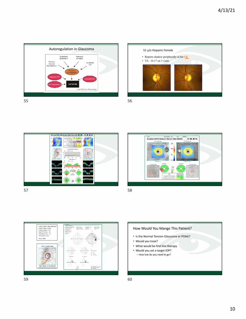
Autoregulation in Glaucoma
55
51 y/o Hispanic Female
• Reports shadow peripherally in her LE• TA: 16-17 on 3 visits
56
57 58
59
How Would You Mange This Patient?
• Is the Normal Tension Glaucoma or POAG?• Would you treat?• What would be first line therapy• Would you set a target IOP?– How low do you need to go?
60

4/13/21
11
51 y/o Hispanic FemaleShadow in LE
• Lumigan 0.01 added – qhs RE• Returned 3 weeks later – not aware of shadow– TA 12/17
• RTC – 4 months
61
Case MC• 73 yo female presents for follow up: GL
Suspect • Past history single elevated IOP• BCVA 20/25 and 20/20• IOP 21 RE 19 LE; – CCT 560u R 565u L
• Anterior segment normal• Mild NS and cortical cataracts
62
The ON• Small optic discs OU• RE c/d ~ 0.6 but– Appeared saucerized infero temporally– Broadening of a vein as it crossed edge disc – ? Small disc hemorrhage
• LE c/d .35
63
Photos
64
65 66

4/13/21
12
67
Summary• Suspicious disc• Borderline IOP• Normal visual field • Normal OCT *• What would you do
68
This was her 15 months earlier
8/2012
69
This was her 15 months earlier
8/2012 10/2013
Is this Significant?
70
Is that change significant?
71
How much change needs to occur on an OCT RNFL for it to be significant?
72

4/13/21
13
How much change needs to occur on an OCT RNFL for it to be significant?
• 5 microns• 10 microns• 20 microns• 25 microns
73
Detecting Glaucoma Progression Using OCT - RNFL
RNFL Thinning on OCTPatient able to do a Reliable VF?
Is thinning > 10 microns
Glaucoma has progressed
Is the thinning > 5 microns?
Progression not confirmedRepeat studies at appropriate intervals
No
No
Is there VF worsening and/ordisc hemorrhage that corresponds to the area of thinning?
yes
yesyes
No
74
• We can measure multiple steps of statistically significant change while a glaucoma suspect still is in the green normal range.
Values shown are for a 69 year old normal.
Fortunately, SDOCT measurements are highly reproducible.
50th percentile = 89 microns
5th percentile = 75 microns
95th percentile = 107 microns
Risk of Disability <50 microns
1st percentile = 67 microns
Normal significance Limits for Average RNFLT
75
Values shown are for a 69 year old normal.3
What This Means For Everyday Clinical Care
50th percentile = 89 microns
5 th percentile = 75 microns
95th percentile = 107 microns
Risk of Disability 50 microns
1st percentile = 67 microns
•Implication 1: SDOCT can now measure 2 to 4 statistically significant RNFL progression steps for the typical glaucoma suspect while the patient is still is in the green zone.
•Implication 2: It may be possible to view SDOCT change from baseline as an early detection strategy in glaucoma suspects.
76
77
• At 95% specificity, up to 35% of eyes had abnormal average RNFL thickness 4 years before development of visual field loss and 19% of eyes had abnormal results 8 years before field loss.
• Conclusions: Assessment of RNFL thickness with OCT was able to detect glaucomatous damage before the appearance of VF defects on SAP. In many subjects, significantly large lead times were seen when applying OCT as an ancillary diagnostic tool.
78

4/13/21
14
Arturo: 50 y/o Russian Male7/25/12• RK 1991 -> 20/20 with hyperopic correction:
+4.00 +1.50X180• TA: 32/18• Pach– 544 μ– 558 μ
• Gonio -CBB
79 80
81
What Would You Do Now?
• Obviously he needs a VF
• But would you start treatment now -> on THIS visit?
82
Would you treat Arturo on this initial visit with IOP 32/18 and normal OCT?
1. Yes2. No
83
20 years of the OHTS
OHTS 11994
OHTS 22002
OHTS 22009
OHTS 32016-2019
84

4/13/21
15
Ocular Hypertension Treatment Study (OHTS)
! Long-term randomized, multicentered controlled, clinical trial
! 1500 OHT pts with moderate risk for POAG randomized! Observation vs stepped medical therapy
! 5 yr minimum follow up! Pts seen 2X/year for IOP check and HVF
85
Ocular Hypertension Treatment Study (OHTS)
! 30-40 clinical centers! Each center randomized minimum of 50 pts! Men and women 40-80 yo! IOP"> 24, < 32 in 1 eye"> 21, < 32 in the fellow eye
86
OHTS: Arch OphthalmolJune 2002;120:701-713
! 1636 participants randomized, followed 60 mo! Observation vs Treatment
! Goal: Reduce IOP 20% or IOP < 24! Treatment: reduction 22.5% + 9.9%! Observation: reduction 4.0 + 11.6%
! Outcome: reproducible visual field defect or Reproducible optic disc deterioration
87
OHTS Results: Arch Ophthalmology June 2002;120:701-713
! Treatment reduced the chance of developing glaucoma by > 50%
! The chance of developing POAG in 5 yrs:! Observation group: 9.5%! Treatment group: 4.4%
! Conclusion: Meds are effective in delaying or preventing the onset of POAG
88
African Americans and Glaucoma
African American Population! Risk of developing POAG doubled
! Treated group: 8.4% developed POAG! Untreated group: 16.1% developed POAG
! Treatment lowered risk of glaucoma by almost 50%
(Archives of Ophthalmol; June 2004)
89
Corneal Thickness and OHTArch Ophthal June 2002:;120:714-720
• Corneal thickness was a strong predictive factor• Corneal thickness of < 555 µ had a 3X greater risk
for developing POAG vs pts with thickness > 588 µ– African Americans had 23.5 µ thinner corneas than other
races – closer to normal– Other races had thicker corneas than normal
90

4/13/21
16
OHT: 5 Yr Risk for POAG
• Baseline IOP of 25.75 mmHg– Ave Corneal thickness < 556 µ: 36% Risk– Corneal thickness 565 to 588 µ: 13%
• Cup-Disc ratio > 0.3– Ave Corneal thickness < 556 µ: 24%– Corneal thickness 565 to 588 µ: 16%
91
POAG Risk Over 5 Years by Central Corneal Thickness and Baseline IOP in Observation Group
92
Vertical C/D Ratio
Central Corneal Thickness (microns)
< 0.30
>0.30 to <0.50
>0.50
< 555 >555 to < 588 >588
15% 1% 4%
26% 16% 4%
22% 16% 8%
POAG Risk Over 5 Years by Corneal Thickness and Baseline Vertical C/D Ratio in Observation Group
93
Risk Factors POAGArch Ophthal June 2002:;120:714-720
• Thin corneas• Age• Cup-disc ratio• IOP• Race – but African Americans had thinner corneas and greater
vertical C/D ratios– Sig in Univariate analyses (59% greater risk), not sig in multivariate
analysis
• Reduced PSD at baseline (need multiple VF’s)
94
Interpreting Risk
• Expert consensus supports the following guidelines based on the 5-year risk of progressing from OHTN to POAG• < 5%: observe• 5%-15%: discuss with patient and consider treatment• > 15%: encourage treatment
Weinreb RN, et al. Am J Ophthalmol. 2004;138(3):458-467.
95
OHTS 3: 20 Years: 2016 to 2019
• 66% Retention of study participants (1078 Pts)(Started with 1636 pts followed 1994 to 96)– 67% from the Med group– 65% from the observation group– 74% (833) there is IOP data (known pressure survivors)• 493 deceased (190 had OHTS visit before they died)
– 1143 total survivors• 30% (488 patients) developed glaucoma by 20 years
96

4/13/21
17
20 Years of OHTS• 30% Developed POAG by 20 years?–Medication group: 220– Observation group: 266
• 64% developed POAG in only 1 eye– Largely a unilateral disease
• 36% developed POAG in 2 eyes• 72% of those in the initial observation group ended up being
on medication
97
OHTS: Putting it all together• About 30% develop glaucoma over a 20 year period• Various risk factors increase may increase that % significantly– Older age– Thinner cornea– Higher IOP
• POAG conversion is largely unilateral• Most patients with OHTN end up with treatment– The risk of converting to glaucoma is about that same as dying (more likely to
occur in >70 yo)
98
Back to Arturo: 50 yo Hispanic MaleDecember 16, 2013
• TA: 27/22
99
Arturo
• Followed without treatment for 4-5 years • Varying IOP’s: Tmax 32/17– RE fluctuated 18 -> 29 (32 max on initial visit)
100
12/16/13
101
2012 2013
102

4/13/21
18
Jan 2013
103
11/3/2015
TA 19/17
104
11/3/1512/16/13
105
4 Months Later TA: 29/16
106
Arturo
• Being followed for OHTN– History of RK
• Variable IOP spikes RE– 3/1/16 visit – TA 29• Suggestion of VF defect• OCT – probably normal…NOT
• What do you do?
107
Arturo• Being followed for OHTN– History of RK
• Variable IOP spikes RE– 3/1/16 visit – TA 29• Suggestion of VF defect• OCT – Thinning in the normal range
What do you do?
• Returned 4/2/16 – TA 20/16
Latanoprost qhs RE started
108

4/13/21
19
Every Day I’m Suffering
Maria
109
57 yo Haitian Female2009• Exam for refractive needs – computer glasses• Has been considered a glaucoma suspect• Father was blind from glaucoma• VA: 20/20• TA 19/16
110
8/23/2009
111
2009
112
113 114
4/13/21
20
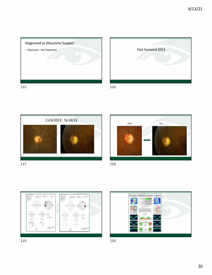
Diagnosed as Glaucoma Suspect
• Observed -> No Treatment
115
Fast Forward 2013
116
11/4/2013: Ta 14/13
117
2009 2013
118
119 120

4/13/21
21
Does she have glaucoma?
Does she need to be treated?
121
Summary: The Pressure is On• We recognize there are other factors besides IOP that influence
the development/progression of glaucoma–We are gaining more and more understanding of these other factors• OPP, Low BP, CSF pressure
• But for now – IOP is still the only thing available to treat• We have great technology to help us diagnose earlier and
detect progression earlier• And maybe even one day soon – we will have a treatment that
doesn’t involve a drop, laser, or taking a medication
122