press - kit cryo-em - ibs.fr · pdf fileibs guy schoehn, group ... single particle cryo-em is...
TRANSCRIPT
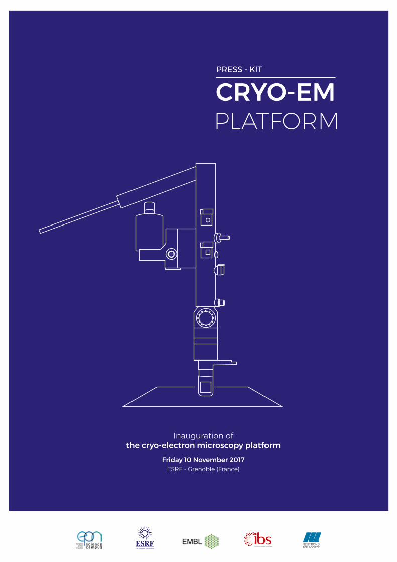
Inauguration of the cryo-electron microscopy platform
Friday 10 November 2017ESRF - Grenoble (France)
CRYO-EMPLATFORM
PRESS - KIT
Characteristics of the ESRF cryo-EM A brand new cryo-electron microscope is now available at the ESRF, the world’s most intense synchrotron, which brings with it a range of novel opportunities for the international user community.
CRYO-EM PLATFORM Inauguration of the cryo-electron microscopy platform
THE STRENGTH OF THIS CRYO-ELECTRON MICROSCOPE PLATFORM LIES IN:
1 - Its mission to serve an international scientific community. The ESRF welcomes about 9000 users from all member countries and from all over the world each year.
2 - Its synergy with synchrotron-based techniques (macromolecular crystallography and BioSAXS) and a large portfolio of state-of-the-art beamlines that allow scientists to better and more fully interpret action and function of complex bio-macromolecules.
3 - Its collaborative nature, with support to users provided by four institutes located on a same site, the EPN Science campus: the European Synchrotron (ESRF), the European Molecular Biology Laboratory (EMBL), the Institut de Biologie Structurale (IBS) and the Institut Laue-Langevin (ILL).
TECHNICAL SPECIFICATIONSThe Titan Krios cryo-electron microscope is equipped with
• K2 Summit direct electron detector, with highest image resolution
• A Quantum LS imaging filter
• A Volta phase plate (VPP)
ACCESSThe microscope will be operated as a beamline with access for the international structural biology community given on scientific merit through rolling-access applications. Proposals can be submitted at any time during the year.
A fully-equipped sample preparation laboratory is in close proximity to the microscope.
OPERATIONThe Cryo-EM platform was fully commissioned from July to November 2017. Regular user operation will start from mid-November 2017.
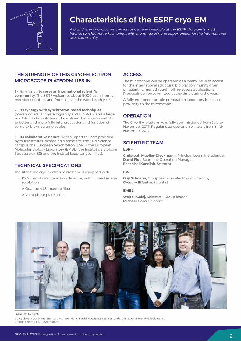
SCIENTIFIC TEAMESRFChristoph Mueller-Dieckmann, Principal beamline scientist David Flot, Beamline Operation Manager Eaazhisai Kandiah, Scientist
IBSGuy Schoehn, Group leader in electron microscopy Grégory Effantin, Scientist
EMBLWojtek Galej, Scientist - Group leader Michael Hons, Scientist
2
From left to right: Guy Schoehn, Grégory Effantin, Michael Hons, David Flot, Eaazhisai Kandiah, Christoph Mueller-Dieckmann.Credits Photos: ESRF/Stef Candé

What is the cryo-EM?Single particle cryo-EM is a technique used for protein structure determination with a resolution limit of up to near atomic resolution. The device uses electrons to image flash-frozen biological molecules and lay bare their molecular shapes.
3
View of the cryo-EM platform.Credit: ESRF/Stef Candé
View of the cryo-EM platform.Credit: The Royal Swedish Academy of Science / Martin Högborn
CRYO-EM PLATFORM Inauguration of the cryo-electron microscopy platform
Cryo-EM is increasingly popular among structural biologists for the exploration of large complex macromolecules. Cryo-EM has built on many decades of developments in the field to reach very high levels of resolution and 3D imaging, making it an innovative tool for the in-depth study of large complexes.
Cryo-EM is being used as a complementary technique with X-ray/neutron diffraction and scattering, with the overall objective to assess a greater number of molecule structures and larger protein complexes, including membrane proteins. Cryo-EM allows scientists to study objects that – because of their size, complexity, or sheer awkwardness – would be virtually impossible to scrutinise with other techniques. Cryo-EM is a powerful tool, especially combined with other techniques.
On the other hand, X-ray crystallography is best suited to smaller and well defined samples. It provides high resolution structures. It has a fast throughput once the crystallization conditions are well established. The cryo-EM will benefit from the facilities provided at the ESRF: a stable environment, data processing and storage. The well-established culture of a large user facility at the ESRF means that access to the different instruments available can and will be streamlined.
The electron microscope’s resolution has radically improved in recent years, from mostly showing blobs to now being able to visualise proteins at atomic resolution.
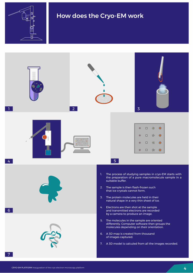
How does the Cryo-EM work
4CRYO-EM PLATFORM Inauguration of the cryo-electron microscopy platform
1. The process of studying samples in cryo-EM starts with the preparation of a pure macromolecule sample in a suitable buffer.
2. The sample is then flash-frozen such that ice crystals cannot form.
3. The protein molecules are held in their natural shape in a very thin sheet of ice.
4. Electrons are then shot at the sample and transmitted electrons are recorded by a camera to produce an image.
5. The molecules in the sample are oriented differently. Computer software then groups the molecules depending on their orientation.
6. A 3D map is created from thousand of images captured.
7. A 3D model is calculed from all the images recorded.
1 2 3
4 5
6
7

Applications and scientific casesIt is often said in biology that “seeing is believing”: It is only by knowing the atom-by-atom arrangement of a biomolecule that researchers can grasp how it works.
The success of X-ray crystallography to reveal structures of macromolecules cannot be left unmentioned. Several Nobel Prizes have been awarded for scientific achievements involving the use of crystallographic methods and techniques. Science continues to delve forever deeper into the mysteries of life and macromolecules are being studied in increasingly complex configurations. For example, today, the ribosome is being studied in its interactions with toxins, antibiotics and other macromolecules.
A picture of these processes is harder to grasp with crystallography, largely due to the fact that these large complexes are difficult to crystallise. The emergence of cryo-EM as a complementary technique to X-ray crystallography is enabling scientists to combine the two methods, looking at the smaller independent molecules with X-rays and at much larger complexes with electrons.
The new cryo-EM will offer a wide range of studies in structural biology:
• Structural analysis of protein complexes involved in biological processes
• Viral proteins: structures and applications to design vaccines
• Structure determination of dynamic molecular complexes
DRUG BINDINGCryo-EM has allowed scientists to see how drugs bind to enzymatic complexes. One complex of particular interest is the AAA ATPase p97, a target for cancer therapeutic development. Because of its flexibility, the full p97 complex has not been amenable to study by X-ray crystallography. Although individual domains of the protein have been crystallized, no single domain could be shown to be affected by drugs that inhibited enzymatic activity. Using cryo-EM, researchers at the National Cancer Institute in the USA were able to directly show how and where exactly the drug blocks the protein’s motion.
DNA REPAIRScientists at the Lawrence Berkeley National Laboratory used cryo-EM to resolve the 3-D structure of a protein complex called transcription factor IIH (TFIIH) that is active in DNA repair. This protein complex is used to unzip the DNA double helix so that genes can be accessed and read during transcription or repair. When TFIIH goes wrong, DNA repair cannot occur, and such a malfunction is associated with severe cancer propensity, premature aging, and other defects. Using this structure - solved with cryo-EM - scientists can now begin to better understand the mode of action or misbehaviour in cells.
HERPES VIRUSHuman cytomegalovirus, or HCMV, is the biggest of all herpes viruses. It is the leading infectious cause of birth abnormalities and a life-threatening pathogen for AIDS and organ transplantation patients. HCMV is composed of an envelope and a protein shell enclosing its double-stranded DNA. Scientists at UCLA in the USA have studied HCMV virus and solved the structure of the protein shells using cryo-EM. The structure gives scientists clues to design countermeasures against HCMV in order to develop therapeutic strategies against HCMV infections.
5CRYO-EM PLATFORM Inauguration of the cryo-electron microscopy platform

Why develop cryo-EM at the ESRF?
At the ESRF, cryo-EM is used as a complementary technique to macromolecular crystallography and bioSAXS. For a protein that can be crystallised, X-ray diffraction (MX) is still the most efficient means to obtain its structure at very high resolution. In contrast, bioSAXS provides low resolution details about the overall shape of a single protein or the special arrangement of a macromolecular complex in solution.
Cryo-EM has been used to obtain sub nanometre-resolution structures of up to ~2 Å for ideal samples including protein complexes and viruses with molecular masses typically of 150 kDa and above. It is commonly used for larger proteins and complexes that are intrinsically difficult to crystallise.
Combining synchrotron science and electron microscopy in one place will allow scientists to push the field of structural biology forward, to yield far-reaching and yet highly-detailed structural information. Thus, scientists will gain a better understanding of the macromolecules and their interactions.
With the addition of this Titan Krios cryo-EM, the ESRF, the world’s most intense X-ray source, offers the international structural biology community the possibility of coordinated access to cutting-edge methods and instrumentation, from preparation and screening to high resolution 3D imaging.
A cryo-EM facility on site will further enhance the scientific attractiveness of the ESRF and reinforce the status of the EPN science campus as a major international centre for integrated structural biology.
IN THE LAST 5 YEARS OF STRUCTURAL BIOLOGY ACTIVITY AT THE ESRF:
6CRYO-EM PLATFORM Inauguration of the cryo-electron microscopy platform
700high impact (IF>9)
peer-reviewed publications
> 4 500structure depositions
in the Protein Data Bank (13 101 since the
beginning of the ESRF)
20%of data collection
done remotely
Eaazhisai Kandiah mounts the sample inside the microscope.Credit: ESRF/Stef Candé

Why develop Cryo-EMat the EPN Science Campus
The ESRF is located on the EPN Science Campus, which includes major scientific institutes such as EMBL, IBS and ILL. Since 2002, the four institutes have merged their expertise in biology in the Partnership for Structural Biology (PSB), based on the same site.
Thanks to the expertise of 350 scientists on a same site, and state-of-the-art instrumentation and laboratories, this platform will offer users in Europe and beyond a holistic approach to their research and a unique global service.
The EPN science campus in a nutshell:
• A long-standing collaboration with the creation of a common platform for integrated structural biology studies, the PSB (Partnership for Structural Biology), established by a Memorandum of Understanding in 2002 by the European Molecular Biology Laboratory (EMBL), the European Synchrotron Radiation Facility (ESRF), the Institut Laue Langevin (ILL) and the Institut de Biologie Structurale (IBS).
• 350 scientists in structural biology on the same site.
• A unique portfolio of 23 technological platforms covering protein expression, 3D structure analysis, sample characterisation and supramolecular studies.
• ~ 200 peer-reviewed articles per year with PSB affiliation.
7CRYO-EM PLATFORM Inauguration of the cryo-electron microscopy platform
Top: Laboratory in the EPN science campus - Credit: ESRF/Pierre JayetBotom: Aerial view of EPN Science Campus - Credit: ESRF/Jocelyn Chavy
EMBL, a partner of the Cryo-EM platformEMBL is Europe’s flagship laboratory for the life sciences. The EMBL is an intergovernmental organisation established in 1974 and supported by over 20 member states.
EMBL performs fundamental research in molecular biology, studying the story of life. EMBL offers services to the scientific community; trains the next generation of scientists and strives to integrate the life sciences across Europe.
8CRYO-EM PLATFORM Inauguration of the cryo-electron microscopy platform
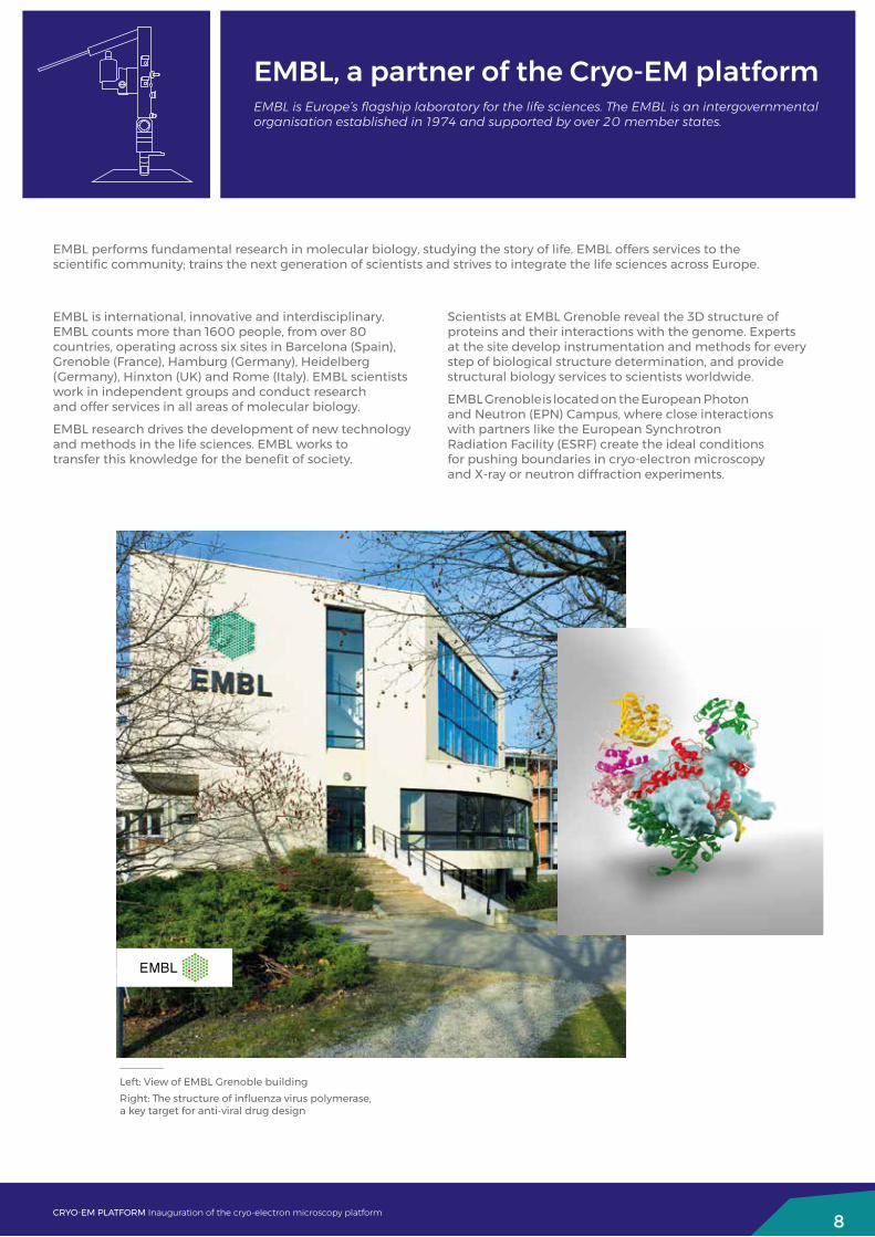
Left: View of EMBL Grenoble buildingRight: The structure of influenza virus polymerase, a key target for anti-viral drug design
EMBL is international, innovative and interdisciplinary. EMBL counts more than 1600 people, from over 80 countries, operating across six sites in Barcelona (Spain), Grenoble (France), Hamburg (Germany), Heidelberg (Germany), Hinxton (UK) and Rome (Italy). EMBL scientists work in independent groups and conduct research and offer services in all areas of molecular biology.
EMBL research drives the development of new technology and methods in the life sciences. EMBL works to transfer this knowledge for the benefit of society.
Scientists at EMBL Grenoble reveal the 3D structure of proteins and their interactions with the genome. Experts at the site develop instrumentation and methods for every step of biological structure determination, and provide structural biology services to scientists worldwide.
EMBL Grenoble is located on the European Photon and Neutron (EPN) Campus, where close interactions with partners like the European Synchrotron Radiation Facility (ESRF) create the ideal conditions for pushing boundaries in cryo-electron microscopy and X-ray or neutron diffraction experiments.
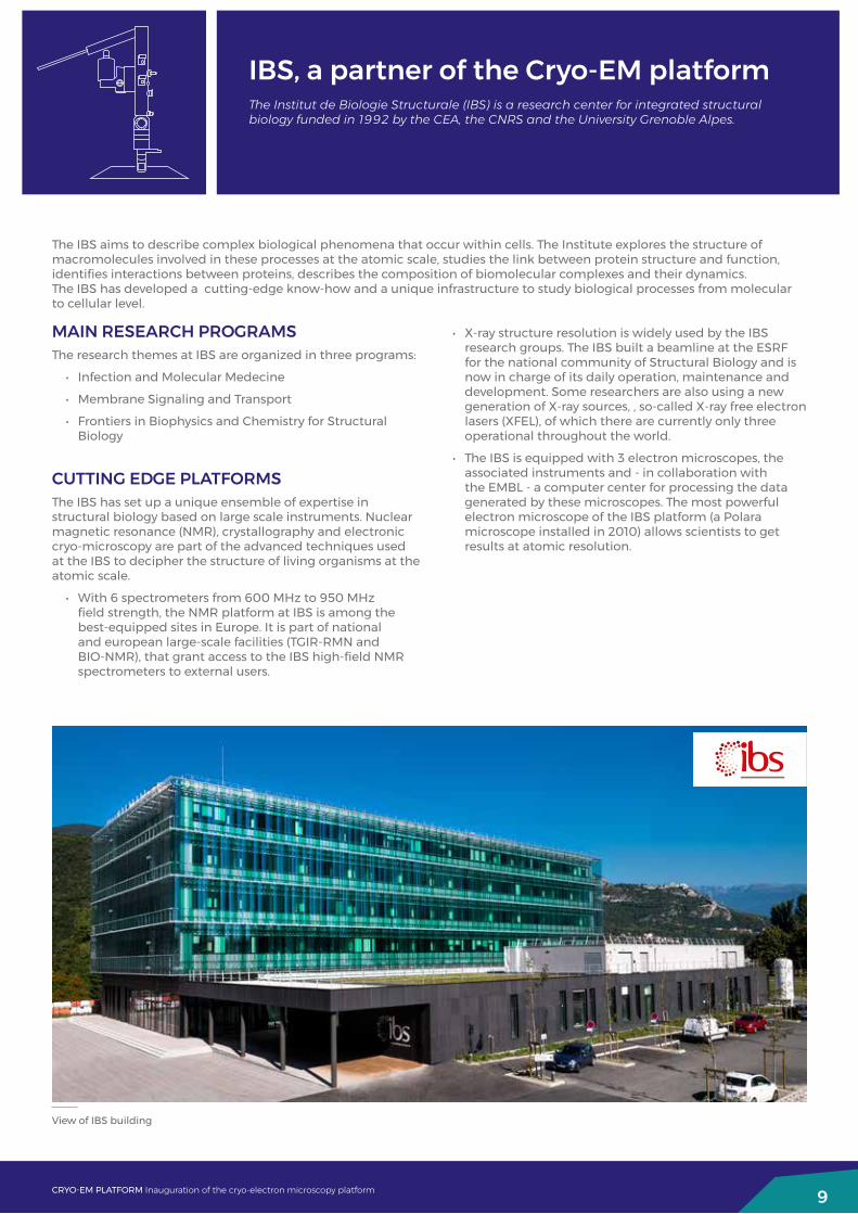
IBS, a partner of the Cryo-EM platformThe Institut de Biologie Structurale (IBS) is a research center for integrated structural biology funded in 1992 by the CEA, the CNRS and the University Grenoble Alpes.
The IBS aims to describe complex biological phenomena that occur within cells. The Institute explores the structure of macromolecules involved in these processes at the atomic scale, studies the link between protein structure and function, identifies interactions between proteins, describes the composition of biomolecular complexes and their dynamics. The IBS has developed a cutting-edge know-how and a unique infrastructure to study biological processes from molecular to cellular level.
9CRYO-EM PLATFORM Inauguration of the cryo-electron microscopy platform
View of IBS building
MAIN RESEARCH PROGRAMSThe research themes at IBS are organized in three programs:
• Infection and Molecular Medecine
• Membrane Signaling and Transport
• Frontiers in Biophysics and Chemistry for Structural Biology
CUTTING EDGE PLATFORMSThe IBS has set up a unique ensemble of expertise in structural biology based on large scale instruments. Nuclear magnetic resonance (NMR), crystallography and electronic cryo-microscopy are part of the advanced techniques used at the IBS to decipher the structure of living organisms at the atomic scale.
• With 6 spectrometers from 600 MHz to 950 MHz field strength, the NMR platform at IBS is among the best-equipped sites in Europe. It is part of national and european large-scale facilities (TGIR-RMN and BIO-NMR), that grant access to the IBS high-field NMR spectrometers to external users.
• X-ray structure resolution is widely used by the IBS research groups. The IBS built a beamline at the ESRF for the national community of Structural Biology and is now in charge of its daily operation, maintenance and development. Some researchers are also using a new generation of X-ray sources, , so‐called X‐ray free electron lasers (XFEL), of which there are currently only three operational throughout the world.
• The IBS is equipped with 3 electron microscopes, the associated instruments and - in collaboration with the EMBL - a computer center for processing the data generated by these microscopes. The most powerful electron microscope of the IBS platform (a Polara microscope installed in 2010) allows scientists to get results at atomic resolution.

ILL, a partner of the Cryo-EM platformThe Institut Laue-Langevin (ILL) is an international research centre based in Grenoble, France. Funded by France, Germany and the United Kingdom, in partnership with 10 other European countries, it has led the world in neutron-scattering science and technology for 45 years.
ILL operates one of the most intense neutron sources in the world, feeding beams of neutrons to a suite of 40 high-performance instruments. Research conducted at ILL covers a wide range of disciplines such as biology, (green) chemistry, materials science, condensed matter physics, as well as fundamental and nuclear physics.
ILL, working in collaboration with Keele University, will engage in the development of cryo electron microscopy on the EPN site. This method, which may provide unparalleled access to high resolution information on large complexes, is one in which a strong complementarity is possible with solution studies using small-angle neutron scattering (SANS), fully exploiting the joint SANS with SAXS platform of the PSB. Keele intends to provide a faculty position located within ILL’s Life Sciences Group and who will work in close collaboration with the cryo-EM team members from ESRF, IBS, and EMBL. The engagement with Keele University Life Sciences Department will introduce research interests relating to host pathogen interactions in parasitology and large complexes of importance in neuroscience. Examples include proteins associated with the sensory hair cells involved in the vertebrate hearing and balance organs and important viral complexes.
10CRYO-EM PLATFORM Inauguration of the cryo-electron microscopy platform
Low resolution EM pictures the precise array of stereocilia in a hair bundle of a hair cell. Specific proteins at the tips of these cilia will be the targets of high resolution cryo-EM work.
Viral protein complexes with the protein prohibitin