presence of toxoplasma gondii infection in brain as a ... · original article presence of...
TRANSCRIPT

ORIGINAL ARTICLE
Presence of Toxoplasma gondii infection in brain as a potentialcause of risky behavior: a report of 102 autopsy cases
Dorota Samojłowicz1 & Joanna Twarowska-Małczyńska2 & Aleksandra Borowska-Solonynko1&
Łukasz A. Poniatowski3,4 & Nipika Sharma5 & Mieszko Olczak1
Received: 8 September 2018 /Accepted: 7 November 2018 /Published online: 23 November 2018# The Author(s) 2018
AbstractToxoplasmosis was linked to impairment in brain function, encompassing a wide range of behavioral and neuropsychiatricchanges. Currently, the precise localization of Toxoplasma gondii in the human brain is limited and the parasite DNAwas notfound in population-based screening of autopsy cases. The aim of proposed study was to identify the presence of parasite DNAwithin the brain and its association with risky behavior and alcohol consumption in postmortem examination. Preliminarily, 102cases with certain circumstances of death at time of forensic autopsy was included. Due to high risk of bias, the females wereexcluded from the analysis and final study group consists 97 cases divided into three groups: risky behavior, inconclusively riskybehavior, and control group. The obtained tissue samples for Nested PCR covered four regions of the brain: symmetric left/rightand anterior/posterior horns of lateral ventricles comprising lining ependyma and hippocampus. The second type of materialcomprised blood evaluated for antibodies prevalence using ELISA and alcohol concentration using HS-GC-FID. Analysisdemonstrated 16.5% prevalence concerning the parasite DNA presence in examined brain tissue samples without specificdistribution and association with age at death or days after death until an autopsy was performed. Results have shown correlationbetween occurrence of risky behavior leading to death and higher proportions of positive parasite DNA presence within the brain.Correlation was not observed between parasite DNA presence and excessive alcohol consumption. Conducted screening dem-onstrated correlation between parasite DNA presence in the brain with risky behavior and provided new information on possibleeffects of latent toxoplasmosis.
Keywords Toxoplasma gondii . Toxoplasmosis . Brain . Risky behavior . Alcohol . Mental health . Drivers
Introduction
Toxoplasma gondii is a common heteroxenous andpolyxenous protozoan parasite which is globally widespread
causing infection in virtually most warm-blooded mammalspecies, including humans [1, 2]. This remarkable virulencecapacity and spread results from the ideal adaptation and com-plex function within the host-parasite axis, as well as the
Nipika Sharma is an independent researcher
* Dorota Samojł[email protected]; http://www.wum.edu.pl; http://www.wum.edu.pl/en/english; http://www.zms.wum.edu.pl/en
Joanna Twarowska-Małczyń[email protected]
Aleksandra [email protected]
Łukasz A. [email protected]
Mieszko [email protected]
1 Department of Forensic Medicine, Center for Biostructure Research,Medical University of Warsaw, Oczki 1, 02-007 Warsaw, Poland
2 Department of General Biology and Parasitology, Center ofBiostructure Research, Medical University of Warsaw,Chałubińskiego 5, 02-004 Warsaw, Poland
3 Department of Experimental and Clinical Pharmacology, Centre forPreclinical Research and Technology (CePT), Medical University ofWarsaw, Banacha 1B, 02-097 Warsaw, Poland
4 Department of Neurosurgery, Maria Skłodowska-Curie MemorialCancer Center and Institute of Oncology, W. K. Roentgena 5,02-781 Warsaw, Poland
5 Warsaw, Poland
European Journal of Clinical Microbiology & Infectious Diseases (2019) 38:305–317https://doi.org/10.1007/s10096-018-3427-z

parasite’s high transmission potential [3]. In humans, parasiteinfection are related with the development one of an omni-present systemic anthropozoonose referred to toxoplasmosis[4]. The epidemiological data shows that T. gondii infectionaffects almost all populations and demographic levels depend-ing on a range of factors, including geographic region, sea-sonal changes, sanitary-hygienic conditions, socioeconomicstatus, and dietary habits [5, 6]. In general, approximately ~30% of the worldwide human population is infected byT. gondii, and the global prevalence is estimated to be in therange of 10–80% consisting a substantial public health prob-lem [6, 7]. For a long time, according to previous publishedstudies, the most common latent form of disease in immuno-competent subjects including pregnant women was thought tobe asymptomatic and clinically normal [8]. Nevertheless, re-cent studies and observations from both human and preclinicalanimal models clearly indicate that this form of disease canexert formerly unrecognized short- and long-lasting negativeconsequences even in immunocompetent subjects [8, 9]. Inparticular, latent T. gondii infection in human populationswas potentially linked to impairment in brain function,encompassing a wide range of behavioral and neuropsychiat-ric changes [10]. Among the behavioral domains, it was ob-served that latent T. gondii infection may trigger changes inimpulsivity control including an increase in general risk-taking behaviors and violence where seropositive female in-dividuals were found to exhibit increased levels of aggression,while male individuals were characterized by excessive im-pulsiveness [11]. Furthermore in regard to neurocognitivefunction, the latent infection was connected with psychomotordeficits, lack of concentration, lower intelligence quotient(IQ), and alternations in personality profile [12–14].Collectively, these attributes may affect personality phenotypeand could ultimately predispose to various life-threateningand health-risk behaviors, including suicide and homicide[15, 16]. Insights afforded from the last two decades also showpotential link between latent T. gondii infection and increasedrisk of schizophrenia, depression, generalized anxiety, andobsessive-compulsive disease [17, 18]. Despite constant prog-ress in both basic and clinical research, the current understand-ing of T. gondii action and influence mechanism in the humanorganism as an intermediate host is still limited [19]. Theobserved cerebral alternations consist a potential result of di-rect presence of T. gondii in different neuroanatomical loca-tions within brain parenchyma [20]. Dopamine (DA) is animportant catecholamine neurotransmitter and regulatorwhich maintains stability and flexibility in a number of func-tions that include, but are not limited to cognition, locomotion,affect, reward, emotional, and neuroendocrine aspects [21,22]. Reported observations from several studies support theconcept that T. gondii infection followed by a local presenceof parasite alters the concentration of neurotransmitters in thebrain revealing one of the potential pathomechanisms leading
to development of behavioral and neuropsychiatric changesby modulation of DA and its metabolite levels [23]. Anotherpotential multiphasic event which contributes to changes inbrain function includes local and systemic inflammatory reac-tions in response to the transposition and spread of T. gondiiinfection through the body [24, 25]. The direct presence ofT. gondiiwithin brain parenchyma is associated with secretionof inflammatory cytokines, chemokines, and mediators byneurons, astrocytes, and microglia, along with activation andrecruitment of immune cells [26]. Persistent localneuroinflammatory reaction seems to also be related with al-ternations of neurotransmitter release [27]. The effect ofT. gondii influence on the behavior of the intermediate hostmay be associated with the location of the parasite in specificregions of the brain, most likely those that are associated withfear and anxiety, mainly within the amygdala [28]. Anotherstudy indicated an increased number of parasites in the cere-bral cortex, diencephalon, and hippocampus [29]. Despitethese findings, the data concerning location of the parasite inthe brains of a population with toxoplasmosis is not well in-vestigated. In the one study conducted to date, in whichT. gondii genetic material presence was evaluated by theNested PCR method postmortem from the frontal cortex ofpsychiatric patients diagnosed during lifetime, the parasiteDNAwas not isolated and detected [30]. Given the evidencelinking parasite infection to several behavioral and neuropsy-chiatric changes, the Department of Forensic Medicine atMedical University of Warsaw in collaboration with otheracademic departments of the university conducted extensivestudies in an attempt to evaluate the precise identification ofT. gondiiDNAwithin the human brain and its association withrisky behavior and alcohol consumption occurrence among ananalyzed population in postmortem examination. Since thispotential correlation has not been presented yet in the avail-able literature, the results of the abovementioned studiespertaining to a correlation between T. gondii infection andrisky behavior along with alcohol consumption have beenpresented in this manuscript.
Materials and methods
Autopsy cases
The study was carried out on samples obtained during externalforensic examinations and autopsies performed and providedby forensic pathologists from the Department of ForensicMedicine at the Medical University of Warsaw over the 3-year period between 2010 and 2013. Cases included in thestudy covered only those whose circumstances surroundingthe death were already known at the time of forensic autopsyor were previously provided by the appropriate Prosecutor’sOffices investigating the respective cases. Bodies presenting
306 Eur J Clin Microbiol Infect Dis (2019) 38:305–317

late postmortem signs as well as cases with incompletemedico-legal data were excluded from the study and did notundergo any further experimental testing. Based on externalforensic examination and autopsy protocols along with dataprovided by the Prosecutor’s Office or in case of inpatientssupplied medical records for each decedent, we collected therespective data: sex, age, circumstances of death, cause ofdeath, period between death and autopsy, and (if provided)alcohol and/or illicit drug abuse status or the presence of psy-chiatric disorder. In the cases of traumatic deaths, the presenceof brain injuries was also noted. Collectively, preliminarily atotal of 102 cases (97 males and 5 females) were included inthis study. The considerable ratio of male cases vs. femalecases in the study sample was in accordance with the generalproportion represented in all performed forensic autopsies inDepartment of ForensicMedicine at theMedical University ofWarsaw, wherein nearly (~ 80%) of all autopsies the decedentis male. Consecutively, due to the high risk of bias, the femaleswere excluded from the analysis and final study group consists97 cases (n = 97). The estimated median age for the studygroup was 49 years (with the youngest case being 18 yearsand the oldest 89 years). The time period between death andautopsy ranged from 1 to 10 days with mean of 4.14 days.After collecting of autopsy reports and medico-legal data in-cluding additional tests, the circumstances of death werereviewed and then all evaluated cases were divided into threeconsecutive groups: risky behavior (RB), inconclusively riskybehavior (IRB), and control (C) group. According to this, theRB group (n = 42) constituted cases wherein the available dataindicated that the death was due to the so-called risky behav-iors. These included drivers who were classified as indisput-able perpetrators of traffic accidents (n = 35), individuals whodied as a result of substance overdose (n = 5), and other indi-viduals who died as a result of disregarding reasonable safetyprecautions (n = 2), where the first died of carbon monoxide(CO) poisoning and the second was a pedestrian. The caseswhose deaths could not be precisely attributed to risky behav-iors after reviewing all available data were included in the IRBgroup (n = 27). These included drivers who could not be clear-ly identified as the perpetrators of the traffic accident thatcaused their death (n = 14) and individuals who died as a resultof disease whose cause could be reasonably suspected to beassociated with chronic long-term alcohol consumption, al-though this fact was not verified (n = 13). The C group (n =28) consisted of individuals whose deaths were not associatedwith risky behaviors, and whomostly died as a result of chron-ic disease.
Tissue dissection and preparation
During the forensic autopsy, two types of material for exam-ination were collected. The first type consisted of brain tissuesamples which were obtained for genetic testing, while the
second sample type comprised of blood used for serologicaltesting. The brain specimens in a volume of approximately ~50 ml were collected to sterile tubes from four distinctivesurrounding regions of brain: the symmetric left (A) and right(B) anterior horn of lateral ventricle comprising liningependyma along with the symmetric left (C) and right (D)posterior horn of lateral ventricle comprising lining ependymaand hippocampus. Collection of brain tissue was excludedfrom areas comprising macroscopic signs of injury such asbrain contusion and laceration, extra- and subdural hematoma,subarachnoid hemorrhage or intraventricular bleeding.Immediately after the dissection, brain tissue samples werestored at the ice cubes and kept away from lights followingits safety transfer to the laboratory where the material wereconsecutively persevered at − 20 °C in special storage until thetime of the preheating step and proper analysis. The bloodsamples (~ 10 ml) were collected directly from the cranialcavity or femoral vein into sterile tubes. After then, sampleswere centrifuged and stored at − 20 °C until the time of thetest. The blood test was not conducted in three cases (maledrivers) from the RB group due to exsanguinations of thebodies causing lack of material.
Nested PCR
Initial digestion
Previously obtained and stored brain tissue samples werethawed at room temperature (20–24 °C) and then drainedof residual blood. Brain tissue were homogenized, pouredwith a warm (37 °C) solution of 0.25% trypsin lyophilizate(3 U/ml) from bovine pancreas (Sigma-Aldrich, St. Louis,USA) which was previously dissolved in 0.9% saline (NaCl)solution and then digested in an oven at 37 °C for 1 h.During digestion the samples were mixed every 5 min.Afterwards, digestion samples were poured into 50-ml tubesand centrifuged at 400×g for 10 min. The supernatant wasremoved and then the sediment was washed twice in 0.9%NaCl followed by its centrifugation. In order to increase theefficiency of the method, the precipitate obtained after theinitial digestion was equally distributed to three 1.5-mltubes. In this way, three digestion products were obtainedcorresponding to each of the collected brain tissue sample.Collectively, a total of 12 DNA digestion products wereobtained from one examined case.
Isolation of DNA
Isolation of DNAwas carried out using the NucleoSpin TissueKit (740952;Macherey-Nagel, Düren, Germany) according tothe manufacturer’s standard protocol. The precipitate wassuspended in 210 μl of buffer T1 and 30 μl of proteinase K,and then was incubated for 24 h in thermomixer at 56 °C.
Eur J Clin Microbiol Infect Dis (2019) 38:305–317 307

When the initial incubation was done, buffer B3 was added ina volume of 230 μl and incubated for 10 min in thermomixerat 70 °C. After this time, 160 μl of 99.8% ethanol was addedto the samples and the material was transferred to the spincolumn and centrifuged at 12,000 rpm for 5 min. The filtratewas removed and 500 μl of buffer BW was added to thecolumn and centrifuged at 12,000 rpm for 5 min. After then,the filtrate was again removed, and 600 μl of buffer B5 wasadded to the column and centrifuged at 12,000 rpm for 5 min.The filtrate removal was repeated and 70 μl of buffer BEwhich was warmed to 70 °C and incubated for 1 min in roomtemperature was added to the column and then centrifuged at12,000 rpm for 3 min. In this way, the obtained DNA isolateswere stored at − 20 °C until the time of further use. In eachcase, isolation control was performed simultaneously. Thesample was incubated and centrifuged under the same condi-tions and with the same reagents as the test samples, withoutthe addition of sediment from the initial digestion.
Amplification of DNA
In order to detect the DNA of the T. gondii in the tested sam-ples, a two-stage amplification was done. The initial amplifica-tion reaction was carried out using forward primer Tox4 (T1)and Tox5 (T2) reverse primer both covering 26 bp which attachto the end of the 3′ and to the 5′ end of the constitutively presentfragment repeated 200–300-fold in the genome of T. gondii(Table 1). These oligonucleotide sequences were recognizedas very specific and sensitive, and therefore remarkably usefulin identifying of parasite. The primer sequences were designedin accordance with well-characterized and established availableliterature data [31]. The amplification reaction of T. gondiiDNA was carried out using Taq PCR Core Kit (201223;Qiagen, Hilden, Germany) according to the manufacturer’sstandard protocol. The amplification reaction mixture (50 μl)contained 5 mM MgCl2, 0.2 mM dNTP mix, 5 μl of PCRbuffer (10×), 5 μl of CoralLoad PCR buffer (10×), 10 pM ofeach primer, 1 unit of Taq DNA polymerase, and 5 μl of DNAtemplate where all was dissolved in sterile water purified fromDNA nucleases. The PCR amplification was performed in aDNA thermal cycler (PTC-200; MJ Research, San Francisco,USA). Cycling conditions were as follows: initial denaturationstep of 7 min at 94 °C, followed by 34 cycles of 1 min at 94 °C(denaturation), 30 s at 56 °C (primer binding), 30 s at 72 °C
(elongation), and a final extension step at 72 °C for 10 min.After the reaction, a 529-bp product was obtained. Due to theinequivalent DNA concentration of the host and parasite in theanalyzed samples, the Nested PCR method was used to detectT. gondii DNA in the obtained isolates. The sequence of theprimers for the second amplification, as well as the optimalreaction conditions, was determined experimentally (Table 1).Subsequent amplification reaction of T. gondii DNAwas car-ried out using the same reagents according to manufacturer’sstandard protocol. Amplification reaction mixture (50 μl)contained 5 mM MgCl2, 0.2 mM dNTP mix, 5 μl of PCRbuffer (10×), 5 μl of CoralLoad PCR buffer (10×), 10 pM ofeach primer, 1 unit of Taq DNA polymerase, and 0.5 μl of firstamplification product where all was dissolved in sterile waterpurified from DNA nucleases. The subsequent PCR amplifica-tion was performed in a DNA thermal cycler (PTC-200; MJResearch, San Francisco, USA). Cycling conditions were asfollows: initial denaturation step of 7 min at 94 °C, followedby 34 cycles of 1 min at 94 °C (denaturation), 20 s at 58 °C(primer binding), 20 s at 72 °C (elongation), and a final exten-sion step at 72 °C for 10 min. After reaction, a 204-bp fragmentcontained in the first amplification product was obtained.During both amplification reactions, a positive and negativecontrol was performed. In the first amplification reaction, theDNA isolated from the T. gondii culture was used for the pos-itive control; in the second amplification, the control DNAfrom the first reaction was used for the positive control. In thenegative control, pure water was added instead of DNA. Thesame reaction mixture as in the test samples was used in allcontrols.
Analysis of the products
The PCR products (10 μl) were separated and analyzed on a2.0% agarose gel (Metaphor, FMC BioProducts, Rockland,USA) and visualized after staining with ethidium bromide.As a reference standard for DNA size marker, Gene Ruler100 bp DNA Ladder Plus (MBI Fermentas, St. Leon-Rot,Germany) was used.
ELISA
The serological prevalence of the T. gondii antibodies (IgG±) was evaluated using an enzyme-linked immunosorbent
Table 1 Primer sequencesdesigned and used for first andsecond amplification ofToxoplasma gondii DNA
Primer Primer sequences (5′-3′) Predicted product size (bp)
T1 5′-CGCTGCAGGGAGGAAGACGAAAGTTG-3′
529
T2 5′-CGCTGCAGACACAGTGCATCTGGATT-3′
T3 5′-GAGCCACAGAAGGGACAGA-3′ 204T4 5′-TTCCGGTGTCTCTTTTCCAC-3′
308 Eur J Clin Microbiol Infect Dis (2019) 38:305–317

assay (ELISA) test. Followed assays were carried out usingan ELISA test kit (Euroimmun, Lübeck, Germany) accord-ing to the manufacturer’s standard protocol. Reaction wellswere added by following (100 ml) aliquots: calibrationserum, positive control serum, negative control serum,and test serum samples diluted at 1:101. Each assay platewas covered and incubated at room temperature for30 min. The wells were consecutively emptied and washedthree times with 300 ml of diluted rinsing solution.Afterwards, anti-human IgG conjugated to peroxidase(100 ml) was added into each reaction well and incubatedat room temperature for 30 min. The wells were washedthree times after incubation. Substrate solution (100 ml)was added into each well and then incubated in a darkenedarea at room temperature for 15 min. The reaction termi-nation was performed by adding 100-ml aliquots of aquench solution to each well in the same order and at thesame speed as that used to add the substrate solution.Photometric assessment of color intensity at a wavelengthof 450 nm (reference wavelength in the range 620–650 nm)was conducted using ELISA microplate reader (Ledetect96; Dynamica, Salzburg, Austria) equipped with MicroWin 2000 software (Biochrom, Cambridge, UK). The in-terpretation of obtained results were done according to theinstructions enclosed within the protocol. Each test wasconducted in two subsequent repetitions.
Analysis of alcohol levels
The alcohol (ethanol) concentration in obtained blood sam-ples was analyzed using headspace gas chromatography-flame-ionization detection (HS-GC-FID) system (7090B;Agilent Technologies, Santa Clara, USA) coupled with aheadspace sampler (7697A; Agilent Technologies, SantaClara, USA). Two separated capillary columns were usedincluding DB-ALC1 (30 m × 0.32 mm × 1.8 μm) and DB-ALC2 (30 m × 0.32 mm × 1.2 μm) column (AgilentTechnologies, Santa Clara, USA). The incubation of sam-ples was performed at 80 °C, the temperature of the transferline was 110 °C, the temperature of the column was 40 °C,and the helium (He) carrier gas purity was 99.999% in a 5:1split where the temperature of the detector was 300 °C.Ethanol levels were calculated from 100 μl of samplesadded to the 20-ml headspace glass vial and mixed withthe 900 μl of internal standard solution (aqueous solutionof 2-butanone). Vials were precisely closed and sealed witha rubber stopper. The obtained results from 90 cases (n = 90)were interpreted based on the definition of alcohol normsincluded in article 115 paragraph 16 point 1 of the PenalCode of the Republic of Poland, according to which indi-viduals with a blood alcohol content equal to or exceeding0.5‰ are considered to be under influence of alcohol.
Statistical analysis
The statistical processing of the obtained data was performedusing R statistical environment (version 3.4.3; RDevelopment Core Team, Vienna, Austria). For group com-parison analysis, Fisher’s exact test was used due to the limit-ed number (n = 97) of available cases. In order to examine thehypothesis that T. gondii infection can be present in the brainand not in blood, the Monte Carlo simulation method wasused. The detailed description of the method and the underly-ing principle is explained in the subsequent BResults^ sec-tions. All the obtained p values were corrected for multiplehypothesis testing using the q value algorithm. The resultswere considered statistically significant when p values wereless than adjusted 0.05 (p < 0.05).
Results
Presence and distribution of the T. gondii DNAwithin brain
The presence of T. gondiiDNA in the brain was detected in 16(n = 16) out of 97 (n = 97) examined cases, which resulted in~ 16.5% prevalence in our total studied population (Fig. 1). Asubsequent analysis was made to determine in which of thefour regions of the brain T. gondii DNA was most often de-tected. As was shown, in 12 (n = 12) out of 16 (n = 16) exam-ined cases, the T. gondii DNAwas detected in more than oneregion of the brain. T. gondii DNA was most frequently de-tected in samples taken from the B and C regions (Fig. 2).Since due to the number of positive cases was too small(n = 16), it is not possible to draw any statistically strong con-clusions regarding brain localization of parasite cysts(Table 2).
Serological prevalence of the T. gondii antibodies
The previously obtained positive test results for T. gondiiDNA were confirmed (IgG+) serologically in ~ 56.3% ofcases (n = 9). In ~ 18.7% of cases (n = 3), no serological testswere performed due to exsanguination of the respective bod-ies, causing lack of material. In other cases, the serologicalscore was negative (IgG−). The 25% of cases (n = 4) in whichthe presence of the T. gondii DNA could not be confirmedserologically were found to be drivers (n = 2) who were clas-sified as the indisputable perpetrators of traffic accidents fromthe RB group, a pedestrian (n = 1) from the RB group, and adriver (n = 1) who could not be clearly identified as the per-petrators of the traffic accident that caused their death from theIRB group. In addition, it is necessary to mention that in 75%of the above cases (n = 3), positive test results for T. gondiiDNAwere confirmed in ≥ 2 regions of the brain.
Eur J Clin Microbiol Infect Dis (2019) 38:305–317 309

Correlation between genetic and serological testing
The obtained results in the case of positive T. gondii DNAdetection in the brain and negative serological antibody testingled to the proposal of a null hypothesis (H0): if serologicaltesting results are negative, then genetic testing results are alsonegative. Given the nature of the relationship between geneticand serological testing, there are several plausible cases:T. gondii antibodies are not present in blood along withDNA in the brain (IgG−/DNA−), T. gondii antibodies arepresent in blood and DNA presence is confirmed in the brain(IgG+/DNA+), and T. gondii antibodies are present in bloodwhile DNA presence is not confirmed in the brain (IgG+/DNA−). In order to determine whether the result thatT. gondii DNA is present in the brain when antibodies arenot present in blood (IgG−/DNA+), is statistically significant,
the Monte Carlo simulation method was used. Since therewere 97 cases (n = 97) of genetic positive results, 9999 ran-dom samples of size 97 were generated from the data. Giventhat there are 4 cases (n = 4) where T. gondii DNAwas detect-ed while serology was negative (out of 97 cases), the objectivewas to examine the number of times it is likely to get this ormore extreme results if random samples from the data aregenerated. Assuming the significance level of 5%, we ob-served that the p value (p < 0.0086) was less than adjusted0.05 in this case. Therefore, we rejected our null hypothesisthat genetic testing will be negative if serological testing re-sults are negative. Hence, we concluded that it is possible thatT. gondiiDNA is present in the brain evenwhen antibodies arenot present in the blood. In cases where T. gondii DNA is notpresent, both positive and negative results of serological test-ing were observed, with the percentage distribution of theseresults being similar, as well as a slight predominance of neg-ative results (58% vs. 42%) in this case.
204 bp
204 bp
a
b
M 1 2 3 4 5 6 7 8 9 10 11 12 C+ IC C-
M 1 2 3 4 5 6 7 8 9 10 11 12 C+ C- IC
Fig. 1 High-resolution agarosegel electrophoresis of NestedPCR products obtained fromamplification of the brain tissuesamples performed for detectionof the Toxoplasma gondiiDNA. asample positive results from thesingle of evaluated cases. bsample negative results from thesingle of evaluated cases. Thedetailed reaction conditions areprovided in the BMaterial andmethods^ section. M ladder, 1–12lanes represent each of the PCRproducts obtained from the singleof evaluated cases, C+ positivecontrol, C− negative control, ICisolation control
IIII
Toxoplasma gondii DNA presence in number of brain regions
Number of brain regions
Perc
enta
ge o
f PC
R p
ositi
ve c
ases
30 -
40 -
20 -
10 -
0 -
1 2 3 4
25.0%
37.5%
12.5%
25.0%
Fig. 2 The schematic representation of the proportion of Toxoplasmagondii DNA presence in number of brain regions. PCR polymerasechain reaction
Table 2 Location of the Toxoplasma gondii DNA presence in brainregions
Brain region
A B C D
Number of PCR positive cases 8 12 11 7
Percentage of PCR positive cases 50 75 68.8 43.8
A—left anterior horn of lateral ventricle comprising lining ependyma;B—right anterior horn of lateral ventricle comprising lining ependyma;C—left posterior horn of lateral ventricle comprising lining ependymaand hippocampus; D—right posterior horn of lateral ventricle comprisinglining ependyma and hippocampus
310 Eur J Clin Microbiol Infect Dis (2019) 38:305–317
Correlation between genetic testing and groupcomparison analysis
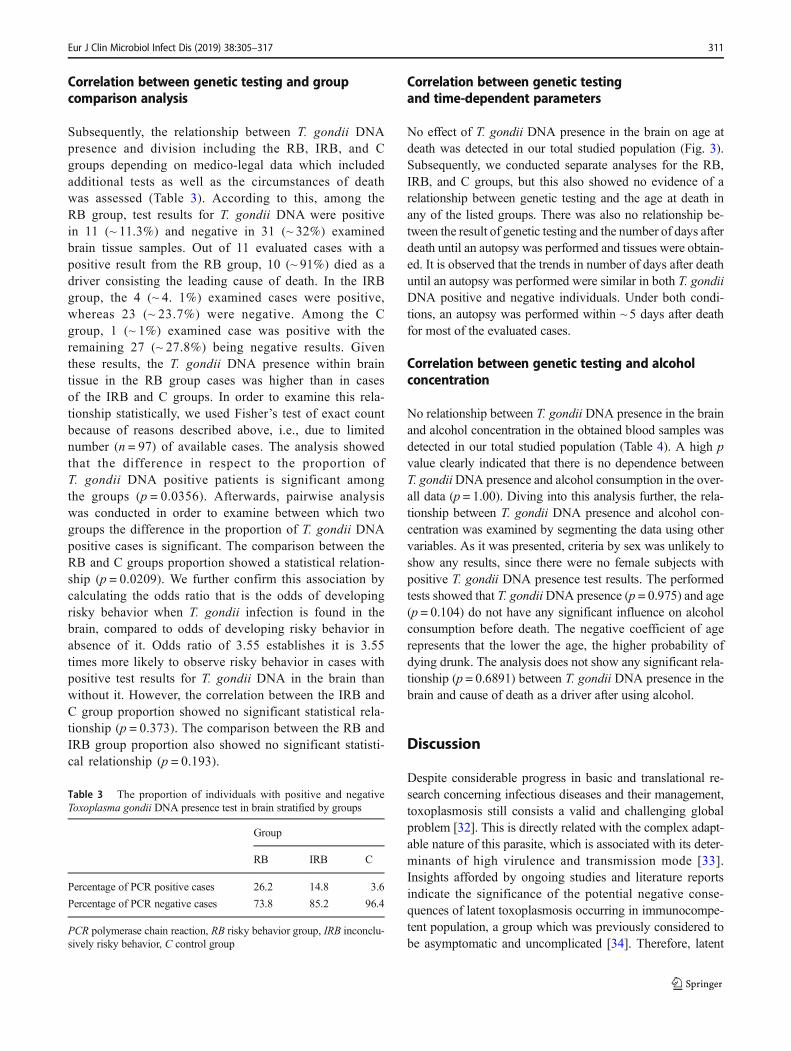
Subsequently, the relationship between T. gondii DNApresence and division including the RB, IRB, and Cgroups depending on medico-legal data which includedadditional tests as well as the circumstances of deathwas assessed (Table 3). According to this, among theRB group, test results for T. gondii DNA were positivein 11 (~ 11.3%) and negative in 31 (~ 32%) examinedbrain tissue samples. Out of 11 evaluated cases with apositive result from the RB group, 10 (~ 91%) died as adriver consisting the leading cause of death. In the IRBgroup, the 4 (~ 4. 1%) examined cases were positive,whereas 23 (~ 23.7%) were negative. Among the Cgroup, 1 (~ 1%) examined case was positive with theremaining 27 (~ 27.8%) being negative results. Giventhese results, the T. gondii DNA presence within braintissue in the RB group cases was higher than in casesof the IRB and C groups. In order to examine this rela-tionship statistically, we used Fisher’s test of exact countbecause of reasons described above, i.e., due to limitednumber (n = 97) of available cases. The analysis showedthat the difference in respect to the proportion ofT. gondii DNA positive patients is significant amongthe groups (p = 0.0356). Afterwards, pairwise analysiswas conducted in order to examine between which twogroups the difference in the proportion of T. gondii DNApositive cases is significant. The comparison between theRB and C groups proportion showed a statistical relation-ship (p = 0.0209). We further confirm this association bycalculating the odds ratio that is the odds of developingrisky behavior when T. gondii infection is found in thebrain, compared to odds of developing risky behavior inabsence of it. Odds ratio of 3.55 establishes it is 3.55times more likely to observe risky behavior in cases withpositive test results for T. gondii DNA in the brain thanwithout it. However, the correlation between the IRB andC group proportion showed no significant statistical rela-tionship (p = 0.373). The comparison between the RB andIRB group proportion also showed no significant statisti-cal relationship (p = 0.193).
Correlation between genetic testingand time-dependent parameters
No effect of T. gondii DNA presence in the brain on age atdeath was detected in our total studied population (Fig. 3).Subsequently, we conducted separate analyses for the RB,IRB, and C groups, but this also showed no evidence of arelationship between genetic testing and the age at death inany of the listed groups. There was also no relationship be-tween the result of genetic testing and the number of days afterdeath until an autopsy was performed and tissues were obtain-ed. It is observed that the trends in number of days after deathuntil an autopsy was performed were similar in both T. gondiiDNA positive and negative individuals. Under both condi-tions, an autopsy was performed within ~ 5 days after deathfor most of the evaluated cases.
Correlation between genetic testing and alcoholconcentration
No relationship between T. gondii DNA presence in the brainand alcohol concentration in the obtained blood samples wasdetected in our total studied population (Table 4). A high pvalue clearly indicated that there is no dependence betweenT. gondiiDNA presence and alcohol consumption in the over-all data (p = 1.00). Diving into this analysis further, the rela-tionship between T. gondii DNA presence and alcohol con-centration was examined by segmenting the data using othervariables. As it was presented, criteria by sex was unlikely toshow any results, since there were no female subjects withpositive T. gondii DNA presence test results. The performedtests showed that T. gondiiDNA presence (p = 0.975) and age(p = 0.104) do not have any significant influence on alcoholconsumption before death. The negative coefficient of agerepresents that the lower the age, the higher probability ofdying drunk. The analysis does not show any significant rela-tionship (p = 0.6891) between T. gondii DNA presence in thebrain and cause of death as a driver after using alcohol.
Discussion
Despite considerable progress in basic and translational re-search concerning infectious diseases and their management,toxoplasmosis still consists a valid and challenging globalproblem [32]. This is directly related with the complex adapt-able nature of this parasite, which is associated with its deter-minants of high virulence and transmission mode [33].Insights afforded by ongoing studies and literature reportsindicate the significance of the potential negative conse-quences of latent toxoplasmosis occurring in immunocompe-tent population, a group which was previously considered tobe asymptomatic and uncomplicated [34]. Therefore, latent
Table 3 The proportion of individuals with positive and negativeToxoplasma gondii DNA presence test in brain stratified by groups
Group
RB IRB C
Percentage of PCR positive cases 26.2 14.8 3.6
Percentage of PCR negative cases 73.8 85.2 96.4
PCR polymerase chain reaction, RB risky behavior group, IRB inconclu-sively risky behavior, C control group
Eur J Clin Microbiol Infect Dis (2019) 38:305–317 311

infection by T. gondiiwas linked to several diseases and statesthat impair the complex functioning of the brain [35]. As wasmentioned before, these potential infection-induced behavior-al and neuropsychiatric changes of the intermediate host arepotentially a result of the direct well-targeted tropism of theparasite in specific neuroanatomical regions within the brain.According to the published observations concerning data ob-tained from preclinical in vitro and rodent models evaluatingthis issue, it was observed that parasite tissue cysts could bedetected in all regions of the brain to a varying extent, withpredilection to cortex, basal ganglia, amygdala, hippocampus,brain stem, and cerebellum [20, 28, 29]. However, in spite ofthese findings and knowledge that the brain is a main organ ofencystment, the data concerning the location of parasite in thebrain of people infected with toxoplasmosis is limited due to
low proportion of neurones contain tissue cysts [35, 36]. Thisdata comes mainly from histopathological studies evaluatingthe presence of toxoplasmosis in people who died of acquiredimmunodeficiency syndrome (AIDS) [37–39]. Despite com-pliance of animal and human studies on the location of para-site within brain of intermediate hosts, they did not providesufficient and accurate information that would indicate onespecific region in which the parasite cysts would be preferen-tially found. The above results indicate a rather random distri-bution of the parasite in various parts of the brain. In thisregard, we have presented data documenting featuresconcerning the expression of T. gondiiDNA in cerebral tissuesamples obtained from a population-based screening of autop-sy cases. According to the available literature reports, thisadopted methodology seems be an innovative approach, be-cause to date, only one study was done, in which an attemptwas made to examine the presence of T. gondii DNA in thefrontal lobe of the brain in autopsy cases of population groupsdiagnosed with schizophrenia, bipolar disease, depression,psychosis, and affective disorder [30]. This attempt was un-successful, as in all of the examined cases a negative resultwas obtained. By general assumption, our study showed ~16.5% prevalence concerning the presence and expression ofT. gondii DNA in the examined brain tissue samples from apopulation-based screening of autopsy cases. To our bestknowledge, this is the first study published in available
III
Groups
RB IRB C
IIII
Age
20 40 60 80
Toxoplasma gondii DNA presence in brain vs
age at death by groups
De
ns
ity
0.025 -
0.020 -
0.015 -
0.010 -
0.005 -
0.000 -
Ag
e
80 -
60 -
40 -
20 -
b
aFig. 3 The proportion ofindividuals with positive andnegative Toxoplasma gondiiDNA presence test in brainstratified by age at death andgroups. a Representation of thedensity function profile of casesby age. b Box plot representation.RB risky behavior group, IRBinconclusively risky behavior, Ccontrol group
Table 4 The proportion of individuals with positive and negativeToxoplasma gondii DNA presence test in brain stratified by detectableand undetectable alcohol concentration
Alcohol concentration
≥ 0.5‰ ≤ 0.5‰
Percentage of PCR positive cases 14.8 14.3
Percentage of PCR negative cases 85.2 85.7
PCR polymerase chain reaction
312 Eur J Clin Microbiol Infect Dis (2019) 38:305–317
literature which successfully indicated the presence ofT. gondii DNAwithin the brain in autopsy cases. We supposethat the initial digestion enabled us to search a larger volumeof the brain tissue due to the increase concentration of parasitecysts after this process. We did not observe any specific dis-tribution of T. gondiiDNA among the examined brains wherethe B and C areas consists the most common regions whereparasite genetic material was found. This type of methodologyhas limitations regarding the assignment of four brain regions(A–D) without precise indication of the structures and areas;this was done in order to increase the probability of findingT. gondii DNAwithin brain tissue due to previous unsuccess-ful attempts. Although the literature reports clearly indicatethat there is a relationship between toxoplasmosis frequencyand age, our collected data and performed analysis did notreveal any correlation between T. gondii DNA presence andtime-dependent parameters including age at death [40]. Thisis, in all cases, consistent with the wide distribution of toxo-plasmosis among all demographic levels [5]. However, ac-cording to the median age for the study group (49 years) withthe youngest case being 18 years and the oldest 89 years, wealso obtained consistent data regarding the fact that amongpopulation parasite, seroprevalence increases with age [5, 6].Therefore, potential damage of the neuroanatomical structuresand areas lining our designated regions is responsible for char-acteristic changes of brain morphology and function in popu-lations with latent toxoplasmosis [41]. In regard to that spe-cific issue, the correlation between T. gondii DNA presencewithin the brain and risky behavior was evaluated. Our anal-ysis and obtained results clearly suggest that the occurrence ofrisky behavior positively correlates with higher proportions ofpositive T. gondii DNA presence within the brain. This is inaccordance with previously conducted studies regarding theinfluence of toxoplasmosis on the induction and occurrence ofbehavioral and neuropsychiatric changes [8, 10, 42]. In refer-ence to the results of previously published reports, the inter-pretation of direct parasite presence confirmed by its DNAdetection and risky behavior relationships in population-based screenings of autopsy cases was first presented in ourstudy and indicates that such a correlation exists. To date,many literature reports have been published that explainhow the parasite can affect the behavior of the intermediatehost [43]. One such potential contributing factor is the effectof inflammatory cytokines produced in response to chronicinvasion of T. gondii in the brain [44, 45]. Inflammatory cy-tokines have been shown to influence the metabolism of tryp-tophan (TRP) leading to a decrease in serotonin (5-HT) levels[46]. Their effects include the increase of indoleamine 2,3-dioxygenase (IDO), which metabolizes TRP into kynurenine(KYN), causing its level reduction in serum and, as a conse-quence, that of 5-HT levels, whose deficiency plays a signif-icant role in the pathomechanism of depression and other psy-chiatric disorders [47, 48]. Studies conducted in recent years
have also shown that neurotropic agents such as T. gondiicreate alterations on the level of neurotransmitters in the brain,including DA levels [49]. Altered neural processes in whichDA production, secretion, or action is abnormal lead to thedevelopment of several neuropsychiatric disorders that in-volve abnormal cognitive and affective function [50].Elevated levels of DA in the brains of rodents with chronicinfection of T. gondii were first noticed and described in 1985on the mouse model [51]. It was found that the respective DAlevel is 14% higher when referring to the control group, whilemice with acute infections also showed a 40% rise inhomovanillic acid (HVA) without changes concerning DAlevels. A similar relationship was also observed in populationswith toxoplasmosis; the results presented indirect proof andwere produced using the Temperament and CharacterInventory (TCI) questionnaire in groups of blood donors andmilitary personnel [14, 52]. It was shown that the study pop-ulation infectedwith T. gondii received fewer points comparedto the respective control group in this questionnaire. A lowernumber of points in the questionnaire could be potentiallyassociated with an increased concentration of DA in the brain.These observations implied that there is a specific mechanismfor the synthesis and packaging of DA mediated by the directpresence of T. gondii in the brain [53]. Another mechanismlinked excessive production of DA in response to interleukin 2(IL-2), released by leukocytes in infected parts of the brainwhere local inflammation occurred [54]. It was noticed thatsimilar results to the TCI questionnaire was obtained by pop-ulations infected with cytomegalovirus (CMV) and herpesvi-rus (HHV), who also had neuroinflammatory foci in theirbrains. Nowadays, it is known that DA accumulates in braincells containing T. gondii cysts, where it is produced and re-leased [23]. It was observed that deregulation of DA levels bythe parasite occurs through extracellular secretion of tyrosinehydroxylase (TH) which consists an enzyme involved in theirsynthesis. Analysis of the T. gondii genome shown that itcontain two genes encoding TH [55]. It collectively seemsthat the alterations in DA levels in the brain of infected indi-viduals leads to behavioral and neuropsychiatric changes, butthe way it affects specific regions in the brain still needs fur-ther investigation. Another mechanism was presented in thestudy conducted by McConkey et al., based on the analysis ofthe pathway for DA biosynthesis [56]. The authors emphasizethat although the TH gene was found in the genome of para-site, the synthesis of this neurotransmitter takes place not inone, but in two enzymatic reactions. Thus, one more enzymesuch as aromatic L-amino acid decarboxylase (AADC) is nec-essary in this case to convert L-3,4-dihydroxyphenylalanine(L-DOPA) into DA in infected cells [57]. According to thisstudy, the L-DOPA released from parasite cysts is secreted intohost neurons and then converted into DA. Therefore, regard-less of the location of the parasite cysts in the brain, DA isalways produced in the same neurons. At present, the precise
Eur J Clin Microbiol Infect Dis (2019) 38:305–317 313

indication of the complex mechanism systems in which theparasite affects the host’s brain is needed, despite the literatureindicating several possibilities of these interactions, includingthe inflammatory and neurochemical aspects. A more preciseanalysis of the individual types of risky behavior revealed thatthere were RB subgroups in which the proportion of positiveT. gondii DNA presence was present, but was not statisticallyhigher in the driver population. However, recently publishedobservations concerning this task suggest a connection be-tween toxoplasmosis and an increased risk of causing a trafficaccident. In the retrospective study of Flegr et al. in a group of146 cases (drivers and pedestrians who contributed to the roadaccident) compared with 446 cases from the control group, astatistically significant relationship between latent toxoplas-mosis and the risk of road accidents was observed [58].Drivers infected with toxoplasmosis also have a prolongedreaction time, and therefore cause more road accidents thanhealthy drivers. Further observations conducted byKocazeybek et al. have also shown an increased risk of caus-ing a car accident in the group of seropositive drivers, com-pared to the control group of drivers who have never had aroad accident [59]. It should be remembered that a road acci-dent often depends on many variables whose contribution canbe divided into all of its participants [60]. Therefore, a resultshowing that almost all cases from the RB group consist ofdrivers with confirmed presence of T. gondii DNAwithin thebrain is not an insignificant result in this case. The tropism ofT. gondii within the brain and its associated influence on spe-cific neuroanatomical regions may have an impact on the per-ception of the environment and could potentially contributesin traffic road accidents. Moreover, it should be stated thatnon-compliance with standards and disregarding rules is oneof the characteristics characterizing people with toxoplasmo-sis [42]. This could lead to the conclusion that the violation oftraffic road regulations, which are intended to guarantee pub-lic security, is the occurrence of some factor that changes theperception of reality and correct assessment of the situation onthe road by drivers which could in this case be the presence ofa T. gondii infection [61]. Simultaneously, such a conclusionshould not be formulated in isolation from cultural conditionsand customs prevailing among the studied population ofdrivers [62]. To date, the available literature reports did notprovide a link between latent toxoplasmosis and alcohol con-sumption. However, we have previously shown a correlationbetween latent toxoplasmosis and excessive alcohol consump-tion; this was observed as a higher proportion of positive se-rological tests in the evaluated cases [63]. Surprisingly, unlikeour previous results, our presently collected data and per-formed analysis did not reveal any correlation betweenT. gondii DNA presence within the brain and excessive alco-hol consumption. According to this incompatibility betweengenetic and serological testing, it is important to resolve thisissue by performing further detailed studies focused on this
potential correlation, as it can have a potential impact in re-search concerning alcohol abuse prevention and treatment[64, 65]. It is worth noticing that there were cases in whichthe presence of T. gondii DNAwas detected in the brain, butthe serologic test was negative. Such a state of affairs can havevarious causes, from technical reasons related to work on theautopsy material, which always involves the possibility ofinfluencing posthumous changes in the obtained results, tonumerous pathophysiological reasons leading to the fact thatthe antibodies were not produced or their level decreased tolow or undetectable values [66]. Due to the very small numberof such cases in this study, unambiguous conclusions cannotbe drawn, and this issue also requires further detailed research.Observations’ finding that T. gondii infection may be presentdespite the negative result of serological tests could have clin-ical significance, especially in cases associated with severeimmunological disorders [67]. It is not possible to explicitlyrule out the existence of immunological disorders in the stud-ied population of the autopsy cases because we did not havefull clinical data on their health status; this consisted the nextlimitation of our study. However, according to our collecteddata and performed analysis, we provided an important con-tribution and filled the gap in research on the effects of thedirect presence of T. gondii within the brain, evaluated in apopulation-based screening of autopsy cases. Most of all, thegenetic test results demonstrated a strong correlation betweenT. gondii DNA presence within the brain and engaging inrisky behavior potentially leading to death. The widespreaddissemination of toxoplasmosis around the entire globe,concerning the research carried out so far, indicates thatT. gondii may contribute to hundreds of thousands of deathsworldwide, including deaths in road accidents, accidents atwork, and suicides [68, 69]. Demonstration of the collectiverelationship between T. gondii infection and risky behaviorwould potentially indicate the need for regular screeningamong people performing responsible professions, such aspilots, air traffic controllers, or professional drivers [70].Adequate treatment of infected people could possibly reducethe number of road accidents and suicides, as well as reducethe risk of developing neuropsychiatric disorders, all of whichdefinitively deserves further attention.
Acknowledgments We highly appreciate the technical assistance andcollaboration with the National Institute of Public Health – NationalInstitute of Hygiene (NIPH-NIH) – Warsaw, Poland. The authors wouldlike to thank Marcin Kruczyk, BS, MS, PhD, for his valuable editorialsuggestions considering statistical analysis.
Funding The analyses performed for this study were conducted as part ofa research and development project for young scientists and participantsof doctoral (PhD) studies financed from the Medical University ofWarsaw internal grant under project number 1MB/PM11D/11 with itscontinuation 1MB/PM11/13 entitled as BToxoplasma gondii DNA detec-tion in samples collected during autopsies of perpetrators of traffic acci-dents and individuals who committed suicide.^ The project was
314 Eur J Clin Microbiol Infect Dis (2019) 38:305–317
implemented with infrastructure financed by the European Union (EU) –The European Regional Development Fund (ERDF) within theBInfrastructure and Environment^ operational program for the years2007–2013. No additional external funding and support was receivedfor this study.
Compliance with ethical standards
Conflict of interest The authors declare that they have no conflict ofinterest.
Ethical approval For this type of study, formal consent is not required.
Informed consent No written informed consent was necessary for thistype of study.
Open Access This article is distributed under the terms of the CreativeCommons At t r ibut ion 4 .0 In te rna t ional License (h t tp : / /creativecommons.org/licenses/by/4.0/), which permits unrestricted use,distribution, and reproduction in any medium, provided you give appro-priate credit to the original author(s) and the source, provide a link to theCreative Commons license, and indicate if changes were made.
References
1. Tenter AM, Heckeroth AR, Weiss LM (2000) Toxoplasma gondii:from animals to humans. Int J Parasitol 30(12–13):1217–1258.https://doi.org/10.1016/S0020-7519(00)00124-7
2. Weiss LM, Dubey JP (2009) Toxoplasmosis: a history of clinicalobservations. Int J Parasitol 39(8):895–901. https://doi.org/10.1016/j.ijpara.2009.02.004
3. Dubremetz JF, Lebrun M (2012) Virulence factors of Toxoplasmagondii. Microbes Infect 14(15):1403–1410. https://doi.org/10.1016/j.micinf.2012.09.005
4. Montoya JG, Liesenfeld O (2004) Toxoplasmosis. Lancet363(9425):1965–1976. https://doi.org/10.1016/S0140-6736(04)16412-X
5. Flegr J, Prandota J, Sovičková M, Israili ZH (2014)Toxoplasmosis–a global threat. Correlation of latent toxoplasmosiswith specific disease burden in a set of 88 countries. PLoS One9(3):e90203. https://doi.org/10.1371/journal.pone.0090203
6. Robert-Gangneux F, Dardé ML (2012) Epidemiology of and diag-nostic strategies for toxoplasmosis. Clin Microbiol Rev 25(2):264–296. https://doi.org/10.1128/CMR.05013-11
7. Pappas G, Roussos N, Falagas ME (2009) Toxoplasmosis snap-shots: global status of toxoplasma gondii seroprevalence and impli-cations for pregnancy and congenital toxoplasmosis. Int J Parasitol39(12):1385–1394. https://doi.org/10.1016/j.ijpara.2009.04.003
8. Flegr J (2013) Influence of latent toxoplasma infection on humanpersonality, physiology and morphology: pros and cons of theToxoplasma-human model in studying the manipulation hypothe-sis. J Exp Biol 216(Pt 1):127–133. https://doi.org/10.1242/jeb.073635
9. Webster JP, McConkey GA (2010) Toxoplasma gondii-altered hostbehaviour: clues as to mechanism of action. Folia Parasitol (Praha)57(2):95–104. https://doi.org/10.14411/fp.2010.012
10. Sugden K, Moffitt TE, Pinto L, Poulton R, Williams BS, Caspi A(2016) Is Toxoplasma gondii infection related to brain and behaviorimpairments in humans? Evidence from a population-representativebirth cohort. PLoS One 11(2):e0148435. https://doi.org/10.1371/journal.pone.0148435
11. Cook TB, Brenner LA, Cloninger CR, Langenberg P, Igbide A,Giegling I, Hartmann AM, Konte B, Friedl M, Brundin L, GroerMW, Can A, Rujescu D, Postolache TT (2015) BLatent^ infectionwith toxoplasma gondii: association with trait aggression and im-pulsivity in healthy adults. J Psychiatr Res 60:87–94. https://doi.org/10.1016/j.jpsychires.2014.09.019
12. Havlícek J, Gasová ZG, Smith AP, Zvára K, Flegr J (2001)Decrease of psychomotor performance in subjects with latent'asymptomatic' toxoplasmosis. Parasitology 122(Pt 5):515–520.https://doi.org/10.1017/S0031182001007624
13. Beste C, Getzmann S, Gajewski PD, Golka K, Falkenstein M(2014) Latent Toxoplasma gondii infection leads to deficits ingoal-directed behavior in healthy elderly. Neurobiol Aging 35(5):1037–1044. https://doi.org/10.1016/j.neurobiolaging.2013.11.012
14. Flegr J, Preiss M, Klose J, Havlícek J, Vitáková M, Kodym P(2003) Decreased level of psychobiological factor novelty seekingand lower intelligence in men latently infected with the protozoanparasite Toxoplasma gondii Dopamine, a missing link betweenschizophrenia and toxoplasmosis? Biol Psychol 63(3):253–268.https://doi.org/10.1016/S0301-0511(03)00075-9
15. Lester D (2010) Brain parasites and suicide. Psychol Rep 107(2):424. https://doi.org/10.2466/12.13.PR0.107.5.424
16. Lester D (2012) Toxoplasma gondii and homicide. Psychol Rep111(1):196–197. https://doi.org/10.2466/12.15.16.PR0.111.4.196-197
17. Torrey EF, Bartko JJ, Yolken RH (2012) Toxoplasma gondii andother risk factors for schizophrenia: an update. Schizophr Bull38(3):642–647. https://doi.org/10.1093/schbul/sbs043
18. Markovitz AA, Simanek AM, Yolken RH, Galea S, Koenen KC,Chen S, Aiello AE (2015) Toxoplasma gondii and anxiety disordersin a community-based sample. Brain Behav Immun 43:192–197.https://doi.org/10.1016/j.bbi.2014.08.001
19. Blader IJ, Saeij JP (2009) Communication between Toxoplasmagondii and its host: impact on parasite growth, development, im-mune evasion, and virulence. APMIS 117(5–6):458–476. https://doi.org/10.1111/j.1600-0463.2009.02453.x
20. Hermes G, Ajioka JW, Kelly KA,Mui E, Roberts F, Kasza K, MayrT, Kirisits MJ,Wollmann R, Ferguson DJ, Roberts CW, Hwang JH,Trendler T, Kennan RP, Suzuki Y, Reardon C, Hickey WF, Chen L,McLeod R (2008) Neurological and behavioral abnormalities, ven-tricular dilatation, altered cellular functions, inflammation, and neu-ronal injury in brains of mice due to common, persistent, parasiticinfection. J Neuroinflammation 5:48. https://doi.org/10.1186/1742-2094-5-48
21. Jaber M, Robinson SW, Missale C, Caron MG (1996) Dopaminereceptors and brain function. Neuropharmacology 35(11):1503–1519. https://doi.org/10.1016/S0028-3908(96)00100-1
22. Nieoullon A (2002) Dopamine and the regulation of cognition andattention. Prog Neurobiol 67(1):53–83. https://doi.org/10.1016/S0301-0082(02)00011-4
23. Prandovszky E, Gaskell E, Martin H, Dubey JP, Webster JP,McConkey GA (2011) The neurotropic parasite toxoplasma gondiiincreases dopamine metabolism. PLoS One 6(9):e23866. https://doi.org/10.1371/journal.pone.0023866
24. Coombes JL, Charsar BA, Han SJ, Halkias J, Chan SW, KoshyAA,Striepen B, Robey EA (2013) Motile invaded neutrophils in thesmall intestine of Toxoplasma gondii-infected mice reveal a poten-tial mechanism for parasite spread. Proc Natl Acad Sci U S A110(21):E1913–E1922. https://doi.org/10.1073/pnas.1220272110
25. Watanabe PDS, Trevizan AR, Silva-Filho SE, Góis MB, Garcia JL,Cuman RKN, Breithaupt-Faloppa AC, Sant Ana DMG, Nogueirade Melo GA (2018) Immunocompetent host develops mild intesti-nal inflammation in acute infection with Toxoplasma gondii. PLoSOne 13(1):e0190155. https://doi.org/10.1371/journal.pone.0190155
Eur J Clin Microbiol Infect Dis (2019) 38:305–317 315

26. Carruthers VB, Suzuki Y (2007) Effects of Toxoplasma gondiiinfection on the brain. Schizophr Bull 33(3):745–751. https://doi.org/10.1093/schbul/sbm026
27. Hsu PC, Groer M, Beckie T (2014) New findings: depression,suicide, and Toxoplasma gondii infection. J Am Assoc NursePract 26(11):629–637. https://doi.org/10.1002/2327-6924.12129
28. Vyas A, Kim SK, Giacomini N, Boothroyd JC, Sapolsky RM(2007) Behavioral changes induced by toxoplasma infection ofrodents are highly specific to aversion of cat odors. Proc NatlAcad Sci U S A 104(15):6442–6447. https://doi.org/10.1073/pnas.0608310104
29. Berenreiterová M, Flegr J, Kuběna AA, Němec P (2011) The dis-tribution of Toxoplasma gondii cysts in the brain of a mouse withlatent toxoplasmosis: implications for the behavioral manipulationhypothesis. PLoS One 6(12):e28925. https://doi.org/10.1371/journal.pone.0028925
30. Conejero-Goldberg C, Torrey EF, Yolken RH (2003) Herpesvirusesand toxoplasma gondii in orbital frontal cortex of psychiatric pa-tients. Schizophr Res 60(1):65–69. https://doi.org/10.1016/S0920-9964(02)00160-3
31. Homan WL, Vercammen M, De Braekeleer J, Verschueren H(2000) Identification of a 200- to 300-fold repetitive 529 bp DNAfragment in toxoplasma gondii, and its use for diagnostic and quan-titative PCR. Int J Parasitol 30(1):69–75. https://doi.org/10.1016/S0020-7519(99)00170-8
32. Furtado JM, Smith JR, Belfort R Jr, Gattey D, Winthrop KL (2011)Toxoplasmosis: a global threat. J Glob Infect Dis 3(3):281–284.https://doi.org/10.4103/0974-777X.83536
33. Turner M, Lenhart S, Rosenthal B, Zhao X (2013) Modeling effec-tive transmission pathways and control of the world’s most success-ful parasite. Theor Popul Biol 86:50–61. https://doi.org/10.1016/j.tpb.2013.04.001
34. Sa Q, Ochiai E, Tiwari A,Mullins J, Shastri N,Mercier C, Cesbron-Delauw MF, Suzuki Y (2017) Determination of a key antigen forimmunological intervention to target the latent stage of Toxoplasmagondii. J Immunol 198(11):4425–4434. https://doi.org/10.4049/jimmunol.1700062
35. Mendez OA, Koshy AA (2017) Toxoplasma gondii: entry, associ-ation, and physiological influence on the central nervous system.PLoS Pathog 13(7):e1006351. https://doi.org/10.1371/journal.ppat.1006351
36. Remington JS, Cavanaugh EN (1965) Isolation of the encystedform of toxoplasma gondii from human skeletal muscle and brain.N Engl J Med 273(24):1308–1310. https://doi.org/10.1056/NEJM196512092732404
37. Lang W, Miklossy J, Deruaz JP, Pizzolato GP, Probst A, SchaffnerT, Gessaga E, Kleihues P (1989) Neuropathology of the acquiredimmune deficiency syndrome (AIDS): a report of 135 consecutiveautopsy cases from Switzerland. Acta Neuropathol 77(4):379–390.https://doi.org/10.1007/BF00687372
38. Strittmatter C, LangW,Wiestler OD, Kleihues P (1992) The chang-ing pattern of human immunodeficiency virus-associated cerebraltoxoplasmosis: a study of 46 postmortem cases. Acta Neuropathol83(5):475–481. https://doi.org/10.1007/BF00310023
39. Laing RB, Flegg PJ, Brettle RP, Leen CL, Burns SM (1996)Clinical features, outcome and survival from cerebral toxoplasmo-sis in Edinburgh AIDS patients. Int J STD AIDS 7(4):258–264.https://doi.org/10.1258/0956462961917933
40. Gajewski PD, Falkenstein M, Hengstler JG, Golka K (2014)Toxoplasma gondii impairs memory in infected seniors. BrainBehav Immun 36:193–199. https://doi.org/10.1016/j.bbi.2013.11.019
41. Horacek J, Flegr J, Tintera J, Verebova K, Spaniel F, Novak T,Brunovsky M, Bubenikova-Valesova V, Holub D, Palenicek T,Höschl C (2012) Latent toxoplasmosis reduces gray matter densityin schizophrenia but not in controls: voxel-based-morphometry
(VBM) study. World J Biol Psychiatry 13(7):501–509. https://doi.org/10.3109/15622975.2011.573809
42. Lafferty KD (2006) Can the common brain parasite, Toxoplasmagondii, influence human culture? Proc Biol Sci 273(1602):2749–2755. https://doi.org/10.1098/rspb.2006.3641
43. Vyas A (2015) Mechanisms of host behavioral change inToxoplasma gondii rodent association. PLoS Pathog 11(7):e1004935. https://doi.org/10.1371/journal.ppat.1004935
44. Schlüter D, Kaefer N, Hof H, Wiestler OD, Deckert-Schlüter M(1997) Expression pattern and cellular origin of cytokines in thenormal and toxoplasma gondii-infected murine brain. Am JPathol 150(3):1021–1035
45. Hwang YS, Shin JH, Yang JP, Jung BK, Lee SH, Shin EH (2018)Characteristics of infection immunity regulated by toxoplasmagondii to maintain chronic infection in the brain. Front Immunol9(158). https://doi.org/10.3389/fimmu.2018.00158
46. Dantzer R, O'Connor JC, Lawson MA, Kelley KW (2011)Inflammation-associated depression: from serotonin to kynurenine.Psychoneuroendocrinology 36(3):426–436. https://doi.org/10.1016/j.psyneuen.2010.09.012
47. Jeon SW, Kim YK (2017) Inflammation-induced depression: itspathophysiology and therapeutic implications. J Neuroimmunol313:92–98. https://doi.org/10.1016/j.jneuroim.2017.10.016
48. Fatokun AA, Hunt NH, Ball HJ (2013) Indoleamine 2,3-dioxygenase 2 (IDO2) and the kynurenine pathway: characteristicsand potential roles in health and disease. Amino Acids 45(6):1319–1329. https://doi.org/10.1007/s00726-013-1602-1
49. Xiao J, Li Y, Prandovszky E, Karuppagounder SS, Talbot CCJ,Dawson VL, Dawson TM, Yolken RH (2014)MicroRNA-132 dys-regulation in toxoplasma gondii infection has implications for do-pamine signaling pathway. Neuroscience 268:128–138. https://doi.org/10.1016/j.neuroscience.2014.03.015
50. Jentsch JD, Roth RH, Taylor JR (2000) Role for dopamine in thebehavioral functions of the prefrontal corticostriatal system: impli-cations for mental disorders and psychotropic drug action. ProgBrain Res 126:433–453. https://doi.org/10.1016/S0079-6123(00)26028-7
51. Stibbs HH (1985) Changes in brain concentrations of catechol-amines and indoleamines in Toxoplasma gondii infected mice.Ann Trop Med Parasitol 79(2):153–157. https://doi.org/10.1080/00034983.1985.11811902
52. Skallová A, Novotná M, Kolbeková P, Gasová Z, Veselý V,Sechovská M, Flegr J (2005) Decreased level of novelty seekingin blood donors infected with Toxoplasma. Neuro Endocrinol Lett26(5):480–486
53. Martin HL, Alsaady I, Howell G, Prandovszky E, Peers C,Robinson P, McConkey GA (2015) Effect of parasitic infectionon dopamine biosynthesis in dopaminergic cells. Neuroscience306:50–62. https://doi.org/10.1016/j.neuroscience.2015.08.005
54. Novotná M, Hanusova J, Klose J, Preiss M, Havlicek J, RoubalováK, Flegr J (2005) Probable neuroimmunological link betweenToxoplasma and cytomegalovirus infections and personality chang-es in the human host. BMC Infect Dis 5(54). https://doi.org/10.1186/1471-2334-5-54
55. Gaskell EA, Smith JE, Pinney JW, Westhead DR, McConkey GA(2009) A unique dual activity amino acid hydroxylase inToxoplasma gondii. PLoS One 4(3):e4801. https://doi.org/10.1371/journal.pone.0004801
56. McConkey GA, Martin HL, Bristow GC, Webster JP (2013)Toxoplasma gondii infection and behaviour - location, location,location? J Exp Biol 216(Pt 1):113–119. https://doi.org/10.1242/jeb.074153
57. Kitahama K, Geffard M, Araneda S, Arai R, Ogawa K, Nagatsu I,Pequignot JM (2007) Localization of L-DOPA uptake anddecarboxylating neuronal structures in the cat brain using dopamine
316 Eur J Clin Microbiol Infect Dis (2019) 38:305–317

immunohistochemistry. Brain Res 1167:56–70. https://doi.org/10.1016/j.brainres.2007.05.081
58. Flegr J, Havlícek J, Kodym P, Malý M, Smahel Z (2002) Increasedrisk of traffic accidents in subjects with latent toxoplasmosis: aretrospective case-control study. BMC Infect Dis 2(11). https://doi.org/10.1186/1471-2334-2-11
59. Kocazeybek B, Oner YA, Turksoy R, Babur C, Cakan H, Sahip N,Unal A, Ozaslan A, Kilic S, Saribas S, Aslan M, Taylan A, Koc S,Dirican A, Uner HB, Oz V, Ertekin C, Kucukbasmaci O, TorunMM (2009) Higher prevalence of toxoplasmosis in victims of traf-fic accidents suggest increased risk of traffic accident intoxoplasma-infected inhabitants of Istanbul and its suburbs.Forensic Sci Int 187(1–3):103–108. https://doi.org/10.1016/j.forsciint.2009.03.007
60. Fridstrøm L, Ifver J, Ingebrigtsen S, Kulmala R, Thomsen LK(1995) Measuring the contribution of randomness, exposure,weather, and daylight to the variation in road accident counts.Accid Anal Prev 27(1):1–20. https://doi.org/10.1016/0001-4575(94)E0023-E
61. Goniewicz K, Goniewicz M, Pawłowski W, Fiedor P (2016) Roadaccident rates: strategies and programmes for improving road trafficsafety. Eur J Trauma Emerg Surg 42(4):433–438. https://doi.org/10.1007/s00068-015-0544-6
62. Nordfjærn T, Şimşekoğlu Ö, Rundmo T (2014) Culture related toroad traffic safety: a comparison of eight countries using two con-ceptualizations of culture. Accid Anal Prev 62:319–328. https://doi.org/10.1016/j.aap.2013.10.018
63. Samojłowicz D, Borowska-Solonynko A, Kruczyk M (2017) New,previously unreported correlations between latent Toxoplasma
gondii infection and excessive ethanol consumption. Forensic SciInt 280:49–54. https://doi.org/10.1016/j.forsciint.2017.09.009
64. Schmitz HP (2016) The global health network on alcohol control:successes and limits of evidence-based advocacy. Health PolicyPlan 31(Suppl 1):i87–i97. https://doi.org/10.1093/heapol/czu064
65. Parry C, Ferreira-Borges C, Poznyak V, Lönnroth K, Rehm J (2013)The international study on alcohol and infectious diseases: threepriorities for research. Addiction 108(1):1–2. https://doi.org/10.1111/j.1360-0443.2012.04000.x
66. Shojania KG, Burton EC, McDonald KM, Goldman L (2003)Changes in rates of autopsy-detected diagnostic errors over time:a systematic review. JAMA 289(21):2849–2856. https://doi.org/10.1001/jama.289.21.2849
67. Montoya JG (2002) Laboratory diagnosis of toxoplasma gondiiinfection and toxoplasmosis. J Infect Dis 185(Suppl 1):S73–S82.https://doi.org/10.1086/338827
68. Flegr J (2013) How and why toxoplasma makes us crazy. TrendsParasitol 29(4):156–163 https://doi.org/10.1016/j.pt.2013.01.007
69. Yagmur F, Yazar S, Temel HO, Cavusoglu M (2010) MayToxoplasma gondii increase suicide attempt-preliminary results inTurkish subjects? Forensic Sci Int 199(1–3):15–17. https://doi.org/10.1016/j.forsciint.2010.02.020
70. Alvarado-Esquivel C, Pacheco-Vega SJ, Hernández-Tinoco J,Salcedo-Jáquez M, Sánchez-Anguiano LF, Berumen-Segovia LO,Rábago-Sánchez E, Liesenfeld O (2015) Toxoplasma gondii infec-tion in interstate truck drivers: a case-control seroprevalence study.Parasit Vectors 8:77. https://doi.org/10.1186/s13071-015-0690-z
Eur J Clin Microbiol Infect Dis (2019) 38:305–317 317