presence of dendritic cells, mcp-1, and activated...
TRANSCRIPT

Presence of Dendritic Cells, MCP-1,and Activated Microglia/Macrophages
in Amyotrophic Lateral SclerosisSpinal Cord Tissue
Jenny S. Henkel, PhD,1 Joseph I. Engelhardt, MD, PhD,2 Laszlo Siklos, PhD,3 Ericka P. Simpson, MD,1
Seung H. Kim, MD, PhD,1 Tianhong Pan, MD, PhD,1 J. Clay Goodman, MD,1,4 Teepu Siddique, MD,5
David R. Beers, PhD,1 and Stanley H. Appel, MD1
Dendritic cells are potent antigen-presenting cells that initiate and amplify immune responses. To determine whetherdendritic cells participate in inflammatory reactions in amyotrophic lateral sclerosis (ALS), we examined mRNA expres-sion of dendritic cell surface markers in individual sporadic ALS (sALS), familial ALS (fALS), and nonneurologicaldisease control (NNDC) spinal cord tissues using semiquantitative and real-time reverse transcription polymerase chainreaction (RT-PCR). Immature (DEC205, CD1a) and activated/mature (CD83, CD40) dendritic cell transcripts weresignificantly elevated in ALS tissues. The presence of immature and activated/mature dendritic cells (CD1a� and CD83�)was confirmed immunohistochemically in ALS ventral horn and corticospinal tracts. Monocytic/macrophage/microglialtranscripts (CD14, CD18, SR-A, CD68) were increased in ALS spinal cord, and activated CD68� cells were demon-strated in close proximity to motor neurons. mRNA expressions of the chemokine MCP-1, which attracts monocytes andmyeloid dendritic cells, and of the cytokine macrophage-colony stimulating factor (M-CSF) were increased in ALS tis-sues. The MCP-1 protein was expressed in glia in ALS but not in control tissues and was increased in the CSF of ALSpatients. Those patients who progressed most rapidly expressed significantly more dendritic transcripts than patients whoprogressed more slowly. These results support the involvement of immune/inflammatory responses in amplifying motorneuron degeneration in ALS.
Ann Neurol 2004;55:221–235
Amyotrophic lateral sclerosis (ALS) is a progressivemotoneuron disease characterized by the degenerationof upper and lower motoneurons, culminating in re-spiratory failure. Although the cause and pathogenesisof ALS are incompletely defined, increasing evidenceindicates the presence of immune/inflammatory reac-tions that could contribute to motor neuron injury inALS. In ALS spinal cord tissue, we and others havedemonstrated the presence of T cells,1– 4 IgG,5 acti-vated microglia,2– 4,7 macrophages,1– 4,6,7 and reactiveastrocytes,8 as well as other indications of inflamma-tion.8 –10 Analyses of blood, skin, and muscle fromALS patients indicate widespread inflammatory re-sponses.11–15 In addition, in the mSOD1 transgenicmouse model of familial ALS (fALS), immune/in-
flammatory responses are present early in disease be-fore any evidence of dysfunction.16 –18 Such data sug-gest that the inflammatory responses could contributeto the pathogenesis of motor neuron injury.
Dendritic cells are the sentinels of the immune sys-tem, controlling both innate and adaptive immunity.Unlike other central nervous system (CNS) antigen-presenting cells, dendritic cells are able to prime naiveT cells. A role for dendritic cells in the CNS has beendemonstrated in multiple sclerosis (MS),19–20 experi-mental autoimmune encephalomyelitis (EAE),21,22
delayed-type hypersensitivity,23 CNS infections,24,25
and after injury.26 CNS dendritic cells can originatefrom infiltrating blood cells, from choroid plexusand/or meninges,24,25,27,28 or from microglia.21,26,29,30
From the 1Department of Neurology, Baylor College of Medicine,Houston, TX; 2Department of Neurology, Albert Szent-GyorgyiSchool of Medicine; 3Department of Biophysics, Biological ResearchCenter, Szeged, Hungary; 4Department of Pathology, Baylor Col-lege of Medicine, Houston, TX; and 5Department of Neurology,Northwestern University Medical School, Chicago, IL.
Received Oct 17, 2002, and in revised form Aug 13 and Sep 3,2003. Accepted for publication Sep 5, 2003.
Address correspondence to Dr Appel, Department of Neurology,Room NB302, Baylor College of Medicine, One Baylor Plaza,Houston, TX 77030. E-mail: [email protected]
© 2003 American Neurological Association 221Published by Wiley-Liss, Inc., through Wiley Subscription Services

Myeloid dendritic cells are most responsive to the che-mokine MCP-1.24,27 Transgenic mice deficient inMCP-1 or its receptor, CCR2, are resistant to EAEand have a significant reduction in T-cell and mono-cyte infiltration.20,31,32 To determine whether imma-ture and mature/activated dendritic cells and other im-mune/inflammatory cells were involved in the CNSinflammatory response in ALS and whether chemokineand cytokine expression levels were increased to recruitor activate such cells, we examined ALS spinal cordtissue and cerebrospinal fluid (CSF) using semiquanti-tative and real-time reverse transcription polymerasechain reaction (RT-PCR), immunohistochemistry, andenzyme-linked immunosorbent assay (ELISA).
Patients and MethodsSporadic Amyotrophic Lateral Sclerosis, FamilialAmyotrophic Lateral Sclerosis, and NonneurologicalDisease Control Postmortem TissuesSpinal cord autopsy tissues obtained from patients at ourMuscular Dystrophy Association (MDA)/ALS Clinic at Bay-
lor College of Medicine (BCM; seven sporadic ALS [sALS],one fALS), the Department of Pathology, BCM (two non-neurological disease control [NNDC]), the NorthwesternUniversity Medical School (four fALS), and the University ofMaryland Brain Bank (two sALS, four NNDC). The clinicaldiagnosis was confirmed pathologically for all sALS patients.No pathological changes were observed in the spinal cords ofNNDC patients. Postmortem times were equivalent betweenALS and NNDC patients (12.9 hours vs 13.4 hours, respec-tively). The relevant clinical data are summarized in Table 1.
Paraffin sections of the lumbar, thoracic, and cervical spi-nal cords were obtained from our MDA/ALS Clinic, BCM(six sALS, one fALS). Sections obtained from the Depart-ment of Pathology, BCM, from seven controls who died ofAlzheimer’s disease (one), ischemic cerebral infarct (three),and NNDC (four), as well as human tonsil, were examined.
In addition, CSF was obtained with informed consentfrom 20 sALS and 20 random control patients (neurodegen-erative and nonneurodegenerative diseases) followed atBCM. The controls consisted of patients with Parkinson’sdisease, Alzheimer’s disease, MS, neuropathy, Bell’s Palsy, ra-diculopathy.
Table 1. Patient Data
PatientNo. Sex
Age atDeath(yr)
Locationof Onset
Length ofALS Disease
SOD1Mutation Cause of Death Analyses
sALS1 M 53 Bulbar 3.25 NA Respiratory failure R,I2 M 70 Arm 0.9 NA Respiratory failure R3 M 70 Arm 16.25 NA Renal failure R4 F 54 Leg 3.0 NA Respiratory failure R5 F 46 BP, Arm 4.5 NA Respiratory failure R6 M 40 Bulbar 4.5 NA Respiratory failure R,I7 M 77 Arm 3.3 NA Respiratory failure R,I8 M 51 Leg 7.75 NA Respiratory failure R,I9 F 71 Leg 0.9 NA Respiratory failure R
10 M 73 Bulbar 2.3 NA Respiratory failure I11 M 47 Arm 7.0 NA Respiratory failure I
fALS12 M 60 Thighs 0.9 A4T Respiratory failure R,I13 M 48 Bulbar 0.75 A4V Respiratory failure R14 M 34 Hand 2.75 V148G Respiratory failure R15 M 69 Abdomen 0.75 A4V Respiratory failure R16 M 55 Leg 3.75 G85R Respiratory failure R
DC17 M 76 NA NA NA MI R18 F 66 NA NA NA Hypertension and sudden death R19 M 58 NA NA NA Head trauma R20 M 33 NA NA NA Asthma with sudden death R21 M 39 NA NA NA CAD with pulmonary embolism R22 F 38 NA NA NA Drug intoxication R23 M 60 NA NA NA Cerebral infarct I24 F 75 NA NA NA Alzheimer’s disease I25 F 64 NA NA NA Pneumonia I26 F 71 NA NA NA Cerebral infarct I27 M 76 NA NA NA Cerebral infarct I28 M 70 NA NA NA MI I29 F 68 NA NA NA Pulmonary embolism I
DC � disease control; M � male; F � female; NA � not applicable; R � reverse transcription polymerase chain reaction; I � immunohis-tochemistry; MI � myocardial infarction; CAD � coronary artery disease; BP � brachial plexus.
222 Annals of Neurology Vol 55 No 2 February 2004

RNA Isolation, Semiquantitative ReverseTranscription Polymerase Chain Reaction,and AnalysesRNA was extracted from homogenized frozen cervical spinalcord specimens according to the manufacturer’s recommen-dations (RNeasy; Qiagen, Chatsworth, CA) and ethanol pre-cipitated. The concentrations were determined spectrophoto-metrically (Beckman Coulter, DU-64, Miami, FL). RT-PCRwas performed on 10ng of RNA according to the manufac-
turer’s recommendations (OneStep RT-PCR; Qiagen). Theprimers, hybridization temperature, and cycles used, selectedto allow amplification within the linear range, are summa-rized in Table 2. The RT-PCR products were resolved byethidium bromide–stained gel electrophoresis and photo-graphed under ultraviolet illumination. The negatives werescanned (HP ScanJet 6300C) and densitometric analyses per-formed (Image J program, National Institutes of Health). Allexpression levels were normalized to �-actin.
Table 2. Semiquantitative Reverse Transcription Polymerase Chain Reaction Primer Pairs and Conditions
Target Primer PairsProductLength
AnnealingTemperature
(°C)No. ofCycles
Reference orAccession No.
�-actin F,5�-GTGGGGCGCCCCAGGCACCA-3� 539 60 28 Draude and Lorenz44
R,5�-CTCCTTAATGTCACGCACGATTTC-3�CD1a F,5�-TTCCGTATACGCACCATTCGG-3� 535 58 40 XM 048792
R,5�-GTTGCGCGGAGATACCATGTC-3�CD123 F,5�-ATGGTCCTCCTTTGGCTCAC-3� 1135 68 45 Leveque and colleagues45
R,5�-CAAGTTTTCTGCACGACCTG-3�CD11b F,5�-CAGAGCGTGGTCCAGCTTCAG-3� 406 63 35 Noguera and colleagues46
R,5�-CCTTCATCCGCCGAAAGTCAT-3�CD11c F,5�-ATGACCAGGACCAGGGCAGCACTC-3� 564 63 40 Uwai and colleagues47
R,5�-AAAACTGGGTGCTGGGTCTCTGGA-3�CD14 F,5�-GGTGCCGCTGTGTAGGAAAGA-3� 453 65 35 Cario and colleagues48
R,5�-GGTCCTCGAGCGTCAGTTCCT-3�CD18 F,5�-TCGTGGACAAGACCGTGCTGC-3� 462 60 37 Luk and colleagues49
R,5�-CTACTGGTCACCGCGAAGGTCG-3�CD40 F,5�-CAGAGTTCACTGAAACGGAATGCC-3� 380 61 40 Lienenluke and colleagues50
R,5�-TGCCTGCCTGTTGCACAACC-3�CD68 F,5�-CATTCCCCTATGGACACCTCA-3� 391 67 39 XM 008237
R,5�-GTCTCCGGATGATGCAGAAAG-3�CD83 F,5�-AAGACCACCTCAGGGGACAGC-3� 573 58 38 XM 052592
R,5�-TGGGATCCCCTTCCAGATGTT-3�DEC-205 F,5�-TGGGCAACTCGAAGACTG-TA-3� 350 58 40 Kato and colleagues51
R,5�-AACATGACCCATGAAAGCC-3�GM-CSF F,5�-TGCAGAGCCTGCTGCTCTTG-3� 390 71 40 Liu and colleagues52
R,5�-CAAGCAGAAAGTCCTTCAGG-3�HLA-DR� F,5�-TGGGACCATCTTCATCATCAAGG-3� 380 61 37 Lichtenberger and col-
leagues53
R,5�-GGGCATTCCATAGCAGAGACAGAC-3�IL-1� F,5�-CGATCACTGAACTGCACGCTCCG-3� 431 71 45 Lesnikov and colleagues54
R,5�-GCCGGTCCTATATTGACTGAAGTGG-3�IL-6 F,5�-CAGCTATGAACTCCTTCTCCACAAG-3� 652 71 40 McGuinness and col-
leagues55
R,5�-TGCCCATGCTACATTTGCCGAAGAG-3�INF F,5�-CAGCTCTGCATCGTTTTGGGT-3� 404 71 45 E 00692
R,5�-AGTGATGGCTGAACTGTCGCC-3�M-CSF F,5�-CAGTTGTCAAGGACAGCAC-3� 670 60 38 Atkins and colleagues56
R,5�-GCTGGAGGATCCCTCGGACTG-3�MCP-1 F,5�-ACTGAAGCTCGCACTCTC-3� 359 58 37 De Rossi and colleagues57
R,5�-CTTGGGTTGTGGAGTGAG-3�MIP-1� F,5�-TCACCTGCTCAGAATCATGC-3� 404 60 45 Hatano and colleagues58
R,5�-TCCATAGAAGAGGTAGCTGTGG-3�RANTES F,5�-ATGAAGGTCTCCGCGGCACGCCT-3� 275 63 40 Hatano and colleagues58
R,5�-CTAGCTCATCTCCAAAGAGTTG-3�SR-A F,5�-CCAGGGACATGGGAATGCAA-3� 365 60 36 Draude and Lorenz44
R,5�-CCAGTGGGACCTCGATCTCC-3�TGF-� F,5�-CAAGTGGACATCAACGGGTT-3� 297 62 39 Koller and colleagues59
R,5�-GCTCCAAATGTAGGGGCAGG-3�TNF-� F,5�-TGCTGCACTTTGGAGTGATC-3� 356 62 39 Lesnikov and colleagues54
R,5�-GTTGACCTTGGTCTGGTAGGAG-3�
Henkel et al: Dendritic Cells and MCP-1 in ALS 223

Real-time Reverse Transcription PolymeraseChain ReactionRT reactions were performed on 2�g of RNA according tothe manufacturer’s recommendations (Omniscript ReverseTranscriptase; Qiagen). Real-time RT-PCR was performedon 1�l of the reverse transcription reaction using IQ Super-mix (Bio-Rad, Richmond, CA) and the iCycler iQ Real-timePCR Detection System (Bio-Rad) according to the manufac-turer’s instructions. The primers and probes listed in Table 3were designed using Primer3 software.
Enzyme-Linked Immunosorbent AssayThe CSF was examined for the following cytokines and che-mokines: tumor necrosis factor (TNF)–� (Quantikine; R&DSystems, Minneapolis, MN), transforming growth factor(TGF)–� (Duo SET, R&D Systems), interleukin (IL)–6(Quantikine; R&D Systems), M-CSF (Quantikine; R&DSystems), granulocyte macrophage (GM)–CSF (Quantikine;R&D Systems), MIP-1� (Duo SET, R&D Systems), andMCP-1� (OpiEI; PharMingen, San Diego, CA) according tothe manufacturers’ instructions. Numerical results were ob-tained using the Microplate autoreader EL311 (Bio-Tek In-struments, Winooski, VT).
Statistical AnalysesRT-PCR data were analyzed using the two-tailed Student’s ttest (Excel software) and analyses of variance (Sigma Statsoftware, Jandel Scientific). Group means were plotted �standard error of the mean; p value up to 0.05 was consid-ered statistically significant. ELISA data were analyzed withthe Wilcoxon-Mann-Whitney test (NCSS software); p valueup to 0.05 was considered statistically significant.
ImmunohistochemistryFor CD68, CD1a, and CD83 staining, spinal cord sampleswere fixed in 10% neutral formaldehyde and embedded inparaffin. Five-micrometer sections were cut, deparaffinized,rehydrated, and rinsed (10mM phosphate-buffered saline[PBS]: pH 7.4). The sections were blocked for endogenousperoxidase activity (0.3% H2O2 in distilled water, 30 min-
utes). Antigen retrieval techniques were used according tothe primary antibody data sheets. The sections were blockedwith 5% normal horse serum (Vector Laboratories, Burlin-game, CA; 1 hour, room temperature). The primary anti-bodies (CD68, CD1a, CD83; Serotec, Bicester, UK), dilutedin PBS containing 3% normal horse serum (1:40, undiluted,1:50, respectively) were incubated (overnight, 4°C). As anegative control, the primary antibodies were omitted. Afterrinsing in PBS, the sections were incubated with biotin la-beled anti-mouse IgG (1:200 dilution in PBS containing 3%normal horse serum; 2 hours, room temperature). Afterwashing in PBS, the sections were incubated further withbiotin–avidin complex containing peroxidase (Vector Elitekit; Vector Laboratories; 1 hour, room temperature). Afterwashing in PBS, the peroxidase reaction was visualized byexposing the sections to Immunopure Metal enhanced DABsubstrate kit (Pierce, Rockford, IL; 15 minutes) and thenwashed. The sections were dehydrated and cleared in xylene.The immunostained sections were examined using a Zeiss(Thornwood, NY) Axioskop microscope equipped with aDXC-970-MD CCD camera (Sony Corp, Japan) and digitalimage analysis system (Optimas 6.2; Optimas Corp, Bothell,WA).
For MCP-1 staining, ALS and control spinal cord sam-ples, snap-frozen in liquid nitrogen and stored at �70°C,were sectioned at 15�m, dried, and fixed in ice-cold acetone(2 minutes). The sections were pretreated as above and in-cubated overnight with 1 to 50 dilution of goat anti–humanMCP-1 (R&D Systems). The sections were processed andphotographed as above.
ResultsDendritic Cell Transcript Levels in AmyotrophicLateral Sclerosis TissuesTo determine levels of dendritic cell surface markermRNA expression in sALS, fALS, and NNDC tissues,we examined expression of the mRNAs encodingCD1a and DEC-205, immature dendritic cell markers;CD83 and CD40, mature dendritic cell markers;
Table 3. Real-time Reverse Transcription Polymerase Chain Reaction Primer Pairs, Probes, and Conditions
Target Primer Pairs and Probes Annealing Temperature (°C) No. of Cycles
�-actin F,5�-GGCATCCACGAAACTACCTTCA-3� 62 45R,5�-GCAGTGATCTCCTTCTGCATC-3�P,5�-TexR-CTGTACGCCAACACAGTGCT-BHQ-2-3�
DEC205 F,5�-ACTGGGAAGAAGCTGAACGA-3� 62 45R,5�-GATCACTCCATTGCCAGGAT-3�P,5�-FAM-TAGCTTCAGCCATGTGGATG-BHQ-1-3�
CD83 F,5�-CACTATCATCAGAAGGGGCAA-3� 62 45R,5�-TGCAGAGTGCACCTGTATGTC-3�P,5�-FAM-AAGGCCCTATTCCCTGAAGA-BHQ-1-3�
CD68 F,5�-GGGAATGACTGTCCTCACAAA-3� 62 45R,5�-GTGGTTTTGTGGCTCTTGGTA-3�P,5�-FAM-ACTTTGCTGCCATCCTTCAC-BHQ-1-3�
MCP-1 F,5�-CCCCAGTCACCTGCTGTTAT-3� 62 45R,5�-TGGAATCCTGAACCCACTTC-3�P,5�-FAM-ATTGTGGCCAAGGAGATCTG-BHQ-1-3�
224 Annals of Neurology Vol 55 No 2 February 2004

CD11c, a myeloid dendritic cell marker; and CD123,a lymphoid dendritic cell marker; normalized to�-actin. Figure 1 demonstrates that CD1a (p � 0.03)and DEC-205 (p � 0.04) mRNA expression was sig-nificantly increased in the fALS tissue compared withcontrols. CD83 and CD40 mRNA expression was sig-nificantly increased in both the sALS (p � 0.05 andp � 0.05, respectively) and fALS (p � 0.0009 andp � 0.007, respectively) tissues compared with con-trols. Although not significant, there was a trend to-ward increased DEC-205 mRNA expression in sALStissues (p � 0.19) and increased CD11c mRNA ex-pression in the fALS tissue (p � 0.07) compared withcontrols. CD123 mRNA was not detected in sALS,fALS, or NNDC tissues, suggesting the absence oflymphoid dendritic cells within ALS spinal cord tissue.
Dendritic Cell Immunohistochemistry in SporadicAmyotrophic Lateral Sclerosis Spinal CordsTo confirm the presence of dendritic cells, we stainedALS and control spinal cord sections for CD1a andCD83 dendritic antigens. In ALS spinal cords, CD1a-immunopositive cells with the morphology of dendriticcells were present both in ventral horns (Fig 2A,C) and
in degenerating corticospinal tracts (see Fig 2B,D). Thenumber of CD1a� cells was less than 5% of the num-ber of CD68� cells (see Fig 5 below). Most of the cellsin the white matter were located proximal or adjacentto vessels (see Fig 2B,D), whereas in the ventral horn,parenchymal CD1a-expressing cells were common.Only a few macrophages and perivascular monocytoidcells were immunostained (see Fig 2). Several CD1a-immunopositive perivascular cells decorated the vesselsin the ventral roots (see Fig 2E). There were clearly nocells that stained positive for CD1a present in theNNDC spinal cords (see Fig 2F). As a positive control,subepithelial dendritic (see Fig 2G) and interfollicularcells showed ample staining in tonsil tissue.
The immunostaining pattern of CD83 antigen wasfound to be similar to CD1a immunostaining. How-ever, the intensity of staining was weaker over cellswith dendritic morphology than seen with the CD1astaining (Fig 3A) but remained strong on perivascularcells with monocytoid morphology (see Fig 3B). Therewere no cells stained in control spinal cords (see Fig3D). As a positive control, interfollicular cells withdendritic shape showed strong immunoreactivity intonsil tissue (see Fig 3C).
Fig 1. Dendritic cell marker tran-scripts increased in amyotrophiclateral sclerosis (ALS) tissues. (A)The products of reverse transcriptionpolymerase chain reactions (RT-PCRs), performed with mRNA fromnine sporadic ALS (sALS), five fa-milial ALS (fALS), and six non-neurological disease control(NNDC) spinal cords and with theCD1a, DEC-205, CD11c, CD83,or CD40 primers pairs listed inTable 2, were separated usingethidium bromide–stained gel elec-trophoresis and photographed underultraviolet illumination. (B) Thebands were scanned and densitomet-ric analyses performed. Expressionlevels were normalized to �-actin.Group means were plotted �SEM.*p 0.05, **p � 0.007, ***p �0.0007.
Henkel et al: Dendritic Cells and MCP-1 in ALS 225

Fig 2. CD1a-immunopositive cells in amyotrophic lateral sclerosis (ALS) sections, not in control sections. (A) Dendritic cells immu-nostained for CD1a (bottom left and top right corner) in the ventral horn of an ALS spinal cord. (B) CD1a-immunostained den-dritic cells in the region of the lateral corticospinal tract. One of them is in close contact with a small vessel. (C) Large CD1a�
macrophage (arrowhead) and a CD1a� dendritic cell in the ventral horn of an ALS spinal cord. (D) CD1a-immunopositivemonocytic (arrow) and macrophage cells (arrowhead), and a dendritic cell (connected to a small vessel) in the lateral corticospinaltract of an ALS spinal cord. (E) A vessel surrounded by CD1a� cells in the ventral root of an ALS spinal cord. (F) Control spinalcord showing no cells immunostained for CD1a. (G) CD1a-positive dendritic cells in the subepithelial layer of the tonsil. (A–G)Peroxidase reaction; (A–D, F) magnification 280; (E) 200.
226 Annals of Neurology Vol 55 No 2 February 2004

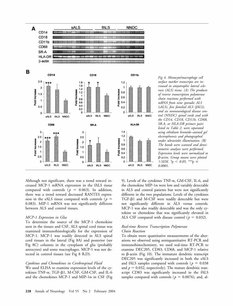
Monocytic/macrophage/microglial cell transcript levelsin ALS tissueTo ascertain the expression levels of activated microgliaand monocyte/macrophage transcripts in ALS tissues,we examined the expression of mRNA encoding thefollowing microglia/monocyte/macrophage cell surfacemarkers in sALS, fALS, and NNDC tissues normalizedto �-actin: CD14, CD18, CD11b, CD68, SR-A, andHLA-DR. As shown in Figure 4, the expression ofCD14, CD18, CD68, and SR-A mRNAs were signif-icantly increased in sALS spinal cord tissue comparedwith controls (p � 0.00017, p � 0.030, p � 0.00010,p � 0.046, respectively). CD18 and CD68 mRNAswere also significantly increased in the fALS spinal cordtissue compared with controls (p � 0.030, p �
0.000053, respectively). Although not significant, therewas a trend toward increased CD14 and SR-A mRNAexpression in the fALS tissue (p � 0.10 and p � 0.19,respectively) and increased HLA-DR mRNA expressionin the sALS tissue (p � 0.066) compared with con-trols. Fc�R� subunit mRNA was not detected in sALS,fALS, or control tissues.
Immunostaining for CD68 AntigenCD68 immunostained microglia were present in thespinal cords of ALS and control sections. In controlspinal cords, CD68 immunostaining was present inramified cells in the ventral horn (Fig 5B) and whitematter (see Fig 5D), but not in proximity to motorneurons, the vasculature, or the corticospinal tracts.However, in the ventral horns of ALS spinal cord sec-tions, there was an increased density of CD68-positivecells with enlarged cell processes, often in close prox-imity to normal appearing motor neurons (see Fig 5A).Several CD68-positive cells laden with lipofuscin alsowere present in proximity to destroyed neurons. Thedensity of ramified cells decreased in regions of the de-generating corticospinal tracts; instead, the cells transi-tioned to rounded macrophages of different sizesmostly with cleared cytoplasm (phagocytized lipids dis-solved during paraffin embedding; see Fig 5C). Cellswith monocyte morphology were also present (see Fig5E). Approximately 5% of the immunostained cellshad dendritic cell morphology, typically in a perivascu-lar location.
Cytokine mRNA LevelsTo determine if cytokines were increased, the expressionlevels of mRNA encoding the following cytokines wereexamined normalized to �-actin: TNF-� TGF-�, IL-1�, IL-6, interferon INF, GM-CSF, and M-CSF. As in-dicated in Figure 6, M-CSF mRNA expression was sig-nificantly increased in the fALS tissue as compared withcontrols (p � 0.017). Although not significant, therewas a trend toward decreased TGF-� and IL-1� ex-pression in sALS tissue ( p � 0.18 and p � 0.11,respectively) compared with controls. IL-6 mRNAwas detected in only two sALS tissues, but not infALS or control tissues. INF-� and GM-CSF mRNAswere not detected, although they were detected inlipopolysaccharide-activated monocytes (positive con-trol). The others were not significantly different fromcontrol levels.
Chemokine mRNA LevelsTo ascertain if there was an increase in chemokinesknown to attract inflammatory cells, we examined therelative expression of MCP-1, MIP-1, and RANTESmRNA normalized to �-actin. Figure 7 demonstratesthat MCP-1 mRNA was drastically increased in thesALS tissue compared with controls (p � 0.000044).
Fig 3. Immunostaining for CD83. (A) CD83-immunopositivedendritic cells in the lateral corticospinal tract. (B) CD83-positive monocytic cells (arrow) close to a vessel in the lateralcorticospinal tract. (C) CD83-positive dendritic cells in theinterfollicular region of the tonsil. (D) Control spinal cordshowing no cells immunostained for CD83. (A–D) Peroxidasereaction, magnification 280.
Henkel et al: Dendritic Cells and MCP-1 in ALS 227
Although not significant, there was a trend toward in-creased MCP-1 mRNA expression in the fALS tissuecompared with controls (p � 0.063). In addition,there was a trend toward decreased RANTES expres-sion in the sALS tissue compared with controls (p �0.083). MIP-1 mRNA was not significantly differentbetween ALS and control tissues.
MCP-1 Expression in GliaTo determine the source of the MCP-1 chemokineseen in the tissues and CSF, ALS spinal cord tissue wasexamined immunohistologically for the expression ofMCP-1. MCP-1 was readily detected in ALS spinalcord tissues in the lateral (Fig 8A) and posterior (seeFig 8C) columns in the cytoplasm of glia (probablyastrocytes) and some macrophages. MCP-1 was not de-tected in control tissues (see Fig 8 B,D).
Cytokines and Chemokines in Cerebrospinal FluidWe used ELISA to examine expression levels of the cy-tokines TNF-�, TGF-�1, M-CSF, GM-CSF, and IL-6and the chemokines MCP-1 and MIP-1� in CSF (Fig
9). Levels of the cytokines TNF-�, GM-CSF, IL-6, andthe chemokine MIP-1� were low and variably detectablein ALS and control patients but were not significantlydifferent in the two populations. Levels of the cytokinesTGF-�1 and M-CSF were readily detectable but werenot significantly different in ALS versus controls.MCP-1 was also readily detectable and was the only cy-tokine or chemokine that was significantly elevated inALS CSF compared with disease control (p � 0.032).
Real-time Reverse Transcription PolymeraseChain ReactionTo obtain more quantitative measurements of the alter-ations we observed using semiquantitative RT-PCR andimmunohistochemistry, we used real-time RT-PCR toexamine DEC205, CD83, CD68, and MCP-1 relativeto �-actin (Fig 10). The immature dendritic transcriptDEC205 was significantly increased in both the sALSand fALS samples compared with controls (p � 0.038and p � 0.032, respectively). The mature dendritic tran-script CD83 was significantly increased in the fALSsamples compared with controls (p � 0.0076), and, al-
Fig 4. Monocyte/macrophage cellsurface marker transcripts are in-creased in amyotrophic lateral scle-rosis (ALS) tissue. (A) The productsof reverse transcription polymerasechain reactions performed withmRNA from nine sporadic ALS(sALS), five familial ALS (fALS),and six nonneurological disease con-trol (NNDC) spinal cords and withthe CD14, CD18, CD11b, CD68,SR-A, or HLA-DR primers pairslisted in Table 2, were separatedusing ethidium bromide–stained gelelectrophoresis and photographedunder ultraviolet illumination. (B)The bands were scanned and densi-tometric analyses were performed.Expression levels were normalized to�-actin. Group means were plotted�SEM. *p 0.05, ***p �0.0001.
228 Annals of Neurology Vol 55 No 2 February 2004

though not significant, there was a clear trend towardincreased expression in the sALS samples (p � 0.066).The microglial transcript CD68 was significantly in-creased in both the sALS and fALS samples comparedwith controls (p � 0.0023 and p � 0.0094). In addi-tion MCP-1 transcripts were increased in the both thesALS (p � 0.00028) and fALS (p � 0.0031) samples
compared with controls. Thus, the real-time RT-PCRverified the results seen with semiquantitative RT-PCR.
Correlation of survival with presence of dendritic cellmarkersTwo sALS patients and three fALS patients died within1 year from first symptom. To determine whether
Fig 5. Immunostaining for CD68. (A) The external membrane (partially tangentially sectioned) of a motoneuron in the ventralhorn of an amyotrophic lateral sclerosis (ALS) spinal cord is decorated by microglial processes immunostained for CD68. (B) Thereis no CD68� reactivity of microglia in proximity of the motoneuron in a control spinal cord. (C) Many of the CD68� microglialcells transitioned to rounded macrophages in the lateral corticospinal tract of an ALS spinal cord (arrowheads). The overall densityof the cells immunostained for CD68 is increased. (D) The density and shape of microglial cells in the lateral corticospinal tract ofa normal spinal cord. (E) The CD68� macrophages and also monocytes tend to gather in the vicinity of a vessel (arrows). (A–E)Peroxidase reaction, A,B, 140; C–E, 180.
Henkel et al: Dendritic Cells and MCP-1 in ALS 229

there was a pattern of expression in these rapidly pro-gressing patients, we ranked the ALS patients from 1 to14 based on their level of expression for each tran-script. The patients who progressed the most rapidlyranked higher in dendritic transcripts, but not in themonocytic/macrophage/microglia transcripts. To assessif this correlation was significant, we grouped the rap-idly progressing patients and the slower progressing pa-tients and compared the expression of dendritic cell,monocytic/macrophage/microglial, cytokine, or chemo-kine transcripts (Fig 11). We performed analyses ofvariances to determine if the rapidly progressing pa-tients expressed an increase in transcripts comparedwith the slower progressing patients. Both the fast andslow progressing ALS groups were significantly differ-ent compared with the controls for dendritic cell (p �0.0000067 and p � 0.0020, respectively), monocytic/macrophage/microglial (p � 0.028 and p � 0.0012,respectively), and chemokine (p � 0.017 and p �0.0058, respectively) transcripts. However, only thedendritic cell transcripts (p � 0.050) were significantly
increased in the rapidly progressing patients comparedwith the slower progressing patients (monocytic/mac-rophage/microglial transcripts: p � 0.355, cytokinetranscripts: p � 0.499, chemokine transcripts: p �0.853). Thus, the more rapidly progressing ALS corre-lated with increased dendritic cell transcripts.
DiscussionDendritic cells are a diverse set of cells that function asmembers of the innate as well as adaptive immune sys-tems and in the latter capacity possess a highly efficientantigen-presenting capacity to stimulate naive T cells.33
Dendritic cells have been demonstrated in the CNS inautoimmune diseases such as MS and the animalmodel, EAE.19–21 A role for dendritic cells has beenestablished in both causing EAE and inducing toler-ance.22,34 To determine whether dendritic cells aresimilarly present in ALS, we assayed for expression ofdendritic cell transcripts in sALS and fALS tissues com-pared with controls. We demonstrated a significant in-crease of CD83 and CD40 (mature/activated dendriticcells markers) mRNA in spinal cord tissue of the sALSpatients and a significant increase of CD83, CD40,CD1a, and DEC-205 (the latter two expressed on den-dritic cells before maturation) mRNA in spinal cordtissue of fALS patients compared with controls. Immu-nohistochemical staining of sALS spinal cord sectionsshowed that both parenchymal and perivascular CD1aand CD83-positive cells were present in the ventralhorn, whereas mostly perivascular-positive cells werepresent in the degenerating corticospinal tract. BothCD1a- and CD83-positive cells were conspicuously ab-
Fig 7. Chemokine mRNA levels in amyotrophic lateral sclerosis(ALS) tissues. (A) The products of reverse transcription poly-merase chain reactions performed with mRNA from nine spo-radic ALS (sALS), five familial ALS (fALS), and six nonneu-rological disease control (NNDC) spinal cords and with theMCP-1, MIP-1�, or RANTES primers pairs listed in Table2 were separated using ethidium bromide–stained gel electro-phoresis and photographed under ultraviolet illumination. (B)The bands were scanned and densitometric analyses were per-formed. Expression levels were normalized to �-actin. Groupmeans were plotted �SEM. ***p � 0.000044.
Fig 6. Cytokine transcription levels in amyotrophic lateral scle-rosis (ALS) tissues. (A) The products of reverse transcriptionpolymerase chain reactions, performed with mRNA from ninesporadic ALS (sALS), five familial ALS (fALS), and six non-neurological disease control (NNDC) spinal cords and withthe TNF-�, TGF�, IL-1�, IL-6, or M-CSF primers pairslisted in Table 2, were separated using ethidium bromide–stained gel electrophoresis and photographed under ultravioletillumination. (B) The bands were scanned and densitometricanalyses performed. Expression levels were normalized to�-actin. Group means were plotted �SEM. *p � 0.017.
230 Annals of Neurology Vol 55 No 2 February 2004

sent in control tissue. The tissues used for the RT-PCRanalyses were frozen and not perfused; thus, mRNAfrom any blood cells present in the spinal cord vesselswould be in the RNA pool. Therefore, the dendriticcell transcripts detected in controls, when no dendriticcells were present in control tissues, could be tran-scripts from the blood cells in the vessels.
The presence of immature dendritic cells in ALS tis-sue, which can take up and process antigen as well asstimulate other cellular constituents of the innate im-mune system (such as microglia or natural killer cells),suggests an involvement in the ongoing immune/in-flammatory reaction. It is also relevant that among thefactors acting on immature dendritic cells are reactiveoxygen intermediates, aggregated IgG, and cytokines.33
All of these are well-known changes present in ALSand could contribute to the enlistment of immaturedendritic cells in CNS degeneration. As dendritic cellsmature, they lose their phagocytic properties and theirability to capture antigen, but express MHC class II aswell as costimulatory molecules and become potentantigen-presenting cells.35 The presence of both imma-ture and mature dendritic cells in ALS, and their ab-sence in control specimens, argues for their involve-ment in accelerating the pathogenesis of motor neuroninjury in ALS.
Our previous results and the results of others haveestablished the presence of monocyte/macrophage cellsand activated microglia in spinal cord tissue of mostALS patients.1,4,6 The increased expression of mRNAsencoding proteins expressed on monocytic/macro-phage/microglial cells (CD14, CD18, and SR-A) inspinal cord clearly confirms the presence of such con-stituents in ALS tissues. In addition, CD68� mono-cyte/macrophage and activated microglia cells wereseen dispersed in degenerating white matter, less exten-sively in the ventral horns, and packed around bloodvessels of ALS spinal cord tissues. Although the specificrole of these monocyte/macrophage cells is unclear,their frequent presence in proximity to normal appear-ing motor neurons in ALS ventral horns would suggestan active role, and not merely a phagocytic role, inmotor neuron injury. The presence of activated micro-glia and the therapeutic benefit of minocycline, an in-hibitor of microglial p38 MAPK, in the presymptom-atic mSOD1 transgenic mouse models of fALS provideadditional evidence for the potential role of microgliain motor neuron injury in ALS.16,17,36–38
To determine whether inflammatory cells are re-cruited to the CNS and/or differentiate from endoge-nous precursor cells, we examined the expression levelsof various cytokine and chemokine mRNAs. The most
Fig 8. MCP-1 immunohistochemistry. Immunohistochemistry was used to detect MCP-1 expression in amyotrophic lateral sclerosis(ALS) and control tissues. (A) MCP-1–immunopositive glia and macrophages in the lateral column of an ALS spinal cord. (C)MCP-1–immunopositive glia in the posterior column of an ALS spinal cord. (B, D) There are no immunolabeled cells for MCP-1in the lateral column or in the posterior column of the control spinal cord, A–D) Peroxidase reaction, 100.
Henkel et al: Dendritic Cells and MCP-1 in ALS 231

significant increase in ALS tissue was the 23-fold in-crease in MCP-1 mRNA compared with controls. Noother mRNA tested was increased as much. We deter-mined that MCP-1 expression was mostly in glia andin some macrophages in the CNS. In addition, MCP-1in the CSF of sALS patients was increased comparedwith controls.
MCP-1 is a potent chemoattractant and activatingpeptide that is expressed mostly in astrocytes but alsoin neurons, microglia, and macrophages after ischemia,hypoxia, or excitotoxicity. Excessive MCP-1 can exac-erbate an injury. It attracts CCR2-expressing myeloiddendritic cells, microglia/monocytes, and activated Tcells but may also regulate Th1 and Th2 responses.Mice deficient in MCP-1 or its receptor, CCR2, areresistant to EAE and have a significant reduction in Tcell and monocyte infiltration.20,31,32 Thus, MCP-1 isimportant in the recruitment of immune/inflammatorycells into the CNS, and more specifically the recruit-ment of dendritic cells and monocytes. Furthermore,the immunohistochemical demonstration of suchperivascular dendritic cells in both ventral horn and
white matter are in accord with recruitment from thecirculation.
A third key finding, in addition to the presence ofimmature and mature/activated dendritic cells in ALStissue and to the increased MCP-1 expression in CSFand tissue in ALS, is that increased expression of den-dritic cell transcripts correlated with the more rapidlyprogressing ALS. Two sALS patients and three fALSpatients died within 1 year from first symptom; theremainder of the patients died between 2.75 and 16.25years after first symptom. The five rapidly progressingpatients expressed significantly more dendritic tran-scripts, compared with the slower progressing patients,but not more monocytic/macrophage/microglial, cyto-kine, or chemokine transcripts. Thus, the correlation ofrapidly progressing ALS and increased dendritic celltranscripts support the involvement of the immune/in-flammatory system in amplifying the disease process.
Profiling mRNAs expressed in sALS and mSOD1transgenic mouse spinal cord tissues has recently beenused to investigate ALS.39–42 As each array examineddifferent sets of genes, the genes identified from each
Fig 9. MCP-1 increased in cerebro-spinal fluid (CSF). ELISA analyseswere performed on CSF samplesfrom 20 amyotrophic lateral sclerosis(ALS) and 20 control patients.MCP-1 levels from ALS patientswere significantly increased over thediseased control group (*p � 0.05);none of the other cytokines or che-mokines were significantly different.The boxes indicate the scores from25 to 75%. The lines in the boxesindicate the median; the smallblack squares indicate the mean.The vertical bars indicate the scoresfrom 5 to 95%. All of the TGF�,M-CSF, and MCP-1 levels werewithin the linear range; 25% of theALS and 15% of the controlTNF-� levels were below the linearrange; 40% of the ALS and 30%of the control IL-6 levels were be-low the linear range; 95% of theALS and 80% of the control GM-CSF levels were below the linearrange; and 100% of the ALS and90% of the control MIP-1� levelswere below the linear range.
232 Annals of Neurology Vol 55 No 2 February 2004

screen were different. Despite this, some categories ofgenes identified were the same. All four of the sALSand mSOD1 transgenic mouse screens showed in-creased expression of genes involved in inflammation.Although the two sALS screens did not identify genesencoding dendritic cell surface proteins, the list ofgenes in the array we were able to obtain did notinclude genes for dendritic cell markers (list providedby Ishigaki). In addition, one of the mSOD1 trans-genic mouse screens detected an increased expressionof CD63, a microglial activation marker that can beexpressed at high levels on a subset of dendriticcells.41,43
In summary, we have demonstrated the presence ofimmature and mature dendritic cells, increased num-bers of activated microglia/macrophages in ALS spinal
cord tissue, and increased levels of the chemokineMCP-1 that can recruit these cells from the systemiccirculation. Although something other than the im-mune system may well have initiated the motor neuroninjury and although macrophages could be playing aphagocytic role, we propose that the presence of im-mature dendritic cells, of the chemokine that attractsthem and their maturation to activated dendritic cells,indicate that dendritic cells may well be exacerbatingthe motoneuron injury in ALS. This is especially ap-parent in those patients who had the most rapid rate ofdecline and expressed higher levels of dendritic cellmarker mRNA. Thus, therapies specifically targeted todendritic cells and to the chemokine MCP-1 and itsreceptors may well offer meaningful pharmacologicalapproaches to ALS.
Fig 10. Real-time reverse transcription polymerasechain reaction (RT-PCR). Real-time RT-PCRwas used to examine DEC205, CD83, CD68,and MCP-1 relative to �-actin. The immaturedendritic transcript DEC205 was significantlyincreased in both the sporadic amyotrophic lateralsclerosis (sALS) and familial ALS (fALS) samplescompared with the nonneurological disease con-trol (NNDC) samples. The mature dendritictranscript CD83 was significantly increased inthe fALS samples compared with the NNDCsamples. The microglial transcript CD68 wassignificantly increased in both the sALS andfALS samples compared with the NNDC sam-ples. In addition, MCP-1 transcripts were in-creased in both the sALS and fALS samples com-pared with the NNDC samples. Thus, the real-time RT-PCR verified the results seen withsemiquantitative RT-PCR. *p 0.05, **p 0.01, *** p 0.005, **** p 0.0005.
Henkel et al: Dendritic Cells and MCP-1 in ALS 233

This study was supported by grants from the Muscular DystrophyAssociation (S.H.A., J.S.H.), the Hamill Foundation (S.H.A.), Drand Mrs G. Kozmetsky (S.H.A.), Methodist (39655, D.R.B.), Na-tional Scientific Research Fund of Hungary (OTKA T 034314,OTKA M 36252, L.S.), Ministry of Health of Hungary (ETT T04/001/2000, L.S.), National Scientific Research Fund of Hungary(OTKA T 026239, J.I.E.), and Ministry of Health of Hungary(ETT T-05/28/2000, FKFP-0032/2000, J.I.E.).
We are grateful to Y. K. Henry for the CSF ELISA assays.
References1. Engelhardt JI, Tajti J, Appel SH. Lymphocytic infiltrates in the
spinal cord in amyotrophic lateral sclerosis. Arch Neurol 1993;50:30–36.
2. Kawamata T, Akiyama H, Yamada T, McGeer PL. Immuno-logic reactions in amyotrophic lateral sclerosis brain and spinalcord tissue. Am J Pathol 1992;140:691–707.
3. Troost D, Van den Oord JJ, Vianney de Jong JMB. Immuno-histochemical characteristics of the inflammatory infiltration inamyotrophic lateral sclerosis. Neuropathol Appl Neurobiol1990;16:401–410.
4. Lampson LA, Kushner PD, Sobel RA. Major histocompatibilitycomplex antigen expression in the affected tissues in amyotro-phic lateral sclerosis. Ann Neurol 1990;28:365–372.
5. Engelhardt JI, Appel SH. IgG reactivity in the spinal cord andmotor cortex in amyotrophic lateral sclerosis. Arch Neurol1990;47:1210–1216.
6. Ince PG, Shaw PJ, Slade JY, et al. Familial amyotrophic lateralsclerosis with a mutation in exon 4 of the Cu/Zn superoxidedismutase gene: pathological and immunocytochemical changes.Acta Neuropathol 1996;92:395–403.
7. McGeer PL, Itagaki S, McGeer EG. Expression of the histo-compatibility glycoprotein HLA-DR in neurological disease.Acta Neuropathol 1988;76:550–557.
8. Sasaki S, Shibata N, Komori T, Iwata M. iNOS and nitroty-rosine immunoreactivity in amyotrophic lateral sclerosis. Neu-rosci Lett 2000;291:44–48.
9. Yasojima K, Tourtellotte WW, McGeer EG, McGeer PL.Marked increase in cyclooxygenase-2 in ALS spinal cord. Neu-rology 2001;57:952–956.
10. Grewal RP, Morgan TE, Finch CE. C1qB and clusterin mRNAincrease in association with neurodegeneration in sporadicamyotrophic lateral sclerosis. Neurosci Lett 1999;271:65–67.
11. Losy J, Michalowska-Wender G. Surface CD2, CD4, CD8markers and IL-2 and TNF-alpha cytokines in amyotrophic lat-eral sclerosis. Neurol Neurochir Pol 2001;35:57–61.
12. Poloni M, Facchetti D, Mai R, et al. Circulating levels of tu-mour necrosis factor-� and its soluble receptors are increased inthe blood of patients with amyotrophic lateral sclerosis. Neuro-sci Lett 2000;287:211–214.
13. Murakami N, McLennan IS, Nonaka I, et al. Transforminggrowth factor-�2 is elevated in skeletal muscle disorders. Mus-cle Nerve 1999;22:889–898.
14. Sekizawa T, Openshaw H, Ohbo K, et al. Cerebrospinal fluidinterleukin 6 in amyotrophic lateral sclerosis: immunologicalparameter and comparison with inflammatory and non-inflammatory central nervous system diseases. J Neurol Sci1998;154:194–199.
15. Ono S, Hu J, Shimizu N, et al. Increased interleukin-6 of skinand serum in amyotrophic lateral sclerosis. J Neuro Sci 2001;187:27–43.
16. Alexianu ME, Kozovska M, Appel SH. Immune reactivity in amouse model of familial ALS correlates with disease progres-sion. Neurology 2001;57:1282–1289.
17. Almer G, Guegan C, Teismann P, et al. Increased expression ofthe proinflammatory enzyme cyclooxygenase-2 in amyotrophiclateral sclerosis. Ann Neurol 2001;49:176–185.
18. Keep M, Elmer E, Fong KS, Csiszar K. Intrathecal cyclosporinprolongs survival of late-stage ALS mice. Brain Res 2001;894:327–331.
19. Hussien Y, Sanna A, Soderstrom M, et al. Glatiramer acetateand IFN-beta act on dendritic cells in multiple sclerosis. J Neu-roimmunol 2001;121:102–110.
20. Huang YM, Kouwenhoven M, Jin YP, et al. Dendritic cellsderived from patients with multiple sclerosis show high CD1aand low CD86 expression. Mult Scler 2001;7:95–99.
21. Fischer HG, Reichmann G. Brain dendritic cells andmacrophages/microglia in central nervous system inflammation.J Immunol 2001;166:2717–2726.
22. Serafini B, Columba-Cabezas S, Di Rosa F, Aloisi F. Intracere-bral recruitment and maturation of dendritic cells in the onsetand progression of experimental autoimmune encephalomyeli-tis. Am J Pathol 2000;157:1991–2002.
Fig 11. Patients with rapidly progressing amyotrophic lateralsclerosis (ALS) expressed significantly more dendritic transcriptsthan those patients who progressed more slowly. Patients whodied within 1 year from first symptom were grouped and com-pared with those patients who progressed more slowly. Theirdendritic transcript expression levels were compared. Patientswith rapidly progressing ALS expressed significantly more den-dritic transcripts than those patients who progressed moreslowly (p � 0.050). Patients with rapidly progressing ALSexpressed equivalent monocytic/macrophage/microglial tran-scripts, cytokine transcripts, and chemokine transcripts as thosepatients who progressed more slowly. Both the rapidly progress-ing and slower progressing patients expressed more dendritictranscripts, monocytic/macrophage/ microglial transcripts, andchemokine transcripts than the nonneurological disease control(NNDC) patients (*).
234 Annals of Neurology Vol 55 No 2 February 2004

23. Matyszak MK, Perry VH. The potential role of dendritic cellsin immune-mediated inflammatory diseases in the central ner-vous system. Neuroscience 1996;74:599–608.
24. Caux C, Ait-Yahia S, Chemin K, et al. Dendritic cell biologyand regulation of dendritic cell trafficking by chemokines.Springer Semin Immunopathol 2000;22:345–369.
25. Pashenkov M, Huang YM, Kostulas V, et al. Two subsets ofdendritic cells are present in human cerebrospinal fluid. Brain2001;124:480–492.
26. Kostulas N, Li HL, Xiao BG, et al. Dendritic cells are presentin ischemic brain after permanent middle cerebral artery occlu-sion in the rat. Stroke 2002;33:1129–1134.
27. Pashenkov M, Teleshova N, Kouwenhoven M, et al. Recruit-ment of dendritic cells to the cerebrospinal fluid in bacterialneuroinfections. J Neuroimmunol 2002;122:106–116.
28. Tran EH, Hoekstra K, van Rooijen N, et al. Immune invasionof the central nervous system parenchyma and experimental al-lergic encephalomyelitis, but not leukocyte extravasation fromblood, are prevented in macrophage-depleted mice. J Immunol1998;161:3767–3775.
29. Santambrogio L, Belyanskaya SL, Fischer FR, et al. Develop-mental plasticity of CNS microglia. Proc Natl Acad Sci USA2001;98:6295–6300.
30. Fischer HG, Bielinsky AK. Antigen presentation function ofbrain-derived dendriform cells depends on astrocyte help. IntImmunol 1999;11:1265–1274.
31. Fife BT, Huffnagle GB, Kuziel WA, Karpus WJ. CC chemo-kine receptor 2 is critical for induction of experimental auto-immune encephalomyelitis. J Exp Med 2000;192:899–905.
32. Izikson L, Klein RS, Charo IF, et al. Resistance to experimentalautoimmune encephalomyelitis in mice lacking the CC chemo-kine receptor (CCR)2. J Exp Med 2000;192:1075–1080.
33. Lipscomb MF, Masten BJ. Dendritic cells: immune regulatorsin health and disease. Physiol Rev 2002;82:97–130.
34. Huang YM, Yang JS, Xu LY, et al. Autoantigen-pulsed den-dritic cells induce tolerance to experimental allergic encephalo-myelitis (EAE) in Lewis rats. Clin Exp Immunol 2000;122:437–444.
35. Banchereau J, Steinman RM. Dendritic cells and the control ofimmunity. Nature 1998;392:245–252.
36. Van Den Bosch L, Tilkin P, Lemmens G, Robberecht W. Mi-nocycline delays disease onset and mortality in a transgenicmodel of ALS. Neuroreport 2002;13:1067–1070.
37. Zhu S, Stavrovskaya IG, Drozda M, et al. Minocycline inhibitscytochrome c release and delays progression of amyotrophic lat-eral sclerosis in mice. Nature 2002;417:74–78.
38. Kriz J, Nguyen MD, Julien JP. Minocycline delays motor neu-ron degeneration and slows down disease progression in amouse model of ALS. J Neurol Sci 2002;199(suppl 1):S34.
39. Ishigaki S, Niwa J, Ando Y, et al. Differentially expressed genesin sporadic amyotrophic lateral sclerosis spinal cords—screeningby molecular indexing and subsequent cDNA microarray anal-ysis. FEBS Lett 2002;531:354–358.
40. Malaspina A, Kaushik N, de Belleroche J. Differential expres-sion of 14 genes in amyotrophic lateral sclerosis spinal cord de-tected using gridded cDNA arrays. J Neurochem 2001;77:132–145.
41. Olsen MK, Roberds SL, Ellerbrock BR, et al. Disease mecha-nisms revealed by transcription profiling in SOD1–G93A trans-genic mouse spinal cord. Ann Neurol 2001;50:730–740.
42. Yoshihara T, Ishigaki S, Yamamoto M, et al. Differential ex-pression of inflammation- and apoptosis-related genes in spinalcords of a mutant SOD1 transgenic mouse model of familialamyotrophic lateral sclerosis. J Neurochem 2002;80:158–167.
43. Brooke GP, Sopp P, Kwong LS, Howard CJ. Molecular clon-ing of cattle CD63 and evidence for high level expression onsubpopulations of dendritic cells. Immunogenetics 1999;49:812–814.
44. Draude G, Lorenz RL. TGF-�1 down regulates CD36 andscavenger receptor A but upregulates LOX-1 in human macro-phages. Am J Physiol Heart Circ Physiol 2000;278:H1042–H1048.
45. Leveque C, Grafte S, Paysant J, et al. Regulation of interleukin3 receptor alpha chain (IL-3R alpha) on human monocytes byinterleukin (IL)-4, IL-10, IL-13, and transforming growth fac-tor beta (TGF-beta). Cytokine 1998;10:487–494.
46. Noguera A, Batle S, Miralles C, et al. Enhanced neutrophil re-sponse in chronic obstructive pulmonary disease. Thorax 2001;56:432–437.
47. Uwai M, Terui Y, Mishima Y, et al. A new apoptotic pathwayfor the complement factor B-derived fragment Bb. J CellPhysiol 2000;185:280–292.
48. Cario E, Rosenberg IM, Brandwein SL, et al. Lipopolysaccha-ride activates distinct signaling pathways in intestinal epithelialcell lines expressing Toll-like receptors. J Immunol 2000;164:966–972.
49. Luk JM, Lai W, Tam P, Koo MW. Suppression of cytokineproduction and cell adhesion molecule expression in humanmonocytic cell line THP-1 by Tripterygium wilfordii polysac-charide moiety. Life Sci 2000;67:155–163.
50. Lienenluke B, Germann T, Kroczek RA, Hecker M. CD154stimulation of interleukin-12 synthesis in human endothelialcells. Eur J Immunol 2000;30:2864–2870.
51. Kato M, Neil TK, Fearnley DB, et al. Expression of multilectinreceptors and comparative FITC-dextran uptake by humandendritic cells. Int Immunol 2000;12:1511–1519.
52. Liu Y, Santin AD, Mane M, et al. Transduction and utility ofthe granulocyte-macrophage colony-stimulating factor gene intomonocytes and dendritic cells by adeno-associated virus. J In-terferon Cytokine Res 2000;20:21–30.
53. Lichtenberger C, Zakeri S, Baier K, et al. A novel high-purityisolation method for human peripheral blood neutrophils per-mitting polymerase chain reaction-based mRNA studies. J Im-munol Methods 1999;227:75–84.
54. Lesnikov V, Lesnikova M, Deeg HJ. Pro-apoptotic and anti-apoptotic effects of transferrin and transferrin-derived glycanson hematopoietic cells and lymphocytes. Exp Hematol 2001;29:477–489.
55. McGuinness PH, Painter D, Davies S, McCaughan GW. In-creases in intrahepatic CD68 positive cells, MAC387 positivecells, and proinflammatory cytokines (particularly interleukin18) in chronic hepatitis C infection. Gut 2000;46:260–269.
56. Atkins GJ, Haynes DR, Geary SM, et al. Coordinated cytokineexpression by stromal and hematopoietic cells during humanosteoclast formation. Bone 2000;26:653–661.
57. De Rossi M, Bernasconi P, Baggi F, et al. Cytokines and che-mokines are both expressed by human myoblasts: possible rel-evance for the immune pathogenesis of muscle inflammation.Int Immunol 2000;12:1329–1335.
58. Hatano Y, Katagiri K, Takayasu S. Increased levels in vivo ofmRNAs for IL-8 and macrophage inflammatory protein-1 alpha(MIP-1 alpha), but not of RANTES mRNA in peripheralblood mononuclear cells of patients with atopic dermatitis(AD). Clin Exp Immunol 1999;117:237–243.
59. Koller MR, Oxender M, Jensen TC, et al. Direct contact be-tween CD34�lin- cells and stroma induces a soluble activitythat specifically increases primitive hematopoietic cell produc-tion. Exp Hematol 1999;27:734–741.
Henkel et al: Dendritic Cells and MCP-1 in ALS 235