preperitoneal space: where and how to access...
TRANSCRIPT
Scholarly Journal of Surgery Volume 1: 1Scholarly J Surg 2018
Preperitoneal Space: where and how to Access during Laparoscopic Total Extraper-itoneal Preperitoneal (TEPP) Inguinal Hernioplasty
Article Information
Maulana Mohammed Ansari1*1Ex-Professor of General Surgery, Department of Surgery, J. N. Medical Col-lege, Aligarh Muslim University, Aligarh, UP, India.
Article Type: ResearchArticle Number: SJS105Received Date: 26 March, 2018Accepted Date: 23 April, 2018Published Date: 30 April, 2018
*Corresponding author: Dr. Maulana Mohammed Ansari, Ex-Professor of General Surgery, Department of Surgery, B-27, Silver Oak Avenue, Street No. 4, Dhaurra Mafi, Aligarh, UP, India. Tel: +0091-9557449212; Email: [email protected].
Citation: Ansari MM (2018) Preperitoneal Space: where and how to Access during Laparoscopic Total Extraperitoneal Preperitoneal (TEPP) Inguinal Hernioplasty. Scholarly J Surg. Vol: 1, Issu: 1 (20-34).
Copyright: © 2018 Ansari MM. This is an open-access article distributed under the terms of the Creative Commons Attribution License, which permits unrestricted use, distribution, and reproduction in any medium, provided the original author and source are credited.
AbstractLaparoscopic revolution made inguinal hernioplasty a topic of intense
study again. Most crucial step in laparoscopic total extraperitoneal preperitoneal (TEPP) repair is creation of an avascular preperitoneal space. Preperitoneal access requires balloon dissection or telescopic dissection with/without prior needle insufflation. Present study prospectively evaluated sixty adult male patients undergoing TEPP hernioplasty (N=65) for inguinal hernia (N=68). Standard 3-midline-port technique with mainly direct telescopic dissection was utilized. Patients were evenly distributed across the 3rd to 8th decade of life and across their professional occupation. Mean patient age was 50.1 ± 17.2 years with a range of 18-80 years, and their mean BMI was 22.6 ± 2.0 kg/m2 with a range of 19.3-31.2 kg/m2). There occurred early conversion to open anterior repair (n=1), open preperitoneal repair (n=1) and transabdominal laparoscopic repair (n=1). Access to avascular preperitoneal plane was made at or just below the level of the middle working port by surgically creating a transverse opening either through transversalis fascia in presence of a classical incomplete posterior rectus sheath with a primary Arcuate line (N=41), or through both posterior rectus sheath and transversalis fascia in presence of a complete posterior rectus sheath without a primary Arcuate line (N=14) or a long incomplete posterior rectus sheath with a low primary Arcuate line (N=10). No conversion was forced secondary to the so-called difficult dissection, and there was no major complication. Unhurried telescopic dissection provided accurate recognition of morphology of posterior wall of posterior rectus canal for its judicious division, invariably resulting in successful access to preperitoneal space for joyful completion of TEPP repair, and the technique with all steps under direct vision, the most fundamental principle of modern laparoscopy, is strongly recommended. Sound understanding of variable multi-layered anatomy of preperitoneal access is essential for seamless TEPP hernioplasty and widespread popularization.
Keywords: Preperitoneal space, Preperitoneal space access, Telescopic dissection, Balloon dissection, laparoscopic hernioplasty, TEP, TEPP.
Abbreviations and AcronymsTEP/TEPP: total extraperitoneal preperitoneal; PRC: posterior
rectus canal; RF: rectusial fascia; PRS: posterior rectus sheath; IC-PRS: incomplete posterior rectus sheath; L-PRS: long incomplete posterior rectus sheath; C-PRS: complete posterior rectus sheath; TF: transversalis fascia; PPF: preperitoneal fascia; ISF: internal spermatic fascia; PPS: preperitoneal space; RA: rectus abdominis muscle; HIS: indirect inguinal hernial sac; DH: direct inguinal hernial sac; DHD: direct hernial defect;

www.innovationinfo.org
Scholarly J Surg 2018 21
V: deep inferior epigastric vessels; GV: gonadal vessels; CM: corona mortis; PL: pectineal ligament; D: vas deferens; CS: cord structures; S: sign of lighthouse; P: plastic working port; P1: level of infraumbilical first (optical) port; P2: level of middle working port; P3: level of suprapubic working port; H: hernia; R: deep inguinal ring; IPT: iliopubic tract; L: cord lipoma.
IntroductionIntroduction of the laparoscopic approach made the
hernia repair a topic of intense study [1]. The crucial step in the laparoscopic total extraperitoneal preperitoneal (TEP/TEPP) repair for the inguinal hernia is the creation of an avascular preperitoneal space, and there are several methods of access to the proper preperitoneal space [2], including balloon dissection and telescopic dissection with/without prior needle insufflation (Dulucq technique) [3]. Even recently in 2009, Edward Felix [4] documented that ‘Initially, the dissection of the extraperitoneal space in the TEP approach tended to be difficult, confusing and therefore hard to learn’, ratifying the early experience of difficulty and time-consuming nature of the TEP hernioplasty [5] and often time-consuming as well as frustrating [6]. Therefore, it is paramount to correctly access a proper avascular preperitoneal plane for seamless creation of ample space for adequate mesh placement [7-9] because neither the Retzius space nor the Bogros space is a single anatomic entity [10-12], and the ‘surgical preperitoneal plane’ between the transversalis fascia and the preperitoneal fascia is the correct plane suitable for dissection for mainly three reasons, viz., firstly, this interfascial plane is easily cleavable and almost totally avascular with occasional perforating vessels between the transversalis fascia and the preperitoneal fascia [13], and the blood supply to the transversalis fascia and structures superficial to it come from the inferior epigastric and external iliac vessels while the blood vessels to the preperitoneal fascia and subjacent tissues originate from the internal iliac vessels [14]; secondly, the loose fissile lateral sub-inguinal space and the medial retro-pubic space are readily communicated in this interfascial anatomical plane; and thirdly, one side can be easily extended to the contralateral side for the simultaneous bilateral hernioplasty [9,11,12,15].
In 1999, Lucas and Arregui [16] prophesied that continued refinement of the techniques and efforts of the cost reduction would help in the greater role of the laparoscopic hernia repair. Therefore, in order to make the TEP procedure more cost-effective and complication-free, controlled direct-vision telescopic dissection is now preferred over to the risky blind balloon dissection [17-20]. However, the exact methods for the access to the preperitoneal space underneath the transversalis fascia are either not described at all or described dubiously against the classical anatomical understanding as well as the current perspectives of the preperitoneal anatomy [9,11,12,21-29]. For example, even recently in 2017, Abd-Raboh et al. [19] stated that ‘As the posterior rectus sheath ends at the line of Douglas (arcuate line), the telescope (0°, 10 mm) passed on top of the posterior rectus sheath will automatically fall
into the extraperitoneal space.’ Exactly similar description was repeated in 2018 by Hisham et al. [20]. Such statements are commonly made by the operating surgeons but in reality they invite four reservations, namely, firstly, the telescope passing over an incomplete posterior rectus sheath, if one is present [26,29,30-33], will not automatically fall into the extraperitoneal space but really result in passing over and anterior to the transversalis fascia which must be surgically opened to enter the extraperitoneal space behind the transversalis fascia, the preperitoneal space [9,34], and secondly, a well-defined arcuate line is often absent [21,23,27,29], and thirdly, the position of the arcuate line is highly variable [23,35,36], and fourthly, the Retzius space is not a single anatomical entity [10-12]. Lack of the sound knowledge of the preperitoneal anatomy has been reported as the most limiting aspect of the laparoscopic hernioplasty [37,38], and a correct access to and dissection in an easily-cleavable avascular plane allows not only rapid identification of the exact anatomy but also facilitates in the creation of the requisite preperitoneal space in a smooth blood-less fashion [7]. Herein, as a fruit of personal expertise and experience, we describe the exact techniques of access to the proper preperitoneal space based on the real-time anatomical planes and landmarks during the TEP inguinal hernioplasty with the unhurried controlled direct telescopic dissection across the variable preperitoneal anatomy of the posterior wall of the posterior rectus canal [9,11,12,22-32,35,36,39].
Materials and MethodsA prospective doctoral research study was carried
out in the Jawaharlal Nehru Medical College Hospital of Aligarh Muslim University, Aligarh, India from April, 2010 to November, 2015 under the institutional ethics committee’s approval and patients’ informed consent. Laparoscopic total extraperitoneal preperitoneal (TEPP) hernioplasty was performed for primary inguinal hernia by a single experienced senior surgeon, the author. The study inclusion criteria were (1) patients’ age 18 years and above, (2) uncomplicated inguinal hernia, (3) ASA grade I-II only of American Society of Anesthesiologists, and (4) informed consent for the laparoscopic repair. The exclusion criteria included (1) lack of consent for laparoscopic repair, (2) age lower than18 years, (3) ASA grade III and IV, (3) recurrent/complicated inguinal hernia, (5) femoral hernia, and (6) past history of lower abdominal surgery. Patients’ body mass index (BMI) was calculated by the Deurenberg’s formula of body weight in Kg divided by square of height in meters.
The surgical technique for the TEPP hernioplasty involved the posterior rectus sheath approach with three ports placed in the midline (one infraumbilical 10-mm port about 2 cm below lower lip of the umbilicus, second 5-mm port between the infraumbilical and the suprapubic ports, and third 5-mm port about 2.5 cm above the upper border of the symphysis pubis). CO2 insufflation was maintained at a pressure of 12 mmHg. Indigenous balloon made of fingerstall of a surgical glove was utilized in first three cases. Direct telescopic initial dissection was used in the remainder of the patients of the study. This was followed by further definitive sharp/blunt dissection by the instruments. Details of the

www.innovationinfo.org
Scholarly J Surg 2018 22
surgical technique were consistently the same as reported earlier by the author [9,11,12,22-29,33,36,40,41].
ResultsA total of 66 adult patients (Age ≥ 18 years) with
inguinal hernia (Male 63; Female 3) were selected for recruitment into the present study. None of the female patients were included for the laparoscopic hernia repair due to one or more exclusion criteria. Three out of 63 male patients had to be excluded from the study due to early conversions [to laparoscopic transabdominal repair (n=1); to open repair (n=2)]. Causes of the conversion were (1) pneumoperitoneum due to peritoneal injury by the very first trocar (optical port), (2) injury to the deep inferior epigastric vessels by the roughened joint of the Maryland dissector after place of the middle working port, and (3) hemodynamic deterioration due to excessive CO2 retention. Thus only 68 primary inguinal hernias (Unilateral 52; Bilateral 8) were repaired successfully by the laparoscopic TEPP approach in the 60 adult male patients (Unilateral TEPP 52; Simultaneous Bilateral TEPP 5; Interval Bilateral TEPP 3). Patients were evenly distributed across the 3rd to 8th decade of life (Table 1). Mean patient age was 50.1 ± 17.2 years with a range of 18-80 years (Table 2), and their mean BMI was 22.6 ± 2.0 kg/m2 with a range of 19.3-31.2 kg/m2) (Table 3). By occupation, the patients were manual labourers (N=24), retired persons (N=9), office workers (N=8), students (N=7), field workers (6), and farmers (N=6).
The types of the posterior rectus sheath were not affected significantly (p>0.05) by the variation in the age (Table 2); however, the overweight/obese patients had the short type of the incomplete PRS significantly (p <0.001) although their number was small in the study (Table 3). Details of these statistics were reported elsewhere by the author [29].
In 30 out of 41 patients with a traditional normal-length incomplete posterior rectus sheath with a classical well-defined primary arcuate line situated at about 1/3rd of the umbilico-pubic distance, the tip of the telescope
itself was used to pierce the transversalis fascia just below the arcuate line by gently gliding the scope in a to-and-fro fashion over the lower of the posterior rectus sheath to enter the preperitoneal space underneath the transversalis fascia. Once the scope was within the preperitoneal space between the transversalis fascia and the preperitoneal fascia, this space was widened as much as easily possible by the to-and-fro and side-to-side movements of the scope, and then the middle working 5-mm port was put in under needle confirmation at the previously marked site, roughly corresponding to the midpoint of the umbilico-pubic distance, so as to enlarge the opening in the transversalis fascia and to widen the preperitoneal space as much as easily possible in an avascular fashion with occasional use of the monopolar electrocautery before division of the fibro areolar tissue strands to keep the field in clean pristine perspective. Whenever telescopic entry into the preperitoneal space was not easily achieved as has happened in 11 out of 41 patients with a normal-length incomplete posterior rectus sheath with classical primary arcuate line, the working 5-mm ports were placed at the previously marked sites under the needle confirmation, and a fine-tipped Maryland dissector or simple curved scissors without cautery was used to make a transverse opening in the transversalis fascia just below the classical well-defined arcuate line to enter the preperitoneal space (Figure 1), and then the preperitoneal space was widened with blunt/sharp instrument dissection. In presence of a narrow posterior rectus canal (N=17) with limitation of the lateral visualization, the lateral end of the classical arcuate line was carefully divided further laterally under the cautery control due to frequent presence of an arterial twig and to avoid injury to the often-adherent peritoneum at the Spigelian line.
In 2 out of 3 cases with a high primary arcuate line (present within 3 cm of the umbilicus), the aforesaid technique was utilised successfully, i.e., the transversalis fascia was perforated with the tip of the telescope at the level of the marked site for the middle working port, roughly corresponding to the midpoint of the umbilico-pubic
S. No.Age Groups Male Female Total
(Years) N % N N %1 18-30 9 15.0 0 9 15.02 31-40 11 18.3 0 11 18.33 41-50 11 18.3 0 11 18.34 51-60 12 20.0 0 12 20.05 61-70 9 15.0 0 9 15.06 71-80 8 13.3 0 8 13.3
Total (18-80) 60 100 0 60 100
Table 1: Age/Sex distribution of patients undergoing laparoscopic total extraperitoneal preperitoneal (TEPP) hernioplasty for primary inguinal hernia.
S. No. PRS Type Hernias Patients Age Mean ± SD (Range) F- Value Sig. (2-Tailed) p- ValueN % N % Years
1. NIC-PRS 41 75.9 35 74.5 50.5 ± 17.9 (18-80)
F3 56 = 0.785 0.507 >0.052. LIC-PRS 10 18.5 9 19.1 55.2 ± 11.6 (40-72)3. SIC-PRS 03 5.6 3 6.4 54.0 ± 12.2 (40-62)4. C-PRS 14 20.6 13 21.7 44.5 ± 19.2 (19-72)
Total 68 100 60 100 50.1 ± 17.2 (18-80)
Table 2: Age distribution of patients with different types of posterior rectus sheath (PRS) according to its extent. PRS, posterior rectus sheath; NIC-PRS, normal-length incomplete PRS; LIC-PRS, long incomplete PRS; SIC-PRS, short incomplete PRS; C-PRS, complete PRS; SD, standard deviation; F, one-way analysis of variance value; Sig., significance; p>0.05, insignificant.

www.innovationinfo.org
Scholarly J Surg 2018 23
distance. In the remaining one patient with a high arcuate line, both the transversalis fascia and the peritoneum got perforated by the inadvertent angulation of the blunt tip of the first trocar just after its placement, forcing conversion to the transabdominal approach (TAPP).
In patients with a complete posterior rectus sheath (N=14) and patients with a long incomplete posterior rectus sheath with a low arcuate line (N=10), entry into the preperitoneal space was made at or just below the level of the middle working port by making a transverse opening in the posterior rectus sheath (with transversely running tendinous/attenuated fibres) with the tip of the Maryland dissector initially without cautery (Figure 2 and 3). The new opening in the posterior rectus sheath (artificial arcuate line) was then enlarged widely in the transverse axis from midline to the Spigelian line (linea semicircularis) under minimal cautery control to visualise the transversalis fascia underneath, which is then opened up cautiously to enter the proper preperitoneal space. Thereafter, the preperitoneal space between the transversalis fascia and the preperitoneal fascia was widened by blunt &/or sharp dissection vertically into the pelvis and horizontally from the midline to the
anterior superior iliac spine (ASIS). In presence of a narrow posterior rectus canal (N=7) with limitation of the lateral visualization, the opening (artificial Arcuate line) in the posterior rectus sheath was carefully enlarged laterally (Figure 4B).
DiscussionGolden words of certain clinical investigators are
worth repeating verbatim to credit them for spreading the intellectual inspiration and to highlight the issue at hand. ‘As anatomic knowledge has increased; the evolution of laparoscopic surgery has paralleled that of open procedures’ [42]. ‘The techniques of laparoscopic hernia repair have evolved in parallel with experience and technology.’ [43]. ‘Major innovations continue to occur in the operative techniques used in hernia operations’ [44]. ‘Since our initial report in 1992 [11], despite earlier bias and criticism inherent to a maturing technique, the evolution of TEP has been apparent’ [45].
Laparoscopic total extraperitoneal preperitoneal (TEP) repair is probably the best laparoscopic technique of inguinal hernioplasty in most situations [3,46], and current
S. No. PRS Type Hernias Patients BMI Mean ± SD (Range) F-Value Sig. (2-Tailed) p- ValueN % N % Kg/m2
1. NIC-PRS 41 75.9 35 58.3 22.2 ± 1.7 (19.3-27.5)
F3 56 = 17.314 0.000 <0.0012. LIC-PRS 10 18.5 9 15.0 21.8 ± 0.7 (20.9-23.2)3. SIC-PRS 03 5.6 3 5.0 28.6 ± 2.4 (26.5-31.2)4. C-PRS 14 20.6 13 21.7 22.7 ± 1.1 (20.9-24.3)
Total 68 100 60 100 22.6 ± 2.0 (19.3-31.2)
Table 3: BMI distribution of patients with different types of posterior rectus sheath (PRS) according to its extent. PRS, posterior rectus sheath; NIC-PRS, normal-length incomplete PRS; LIC-PRS, long incomplete PRS; SIC-PRS, short incomplete PRS; C-PRS, complete PRS; SD, standard deviation; F, one-way analysis of variance value; Sig., significance; p <0.001, significant.
Figure 1: Access to Preperitoneal Space during Posterior Rectus sheath Approach of Right Laparoscopic Total Extraperitoneal Preperitoneal Inguinal Hernioplasty in a Patient with Incomplete Posterior Rectus Sheath: (A) Dissection in posterior rectus canal bounded anteriorly by Rectusial fascia (RF) covering rectus abdominis muscle and posteriorly by incomplete posterior rectus sheath (IC-PRS) in upper part and transversalis fascia (TF) in lower part; (B-F) creation of an opening in transversalis fascia (TF) just below Arcuate Line (arrow) to enter preperitoneal space; (Reproduced with permission from Ansari’s Thesis [41]).

www.innovationinfo.org
Scholarly J Surg 2018 24
NICE guidance also prefers TEP over TAPP (transabdominal preperitoneal) where feasible depending upon surgeon’s preference and patient choice [47]. However, its technical complexity, higher cost and long learning curve are regarded as the main cause for the criticism of the laparoscopic TEP hernioplasty [48-52].
Establishing a preperitoneal space is the most crucial step in the laparoscopic TEP inguinal hernioplasty [53]. During TEP hernioplasty, the preperitoneal space is created most commonly by the technique of balloon dissection because of its ease and rapidity [1,19,20,54], but a balloon dissector is not always necessary because the preperitoneal space can be safely created without difficulty through a meticulous dissection under excellent magnification of modern laparoscopy by an experienced competent surgeon [55]. Moreover, the branded disposable dissection balloons are really expensive, adding to the high cost of the procedure in addition to the disposable mesh-fixation devices [3], although cheaper alternatives to using a relatively expensive
balloon dissector have been described [56]. Balloon dissectors, branded or indigenous, are commonly used in a blind manner with associated risks of failure, inadequate dissection, wrong-plane dissection, bringing down of the deep inferior epigastric vessels, balloon rupture, bleeding, and bladder neck rupture [17,18,20,34,55]; risk of injury to vessels, peritoneum, bowel and bladder is especially present in patients with history of previous lower abdominal surgery [55,57]. Furthermore, ‘When the balloon is inflated, almost all of the soft tissues and minor vessels are compressed by the balloon; it crushes the surrounding tissue indiscriminately, and the soft tissue fragments and oozing from the minor vessels can obscure the operative field’ [55]. See-through balloons are much costlier, and they do allow vision across the inflating balloon but tissue dissection remains still uncontrolled and essentially blind. It is common experience that the telescopic dissection is feasible in the experienced hands while balloon dissection is easier for the beginners because the direct telescopic dissection is often cumbersome, time-
Figure 2: Access to Preperitoneal Space during Posterior Rectus sheath Approach of Right Laparoscopic Total Extraperitoneal Preperitoneal Inguinal Hernioplasty in a Patient with Complete Posterior Rectus Sheath: (A) Dissection in posterior rectus canal bounded anteriorly by Rectusial fascia (RF) covering rectus abdominis muscle and posteriorly by grossly-attenuated complete posterior rectus sheath (C-PRS) extending upto pubic bone; (B-F) creation of an transverse opening (artificial arcuate line) in complete posterior rectus sheath at about the level of the middle port (P1); (G-H) Dissection underneath posterior rectus sheath and placement of second working port (P2) after needle confirmation (N); (I-J) Creation of an opening in transversalis fascia (TF) to enter preperitoneal space; (K) Entry into lateral preperitoneal space underneath transversalis fascia (TF); (L) Division of transversalis fascia (TF) to join the lateral interfascial preperitoneal space and the medial prefascial (anterior to transversalis fascia) preperitoneal space; (Reproduced with permission from Ansari’s Thesis [41]).

www.innovationinfo.org
Scholarly J Surg 2018 25
consuming and frequently messy [44,46]. It simply means that we do not really understand the real-time anatomy at hand as lamented by Arregui [58] and certified by a number of investigators [44,51], thereby approving a lame rationale for the use of balloon dissectors. In 1994, James Rosser [38] rightly pointed out that ‘the textbook description does not provide an organized, common-sense-based map of the preperitoneal anatomy to help the surgeon to perform the laparoscopic hernia repair with ease and safety’.
In our study, the direct telescopic dissection under CO2 insufflation through the infraumbilical port afforded excellent view of the anatomical planes and surgical landmarks in majority (92%) of our patients. Grade I blood staining of Kang’s classification [55] was easily attainable with initial unhurried controlled telescopic dissection within the posterior rectus canal, which made it possible for clear visualization of the posterior wall of the posterior rectus canal for timely recognition of its varied morphology and judicious further dissection. Moreover, no major complication occurred during and after the procedure in the present study, and there was no conversion secondary to the so-called difficult dissection.
It was observed in the presented study that the lower part of the posterior rectus canal and conventional Retzius space bounded posteriorly by the transversalis fascia is not the proper preperitoneal space, and this anatomic disposition has been reported earlier by the author [9], confirming the
observations of Lange et al. [34]. It is strange and of interest to note that those who investigated TEP hernioplasty without balloon dissection [19,20,55] failed to describe the exact anatomy and technique of the crucial initial access to the proper preperitoneal space while working within the posterior rectus canal bounded posteriorly either by the incomplete posterior rectus sheath in upper part and the transversalis fascia in the lower part, or only by the complete posterior rectus sheath in upper part as well as the lower part and lined underneath by the transversalis fascia [11,26,29]. The statement that the telescope passing over the posterior rectus sheath automatically enters into the preperitoneal space as described by a number of clinical investigators including Lange et al. [34], Abd-Raboh et al. [19] and Hisham et al. [20], is anatomically erroneous and unacceptable but perpetuated verbatim in the literature; instead such a dissection technique leads to a dissection in the prefascial plane of the classical Retzius space anterior to the transversalis fascia and not into the requisite proper preperitoneal space [9]. Entry into the proper preperitoneal space is feasible only when the transversalis fascia below the classical arcuate line of the incomplete posterior rectus sheath is opened up surgically (Figure 1 and 6). The present study confirmed the observations of Lange et al. [34] that the telescope present within the posterior rectus canal and the Retzius space cannot enter the requisite preperitoneal space without first breaking the posterior fascial boundary of the posterior rectus canal or the Retzius space (Figure 6 and 7).
Figure 3: Access to preperitoneal space during posterior rectus sheath approach of laparoscopic total extra-peritoneal preperitoneal inguinal hernioplasty in three different patients: (1A) shows a complete membranous posterior rectus sheath (C-PRS) and absent arcuate line; (1B) shows a transverse opening (double-headed arrow) or artificial arcuate line (thick short single-headed arrows) made surgically in the C-PRS at about the level of middle working port; (2A) shows a long aponeurotic posterior rectus sheath (L-PRS) with a very low arcuate line; (2B) shows a transverse opening (double-headed arrow) or artificial arcuate line (green single-headed arrows) made surgically in the L-PRS at about the level of the middle working port; (3A) shows a complete aponeurotic posterior rectus sheath (C-PRS) with absent arcuate line; (3B) shows a transverse opening (double-headed arrow) or artificial arcuate line (thick short single-headed arrows) made surgically in the C-PRS at about the level of middle working port; RA, rectus abdominis (visible partly); RF, rectusial fascia (variably condensed posterior epimysium) covering rectus abdominis; S, sign of lighthouse faintly visible at the end of the posterior rectus canal; P, plastic working port; Black thin short single-headed arrows, indicate the low primary Arcuate line; 1, proximal part of posterior rectus sheath; 2, distal part of posterior rectus sheath (Reproduced under Creative Commons Attribution License of Open Access from Ansari [25]).
www.innovationinfo.org
Scholarly J Surg 2018 26
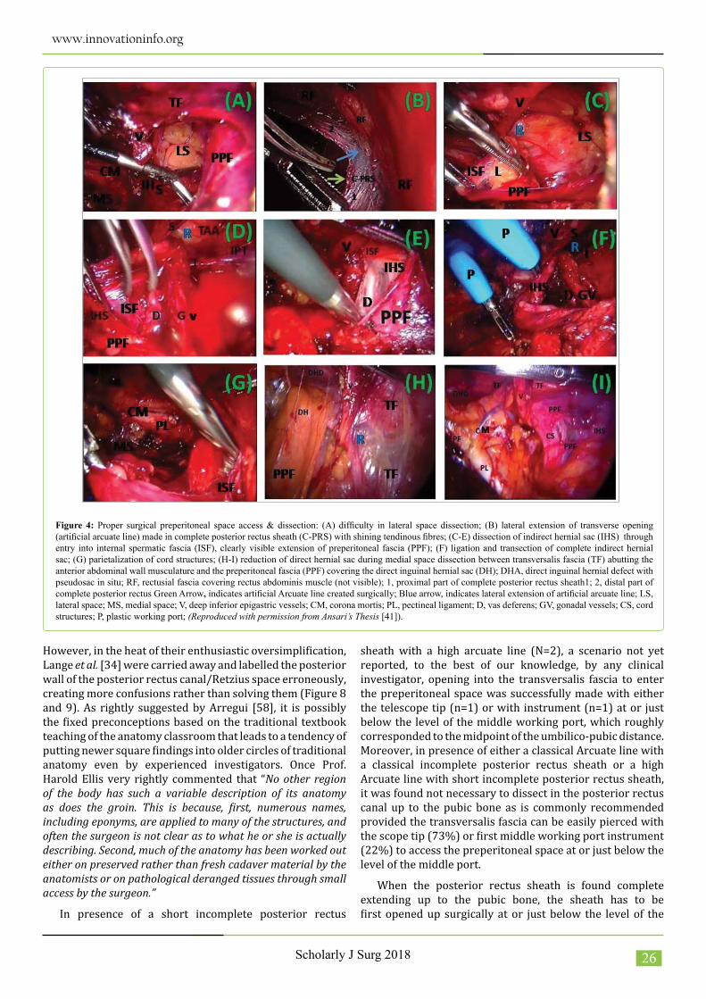
However, in the heat of their enthusiastic oversimplification, Lange et al. [34] were carried away and labelled the posterior wall of the posterior rectus canal/Retzius space erroneously, creating more confusions rather than solving them (Figure 8 and 9). As rightly suggested by Arregui [58], it is possibly the fixed preconceptions based on the traditional textbook teaching of the anatomy classroom that leads to a tendency of putting newer square findings into older circles of traditional anatomy even by experienced investigators. Once Prof. Harold Ellis very rightly commented that “No other region of the body has such a variable description of its anatomy as does the groin. This is because, first, numerous names, including eponyms, are applied to many of the structures, and often the surgeon is not clear as to what he or she is actually describing. Second, much of the anatomy has been worked out either on preserved rather than fresh cadaver material by the anatomists or on pathological deranged tissues through small access by the surgeon.”
In presence of a short incomplete posterior rectus
sheath with a high arcuate line (N=2), a scenario not yet reported, to the best of our knowledge, by any clinical investigator, opening into the transversalis fascia to enter the preperitoneal space was successfully made with either the telescope tip (n=1) or with instrument (n=1) at or just below the level of the middle working port, which roughly corresponded to the midpoint of the umbilico-pubic distance. Moreover, in presence of either a classical Arcuate line with a classical incomplete posterior rectus sheath or a high Arcuate line with short incomplete posterior rectus sheath, it was found not necessary to dissect in the posterior rectus canal up to the pubic bone as is commonly recommended provided the transversalis fascia can be easily pierced with the scope tip (73%) or first middle working port instrument (22%) to access the preperitoneal space at or just below the level of the middle port.
When the posterior rectus sheath is found complete extending up to the pubic bone, the sheath has to be first opened up surgically at or just below the level of the
Figure 4: Proper surgical preperitoneal space access & dissection: (A) difficulty in lateral space dissection; (B) lateral extension of transverse opening (artificial arcuate line) made in complete posterior rectus sheath (C-PRS) with shining tendinous fibres; (C-E) dissection of indirect hernial sac (IHS) through entry into internal spermatic fascia (ISF), clearly visible extension of preperitoneal fascia (PPF); (F) ligation and transection of complete indirect hernial sac; (G) parietalization of cord structures; (H-I) reduction of direct hernial sac during medial space dissection between transversalis fascia (TF) abutting the anterior abdominal wall musculature and the preperitoneal fascia (PPF) covering the direct inguinal hernial sac (DH); DHA, direct inguinal hernial defect with pseudosac in situ; RF, rectusial fascia covering rectus abdominis muscle (not visible); 1, proximal part of complete posterior rectus sheath1; 2, distal part of complete posterior rectus Green Arrow, indicates artificial Arcuate line created surgically; Blue arrow, indicates lateral extension of artificial arcuate line; LS, lateral space; MS, medial space; V, deep inferior epigastric vessels; CM, corona mortis; PL, pectineal ligament; D, vas deferens; GV, gonadal vessels; CS, cord structures; P, plastic working port; (Reproduced with permission from Ansari’s Thesis [41]).

www.innovationinfo.org
Scholarly J Surg 2018 27
middle port, i.e., an artificial arcuate line is created first in the complete posterior rectus sheath [25-27], followed by making an opening into the transversalis fascia underneath. Spitz and Arregui [59] also recommended that the posterior rectus sheath, always complete extending below the arcuate line up to the pubic bones in their observations [60], must be opened up to enter the preperitoneal space although they did not elaborate its technique-how and where. The novel concept of the ‘effective rectus sheath canal (ERSC)’ reported earlier by the author [40] becomes an important guiding principle to decide the level of entry into the preperitoneal space so as to keep the ERSC as short as possible. Hence in order to keep the best forward ergonomic working with minimal ERSC, one has to enter the preperitoneal space at or just below the level of the middle working port, which roughly corresponded to the midpoint of the umbilico-pubic distance. This technique proved extremely effective in our study as there was no conversion secondary to the so-called difficult dissection and no major complication except one episode of epigastric vessel injury by the roughened joint of the dissecting instrument, i.e., Maryland dissector. Access to the preperitoneal space at about the level of the interspinous
line (between two anterior superior iliac spines) as advocated by Dulucq et al. [61] on the basis of their own experience appears to be too low to be advantageous in terms of the best ergonomics [28] and hence not popular and therefore cannot be recommended for the general use. Moreover, the misconception that the preperitoneal space cannot be accessed easily in presence of a complete posterior rectus sheath extending up to the pubic symphysis [62,63] was dispelled by adopting a judicious anatomy-based surgical technique in the present study, i.e., the posterior rectus canal is first opened up by the telescope’s to-and-fro movements under CO2 insufflation for placement of one or both working ports and the instrument dissection is then used to access the preperitoneal space at or just below the middle port.
During the initial phase of dissection, access to the lateral preperitoneal space of the inguinal region is often limited due to the lateral firm attachment of the posterior rectus sheath to the Spigelian fascia at the linea semicircularis, especially in presence of a narrow posterior rectus canal. Access can be improved by taking down the lateral tendinous fibres of the arcuate line, preferably with a hook scissors, i.e., by
Figure 5: Proper avascular preperitoneal space created widely between transversalis fascia and preperitoneal fascia from midline to anterior superior iliac spine in 4 different patients: (A) left indirect inguinal hernia; (B) right indirect inguinal hernia; (C) combined right direct and indirect inguinal hernia; (D) right indirect inguinal hernia; MS, medial preperitoneal space; LS, lateral preperitoneal space; TF, transversalis fascia; PPF, preperitoneal fascia; ISF, internal spermatic fascia covering indirect hernial sac and cord structures; L, cord lipoma; V, deep inferior epigastric vessels; R, deep inguinal ring; IPT, iliopubic tract; DH, direct hernial sac covered within PPF; DHD, direct hernial defect; (Reproduced with permission from Ansari’s Thesis [41]).

www.innovationinfo.org
Scholarly J Surg 2018 28
the lateral extension of the primary Arcuate line [57,60], as was also successfully done in the present study in 41% of cases with incomplete posterior rectus sheath with narrow posterior rectus canal. However, the technique of the lateral extension of the primary Arcuate line is not risk-free because of the presence of an arterial twig and the adherent peritoneum at the Spigelian line.
Long tunnel of the posterior rectus canal produced by a long incomplete posterior rectus sheath with a low Arcuate line creates a narrow working space with marked ergonomic limitation and impairment of the endoscopic vision, and Edward Felix [64] recommended that the posterior rectus sheath should be cut back with an endo-scissors but he did not elaborate its exact method. Similarly, Meyer et al. [65,66] also recommended a short incision with an endo-scissors without cautery in case of low Arcuate line, but they also failed to elaborate the exact details of how and where, keeping both the inexperienced youngsters and the seasoned senior beginners guessing. The author successfully achieved the objective of safe ergonomic preperitoneal entry by surgically creating an artificial transverse arcuate line in 24 cases with non-classical posterior rectus sheath (10 long incomplete posterior rectus sheath with low primary arcuate line, and 14 complete posterior rectus sheath with absent primary arcuate line) at or just below the level of the middle working port which roughly corresponded to the midpoint of the umbilico-pubic distance. Liberal lateral extension of the artificial arcuate line easily facilitated the safe creation of an adequate lateral preperitoneal space up to the anterior superior iliac spine (Figure 4). Vertical division in the long incomplete posterior rectus sheath with a low arcuate line [65,66] or complete posterior rectus sheath with absent arcuate line is likely to result in unsupported medial and lateral flaps hanging like a curtain across the operation field and hampering the smooth working. Similar situation is likely to arise by a vertical incision in the transversalis fascia with an additional high risk of falling down of the deep inferior epigastric vessels now left invested within the lateral flap of the divided transversalis fascia, that may hang across the field like a cord, resulting in the so-called ‘cord sign’ as is sometimes seen with the balloon dissection [34]. Transverse incision keeps the distal part of the sheath and fascia intact abutting the anterior abdominal wall musculature and the preperitoneal space underneath it is safely dissected with ease and rapidity, and this technique proved efficacious in our study.
Any laparoscopic hernia study that does not include the current perspective of the preperitoneal surgical anatomy and its frequent variations [9,11,12,21-33,35,36,39,67], especially of the posterior rectus sheath, would be grossly deficient in impacting the sound understanding, learning and teaching of the TEP inguinal hernioplasty, because ‘during the learning phase, one is often struck by how difficult it is to handle the approach to the preperitoneal space’ [34]. This holds true even for currently recommended modified technique of the TEP repair by Dr. George Ferzli and associates [45]. In the light of current knowledge of the multi-layered preperitoneal anatomy [9,11,12,22-29,36], their technique of the blind finger dissection first followed by
the under-vision instrument dissection of the preperitoneal space appears feasible only in a few uncommon patients with a high Arcuate line of a short incomplete posterior rectus sheath [9,35], with a greater risk of peritoneal injury; moreover, these reputed authors failed to mention the anatomical plane of the preperitoneal space [9] and the different fascial layers across which the dissection proceed to the requisite preperitoneal space [11,12,24,26,29].
In nutshell, the method of the access to the preperitoneal space during the posterior rectus sheath approach of the laparoscopic TEPP inguinal hernioplasty without balloon dissection is based on two fundamental principles, viz., firstly, the effective rectus sheath canal (ERSC) should be ‘minimum’ in order to obtain the least fulcrum effect, and secondly, the ergonomic function should be ‘maximum’ in order to achieve best working in a limited closed space [40]. Key to the seamless TEPP hernioplasty warrants choreographic repetition of the identical moves under a detailed ‘step-by-step’ scheme [57]. In order to provide an organized, common-sense-based Rosser’s map [38] of the preperitoneal anatomy to help the surgeon to learn, perform and teach the laparoscopic TEPP hernia repair with ease and safety, the real-time-anatomy-based techniques for access to the proper preperitoneal space are summarized with schematic illustrations (Figure 6 and 10-12) as under:
1. When a classical normal-length incomplete posterior rectus sheath (single-/double-layered) [29] with a primary arcuate line (well-defined/ill-defined) situated at ⅓rd to ½ of the umbilico-pubic distance is encountered, the transversalis fascia is pierced with the to-and-fro movements of the telescope tip or opened up transversely by the spreading-out action of the Maryland dissector tip (without cautery) below the arcuate line to enter the ‘surgical preperitoneal space’ between the transversalis fascia (invariably single-layered) [24] and the membranous preperitoneal fascia (single-/double-layered) [24] (Figure 6).
2. When a complete posterior rectus sheath is encountered extending up to the pubic bone with/without a variable secondary arcuate line [29], an artificial arcuate line is surgically created in the transverse axis (tendinous fibres largely running transversely) at or just below the level of the middle port, and then dividing the transversalis fascia underneath it to enter the ‘surgical preperitoneal space’ (Figure 10).
3. When a long incomplete posterior rectus sheath with a low primary arcuate line is encountered [23,29], the ‘surgical preperitoneal space’ is entered first by transversely creating an artificial arcuate line in the posterior rectus sheath more proximally at or just below the level of the middle port, and then dividing the transversalis fascia underneath it to enter the ‘surgical preperitoneal space’ in a fashion similar to the case of the complete posterior rectus sheath (Figure 11).
In all three clinical situations mentioned above, once the proper avascular ‘surgical preperitoneal space’ between the transversalis fascia and the preperitoneal fascia is accessed correctly (Figure 4A), adequate wide space is first created by the sharp and/or blunt instrument dissection from the midline in the retro-pubic area to the anterior

www.innovationinfo.org
Scholarly J Surg 2018 29
Figure 6: Schematic illustration of longitudinal section of anterior abdominal wall with classical incomplete posterior rectus sheath and well-defined arcuate line : Purple arrows, indicate the trajectory of access to the preperitoneal space between transversalis fascia and preperitoneal space; P1, level of infraumbilical first (optical) port; P2, level of middle working port; P3, level of suprapubic working port; Dashed line, indicates occasional presence; (adopted with permission from Ansari’s Thesis [41]).
superior iliac spine in the sub-inguinal region for the mesh placement of about 15x10 cm (Figure 5), and only then the preperitoneal fascia forming the internal spermatic fascia around the intra-abdominal part of the spermatic cord structures is entered to identify and dissect the indirect hernial sac [24,34,58,68,69] (Figure 4C-E). After inversion/transection of the indirect hernial sac (Figure 4F), adequate parietalization of the cord structures with the dorsal part of the internal spermatic fascia is finally carried out (Figure 4G) to enlarge the preperitoneal space inferiorly sufficient enough to accommodate a requisite large mesh without edge curling/infolding (Figure 4G). In case of a direct hernial sac, the sac is pulled in along with the preperitoneal fascia during widening of the ‘surgical preperitoneal space’ by separating it from the transversalis fascia, leaving behind the ‘pseudo sac’ of the direct hernia in the hernial defect along the parietes (Figure 4H-I), and the dissection of the direct hernial sac is usually not required and hence its covering the preperitoneal fascia is also not disturbed or dissected out.
Preperitoneal access techniques described here remain essentially the same even if the operating surgeon believes in the hybrid technique of the true preperitoneal dissection laterally to safeguard with additional caution against the nerve injuries and the interfascial dissection medially to safeguard against the urinary bladder injury [6,14,59]
although the perceived notion of greater risk of the nerve irritation/injury in the ‘triangle of pain’ is not really proved in our study [9], provided a gentle meticulous dissection is performed in the lateral ‘surgical preperitoneal plane’ and the ilio-psoas muscle is not bared posteriorly by the excessive dissection of the loose areolar tissues and fascia covering the nerves which run on the anterior surface of the ilio-psoas muscle, i.e., in the posterior wall of the ‘surgical preperitoneal space’.
ConclusionDirect telescopic dissection under CO2 insufflation
in an unhurried controlled fashion provided accurate recognition of the morphology of the posterior wall of posterior rectus canal (posterior rectus sheath, transversalis fascia, preperitoneal fascia and peritoneum) during the laparoscopic total extraperitoneal preperitoneal (TEPP) inguinal hernioplasty. Lower part of the posterior rectus canal and conventional Retzius bounded posteriorly by the transversalis fascia is not the proper preperitoneal space. Careful judicious transverse division of either only the transversalis fascia in presence of a classical (n=41) and high (n=2) incomplete posterior rectus sheath, or long incomplete (n=10) and complete (n=14) posterior rectus sheath at or just below the level of the middle port which approximately corresponded to the midpoint of the umbilico-

www.innovationinfo.org
Scholarly J Surg 2018 30
Figure 7: Breaking of the posterior wall of the posterior rectus canal to enter the preperitoneal space: VC, really represent the transversalis fascia proper and not the ventral lamina of the preperitoneal fascia as defined by the original authors; H, indicates hernia; (Reproduced under Creative Commons Attribution License of Open Access from Lange et al. [34]).
Figure 8: Schematic longitudinal section of anterior abdominal wall: Original authors’ illustration is correct but the labelling of various layers is partly erroneous; A, peritoneum; B, preperitoneal fascia; C, transversalis fascia, erroneously labelled as ventral component of preperitoneal fascia/posterior lamina of transversalis fascia; E, indicates Rectusial fascia (variably condensed posterior epimysium of rectus abdominis muscle), erroneously labelled as the rectus abdominis muscle; (Reproduced under Creative Commons Attribution License of Open Access from Lange et al. [34]).
pubic distance, resulted invariably in successful access to the surgical preperitoneal space and joyful completion of the seamless TEP repair with no conversion secondary to the so-called difficult dissection. Author’s real-time-anatomy-based technique with all steps under direct vision, the most fundamental principle of the modern laparoscopy, is strongly recommended.
Sound precise knowledge of the inguino-abdominal anatomy beyond the traditional textbooks of anatomy & surgery and keen observation during unhurried preperitoneal laparoscopy is, no doubt, essential for the rapid smooth execution of the TEPP hernioplasty with
safety. However, the author strongly feels that the lack of a well-choreographed strategy of ‘one-step-built-upon-the-other’ based on the real-time anatomy with all steps under direct vision, a hallmark principle of the current advanced laparoscopy, is the single most important factor not only for the poor understanding of the preperitoneal anatomy but also for the prevailing unpopularity of the laparoscopic TEPP repair secondary to the hard-to-learn nature of the apparently straightforward procedure. The author tried to develop the highly desirable Rosser’s common-sense map based on current scientific knowledge of the preperitoneal anatomy to overcome the peer reluctant adoption of the laparoscopic TEPP approach, and further meticulous

www.innovationinfo.org
Scholarly J Surg 2018 31
Figure 9: Schematic longitudinal section of anterior abdominal wall under co2 insufflation: VC, really represent the transversalis fascia proper and not the ventral lamina of the preperitoneal fascia/posterior lamina of transversalis fascia as defined by the original authors; H, indicates hernia; (Reproduced under Creative Commons Attribution License of Open Access from Lange et al. [34]).
Figure 10: Schematic illustration of longitudinal section of anterior abdominal wall with complete posterior rectus sheath and absent arcuate line: Purple arrows, indicate the trajectory of access to the preperitoneal space between transversalis fascia and preperitoneal space; P1, level of infraumbilical first (optical) port; P2, level of middle working port; P3, level of suprapubic working port; Dashed line, indicates occasional presence; (Reproduced with permission from Ansari’s Thesis [41]).

www.innovationinfo.org
Scholarly J Surg 2018 32
Figure 11: Schematic illustration of longitudinal section of anterior abdominal wall with long incomplete posterior rectus sheath and low arcuate line: Purple arrows, indicate the trajectory of access to the preperitoneal space between transversalis fascia and preperitoneal space; P1, level of infraumbilical first (optical) port; P2, level of middle working port; P3, level of suprapubic working port; Dashed line, indicates occasional presence; (Reproduced with permission from Ansari’s Thesis [41]).
Figure 12: Flow chart showing access to proper preperitoneal space based on real-time anatomy in a patient undergoing total extraperitoneal preperitoneal (TEP) hernioplasty.

www.innovationinfo.org
Scholarly J Surg 2018 33
research on the laparoscopic live surgical anatomy by the dedicated clinical investigators will go a long way to popularize the cost-effective TEPP hernioplasty for the larger good of the masses afflicted with the common malady of the inguinal hernia.
AcknowledgementAll figures of this article are reproduced with permission
from ‘Ansari MM. A Study of Laparoscopic Surgical Anatomy of Infraumbilical Posterior Rectus Sheath, Fascia Transversalis & Pre-Peritoneal Fat/Fascia during TEPP Mesh Hernioplasty for Inguinal Hernia., Doctoral Thesis for PhD (Surgery), Aligarh Muslim University, Aligarh, India, 2016 [41].
Supplementary Information: Question: Three ways to create a transverse opening
according to the condition of posterior rectus sheath is completed?
Answer: Author’s Response: Yes, the same surgical technique of transverse opening (artificial arcuate line) was utilized in the three different types of complete, long and short posterior rectus sheath. Opening in the transverse axis was found most ergonomic for easy and safe access to the preperitoneal space and for further dissection readily for ample space creation for the mesh placement.References
1. Davis CJ, Arregui ME (2003) Laparoscopic repair for groin hernias. Surg Clin N Am 83: 1141-1161.
2. Kakadia CB, Choksi DB, Bhedi A, Damor S (2017) Comparison of conventional balloon method and Dulucq method for Extraperitoneal access for laparoscopic total extraperitoneal repair of Inguinal hernia. Int Surg J. 4: 2791-2795.
3. Abbasi MR, Shaikh A, Sangrasi AK, Shaikh NA, Shaikh U (2015) Inguinal hernioplasty: laparoscopic TEP with & without dissection balloon. Professional Med J. 22: 782-786.
4. Felix EL (2009) Laparoscopic Inguinal Hernia Repair. In: Nathaniel J. Soper, Lee L. Swanstrom, W. Stephen Eubanks (eds.). Mastery of Endoscopic and Laparoscopic Surgery, 3rd Edition, Chapter 53, Philadelphia: Lippincott Williams & Wilkins, pp: 523-537.
5. Wishart GC, Wright D, O’Dwyer PJ (1995) Use of a Foley catheter to dissect the preperitoneal space for extraperitoneal endoscopic hernia repair. J Laparoendosc Surg 5: 27-29.
6. Christensen BJ, Fisher KS (1998) Laparoscopic Access to the Preperitoneal Space. J Soc Laparoendosc Surg 2: 97-98.
7. Folscher DJ, Leroy J, Jamali FR, Marescaux J (2000) Totally extrafasdal endoscopie preperitoneal hernia repair: a merger of anatomy and surgery. The exact description to endoscopkally dissect the spermatic fascia. Hernia 4: 223-227.
8. Mirilas P, Mentessedou A, SKandalais JE (2008) Secondary internal inguinal ring and associated surgical planes: surgical anatomy, embryology, applications. J Am Coll Surg 206: 561-570.
9. Ansari MM (2018) Surgical preperitoneal space: holy plane of dissection between transversalis fascia and preperitoneal fascia for TEPP inguinal hernioplasty. MedCrave Online J Surg 6: 26-33.
10. Hayes MA (1950) Abdominopelvic Fascia. Am J Anat 1950; 87: 119-161.
11. Ansari MM (2017) Retzius and Bogros Spaces: A Prospective Laparoscopic Study and Current Perspectives. Ann Int Med Dent Res 3: SG25-31.
12. Ansari MM (2017) Retzius Space: Not A Single Anatomical Entity: New Insights, Simplified & Illustrated in A Laparoscopic Study during TEPP
Hernioplasty for Inguinal Hernia. Ann Int Med Dent Res 4: SG63-73.
13. Rabea G (2016) Laparoscopic anatomy of the inguinal region. SlideShare (https://www.slideshare.net/GergisRabea/laparoscopic-anatomy-of-inguinal-canal) [Accessed on: 25.03.2018]
14. Arregui ME, Navarrete J, Davis CJ, Castro D, Nagan RF (1993) Laparoscopic inguinal herniorrhaphy: Techniques and controversies. Surg Clin North Am 73: 513-27.
15. Hureau J (2001) The space of Bogros and the interparietoperitoneal spaces. In: Robert Bendavid, Jack Abrahamson, Maurice E. Arregui, Jean Bernard Flament, Edward E. Phillips (eds.) Abdominal Wall Hernias: Principles and Management, Chapter 11, Spinger-Verlag, New York, pp: 101-106.
16. Lucas SW, Arregui ME (1999) Minimally invasive surgery for inguinal hernia. World J Surg 23: 350-355.
17. Misra C, Kumar S, Bansal V (2008) Total extraperitoneal (TEP) mesh repair of inguinal hernia in the developing world: comparison of low-cost indigenous balloon dissection versus direct telescopic dissection: a prospective randomized controlled study. Surg Endosc 22: 1947-1958.
18. Ishita T, Sato T, Iino T (2013) The new approach for total extraperitoneal (TEP) laparoscopic inguinal hernia repair using optical method and needle-like forceps. SAGES Abstracts Archive: P279.
19. Abd-Raboh OH, Ismael TA, Mohammed HAH, El-Sheikh MM (2017) Outcome of the laparoscopic total extraperitoneal approach with direct dissection and mesh hernioplasty in the treatment of inguinal hernias. Egypt J Surg 36: 124-130.
20. Hisham Y, Ranjith M, Nabeel TP, Sidhic KA (2018) Outcome of laparoscopic total extraperitoneal approach with direct dissection and mesh hernioplasty in the treatment of inguinal hernias. Int Surg J 5: 248-252.
21. Rosen MJ, Petro CC, Stringer MD (2016) Anterior Abdominal Wall. In: Susan Standring (ed.) Gray’s Anatomy: The Anatomical Basis of Clinical Practice, 41st Edition, Chapter 61, Elsevier, UK, pp: 1069-1082.
22. Ansari MM (2017) Rectusial Fascia: A New Entity of Laparoscopic Live Surgical Anatomy. Open Access J Surg 3: 1-5.
23. Ansari MM (2017) Arcuate Line of Douglas: Localization from Surface Anatomic Landmarks of Anterior Abdomen during Laparoscopic TEPP Hernioplasty. Int J Sci Res Publ 7: 575-580.
24. Ansari MM (2017) Transversalis Fascia and Preperitoneal Fascia: A Laparoscopic Study of Live Surgical Anatomy during TEPP Hernioplasty-Final Report and Literature Review. Ann Int Med Den Res 3: SG19-32.
25. Ansari MM (2017) Artificial Arcuate Line: Surgical Creation during TEPP Hernioplasty. Clin Surg 2: 1698.
26. Ansari MM (2018) Posterior rectus sheath variations: surgical significance and clinical implications for laparoscopic hernia surgeons. Int Surg J 5: 683-694.
27. Ansari MM (2017) Arcuate Line Variations: Surgical Significance and Clinical Implications during TEPP Hernioplasty. J Surg Clin Interventions 1: 1-8.
28. Ansari MM (2017) Interspinous Line: Relations with Other Landmarks (Xiphisternum, Umbilicus, Pubic Symphysis and Arcuate Line of Douglas) and Importance for Surgeons. ARC J Clin Cas Rep 3:18-30.
29. Ansari MM (2018) Posterior Rectus Sheath: A Prospective Study of Laparoscopic Live Surgical Anatomy during TEPP Hernioplasty. World J Lap Surg 11: 1-13.
30. Askar OM (1877) Surgical anatomy of the aponeurotic expansions of the anterior abdominal wall. Ann R Coll Surg Eng 59: 313-321.
31. Rizk NN (1991) The arcuate line of the rectus sheath-does it exist? J Anat 1991; 175:1-6.
32. Mwachaka PM, Saidi HS, Odula PQ, Awori KO, Kaisha WO (2010) Locating the arcuate line of Douglas: is it of surgical relevance. Clin Anat 23: 84-86.
33. Ansari MM (2014) Complete posterior rectus sheath and total extra-peritoneal hernioplasty. Saudi Surg J 2(4): 80-83.
34. Lange JF, Rooijen PPGM, Koppert S, Kleinrensink GJ (2002) The Preperitoneal tissue dilemma in totally extraperitoneal (TEP) laparoscopic

www.innovationinfo.org
Scholarly J Surg 2018 34
hernia repair. Surg Endosc 16: 927-930.
35. Monkhouse WS, Khalique A (1986) Variations in the composition of the human rectus sheath: a study of the anterior abdominal wall. J Anat 145: 61-66.
36. Ansari MM (2018) Arcuate Line Position: Current Perspective and Revised Ansari Classification. Sci Fed J Surg 1: Pages 1-9.
37. Spaw AT, Ennis BW, Spaw LP (1991) Laparoscopic hernia repair: the anatomic basis. J Laparoendosc Surg 1: 269-277.
38. Rosser J (1994) The anatomical basis for laparoscopic hernia repair revisited. Surg Laparosc Endosc 4: 36-44.
39. Mwachaka P, Odula P, Awori K, Kaisha (2009) Variations in the Pattern of Formation of the Abdominis Rectus Muscle Sheath among Kenyans. Int J Morphol 27: 1025-1029.
40. Ansari MM (2013) Effective Rectus Sheath Canal: Does It Affect TEP Approach for Inguinal Mesh Hernioplasty. J Exp Integr Med 3: 73-76.
41. Ansari MM (2016) A Study of Laparoscopic Surgical Anatomy of Infraumbilical Posterior Rectus Sheath, Fascia Transversalis & Pre-Peritoneal Fat/Fascia during TEPP Mesh Hernioplasty for Inguinal Hernia. Doctoral Thesis for PhD (Surgery), Aligarh Muslim University, Aligarh, India.
42. Mirilas P, Colborn GL, McClusky-III DA, Skandalakis LJ, Skandalakis PN, et al. (2005) The history of anatomy and surgery of the preperitoneal space. Arch Surg 140: 90-94.
43. Chowbey PK, Khullar R, Sharma A, Soni V, Baijal M (2006) Totally extraperitoneal repair of inguinal hernia: Sir Ganga Ram Hospital technique. J Minim Access Surg 2: 160-164.
44. Swadia ND (2011) Laparoscopic totally extra-peritoneal inguinal hernia repair: 9 years’ experience. Hernia 15: 273-279.
45. Bowne WB, Morgenthal CB, Castro AE, Shah P, Ferzli GS (2007) The role of endoscopic extraperitoneal herniorrhaphy: where do we stand in 2005? Surg Endosc 21: 707-712.
46. Felix EL, Michas CA, Gonzalez MH Jr (1995) Laparoscopic hernioplasty. TAPP vs TEP. Surg Endosc 9: 984-989.
47. Hanif Z, Sajid MA, Pandya R, Shanmugarajah K, Mahmud S (2017) Modification of standard laparoscopic total extra peritoneal hernia repair technique: Methods to improve feasibility in the UK health service. Int J Surg Open 9: 45-47.
48. Liem MS, van Steensel CJ, Boelhouwer RU (1996) The learning curve for totally extraperitoneal laparoscopic inguinal hernia repair. Am J Surg 171: 281-285.
49. Voitk AJ (1998) The learning curve in laparoscopic inguinal hernia repair for the community general surgeon. Can J Surg 41: 446-450.
50. Wright D, O’Dwyer PJ (1998) The learning curve for laparoscopic hernia repair. Semin Laparosc Surg 5: 227-232.
51. Bringman S, Ek A, Haglind E, Heikkinen T (2001) Is a dissection balloon beneficial in totally extraperitoneal endoscopic hernioplasty (TEP)? A randomized prospective multicenter study. Surg Endosc 15: 266-270.
52. Lau H, Patil NG, Yuen WK, Lee F (2002) Learning curve for unilateral endoscopic totally extraperitoneal (TEP) inguinal hernioplasty. Surg Endosc 16: 1724-1728.
53. Jiang H, Ma R, Zhang X (2016) Novel retrograde puncture method to
establish preperitoneal space for laparoscopic direct inguinal hernia repair with internal ring suturing. Braz J Med Biol Res 49: e5247.
54. Fiennes AG (1994) The Kieturakis balloon dissector-an aid to the extraperitoneal approach for laparoscopic repair of groin hernias? Endosc Surg Allied Technol 2: 221-225.
55. Kang AY, Lee SR, Son BH, Jung KU (2014) Achieving the preperitoneal space in totally extraperitoneal inguinal hernia repair: dissection with or without a balloon dissector. J Minim Invas Surg 17: 62-68.
56. Kumar S (2009) A new balloon dissector for totally extraperitoneal hernia repair. J Minim Access Surg 5: 22-24.
57. Putnis S, Berney CR (2012) Totally extraperitoneal repair of inguinal hernia: techniques and pitfalls of a challenging procedure. Langenbeck’s Arch Surg 397: 1343-1351.
58. Arregui ME (1997) Surgical anatomy of the pre-peritoneal fasciae and posterior transversalis fasciae in the inguinal region. Hernia 1: 101-110.
59. Spitz JD, Arregui ME (2001) Laparoscopic Totally Extraperitoneal Repair of Inguinal Hernias (TEP): Part II. In: Robert Bendavid, Jack Abrahamson, Maurice E. Arregui, Jean B. Flament, Edward H. Phillips (eds.) Abdominal Wall Hernias: Principles and Management, 1st Edition (Reprint), Chapter 70, Springer Science-Business Media, New York, pp: 471-480.
60. Spitz JD, Arregui ME (2001) Fascial anatomy of the inguinal region. In: Robert Bendavid, Jack Abrahamson, Maurice E. Arregui, Jean B. Flament, Edward H. Phillips (eds.) Abdominal Wall Hernias: Principles and Management, 1st Edition (Reprint), Chapter 8, Springer Science-Business Media, New York, pp 86-91.
61. Dulucq JL, Wintringer P, Mahajna A (2009) Laparoscopic totally extraperitoneal inguinal hernia repair: lessons learned from 3,100 hernia repairs over 15 years. Surg Endosc 23: 482-486.
62. Simons MP, Aufenacker T, Bay-Nielsen M, et al (2009) European Hernia Society guidelines on the treatment of inguinal hernia in adult patients. Hernia 13: 343-403.
63. Tran H, Tran K, Turingan I, Zaskoowaska M, Lam V, et al. (2015) Single-incision laparoscopic inguinal herniorraphy with telescopic extraperitoneal dissection: technical aspects and potential benefits. Hernia 19: 407-416.
64. Felix EL (2001) Laparoscopic Totally Extraperitoneal Hernioplasty (TEP): Part I In: Robert Bendavid, Jack Abrahamson, Maurice E. Arregui, Jean B. Flament, Edward H. Phillips (eds.) Abdominal Wall Hernias: Principles and Management, 1st Edition (Reprint), Chapter 69, Springer Science-Business Media, New York, pp: 464-471.
65. Meyer ALM, Bellandi DM, Delacoste F (2010) Laparoscopic Totally Extraperitoneal Inguinal Hernia Repair: Nonfixation of Three-Dimensional Mesh. Bras J Video-Sur 3: 19-23.
66. Meyer A, Dulucq JL, Mahajna A (2013) Laparoscopic totally extraperitoneal hernioplasty with nonfixation of three-dimensional mesh-Dulucq´s technique. ABCD Arq Bras Cir Dig 26:59-61.
67. Cunningham SC, Rosson GD, Lee RH (2004) Localization of the arcuate line from surface anatomic landmarks: a cadaveric study. Ann Plast Surg 53: 129-131.
68. Yang XF, Liu JL (2016) Anatomy essentials for laparoscopic inguinal hernia repair. Ann Transl Med 4: 372.
69. Mirilas P, Mentessedou A, SKandalais JE (2008) Secondary internal inguinal ring and associated surgical planes: surgical anatomy, embryology, applications. J Am Coo Surg 206: 561-570.
Citation: Ansari MM (2018) Preperitoneal Space: where and how to Access during Laparoscopic Total Extraperitoneal Preperitoneal (TEPP) Inguinal Hernioplasty. Scholarly J Surg. Vol: 1, Issu: 1 (20-34).