preparation of histological slide
TRANSCRIPT

PREPARATION OF HISTOLOGICAL SPECIMENS

• Histology is the study of tissues and how these tissues are arranged into organs
• Histo in Greek means tissue or web
• Tissues consist of cells and extra cellular matrix
• The function of the tissue depends on the interaction between cells and extracellular matrix.
• The small size of cells and matrix content make the study of tissues dependent on microscope and other advances in biological techniques
TISSUE PREPARATION• The most widely used method of studying
tissues is using histological slides.• The tissue in the slide must reflect the actual
nature of the tissue in the body• To insure that, tissues to be studied must
pass through a series of steps before examination
• These steps are in sequential order• Fixation is a complex series of chemical
events that differ for the different groups of substance found in tissues.

TISSUE FIXATION Aim of Fixation:
1- To prevent autolysis and bacterial attack.
2- To fix the tissues so they will not change their volume and shape during processing.
3- To prepare tissue and leave it in a condition which allow clear staining of sections.
4- To leave tissue as close as their living state as possible, and no small molecules should be lost.
•Fixation is a reaction between the fixative and proteins in the specimen which form a gel, so keeping every thing as their in vivo relation to each other.

Types of Fixative
• Acetic acid• Formaldehyde 10%• Ethanol• Glutaraldehyde• Methanol • Picric acid• Osmic acid (Osmium tetroxide)

TISSUE PROCESSING
• Aim:Is to embed the tissue in a solid medium firm enough to support the tissue and give it sufficient rigidity to enable thin sections to be cut , and yet soft enough not to damage the knife or tissue.
• Stages of processing:1.Dehydration.
2- Clearing.
3- Embedding.
Dehydration• To remove fixative and water from the tissue
and replace them with dehydrating fluid.
• Delicate specimens are dehydrated in a graded ethanol series from water through 10%-20%-50%-95%-100% ethanol to minimize tissue distortion from diffusion currents.
• In the paraffin method, dehydration from aqueous fixatives is usually initiated in 60%-70% ethanol, progressing through 90%-95% ethanol, absolute ethanol before proceeding to the clearing stage.

Types of dehydrating agents
• Ethanol• Methanol• Acetone
• Tissues may be held and stored indefinitely in 70% ethanol without harm

Clearing
• Replacing the dehydrating fluid with a fluid that is totally miscible with both the dehydrating fluid and the embedding medium.
• Choice of a clearing agent depends upon many factors
Types of Clearing Agents
• Xylene.
• Toluene.
• Chloroform.
• Benzene.
• Propylene oxide

Embedding
• Is the process by which tissues are surrounded by a medium such as agar, gelatin, or wax which when solidified will provide sufficient external support during sectioning.
• A substances added alone or in combination to the wax to:
Improve ribboning. Increase hardness. Decrease melting point Improve adhesion between specimen and wax

Embedding Moulds
A. paper boat mouldB. metal boat mould C. Dimmock embedding
mouldD. Peel-a-way disposable
mould E. Base mould used with
embedding ring ( F) or cassette bases (G)

CUTTING
• using the microtome

• A microtome is a mechanical instrument used to cut biological specimens into very thin sections for microscopic examination.
• Most microtomes use a steel blade and are used to prepare sections of animal or plant tissues for histology.

Microtome knives
• STEEL KNIVES• NON-CORROSIVE KNIVES FOR CRYOSTATS • DISPOSABLE METAL BLADES• GLASS KNIVES • DIAMOND KNIVES

STAINING
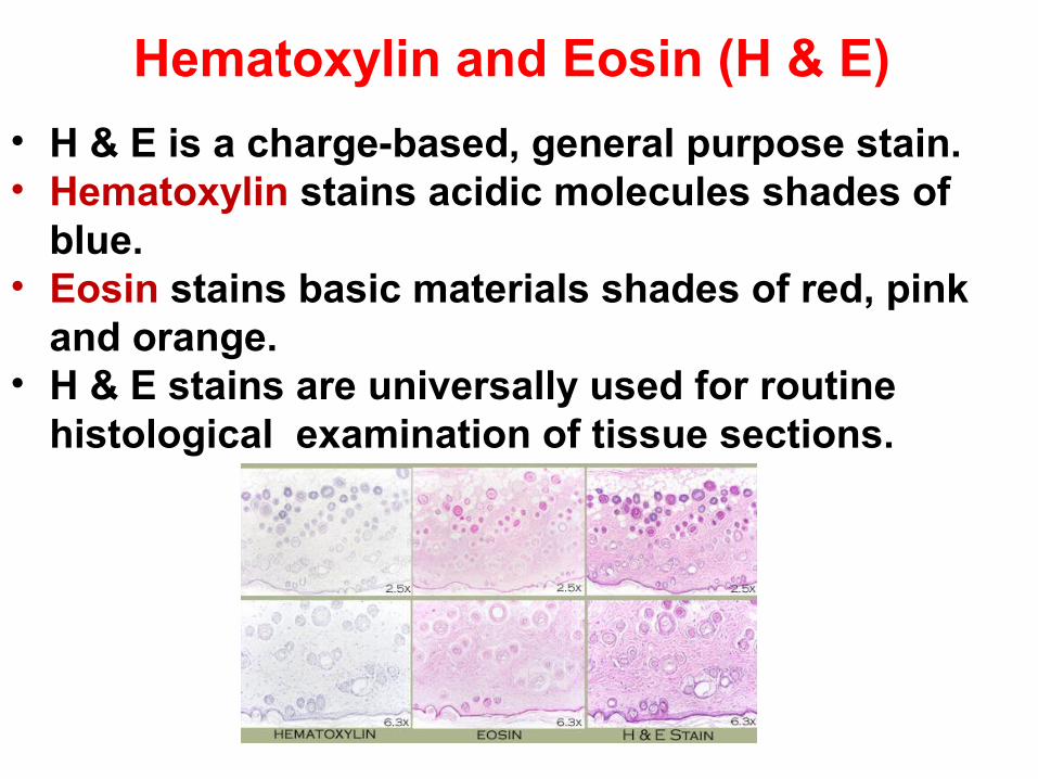
Hematoxylin and Eosin (H & E)
• H & E is a charge-based, general purpose stain.• Hematoxylin stains acidic molecules shades of
blue.• Eosin stains basic materials shades of red, pink
and orange.• H & E stains are universally used for routine
histological examination of tissue sections.
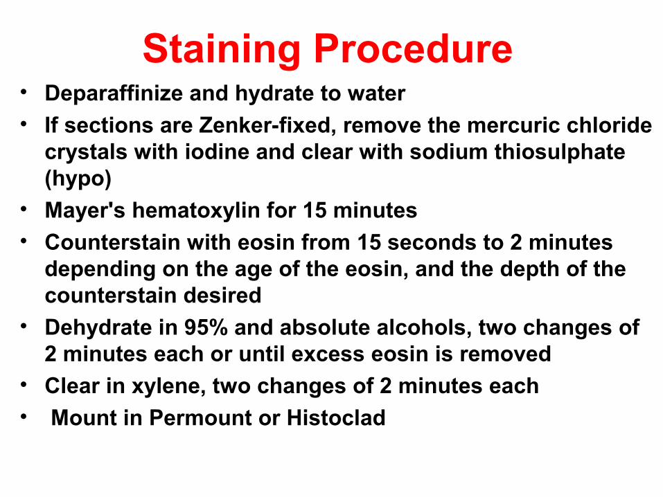
Staining Procedure • Deparaffinize and hydrate to water
• If sections are Zenker-fixed, remove the mercuric chloride crystals with iodine and clear with sodium thiosulphate (hypo)
• Mayer's hematoxylin for 15 minutes • Counterstain with eosin from 15 seconds to 2 minutes
depending on the age of the eosin, and the depth of the counterstain desired
• Dehydrate in 95% and absolute alcohols, two changes of 2 minutes each or until excess eosin is removed
• Clear in xylene, two changes of 2 minutes each
• Mount in Permount or Histoclad

Staining machine

• Basic dyes stain:HeterochromatinNucleic acidsRibosomesCartilage
• Acidic dyes stain:FilamentsMitochondriaCollagenMuscle fibers

Additional Dyes
• Many tissue components can not be stained with (Hematoxylene and Eosin).
• Other dyes are used to specifically stain certain tissue components
Resorcin-Fuchsin for elastic fibers
Silver stain for reticular fibers and basement membrane
Periodic-Acid Schiff (PAS) Reaction for CHO

