prehospital evaluation for large vessel occlusions
TRANSCRIPT
The Prehospital VAN Stroke Assessment
Prehospital evaluation for large vessel occlusions
James Chad Curry LP, FPCTraining Chief, UMC EMS
Chad Curry is a Licensed Paramedic with the state of Texas, a certified
Texas EMS Instructor, and a certified Flight Paramedic (FP-C). He is the
Training Chief for University Medical Center’s Emergency Medical Service
(UMC EMS) in Lubbock, Texas and has been employed with UMC EMS for
over 23 years. His experience includes over a decade of employment with
AeroCare as a flight paramedic, and he continues to work with them today.
As the Clinical Coordinator for UMC EMS’ licensed continuing education
program, Chad oversees and teaches both in-house education and
community outreach programs to EMS providers. He is a co-author to
several research posters presented across the state and nation, generating
interest and helping other EMS services advance their treatment protocols
based on UMC EMS’ experiences. Chad also directs the UMC EMS
Outreach CE program that offers free, approved online continuing
education to EMS providers in Texas and NREMT recertification
candidates throughout the United States.
Closer to home, he and his team of educators teach the American College
of Surgeons’ “Stop the Bleed” campaign to schools, organizations, and
communities across Lubbock and the South Plains. Away from home,
Chad represents EMS at the Governor’s EMS and Trauma Advisory
Council (GETAC). In addition, he has conveyed the concerns of many
prehospital providers by testifying to the United States Sentencing
Commission in Washington D.C. about the difficulties faced by synthetic
cannabinoid use by the public. He continues to speak to students, parents,
and the general public about these dangers that may be lurking within their
own homes.

DISCLOSURES
FINANCIAL DISCLOSURE:
No financial relationships to disclose
UNLABELED/UNAPPROVED USES DISCLOSURE:
None to disclose

A review of large cerebral vessel occlusions
The cerebral arteries are similar any other arterial network in the body: A few large vessels branch out from the base and feed the many smaller diameter vessels winding throughout the brain.
Anterior
Posterior
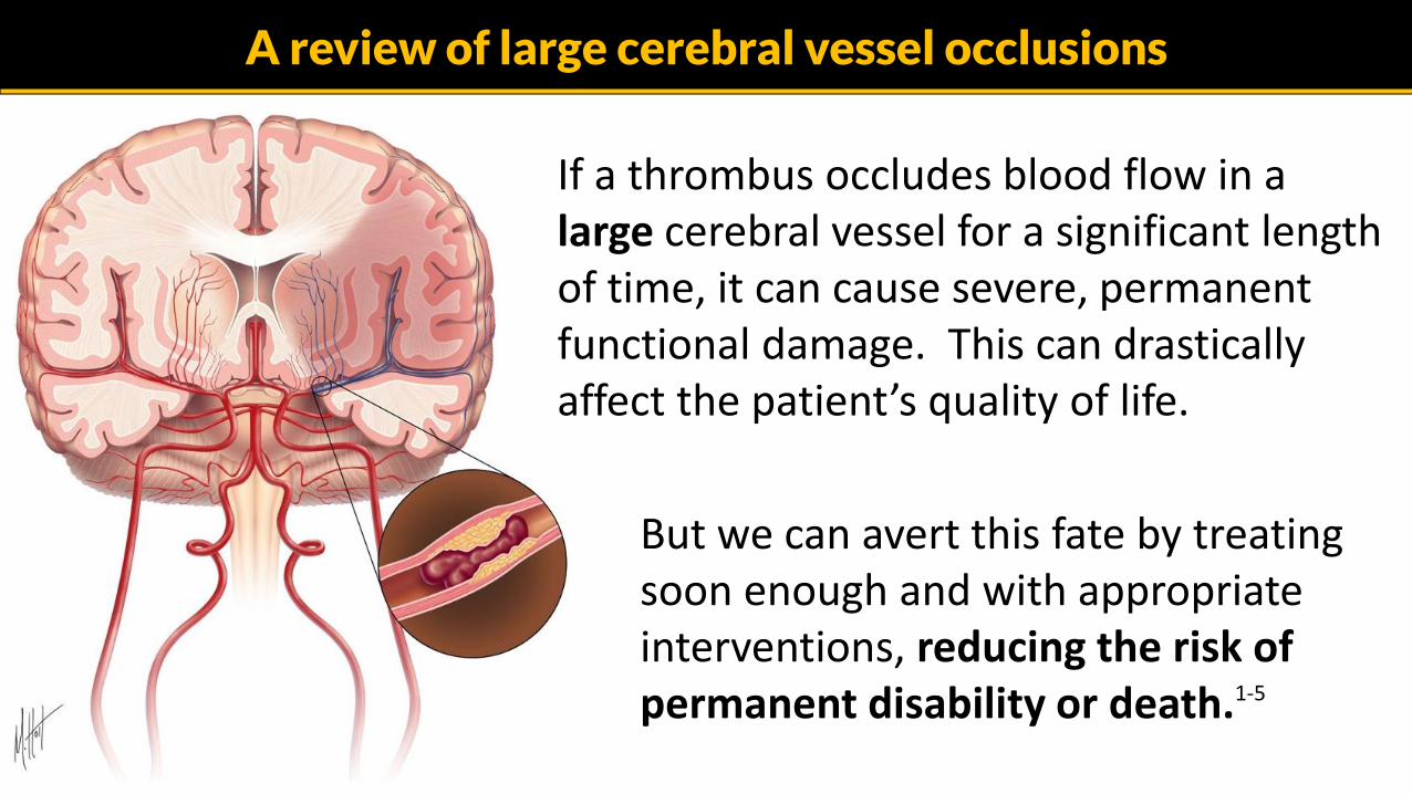
But we can avert this fate by treating soon enough and with appropriate interventions, reducing the risk of permanent disability or death.1-5
A review of large cerebral vessel occlusions
If a thrombus occludes blood flow in a large cerebral vessel for a significant length of time, it can cause severe, permanent functional damage. This can drastically affect the patient’s quality of life.

One treatment pathway for large vessel clot extraction starts with a controlled IV infusion of tPA to help begin the thrombus-breakdown possess, which is then followed with mechanical or aspiration thrombectomy.
Both thrombectomy techniques introduce a catheter into the cerebral artery and physically remove the clot, facilitated by suction.
Treatment of large vessel occlusions

One local stroke center uses the Penumbra® system with ACE catheters.
The catheter is threaded into the cerebral artery via femoral access. The “separator” wire residing within the catheter is pushed in and out of the clot to break it into smaller pieces. The clot pieces are then drawn out of the vessel and into the catheter with suction.
Treatment of large vessel occlusions
Separator
Penumbra® system using a separator and catheter aspiration for clot retrieval

The mechanical stent retrieval devices used by our local stroke centers include the Solitaire™
revascularization device and the Trevo XP Provue® system.
Basically, the device is threaded through the cerebral artery and the closed stent is then introduced through the clot. The stent expands, reperfuses the vessel, and the clot is dragged back into the catheter with suction assistance.
Treatment of large vessel occlusions
Mechanical stent retrieval device
1 2
3 4

EMS now uses the VAN stroke scale to recognize the potential for a large vessel occlusion. This allows for early, specific emergency center notification for mobilization of appropriate teams and resources.
The emergency center is now notified of both a Cincinnati Stroke Scale assessment and a “positive VAN” alert, if the patient meets the criteria.
New hospital notification alerts

Large vessel occlusion assessment tools: VAN
So… what is VAN?
Before explaining the VAN assessment procedure, take a look at the some of the current stroke assessment scales. See why this one was selected over the other prehospital stroke scale options.

The National Institute of Health Stroke Scale (NIHSS)
The NIHSS is used in the hospital setting to determine the severity of stroke signs and to help guide stroke therapy decisions.
1 to 4: Minor stroke
5 to 15: Moderate stroke
16 to 20: Moderate to severe stroke
21 to 42: Severe stroke
It is an 11 item patient assessment with a maximum score of 42. A score of zero suggests no deficits.

NIHSS is not quite prehospital friendly.
Benefits:
• Rapid• Easy to remember• No calculations
But, the NIHSS is not a rapid test, so becomes a problem for the prehospital setting. The Cincinnati Prehospital Stroke Severity Scale (CPSSS) was developed as a three item scale based on the NIHSS. It was found to be very useful in the prehospital setting, with particular sensitivity towards anterior circulation strokes.6
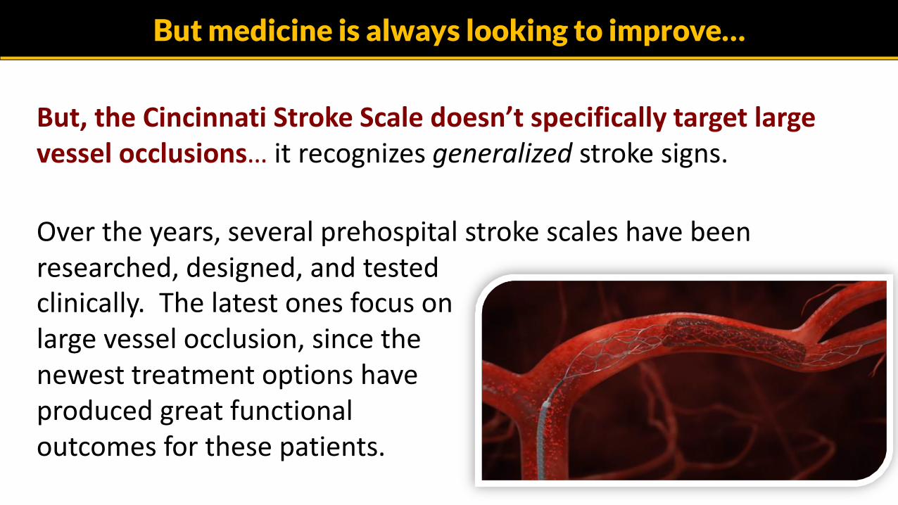
But, the Cincinnati Stroke Scale doesn’t specifically target large vessel occlusions… it recognizes generalized stroke signs.
But medicine is always looking to improve…
Over the years, several prehospital stroke scales have been researched, designed, and testedclinically. The latest ones focus on large vessel occlusion, since the newest treatment options have produced great functional outcomes for these patients.

Lots of stroke scales out there. Some are complex.
Some of these other stroke scales still require adding up scores, others take some extra time to assess, and a few don’t offer an easy way to remember the assessment points.
Checklists and score boxes are not ideal solutions for the prehospital environment.

Looking at a few of the stroke assessment tools out there:
Acronym: Stroke Assessment Tools:
3I-SS Three Item Stroke Scale
CPSSS Cincinnati Prehospital Stroke Severity Score
FAST-ED Field Assessment Stroke Triage for Emergency Destination
Hemiparesis Assessment for unilateral paralysis
LAMS Los Angeles Motor Scale
LEGS Texas Stroke Intervention Prehospital Stroke Severity Scale
RACES Rapid Arterial Occlusion Evaluation Scale
VAN Vision, Aphasia, and Neglect
And of course, the emergency department standard for stroke assessment scores:
NIHSS National Institute of Health Stroke Scale

MOTOR FUNCTION AREA: Initiation of voluntary muscle use. Is a component of ALL stroke scales.7,8
PARIETAL LOBE: Sensation from muscles and skin. Used by VAN, FAST-ED, and RACE.
CEREBRAL CORTEX: Controls eye movement and orientation. Used by VAN, 3I-SS, CPSSS, FAST-ED, LEGS, and RACE.
BROCA’S AREA: Muscle control area for speech. Used by VAN, CPSSS, FAST-ED, LEGS, and RACE.
OCCIPITAL LOBE: Sight, image perception/recognition. Used by VAN, LEGS, and RACE.
WERNICKE’S AREA: Written and spoken language comprehension. Used by VAN, CPSSS, LEGS, and RACE.
What do the different stroke scales assess?

Assessing the patient using VAN
The VAN scale was selected as our large vessel occlusion determination scale because it’s rapid (30 seconds), no math, no checklists, and:
• The first assessment involves motor function, just like the Cincinnati Stroke Scale. If the patient has no arm drift, paralysis, or unilateral weakness, the test is over. This rapidly triages out a group of patients since a large vessel occlusion affects motor function.
• If arm drift is present, you’ll move on to the VAN portion of the assessment…
Both arms extended out, palms up, and her eyes must be closed for 10 seconds to assess for drift.

The VAN Assessment Procedure
VVisual Disturbance. Does the patient report double-vision, field cut, or loss of vision? Is it difficult for her to see your fingers clearly in a visual quadrant? If “Yes”, the test is done at this point and she’s “VAN positive”. If not, move to the next test.
Two examples of field cut (hemianopsia) below, where a portion of the patient’s vision is blurred or blinded. It’s helpful to learn which side is affected and report that to the receiving staff.
Or…

The VAN Assessment Procedure
AAphasia. Any difficulty forming words? Can she repeat a short sentence? Can she recognize two objects correctly? Follows simple commands (and there’s no language barrier present)?
Do not count slurred speech or baseline aphasia as a positive sign… this could be a smaller vessel occlusion or from another cause. If any of the listed aphasia criteria are met, the test is done at this point and she’s “VAN positive”. If not, move on to the next test.
Good dog.

The VAN Assessment Procedure
NNeglect. Neglect refers to the patient’s senses and gaze. Does the patient present with an acute “forced gaze”/conjugate gaze palsy? Are her eyes unable to track your pen to one side? Ask her to close her eyes: Is she unable to feel sensation to an arm or leg when
It’s important to assess for all directions of tracking, as eye deviation may be noticeable in one direction only.
one or both are stimulated? If “Yes” to any of these, she is “VAN positive”. If not, her VAN assessment is negative for a large vessel occlusion.

Video: Dr. Mohamed Teleb. Stroke VAN. Web site: https://www.strokevan.com/learn-van/
Video of the VAN assessment

The VAN stroke assessment is new and hasn’t been validated in a large, multicenter study yet. However, early results from one emergency department’s stroke activations offers promising results:
How accurate is the VAN stroke scale?
Results from a single center VAN pilot study 8
Total number of patients who were stroke activations: 62
Of those 62 patients, how many were VAN positive on assessment? 19
Of those 19 patients, how many had a large vessel occlusion (LVO)? 14
Note: All 14 of those LVO patients were VAN positive in this study. 100% sensitivity
There were 5 false-positives (patients who were VAN+, but not having a LVO stroke). 90% specificity
The VAN assessment did not miss any LVO strokes. No false negatives

EMS: Both the Cincinnati Stroke Scale and the VAN assessment need to be performed for any suspected stroke patient. However, this is easy to
do since both share two elements: Arm drift and a speech assessment.
Protocol revision for EMS
Keep in mind that computed tomography (“the head CT”) still remains as the goldstandard for stroke diagnosis.

This box now lists that both the Cincinnati Stroke Screen and VAN stroke screen should be performed.
The hospital should be alerted to any positive VAN assessments.

The VAN Stroke Assessment isn’t just for EMS
Where else can the VAN scale be used?
• Emergency Center Triage
• Hospital ICUs, step-down units, anywhere a patient occupies a bed or a seat and suddenly develops acute stroke-like signs and symptoms.

“Wake Up” Strokes
A “wake-up stroke” occurs when a patient awakens with stroke-like symptoms that were not present before falling asleep. They remain a mystery, but are likely the result of circadian changes in coagulability, serum catecholamine levels, and autonomic tone.9
They account for roughly 1 in 5 acute ischemic strokes 9.

The Future for “Wake Up” Stroke Patients
There are ongoing endovascular stroke trials that will allow for enrollment of patients with an unknown time of symptom onset.
In one trial using the Trevo® stent retriever, the time window for treatment is 6 to 24 hours after symptom onset or last seen well, respectively. The trial started in 2014 with a completion date expected in 2017.10

Questions?

1. Berkhemer OA, Fransen PS, Beumer D, et al. MR CLEAN Investigators. A randomized trial of intra-arterial treatment for acute ischemic stroke. N Engl J Med. 2015;372:11–20. doi: 10.1056/NEJMoa1411587.
2. Saver JL, Goyal M, Bonafe A, et al. SWIFT PRIME Investigators. Stent-retriever thrombectomy after intravenous t-PA vs. t-PA alone in stroke. N Engl J Med. 2015;372:2285–2295. doi: 10.1056/NEJMoa1415061.
3. Campbell BC, Mitchell PJ, Kleinig TJ, et al. EXTEND-IA Investigators. Endovascular therapy for ischemic stroke with perfusion-imaging selection. N Engl J Med. 2015;372:1009–1018. doi: 10.1056/NEJMoa1414792.
4. Goyal M, Demchuk AM, Menon BK, et al. ESCAPE Trial Investigators. Randomized assessment of rapid endovascular treatment of ischemic stroke. N Engl J Med. 2015;372:1019–1030. doi: 10.1056/NEJMoa1414905.
5. Jovin TG, Chamorro A, Cobo E, et al. REVASCAT Trial Investigators. Thrombectomy within 8 hours after symptom onset in ischemic stroke. N Engl J Med. 2015;372:2296–2306. doi: 10.1056/NEJMoa1503780.
6. Kothari RU, Pancioli A, Liu T, et al. Cincinnati Prehospital Stroke Scale: reproducibility and validity. Ann Emerg Med. 1999 Apr;33(4):373-378.
7. Lima FO, Silva GS, Furie KL, et al. Field assessment stroke triage for emergency destination: A simple and accurate prehospital scale to detect large vessel occlusion strokes. Stroke. 2016;47:1997-2002. doi: 10.1161/STROKEAHA.116.013301.
8. Teleb MS, Ver Hage A, Carter J, et al. Stroke vision, aphasia, neglect (VAN) assessment — a novel emergent large vessel occlusion screening tool: pilot study and comparison with current clinical severity indices. NeuroIntervent Surg 2016;0:1–5. doi:10.1136/neurintsurg-2015-012131
References:

9. Rubin MN and Barrett KM. What to do with wake-up stroke. Neurohospitalist. 2015 Jul;5(3):161-172. doi: 10.1177/1941874415576204.
10. Thomalla G and Gerloff C. Treatment Concepts for Wake-Up Stroke and Stroke With Unknown Time of Symptom Onset. Stroke. 2015 Sep;46(9):2707-13. doi: 10.1161/STROKEAHA.115.009701.
References: