pregnancy and the relationship to age-related macular
TRANSCRIPT

Louisiana State UniversityLSU Digital Commons
LSU Master's Theses Graduate School
2012
Pregnancy and the relationship to age-relatedmacular degenerationAnn Hardin ShawLouisiana State University and Agricultural and Mechanical College, [email protected]
Follow this and additional works at: https://digitalcommons.lsu.edu/gradschool_theses
Part of the Human Ecology Commons
This Thesis is brought to you for free and open access by the Graduate School at LSU Digital Commons. It has been accepted for inclusion in LSUMaster's Theses by an authorized graduate school editor of LSU Digital Commons. For more information, please contact [email protected].
Recommended CitationShaw, Ann Hardin, "Pregnancy and the relationship to age-related macular degeneration" (2012). LSU Master's Theses. 1895.https://digitalcommons.lsu.edu/gradschool_theses/1895

PREGNANCY AND THE RELATIONSHIP TO AGE-RELATED
MACULAR DEGENERATION
A Thesis
Submitted to the Graduate Faculty of the Louisiana State University and
Agricultural and Mechanical College in partial fulfillment for the
requirements for the degree of Master of Science
in
The School of Human Ecology
by
Ann Hardin Shaw B.B.A., University of Georgia, 2003
O.D., Southern College of Optometry, 2008 May 2012

ii
ACKNOWLEDGMENTS
I would like to thank Dr. Carol Lammi-Keefe for her guidance with my thesis. I
would especially like to thank Dr. Holiday Durham for all of her time and patience with
me while completing my thesis. I would like to thank my committee members Dr.
Georgianna Tuuri and Dr. Liu Zhijun. I would also like to thank my parents for their
support while I was in graduate school. Finally, I would like to thank Drs. Roger Shaw,
Donald Peavy, Stephen Breaud, and all of their staff for helping me collect the data for
the macular study.

iii
TABLE OF CONTENTS
ACKNOWLEDGEMENTS……………………………………………………………….ii ABSTRACT………………………………………………………………………….….iv CHAPTER 1 INTROUCTION………………………………………………………………...1 Justification………………………………………………………..............2 Assumptions……………………………………………………….............2 Research Hypothesis……………………………………………………....2 Objectives…………………………………………………………............2 Limitations………………………………………………………………...2 2 LITERATURE REVIEW……………………………………………………….3 Lipids……………………………………………………………………...3 Essential Fatty Acids and the Fatty Acid Derivatives…………………….4 DHA and Pregnancy……………………………………………………..10 The Retina………………………………………………………………..13 Absorption of Light and Age-Related Macular Degeneration…...………16 DHA (Nutrition) and Macular Protection………………………………..18 3 MATERIALS AND METHODS………………………………………………23 Participants, Recruitment, Consenting, and Procedure………………......23 Data Analysis…………………………………………………………….24 4 RESULTS…………………….……………………………………………......26
5 DISSCUSSION………………………………………………………………...31
6 CONCLUSION………………………………………………………………...35 LITERATURE CITED…………………………………………………………………..36 APPENDIX A “WE NEED YOU” CARD …………………………………………………...41 B CONSENT FORM…………………………………………………………….42 C QUESTIONNAIRE (HEALTH HISTORY FORM)…………………………44 D “NURSES SCRIPT/DIRECTIONS”………………………………………….46 VITA……………………………………………………………………………………..4
ABSTRACT

iv
The Macular Study was a case control study that evaluated if parity and other
participant characteristics predicted the diagnosis of age-related macular degeneration
(AMD). Women, compared to men, are at higher risk for AMD. AMD is one of the
leading causes of blindness in the elderly population [1]. Docosahexaenoic acid (DHA,
22:6n-3) is a long-chain fatty acid that is essential for the structure and function of the
eye. During pregnancy the growing fetus depletes the maternal stores of DHA through
placental transfer. The fetus needs an ample supply of DHA for proper retinal and central
nervous system development. To date there is no research evaluating the number of
pregnancies and their effect on development of AMD. We posed the question: “Does the
number of pregnancies have an effect on the development of AMD in women?” Degree
of AMD was documented and evaluated by four different eye doctors in Baton Rouge for
501 women. The women in the study completed a health history form that included
demographic information, information about past pregnancies, and general health.
Using analysis of variance (ANOVA), women with a higher number of births
were more likely to be diagnosed with early, intermediate or advanced AMD versus those
women never diagnosed (3.27 + 0.19, 3.64 + 0.22, 3.33 + 0.24 versus 2.53 + 0.15,
number of children p < 0.0001). Numerous risk factors were considered, along with
parity, in subsequent analyses; these were age, race, eye color, smoking history, vitamin
intake, fish oil intake, family history of AMD, history of hypertension, and body mass
index (BMI). Using backwards-stepwise regression the most significant risk factors
predicting the diagnosis of AMD were determined (p < 0.01) and entered into a logistic
regression model. Age, parity, BMI, and BMI by parity significantly predicted the
v
diagnosis of AMD. As age, BMI and the number of pregnancies increased, the
probability of being diagnosed with AMD also increased.
In conclusion, older women, with a higher BMI, who have had more pregnancies,
were more likely to have AMD compared to younger women with a lower BMI and
fewer pregnancies. It is important that future studies consider parity as a possible risk for
AMD, especially as it relates to other participant characteristics. Such studies may
provide insight as to why women are at greater risk for AMD.
1
CHAPTER 1
INTRODUCTION
Age related macular degeneration (AMD) is one of the leading causes of
blindness in the elderly population [1]. AMD is the degeneration of the macula area in
the eye. It leads to distorted central vision and can eventually lead to blindness in more
advanced forms. AMD is currently untreatable and currently there are only
recommendations that have not been completely proven to slow down the development of
AMD. Women have been shown to be at higher risk for development of AMD and here
we speculate that this high risk could be associated with pregnancy.
During pregnancy the fetus depends on the mother for all nutrients to develop.
Docosahexaenoic acid (DHA, 22:6n-3), a long-chain n-3 fatty acid is an important
nutrient that the fetus must obtain directly through placental transfer from the mother [2].
DHA is necessary for the fetus to have proper retinal and brain development [3]. It is
found in high concentrations in the retinal photoreceptors where it is needed for
photoreceptors to function properly. However, consumption of DHA in the average
American woman’s diet, especially pregnant women, is low [4] and this may cause harm
to the mother’s eye sight in the future, especially in women who have had multiple
pregnancies. The relationship between number of pregnancies and retinal health was
explored in the current study.
JUSTIFICATION
DHA’s role in the eye is not completely understood. DHA is an important long-
chain polyunsaturated fatty acid that is recognized for its role in proper brain and retinal
development. A growing fetus obtains DHA through placental transfer from the mother.
2
This transfer depletes maternal DHA stores. After pregnancy it can be difficult for the
mother to return her DHA stores back to pre-pregnancy levels [5].
In the retina, DHA protects the photoreceptors and offers neuroprotective
properties [6]. Although little is known about how DHA protects the macular pigment,
Johnson et al. [7] hypothesized that the mechanism involves DHA’s role in the transport
of lutein and zeaxanthin through the circulation via high-density lipoproteins (HDL) to
the macula. The macular pigment (MP) is composed of the carotenoids lutein and
zeaxanthin which help protect the macula by filtering blue light which causes damage to
the photoreceptors [8].
ASSUMPTIONS
It is assumed that women who participate in the study will give correct
information about their health and past pregnancies to the nurses recording the
information. It is also assumed that the nurses and the doctors will document the correct
information following the guidelines in the study.
RESEARCH HYPOTHESIS
Women who have had a higher number of pregnancies will have a higher risk of
diagnosis with AMD.
OBJECTIVE
To evaluate parity and other known predictors of AMD among women diagnosed
with AMD.
LIMITATIONS
1. Self‐reported health history.
2. Observational study at one time point; not a longitudinal study.
3
CHAPTER 2
LITERATURE REVIEW
LIPIDS
Lipids are hydrophobic components comprised of carbon, hydrogen, and oxygen
atoms [9]. Major classes of lipids include fatty acids, triacylglycerols, phospholipids, and
sterols [9] . Lipids are important sources of energy, constituents of cells, membranes,
hormones, and mediators of electron transport [10]. Fatty acids are the most abundant
class of lipids that are essential to the function and structure of the eye.
Fatty acids contain a carboxylic acid group (-COOH) at one end and a methyl
group (-CH3) at the other end. The different groups at the opposite ends of the fatty acid
allow fatty acids to have polar (-COOH) as well as non-polar, hydrophobic (-CH3)
charcteristics [10]. Fatty acids classified as saturated fatty acids (SFA) are those that
contain only carbon-carbon single bonds, while unsaturated fatty acids contain carbon-
carbon double bonds. The double bonds imbue the structure with flexibility.
Monounsaturated fatty acids (MUFA) contain one carbon-carbon double bond.
Polyunsaturated fatty acids (PUFA) contain more than one carbon-carbon double bond
[9].
Fatty acids are also further classified according to chain length. Fatty acids
exceeding a 12-carbon chain are considered long chain fatty acids [9]. Longer chain fatty
acids have higher melting and boiling points. The longer chain fatty acids are also more
water insoluble, underlining the need for complex processes for digestion, absorption,
transport, and circulation of long chain fatty acids [9].
4
Fatty acids exist in either the “cis” or “trans” configuration. The majority of
naturally occurring fatty acids are found in the “cis” form, which means that the
hydrogen atoms are positioned on the same side of the double bond [9]. If the hydrogen
atoms are on opposite sides of the double bond, the fatty acid is in the “trans”
configuration [9]. Dietary “trans” fatty acids usually result from the partial
hydrogenation of fats and oils during processing [10].
The omega “ω” or “n” nomenclature is used in nutritional sciences to name fatty
acids. The omega nomenclature categorizes fatty acids into groups based on where the
first double bond is located relative to the methyl group [9]. For example, in the case of
docosahexaenoic acid (DHA, 22:6n-3), the 22 indicates the total number of carbons, 6
represents the total number of double bonds, and the location of the first double bond
from the methyl group is signaled by the number following n. A methylene group
(-CH2-) separates double bonds.
ESSENTIAL FATTY ACIDS AND THE FATTY ACID DERIVATIVES
Essential fatty acids (EFA) are fatty acids that are needed by the body for growth
and development. The two EFAs linoleic acid (LA, 18:2n-6) and alpha linolenic acid
(ALA, 18:3n-3) must be obtained from the diet because the body does not make them.
Humans cannot make these fatty acids because humans lack specific enzymes to
introduce additional double bonds to the carbon chain before the 9 position from the
methyl group [10]. The longer chain polyunsaturated fatty acid (LCPUFA) arachidonic
acid (AA, 20:4n-6) can either come from the shorter 18-carbon chain acid (18:2n-6) or
can be obtained directly from the diet. The LCPUFA DHA comes primarily from the diet

5
because enzyme competition results in very little DHA being synthesized from the
shorter 18-carbon chain acid (ALA).
The shorter chain fatty acids, LA and ALA, cannot be synthesized by humans but
once consumed they can be desaturated and elongated to the LCPUFA’s through a
process taking place in the liver (Figure 1). Although ALA and LA are both needed in
the human diet, studies of the average American diet indicate that people consume
significantly more omega 6 than omega 3 fatty acids [11]. Additionally, as noted here,
very little ALA is converted to the long chain derivatives.
Figure 1: Pathways of LA and ALA and enzymes involved (Adopted from [12]).
6
LA is converted to longer chain fatty acids, including AA. AA is very abundant
in the American diet because it is found in animal meats and processed foods made from
animal fats. Arachidonic acid is involved in the cell signaling pathways and cell division
[4]. It is an eicosanoid precursor that plays an important role in inflammation, and
cyclooxygenase and lipoxygenase pathways, which modulate platelet aggregation,
smooth muscle contraction, and vascular constriction [10]. LA is first converted to
gamma- linolenic acid by n-6 desaturase and then elongated to dihomo-gamma-linolenic
acid (DGLA) [11]. DGLA is converted to AA, which produces the pro-inflammatory
prostaglandins.
ALA can be converted to the longer chain fatty acid including eicosapentaenoic
acid (EPA, 20:5, n-3) and DHA. EPA is an eicosanoid precursor involved with anti-
inflammatory prostaglandins and DHA is a central determinant of brain and visual system
development [4]. ALA is converted to stearidonic acid by n-6 desaturase, which is then
elongated to eicosatetraenoic acid [11]. Eicosatetraenoic acid is converted to EPA by n-5
desaturase [11]. EPA is elongated to docosapentaenoic acid, which is further elongated,
desaturated, and β-oxidized to produce DHA [11]. However, the conversion of ALA to
EPA and particularly DHA, lacks efficiency due to the rate limitation introduced by the
n-6 desaturase during the process of long chain conversion of PUFA precursors [13].
Moreover, if a person has higher plasma levels of LA (typical in the diets of Americans),
research has shown that there may be a decrease in the conversion of ALA to EPA and
DHA, and an increase in the conversion of LA to AA because of the competition for the
n-6 desaturase [14]. It is estimated that adults convert only about 0.2% to 9% of ALA to
EPA [4]. Therefore, it is important that pregnant women obtain their nutritional

7
requirements for EPA and DHA directly from the diet as preformed acids to meet their
needs, as well as the needs of the growing fetus.
Physiologically, EPA and AA are precursors to eicosanoids. Eicosanoids are
hormone-like molecules that are necessary for numerous physiological processes in the
human body, such as movement of calcium, muscle contraction and relaxation, control of
fertility, cell division, and growth [15]. The eicosanoids for EPA and AA produce
different subgroups of substances know as prostaglandins, leukotrienes, and
thromboxanes (Figure 2) [15]. The eicosanoid subgroups produced by EPA tend to have
an anti-inflammatory effect and the subgroups produced by AA tend to have more of an
inflammatory response. However, the body requires the production of both types of
eicosanoid subgroups because anti-inflammatory properties as well as pro-inflammatory
properties are required by the body to function properly.
Figure 2: Eicosanoid pathways from n-3 and n-6 (Adopted from [16]).

8
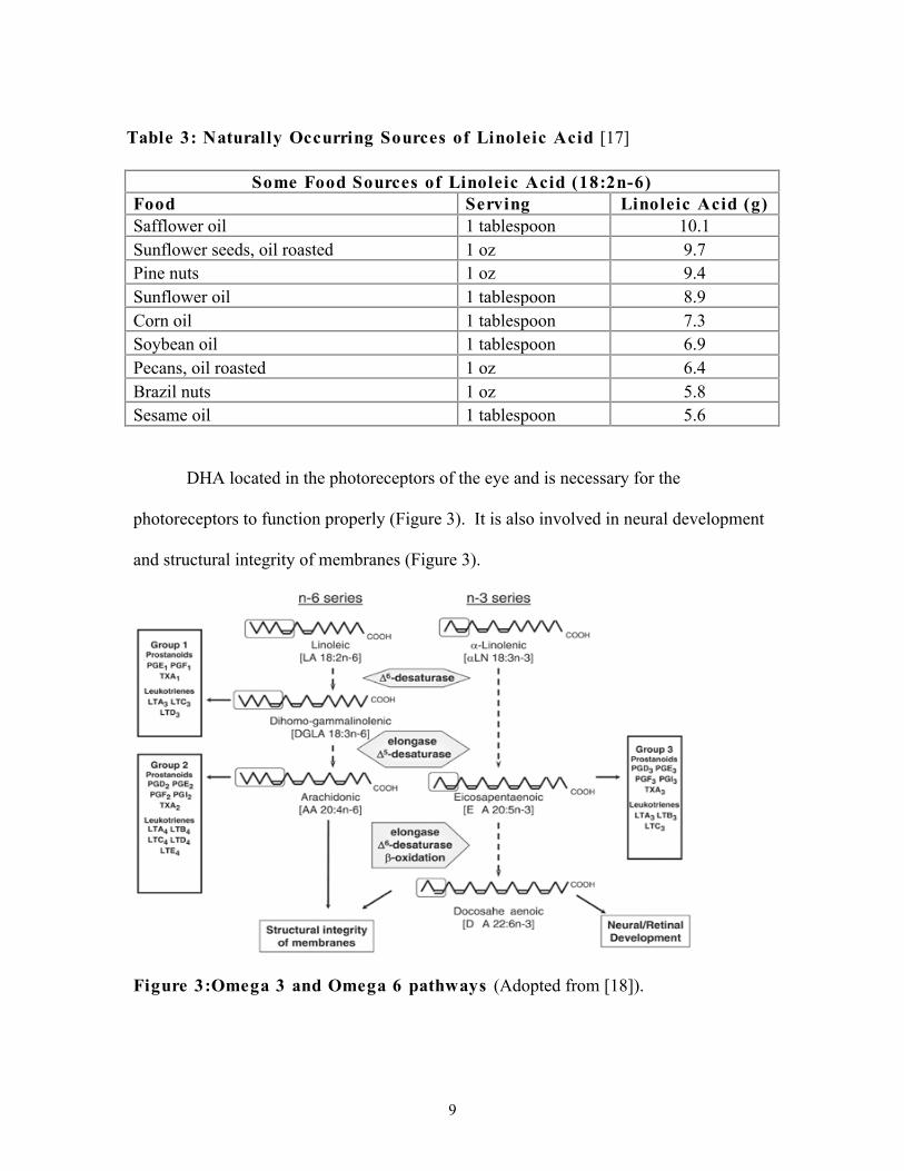
Some sources of ALA are walnuts, firm tofu, and flaxseed oil (Table 1). DHA is
found naturally in deep-water fish because they eat algae. Table 2 list different types of
fish that are high in DHA such as salmon and tuna. Some sources of LA are pine nuts,
corn oil, and sunflower oil (Table 3).
Table 1: Naturally Occurring Sources of Alpha-linolenic Acid [17]
Some Food Sources of Alpha-linolenic Acid (18:3n-3) Food Serving Alpha-Linolenic acid (g) Flaxseed oil 1 tablespoon 7.3 Walnuts, English 1 oz 2.6 Flaxseeds, ground 1 tablespoon 1.6 Walnut oil 1 tablespoon 1.4 Canola oil 1 tablespoon 1.3 Soybean oil 1 tablespoon 0.9 Mustard oil 1 tablespoon 0.8 Tofu, firm ½ cup 0.7 Walnuts, black 1 oz 0.6
Table 2: Naturally Occurring Sources of EPA and DHA [17]
Some Food Sources of EPA (20:5n-3) and DHA (22:6n-3)
Food Serving EPA (g)
DHA (g)
Amount providing 1 g
of EPA + DHA
Herring, Pacific 3 oz* 1.06 0.75 1.5 oz Salmon, Chinook 3 oz 0.86 0.62 2 oz Sardines, Pacific 3 oz 0.45 0.74 2.5 oz Salmon, Atlantic 3 oz 0.28 0.95 2.5 oz Oysters, Pacific 3 oz 0.75 0.43 2.5 oz Salmon, sockeye 3 oz 0.45 0.60 3 oz Trout, rainbow 3 oz 0.40 0.44 3.5 oz Tuna, canned, white 3 oz 0.20 0.54 4 oz Crab, Dungeness 3 oz 0.24 0.10 9 oz Tuna, canned, light 3 oz 0.04 0.19 12 oz *A 3-oz serving of fish is about the size of a deck of cards.
9
Table 3: Naturally Occurring Sources of Linoleic Acid [17]
Some Food Sources of Linoleic Acid (18:2n-6) Food Serving Linoleic Acid (g) Safflower oil 1 tablespoon 10.1 Sunflower seeds, oil roasted 1 oz 9.7 Pine nuts 1 oz 9.4 Sunflower oil 1 tablespoon 8.9 Corn oil 1 tablespoon 7.3 Soybean oil 1 tablespoon 6.9 Pecans, oil roasted 1 oz 6.4 Brazil nuts 1 oz 5.8 Sesame oil 1 tablespoon 5.6
DHA located in the photoreceptors of the eye and is necessary for the
photoreceptors to function properly (Figure 3). It is also involved in neural development
and structural integrity of membranes (Figure 3).
Figure 3:Omega 3 and Omega 6 pathways (Adopted from [18]).

10
DHA AND PREGNANCY The developing fetus requires LCPUFAs, which are critical for fetal development,
especially during the third trimester [2, 3]. The essential fatty acids, LA and ALA, are
inefficiently transferred from the mother to the fetus; if transferred to the fetus, the fetus
has limited ability to convert the shorter chain fatty acids into the LCPUFAs because of
enzyme competition [3]. The fetus normally efficiently obtains DHA and AA directly
from the mother via placenta transfer because the human placental tissue and the fetus
lack the n-6 and n-5 desaturase enzymes necessary for the formation of longer chain fatty
acids [2]. Therefore, a pregnant mother requires LCPUFAs such as AA and DHA not
only for herself but also for her growing baby. DHA is required for the development of
the central nervous system and the retina of the fetus. During the last trimester, it is
estimated that the fetus needs about 12 g DHA per week for proper brain development
[19]. During pregnancy the maternal stores of DHA decline because of the mobilization
of DHA from the mother to the growing fetus [20]. Through placental transfer the fetus
obtains the LCPUFAs, such as DHA, EPA, and AA. Therefore, the mother needs to
consume enough LCPUFAs, especially DHA to ensure proper fetal growth and
development. Since the fetus requires an ample supply of LCPUFAs, there is a concern
that the majority of pregnant women are not getting enough DHA for themselves or their
fetuses [4].
For placental transfer of fatty acids to occur the fatty acids cross the
syncytiotrophoblast, which is the outer fetal component of the placenta that separates the
maternal microvillus vasculature from the fetal basal membrane vasculature [21, 22].
The syncytiotroblast consist of syncytiotroblast cells that are responsible for nutrient

11
exchange, such as fatty acids [22]. The essential and nonessential free fatty acids cross
the placenta by simple diffusion and/or fatty acid binding proteins (FABPs) [22]. The
placental plasma membrane fatty acid proteins (p-FABPpm) are located on the maternal
microvillus membrane and allow preferential binding of the LCPUFAs and aid in the
unidirectional flow to the fetus [2]. Research by Haggarty et al. [23] showed that when
the placenta is presented with a mixture of fatty acids the order of uptake is
AA>DHA>ALA>LA. However, they also determined that when the placenta is involved
in transferring fatty acids to the fetus the placenta retains AA and the order of transferring
is DHA>ALA>LA>AA.
Otto et al. [5] looked at the postpartum DHA plasma phospholipid profiles of
lactating and non-lactating women. The plasma phospholipids profiles showed that after
parturition DHA values in maternal plasma were decreased significantly in both lactating
and non-lactating women [5]. When compared to non-pregnant women the decline in
DHA levels was more significant for the lactating group than the non-lactating group [5].
Therefore, the DHA stores not only decrease during pregnancy but they remain decreased
after parturition for at least 16 weeks and are decreased further if the mother breastfeeds.
To maintain DHA status during and after pregnancy, women who plan on
becoming pregnant need to get DHA in their diet. Because of the high fetal demand for
DHA, the mother’s stores will be drained especially during the third trimester [24]. This
decrease will continue after parturition and replenishing the DHA stores can take several
months if the mother is lactating [5, 20]. If the mother does not replenish her DHA stores
then she puts herself at risk for “DHA deficiency”. N-3 fatty acid deficiency during
pregnancy can increase the chances of preterm deliveries and postpartum depression [25].

12
High amounts of n-3 LCPUFAs are thought to possibly prolong gestation because they
inhibit specific prostaglandins that are mediators of uterine contractions and cervical
ripening [26].
Two studies have been conducted to determine the effects of n-3 LCPUFA
supplementation and postpartum depression. Mozurkewich et al. [27] performed a
double blind, placebo-controlled study using EPA and DHA. The study concluded that
molecularly distilled n-3 fatty acid preparations provided a way for pregnant women to
reduce their risk for depression [27]. Because n-3 fatty acids are critical to the nervous
system a depletion in n-3 fatty acids could possibly affect certain neurotransmitters’
biosynthesis, signaling transduction, and uptake of certain hormones resulting in
depression [28]. However, further research is needed to determine if dietary
supplementation of n-3 fatty acids reduces the prevalence of post-partum depression [28].
Currently, there is not a recommended dietary allowance (RDA) for DHA during
pregnancy or lactation. However, much research has shown the need to increase DHA
intake before, during, and after pregnancy. The typical American diet lacks sufficient
amounts of DHA needed for pregnant women [29]. It is recommended that pregnant
women get at least 200 mg of DHA a day [30]. Because the western diet is high in meats
and processed foods that tend to be high in n-6 LCPUFAs, the ratio of dietary n-6 to n-3
is higher in the majority of people and including pregnant women when the fetus depletes
the maternal stores [4].
Pregnant women should consume about 200 mg of DHA a day by a supplement or
consumption of one to two servings of oily fish per week [30]. However, due to the high
mercury content in some of the deep-water oily fish, pregnant women need to be careful

13
of the fish they choose to consume. Predatory fish that are larger tend to contain more
mercury [31]. The predatory fish obtain the mercury from the ocean sediment that enters
the fish through their gills as they swim and their digestive tracts as they feed and from
the numerous smaller fish they consume [31, 32]. Some examples of predatory fish that
are high in mercury are swordfish, shark, and orange roughy.
The fetus is most affected by mercury during the second trimester of pregnancy
and the negative effect it can have on development, such as delayed walking and talking,
may not be noticed until later in life [31]. Fish such as salmon and chunk light canned
tuna are both good sources of DHA without high mercury toxicity. Pregnant women can
also add a fish oil supplement to their daily routine to increase DHA intake.
THE RETINA
The retina is part of the central nervous system and the majority of humans
sensory inputs transmit through the retina [33]. The retina is responsible for the
transformation of light into a neural signal through the process of phototransduction,
which is carried out by a number of different cells in the retina. The three main cells in
the retina involved in the phototransduction process are the photoreceptors, the bipolar,
and the ganglion cells (CAVS). There are two types of photoreceptors: rods and cones.
The rods are larger, located more peripherally in the retina, and are important in dim light
(scotopic vision). Humans have more rod photoreceptors than cones. The cones are
smaller, located more centrally in the retina, are important for well-lit conditions, and are
responsible for our central vision. The cone photoreceptors make up the macula and are
used for central vision; these are the photoreceptors that are affected most in macular

14
degeneration. The rods and the cones contain photopigment that absorbs photons of light
to aid in our vision [34].
The composition of the rods and cones is somewhat similar in that they both
contain an outer segment consisting of a stalk, the cilium, the inner segment, and the
outer fiber, cell body, and inner fiber, which terminate at the synaptic terminal [34]. The
photoreceptor outer segments (POS) are made up of lipid filled membranous discs that
contain visual pigment that converts light into a neural signal [34].
Light absorbed by the eye causes damage to the tips of the outer segments of the
photoreceptors, thereby requiring them to be replaced on a daily basis [33]. The rod
outer segments contain DHA and rhodopsin in their discs. The rod outer segment (ROS)
tips are shed in the morning and the replacement proteins are synthesized in the inner
segments, eventually making their way to the outer segments [33]. The cone outer
segments contain three different types of visual pigments and not as much DHA as the
ROS’s [34]. The cone outer segments are shed in the evening, but little is known about
the mechanism involved [33]. The regeneration of the outer segment tips is important for
the survival of the photoreceptors because the buildup of light-damaged outer segment
tips accumulate waste products such as lipofuscins, oxidized lipid end products that can
lead to damage in the eye [33].
The retinal pigment epithelium (RPE) is the most anterior layer of the retina,
located between the POS and Bruch’s membrane. The cells in the RPE have many
different functions that aid in the maintenance of the photoreceptors; they conduct the
daily shedding, internalization, and degradation of the photoreceptors [6]. The RPE cells
aid in the absorption of light and they transport nutrients as well as fatty acids to the

15
photoreceptors. They are responsible for the reisomerization of all-trans-retinal to 11-cis-
retinal that is needed by the photoreceptors for the visual cycle [35]. They stabilize the
ion composition in the sub-retinal space, which is also required for photoreceptors to
function properly [35]. Another important function of the RPE cells is the phagocytosis
of the shed POS. This is especially important because build up of POS can lead to waste
that can affect vision. When the RPE cells phagocytose the POS, they send the essential
components, such as retinal and DHA, back to the photoreceptors to be reused [35]. The
DHA is redelivered to the photoreceptors as the fatty acid and the retinal is redelivered as
11-cis-retinal [35].
Finally, the RPE cells secrete specific growth factors that are required for
photoreceptor functioning [35]. The loss of any of these functions can lead to retinal
degeneration and eventually loss of vision [35].
Bruch’s membrane located below the RPE and above the choroid, is the
passageway for nutrients to the retina (Figure 4). The build up of basement membrane
material in the Bruch’s membrane, is referred to as “drusen” [34]. AMD involves the
degeneration of the retina-choroid interface [34]. As a person ages, phospholipids
accumulate, causing a barrier that prevents the passage of metabolites and water, and
subsequently resulting in loss of nutrients to the retina [34]. If the retina is no longer
receiving the nutrients it needs to function, there is neovascularization and retinal atrophy
leading to AMD [34].
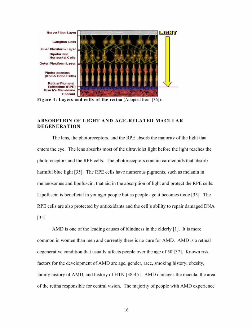
16
Figure 4: Layers and cells of the retina (Adopted from [36]).
ABSORPTION OF LIGHT AND AGE-RELATED MACULAR DEGENERATION
The lens, the photoreceptors, and the RPE absorb the majority of the light that
enters the eye. The lens absorbs most of the ultraviolet light before the light reaches the
photoreceptors and the RPE cells. The photoreceptors contain carotenoids that absorb
harmful blue light [35]. The RPE cells have numerous pigments, such as melanin in
melanosomes and lipofuscin, that aid in the absorption of light and protect the RPE cells.
Lipofuscin is beneficial in younger people but as people age it becomes toxic [35]. The
RPE cells are also protected by antioxidants and the cell’s ability to repair damaged DNA
[35].
AMD is one of the leading causes of blindness in the elderly [1]. It is more
common in women than men and currently there is no cure for AMD. AMD is a retinal
degenerative condition that usually affects people over the age of 50 [37]. Known risk
factors for the development of AMD are age, gender, race, smoking history, obesity,
family history of AMD, and history of HTN [38-45]. AMD damages the macula, the area
of the retina responsible for central vision. The majority of people with AMD experience

17
central vision distortion and therefore have to rely on their peripheral vision. There are
two types of AMD: dry AMD and wet AMD. Dry AMD is more common and less
visually disabling. Dry AMD can be classified as early, intermediate, or advanced. Dry
AMD is characterized by drusen, which is formed from the build up of basement
membrane material in Bruch’s membrane [35]. The classification of dry AMD depends
on the size and amount of the drusen. In advanced cases, the individual can develop
geographic atrophy and this causes more severe central vision loss. The other type of
AMD is called wet or neonvascular AMD. In wet AMD, the individual develops new
vessel growth in the macular area that can lead to unwanted bleeding. Wet AMD is more
visually devastating [37].
Although many theories exist to explain the pathogenesis of AMD, it remains a
complex disease that is poorly understood, with no effective therapy or prevention
currently available [45]. Recent research suggests that both genetic and exogenous
factors contribute to the pathogenesis of AMD [45]. AMD is believed to develop from
increased oxidative stress as a result of an increase in the number of reactive oxygen
species in the RPE [35]. The increase in stress and reactive oxygen species leads to
degeneration of the functioning of the RPE, causing vision loss with the macula most
affected. Increased oxidative stress can result in the build-up of toxic aged pigment
(lipofuscin) as a person ages or the reduction of melanosomes, which are needed for
protection from oxidative stress [35]. One main effect of the buildup of lipofuscin is
decreased ability of RPE to convert all-trans-retinal into 11-cis-retinal which is required
for the visual cycle [35]. Many different theories exist on how the retinoid from
lipofuscin, lipophilic cation N-retinyl-N-retinylidene ethanolamine (A2E), causes AMD,

18
with most studies finding that it increases the sensitivity of the RPE to blue light and it
induces apoptosis in RPE cells [35, 45]. A2E accumulates in the mitochondria of the
RPE cells leading to the release of different proapoptotic proteins that lead to further
retinal degeneration [45]. The destruction of RPE cells leads to the formation of drusen,
one of the most important symptoms of AMD [35]. Drusen can be formed between RPE
in Bruch’s membrane or within the Bruch’s membrane. These deposits consist of
metabolic products such as lipoproteins [35].
DHA (NUTRITION) AND MACULAR PROTECTION
DHA is found in very high concentrations in both the rod and cone photoreceptors
in the retina. These concentrations of DHA are highest in the rods, due in part to the
cones’ heightened exposure to harmful light rays that can destroy DHA. DHA acts as an
essential structural component in the retina with many protective functions [46]. DHA
protects against inflammation, oxidative damage, and ischemia, all of which are possible
pathogenic factors for the development of AMD [46]. The main dietary source of DHA
is fish oil, which is especially high in tuna, salmon, and herring. These deep-water fish
get DHA from the algae they consume. Our understanding of DHA’s role in the retina
remains limited but it appears to play a role in photoreceptor cell survival and function
[6]. DHA is found in the lipid-filled discs in the POS and it is constantly recycled from
the RPE to the POS [6]. Figure 5 shows the extracellular trafficking of DHA [6]. For
dietary DHA to reach the photoreceptors it is first taken up by the liver, where it is
esterified into phospholipids [6]. Circulating lipoproteins deliver DHA phospholipids to
the RPE and the brain; this is considered the long loop [6]. The short loop recycles the
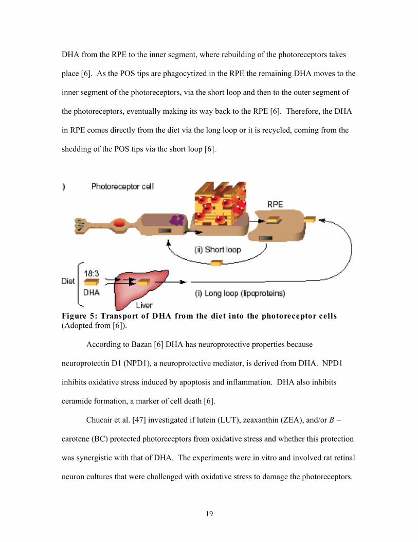
19
DHA from the RPE to the inner segment, where rebuilding of the photoreceptors takes
place [6]. As the POS tips are phagocytized in the RPE the remaining DHA moves to the
inner segment of the photoreceptors, via the short loop and then to the outer segment of
the photoreceptors, eventually making its way back to the RPE [6]. Therefore, the DHA
in RPE comes directly from the diet via the long loop or it is recycled, coming from the
shedding of the POS tips via the short loop [6].
Figure 5: Transport of DHA from the diet into the photoreceptor cells (Adopted from [6]).
According to Bazan [6] DHA has neuroprotective properties because
neuroprotectin D1 (NPD1), a neuroprotective mediator, is derived from DHA. NPD1
inhibits oxidative stress induced by apoptosis and inflammation. DHA also inhibits
ceramide formation, a marker of cell death [6].
Chucair et al. [47] investigated if lutein (LUT), zeaxanthin (ZEA), and/or B –
carotene (BC) protected photoreceptors from oxidative stress and whether this protection
was synergistic with that of DHA. The experiments were in vitro and involved rat retinal
neuron cultures that were challenged with oxidative stress to damage the photoreceptors.

20
The cultures were treated with BC, DHA, LUT, ZEA, or DHA combined with ZEA. The
results indicated that BC, DHA, LUT, ZEA, and DHA combined with ZEA all reduced
oxidative stress-induced apoptosis in photoreceptors, preserved the mitochondrial
potential and prevented the release of cytochrome c [47]. These results point to a
protective role for DHA in the retina.
DHA has also been shown to increase the macular pigment optical density
(MPOD). MPOD is the measurement of the macular pigment, which represents a ratio of
the different amounts of light absorbed in the retina. If a person has a high MPOD then
they are less likely to develop AMD because they are more protected by the increase in
macular pigment. An investigation by Johnson et al. [7] found that women, aged 60-80
years, supplemented with 800 mg per day of DHA had an increase in MPOD centrally in
the retina therefore creating more protection for the macula. These authors also looked at
DHA’s effect on lipoproteins and found that DHA aided in the transport of carotenoids,
such as lutein, which promoted the uptake of the protective carotenoids to the retina.
Chong et al. [46] performed a meta-analysis using numerous databases to
determine the association between fish consumption and development of early and late
AMD. The results of their investigation showed that a high dietary intake of n-3 fatty
acids (ALA, DHA and EPA) with supplementation or fish was associated with a 38
percent reduction in the risk of late AMD and that consumption of fish rich in n-3 fatty
acids twice or more a week was associated with decreased risk of development of early
and late AMD [46]. Other studies that supported their results, including the Blue
Mountains Eye Study [48] and the Nurses’ Health Study and the Health Professional
Follow-up Study [49] all showed a protective effect for increase n-3 fatty acid
21
consumption for early AMD [46]. The Blue Mountains Eye Study assessed the
associations between dietary fatty acids and incidence of age-related maculopathy (ARM)
[48]. Chua et al. [48] concluded that weekly intake of fish high in n-3 LCPUFAs offers
protection against both early and late ARM. In the Nurses’ Health Study and the Health
Professional Follow-up Study the authors looked at intake of total and specific types of
fat and their relationship to AMD [49]. Cho et al. [49] concluded that a high intake of
fish possibly reduced the risk of AMD. Chong et al. [46] also reported from prospective
cohort studies that fish intake of twice or more per week was associated with a 37 percent
decrease in risk of early AMD compared to intake of fish less than once per month.
Augood et al. [50] showed that increased consumption of oily fish high in DHA
and EPA at least once a week decreased the chances of development of neovascular
AMD. These investigators conducted their study using fundus photography and food-
frequency questionnaires. The fundus photos where graded by the International
Classification System for Age Related Maculopathy [50]. The data from the
questionnaires were converted into nutrient intakes with the use of food-composition
tables for comparison [50]. These data revealed that individuals with higher weekly
intakes of oily fish were significantly less likely to have neovascular AMD.
SanGiovanni et al. [51] performed a multicenter clinic-based prospective cohort
study from the Age-Related Eye Disease Study participants [52, 53]. They compared
fundus photos with nutritional intake data obtained from food frequency questionnaires.
They determined that consumption of n-3 LCPUFA (DHA and/or EPA) reduced the risk
of progression of bilateral drusen into central geographic atrophy, a precursor of AMD
[51].
22
A more recent study by Christen et al. [54], examined the effects of n-3 fatty
acids, particularly EPA and DHA, on incidence of AMD in women. All the women who
entered the study were free of AMD (n = 39,876) and by the completion of the 10-year
follow up study 235 of the original women were diagnosed with AMD, which could have
been low because diagnosis of AMD was self-reported. The examiners used food
frequency questionnaires to determine the amount of EPA and DHA the women
consumed. The results of the study suggested that regular consumption of EPA and DHA
significantly reduced the risk of incident AMD [54].
DHA has been shown in numerous studies to reduce the risk of development of
different forms of AMD [7, 46, 48-51]. However, despite evidence that DHA plays a
protective and preventative role in AMD, more research must be undertaken to advance
concrete recommendations for DHA intake for AMD prevention. Further research
involving large sample sizes to examine the relationship between DHA and AMD is
needed.
23
CHAPTER 3
MATERIALS AND METHODS
PARTICIPANTS, RECRUITMENT, CONSENTING, AND PROCEDURE
Women in the Baton Rouge area diagnosed with AMD and without AMD who
were 50 years or older, not incarcerated and were patients at Southern Eye Centers,
Vitreoretinal Institute, or Baton Rouge Eye Physicians, all located in Baton Rouge, LA,
were recruited to participate in this case controlled study. The doctors and nurses at these
offices obtained all study information from the subjects during their regularly scheduled
eye doctors’ visits. The Institutional Review Board of Louisiana State University’s
Agricultural Center approved the protocol.
When the women checked in for their appointments at one of these offices they
were given a “We Need You!” card that explained the study (Appendix A). Once inside
the private examination room they were then given a verbal explanation of the study and
the consent form (Appendix B). If the individual was interested in participating in the
study, the nurse consented the individual and proceeded to the questionnaire (Appendix
C). Participants were asked questions pertaining to their physical health, retinal health,
and past pregnancies. Family history of AMD, vitamin supplementation, and fish oil
supplementation were self-reported. Many subjects could not remember the exact brand
of vitamins and/or fish oil they took nor could they remember the exact number of years
they had been taking their supplements. The nurses at each of the offices followed the
“Nurse script/directions” provided by the researcher to maintain consistency between
offices (Appendix D). They were also provided demonstrations of the exact procedure to
follow.

24
The doctors evaluated the health of the macula by using a direct ophthalmoscope,
a binocular indirect ophthalmoscope, or a 90-diopter lens with a slit lamp. The degree of
macular degeneration was recorded as ‘no AMD’, ‘early’, ‘intermediate’, or ‘advanced.’
These categories of AMD are based on the description in the Will Eye Manual [37].
Early AMD was defined as small or medium size drusen with positive or negative
changes in pigment and no symptoms of vision loss [37]. Intermediate AMD was
defined as medium size drusen or one or more large drusen, may have some blurry vision
and a need for more light at near task [37]. Advanced AMD was defined as large drusen
and geographic atrophy with blurred areas in the central vision [37].
Once all of the questions were answered and the doctor agreed with the degree of
macular degeneration diagnosed, a label was put on the chart to ensure that the patients
were not recruited again. The signed consent form and the completed questionnaire were
stapled together and put in a locked file cabinet at the respective offices. The completed
forms were picked up weekly and brought to LSU where they were kept in a locked file
cabinet in Knapp Hall.
DATA ANALYSIS
Data were analyzed using SAS 9.2 (2011 SAS Institute, Cary NC). Analysis of
variance (ANOVA) and χ2 analyses were used to determine differences in participant
characteristics. Participant characteristics considered independent variables: included
age, parity, eye color, smoking history, family history of AMD, history of HTN, fish oil
intake, vitamin intake, and BMI. Diagnosis of AMD, as having or not having AMD, was
considered the dependent outcome variable. Using a backwards-stepwise regression,

25
non-significant variables were dropped from the model (p < 0.01). In the final binary
logistic regression model, only the significant variables from the backwards-stepwise
regression analysis were entered into the model (p < 0.05). The total effects of the
independent variables on the predictor of the outcome variable were reported. The
significance level was set to p < 0.05.

26
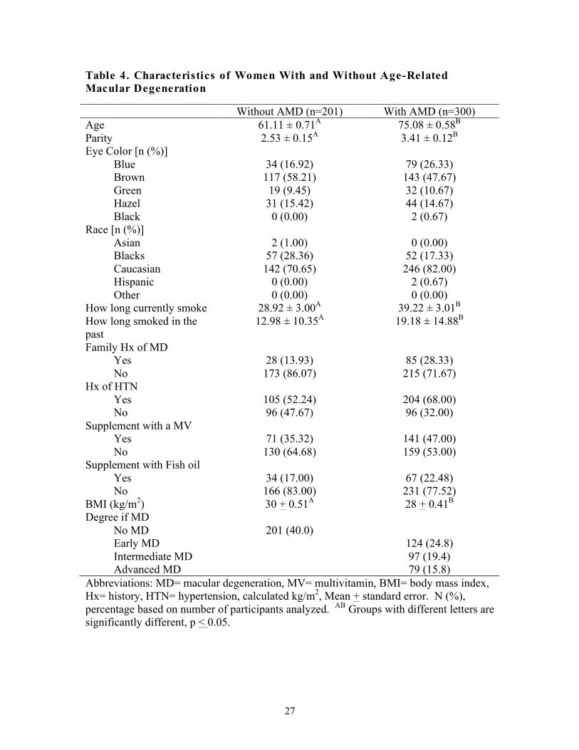
CHAPTER 4
RESULTS
Over a five-month period, 501 women were recruited and completed the study on
pregnancy and the relationship to AMD. As shown in Table 4, 40% of the women had
not been diagnosed with AMD and 60% were diagnosed with varying degrees of AMD.
Of the 300 women who were diagnosed with AMD 24.8% were diagnosed with early
stage AMD, while 19.4% were diagnosed with intermediate stage AMD and 15.8% were
diagnosed with advanced AMD. Participants diagnosed with AMD were significantly
older than those who had never been diagnosed (61.11 + 0.71 versus 75.08 + 0.58 years5,
p < .0001). The mean age of participants increased with more advanced forms of AMD
(70.64 + 0.85, 75.04 + 0.96, 82.11 + 1.07, age p < 0.001) (Table 5).
The majority of the women in this study had brown (52%) or blue (22.6%) eyes
and were predominately Caucasian (77.6%). Women with AMD were more likely to
have currently smoked for more years compared to women without AMD (39.22 + 3.01,
28.92 + 3.00 years, p = 0.0205). There was also a significant difference in past smoking
history between women with AMD versus those without AMD (19.18 + 14.88, 12.98 +
10.35 years past, p = 0.0084). Eye color, race, family history of AMD, and history of
HTN were not significantly different between women diagnosed with AMD and those
without AMD. There was no significant difference between vitamin and fish oil
supplementation between women with AMD and those without AMD.
27
Table 4. Characteristics of Women With and Without Age-Related Macular Degeneration
Without AMD (n=201) With AMD (n=300) Age 61.11 ± 0.71A 75.08 ± 0.58B
Parity 2.53 ± 0.15A 3.41 ± 0.12B
Eye Color [n (%)] Blue 34 (16.92) 79 (26.33) Brown 117 (58.21) 143 (47.67) Green 19 (9.45) 32 (10.67) Hazel 31 (15.42) 44 (14.67) Black 0 (0.00) 2 (0.67) Race [n (%)] Asian 2 (1.00) 0 (0.00) Blacks 57 (28.36) 52 (17.33) Caucasian 142 (70.65) 246 (82.00) Hispanic 0 (0.00) 2 (0.67) Other 0 (0.00) 0 (0.00) How long currently smoke 28.92 ± 3.00A 39.22 ± 3.01B
How long smoked in the past
12.98 ± 10.35A 19.18 ± 14.88B
Family Hx of MD Yes 28 (13.93) 85 (28.33) No 173 (86.07) 215 (71.67) Hx of HTN Yes 105 (52.24) 204 (68.00) No 96 (47.67) 96 (32.00) Supplement with a MV Yes 71 (35.32) 141 (47.00) No 130 (64.68) 159 (53.00) Supplement with Fish oil Yes 34 (17.00) 67 (22.48) No 166 (83.00) 231 (77.52) BMI (kg/m2) 30 + 0.51A 28 + 0.41B
Degree if MD No MD 201 (40.0) Early MD 124 (24.8) Intermediate MD 97 (19.4) Advanced MD 79 (15.8) Abbreviations: MD= macular degeneration, MV= multivitamin, BMI= body mass index, Hx= history, HTN= hypertension, calculated kg/m2, Mean + standard error. N (%), percentage based on number of participants analyzed. AB Groups with different letters are significantly different, p < 0.05.

28
Table 5. Characteristics of Women with Early, Intermediate, and Advanced Age-Related Macular Degeneration.
No AMD (n=201)
Early AMD (n=124)
Intermediate AMD (n= 97)
Advanced AMD (n= 79)
Age 61.11 ± 0.67A
70.64± 0.85B 75.04 ± 0.96C 82.11 ± 1.07D
Parity 2.53 ± 0.15A
3.27 ± 0.19B 3.64 ± 0.22B 3.33 ± 0.24B
Eye color [n (%)] Blue 34 (16.92) 34 (27.42) 24 (24.74) 21 (26.58) Brown 117 (58.21) 54 (43.55) 52 (53.61) 37 (46.84) Green 19 (9.45) 22 (17.74) 7 (7.22) 3 (3.80) Hazel 31 (15.42) 14 (11.29) 12 (12.37) 18 (22.78) Black 0 (0.00) 0 (0.00) 2 (2.06) 0 (0.00) Race [n (%)] Asian 2 (1.00) 0 (0.00) 0 (0.00) 0 (0.00) Blacks 57 (28.36) 26 (20.97) 18 (18.56) 8 (10.13) Caucasian 142 (70.65) 97 (78.23) 78 (80.41) 71 (89.87) Hispanic 0 (0.00) 1 (0.81) 1 (1.03) 0 (0.00) Other 0 (0.00) 0 (0.00) 0 (0.00) 0 (0.00) How long currently smoke
28.92 ± 2.84A
30.38 ± 4.91A
40.00 ± 4.19AB 53.75 ± 6.95B
How long smoked in the past
12.98 ± 1.78A
16.95 ± 2.34AB
19.11 ± 2.76AB 22.11 ± 2.64B
Family Hx of MD Yes 28 (13.93) 27 (21.77) 25 (25.77) 33 (41.77) No 173 (86.07) 97 (78.23) 72 (74.23) 46 (58.23) Hx of HTN Yes 105 (52.24) 81 (65.32) 69 (71.13) 54 (68.35) No 96 (47.76) 43 (34.68) 28 (28.87) 25 (31.65) Supplement with MV
Yes 71 (35.32) 44 (35.48) 51 (52.58) 46 (58.23) No 130 (64.68) 80 (64.52) 46 (47.42) 33 (41.77) Supplement with fish oil
Yes 34 (17.00) 28 (22.76) 22 (22.92) 17 (21.52) No 166 (83.00) 95 (77.24) 74 (77.08) 62 (78.48) BMI (kg/m2) 29.54 ±
0.51A 28.27 ± 0.64AB
28.91 ± 0.73AB 26.82 ± 0.81B
Abbreviations: MD= macular degeneration, MV= multivitamin, BMI= body mass index, Hx= history, HTN= hypertension, calculated kg/m2, Mean + standard error. N (%), percentage based on number of participants analyzed. ABCD Groups with different letters are significantly different, p < 0.05.
29
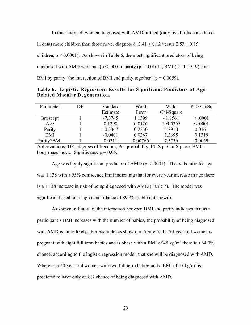
In this study, all women diagnosed with AMD birthed (only live births considered
in data) more children than those never diagnosed (3.41 + 0.12 versus 2.53 + 0.15
children, p < 0.0001). As shown in Table 6, the most significant predictors of being
diagnosed with AMD were age (p < .0001), parity (p = 0.0161), BMI (p = 0.1319), and
BMI by parity (the interaction of BMI and parity together) (p = 0.0059).
Table 6. Logistic Regression Results for Significant Predictors of Age-Related Macular Degeneration.
Parameter DF Standard Estimate
Wald Error
Wald Chi-Square
Pr > ChiSq
Intercept 1 -7.3745 1.1399 41.8561 < .0001 Age 1 0.1290 0.0126 104.5265 < .0001
Parity 1 -0.5367 0.2230 5.7910 0.0161 BMI 1 -0.0401 0.0267 2.2695 0.1319
Parity*BMI 1 0.0211 0.00766 7.5736 0.0059 Abbreviations: DF= degrees of freedom, Pr= probability, ChiSq= Chi-Square, BMI= body mass index. Significance p = 0.05.
Age was highly significant predictor of AMD (p < .0001). The odds ratio for age
was 1.138 with a 95% confidence limit indicating that for every year increase in age there
is a 1.138 increase in risk of being diagnosed with AMD (Table 7). The model was
significant based on a high concordance of 89.9% (table not shown).
As shown in Figure 6, the interaction between BMI and parity indicates that as a
participant’s BMI increases with the number of babies, the probability of being diagnosed
with AMD is more likely. For example, as shown in Figure 6, if a 50-year-old women is
pregnant with eight full term babies and is obese with a BMI of 45 kg/m2 there is a 64.0%
chance, according to the logistic regression model, that she will be diagnosed with AMD.
Where as a 50-year-old women with two full term babies and a BMI of 45 kg/m2 is
predicted to have only an 8% chance of being diagnosed with AMD.

30
Table 7. Odd Ratio for Age and Diagnosis of Age-Related Macular Degeneration.
Effect Point Estimate Confidence Interval Age 1.138 1.110 - 1.166
Figure 6: Probability being diagnosed with MD for 50-year-old women based on BMI and Parity. Based on the equation that probability = EXP (-7.3745+0.129*50-0.0401*BMI-0.5367*Parity+0.0211*BMI*Parity).
31
CHAPTER 5
DISCUSSION
To the best of our knowledge, this is the first study to examine the relationship
between parity and diagnosis of AMD. Results from the current study showed that the
women having more children were more likely to be diagnosed with early, intermediate,
and advanced stage AMD. In addition to parity, other risk factors commonly associated
with having AMD were considered, such as age eye color, race, family history of AMD,
history of HTN, and BMI [38-45]. Like other studies [39, 40, 45] age was considered a
significant predictor of AMD. However, this study uniquely found that BMI and parity
interacted together as a predictor for diagnosis of AMD.
Increased age is a well-established risk factor for AMD [39, 40, 45]. According
to Suter et al. [45] older individuals are at increased risk for developing AMD. Despite
the fact that the pathogenesis of AMD is poorly understood, as an individual ages there is
an increase in the accumulation of age pigment lipofuscin in lysosomes of the retinal
pigment epithelium (RPE) and this is thought to contribute to the development of AMD
lesions in the retina [45]. The lesions result from increased oxidative stress from the
build-up of toxic age pigment (lipofuscin) as the person ages [45]. The retinoid produced
from the lipofuscin, lipophilic cation N-retinyl-N-retinylidene ethanolamine (A2E),
causes AMD, with most studies reporting that it increases the sensitivity of the RPE to
blue light, inducing apoptosis in RPE cells [39, 45]. A2E accumulates in the
mitochondria of the RPE cells leading to the release of different proapoptotic proteins
that lead to further retinal degeneration [45]. The destruction of RPE cells from the
lesions (retinal degeneration) leads to the formation of drusen, one of the most significant

32
symptoms of AMD [35]. Therefore, our finding that age is an important risk factor is
supported by previous research.
Unlike most studies [43, 44], we did not find that BMI was a significant
independent predictor of being diagnosed with AMD in women, it was only considered
significant because of its interaction with parity. This may be explained in part because
height and weight were self-reported. Other studies have considered BMI, waist
circumference, and hip-to-waist ratios and it has been reported that increase in BMI is
associated with an increase in developing AMD. Seddon et al. [44] found that a BMI
greater than 30 kg/m2 was associated with an increase in the progression to more
advanced forms of AMD [44] in comparison to those individuals with a BMI less than 25
kg/m2. Those investigators also found similar results with increased waist circumference
and increased hip-to-waist ratio.
It is hypothesized that obesity (BMI > 30 kg/m2) is a risk factor for AMD because
it is associated with increased diagnosis of vascular diseases, decreased intake of
important nutrients, and increased inflammation, all of which can affect macular health
[44]. Peeters et al. [43] found that a reduction in waist-hip ratio in obese individuals
decreased the likelihood of having AMD. It is hypothesized that a decrease in waist-hip
ratio leads to improved cardiovascular health, which affects macular health [43]. While
the current exploratory study did not measure other physiological factors associated with
BMI, future studies should.
In the current study, parity was a significant predictor of AMD. To date no other
studies have evaluated the effects of pregnancy on the risk for developing AMD.
Because women are at higher risk for AMD, it is possible pregnancy may impact macular

33
health and it should therefore be considered when evaluating the risk factors associated
with being diagnosed with AMD. Herein we have provided evidence for the relationship
between parity and AMD.
During pregnancy there is a transfer of DHA from the mother to the growing fetus
[20]. DHA is a critical component found in high concentrations in both the rod and cone
photoreceptors [20]. DHA acts as an essential structural component in the retina with
many protective functions [46]. DHA protects against inflammation, oxidative damage,
and ischemia, all of which are possible pathogenic factors for the development of AMD
[46]. A concern regarding DHA and pregnancy is that the majority of women are not
getting sufficient amounts of DHA during pregnancy because the typical American diet
lacks DHA [29]. The fetus efficiently obtains DHA and AA directly from the mother
through placental transfer, the fetus lacking the n-6 and n-5 desaturase enzymes
necessary for the formation of longer chain fatty acids from the 18 carbon precursors [2].
Because of the transfer of DHA from the mother to the growing fetus and the important
roles that DHA has in the human body, DHA had been added to many prenatal vitamin
supplements in recent years. Therefore, the possible impact that pregnancy has on
macular health should be considered when evaluating the risk factors associated with the
diagnosis of AMD.
It is of interest to better understand how parity and other subject characteristics
may function together to impact an individual’s risk for AMD. In this study, we found
that BMI and parity significantly interact with one another and predict the likelihood of a
woman being diagnosed with AMD. We showed that the probability of being diagnosed
with AMD dramatically increased with increased BMI and number of pregnancies.
34
These results indicate the combined effects of the increased in diagnosis of vascular
diseases, decreased intake of carotenoids (research has shown that people with high
BMI’s tend to have a decrease in certain nutrients) [44], and increased in inflammation
possibly due to a high BMI >25 kg/m2 in conjunction with a decrease in DHA supply due
to pregnancy all together may significantly impact the diagnosis of AMD. While dietary
information was not collected in this population, evidence indicates that an increase in
consumption of DHA among women decreases the chance of being diagnosed with AMD
[7, 38, 40-44]. Therefore, it may be possible to improve macular health and decrease the
likelihood of being diagnosed with AMD in light of increased BMI and pregnancies by
consuming foods high in DHA. It remains to be studied, but supplementation with DHA
during pregnancy may be protective against future AMD. However, more research is
needed to make this association.

35
CHAPTER 6
CONCLUSION
The purpose of this study was to determine if parity is a risk factor for AMD in a
group of women over 50 years of age. This exploratory study has provided data that
suggest that parity is an important risk factor for diagnosis of AMD. Other significant
predictors were age and the interaction between BMI and parity. Age and BMI have
previously been researched and shown to be significant risk factors for AMD in women.
This current study is the first to describe parity as it relates to diagnosis of AMD. The
significant interaction between BMI and parity is an important finding and warrants
further investigation for a full understanding of the association with macular health and
the high risk of AMD in women.

36
LITERATURE CITED 1. National Plan for Eye and Vision Research
[http://www.nei.nih.gov/strategicplanning/nationalplan1.pdf] 2. Dutta-Roy AK: Transport mechanism for long-chain polyunsaturated fatty acid
requirements during pregnancy and lactation. Am J Clin Nutr 2000, 71:307S-311S.
3. Judge MP, Harel O, Lammi-Keefe CJ: Maternal consumption of a
docosahexaenoic acid-containing functional food during pregnancy: benefit for infant performance on problem-solving but not on recognition memory tasks at age 9 mo. Am J Clin Nutr 2007, 85:1572-1577.
4. Greenberg JA, Bell SJ, Ausdal WV: Omega-3 Fatty Acid supplementation during
pregnancy. Rev Obstet Gynecol 2008, 1:162-169. 5. Otto SJ, van Houwelingen AC, Badart-Smook A, Hornstra G: Comparison of the
peripartum and postpartum phospholipid polyunsaturated fatty acid profiles of lactating and nonlactating women. Am J Clin Nutr 2001, 73:1074-1079.
6. Bazan NG: Cell survival matters: docosahexaenoic acid signaling,
neuroprotection and photoreceptors. Trends Neurosci 2006, 29:263-271. 7. Johnson EJ, Chung HY, Caldarella SM, Snodderly DM: The influence of
supplemental lutein and docosahexaenoic acid on serum, lipoproteins, and macular pigmentation. Am J Clin Nutr 2008, 87:1521-1529.
8. Loane E, Stack J, Beatty S, Nolan JM: Measurement of macular pigment optical
density using two different heterochromatic flicker photometers. Curr Eye Res 2007, 32:555-564.
9. McGuire M, Beerman KA: Nutritional Sciences: From Fundamentals to Food.
Thomson Higher Education Inc.; 2007. 10. Gropper SS, Smith JL, Groff JL: Advanced Nutrition and Human Metabolism 5th
edition. Wadsworth Inc.; 2009. 11. Biochemisty Omega 3 and Omega 6 Fatty Acids
[http://www.livehealthytoday.org/pages/omega-fattyacids.php] 12. Chavarro JE, Stampfer MJ, Li H, Campos H, Kurth T, Ma J: A prospective study
of polyunsaturated fatty acid levels in blood and prostate cancer risk. Cancer Epidemiol Biomarkers Prev 2007, 16:1364-1370.

37
13. Hussein N, Ah-Sing E, Wilkinson P, Leach C, Griffin BA, Millward DJ: Long-chain conversion of [13C]linoleic acid and alpha-linolenic acid in response to marked changes in their dietary intake in men. J Lipid Res 2005, 46:269-280.
14. Louis YA, King DJ, Zibrik D, Innis SM: Decreasing linoleic acid with constant
alpha-linolenic acid in dietary fats increases (n-3) eicosapentaenoic acid in plasma phospholipids in healthy men. J Nutr 2007, 137:945-952.
15. Maclean CH, Issa AM, Newberry SJ, Mojica WA, Morton SC, Garland RH,
Hilton LG, Traina SB, Shekelle PG: Effects of omega-3 fatty acids on cognitive function with aging, dementia, and neurological diseases. Evid Rep Technol Assess (Summ) 2005:1-3.
16. Din JN, Newby DE, Flapan AD: Omega 3 fatty acids and cardiovascular disease--
fishing for a natural treatment. Br Med J 2004, 328:30-35. 17. Essential Fatty Acids [http://lpi.oregonstate.edu/infocenter/othernuts/omega3fa/] 18. Haggarty P: Fatty acid supply to the human fetus. Annu Rev Nutr 2010, 30:237-
255. 19. Clandinin MT, Chappell JE, Heim T, Swyer PR, Chance GW: Fatty acid
utilization in perinatal de novo synthesis of tissues. Early Hum Dev 1981, 5:355-366.
20. Hornstra G, Al MD, van Houwelingen AC, Foreman-van Drongelen MM:
Essential fatty acids in pregnancy and early human development. Eur J Obstet Gynecol Reprod Biol 1995, 61:57-62.
21. Campbell FM, Dutta-Roy AK: Plasma membrane fatty acid-binding protein
(FABPpm) is exclusively located in the maternal facing membranes of the human placenta. FEBS Lett 1995, 375:227-230.
22. Haggarty P: Placental regulation of fatty acid delivery and its effect on fetal
growth--a review. Placenta 2002, 23 Suppl A:S28-38. 23. Haggarty P, Page K, Abramovich DR, Ashton J, Brown D: Long-chain
polyunsaturated fatty acid transport across the perfused human placenta. Placenta 1997, 18:635-642.
24. Carlson SE WS, Rhodes PG, Tolley EA: Visual-acuity development in healthy
preterm infants: effects of marine-oil supplementaion. Am J Clin Nutr 1993, 58:35-42.
25. Makrides M, Gibson RA: Long-chain polyunsaturated fatty acid requirements
during pregnancy and lactation. Am J Clin Nutri 2000, 71:307S-311S.
38
26. Olsen SF, Hansen HS, Sorensen TI, Jensen B, Secher NJ, Sommer S, Knudsen LB: Intake of marine fat, rich in (n-3)-polyunsaturated fatty acids, may increase birthweight by prolonging gestation. Lancet 1986, 2:367-369.
27. Mozurkewich E, Chilimigras J, Klemens C, Keeton K, Allbaugh L, Hamilton S,
Berman D, Vazquez D, Marcus S, Djuric Z, Vahratian A: The mothers, Omega-3 and mental health study. BMC Pregnancy Childbirth 2011, 11:46.
28. Hibbeln JR, Salem N, Jr.: Dietary polyunsaturated fatty acids and depression:
when cholesterol does not satisfy. Am J Clin Nutr 1995, 62:1-9. 29. Omega-3 Fish Oil and Pregnancy
[http://ww.americanpregnancy.org/pregnancyhealth/omega3fishoil.html] 30. Koletzko B, Cetin I, Brenna JT: Dietary fat intakes for pregnant and lactating
women. Br J Nutr 2007, 98:873-877. 31. Mercury in Fish
[http://www.betterhealth.vic.gov.au/bhcv2/bhcarticles.nsf/pages/Mercury_in_fish] 32. Methymercury in Sport Fish: Information for Fish Consumers
[http://oehha.ca.gov/fish/hg/index.html] 33. Kaufman PL, Albert A: Adler's Physiology of the Eye Clinical Application Tenth
Edition. St. Louis: Mosby Inc.; 2003. 34. Remington LA: Clinical Anatomy of the Visual System. Newton: Butterworth-
Heinemann; 1998. 35. Strauss O: The retinal pigment epithelium in visual function. Physiol Rev 2005,
85:845-881. 36. Anatomy of the Eye: The Retinal Layers [http://www.mdsupport.org/anatomy.html] 37. Ho AB, Brown GC, McNamara JA, Recchia FM, Regillo, Vebder JF: Color Atlas
& Synopsis of Clinical Ophthalmology Wills Eye Hospital: Retina. 2003. 38. National Eye Instituute [http://www.nei.nih.gov/health/maculardegen/armd_facts.asp -
2b] 39. Curcio CA, Johnson M, Huang JD, Rudolf M: Aging, age-related macular
degeneration, and the response-to-retention of apolipoprotein B-containing lipoproteins. Prog Retin Eye Res 2009, 28:393-422.

39
40. Curcio CA, Johnson M, Huang JD, Rudolf M: Apolipoprotein B-containing lipoproteins in retinal aging and age-related macular degeneration. J Lipid Res 2010, 51:451-467.
41. Hyman L SA, He Q, Lesle MC: Hypertension, cardiovascular disease, and ag-
related macular degeneration. Age-Related Macular Degeneration Risk Factors Study Group. Arch Ophthalmol 2000, 118:351-358.
42. Mitchell P SW, Wand JJ: Iris color, skin sun sensitivity, and age-related
maculopathy. The Blue Mountains Eye Study. Ophthalmol 1998, 105:1359-1363. 43. Peeters A, Magliano DJ, Stevens J, Duncan BB, Klein R, Wong TY: Changes in
abdominal obesity and age-related macular degeneration: the Atherosclerosis Risk in Communities Study. Arch Ophthalmol 2008, 126:1554-1560.
44. Seddon JM, Cote J, Davis N, Rosner B: Progression of age-related macular
degeneration: association with body mass index, waist circumference, and waist-hip ratio. Arch Ophthalmol 2003, 121:785-792.
45. Suter M, Reme C, Grimm C, Wenzel A, Jaattela M, Esser P, Kociok N, Leist M,
Richter C: Age-related macular degeneration. The lipofusion component N-retinyl-N-retinylidene ethanolamine detaches proapoptotic proteins from mitochondria and induces apoptosis in mammalian retinal pigment epithelial cells. J Biol Chem 2000, 275:39625-39630.
46. Chong EW, Kreis AJ, Wong TY, Simpson JA, Guymer RH: Dietary omega-3
fatty acid and fish intake in the primary prevention of age-related macular degeneration: a systematic review and meta-analysis. Arch Ophthalmol 2008, 126:826-833.
47. Chucair AJ, Rotstein NP, Sangiovanni JP, During A, Chew EY, Politi LE: Lutein
and zeaxanthin protect photoreceptors from apoptosis induced by oxidative stress: relation with docosahexaenoic acid. Invest Ophthalmol Vis Sci 2007, 48:5168-5177.
48. Chua B, Flood V, Rochtchina E, Wang JJ, Smith W, Mitchell P: Dietary fatty
acids and the 5-year incidence of age-related maculopathy. Arch Ophthalmol 2006, 124:981-986.
49. Cho E, Hung S, Willett WC, Spiegelman D, Rimm EB, Seddon JM, Colditz GA,
Hankinson SE: Prospective study of dietary fat and the risk of age-related macular degeneration. Am J Clin Nutr 2001, 73:209-218.
50. Augood C, Chakravarthy U, Young I, Vioque J, de Jong PT, Bentham G, Rahu
M, Seland J, Soubrane G, Tomazzoli L, Topouzis F, Vingerling JR, Fletcher AE:
40
Oily fish consumption, dietary docosahexaenoic acid and eicosapentaenoic acid intakes, and associations with neovascular age-related macular degeneration. Am J Clin Nutr 2008, 88:398-406.
51. SanGiovanni JP, Chew EY, Agron E, Clemons TE, Ferris FL, 3rd, Gensler G,
Lindblad AS, Milton RC, Seddon JM, Klein R, Sperduto RD: The relationship of dietary omega-3 long-chain polyunsaturated fatty acid intake with incident age-related macular degeneration: AREDS report no. 23. Arch Ophthalmol 2008, 126:1274-1279.
52. Clemons TE MR, Klein R, Seddon JM, Ferris FL 3rd: Risk factors for the
incidence of Advanced Age-Related Macular Degeneration i the Age-Related Eye Disease Study (AREDS): AREDS report No. 19. Ophthalmol 2005, 112:533-539.
53. SanGiovanni JP CE, Clemons TE, Davis MD, Ferris FL 3rd, Gensler GR, Kurinij
N, Lindblad AS, Milton RC, Seddon JM, Sperduto RD: The relationship of dietary lipid intake and age-related macular degeneration in a case-control study: AREDS Report No. 20. Arch Ophthalmol 2007, 125:671-679.
54. Christen WG SD, Glynn RJ, Buring JE.: Dietary ω-3 fatty acid and fish intake
and incident age-related macular degeneration in women. Arch opthamol 2011, 34:E1-E9.

41
APPENDIX A: “WE NEED YOU” CARD
The LSU Division of Human Nutrition and Food is conducting a study to evaluate eye health and its relationship to pregnancy. With your help, LSU will better understand eye health of women. If you are interested in participating in the study, you will be asked a few questions concerning your health and previous pregnancy history. If you choose to participate, the doctor or nurse will explain the study and answer any questions you have about the study.

42
APPENDIX B: CONSENT FORM
SUBJECT CONSENT FORM
Title of Research Study : Eye Health as it May Relate to Number of Pregnancies Performance Sites: 202S Knapp Hall, Louisiana State University, Baton
Rouge, LA Contact: Ann Shaw, Graduate Student Telephone: (225) 578-7160 or email at [email protected] 202S Knapp Hall, Louisiana State University
Carol J. Lammi-Keefe, Ph.D., R.D. Professor Principal Director Telephone: 225 578-1518 297B Knapp Hall, Louisiana State University Baton Rouge, LA 70803 Purpose of the Study : This study will assess if the number of pregnancies a
woman has experienced is related to her eye health. Subjects: Females, 50 years or older Study Procedures : You will be asked a few health-related questions and your
eye care doctor will provide some information about your current eye health.
Benefits : In the long term, you will help us establish if there is a
relationship between pregnancy and eye health. Risks/Discomforts : There are no major identifiable risks. You may be
uncomfortable about questions involving your previous pregnancies.
If you wish to discuss any possible discomforts you might
experience, you may contact the research assistant or the principal investigator listed on this form.
Right to Refuse : Your participation in this study is entirely voluntary. You
may change your mind and withdraw from the study at any time without penalties or consequences.
Privacy: Your private information will be kept confidential as
required by law. You will be assigned an ID code and all

43
(name of subject)
data will be labeled only with the code number and not your name. All of your data will be kept in a locked file in 202S Knapp Hall, LSU.
The results from this study may be incorporated into research papers, presented at scientific meetings, or published in professional journals. Any information gathered will be protected from unauthorized access by the principal investigator. Data will be kept confidential unless release is legally required.
Removal : The principal investigator reserves the right to remove a
subject from the research if the subject fails to meet the requirements of the study protocol.
Questions: If you have any questions about this study, we will be
happy to answer these; you may contact the principal investigator, Carol Lammi-Keefe Ph.D., R.D. at (225) 578-1518. If you have questions about your rights as a subject or other concerns, you can contact Michael Keenan, Chairman, LSU AgCenter Institutional Review Board at (225) 578-1708.
Authorization: I have read this form and decided that _______________________ will participate in the
project described above. Its general purposes, procedures and risks have been explained
to my satisfaction. My signature also indicates that I have received a copy of this consent
form.
______________________ ______ _________________________ Signature of Subject Date Name of Subject (print) ______________________ ______ _________________________ Signature of Person Date Name of Person Obtaining Consent Obtaining Consent (print)
44
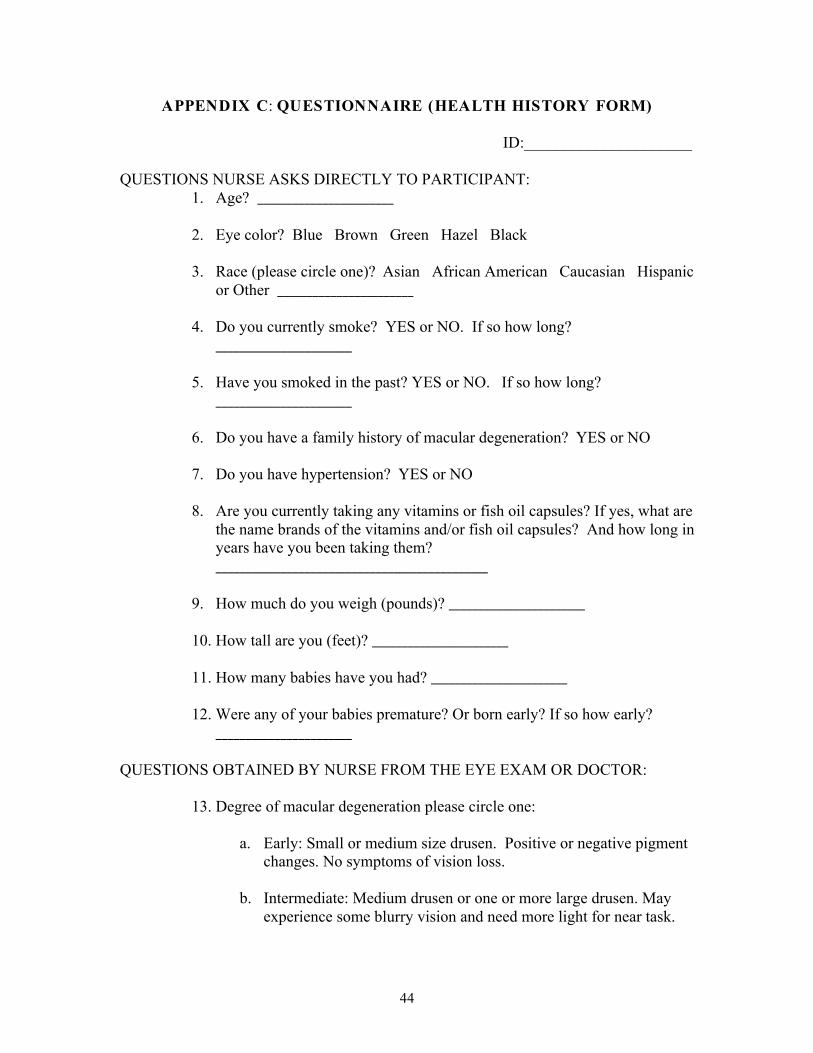
APPENDIX C: QUESTIONNAIRE (HEALTH HISTORY FORM)
ID:_____________________ QUESTIONS NURSE ASKS DIRECTLY TO PARTICIPANT:
1. Age? _______________________
2. Eye color? Blue Brown Green Hazel Black
3. Race (please circle one)? Asian African American Caucasian Hispanic or Other _______________________
4. Do you currently smoke? YES or NO. If so how long? _______________________
5. Have you smoked in the past? YES or NO. If so how long? _______________________
6. Do you have a family history of macular degeneration? YES or NO
7. Do you have hypertension? YES or NO
8. Are you currently taking any vitamins or fish oil capsules? If yes, what are the name brands of the vitamins and/or fish oil capsules? And how long in years have you been taking them? ______________________________________________
9. How much do you weigh (pounds)? _______________________
10. How tall are you (feet)? _______________________
11. How many babies have you had? _______________________
12. Were any of your babies premature? Or born early? If so how early?
_______________________ QUESTIONS OBTAINED BY NURSE FROM THE EYE EXAM OR DOCTOR:
13. Degree of macular degeneration please circle one:
a. Early: Small or medium size drusen. Positive or negative pigment changes. No symptoms of vision loss.
b. Intermediate: Medium drusen or one or more large drusen. May experience some blurry vision and need more light for near task.

45
c. Advance: Large drusen and geographic atrophy. Blurred areas in the central vision.
14. How long has the patient been diagnosed with macular degeneration? _______________________

46
APPENDIX D: “NURSES SCRIPT/DIRECTIONS” 1) Before entering the exam room, make sure you have three forms two copies of the consent forms and one questionnaire. The questionnaire will be coded with a number. 2) Nurse states to the patient, "LSU Division of Human Nutrition and Food, is conducting a study involving eye heath and it's relationship to pregnancy. Your participation is completely voluntary and your name will not be associated with any of your answers to the questions. All the information that we obtain today will be kept completely confidential and you will not be contacted again after today." 3) Next, show the consent form to the patient and ask if there are any questions. If the patient agrees to participate in the study, ask her to sign the consent form. The patient needs to sign two of the same consent form -one for the patient’s records and one will be kept for LSU’s records. Then pick up the card that was handed to the patient at the front desk. 4) Once the signatures are obtained, then proceed to the questions. Some of the questions can be answered from the chart and other questions will need to be asked directly to the patient. Once the questions are filled out and the doctor agrees to the degree of macular degeneration, put a designated label on the chart so the patient is not recruited twice.

47
VITA
Ann Hardin Shaw is an optometrist who decided to extend her education by
getting her master’s at Louisiana State University in human nutrition in fall 2009. She
did her under graduate at University of Georgia in Athens, Georgia. She got her under
graduate degree in international business with a concentration in marketing. She also
took all the pre-optometry courses at University of Georgia. In 2008, she graduated from
Southern College of Optometry in Memphis, Tennessee. While in optometry school she
worked at Bethesda Navy Hospital in Maryland and with Dr. Charles Shidlofsky in
Dallas, Texas. In May of 2008, she joined her father, Dr. Roger F. Shaw at Southern Eye
Centers. She currently works at both offices seeing a variety of patients. She hopes that
her extended education in nutrition will better help her serve her patients because of the
strong association between eye health and nutrition.