predictability of fetal doppler, biophysical profile, and ... · predictability of fetal doppler,...
TRANSCRIPT

ORIGINAL ARTICLE
Predictability of Fetal Doppler, Biophysical Profile,and Cardiotocography for Fetal Acidosis at Birth
Ashutosh Gupta • Sanjay Mehta • Tauqeer Syed Fazal •
Renu Raina Sehgal • Ashima Gogia
Received: 28 September 2014 / Accepted: 8 December 2014 / Published online: 18 December 2014
� Society of Fetal Medicine 2014
Abstract Fetal Doppler has allowed evaluation of the
fetus in both physiological and pathological conditions
which has helped in establishing the relationship between
Doppler and fetal oxygenation. It is difficult to define
pathological acidosis, but the threshold pH of\7 is the best
independent predictor for unexplained seizures. Most
infants tolerate acidemia well and recover without any
remarkable long term sequelae. Worsening umbilical artery
acidosis is directly and adversely related to worsening of
neurological outcome, hypoxic ischemic encephalopathy,
and multiorgan involvement with permanent neurologic
injury. Hypoxic ischemic encephalopathy events are not
limited to high-risk pregnancies but may occur in about
50 % of the low-risk population. Combination of low pH at
birth with other abnormal clinical parameters, e.g.,
requirement for intubation, 5 min Apgar score B5 has
80 % positive predictability of seizures. Predictability of
fetal Doppler examination for asphyxiated fetuses is in the
tune of 86 %. High-risk pregnancies are screened antena-
tally by fetal Doppler, biophysical profile, and CTG to
identify at-risk fetuses which are confirmed by the ABG
analysis of cord blood immediately after birth. All these
noninvasive modalities complement each other to identify,
at the earliest, any clinical deterioration. Isolated abnormal,
e.g., absent end diastolic flow in umbilical artery, abnormal
biophysical profile, or nonreactive CTG are not adequately
sensitive in identifying these fetuses which was observed in
the present cohort. Thus, fetal Doppler in combination with
biophysical profile supplemented with cardiotocograph
helps in identifying at-risk fetuses for fetal acidosis and
encephalopathy and helps in considering early intervention.
Keywords Fetal Doppler � Biophysical �Cardiotocograph � Hypoxemia � Acidosis � Apgar score �Predictability � Seizures
Introduction
Fetal Doppler has allowed evaluation of the fetus in both
physiological and pathological conditions which has
helped in establishing the relationship between Doppler
and fetal oxygenation. It is known that increased imped-
ance to uteroplacental circulation is directly related to fetal
hypoxemia. It stimulates preferential bloodflow to the
brain, heart and adrenals; however, it does not necessarily
correlate with hypercapnia and acidosis [1]. But, when this
compensatory mechanism fails, it leads to increased pul-
satility in the venous circulation which helps in optimizing
the timing of delivery [2].
Acidosis is usually well tolerated by the fetus without
any sequela. When fetal cord blood pH is [7.0 or base
excess is less than minus 12 it does not necessarily have
an adverse effect on the fetal cognition or long term
complications [3]. It is difficult to define pathological
acidosis, but Williams et al. have recommended the
threshold pH of \7 as the best independent predictor for
unexplained seizures [4].
A. Gupta (&)
Department of Fetal Medicine & Clinical Genetics, Max Super
Speciality Hospital, West Block, 1 Press Enclave Road, Saket,
New Delhi 110017, India
e-mail: [email protected]
S. Mehta � T. S. Fazal
Department of Radiology, Artemis Health Institute, Gurgaon,
Haryana, India
R. R. Sehgal � A. Gogia
Department of Obstetrics & Gynecology, Artemis Health
Institute, Gurgaon, Haryana, India
123
J. Fetal Med. (September 2014) 1:143–149
DOI 10.1007/s40556-014-0024-9

Materials and Methods
Umbilical vein sampling was done immediately after the
baby was born after doubly ligating the umbilical cord.
Cord blood arterial blood gas analysis (ABG) was done to
identify fetal acidosis after birth. ABG included pH, pCo2,
base excess and lactate. Fetal carbon di-oxide may be
removed from the umbilical arterial blood and venous
circulation has shown to have slightly higher pH and lower
pCo2 than the arterial blood [5]. Restriction of blood flow
in the umbilical cord adversely affects the blood gases, as
in cases of tight nuchal cord [6], cord prolapsed [7], or
placental abruption with cord obstruction [8]. Even though
these techniques have their limitations, yet if used within
their specific domain, these may be of immense help to
identify at-risk fetuses and to take appropriate measures to
prevent neurological impairment.
Results
High-risk antenatal cases were followed-up prospectively
in the department of Fetal Medicine at Artemis hospital
with fetal Doppler, biophysical profile, and antenatal or
intrapartum cardiotocograph (CTG). ABG analysis was
done after birth in all the cases. All these antenatal
screening tools were utilized for predicting worsening of
the clinical situation and to identify at an earliest, the
fetuses at risk of fetal hypoxemia and acidosis (Fig. 1).
This cohort of eight high-risk cases with their clinical
presentation is depicted in Table 1; the fetal Doppler
assessment and ABG analysis after birth are shown in
Tables 2 and 3, respectively.
Cohort of high risk was defined by the fetal abdominal
circumference or fetal weight \25th centile for the gesta-
tional age; abnormal Doppler when umbilical or uterine
artery pulsatility index (PI) is [95th centile for the
gestational age; abnormal biophysical profile by any score
of 4/8 or less and nonreactive CTG (Figs. 2, 3).
Intrauterine growth restriction (IUGR) was identified in
4 cases (case no. 2, 6, 7, and 8). However, acidosis was
identified in only 1 case (case no. 8), when the biophysical
score dipped to 4/8 with nonreactive CTG. Two cases (case
no 6 and 8) had biophysical score of 4/8 and nonreactive
CTG (case no 4 and 8) but only 1 (case no. 4) had both of
them abnormal and had severe respiratory acidosis. One
case (case no. 6) with biophysical score of 4/8 had a
reactive CTG and the other one (case no. 4) with normal
biophysical score had nonreactive CTG but both of them
had normal pH.
Absent end diastolic flow in the umbilical artery was
identified in 2 preterm fetuses (case no. 2 and 3), but both
of them had normal CTG and normal pH at birth. Fetal
acidosis was identified more commonly in term pregnan-
cies (case no. 4 and 8) as compared to preterm fetuses.
Identification of absent end diastolic flow led to the
decision to expedite the delivery before ominous venous
Doppler appeared to prevent irreversible fetal outcome.
One case with nuchal cord with high resistance in the cord
was identified to have fetal acidemia (case no. 4). Both the
cases of fetal acidemia had high levels of lactate (cases no.
4 and 8, Table 2) depicting an intrinsic metabolic
derangement.
Discussion
Predictability of fetal Doppler examination for asphyxiated
fetuses is in the tune of 86 % [9]. Even then, most infants
tolerate acidemia well and recover without any remarkable
long term sequelae [10]. Worsening umbilical artery aci-
dosis is directly and adversely related to worsening of
neurological outcome, hypoxic ischemic encephalopathy,
multiorgan involvement with permanent neurologic injury.
Fig. 1 Increased resistance to
blood flow in the umbilical
artery in nuchal cord and normal
blood flow in the free loop of
umbilical cord in case no. 4
(Table 1)
144 J. Fetal Med. (September 2014) 1:143–149
123

Ta
ble
1C
lin
ical
pro
file
so
fth
eca
ses
wit
hth
eir
feta
lD
op
ple
ras
sess
men
t,b
iop
hy
sica
lsc
ore
,C
TG
,an
dA
BG
anal
ysi
s
Cas
e
no
.
Per
iod
of
ges
tati
on
(wee
ks)
Co
nfo
un
din
gfa
cto
rsA
FI
Fet
alD
op
ple
rB
iop
hy
sica
l
pro
file
CT
GA
BG
Bir
th
wei
gh
t
(kg
)
Mo
de
of
del
iver
y
1T
erm
;3
7?
6N
oO
lig
oh
yd
ram
nio
s;A
FI
6.2
cmB
rain
spar
ing
No
rmal
Rea
ctiv
ep
H7
.35
53
.36
LS
CS
;em
erg
ency
2P
re-t
erm
;3
2?
6IU
GR
No
rmal
;1
2.9
cmA
bse
nt
end
dia
sto
lic
flo
w
No
rmal
Rea
ctiv
ep
H7
.32
1.3
9L
SC
S;
emer
gen
cy
3P
re-t
erm
;3
3G
DM
;h
igh
MS
AF
Pat
qu
adru
ple
test
ing
—2
.7
Mo
M
No
rmal
;1
3cm
Ab
sen
ten
dd
iast
oli
c
flo
w
No
rmal
Rea
ctiv
ep
H7
.47
1.2
LS
CS
;em
erg
ency
4T
erm
;3
7?
6ID
DM
;h
yp
oth
yro
idis
m;
cho
lest
asis
;P
IH
No
rmal
;1
1.9
cmH
igh
um
bil
ical
arte
ryre
sist
ance
in
loo
par
ou
nd
nec
k
No
rmal
No
nre
acti
ve;
thic
k
mec
on
ium
pH
7.2
62
.76
Ind
uct
ion
5T
erm
;3
8H
yp
oth
yro
idis
m;
cho
lest
asis
Oli
go
hy
dra
mn
ios;
AF
I9
.0cm
No
rmal
No
rmal
Rea
ctiv
ep
H7
.30
3.2
Vag
inal
6P
re-t
erm
;3
6?
3S
ym
met
rica
lIU
GR
;
pla
cen
tap
rev
iaty
pe
IV;
abse
nt
feta
lm
ov
emen
ts
An
hy
dra
mn
ios
Bra
insp
arin
gS
core
of
4/8
Rea
ctiv
ep
H7
.30
2.2
LS
CS
;em
erg
ency
;
pla
cen
tap
rev
ia
7P
re-t
rem
;3
5?
1S
ym
met
rica
lIU
GR
;
cho
lest
asis
Oli
go
hy
dra
mn
ios;
AF
I8
.5cm
No
rmal
No
rmal
Rea
ctiv
ep
H7
.30
2.9
Ele
ctiv
eL
SC
S
8T
erm
;3
8H
yp
oth
yro
idis
m;
Ch
ole
stas
is;
sym
met
rica
l
IUG
R
Oli
go
hy
dra
mn
ios;
AF
I9
.2cm
No
rmal
Sco
reo
f4
/8N
on
reac
tiv
ep
H7
.19
2.9
LS
CS
;em
erg
ency
;
imp
end
ing
ecla
mp
sia
AB
Gar
teri
alb
loo
dg
asan
aly
sis,
AF
Iam
nio
tic
flu
idin
dex
,C
TG
card
ioto
cog
rap
h,
GD
Mg
esta
tio
nal
dia
bet
esm
elli
tus,
IDD
Min
suli
nd
epen
den
td
iab
etes
mel
litu
s,IU
GR
intr
aute
rin
eg
row
th
rest
rict
ion
,M
SA
FP
mat
ern
alse
rum
alp
ha
feto
pro
tein
,P
IHp
reg
nan
cyin
du
ced
hy
per
ten
sio
n
J. Fetal Med. (September 2014) 1:143–149 145
123
Combination of low pH at birth with other abnormal
clinical parameters, e.g., requirement for intubation, 5 min
Apgar score B5 has 80 % positive predictability of seizures
[11]. However, in the present series, both the acidotic
fetuses responded very well to the treatment and recovered
without any sequela.
Noninvasive antenatal techniques to predict mortality in
preterm fetuses is almost similar to that of term fetuses.
Inspite of absent end diastolic flow in umbilical artery in
two preterm fetuses, none of them had acidosis stresses, the
fact that surviving preterm fetuses tend to have higher
umbilical artery pH as compared to those who die [12].
Cord blood lactate has been identified to be almost fetal
in origin and since it does not cross the placenta, it cor-
relates very well with pH and base excess [13]. In the
present series, both the cases of fetal acidemia had high
lactate, which is a very specific sign of anaerobic metab-
olism and has high sensitivity (approaching 100 %) and
specificity (95.4 %) for predicting neonatal encephalopathy
[14].
In this series, none of the cases with absent end diastolic
flow in umbilical artery had acidosis which is in concor-
dance with the fact that markedly reduced or absent end
diastolic flow of umbilical artery is an independent risk
factor for fetal hypoxia and acidosis [15]. However, both
the cases were delivered before the reversal of arterial flow
or the venous changes appeared, as the fetuses with
abnormal venous Doppler have worse prognosis [16] with
almost all of them at risk of being asphyxiated [17].
Predictability of fetal biophysical score of B6 [18] and
abnormal CTG trace for fetal acidosis (pH \ 7.25) is only
50–65 % [19]. However, in the present series, when both
low biophysical score was associated with loss of vari-
ability on CTG, the fetus was found to be acidotic. Growth
Fig. 2 Nonreactive cardiotocograph (CTG) with loss of baseline
variability with arterial blood gas (ABG) analysis of the fetus
showing mixed acidosis in case no. 4 (Table 1)
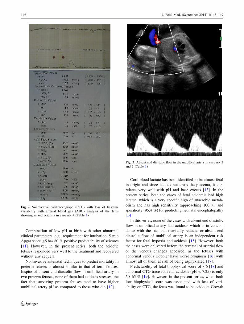
Fig. 3 Absent end diastolic flow in the umbilical artery in case no. 2
and 3 (Table 1)
146 J. Fetal Med. (September 2014) 1:143–149
123

Ta
ble
2D
epic
tin
gfe
tal
Do
pp
ler
asse
ssm
ent
of
the
hig
h-r
isk
case
s
Cas
en
o.
Ges
tati
on
(wee
ks)
Rig
ht
MC
AU
mb
ilic
alar
tery
Rig
ht
ute
rin
ear
tery
Lef
tu
teri
ne
arte
ryU
mb
ilic
alv
ein
Dia
sto
lic
flo
wC
om
men
t
S/D
rati
oP
IS
/Dra
tio
PI
(no
rmal
\3
)R
IP
IR
IP
I
13
7?
62
.71
.13
2.9
1.2
0.4
80
.77
0.5
60
.99
Co
nti
nu
ou
sfo
rwar
dfl
ow
Pre
sen
tB
rain
spar
ing
2.9
1.1
3
2.8
1.1
0a
23
2?
63
.41
.30
2.1
20
.54
0.8
20
.59
1.0
7C
on
tin
uo
us
forw
ard
flo
wA
bse
nt
Ab
sen
ten
dd
iast
oli
cfl
ow
2.1
2
2.1
9
2.0
3a
33
33
.91
.49
2.4
80
.62
1.2
90
.74
1.4
8C
on
tin
uo
us
forw
ard
flo
w;
pre
load
ind
exin
feri
or
ven
aca
va—
0.2
3
(No
rmal
)
Ab
sen
tA
bse
nt
end
dia
sto
lic
flo
w
wit
hh
igh
ute
rin
ear
tery
resi
stan
ce
43
7?
62
.20
.93
.31
.24
0.5
40
.99
0.5
80
.95
Co
nti
nu
ou
sfo
rwar
dfl
ow
Pre
sen
tH
igh
um
bil
ical
arte
ry
resi
stan
cein
nu
chal
cord
;
no
rmal
flo
ws
infr
eelo
op
S/D
rati
o=
2.7
;
PI
=1
.11
3.0
1.1
64
.41
.20
3.4
1.6
4
3.6
a1
.32
53
88
.52
.52
.40
.85
0.5
20
.76
0.4
90
.80
Co
nti
nu
ou
sfo
rwar
dfl
ow
Pre
sen
tH
igh
ute
rin
ear
tery
resi
stan
ce
63
6?
32
.51
.02
.60
.93
0.4
10
.74
0.5
20
.79
Co
nti
nu
ou
sfo
rwar
dfl
ow
Pre
sen
tB
rain
spar
ing
2.5
0.9
7
2.6
a0
.98
73
5?
18
.62
.02
.71
.00
.47
0.7
30
.51
0.8
0C
on
tin
uo
us
forw
ard
flo
wP
rese
nt
No
rmal
Do
pp
ler
83
82
.91
.15
2.3
0.8
41
.00
.63
1.2
Co
nti
nu
ou
sfo
rwar
dfl
ow
Pre
sen
tB
rain
spar
ing
aT
hes
ed
iffe
ren
tv
alu
esw
ere
reco
rded
30
min
apar
tin
case
sw
ith
abn
orm
alD
op
ple
rv
alu
es
MC
Am
idd
lece
reb
ral
arte
ry,
PI
pu
lsat
ilit
yin
dex
,R
Ire
sist
ive
ind
ex,
S/D
rati
osy
sto
lic-
dia
sto
lic
rati
o
J. Fetal Med. (September 2014) 1:143–149 147
123

restricted fetuses with brain sparing may have hypoxemia
and, if complicated with abnormal fetal Doppler and
ominous CTG, may have associated fetal acidosis.
Conclusion
As early as 1958, umbilical cord blood gas analysis was
being utilized to identify fetal hypoxic stress [20]. Ever
since, it has been widely accepted and now recommended
by both American and British College of Obstetricians and
Gynecologists to all the high-risk deliveries with its
increasing clinical and medico-legal importance [21, 22].
Hypoxic ischemic encephalopathy events are not limited to
high-risk pregnancies. They may occur in about 50 % of
the low-risk population [23], and thus is recommended in
all high-risk deliveries.
High-risk pregnancies are screened antenatally by fetal
Doppler, biophysical profile, and CTG to identify at-risk
fetuses which is confirmed by ABG analysis of cord blood
immediately after birth. All these noninvasive modalities
complement each other to identify at the earliest any clinical
deterioration. Isolated abnormal, e.g., absent end diastolic
flow in umbilical artery, abnormal biophysical profile, or
nonreactive CTG are not adequately sensitive in identifying
these fetuses which is observed in the present cohort.
Fetal Doppler in combination with biophysical profile
supplemented with CTG helps in identifying at-risk fetuses
for fetal acidosis. Umbilical cord blood gas analysis in
combination with other clinical parameters, Apgar scores,
need for ventilation, cardiopulmonary compromise, helps
in identifying at-risk infants for encephalopathy and to
consider early intervention.
Conflict of interest None.
References
1. Bilardo CM, Nicolaides KH, Campbell S. Doppler measurements
of foetal and uteroplacental circulations: relationship with
umbilical venous blood gases measured at cordocentesis. Am J
Obstet Gynecol. 1990;162(1):115–20.
2. Hecher K, Snijders R, Campbell S, Nicolaides K. Foetal venous,
intracardiac, and arterial blood flow measurements in intrauterine
growth retardation: relationship with foetal blood gases. Am J
Obstet Gynecol. 1995;173(1):10–5.
3. Svirko E, Mellanby J, Impey L. The association between cord pH
at birth and intellectual function in childhood. Early Hum Dev.
2008;84(1):37–41.
4. Williams KP, Singh A. The correlation of seizures in newborn
infants with significant acidosis at birth with umbilical artery cord
gas values. Obstet Gynecol. 2002;100(3):557–60.
5. Westgate J, Garibaldi JM, Greene KR. Umbilical cord blood gas
analysis at delivery: a time for quality data. Br J Obstet Gynaecol.
1994;101(12):1054–63.
6. Martin GC, Green RS, Holzman IR. Acidosis in newborns with
nuchal cords and normal Apgar scores. J Perinatol. 2005;
25(3):162–5.
7. Johnson JW, Richards DS. The etiology of foetal acidosis as
determined by umbilical cord acid-base studies. Am J Obstet
Gynecol. 1997;177(2):274–80.
8. Pomerance J. Umbilical cord blood gas casebook. Interpreting
umbilical cord blood gases, IX. J Perinatol. 2001;21(7):469.
9. Stark JE, Seibert JJ. Cerebral artery Doppler ultrasonography for
prediction of outcome after perinatal asphyxia. J Ultrasound Med.
1994;13(8):595–600.
10. King TA, Jackson GL, Josey AS, et al. The effect of profound
umbilical artery acidemia in term neonates admitted to a newborn
nursery. J Pediatr. 1998;132(4):624–9.
11. Perlman JM, Risser R. Can asphyxiated infants at risk for neo-
natal seizures be rapidly identified by current high-risk markers?
Pediatrics. 1996;97(4):456–62.
12. Hibbard JU, Hibbard MC, Whalen MP. Umbilical cord blood
gases and mortality and morbidity in the very low birth weight
infant. Obstet Gynecol. 1991;78(5 Pt 1):768–73.
13. Kruger K, Kublickas M, Westgren M. Lactate in scalp and cord
blood from foetuses with ominous foetal heart rate patterns.
Obstet Gynecol. 1998;92(6):918–22.
14. Chou YH, Tsou Yau KI, Wang PJ. Clinical application of the
measurement of cord plasma lactate and pyruvate in the assess-
ment of high-risk neonates. Acta Paediatr. 1998;87(7):764–8.
15. Tyrrell S, Obaid AH, Lilford RJ. Umbilical artery Doppler ve-
locimetry as a predictor of foetal hypoxia and acidosis at birth.
Obstet Gynecol. 1989;74:332.
16. Baschat AA, Gembruch U, Reiss I, et al. Relationship between
arterial and venous Doppler and perinatal outcome in foetal
growth restriction. Ultrasound Obstet Gynecol. 2000;16(5):
407–13.
17. Brar HS, Platt LD. Reverse end-diastolic flow velocity on
umbilical artery velocimetry in high-risk pregnancies: an omi-
nous finding with adverse pregnancy outcome. Am J Obstet
Gynecol. 1988;159(3):559–61.
18. Yoon BH, Romero R, Roh CR, et al. Relationship between the
foetal biophysical profile score, umbilical artery Doppler veloc-
imetry, and foetal blood acid-base status determined by cordo-
centesis. Am J Obstet Gynecol. 1993;169(6):1586–94.
19. Parer JT. Efficacy and safety of intrapartum electronic foetal
monitoring: an update. Obstet Gynecol. 1996;87(3):476–7.
20. James LS, Weisbrot IM, Prince CE, et al. The acid-base status of
human infants in relation to birth asphyxia and the onset of res-
piration. J Pediatr. 1958;52(4):379–94.
Table 3 Depicting arterial blood gas analysis of the cases
Case
no.
pH pCO2 Lactate HCO3 Base
excess
Interpretation
1 7.3 44.0 1.5 23.9 -1.4 Mild mixed
acidosis
2 7.3 45.0 2.0 22.6 -3.2 Mild mixed
acidosis
3 7.4 40.0 3.5 23.5 0.4 Normal pH
4 7.2 52.5 2.9 23.2 -4.1 Mixed acidosis
5 7.36 45.0 1.3 25.3 0.2 Normal pH
6 7.3 42.2 2.0 18.0 -1.1 Metabolic acidosis
7 7.5 41.5 1.5 32.0 -2.4 Metabolic
alkalosis
8 7.1 63.7 3.7 23.8 -6.1 Severe respiratory
acidosis
148 J. Fetal Med. (September 2014) 1:143–149
123

21. Goddard R. Electronic foetal monitoring is not necessary for low
risk labours. BMJ. 2001;322(7300):1436–7.
22. ACOG Committee on Obstetric Practice. ACOG Committee
Opinion No. 348, November 2006: umbilical cord blood gas and
acid-base analysis. Obstet Gynecol. 2006;108(5):1319–22.
23. Sameshima H, Ikenoue T, Ikeda T, et al. Unselected low-risk
pregnancies and the effect of continuous intrapartum foetal heart
rate monitoring on umbilical blood gases and cerebral palsy. Am
J Obstet Gynecol. 2004;190(1):118–23.
J. Fetal Med. (September 2014) 1:143–149 149
123