precordial st-segment elevation in acute right ventricular ... · 496·precordial st elevation in...
TRANSCRIPT

495
CASE REPORT
Korean Circ J 2008;38:495-499 Print ISSN 1738-5520 / On-line ISSN 1738-5555
Copyright ⓒ 2008 The Korean Society of Cardiology
Precordial ST-Segment Elevation in Acute Right Ventricular Myocardial Infarction Nae Sun Ryou, MD, Min Hyoung Cho, MD, Dae-Hee Shin, MD, Sang-Sig Cheong, MD and Sang-Yong Yoo, MD Division of Cardiology, Department of Internal Medicine, Gangneung Asan Hospital, Gangneung, Korea ABSTRACT
It is rare to observe ST-segment elevations in the precordial leads that are caused by an occlusion of the right co-ronary artery and/or its branches. We report here on two cases of acute occlusion of the right coronary artery or its branches that caused acute right ventricular myocardial infarction with ST-segment elevations in the anterior pre-cordial leads. These cases should remind us that the presence of diffuse ST-segment elevations in the precordial leads could be due to acute occlusion of the right coronary artery. (Korean Circ J 2008;38:495-499) KEY WORDS: Myocardial infarction; Right ventricle; Electrocardiography.
Introduction
Clinically observing acute ST-segment elevations in
the precordial leads generally suggests acute anterior myocardial infarction (MI) that is caused by occlusion in one of the branches of the left coronary artery. How-ever, there have been several reported cases of right ven-tricular myocardial infarction (RVMI) presenting with precordial ST-segment elevations on the electrocardi-ography (ECG).1-3) We describe here two cases of marked ST-segment elevations in the precordial leads, and this was caused by acute occlusion of the proximal segment of the right coronary artery (RCA) or its major side bran-ches. These cases should remind us that the presence of diffuse ST-segment elevations in the precordial leads could be due to acute RCA occlusion.
Case Case 1
A 72-year-old male patient was admitted to our hos-pital with the symptoms of chest pain upon effort for the previous year. He had a 10-year history of hyper-tension. The man’s blood pressure was 130/90 mmHg and his pulse rate was 65 beats/minute. The ECG on ad-
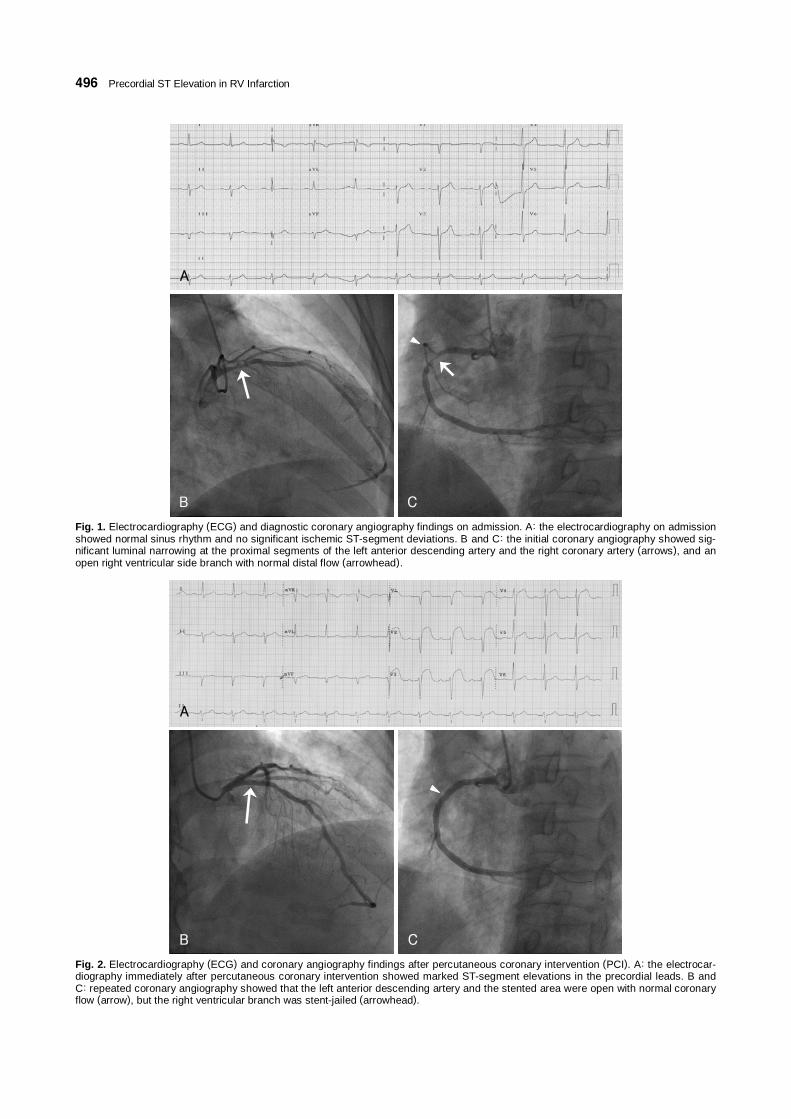
mission (Fig. 1A) showed normal sinus rhythm and no significant ischemic ST-segment changes. The ech-ocardiography revealed normal systolic function without any abnormal regional wall motion. Diagnostic coronary angiography revealed significant luminal narrowing at the proximal segments of the RCA and the left anterior descending (LAD) artery (Fig. 1B and C).
After performing pre-dilation using a balloon cath-eter, a stent (TAXUSTM 4.5×24 mm, Boston Scientific, Boston, USA) was implanted into the proximal seg-ment of the RCA. Soon after performing percutaneous coronary intervention (PCI) for the RCA, he complained of prolonged chest pain. We thought that this chest pain might be caused by stent-induced stretch of the vessel and we neglected to properly assess the man’s condition. After successfully implanting a TAXUSTM into the proximal segment of the LAD, he still con-tinuously suffered from squeezing chest pain. An ECG and bedside echocardiography were performed immedi-ately. The ECG showed marked ST-segment elevations in the precordial leads (Fig. 2A), and the echocardio-graphy showed no regional wall motion abnormalities in the left ventricle (LV), but hypokinesia was noted in the free wall of the right ventricle, and this was com-patible with RVMI. The patient was again transferred to the catheterization laboratory for revaluating the LAD and RCA anatomy. Repeated coronary angiogra-phy was performed and this showed a normal Throm-bolysis In Myocardial Infarction (TIMI) flow and no change in the degree of stenosis in the proximal LAD; however, a right ventricular branch stent-jailed, and this had been missed during the PCI for the RCA (Fig. 2B and C).
Received: May 1, 2008
Accepted: June 16, 2008 Correspondence: Sang-Yong Yoo, MD, Division of Cardiology, Department
of Internal Medicine, Gangneung Asan Hospital, 415 Bangdong-ni, Sacheon-
myeon, Gangneung 210-711, Korea
Tel: 82-33-610-3130, Fax: 82-33-641-8130
E-mail: [email protected]
496·Precordial ST Elevation in RV Infarction
A
B C
Fig. 1. Electrocardiography (ECG) and diagnostic coronary angiography findings on admission. A: the electrocardiography on admission showed normal sinus rhythm and no significant ischemic ST-segment deviations. B and C: the initial coronary angiography showed sig-nificant luminal narrowing at the proximal segments of the left anterior descending artery and the right coronary artery (arrows), and an open right ventricular side branch with normal distal flow (arrowhead).
Fig. 2. Electrocardiography (ECG) and coronary angiography findings after percutaneous coronary intervention (PCI). A: the electrocar-diography immediately after percutaneous coronary intervention showed marked ST-segment elevations in the precordial leads. B and C: repeated coronary angiography showed that the left anterior descending artery and the stented area were open with normal coronaryflow (arrow), but the right ventricular branch was stent-jailed (arrowhead).
A
B C

Nae Sun Ryou, et al.·497
Case 2 A 59-year-old male patient was admitted to our hos-
pital with the symptom of squeezing chest pain that had lasted for an hour. He had no specific cardiovas-cular risk factor except for a current 40-pack-year smok-ing habit. The blood pressure was 70/41 mmHg and the pulse rate was 45 beats/minute. The ECG on ad-mission showed atrio-ventricular block and marked ST segment elevations in both the inferior (II, III, aVF) and precordial leads (V1 to V5) (Fig. 3A). The max-imum magnitude of the ST-segment elevation in the precordial leads was 15 mm in the V4 lead and that in the inferior leads was 5 mm in lead III. The morphol-ogy of the ST-segment elevations in the precordial leads was a rounded ST-segment and T-wave, and the mor-phology of the ST-segment elevations in the inferior leads was convex upward. We also found 1 mm-ST seg-ment elevation from leads V4R to V6R. Echocardiog-raphy showed a D-shaped LV and akinesia in both the right ventricular free wall and the inferior wall of the LV, and the echocardiography also showed the left to right intracardiac shunt at the atrial level. The patient was immediately taken to the catheterization laboratory for PCI. The patient underwent primary PCI and the coronary angiography revealed total thrombotic occlu-sion at the proximal portion of the RCA with TIMI 0 flow and nearly normal left coronary arteries (Fig. 3B
and C). The lesion of the proximal RCA was pre-di-lated with a 2.5×15 mm balloon catheter and a stent (Endeavor® 3.0×24 mm, Medtronic Vascular, Santa Rosa, USA) was deployed and placed (Fig. 4B). At 7 days after successful PCI, transesophageal echocardi-ography (Fig. 4C) revealed the secundum type of atrial septal defect (2.0×2.0 cm) with a left to right shunt. The in-hospital course of the patient was uneventful, and we are planning closure for the atrial septal defect.
Discussion
Acute ST-segment elevations in the precordial leads generally indicate acute occlusion in one of the branches of the left coronary artery. However, there have been some reported cases of precordial ST-segment elevations that were seen on ECG and these elevations were sec-ondary to occlusion of the RCA and/or its branches.1-3) Recording of the right ventricular electrical potentials in the left precordial leads depends on the degree of clockwise rotation of the heart in the horizontal plane and in relation to the body’s geometry.4) The dominant electric forces generated by the ischemia of the inferior wall suppress the changes caused by the ischemia of the right ventricle because the inferior wall has a larger mass of myocardium compared with the thin right ventricular wall; therefore, the left precordial ECG manifestations
Fig. 3. Electrocardiography (ECG) and diagnostic coronary angiography findings on admission. A: the initial electrocardiography on admis-sion showed atrio-ventricular block and marked ST-segment elevations in both the inferior and precordial leads and 1 mm ST-segment ele-vations from V4R to V6R. B: the initial coronary angiography revealed no significant luminal narrowing of the left coronary arteries. C: notethe total occlusion at the proximal segment of the right coronary artery with TIMI 0 flow (arrowhead). TIMI: Thrombolysis In Myocardial Infarction.
V6R
V5R
V3R
V4R
A
B C

498·Precordial ST Elevation in RV Infarction
of RV injury are absent in most patients.5) These op-posing currents are too weak to be offset by each other and this is true not only in the case of an isolated occlu-sion of the RV branch, but also in the case of a proxi-mal occlusion of the non-dominant small RCA, which produces a ECG pattern that’s similar to that in isolated RVMI, that is, ST-elevation in leads V4R and V1-3.6)
Eskola et al.7) reported a case of isolated temporary occlusion of the major side branch of the RCA that was caused by the stent during PCI in a patient with unstable angina, and this was recognized by the angiog-raphy findings and the ST-segment elevations in the precordial leads (V1-V3) and lead V4R, which is sim-ilar to that of our first case. Isolated occlusion of the large right ventricular branches of the RCA appears to be extremely rare. According to the report by van der Bolt et al.8) among the 10,000 patients who underwent PCI, 23% underwent PCI for a stenosis in the RCA and only nine patients revealed an isolated occlusion of a large right ventricular branch. The patient with the ECG pattern described in this first case might be mis-takenly regarded as suffering from anterior MI caused by acute stent thrombosis in the proximal LAD.
The patient of our second case had total occlusion of the dominant proximal RCA to the right ventricular branch, and this caused inferior and RVMI. In such a case, the expected ECG findings might possibly display
marked ST-segment elevations in the inferior leads and reciprocal changes in the precordial leads. However, there were marked ST-segment elevations in the inferior and precordial leads, while there were ST-segment depres-sions in the lateral leads (I, aVL). These findings are contrary to that of a classic RVMI complicating an in-ferior wall MI. These unusual findings were presumed to come about because the dominant electric forces gen-erated by the ischemia of the inferior ventricular wall couldn’t suppress the changes caused by the ischemia of the right ventricular wall due to of the right ventricular hypertrophy secondary to the untreated, large atrial sep-tal defect (ASD).
Although reciprocal ST-segment depressions (RSTDs) in patients with acute inferior MI are common ECG findings,9) controversy still exists about the significance of this. A recent published study showed how ana-tomical interpretation of an ECG that’s recorded dur-ing chest pain can help to stratify the risk for patients with inferior ST-segment elevation MI.6) According to their findings, ST-segment elevations in the inferior leads, with these being maximal in lead III, suggest that the RCA is the infarct-related artery. RSTDs in leads I and aVL and isoelectric or minimally depressed (0.5 mm) ST segments in leads V5 and V6 indicate that the RCA is a non-dominant artery. ST segment elevations in leads V1 and V4R suggest that the culprit lesion is
RA
LA
A
B C
Fig. 4. Electrocardiography (ECG), coronary angiography, and transesophagreal echocardiography (TEE) findings after percutaneous coronary intervention (PCI). A: the electrocardiography after percutaneous coronary intervention showed resolution of the ST-segment elevation in the precordial leads. B: the coronary angiography, following percutaneous coronary intervention, showed a good appearancewith TIMI 3 flow (arrows). C: the transesophageal echocardiography revealed the secundum type of atrial septal defect with left to right shunt. LA: left atrium, RA: right atrium, TIMI: Thrombolysis In Myocardial Infarction.

Nae Sun Ryou, et al.·499
proximal to the right ventricular branch. Thus, despite that a physician can observe dominant precordial ST-segment elevations, the inferior ST-segment elevations that are maximal in lead III and the reciprocal ST-seg-ment depressions (RSTDs) in leads I and aVL suggest the possibility of an RCA lesion.10)
Although clinically observing ST-segment elevation in the precordial leads is not common, and this mimics an anterior wall MI that’s caused by RVMI, we must keep in mind the possibility that marked ST-segment elevations in the precordial leads are due to occlusion of the right coronary artery and/or its branches.
REFERENCES 1) Porter A, Herz I, Strasberg B. Isolated right ventricular infarc-
tion presenting as anterior wall myocardial infarction on electro-cardiography. Clin Cardiol 1997;20:971-3.
2) Saw J, Amin H, Kiess M. Right ventricular ischemia mimicking acute anterior myocardial infarction. Can J Cardiol 1999;15: 1143-6.
3) Khan ZU, Chou TC. Right ventricular infarction mimicking acute anteroseptal left ventricular infarction. Am Heart J 1996;132: 1089-93.
4) Lew AS, Maddhai J, Shah PK, et al. Factors that determine the
direction and magnitude of precordial ST-segment deviations during inferior wall acute myocardial infarction. Am J Cardiol 1985;55:883-8.
5) Geft I, Shah P, Rodriguez L, et al. ST elevations in leads V1 to V5 may be caused by right coronary artery occlusion and acute right ventricular infarction. Am J Cardiol 1984;53:991-6.
6) Eskola MJ, Nikus KC, Niemela KO, Sclarovsky S. How to use ECG for decision support in the catheterization laboratory: cases with inferior ST elevation myocardial infarction. J Electrocardiol 2004;37:257-66.
7) Eskola MJ, Kosonen P, Sclarovsky S, Vikman S, Nikus KC. The ECG pattern of isolated right ventricular infarction during per-cutaneous coronary intervention. Ann Noninvasive Electrocardiol 2007;12:83-7.
8) van der Bolt CL, Vermeersch PH, Plokker HW. Isolated acute occlusion of a large right ventricular branch of the right coro-nary artery following coronary balloon angioplasty: the only true ‘model’ to study ECG changes in acute, isolated right ventri-cular infarction. Eur J Heart 1996;17:247-50.
9) Kim MS, Han JK, Lee SE, et al. Cases of right ventricular myo-cardial infarction in patients with an absent or hypoplastic right coronary artery. Korean Cir J 2007;37:84-6.
10) Chia BL, Yip JW, Tan HC, Lim YT. Usefulness of ST elevation II/III ratio and ST deviation in lead I for identifying the culprit artery in inferior wall acute myocardial infarction. Am J Cardiol 2000;86:341-3.