pre med iii genetics guri tzivion, phd [email protected] extension 506 summer 2015 windsor...
TRANSCRIPT
Pre Med III Genetics
Guri Tzivion, PhD
Summer 2015Windsor University School of Medicine

Pre Med III GeneticsClass 14
Review for Block 2 exam

Review for Block 2 exam
The exam will cover:
RNA & Transcription
Proteins and Translation
Mutations and DNA Repair
Regulation of Gene Expression:
Prokaryotes & Eukaryotes
Recombinant DNA & Molecular Biology Methods

RNA & Transcription

DNA RNA protein
transcription translation replication
reverse transcription
Central dogma

MAJOR CLASSES OF RNA
rRNA (ribosomal RNA) mRNA (messenger RNA) tRNA ( transfer RNA)
snRNA (small nuclear RNA)hn RNA (heterogeneous nuclear
RNA) miRNA (micro RNA)

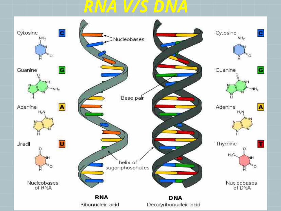
RNA V/S DNA

replication transcription
template double strands single strand
substrate dNTP NTP
primer yes no
Enzyme DNA polymerase RNA polymerase
product dsDNA ssRNA
base pair A-T, G-C A-U, T-A, G-C
Differences between replication and transcription

Transcription in Bacteria
mRNA transcript is synthesized by RNA polymerase in the 5’ to 3’ direction.
Template strand:DNA strand that is read (transcribed) by RNA
polymerase.
Non-template strand:DNA strand that is not read by RNA polymerase.
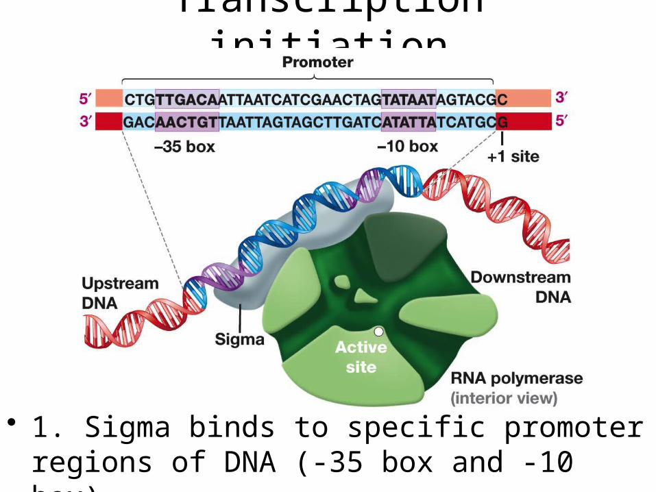
Transcription initiation
• 1. Sigma binds to specific promoter regions of DNA (-35 box and -10 box).

Phase 3: Transcription termination
• RNA polymerase encounters a termination signal within the DNA template, which codes for a hairpin loop structure in the RNA.
• The hairpin causes the RNA polymerase to separate from the RNA transcript, ending the transcription.

DNA
Cytoplasm
Nucleus
Eukaryotic Transcription
ExportG AAAAAA
RNA
Transcription
Nuclear pores
G AAAAAA
RNAProcessing
mRNA

Eukaryotic mRNA Processing
• After transcription, specific regions of the primary RNA transcript are spliced out and degraded during RNA processing.
• Exon: Contains sequences required for protein synthesis and is NOT spliced out during RNA processing. Is part of the final mature spliced RNA transcript.
• Intron: Spliced out during RNA processing and degraded.
Exon 1 Exon 2 Exon 3

Eukaryotic mRNA Processing
• Besides splicing, other steps involved in mRNA processing include:
1. Addition of a “cap” at the 5’ end of the mRNA. The “5’ cap” serves as a recognition signal for the translation machinery of the cell.
2. Addition of poly (A) tail at the 3’ end of the mRNA. Serves to stabilize the mRNA by protecting it from enzymatic degradation.

Proteins and Translation

From messenger RNA to protein: Translation
The mRNA is translated by ribosomes as series of 3 letter codes designated codons

Second mRNA base
Fir
st m
RN
A b
a se
(5¢
e nd
)
Th
ird
mR
NA
bas
e (3
¢ en
d)
The Genetic Code

The Genetic Code

General tRNA structure: ~ 80 nt long

Met
GTPInitiator tRNA
mRNA
5¢3¢
mRNA binding site
Smallribosomalsubunit
Start codon
P site
5¢3¢
Translation initiation complex
E A
Largeribosomalsubunit
GDP
Met
• The initiation process involves the association of the mRNA, the initiator methionine-tRNA and the small ribosomal subunit. Several additional “initiation factors” -are also involved. The large ribosomal subunit then joins the complex.
Initiation in prokaryotes

Schematic model showing the binding sites on the ribosome
P site (Peptidyl-tRNAbinding site)
E site (Exit site)
mRNAbinding site
A site (Aminoacyl-tRNA binding site)
Largesubunit
Smallsubunit
E P A
The assembled ribosome has one exit site and two tRNA-binding sites, which are called A- and P-site, for aminoacyl and peptidyl sites respectively.
Only fMet-tRNAfMet can be used for initiation by 30S subunits; all other aminoacyl-tRNAs are used for elongation by 70S ribosomes.

Polypeptide
tRNA withamino acidattached
Ribosome
tRNA
Anticodon
3¢5¢
mRNA
Aminoacids
Codons
Elongation
- Amino acids are added one by one to the preceding amino acid
-Elongation factors facilitate
- codon recognition
- peptide bond formation
- translocation

Ribosome ready fornext aminoacyl tRNA
mRNA
5¢
Amino endof polypeptide
E
Psite
Asite
3¢
2
2 GDP
E
P A
GTP
GTP
GDP
E
P A
E
P A
1. Recognition
2. Peptide bondformation3. Translocation

3¢
The release factor hydrolyzes thebond between the tRNA in theP site and the last amino acid of thepolypeptide chain. The polypeptideis thus freed from the ribosome.
The two ribosomal subunitsand the other componentsof the assembly dissociate.
Releasefactor
Stop codon(UAG, UAA, or UGA)
5¢
3¢
5¢
3¢
5¢
Freepolypeptide
When a ribosome reaches a stopcodon on mRNA, the A site of theribosome accepts a protein calleda release factor instead of tRNA.
Termination

Mutations and DNA Repair

Mutations, definition
• Mutation - any change made to the DNA sequence or chromosome structure.
1) Inherently can either have beneficial or negative effect or have no significance. For example, they can lead to disease or death or promote evolution by generating new alleles.
2) They are permanent – can’t be removed or repaired (damage versus mutation)
3) They do not occur selectively and are random

3) The type of the cell that contains the mutated DNA:
a) Somatic mutations, arise in the DNA of somatic cells (normal diploid cells), do not pass
to the next generation. b) Germ-line mutations: arise in the DNA of gamete-
forming tissue (those cells that produce sperm and eggs). Are transmitted to the offspring and pass to the future generations.
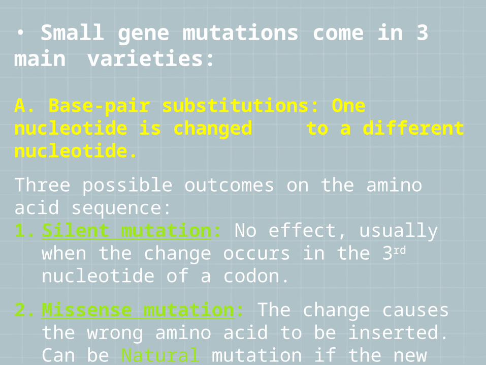
• Small gene mutations come in 3 main varieties:
A. Base-pair substitutions: One nucleotide is changed to a different nucleotide.
Three possible outcomes on the amino acid sequence: 1. Silent mutation: No effect, usually when the change
occurs in the 3rd nucleotide of a codon.
2. Missense mutation: The change causes the wrong amino acid to be inserted. Can be Natural mutation if the new amino acid has a similar structure to the previous aa.
3. Nonsense mutation: Change turns the codon into a stop codon. Results in a truncated protein.

A. Base-pair substitutions:
1. Silent mutation: No effect, usually when the change occurs in the 3rd nucleotide of a codon.
2. Missense mutation: The change causes the wrong amino acid to be inserted. Can be Natural mutation if the new amino acid has a similar structure to the previous aa.
3. Nonsense mutation: Change turns the codon into a stop codon. Results in a truncated protein.

B. Insertion/deletion: An extra nucleotide gets added or removed, causing a frame-shift. All amino acids after the insertion/deletion site will be altered!!

Excision repairTwo major types of excision repair:
I. Base-Excision repair: Remove abnormal or modified bases from DNA.
II. Nucleotide-Excision Repair: Remove larger defects like thymine dimers.
Base- Excision Repair:
Initiated by a group of enzymes called DNA glycosylases (recognize abnormal bases in DNA).
The glycosylases cleave glycosidic bonds between the abnormal base and the 2-deoxyribose.

Base Excision Repair
There are different DNA glycosylases, for different types of damaged bases.
AP endonuclease recognizes sites with a missing base; cleaves sugar-phosphate backbone.
Deoxyribose phosphodiesterase removes the sugar-phosphate lacking the base.

Nucleotide Excision Repair

Base excision repair Nucleotide excision repair

Mismatch repair system

Sample Questions
1. Mutations:
a. Are permanent changes in the DNA sequence or structure.
b. Produce allelic variation.
c. Are more likely to be harmful than beneficial.
d. All of the above.
e. None of the above.

Sample Questions
1. Mutations:
a. Are permanent changes in the DNA sequence or structure.
b. Produce allelic variation.
c. Are more likely to be harmful than beneficial.
d. All of the above.
e. None of the above.

Regulation of Gene
Expression
Prokaryotes & Eukaryotes

Regulation of Gene Expression
Gene expression can be regulated During transcription (transcriptional control). During translation (translational control). After translation (post-translational control).

Positive Control of Transcription
Positive control occurs when a regulatory protein (activator) binds to DNA and increases the rate of transcription of downstream genes.
Negative Control of Transcription
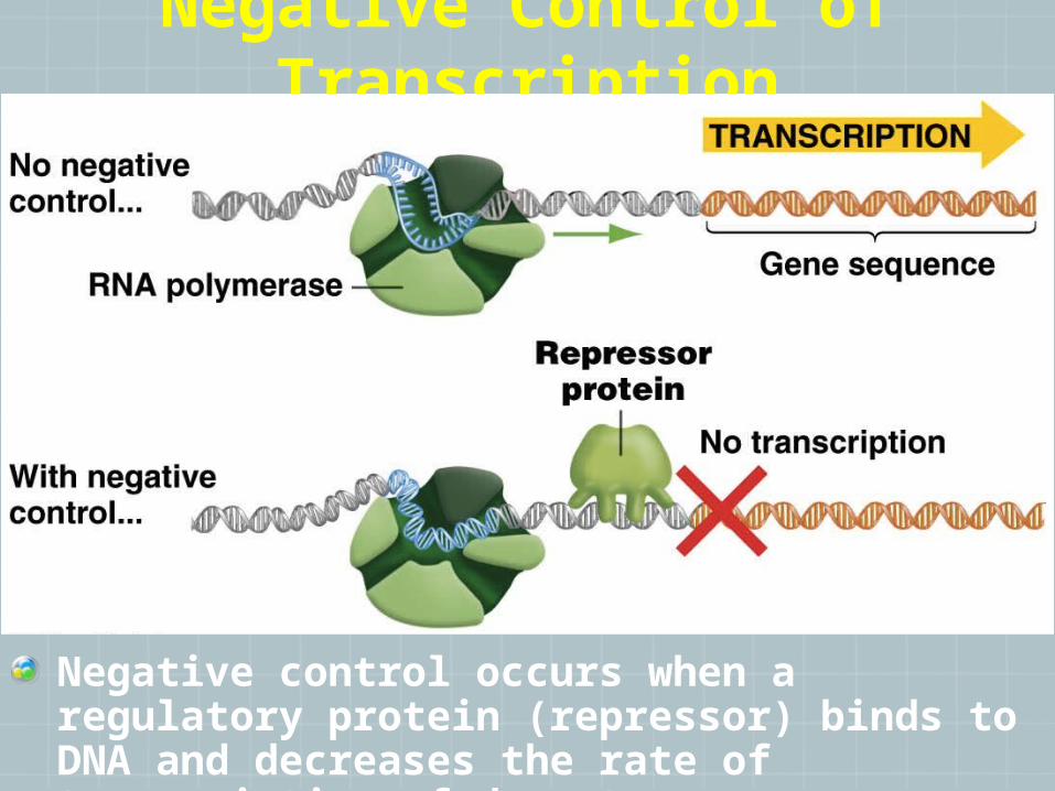
Negative control occurs when a regulatory protein (repressor) binds to DNA and decreases the rate of transcription of downstream genes.
Copyright © 2006 by Elsevier, Inc.
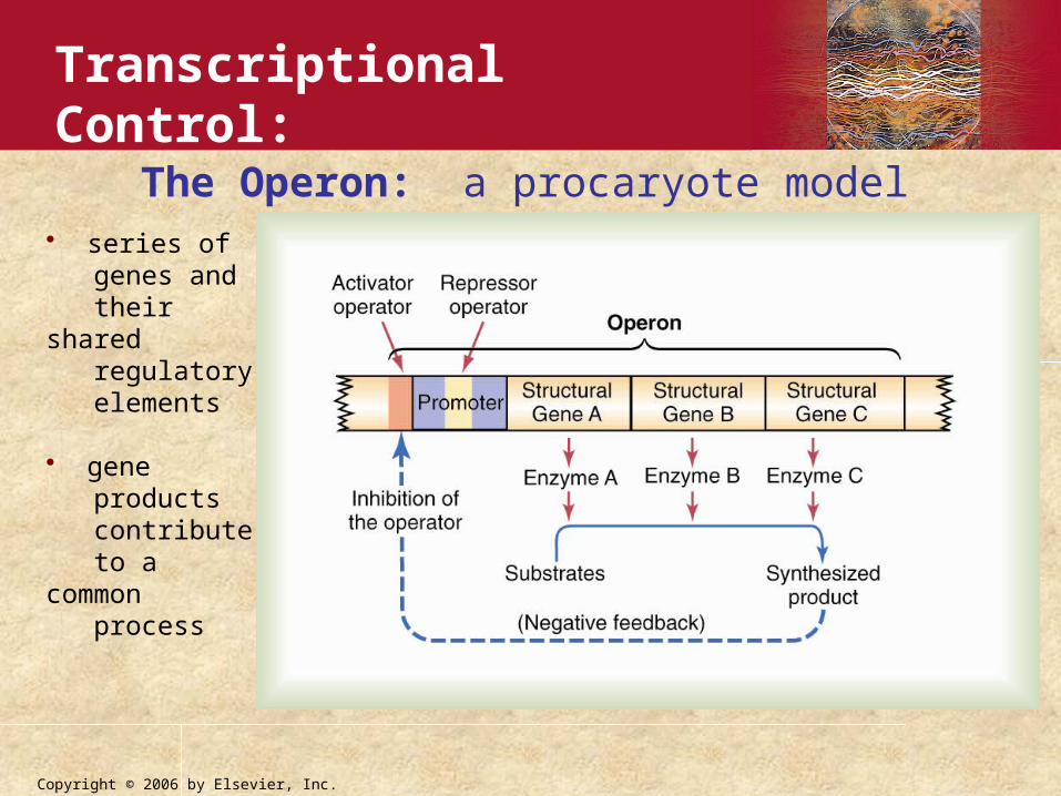
The Operon: a procaryote model• series of genes and their shared regulatory elements
• gene products contribute to a common process
Transcriptional Control:
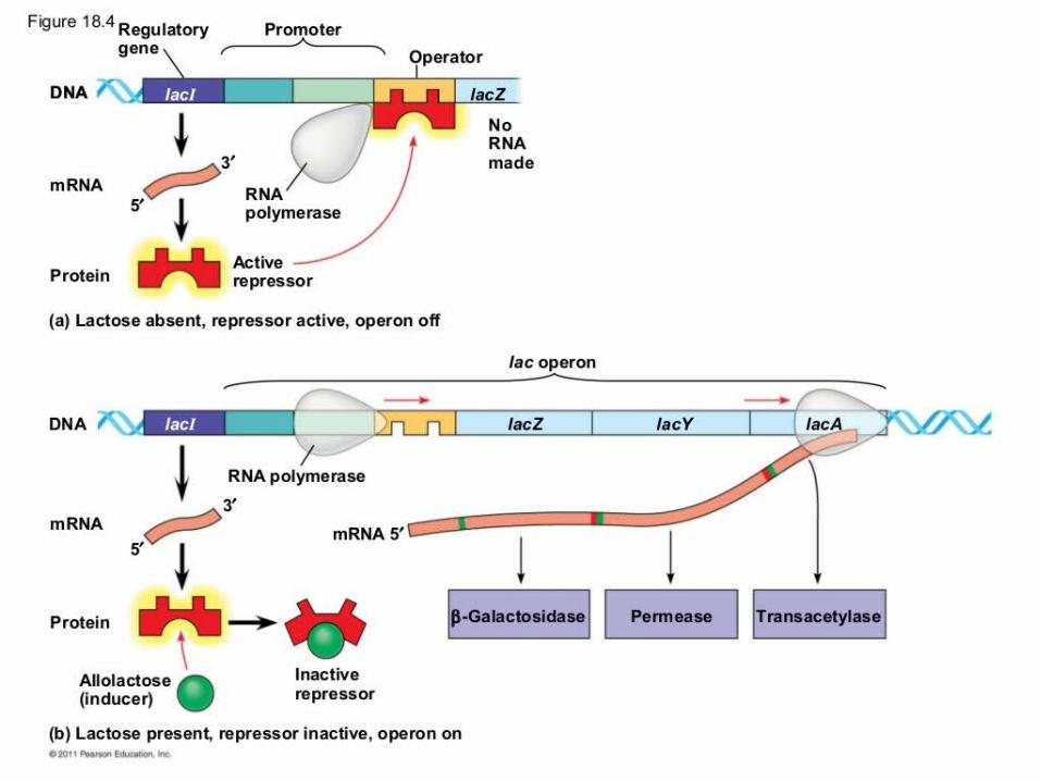


The lac and trp operons

Glucose regulates cAMP levels

Positive control of the lac operon
When cAMP levels are high, CAP is activated, inducing its binding to the CAP site and promoting efficient transcription.

Summary of the lac operon

Examples of gene expression regulation
Transcriptional control:Regulatory proteins affect the ability for the RNA polymerase to bind to or transcribe a particular gene.
Translational control:Regulatory proteins can affect the rate of translation.Enzymes can affect the stability of the mRNA.
Post-translational control:Translated protein may be modified by phosphorylation or other modifications that alter the protein’s activity, folding or stability.

AP Biology
Points of control The control of gene
expression can occur at any step in the pathway from gene to functional protein1. Packing/unpacking of DNA
2. Transcription
3. mRNA processing
4. mRNA transport
5. Translation
6. Protein processing
7. Protein degradation


• Histone deacetylases (HDACs) are negative regulators of transcription
• Histone acetylases (HATs) are positive regulators of transcription
In general…
+
-
CBP/P300
HDAC1/2

DNA methylation• DNA can be modified by methylation of adenine and cytosine bases
Methylated Base Methylation Sequence
C5-methylcytosine (5-mC) CpG
C5-hydroxymethylcytosine (5-hmC)
CpG, CpHpG1, CpHpH1
H = Adenine, Cytosine, or Thymine
"p" in CpG refers to the phosphodiester bond

Components
of epigenetic
inheritence
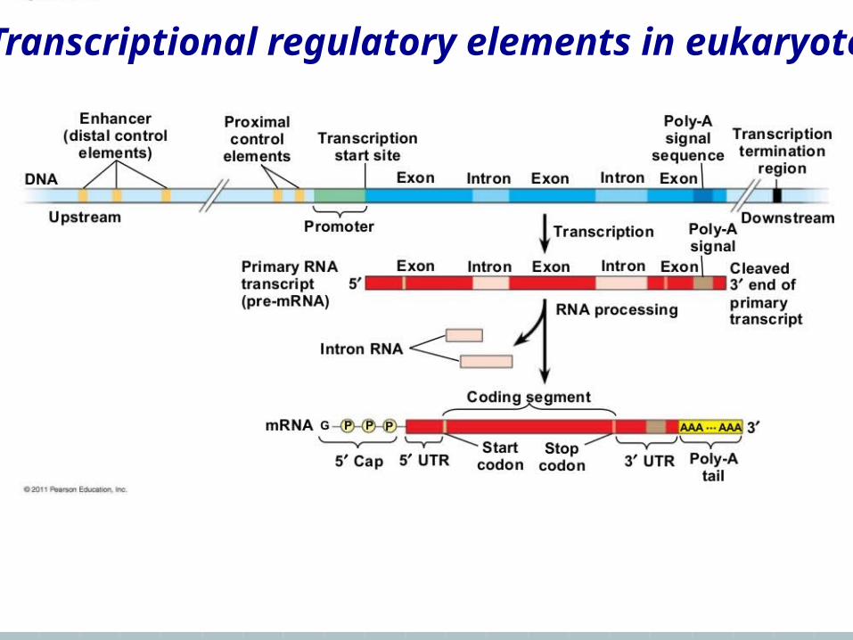
Transcriptional regulatory elements in eukaryotes


Control of transcription: promoters, enhancers, repressors….
Control of transcription in eukaryotes depends on both cis- and trans-acting factors, including: regulatory & promoter sequences, RNA polymerase, the general transcription factors and gene specific activators and repressors

Cis- versus trans-acting factors
Cis: includes all the elements that are present on the same DNA strand as the regulated gene: promoter, enhancer, intron/exons etc. (addition of an acetyl - CH3CO group) for example, results in decreased condensation of the DNA and increased transcription of genes in that region.
Trans: includes all the rest regulatory elements such as transcription activators and repressors.


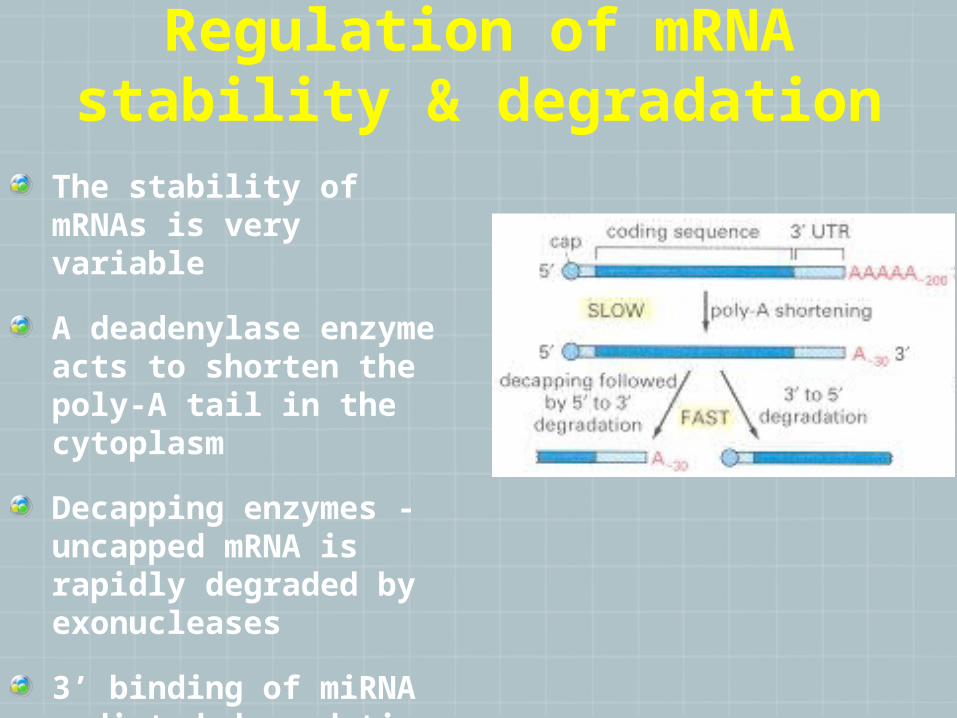
Regulation of mRNA stability & degradation
The stability of mRNAs is very variable
A deadenylase enzyme acts to shorten the poly-A tail in the cytoplasm
Decapping enzymes - uncapped mRNA is rapidly degraded by exonucleases
3’ binding of miRNA mediated degradation
RNAi mediated degradation

Common methods in genetics
DNA gel electrophoresis
PCR: Polymerase Chain Reaction
Conventional DNA sequencing & High-throughput sequencing
Micro-array hybridization
RFLP: Restriction Enzyme Fragment Polymorphism:

Blotting techniques
Southern Blot
Used to detect DNA
Northern Blot
Used to detect RNA
Western blot
Used to detect protein
TYPES OF BLOTTING TECHNIQUES

Restriction Endonuleases
Restriction endonucleases are enzymes that cleave DNA
molecules at specific nucleotide sequences depending on
the particular enzyme used. Enzyme recognition sites
are usually 4 to 6 base pairs in length.
The recognition sequences are randomly distributed
through the DNA.
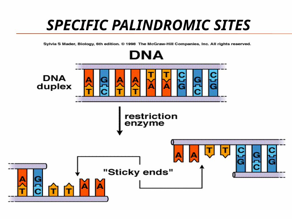
SPECIFIC PALINDROMIC SITES

CLONING PROCESS Gene of interest is
cut with specific RE
Host plasmid is cut with the same RE
Gene annealed with the plasmid and ligated with ligase
New plasmid inserted into bacterium (transformation)

STEP 3: LIGATION OF DNA SAMPLE AND PLASMID DNA

Cloning a segment o f DNA into a plasmid vect or
• bacteria are “transformed” with the recombinant plasmid• colonies that grow in tetracycline, but not in ampicillin are isolated
PstIHuman DNA cut with PstI
P
P
pBR322 ampR, tetR
pBR322 (human clone) tetR
P
P
ampR tetR
pBR322 DNA cut with PstIinactivating the ampR gene
tetR
tetR
combineand
ligate

STEP 5: GROWTH ON AGAR PLATES

DNA-Polymerase + Nucleotides
Primers
Denaturation 95°C
Annealing 50-60°C
Extension 68°C
Denaturation, annealing
Extension
x30
Steps in PCR


Microarray Technology: Detection of differentially-expressed genes

Sample Questions
1. DNA molecules can be cut into sections by using:
a. ATP
b. Gel electrophoresis
c. Restriction endonucleases
d. Plasmids
e. A probe
Sample Questions
1. DNA molecules can be cut into sections by using:
a. ATP
b. Gel electrophoresis
c. Restriction endonucleases
d. Plasmids
e. A probe

Good Luck !!!