pre-lab e11: protein agarose gel ( 10 pts) 2281, expt. 11 dr.wenju lin, spring 2016 1 experiment 11:...
TRANSCRIPT

Pre-Lab E11: Protein Agarose Gel ( 10 pts) Name: _______________________________; Lab Section: _______________; Grade: _______
1. (2 pts) The isoelectric point of an amino acid is defined as ___________________ _________________________________________________________________.
2. If amino acid Lysine with a pI of 9.7 is dissolved in a gel running buffer of pH 5.5; at this pH Lysine is an (anion or cation) and it will migrate toward (Anode or cathode) during the native agarose gel electrophoresis (2 pt, circle the right answer)
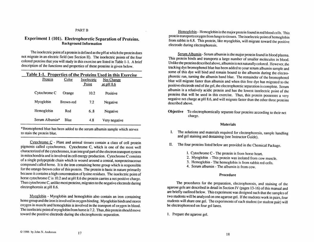
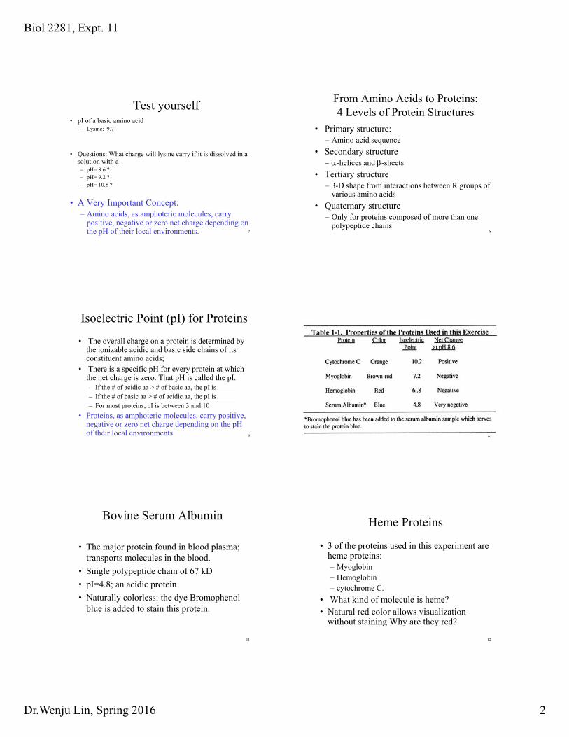
3. Which of the following four proteins in Part A of today’s experiment is the most acidic
protein? (1 pt) a) cytochrome C (pI =10.2) b) myoglobin (pI =7.2) c) hemoglobin (pI= 6.8) d) bovine serum albumin (pI = 4.8)
4. What is the basis of separation of the HbA and HbS in Part B of today’s experiment? (1 pt)
a) size difference b) net charge difference at a particular pH c) shape difference
5. Short Answer: How is the separation of protein molecules in protein agarose gel different from the separation of DNA molecules in a DNA agarose gel? (2 pts)
6. Preparation of gel solution (2 pts)
If your group is assigned to prepare 1.3 % agarose gel solution for part A, you need to use 50 ml cylinder to dispense 45 ml of electrophoresis buffer with pH at __________ into a 125 ml flask and add ________ g of agarose ( show calculation below to get full credit, 1% : 1 gram/ 100 ml buffer).

Biol 2281 Spring 2016 Protein Agarose Gel Procedure/Report
1
Experiment 11: Protein Agarose Gel Electrophoresis
Objectives: At the end of the exercise, you should be able to
1. understand the concepts of protein structures, isoelectric point of amino acids and proteins.
2. understand the principle of separation in protein agarose gel electrophoresis. 3. demonstrate laboratory techniques used in running a protein agarose gel (preparing
gels, loading samples with micropippets, and electrophoresis). 4. Understand the molecular difference between normal and mutant forms of beta-
hemoglobin in patients with sickle cell disease, and how protein agarose gel electrophoresis can be used in disease diagnosis.
Introduction:
Important background information for this experiment is provided at the end of this handout. This experiment consists of two parts, Part A and Part B.
Part A: Comparing Migration Directions and Rates of Four Proteins (cytochrome C,
myoglobin, hemoglobin and albumin) at pH 8.6 Part B: Comparing Migration Rates of Hemoglobins Isolated from Normal, Heterozygous
and Sickle cell anemia Individuals at pH 9.2 EVERT TWO STUDENTS form a group. Your group will be assigned to run either Part A or Part B. Each student will have access to the results of both experiments and therefore, will be responsible for understanding both parts.
Materials at the supply bench:
1) gel running apparatus (lid matches the tank); one comb. 2) flasks for preparing agarose gel (125 ml) 3) micropippets (P20) and yellow tip boxes 4) pH 8.6 buffer for part A 5) pH 9.2 buffer for part B 6) 4 protein samples for part A (on ice) 7) 3 protein samples for part B (on ice) 8) agarose powder, balance, cylinders 9) plastic waste cup, markers and microcentrifuge tubes
Procedure for Pouring an Agarose Gel:
1. Place gel casting tray on the top of gel support deck, the open ends of the tray should be placed next to the sides of the chamber. Insert the comb into the middle casting tray slots.
2. Read the labels of pH value on flasks, bottles and cylinders. Using 50 ml cylinder to dispense 45 ml of electrophoresis buffer (see below) into a 125 ml flask and add ____ g of agarose to make the final concentration of 1.2% ( 1% : 1 gram/ 100 ml buffer).
• Part A: pH 8.6, 1X buffer • Part B: pH 9.2, 1X buffer
Biol 2281 Spring 2016 Protein Agarose Gel Procedure/Report
2
3. Microwave until the suspension comes to a low boil. Make sure the agarose is completely dissolved: the suspension needs to be completely clear. Cool at room temperature for about 5 min.
4. Pour the melted agarose into the casting tray. Rinse the flask with hot water immediately. Return the flask to your tray.
5. After the gel is solidified (about 15 min), lift up the tray from the tank and rotate it by 90 degrees, and place it back on the supporting deck.
6. Slowly fill the electrophoresis chamber with the electrophoresis buffer until the gel is completely submerged. The buffer should be about 3-4 mm above the gel surface.
• Part A: pH 8.6, 1X buffer • Part B: pH 9.2, 1X buffer
7. While the gel is submerged in the buffer gently lift the comb straight up and out of the gel. Rinse the comb immediately and return it to your tray. Return the cylinder to the assigned supply bench.
Procedure for Loading Protein Samples into the Agarose Gel
1. (TA) Transfer the protein samples into separate microcentrifuge tubes. 2. Use micropipets (P20) to draw each of the protein samples into the yellow-tip.
• Part A Samples to be loaded into the pH 8.6 gel
i. Cytochrome C: pI =10.2 10 ul ii. Myoglobin: pI=7.2 10 ul iii. Hemoglobin: pI=6.8 10 ul iv. Serum albumin: pI=4.8 10 ul
Which of the above 4 proteins will migrate to the cathode?____________ • Part B Samples to be loaded into the pH 9.2 gel
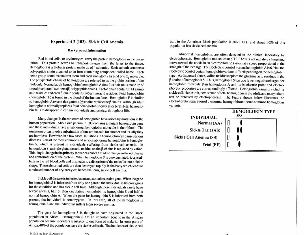
i. HbA HbA (normal human hemoglobin) 20 ul ii. HbA HbS (heterozygous, or Trait) 20 ul
iii. HbS HbS (sickle cell hemoglobin) 20 ul
HbS has two fewer negative charges per molecule than the HbA. What is your prediction for the migration rate of HbS relative to HbA at pH 9.2? ____________________________________________________
3. Carefully direct the tip of the micropipette into the top of the sample well and slowly eject
the sample into the well. The sample is already mixed with 10-20% glycerol to ensure that it sediments at the bottom of the well.
4. Discard the used tip into the plastic waste cup. 5. Do not leave any gap between samples. Repeat to load each additional
sample. Return the empty microcentrifuge tubes to supply bench. 6. You can practice loading with 10 ul of 1X loading dye on empty lanes.
Procedure for Electrophoresis
1. Place the lid of the electrophoresis chamber in position. 2. With the power supply off, connect the cables from the tank to the power supply, red to
red (positive electrode) and black to black (negative electrode).
Biol 2281 Spring 2016 Protein Agarose Gel Procedure/Report
3
3. Turn on the power supply and adjust the voltage to “120V”. Press the “Run” button on the front of the power supply to start running.
4. Electrophorese until: • Part A: the bromophenol blue in the serum albumin sample has migrated to
within 1 cm of the positive electrode end of the gel. ABOUT 35-40 minutes. • Part B: the two bands in heterozygous sample are well separated. ABOUT 30
minutes. 5. Prepare the illustration of two protein gels after electrophoresis (Part A AND B) with
each lane clearly labeled with the name of the samples loaded.
Cleaning Up:
1. Return the protein microcentrifuge tubes to supply bench. 2. Dispose the tips, agarose gels into big biohazard trashcan. 3. Transfer the gel running buffer into the beaker labeled with correct pH value. 4. Clean up the tank, the comb, the gel tray with tap water. Place the tray inside the tank
and make sure the lid matches the tank. 5. Refill yellow tip box.
Post Lab Report (20 points total, include title page, questions): (1 pt : typed answers for q1-q6)
1. ( 2 points) What is the basis of separation of the four proteins in Part A of the experiment? Relate your answer to the pI of these proteins.
2. (2 points) How would the separation of the four proteins in Part A of the experiment
change if the students mistakenly used the pH 9.2 buffer instead of the pH 8.6 buffer? Explain.
3. (2 points) What is the basis of separation of the HbA and HbS hemoglobin in Part B of
the experiment? Relate your answer to the mutation found in HbS. 4. Define the four levels of protein structures. ( 2 points) 5.
a) The pI of HbA is about 6.8. Do you expect the pI of HbS to be less than 6.8 or greater than 6.8 based on the fact that HbA carries two more negatively charged amino acids per molecule than HbS? (2 points)
b) If Part B of the experiment was carried out at pH 8.6 instead of pH 9.2, do you expect
to see similar separation of the two forms of hemoglobin? Why or Why not? (2 points)
6. (True or False) The net electric charge carried by a protein varies at different pHs of the
environment. (1 points)
7. Use your biology textbook as a guide, hand-draw sample structures of the following types of amino acids in pencil: (2 pts) (DO NOT copy and paste image files)
i. one acidic amino acid ii. one basic amino acid iii. one non-polar amino acid iv. one polar, uncharged amino acid
Biol 2281 Spring 2016 Protein Agarose Gel Procedure/Report
4
8. ( 4 points) Present the illustration of two protein gels after electrophoresis (Part A AND
B) with each lane clearly labeled with the name of the samples loaded (wells, anode and cathode).
Biol 2281, Expt. 11
Dr.Wenju Lin, Spring 2016 1
Experiment 11: Protein Separation with Agarose
Gel Electrophoresis
1
Amino acids/Protein StructureAmino acids/Protein Structure
ElectrophoresisElectrophoresis
2
3
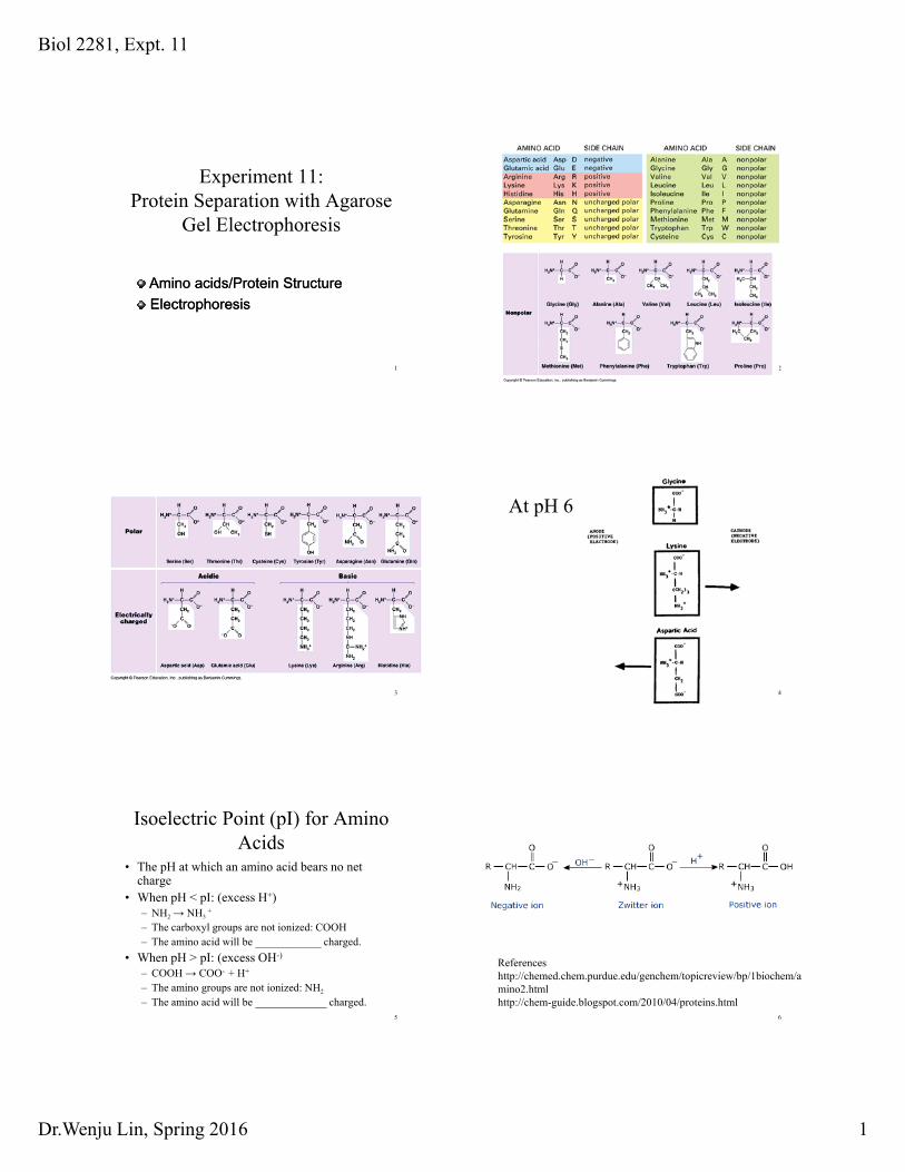
At pH 6
4
Isoelectric Point (pI) for Amino Acids
• The pH at which an amino acid bears no net charge
• When pH < pI: (excess H+)NH NH +
5
– NH2 → NH3+
– The carboxyl groups are not ionized: COOH– The amino acid will be ____________ charged.
• When pH > pI: (excess OH-)
– COOH → COO- + H+
– The amino groups are not ionized: NH2
– The amino acid will be _____________ charged.
Referenceshttp://chemed.chem.purdue.edu/genchem/topicreview/bp/1biochem/amino2.htmlhttp://chem-guide.blogspot.com/2010/04/proteins.html
6
Biol 2281, Expt. 11
Dr.Wenju Lin, Spring 2016 2
Test yourself• pI of a basic amino acid
– Lysine: 9.7
• Questions: What charge will lysine carry if it is dissolved in a solution with a
7
– pH= 8.6 ?– pH= 9.2 ? – pH= 10.8 ?
• A Very Important Concept:– Amino acids, as amphoteric molecules, carry
positive, negative or zero net charge depending on the pH of their local environments.
From Amino Acids to Proteins: 4 Levels of Protein Structures
• Primary structure:– Amino acid sequence
• Secondary structure– -helices and -sheets
8
helices and sheets
• Tertiary structure– 3-D shape from interactions between R groups of
various amino acids
• Quaternary structure– Only for proteins composed of more than one
polypeptide chains
Isoelectric Point (pI) for Proteins
• The overall charge on a protein is determined by the ionizable acidic and basic side chains of its constituent amino acids;
• There is a specific pH for every protein at which
9
• There is a specific pH for every protein at which the net charge is zero. That pH is called the pI.– If the # of acidic aa > # of basic aa, the pI is _____– If the # of basic aa > # of acidic aa, the pI is _____– For most proteins, pI is between 3 and 10
• Proteins, as amphoteric molecules, carry positive, negative or zero net charge depending on the pH of their local environments 10
Bovine Serum Albumin
• The major protein found in blood plasma; transports molecules in the blood.
• Single polypeptide chain of 67 kD
11
Single polypeptide chain of 67 kD
• pI=4.8; an acidic protein
• Naturally colorless: the dye Bromophenol blue is added to stain this protein.
Heme Proteins
• 3 of the proteins used in this experiment are heme proteins: – Myoglobin
12
– Hemoglobin– cytochrome C.
• What kind of molecule is heme?• Natural red color allows visualization
without staining.Why are they red?

Biol 2281, Expt. 11
Dr.Wenju Lin, Spring 2016 3
Protoporphyrin and Heme
13Heme absorbs green and yellow wavelengths; reflects orange and red.
Cytochrome C Protein• Utilized in the mitochondrial and chloroplast membranes
as part of the electron transport chain.• Single polypeptide chain with a heme center (12.5 kD)• Heme is responsible for the orange-brown color• pI = 10.2; a basic protein; contains lots of lysines
14
Horse Heart Cyt C
Myoglobin
• Single polypeptide chain (17kD); structurally similar t h i f
15
to -chain of hemoglobin
• binds and stores O2
in muscle
• pI =7.2
Oxygen-binding site
• Oxygen: green
• Fe(II): orange
• The amino acid side
16
chain of His-64, Val-68 and Phe-43 contribute to the hydrophobic environment of the site
two chains
two chains
Each chain contains an identical heme group.
17
•In lungs: Hb + 4 O2 ----> Hb.O8
•In tissues:
Hb.O8 ---> Hb + 4 O2
Normal and Mutant Forms of -Hb
18
Biol 2281, Expt. 11
Dr.Wenju Lin, Spring 2016 4
What is electrophoresis?
• a separation technique in which an electrical field causes charged molecules to move through a matrix (usually a gel).
• routinely used to separate DNA, protein
19
routinely used to separate DNA, protein and other polymeric molecules.
• Separation can be based on– Sizes– Net charges– shapes
Agarose Protein Gel Electrophoresis
• Separation of Protein using "Native" or "non-denaturing" gel electrophoresis is Based on NET CHARGE
Th N Ch f P i i d d h
20
The Net Charge of a Protein is dependent on the relative relationship between its pI and the pH of the gel running system
When pH < pI: it is _________ charged;
When pH > pI: it is _________ charged;
Separation of DNA molecules by agarose gel electrophoresis is Based on ___
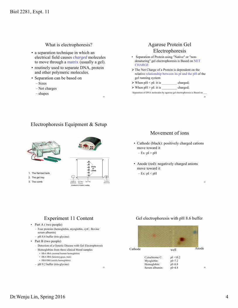
Electrophoresis Equipment & Setup
21
1. The flat-bed tank
2. The gel tray
3. The comb
Movement of ions
• Cathode (black): positively charged cations move toward it – Ex: pI > pH
22
Ex: pI pH
• Anode (red): negatively charged anions move toward it – Ex: pI < pH
Experiment 11 Content• Part A ( two people)
– Four proteins (hemoglobin, myoglobin, cytC, Bovine serum albumin)
– pH 8.6 buffer (tris-glycine)
23
• Part B (two people)– Detection of a Genetic Disease with Gel Electrophoresis
– Hemoglobins from three clinical blood samples• HbA HbA (normal human hemoglobin)
• HbA HbS (heterozygous, trait)
• HbS HbS (sickle hemoglobin)
– pH 9.2 buffer (tris-glycine)
Gel electrophoresis with pH 8.6 buffer
24
AnodeCathode
Cytochrome C: pI =10.2Myoglobin: pI=7.2Hemoglobin: pI=6.8Serum albumin: pI=4.8
well
Biol 2281, Expt. 11
Dr.Wenju Lin, Spring 2016 5
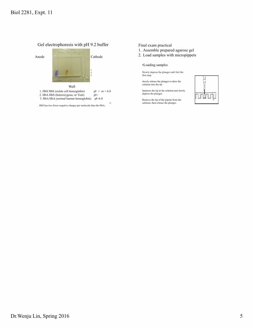
Gel electrophoresis with pH 9.2 buffer
CathodeAnode
1
2
25
1. HbS HbS (sickle cell hemoglobin) pI > or < 6.82. HbA HbS (heterozygous, or Trait) pI=3. HbA HbA (normal human hemoglobin) pI=6.8
HbS has two fewer negative charges per molecule than the HbA.
Well
2
3
•Loading samples
Slowly depress the plunger until feel the
Final exam practical 1. Assemble prepared agarose gel 2. Load samples with micropippets
26
y p p gfirst stop.
slowly release the plunger to draw the solution into the tip
Immerse the tip in the solution and slowly depress the plunger.
Remove the tip of the pipette from the solution, then release the plunger.