practical roadmap - university of the...
TRANSCRIPT

PRACTICAL ROADMAP
CONNECTIVE TISSUE PROPER
A. JOVANOVIĆ
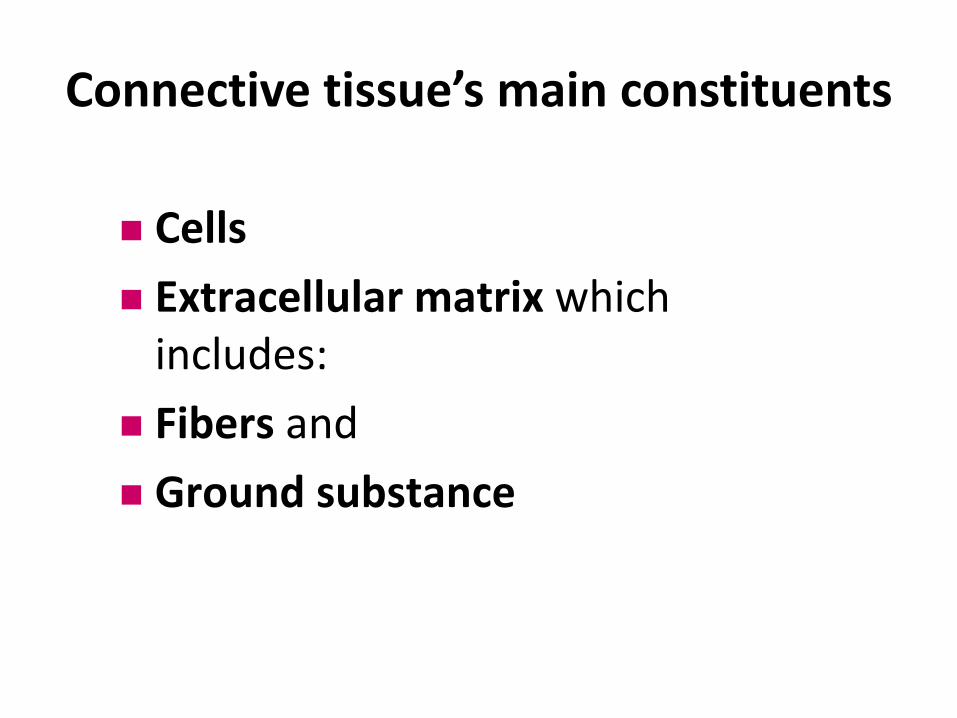
Cells
Extracellular matrix which includes:
Fibers and
Ground substance
Connective tissue’s main constituents
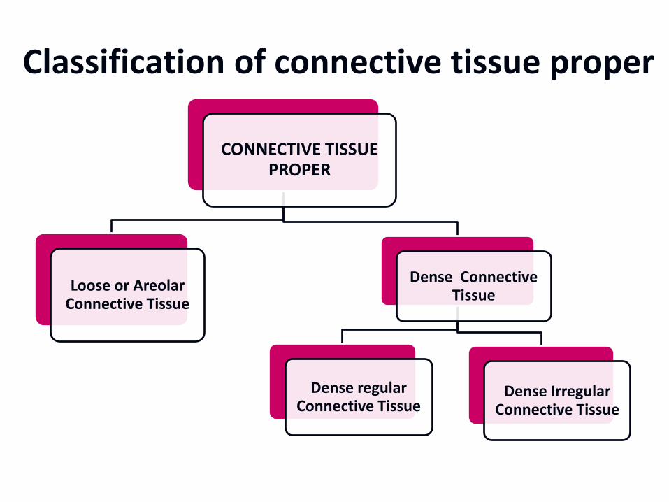
Classification of connective tissue proper
CONNECTIVE TISSUE PROPER
Loose or Areolar Connective Tissue
Dense Connective Tissue
Dense regular Connective Tissue
Dense Irregular Connective Tissue

• Slides: 120 – 1, 120 – 2, 120 – 3
Loose (areolar) connective tissue – Mesentery spread
Stain: Haematoxylin, acid fuchsin and elastic stain
This is a whole mount spread of mesenchymal tissue. It consists of a sheet of loose connective tissue with simple squamous epithelium - mesothelium on each side and a thin “filling” of loose connective tissue in between them. Look at the schematic drawing of the mesentery available in your histology practical manual in order to understand the “sandwich” concept!
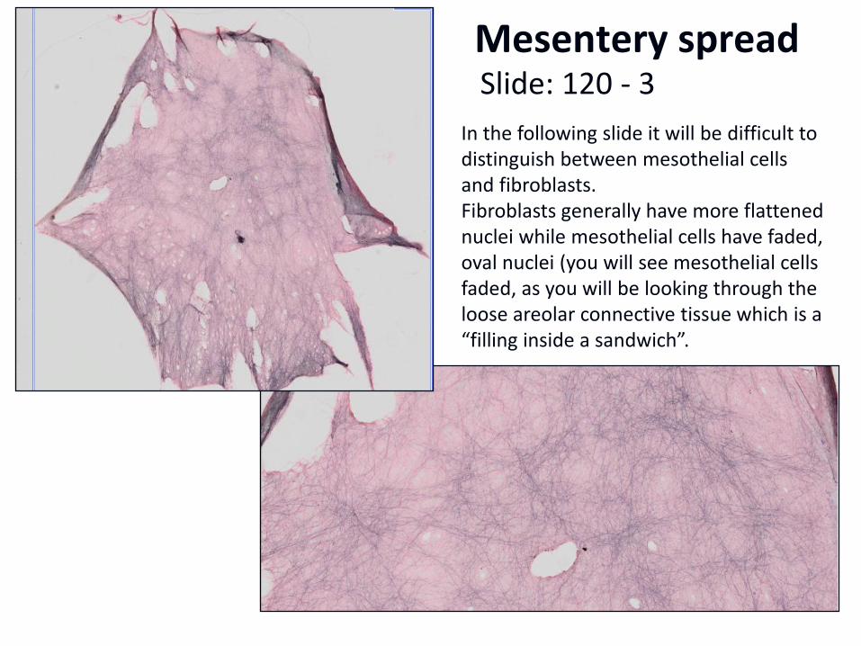
Mesentery spread Slide: 120 - 3
In the following slide it will be difficult to distinguish between mesothelial cells and fibroblasts. Fibroblasts generally have more flattened nuclei while mesothelial cells have faded, oval nuclei (you will see mesothelial cells faded, as you will be looking through the loose areolar connective tissue which is a “filling inside a sandwich”.
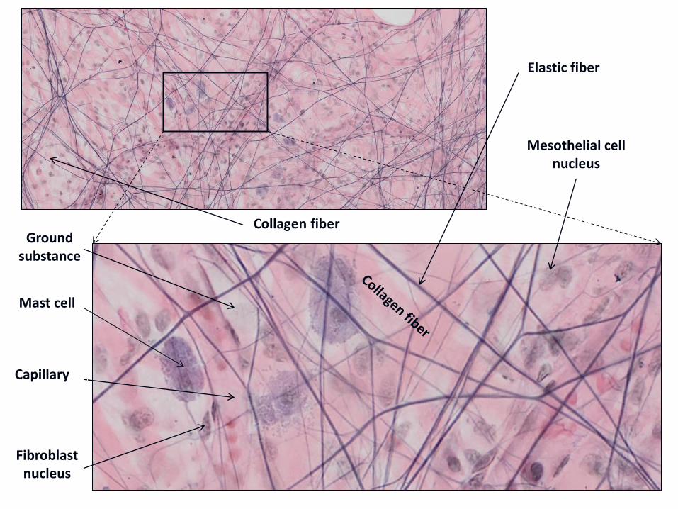
Elastic fiber
Mast cell
Ground substance
Capillary
Fibroblast nucleus
Mesothelial cell nucleus
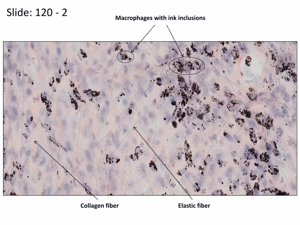
Slide: 120 - 2 Macrophages with ink inclusions
Elastic fiber Collagen fiber
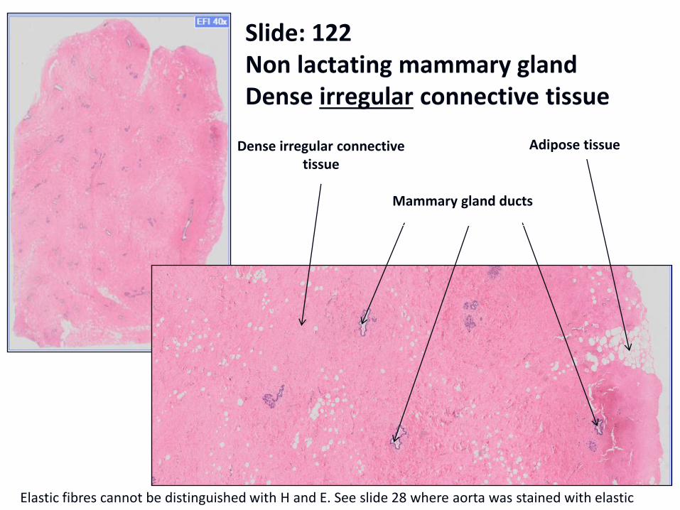
Slide: 122 Non lactating mammary gland Dense irregular connective tissue
Mammary gland ducts
Adipose tissue Dense irregular connective tissue
Elastic fibres cannot be distinguished with H and E. See slide 28 where aorta was stained with elastic stain.
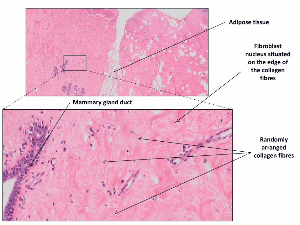
Adipose tissue
Randomly arranged
collagen fibres
Fibroblast nucleus situated on the edge of
the collagen fibres
Mammary gland duct
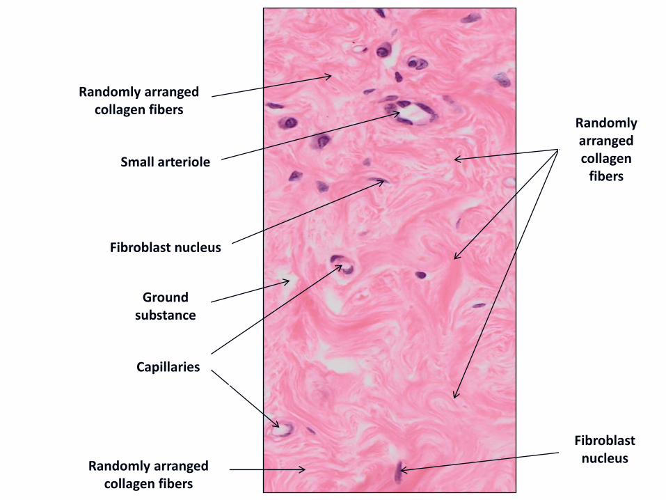
Small arteriole
Capillaries
Fibroblast nucleus
Randomly arranged collagen fibers
Randomly arranged collagen fibers
Ground substance
Randomly arranged collagen
fibers
Fibroblast nucleus
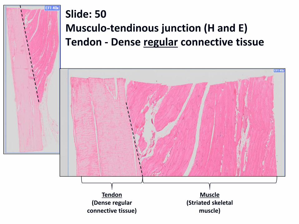
Slide: 50 Musculo-tendinous junction (H and E) Tendon - Dense regular connective tissue
Tendon (Dense regular
connective tissue)
Muscle (Striated skeletal
muscle)
Note the difference in fibre density, ground substance and cellularity seen in the basic three types of connective tissue proper. These criteria are used to classify the type of connective tissue.
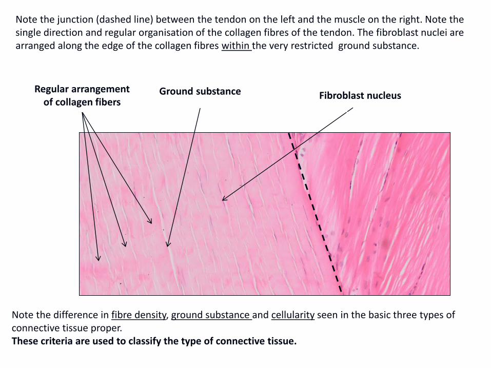
Regular arrangement of collagen fibers
Ground substance Fibroblast nucleus
Note the junction (dashed line) between the tendon on the left and the muscle on the right. Note the single direction and regular organisation of the collagen fibres of the tendon. The fibroblast nuclei are arranged along the edge of the collagen fibres within the very restricted ground substance.
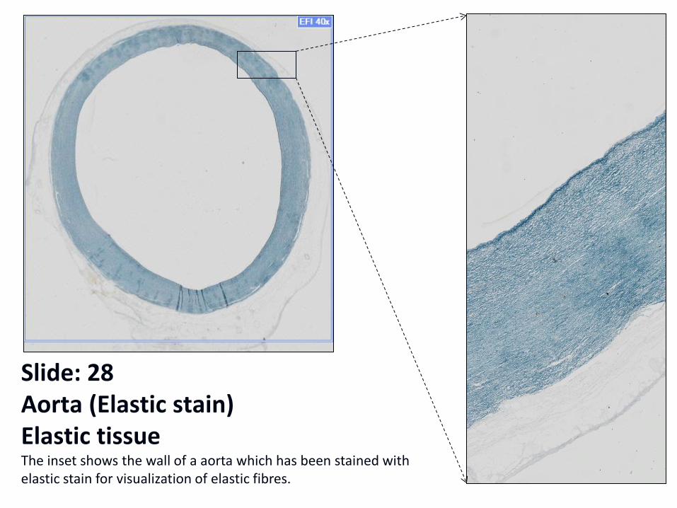
Slide: 28 Aorta (Elastic stain) Elastic tissue The inset shows the wall of a aorta which has been stained with elastic stain for visualization of elastic fibres.
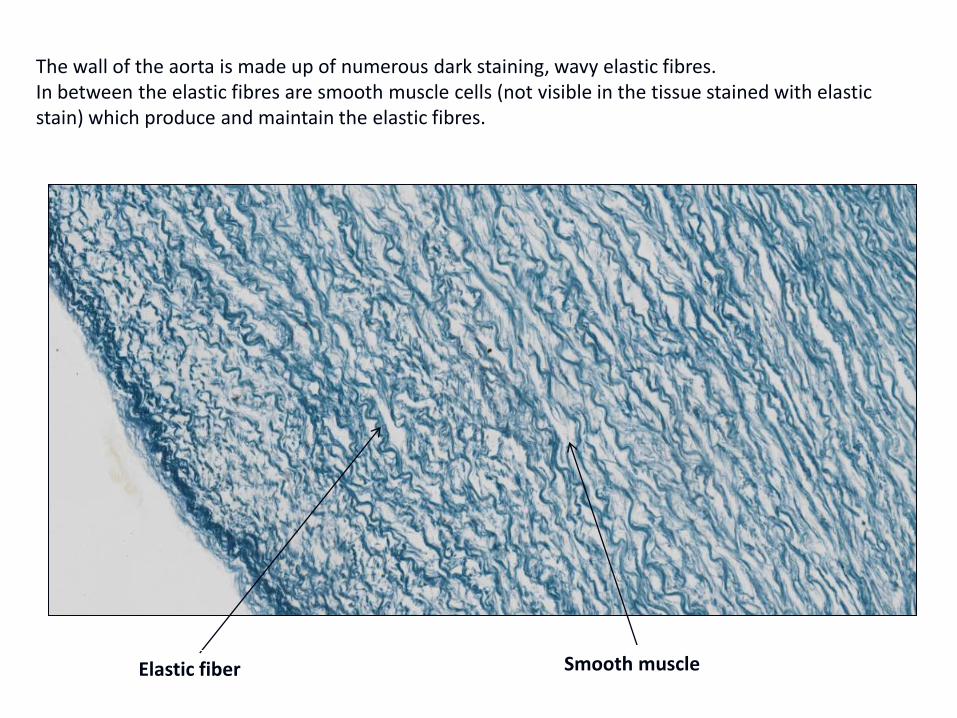
The wall of the aorta is made up of numerous dark staining, wavy elastic fibres. In between the elastic fibres are smooth muscle cells (not visible in the tissue stained with elastic stain) which produce and maintain the elastic fibres.
Elastic fiber Smooth muscle
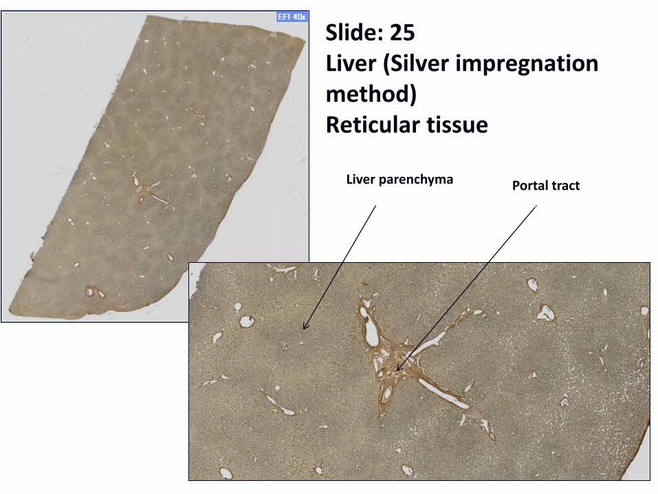
Slide: 25 Liver (Silver impregnation method) Reticular tissue
Liver parenchyma Portal tract
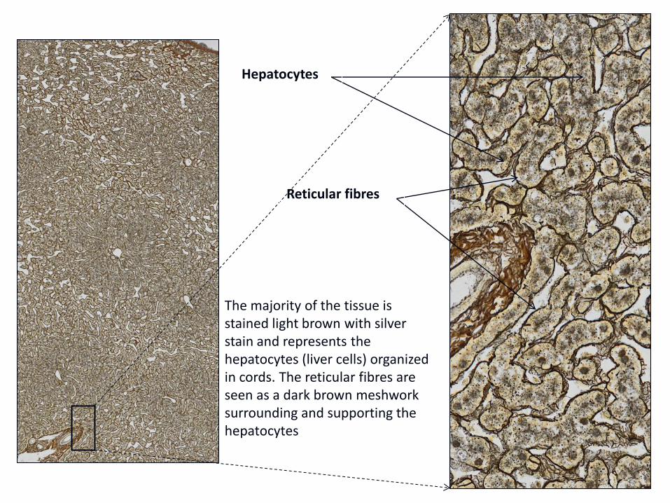
The majority of the tissue is stained light brown with silver stain and represents the hepatocytes (liver cells) organized in cords. The reticular fibres are seen as a dark brown meshwork surrounding and supporting the hepatocytes
Hepatocytes
Reticular fibres