practical lab manual - s a raja pharmacy collegesarajapharmacycollege.com/downloads/pm.pdf5. hot air...
TRANSCRIPT
S.A.RAJA PHARMACY COLLEGE VADAKKANGULAM 627 116
SUBJECT: PHARMACEUTICAL MICROBIOLOGY
III SEMESTER B.PHARM
PRACTICAL LAB MANUAL
Experiment No – 01
INTRODUCTION AND STUDY OF DIFFERENT EQUIPMENTS AND PROCESSING
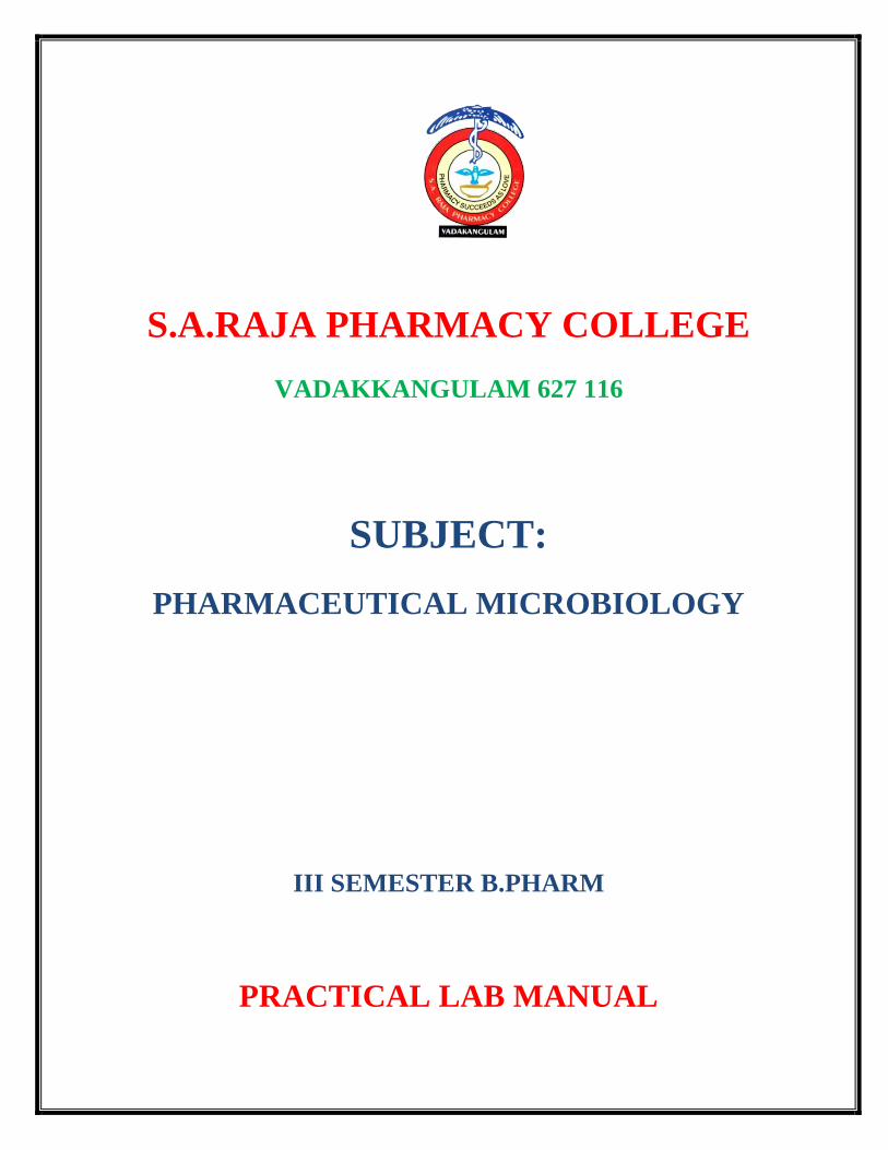
1. BOD INCUBATOR:
It is often called low temperature incubators are one of the most important lab equipment in
many research centers, hospitals and other pharmaceutical labs. BOD incubators is the most
versatile and reliable low temperature incubator which is designed to maintain at 20’C ,
necessary for biological oxygen demand/ biochemical oxygen demand determination. BOD
incubators provide controlled temperature conditions accelerated tests and exposures.
Applications
It measuring waste loadings to treatment plants and in evaluating the BOD removal
efficiency of such treatment systems
It measures the molecular oxygen utilized during a specified incubation period for the
biochemical degradation of organic materials and the oxygen used to oxidized in organic
materials such as sulfides and ferrous iron.
2. ASEPTIC HOOD
The air of laboratory is full of microorganism. Air borne micro organism may contaminate
sterile medium while handling pure culture during inoculation. Thus , an inoculation procedure is
usually carried out under a hood or special cabinet. To have aseptic environment, two types of
the devices are placed in practice – laminar air flow aseptic cabinets.

The inoculation hood is used to create bacteria, fungal free atmosphere in the chamber with
ultra violet germicidal light and it is used in biological culture studies.
3. LAMINAR FLOW CABINET
Laminar hood sometimes also known as laminar air flow is an enclosed bench designed to
prevent contamination like biological particles or any particle sensitive device
A laminar flow hood consist of a fan, HEPA filter. The fan sucks the air through the filter
pad where dust is trapped , after that the pre filtered air has to pass the HEPA filter where
contaminating fungi, bacteria, dust etc are removed. Now the sterile air flows into the working
areas where you can do all your flasking work without risk of contamination.
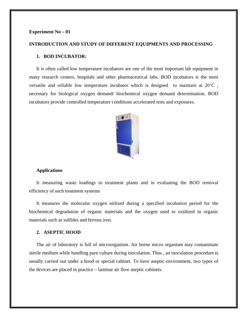
4. AUTOCLAVE
It is laboratory equipment used to sterilize the equipment culture medium, aprons, rubber
tubing’s etc. by steam under pressure technique. It is also used to sterilize infected
materials like contaminated dressings, used culture medium etc., a process known as
decontamination.
Types of autoclave:
1. Stove top autoclaves: they are considered to be modified pressure cookers and
require outside heat source.
2. Front load auto claves: they are most widely used for sterilization being more
convenient as compared to stove top[ autoclaves. They are box shaped and
self contained, equipped with heating unit to turn water into steam sterilization
. the various controls allow the operator to set the desired temperature. They
are also equipped with temperature and pressure gauges to note the respective
parameters.
5. HOT AIR OVEN
It is electrically operated equipment used for the sterilization of glass ware such as syringes,
pipettes, Petri dishes, test tubes, liquid paraffin, oils, powders and flasks.
Precautions:
• The glass ware should be clean and dry before loading the articles for
sterilization .

• The glass ware like test tube , flasks and pipettes must be wrapped in a paper or
aluminum foil before sterilization.
• The temperature should not be allowed to increase above 180’C
• All sterilized articles must be removed only after they have cooled down to room
temperature.
6. DEEP FREEZERS FOR MEDICAL LABORATORIES
Deep freezers are the testing equipment that are used in scientific laboratories to preserve
and store medical equipment, blood samples, medicines and injections, etc for a long period of
time. There are numerous types of deep freezers such as blood bank refrigerators, freezer drier,
ultra low deep freezer. These devices are available indifferent sizes and shapes sometimes it is
designed with compact designs and sometimes with regular designs. The specifications and
functions of the instruments varies as per the requirements of the test application.
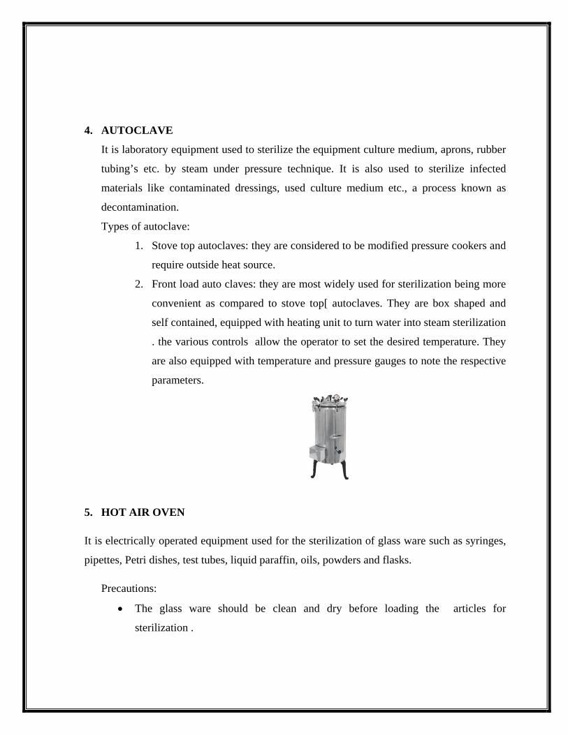
7. COMPOUND MICROSCOPE:
The compound microscope is a basic tool for scientific education and research without which
world of micro organism would have remained un explored. A compound microscope helps in
magnification an image in two stages. It uses an objective lens that has many different powers
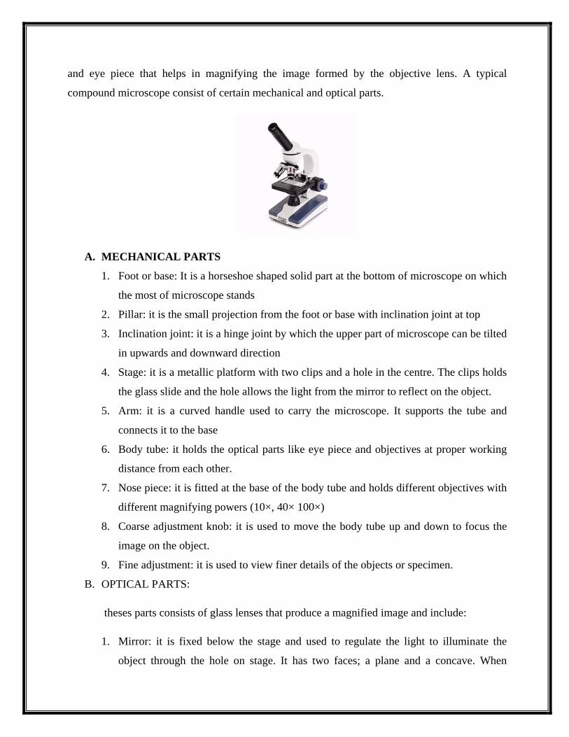
and eye piece that helps in magnifying the image formed by the objective lens. A typical
compound microscope consist of certain mechanical and optical parts.
A. MECHANICAL PARTS
1. Foot or base: It is a horseshoe shaped solid part at the bottom of microscope on which
the most of microscope stands
2. Pillar: it is the small projection from the foot or base with inclination joint at top
3. Inclination joint: it is a hinge joint by which the upper part of microscope can be tilted
in upwards and downward direction
4. Stage: it is a metallic platform with two clips and a hole in the centre. The clips holds
the glass slide and the hole allows the light from the mirror to reflect on the object.
5. Arm: it is a curved handle used to carry the microscope. It supports the tube and
connects it to the base
6. Body tube: it holds the optical parts like eye piece and objectives at proper working
distance from each other.
7. Nose piece: it is fitted at the base of the body tube and holds different objectives with
different magnifying powers (10×, 40× 100×)
8. Coarse adjustment knob: it is used to move the body tube up and down to focus the
image on the object.
9. Fine adjustment: it is used to view finer details of the objects or specimen.
B. OPTICAL PARTS:
theses parts consists of glass lenses that produce a magnified image and include:
1. Mirror: it is fixed below the stage and used to regulate the light to illuminate the
object through the hole on stage. It has two faces; a plane and a concave. When
natural light is available , plane mirror may be used and in artificial light, concave
mirror is used.
2. Iris/Diaphragm: it is located between the mirror and the object . it has an aperture
which can be widened or narrowed as required. Thus it regulates the intensity of light
that reaches the specimen
3. Condenser: it is fixed between the mirror and stage. It concentrates or focuses the
light rays on the objects
4. Objectives: low power objectives, high power objectives, oil immersion objectives
5. Eye piece : it consist of a lens placed at the upper end of the body tube.
Experiment No: 2
STERILIZATION OF GLASSWARE, PREPARATION AND
STERILIZATION OF MEDIA
Sterilizing methods for glassware in a laboratory hot air oven
Sterilizing glassware such as bottles, Petri dishes and test tubes, dry heat is required and this is
carried out in a hot air oven. The ideal temperature of the oven needs to reach at least 160’c and
the content need to regulated at this 45 to 50 min . the content must not be removed from oven
immediately as a slow cooling period is necessary when the temperature has reduced down 50’c
AUTOCLAVING LAB GLASSWARE
Autoclave are widely used to sterilize instruments , glassware and plastic ware, solutions and
media and to decontaminate biological wastes. Because of the physical hazards associated with
autoclaving , extra care must be taken to ensure their safe use. The following safety practices are
followed when autoclaving laboratory glassware:
• Never autoclave items containing corrosives or radio active materials
• Use only borosilicates, glassware’s, which can be better withstand the stresses of high
autoclave temperatures and pressures.
• Load the auto clave properly as per the manufacturers recommendations.
• Individual glassware vessels should be placed within a heat resistant plastic or metal tray
on a shelf or rack and never placed directly on the autoclave bottom or floor.
• Add ¼ to ½ inch of water to the tray so the glassware will heat more evenly
• Check any plastic caps, tubing or other items to ensure they can be safely autoclaved with
the glassware
• Fill glassware only half full with liquids to be sterilized. Take into account the volume of
liquid to be autoclaved.

PREPARATION AND STERILIZATION OF CULTURE MEDIA
1. The constituents or ingredients of that particular medium are weighed accurately
2. The required amount of water is measured and poured into stainless steel pan or heat
resistant beaker and the weighed ingredients are putted one by one into it.
3. The pan is kept on heat source and each ingredients is gradually dissolve by contsnt
stirring with glass rod and heat is reduced to prevent boiling over or burning of the
medium.
4. Small amount of preparation is taken out, cooled and pH is checked and if necessary pH
is adjusted.
5. The medium is now poured into test tubes usually in 10 ml aliquots in 500 ml erlemeyer
flasks or other containers. While in liquefied state, solid media can be added in test tubes,
which are allowed to cool and harden in a slanted position producing agar slants.
6. The container are now closed with cotton plugs, screw caps or cap all covers.
7. The medium is now sterilized in an auto clave usually at 121’C for 15 min and allowed to
cool to be used for the purpose.

Experiment No -3
SUB CULTURING OF BACTERIA & FUNGUS
SUB CULTURING
After incubation has been completed in streak plate , pour plate or spread plate techniques and
appearance of the discrete, well separated colonies has been examined, the next step is to
subculture some of the cells from one of the colonies to separate agar plates or nutrient agar
slants with a sterilized needle or loop for further examination and use. Each of these new cultures
represents the growth of a single species called a pure culture or stock culture. Sub culturing
term is the term used to describe the procedure of transferring o microorganism from their parent
growth source to a fresh one or from one medium to another medium
REQUIREMENTS
Mixed nutrient agar streaks, pour plate and spread preparations of nay refrences bacteris
Nutrient agar slants or nutrient agar plates
Inoculating loop
Wax marking pencil
Microscope

PROCEDURE:
1. With a wax pencil label the nutrient agar slants and agar plates as bacteria A & B.
sterilize the inoculating loop by holding it in the hottest portion of the Bunsen burner
flame
2. Flame until entire wire become red hot
3. Allow the loop to cool for a few seconds or cool it by dipping In a fresh agar plate
4. Touch the tip of the loop to the surface of a selected discrete colony or the agar streak
plate or the pour plate
5. Remove the plug of the agar slants, grasp the plug with thee little finger of the left hand
and pass the neck tube rapidly over the Bunsen burner flame. Inset the loop into the
subculture tube rapidly over the Bunsen burner flame. Inset the loop into the subculture
tube and inoculate it lightly over the hardened surface in a straight or zig zag line and
recap tube.
6. Reflame the inoculating loop/ needle to destroy existing organism
7. Incubate the culture for 48-72hrs.
Observations:
After incubation, observe the slants or plates for the growth of pure colonies.

Experiment – 04
PREPARATION OF NUTRIENT SLAB AND SLANT
Preparation of agar slant
The sterilized agar medium in test tube is kept in an inclined position while hot making a slope
of 20’ angle or less. The tubes are allowed to cool in that position to make slants. The sloped
surface provides more surface area for the growth of the inoculated organism. Which is easily
inoculated with loop or needle, for each stab culture a straight needle is used and stabbed down
to the bottom of the agar medium called “butt”
Preparation of culture plates
Procedure:
1. The hot sterilized medium in test tubes is allowed to cool to 55’C and poured into
sterilized petri dishes under aseptic conditions.
2. The medium is allowed to solidify in flat position and the plates are left overnight at
room temperature in an inverted position. This prevents the condensation on the lid and
keeps the plates clear for viewing.
3. The plates are stored in refrigerator or in a cool place in inverted position, avoiding
drying of the medium by keeping them in plastic bags.

Experiment – 05
STAINING METHODS – SIMPLE STAINING
Staining methods:
Normally there two staining procedures for light microscopy
a. Simple attaining
b. Differential staining
Simple staining: In positive, the stain is basic having positive charge and attaches to the surface
of the object that is negatively charged
Negative staining: in this procedure, more than one staining reagents are used and specific
objects exhibit different staining reactions which are readily distinguishable. Two most widely
differential procedures are gram staining an acid fast staining.
SIMPLE STAINING
Principle :
simple staining involves single dye or staining reagents. The purpose of staining is to
demonstrate cell size, shape and arrangement of bacterial cells. Since bacterial cells usually have
a negative charge on their surface, they are most readily colored by basic stains. These
compounds will either give up, which is attracted to the negatively charged cell surface.
Reagents: Loeffler’s methylene blue solution

Procedure: prepare a smear of a given culture or materials by spreading a thin film on a clean
glass slide
Dry it by waving in air and then heat fix by passing the slides 2 to 3 times through the flame with
the smeared slide facing upwards
Stain the smear by flooding it with one of the staining solutions and allowing it to remain
covered with the stain for the time designated below.
• Methylene blue – 1 min
• Crystal violet- 30 sec
• Carbol fuchsin – 20 sec
Wash the slide gently with running water to remove the stain Air dry the slide or blot with
blotting paper
Apply oil directly to the smear, and focus the smear first under low power objective and then
under oil immersion objectives.
Experiment – 06
STAINING METHODS –GRAM STAINING
The technique was developed by a Danish physician Dr. Hanes Christian gram . this is useful
differential staining procedure in bacteriology which besides determining gross morphology,
differentiates bacteria into two major distinct groups:
Gram positive bacteria
Gram negative bacteria
The differentiation assists in determining subsequent biochemical tests and the respective media
for their culture in laboratory
The technique involve 6 basic steps
• Smear preparation
• Heat fixation
• Staining with crystal violet
• Use of iodine/lugol’s soln
• Treatment with acetone alcohol mix
• Use of safranin
Principle:
The peculiar response towards the staining is related to physical and chemical difference
in the cell walls of the two groups of bacteria . in gram negative bacteria, the cell wall is thin ,
multi layered containing high lipid contents which are readily dissolved by alcohol , resulting in
pore formation in the cell wall facilitating the leakage of the crystal violet iodine complex and
resulting in discolouration of gram negative bacteria which takes safranin and appears red. On
the other hand cell walls of gram positive bacteria are thick composed mainly proteins and cross
linked mucopeptides. On application of decolorizing agent, dehydration is caused resulting in
closure of pores of the cell wall therby retaining the CV-I complex and do appear blue or purple.

Procedure:
1. Make smears of the given culture on a clean glass lside
2. Air dry the smear and heat fix it
3. Cover the smear completely with crystal violet stain and leave the stain on the slide for
one min
4. Wash the slide gently with distilled water or tap water
5. Flood the smear with gram iodine solution and wait for one min
6. Wash with tap water gently and drain carefully
7. Add ethyl alcohol or alcohol acetone (1:1) solution drop by drop until the smear becomes
free from any colorization
8. Wash the slide gently under running tap water and drain
9. Now counter stain with safranin and wait for 30 sec
10. Wash again and blot dry with blotting paper or simply air dry the slide and observe under
oil immersion objectives.
RESULT:
Bacteria that appear blue/ violet/ purple are assigned as gram positive bacteria
Bacteria that appear red/ pink are assigned as gram negative bacteria

Experiment – 07
STAINING METHODS –ACID FAST STAINING
The technique was developed by paul ehrlich (1882) and was modified later by ziehl- neelsen
and therefore also known as Ziehl Neelsen staining. This is a differential staining used to identify
mainly the members of mycobacterium tuberculosis and leprae. These organism are difficult to
stain by ordinary staining methods due to presence of high lipid content in their cell wall.
Bacteria are classifiedas
1. ACID FAST: if they retain the primary stain after the application of strong acid and
appeared red.
2. NON ACID FAST: if they do not retain the primary stain and are counterstained by
methylene blue.
Reagents: 1. Carbol fuchsin solution, Acid alcohol solution (3% Hcl in alcohol)
Alternatively: sulphuric acid solution (20-25% v/v in water)
Methylene blue counter stain (0.3% w/v aq)
Procedure:
1. Prepare a smear of purulent portion of the specimen on a clean glass slide
2. Air dry and heat fix the smear
3. Flood the smear with freshly filtered carbol fuchsin. Heat gently until steam rises
4. Continue to heat for 5 min so that steam is seen but without boiling. Do not allow the side
to dry and add more stain from time to time to prevent this drying
5. Cool and wash the stain of the slide with water.
6. Cover the slide with acid alcohol solution for 3 min. wash with running water and drain.
Repeat de colorization process until smear becomes faint pink in color.
7. Cover the slide with methylene blue stain and leave it for 2 min
8. Wash with tap water , blot dry air the slide under the oil immersion object
Results: Acid fast organism will appear bright red on a blue background while non acid fast
organism will appear dark blue in color.

Experiment – 08
ISOLATION OF PURE CULTURE OF MICRO ORGANISM
A pure culture contains only one kind of micro organism and involves not only the isolation of
individual micro organism from a mixed population but also the maintenance of such individuals
and their progenies in artificial media. Pure culture are essential in order to study; colony
characteristics, biochemical characteristics, morphology, staining reactions and immunological
reactions or the susceptibility to antimicrobial agents of a particular strain of bacterium or fungus
or actinomycetes
The commonly used methods are.
1.Streak plate method
2.Pour plate method
3.Spread plate method
4. Roll tube method
5.Micromanipulator method
I. STREAK PLATE METHOD
Principle
The method is based on the principle that by streaking , a dilution gradient gets established
across the surface of the Petri plate as bacterial cells are deposited on the agar surface. Because
of this dilution gradient, confluent growth takes place on the part of the medium where few
bacteria are deposited. Each colony is the progeny of a single microbial cell, thus representing a
clone of pure culture. Such colonies may be picked aseptically and re streaked into fresh media
to ensure purity of a particular strain.
Procedure:
1. Place a loopful of the inoculums near the periphery of the petri dish and cover with the
close parallel streaks.
2. Turn the plate at right angles and streak approx one half of the remaining portion
3. Without overlapping the previous streaks
4. Turn the plate to 180’c and streak the reminder of the plate, avoiding previous streaked
areas.
II.POUR PLATE METHOD
Principe
The main principle is to dilute the inoculums in successive tubes containing liquefies agar
medium so as to permit a through distribution of bacterial cells with the medium. The mixed
culture of bacteria is diluted through distribution of bacterial cells with the medium. The mixed
culture of bacteria is diluted directly in tubes containing melted agar medium maintained in
liquid state. When bacterial colonies develop, the isolated colonies developed both within the agr
medium and on the medium . These isolated colonies can be picked up streaked onto another
agar plate to ensure purity of the strain.
Procedure:
1. Prepare a liquefied agar tube and allow to cool to 45’C. prepare a water blank
2. Transfer 1 ml of a suspension of a mixed culture of organism to water blank tube
3. Transfer 1 ml of the suspension from tube 1 to tube 2 and repeat the same process up to
tube 5 to get the appropriate dilutions.
4. Transfer 1 ml of the bacterial suspension each from tubes 1-5 sterile petri plates using
sterile pipettes.
5. Pour the liquefied agar medium in tubes into each petri plates using sterile pipettes.
6. Pour the liquefied agar medium in tubes into each petri plate containing 1 ml of diluted
suspension.
7. Rotate the plates gently to ensure uniform distribution of bacterial cells in the medium
and allow the medium to solidify.
8. Incubate the plates for 24-48 hours at 37’C in an inverted position.
SPREAD PLATE METHOD:
Principle:
In this method, micro organism are spread over the solidified agar medium with a sterile
L shaped glass rod called spreader when the petri plate is spinning on a turn table.
Procedure:
1. Take three nutrient agar plates and label them with name of the organism to be inoculated
2. Aspectically inoculate the plates with a loopful of the given organism
3. Place plate 1 on the turn table
4. Sterilize the spreader by putting it first in ethanol in a beaker , then on the flame of
Bunsen burner and cool the rod for 30 sec.
5. Remove the lid of plate and spin the turn table.
6. Touch the spreader gently on the surface of agar and move it forth and back to spread
bacterial cells on the agar surface when the turn table is spinning
7. When turn table stops spinning put the lid over the lower half of petri dish
8. Sterilize the spreader again and repeat same process for the other two plates.
9. Incubate all the plates at 37’C for 24 hrs.
ROLL TUBE METHOD
1. This method is used for isolation of obligate anaerobes. A stoppered anaerobic culture
tube coated with a pre-reduced agar medium containing oxygen free nitrogen is used for
isolation. When the stopper is removed the tube is kept anaerobic continuously flusing it
with oxygen free carbon di oxide from a gas cannula. Inoculation is done with transfer
loop held against the agar surface as the tube is being rotated by a motor.
MICROMANIPULATOR METHOD
1. This device that can pick up a single microbial cell from a colony of mixed culture and
are used in conjuction with microscopes. The single microbial cell is gently sucked into a
micro pipette and transferred into a large drop of sterile medium on another cover slip.
Experiment :9
MICROBIOLOGICAL ASSAY OF ANTIBIOTICS BY CUP PLATE METHODS
The microbiological assay is based upon a comparison of the inhibition of growth of
micro organism by measured concentration of the standard preparation of the anti biotics to be
examined with that produced by known concentration of a standard preparation of the antibiotic
having a known activity.
1. Cup plate method or cylinder plate
2. Turbidimetric or tube assay method
CUP PLATE METHOD
This method depends upon diffusion of an anerobic from a vertical cavity , through the solidified
agar layer in a petri plate. The growth test of micro organism is inhibited entirely in a circular
area or zone around the cavity of the anti biotics
A liquefied assay medium is inoculated by suspension of test micro organism and the inoculated
medium is poured into sterile petri plate by using an assay medium and then spread the test
culture or micro organisms on the surface of plates
Solution from known concentration of the standard preparation and the test antibiotics are
prepared in appropriate solutions. Preparation of the standard solutions and potency of antibiotics
for assay of penicillin and assay streptomycin .
The volume of solution added toe ach cavity or cylinder must be uniform and sufficient to fill the
holes. When paper discs are used, theses discs should be sterilize first and then dipped in the
standard solutions on the surface of the medium.
The plates are left standing for 1 to 2 hours at room temperature or 4’C as a period of pre-
incubation diffusion to minimize to maximize the effects of variation in time between the
application of different solutions. All plates are then incubated for about 18 to 24 hrs.teh
diameter or areas of the circular inhibition zones produced by standard and test anti biotics
solutions are accurately measured. The graph which relates zone diameter to the logarithm of the
concentration of anti biotics is plotted and the unknown concentration of test antibiotics is
calculated
II. TURBIDIMETRIC METHOD
This method depends upon the growth of a microbial culture in a uniform solution of the
antibiotics in a fluid medium that is favourable to its rapid growth in the absence of the
antibiotics. This method has the advantage of shorter incubation period for the growth of the test
microorganism .however the presences of solvent residues or other inhibitory subastances affect
this assay more than cup plate assay. This method is not recommended for cloudy or turbid
preparation
Five different concentration of the standard solution are prepared by diluting the stock solution
for making the standard curve. A medium concentration is selected and test sample of the
antibiotic solution is adjusted by dilution to obtain approximately this concentration . one ml of
each concentration of solution and of the sample solution are placed in each of the tubes in
duplicate . to each tube 9 ml, of nutrient medium previously seeded with appropriate test micro
organism is added.
All the tubes are placed in an incubator at the specified temperature .after incubation add 0.5 ml
of dilute formaldehyde solution to each tube. The growth of the test micro organism is measured
by determining the absorbance at about 530 ml of each of the solutions in the tubes against the
blank.

Experiment : 10
MOTILITY DETERMINATION BY HANGING DROP METHOD
Hanging drop method is used to examine the motility of bacteria in a given culture. This
method is most frequently used in examination of stool specimen of suspected cholera patients. It
is a method in which a drop of bacterial suspension is enclosed in an air tight chamber prepared
in a special depression/ concavity slide
Method:
1. Hold a clean cover slip by its edges and carefully apply on its corner using a
toothpick
2. Place a loopful of the culture to be tested in the centre of the prepared coverslip
3. Turn the clean concavity slide upside down over the drop on the cover slip so that the
Vaseline seals the cover slip to the slide around the concavity and the drop remains
hanging in the depression of slide.
4. Turn the slide over so the cover slip is on top and the drop can be observed banging
from the cover slip over the concavity
5. Place the preparation in the microscope slide holder and align it using the naked eye
so an edge of the drop is under the low power objectives
6. Turn the objective to its lowest position using the coarse adjustment and close the
diaphragm
7. Look through the eye piece and raise the objectives slowly using the coarse
adjustment knob until the edge of the drop is observed as irregular line crossing the
field
8. Focus the edge of the drop carefully and look at each side of that line for very small
objects that are the bacteria. The cells will look either like dark or slightly greenish ,
very small rods or spheres.
9. Adjust the light using the diaphragm level to maximize the visibility of the cells.

Experiment : 11
STERILITY TESTING OF PHARMACEUTICALS
Sterility test can be carried out by using the following two method
MEMBRANE FILTRATION : the method is to be preferred where the substances examined is
an oil or ointment that can be put into a solution or non bacteriostatic solid not readily soluble in
the culture medium and a soluble powder or a liquid that possesses inherent bacteriostatic and
fungi static properties
This method needs goods skill and special knowledge and it also calls for the routine use of
positive and negative controls. A positive control is small number of micro organism specified in
separate portion of each medium.
Apparatus
The sterility test apparatus consists of a closed reservoir and a container to collect the filtrate ,
between which a property supported membrane of appropriate porosity is placed. Membrane
generally suitable for sterility testing has nominal porosity of 0.45 micrometer , diameter about
50 mm , flow rate 55-75 ml of water . minute at a pressure of 70 mm of mercury . Cellulose
nitrate are used for aqueous , oily and weakly alcoholic solutions and cellulose acetate filters for
strongly alcoholic soln. complete unit should be free from microganism including the membrane
, and operation should be carried out aseptically. Preferably assemble and sterilize the entire with
the membrane in place prior to use.
DIRECT INOCULATION
The quantity of the substances or preparation being examined which is to be used for
inoculation in the media varies according to the quality in each container.
Method of Test
1. For aqueous and suspension: remove the liquid from the test container with a sterile
pipette or syringe . transfer the quality of the preparation under examination directly into
the culture medium so that the volume of the preparation under examination is not more

than 10 % of the volume of the medium , unless otherwise prescribed. When the
quantity in a single container is insufficient to carry out the tests,the inoculated medium
is incubated for days at 14 days at 30 to 35’C in the case of fluid thioglycollate medium
at 20 to 25’C in the case of soyabean – casein digest medium.

EXPERIMENT : 12
IMViC Test
Each of the letters in “IMViC” stands for one of these tests. “I” is for indole; “M” is for methyl
red; “V” is for Voges-Proskauer, and “C” is for citrate, lowercase “i” is added for the ease of
pronunciation. IMViC is an acronym that stands for four different tests
Indole test
Methyl red test
Voges-Proskauer test
Citrate utilization test
To obtain the results of these four tests, three test tubes are inoculated: tryptone broth (indole
test), methyl red – Voges Proskauer broth (MR-VP broth), and citrate. IMViC tests are employed
in the identification/differentiation of members of family enterobacteriaceae.
General procedure for performing IMViC Tests and their interpretations: 8 hours at 37°C and the
respective tests can be performed:
Indole test
It is performed on sulfide-indole-motility (SIM) medium or in tryptophan broth, or in motility
urease indole (MIU) medium. Result is read after adding Kovac’s reagent.
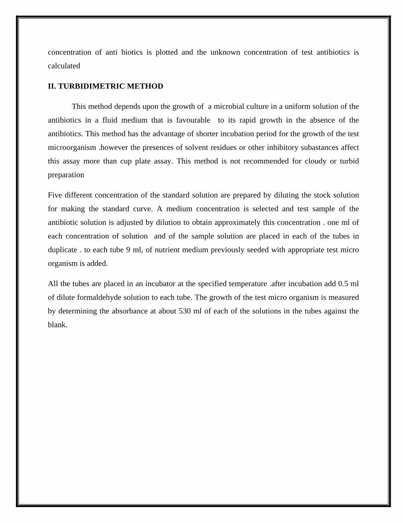
1. The positive result is indicated by the red layer at the top of the tube after the addition of
Kovács reagent.
2. A negative result is indicated by the lack of color change at the top of the tube after the
addition of Kovács reagent.
Indole Test Results: Positive-development of Red-ring
Methyl red test and Voges-Proskauer test both are done in methyl red–Voges-Proskauer (MR-
VP) broth, but the reagents that are added varies according to the test
Methyl Red (MR)Test:
Positive methyl red test are indicated by the development of red color after the addition of
methyl red reagent.
A negative methyl red test is indicated by no color change after the addition of methyl red
reagent
Voges-Proskauer (VP) test:
1. Negative test is indicated by lack of color change after the addition of Barritt’s A and
Barritt’s B reagents.
2. A positive Voges-Proskauer test is indicated by the development of red-brown color after
the addition of Barritt’s A and Barritt’s B reagents.
Citrate utilization test
The test is performed on Simmons citrate agar:
1. Negative citrate utilization test is indicated by the lack of growth and color change in the
tube
2. A positive citrate result as indicated by growth and a blue color change.
IMViC Test results of Some Genera of Enterobacteriaceae:
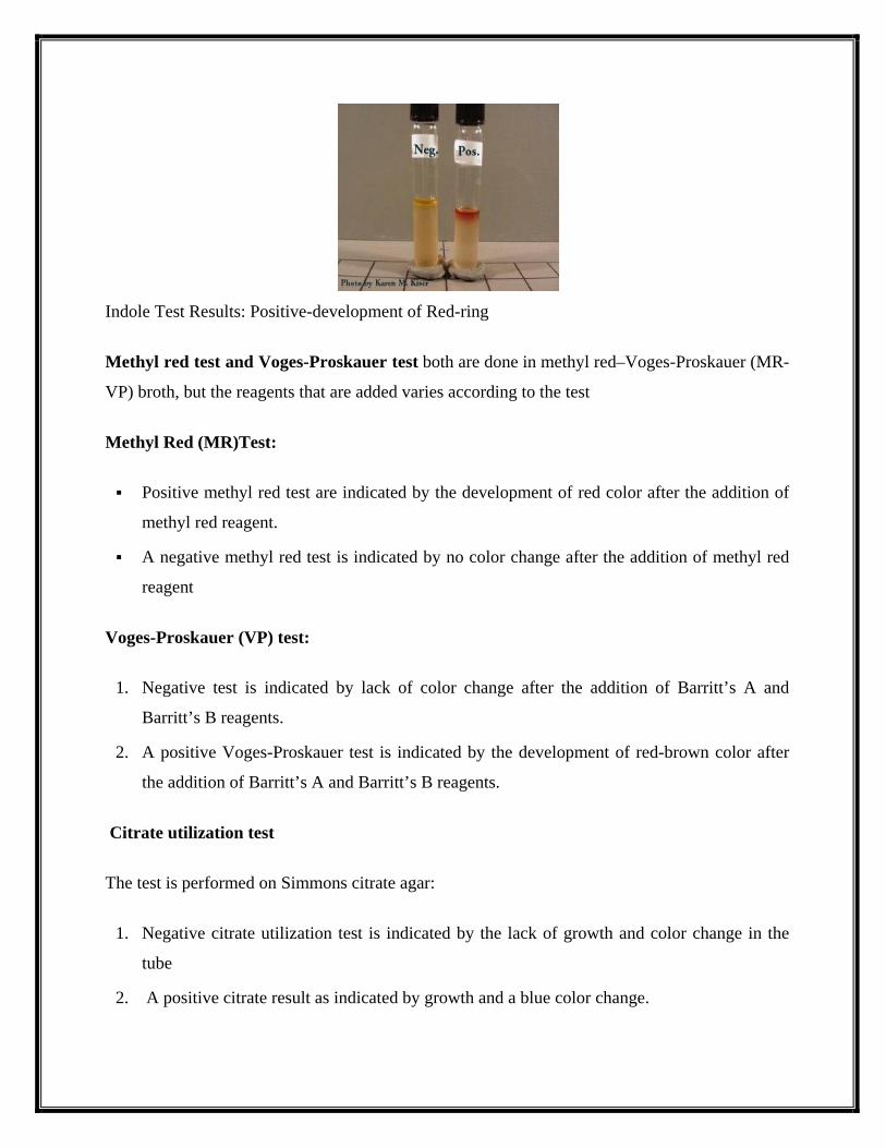
1. IMViC tests of Escherichia coli
1. Indole: Positive
2. Methyl-Red: Positive
3. Voges-Proskauer test: Negative
4. Citrate test: Negative
IMViC Test of Ecoli: ++– (Photo source and copyright: ASM)
2. IMViC tests of Enterobacter aerogenes 1. Indole: Negative 2. Methyl-Red: Negative 3. Voges-Proskauer test: Positive 4. Citrate test: Positive
3. IMViC tests of Proteus vulgaris 1. Indole: Positive 2. Methyl-Red: Positive 3. Voges-Proskauer test: Negative 4. Citrate test: Negative
4. IMViC tests of Citrobacter freundii 1. Indole: Negative 2. Methyl-Red: Positive 3. Voges-Proskauer test: Negative 4. Citrate test: Positive