practical genetic counseling 7th ed
TRANSCRIPT


PRACTICAL
GENETICCOUNSELLING

This page intentionally left blank

PRACTICAL
GENETICCOUNSELLING
SEVENTH EDITION
Peter S. HarperUniversity Research Professor (Emeritus) in Human Genetics
Institute of Medical GeneticsCardiff University
UK

First published in Great Britain in 1981 by John Wright LtdReprinted 1982 Second edition 1984Third edition 1988Reprinted 1991Fourth edition 1993 by Butterworth Heinemann LtdFifth edition 1998Reprinted in 2001 by ArnoldSixth edition 2004This seventh edition published in 2010 by Hodder Arnold, an imprint of Hodder Education, an Hachette UK Company, 338 Euston Road, London NW1 3BH
http://www.hodderarnold.com
© 2010 Edward Arnold (Publishers) Ltd
All rights reserved. Apart from any use permitted under UK copyright law, this publication may only be reproduced, stored or transmitted, in any form, or by any means with prior permission in writing of the publishers or in the case of reprographic production in accordance with the terms of licences issued by the Copyright Licensing Agency. In the United Kingdom such licences are issued by the Copyright Licensing Agency: Saffron House, 6–10 Kirby Street, London EC1N 8TS.
Hachette UK’s policy is to use papers that are natural, renewable and recyclable products and made from wood grown in sustainable forests. The logging and manufacturing processes are expected to conform to the environmental regulations of the country of origin.
Whilst the advice and information in this book are believed to be true and accurate at the date of going to press, neither the author[s] nor the publisher can accept any legal responsibility or liability for any errors or omissions that may be made. In particular (but without limiting the generality of the preceding disclaimer) every effort has been made to check drug dosages; however it is still possible that errors have been missed. Furthermore, dosage schedules are constantly being revised and new side-effects recognized. For these reasons the reader is strongly urged to consult the drug companies’ printed instructions before administer-ing any of the drugs recommended in this book.
British Library Cataloguing in Publication DataA catalogue record for this book is available from the British Library
Library of Congress Cataloging-in-Publication DataA catalog record for this book is available from the Library of Congress
ISBN-13 978 0 340 990 698
1 2 3 4 5 6 7 8 9 10
Commissioning Editor: Caroline MakepeaceProject Editor: Sarah PennyProduction Controller: Kate HarrisCover Designer: Lynda King
Typeset in 10/13 pt Minion by MPS Limited, A Macmillan Company, Chennai, IndiaPrinted and bound in the UK by CPI Antony Rowe Ltd
What do you think about this book? Or any other Hodder Arnold title? Please visit our website: www.hodderarnold.com

ContentsPreface viiAcknowledgements viii
PART I General Aspects of Genetic Counselling 1. Genetic Counselling: an Introduction 3 2. Genetic Counselling in Mendelian Disorders 23 3. Common Disorders and Genetic Counselling 54 4. Chromosome Abnormalities 63 5. Molecular Genetics and Genetic Counselling 81 6. Dysmorphology and Genetic Syndromes 91 7. Carrier Testing and Genetic Prediction 102 8. Prenatal Diagnosis and Reproductive Aspects of Medical Genetics 115 9. Special Issues in Genetic Counselling 135 10. The Genetic Counselling Clinic 150
Part II Genetic Counselling: Specific Organ Systems 11. Neuromuscular Disorders 167 12. Central Nervous System Disorders 179 13. Disorders of Mental Function 201 14. Disorders of Bone and Connective Tissue 216 15. Oral and Craniofacial Disorders 234 16. The Skin 242 17. The Eye 253 18. Deafness 265 19. Cardiovascular and Respiratory Disorders 271 20. The Gastrointestinal Tract 287 21. Renal and Urinary Tract Disease 297 22. Endocrine and Reproductive Disorders 305 23. Inborn Errors of Metabolism 317 24. Disorders of Blood and Immune Function 325 25. Genetic Risks in Cancer 334 26. Environmental Hazards 348
Part III Genetic Counselling: the Wider Picture 27. Population Aspects of Genetic Counselling and Genetic Screening 361 28. Genetics and Society 375
Appendix: Useful Information in Connection with Genetic Counselling 385Glossary 389Index 395

ToElaineand to
MatthewEmma Jane
NicholasKaty Thi
Lucy and
Osiris
PrefaceThis seventh edition of Practical Genetic Counselling is also the final one, at least in its present form. Most of the book’s value over the years has come from it reflecting the experience of someone directly involved in genetic counselling and its problems. Since I am no longer in active clinical practice, it seems best to let this final edition stand for what I consider to be the main foundations of present-day genetic counselling, rather than attempt myself to continue it into the future.
It is now 30 years since I wrote the first edition of the book, and more than 40 years since I began to practice Medical Genetics. During this time it has been truly amazing to see what has altered in terms of what we are able to do. Looking back, it seems almost unbelievable that when I started in the field there was no prenatal diagnosis, almost no carrier detection, let alone presymptomatic testing, while human molecular genetics was non-existent, as were its practical applications of mutation detection and linked DNA markers.
At another level, though, much has remained unchanged. The problems and ques-tions that families bring to us are largely the same, and so are the main approaches used in genetic counselling to try to help them. New laboratory advances have greatly extended what we are able to do, but have not removed the need for the practice of genetic counselling, and of clinical genetics generally, to rest on these long established foundations, and on sound psychological principles for the way in which we interact with those we see.
I wrote the original edition of this book primarily for those who were not experts or specialists in the field, and have been surprised to find that those working in Medical Genetics have also found it useful, particularly trainees and, increasingly, non-medical genetic counsellors. I suspect that this is because the book is short and written in simple language, and has never attempted to provide the details that can be found in larger volumes.
One final aspect that has brought me particular pleasure has been the book’s wide use, often in translation, across the world, including countries where previously genetic counselling was non-existent or very different in its approaches. I have often felt humbled by the appreciation that people from such countries have shown to me and I hope that, in turn, Practical Genetic Counselling has made a contribution to forging even closer links between members of the international community of workers in the field.
Peter Harper, Cardiff, February 2010

AcknowledgementsAs with previous editions, I owe a great debt to my Cardiff colleagues, who have provided suggestions and comments and especially to Angus Clarke, who has contrib-uted greatly to the ‘Further reading’ given at the end of each chapter. Selwyn Roberts has again helped with cytogenetic advice and Buddug Williams with web information.
A number of people have sent me helpful suggestions and corrections and I feel par-ticularly honoured to have received detailed comments from Dr F. Clarke Fraser, one of the pioneers of Medical Genetics.
The publishers, Hodder Arnold, have been unfailingly helpful and efficient in seeing this volume through to its final form, as has Oxford University Press in the United States, while I am most grateful to Joanne Richards for her help in reorganizing the text and preparing it for publication.

General Aspectsof Genetic Counselling
PART I

This page intentionally left blank

What do we mean by ‘genetic counselling’? . . . . . . . . . . . . . . . . . . . . . . . . . . . . . . . . . . . . . . . 3
The history and development of genetic counselling . . . . . . . . . . . . . . . . . . . . . . . . . . . . . . 4
Constructing a family tree . . . . . . . . . . . . . . . . . . . . . . . . . . . . . . . . . . . . . . . . . . . . . . . . . . . . . . . . 5
Diagnostic information . . . . . . . . . . . . . . . . . . . . . . . . . . . . . . . . . . . . . . . . . . . . . . . . . . . . . . . . . . . 8
Genetic risk estimation . . . . . . . . . . . . . . . . . . . . . . . . . . . . . . . . . . . . . . . . . . . . . . . . . . . . . . . . . . 10
The basis of risk estimation . . . . . . . . . . . . . . . . . . . . . . . . . . . . . . . . . . . . . . . . . . . . . . . . . . . . . . . 13
Communication and genetic counselling . . . . . . . . . . . . . . . . . . . . . . . . . . . . . . . . . . . . . . . . . 16
The back-up to genetic counselling . . . . . . . . . . . . . . . . . . . . . . . . . . . . . . . . . . . . . . . . . . . . . . 18
Support in the context of genetic counselling . . . . . . . . . . . . . . . . . . . . . . . . . . . . . . . . . . . . 20
Further reading . . . . . . . . . . . . . . . . . . . . . . . . . . . . . . . . . . . . . . . . . . . . . . . . . . . . . . . . . . . . . . . . . 20
Genetic Counselling: an Introduction
CHAPTER 1
WHAT DO WE MEAN BY ‘GENETIC COUNSELLING’?
Although most people working in the field of medicine are familiar with the term ‘genetic counselling’ and have some idea what it means, it is surprisingly rare to see it actually defined. Closer enquiry among patients and colleagues shows a wide variation in people’s concepts of what the process of genetic counselling actually entails. Some envisage an essentially supportive – even psychotherapeutic – role, akin to that of coun-selling processes in the social field; others see genetic counselling as primarily concerned with special diagnostic tests in inherited disease; yet others regard it as a complex mathematical process involving the estimation of risk.
All these views of genetic counselling contain a considerable element of truth, but none fully identifies what the overall process of genetic counselling actually involves. Even within the group of professionals for whom genetic counselling is a major activity, there are varied opinions as to its proper role and scope, but in essence it is a composite activity, made up of a series of key elements that individually are very different, but which together constitute a process that is highly distinctive in its character and its ethos.
Previous editions of this book have given various definitions of genetic counselling, but all of them are cumbersome and unsatisfactory. It is more meaningful to list here the principal elements of genetic counselling, and these are given in Box 1.1.
This chapter outlines these main elements, which are then dealt with in more detail in subsequent chapters of the book. It is the satisfactory synthesis of these various aspects that makes up genetic counselling as a specific process. A thoughtful and valuable dis-cussion of the process of genetic counselling is given by Clarke (see ‘Further reading’).

44 Practical Genetic Counselling
Box 1.1 Genetic counselling: the main elements
Diagnostic and clinical aspectsDocumentation of family and pedigree informationRecognition of inheritance patterns and risk estimationCommunication and empathy with those seenInformation on available options and further measuresSupport in decision-making and for decisions made
THE HISTORY AND DEVELOPMENT OF GENETIC COUNSELLING
The origins of genetic counselling need to be seen within the context of the overall history of human genetics, a topic until recently neglected, but which is given by McKusick’s introductory chapter for Emery and Rimoin’s textbook and more recently in my own Short History of Medical Genetics (see ‘Further Reading’).
Although human genetics research had begun to develop strongly in the first half of the twentieth century, its application at that time was confused and, to an extent, dis-credited by the abuses of eugenics. It was not until the Second World War that the first genetic counselling clinics were opened in America, in Michigan in 1940 and in Minnesota in 1941. In the UK, the Hospital for Sick Children in Great Ormond Street, London, developed the first such clinic in 1946. By 1955 there were over a dozen centres in North America and there has been a steady development since that time. As with many pioneering developments, the early centres were often the work of far-sighted eccentrics. Sheldon Reed, in his book Counselling in Medical Genetics, first published in 1955, gives a delightful description of Edward Dight, responsible for endowing the Dight Clinic in Minneapolis, who lived in a house built in a tree and who failed to file income tax returns. Reed also wrote a brief historical article on genetic counselling.
Reed’s book gives a vivid picture of the main areas covered in the early years of genetic counselling, and it was Reed himself who first introduced the term. Many of the problems are unchanged today and his examples of individual cases show that the fears and concerns of families have altered little. In other respects, there have been profound changes in the 50 years since the book was written. Carrier detection was rarely possible and prenatal diagnosis entirely non-existent, as was oral contraception, so the options open to patients at risk were limited; either they took the risk or they did not. An even more important change has been that of the general climate of opinion among the public and the medical profession, in particular a greater openness in relation to family disorders.
Reed’s case histories illustrate the background of ignorance and prejudice with which his patients had to cope, and it is no wonder that he found them grateful, even when he could only give them pessimistic advice. He comes across as a caring and sensitive person, upholding the concept of non-directiveness and turning his back completely on eugenics.
It is of interest that the most common cause of referral to the Dight Clinic was regard-ing skin colour and whether a child for adoption would ‘pass for white’. Several other problems among the 20 most common causes for referral listed by Reed are infrequently encountered today, including eye colour, twinning and rhesus haemolytic disease.

55Genetic Counselling: an Introduction
The last of these provides a real example of advance in treatment and prevention; the others reflect changes in social attitudes. Many others of Reed’s most common problems remain equally important today, including mental handicap, schizophrenia, facial clefting, neural tube defects and Huntington’s disease.
Most of the early genetic counselling clinics were run by non-medical scientists (like Reed himself) or by those who were not experienced clinicians. With the growth in knowl-edge of genetic disorders and the appearance of medical genetics as a distinct specialty in the 1960s, genetic counselling progressively became medicalized, representing one of the key components of clinical genetics. It was not, though, until later that the importance of a firm psychological basis was recognized and became an essential part of genetic counselling, the writings of Seymour Kessler making a particular contribution to this.
From around 1970, beginning in America, non-medical genetic counsellors with a specific training in the field have become increasingly prominent, the graduate course based at Sarah Lawrence College, New York, becoming the model for other centres in America and Britain. As the demand for genetic counselling has grown, it has become clear that not all consultations require the clinical expertise of the medical geneticist, though careful coordination and mutual respect of the two groups are essential for an optimal genetic counselling service.
At the time when I wrote the first edition of this book, 30 years ago, I had in mind the general hospital clinician or family doctor as the main likely reader and the main provider of genetic counselling, at least for relatively straightforward situations. It has been inter-esting that only a few general clinicans have developed such a role; this is perhaps in part because of lack of time, the most precious commodity for good genetic counselling. Also it may reflect the fact that many clinicians wish to spend most of their time seeing and managing sick patients, whereas much of genetic counselling involves the problems of entirely healthy relatives. As genetics progressively spreads out in its applications beyond specialist centres, there is a growing need for clinicians in different medical fields to engage actively with the genetic counselling needs of those whom they see and also to link more closely with their local medical genetics and genetic counselling centre.
CONSTRUCTING A FAMILY TREE
Collecting genetic information is the first and most important step in genetic counsel-ling, and is best achieved by drawing up a family tree or pedigree. The use of clear and consistent symbols allows genetic information to be set out much more clearly than does a long list of relatives.
Drawing a satisfactory pedigree is not difficult, although it is remarkable how rarely those clinicians without an interest in genetics will attempt the process. A clearly drawn pedigree has a certain aesthetic appeal, but its chief value is to provide an unambiguous and permanent record of the genetic information in a particular family. Although computer programs exist, they are no substitute, in the author’s view, for a clearly drawn pedigree constructed by hand at the time of the interview. A recent publication from the UK Genetics Education Centre gives a clear and helpful guide to drawing a pedigree, largely similar to the account given here.

66 Practical Genetic Counselling
Figure 1.1 shows the main symbols used in constructing pedigrees. The symbols shown for the sexes (�, �) are preferred to the alternatives ( , ), which tend to be confused at a distance. Heterozygous carriers can be denoted by half-shaded symbols or, in the case of an X-linked disorder, by a central dot. Although the sign for an early abortion (spontaneous or induced) can also be used for a stillbirth, it is preferable to denote the sex of the latter with an appropriate symbol and indicate beneath the symbol that it was a stillbirth. The previous use of a broken line for an offspring from outside marriage is no longer appropriate. ‘Illegitimacy’ is no longer a meaningful concept in most Western societies, but employing a broken line in a pedigree is still useful to repre-sent the situation where parentage is unknown or unacknowledged.
The proband – also called the propositus (male) or proposita (female) – should be clearly indicated with an arrow. The proband is the individual through whom the family is ascertained. Large families will commonly have several probands. The proband is generally an affected individual, but the person primarily seeking advice may well not be affected. The term ‘consultand’ is conveniently used for this individual.
Multiple marriages and complex consanguinity can cause problems in constructing a pedigree, and artistry will have to be sacrificed for accuracy in such cases. It is usually
3
Male, female (unaffected)
Sex unknown
Affected male and female
Three unaffected males
Examined personally
Deceased (and affected)
Individual without offspring
Consanguineous marriage
Offspring with parentage unacknowledged or different from expected
Abortion (spontaneous or induced)
Twins
Monozygotic twins
Heterozygote (autosomal recessive)
Heterozygote (X-linked)
Propositus
Figure 1.1 Symbols used in drawing a pedigree.

77Genetic Counselling: an Introduction
wise to start near the middle of the pedigree sheet and to leave more room than one thinks will be needed, so that particularly prolific family branches do not become crowded out. Figure 1.2 shows examples of the ‘working pedigree’, one simple and one more complex.
The following practical points deserve emphasis.
• Enquire specifically about infant deaths, stillbirths and abortions. These may be highly relevant, especially if structural abnormalities prove to have been present; the fact that the information had not been volunteered may be significant. Thus two children ‘lost at birth’ by the mother of a woman seen for counselling proved to have both had spina bifida, a fact that considerably altered the risks.
Figure 1.2 Two examples of the ‘working pedigree’. These two pedigrees – one simple, the other more extensive – show how family data can be easily and clearly recorded at the time of the interview. A simple lined sheet is used; more detailed information on individuals can be recorded at the foot of the pedigree or on the back. Identifying details have been deleted.

88 Practical Genetic Counselling
• Consanguinity should be directly asked about and may be the clue that suggests autosomal recessive inheritance (see Chapter 9). Equally, though, the background level for consanguinity in the general population concerned must be taken into account before attributing the problem to consanguinity.
• Mistaken or unacknowledged paternity must be borne in mind, especially in a puzzling situation. A family doctor or nurse, particularly in a small community, may well be able to clarify this possibility, but increasingly families are more open about it, recognizing its importance in the context of genetic risk. Definitive tests of paternity based on DNA (see Chapter 5) can help to resolve these problems more easily, but DNA-based diagnostic tests may equally produce new difficulties by the detection of unsuspected non-paternity.
• Always take at least basic details about both sides of the family, even in a dominantly inherited disorder clearly originating from one side. Unexpected findings may emerge. The family that insists that there is ‘nothing on our side’ should be regarded with suspicion until this is verified. Taking details about both sides may also help to avoid feelings of guilt or blame resting exclusively on one member of a couple, always an important factor, but particularly in some cultural and social situations.
• Record dates of birth where possible rather than ages. Note the date when the pedigree was drawn up.
• Record maiden names of women. This is especially significant for X-linked disorders, where the surname of affected members is likely to change with each generation.
• Note the addresses of key relevant members, though this is best done on a separate sheet. This may prove invaluable in obtaining hospital records or in later contact with relatives.
Most of the above points are obvious, yet it is surprising how often vital information is not obtained unless a systematic approach is used.
In constructing a pedigree, it is not generally necessary to trace a person’s ancestry back more than three or four generations; medical details often become inaccurate at this early period. Sometimes, though, it may be important to link kindreds or to establish a common ancestor, in which case genealogical records will be useful. These are surprisingly abundant in many European countries, especially Scandinavia. In the UK, a useful guide to the different sources has been produced (see Bevan and Duncan, ‘Further reading’). Even in mobile populations such as in America, the growth in interest in family history has considerably increased people’s knowledge of their ancestors.
DIAGNOSTIC INFORMATION
It has already been emphasized that a clear diagnosis is the essential basis for accurate genetic counselling. Unfortunately, this basis is all too often a shaky one, and one of the principal tasks of anyone involved in genetic counselling is to ensure that it is made as

99Genetic Counselling: an Introduction
firm as possible before risk estimates are given to those seeking advice. Common reasons for lack of a clear diagnosis include the following.
• The affected individual may have lived a considerable time ago, when relevant diagnostic investigations were not available. There is little that can be done about this, but it is surprising how much detailed information may be obtained by questioning close relatives who were involved in caring for the patient. Even if an exact diagnosis cannot be established, it may be possible to exclude a disorder. Thus a man with muscular dystrophy who lived to the age of 40 years clearly would not have had the Duchenne type. Old photographs may show typical facial features of a condition (e.g. myotonic dystrophy).
• The affected individual may have died without essential investigations having been done, or without autopsy being performed. This is all too often the case and is inexcusable. Reasons usually offered are reluctance to trouble the parents in distressing circumstances, or the fact that investigations would not have altered the patient’s management; but frequently the real reason is that those involved have not taken the trouble to undertake the studies, or to make arrangements with those who can undertake them. The recent sharp reduction in the frequency of autopsy has made the situation worse. The tragic consequences of such inertia only become apparent when the question of risk to further family members arises.
• A firm diagnosis cannot be reached, even with the affected individual living. This is inevitable in some cases, since our knowledge of many genetic disorders remains very incomplete, but a considerable degree of help can be obtained by enlisting the efforts of colleagues, even at a distance. Photographs, radiographs and samples of urine, blood, DNA and cultured skin fibroblasts can all be sent to distant parts of the world for experts to study, and developments in ‘telemedicine’ now extend greatly the scope of what can be done in this way, especially for imaging. Presentation of puzzling cases at clinical meetings, notably national or international groups for malformation syndromes and bone dysplasias, may often result in a diagnosis being provided. Even if it does not, one can feel happier that one is not overlooking a recognizable disorder if one has sought the advice of those most likely to know, and families also appreciate these efforts. Wherever possible, one should store (with consent) appropriate samples for future biochemical or DNA analysis.
• The diagnosis may be wrong. This is a much more dangerous situation than when the diagnosis is uncertain, as it may lead to false confidence. It is extremely difficult to know how far to rely on other people’s diagnoses and how far to insist on confirming them oneself. Clearly, neither a medical geneticist nor any other clinician can be an expert diagnostician in every speciality, and one will frequently have to rely on colleagues’ advice. Nevertheless, it is essential for all clinicians involved in genetic counselling to have a wide range of diagnostic ability, to know their limitations – and those of their colleagues – and to develop a healthy scepticism in diagnostic matters and a sensitivity for where error may lie. For non-medical genetic counsellors, it is essential to work in the closest cooperation with clinical geneticists if major problems are to be avoided.
1010 Practical Genetic Counselling
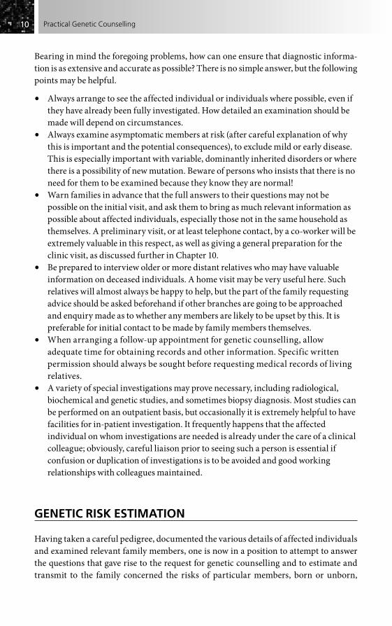
Bearing in mind the foregoing problems, how can one ensure that diagnostic informa-tion is as extensive and accurate as possible? There is no simple answer, but the following points may be helpful.
• Always arrange to see the affected individual or individuals where possible, even if they have already been fully investigated. How detailed an examination should be made will depend on circumstances.
• Always examine asymptomatic members at risk (after careful explanation of why this is important and the potential consequences), to exclude mild or early disease. This is especially important with variable, dominantly inherited disorders or where there is a possibility of new mutation. Beware of persons who insists that there is no need for them to be examined because they know they are normal!
• Warn families in advance that the full answers to their questions may not be possible on the initial visit, and ask them to bring as much relevant information as possible about affected individuals, especially those not in the same household as themselves. A preliminary visit, or at least telephone contact, by a co-worker will be extremely valuable in this respect, as well as giving a general preparation for the clinic visit, as discussed further in Chapter 10.
• Be prepared to interview older or more distant relatives who may have valuable information on deceased individuals. A home visit may be very useful here. Such relatives will almost always be happy to help, but the part of the family requesting advice should be asked beforehand if other branches are going to be approached and enquiry made as to whether any members are likely to be upset by this. It is preferable for initial contact to be made by family members themselves.
• When arranging a follow-up appointment for genetic counselling, allow adequate time for obtaining records and other information. Specific written permission should always be sought before requesting medical records of living relatives.
• A variety of special investigations may prove necessary, including radiological, biochemical and genetic studies, and sometimes biopsy diagnosis. Most studies can be performed on an outpatient basis, but occasionally it is extremely helpful to have facilities for in-patient investigation. It frequently happens that the affected individual on whom investigations are needed is already under the care of a clinical colleague; obviously, careful liaison prior to seeing such a person is essential if confusion or duplication of investigations is to be avoided and good working relationships with colleagues maintained.
GENETIC RISK ESTIMATION
Having taken a careful pedigree, documented the various details of affected individuals and examined relevant family members, one is now in a position to attempt to answer the questions that gave rise to the request for genetic counselling and to estimate and transmit to the family concerned the risks of particular members, born or unborn,

1111Genetic Counselling: an Introduction
developing the particular disorder. The fact that the process of recording information will probably have taken a considerable time is in some ways an advantage, particularly if the family is not under one’s regular care but is being seen specifically for genetic counselling. From the way in which information is given (or not given) and from the reaction to questions, much can be learned about the general attitude of the individuals being counselled to the family disorder.
• Did they themselves initiate the request for genetic counselling, or did someone else?
• Is there an unspoken and perhaps exaggerated fear of the disorder?• Do feelings of guilt or hostility exist between parents?• Is the rest of the family supportive, or are there tensions between the generations?• Is an affected child valued and loved, or regarded as a burden?
Much information on these and other important issues can be obtained by a person who is sensitive and observant, without the need for direct questioning.
It is also possible during this preliminary stage to assess the way in which informa-tion is to be most suitably transmitted. Some couples will be unable to grasp more than the simplest concepts of ‘high risk’ or ‘low risk’, while others will require a precise risk figure and even a detailed explanation of the mode of inheritance. In my own experi-ence I have noted that it is common for those undertaking genetic counselling to overestimate the level of complexity and amount of information that can be absorbed in a consultation.
Information on genetic risks is rarely an absolute ‘yes’ or ‘no’, and in medical genetics, more perhaps than in any other branch of medicine, one thinks and works almost entirely in terms of probabilities or odds. Colleagues in other specialties fre-quently find this unsatisfactory, preferring to accept only a ‘definite’ conclusion. Yet when examined closely, there is often as much, if not more, uncertainty in the appar-ently ‘definite’ specialities than there is in medical genetics. Thus the chance that a definitely inflamed appendix will be found at appendicectomy is far from 100 per cent, while the entire process of clinical diagnosis is based on the combination of numerous pieces of information, each with a degree of uncertainty, although this is often unappre-ciated by those involved. The same applies to the ‘normal ranges’ of most laboratory investigations. It is perhaps only because uncertainty is well recognized in genetics that methods of measuring it and defining its limits have been generally used, as exempli-fied in genetic counselling. More recently, other areas of medicine have started to use these approaches.
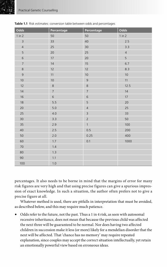
Risk figures in genetic counselling may be given either as odds or as percent-ages. Some people prefer to use odds and to quote risks as 1 in 10, 1 in 50, 1 in 100, etc. Others prefer to use such figures as 10 per cent, 2 per cent, 1 per cent. The author admits to inconsistency in this, both in practice and in this book, and for this reason, and because others are equally inconsistent, a selection of conversions is given (Table 1.1), which should allow ready exchange between the two approaches. It is often necessary to adapt whichever is used to a particular situation, for some people simply do not understand odds, while others are more confused with
1212 Practical Genetic Counselling
percentages. It also needs to be borne in mind that the margins of error for many risk figures are very high and that using precise figures can give a spurious impres-sion of exact knowledge. In such a situation, the author often prefers not to give a precise figure at all.
Whatever method is used, there are pitfalls in interpretation that must be avoided, as described below, and this may require much patience.
• Odds refer to the future, not the past. Thus a 1 in 4 risk, as seen with autosomal recessive inheritance, does not mean that because the previous child was affected the next three will be guaranteed to be normal. Nor does having two affected children in succession make it less (or more) likely for a mendelian disorder that the next will be affected. That ‘chance has no memory’ may require repeated explanation, since couples may accept the correct situation intellectually, yet retain an emotionally powerful view based on erroneous ideas.
Table 1.1 Risk estimates: conversion table between odds and percentages
Odds Percentage Percentage Odds
1 in 2 50 50 1 in 2
3 33 40 2.5
4 25 30 3.3
5 20 25 4
6 17 20 5
7 14 15 6.7
8 12 12 8.3
9 11 10 10
10 10 9 11
12 8 8 12.5
14 7 7 14
16 6 6 17
18 5.5 5 20
20 5.0 4 25
25 4.0 3 33
30 3.3 2 50
35 2.9 1 100
40 2.5 0.5 200
50 2.0 0.25 400
60 1.7 0.1 1000
70 1.4
80 1.3
90 1.1
100 1.0

1313Genetic Counselling: an Introduction
• It is embarrassingly easy for odds to be reversed. Thus a patient seen by the author with one spina bifida child, having been correctly advised by her obstetrician that there was a 1 in 20 recurrence risk, came seeking termination of her next pregnancy because she considered that ‘a chance of 1 in 20 of a normal child was far too low’.
• Odds of 1 in 2 (1/2) are not the same as 1 to 2 (1/3). This may be misinterpreted by those used to betting. Fortunately, the difference is only considerable for the highest risks.
• Many people do not have a clear idea of what constitutes a high or low risk. Thus some couples who are given a low risk (e.g. 1 in 200) express the view that this is far too high to be acceptable, whereas others seen by the author have been greatly relieved by a risk of 50 per cent. Clearly the nature and severity of the disorder will determine what risk is acceptable, but it is helpful to be able to give some kind of reference point for comparison, such as the fact that one child in 50 in the population is born with a significant disability, or that the population frequency of the disorder in question is, say, 1 in 2000. Some useful data of this type are summarized in Box 1.2.
THE BASIS OF RISK ESTIMATION
The ways in which risks can be estimated and the results of these estimates form the basis of this book and are considered in detail in later chapters. It is important from the outset, though, to recognize that not all risk estimates are of the same type. They may be based on different sorts of information and may be of greater or lesser reliability. The main categories discussed below can be recognized.
Empirical risks
Here the estimate is based on observed data rather than theoretical predictions (Fig. 1.3; see also Chapter 3). This is the form of risk estimate available for most of the more common non-mendelian or chromosomal disorders. The information is usually reliable provided it has been collected in an unbiased manner (often not easy), and provided the population from which the individual receiving genetic counselling comes is compara-ble to the one on which the data were established. Sadly, there is a dearth of recent empirical risk studies and the use of older ones is often made more complex as the result
Box 1.2 Risk of abnormalities in the ‘normal’ population (approximate)
Risk of a child being born with some congenital abnormality 1 in 30Risk of child being born with a serious physical or mental handicap 1 in 50Risk of a pregnancy ending in a spontaneous abortion 1 in 8Risk of perinatal death* 1 in 120Risk of a child dying in the first year of life after the first week* 1 in 300Risk that a couple will be infertile 1 in 10
*Figure for ‘developed’ countries; there is great geographical variation.

1414 Practical Genetic Counselling
of changes in classification following recognition of genetic heterogeneity or identifica-tion of specific genes, as well as by genuine biological changes in disease frequency (e.g. neural tube defects).
Mendelian risks
Mendelian risk estimates can be given only when a clear basis of single gene inheritance can be recognized for a disorder (Fig. 1.4; see also Chapter 2). They are perhaps the most satisfactory form of risk estimate because they commonly allow a clear differentiation into categories of negligible risk (e.g. offspring of healthy sibs in a rare autosomal reces-sive disorder) and high risk (e.g. offspring of an individual affected with an autosomal dominant disorder). It must be remembered that it is the genotype that follows mende-lian inheritance, which will not necessarily give the same risk for the actual disease phenotype. There often remains the problem of achieving greater certainty in the indi-vidual at high risk (e.g. a person at 50 per cent risk of developing Huntington’s disease), and information from the next two risk categories may be helpful in this situation.
Modified genetic risks
Non-geneticists may find modified genetic risk estimates (Fig. 1.5) difficult to use ini-tially; they are particularly applicable in X-linked recessive inheritance, where fully
?
Figure 1.3 Empirical risk estimate: one child is affected with spina bifida. The risk of a subsequent child being affected by a neural tube defect is around 3 per cent in an area of high risk (e.g. South Wales) and with no other affected family members. The risk estimate would be different in an area of low incidence and would be altered by the presence of other affected relatives. It has also changed with time, being less than in earlier surveys decades ago.
Figure 1.4 Mendelian risk estimate: a family with myotonic dystrophy (an autosomal dominant disorder). The risk for the offspring of affected individuals is 50 per cent regardless of the incidence of the disorder and the number of affected individuals in the family. See Chapter 11 for the relevance of genetic instability to this disorder.

1515Genetic Counselling: an Introduction
worked-out examples are given (see Chapter 2). The essential feature is that a ‘prior’ genetic risk, based usually on mendelian inheritance, may be modified by ‘conditional’ information, usually genetic, but sometimes from other sources. Thus the modified risk of a man developing Huntington’s disease, whose grandparent was affected, is not the same as the prior risk of 1 in 4, but is reduced by his own age and by the fact that the intervening parent was unaffected. It may also be reduced by the number of unaffected sibs, if these have reached an advanced age. Such modifying information may drasti-cally alter the risk estimate and should always be used when available, especially if presymptomatic or prenatal genetic tests are being considered, where it may have a con-siderable influence on decisions.
Risk estimates from independent evidence
Where special investigations can be utilized, these may greatly alter the risk estimates. Thus a normal serum immunoreactive trypsin test in an infant having a sib with cystic fibrosis will considerably reduce the chance of the disorder being present.
Carrier detection in such disorders as haemophilia and Duchenne muscular dystro-phy provides comparable information, as do linked DNA markers in many mendelian disorders. However, a strong caution must be given here: the results of these investiga-tions are rarely so clear-cut that they can be used in isolation; they require combination with the prior genetic risk, along with other modifying information. Failure to appreci-ate this may lead to serious error, especially when investigations are being applied as screening procedures in situations of low prior risk. Thus, in the example given of cystic fibrosis, the final risk, and very possibly the actions taken, would depend very much on whether there was a close family history of cystic fibrosis or whether the abnormality resulted from a general population screening test.
Composite risks
Most empirical risks really fall into this category, but in some instances it is obvious that one is dealing with a mixed situation that cannot be satisfactorily resolved. Thus
Figure 1.5 Modified risk estimate: Duchenne muscular dystrophy. The grandmother of the individual seeking advice (consultand) is an obligatory carrier; prior risks of the mother and the consultand being carriers are thus 50 per cent and 25 per cent, respectively. These risks are, however, greatly reduced by the fact that the mother has had four healthy sons and no affected sons. See Chapter 2 for further details.

1616 Practical Genetic Counselling
isolated cases of a disorder such as osteogenesis imperfecta congenita are composed of a large number of cases representing new dominant mutations, with a minimal recur-rence risk to sibs, and a very small number of autosomal recessive cases, with a recurrence risk of 1 in 4. Because the two situations cannot always be reliably distin-guished, one ends up with an intermediate risk depending on the relative frequency of the two groups. Obviously this intermediate risk does not really exist at all – the family must represent one or other of the extreme positions. Such a composite risk estimate is an unsatisfactory one and should be regarded as a temporary measure.
With improved resolution of genetic heterogeneity, to which molecular analysis is now contributing, it may be possible to distinguish the individual components, while even within a single family, additional information may resolve the situation. Thus the birth of a further affected child in the example given of osteogenesis imperfecta would make it almost certain that autosomal recessive inheritance (or possibly gonadal mosai-cism) is operating in this family, with at least a 1 in 4 risk for further children.
The recognition of mendelian subsets within a disorder generally considered to be ‘multifactorial’ (e.g. congenital heart disease, breast cancer) is also proving important, as discussed in Chapter 3.
COMMUNICATION AND GENETIC COUNSELLING
No matter how well one may have confirmed a genetic diagnosis, utilized appropriate genetic tests and established an accurate risk estimate, all this will count for little if one cannot communicate satisfactorily with the family that one is seeing. The constant and unending variety provided by the interactions inherent in genetic counselling is one of the chief reasons why those involved find it such a rewarding activity, but even the most ‘natural’ communicators need training to optimize their skills, while a basic knowledge of the main theoretical aspects underpinning counselling skills is also of the greatest help. As indicated later in the chapter, these skills can also help to turn what may start out as an essentially information-giving interview into one that may be therapeutic in nature.
Genetic counselling and non-directiveness
It will have been noted that the emphasis so far has been placed on ensuring that a correct diagnosis and risk estimate have been reached and that those being counselled have correctly understood the situation. Nothing has been said about recommending a particular line of action or of advising couples against having children in high-risk situ-ations, and it may surprise some readers to learn that the author, in common with most professionals involved in genetic counselling, rarely if ever adopts a ‘directive’ approach. A survey of American genetic counselling centres has shown that a similarly non-directive approach is almost universal, although this has not always been the case in eastern Europe until recently, and may be difficult to apply in relation to some cultural and ethnic groups used to a more authoritarian situation.

1717Genetic Counselling: an Introduction
This may appear all the more surprising since many doctors with little experience of genetics do frequently give directive advice. Remarks such as ‘We were told not to have further children’ or ‘The doctors said I should have a termination’ are still commonly heard at genetic counselling clinics, and in many cases great distress has been caused to the couples involved, particularly because the advice has not been accompanied by an explanation of why it has been given or how great the risk really is.
The author’s view is that it is not the duty of a doctor or genetic counsellor to dictate the lives of others, but to ensure that individuals have the facts to enable them to make their own decisions. This includes not simply a knowledge of the genetic risks, but a clear appreciation of the consequences, long-term as well as short-term, that may result from a particular course of action. In any case, it seems likely (although not proven) that directive counselling may be counterproductive. Intelligent couples may resent being told what to do in a situation where they have already spent much troubled thought over the alternatives. Among the less privileged, there is often a strong resentment of being dictated to by authority and the author’s experience with Huntington’s disease suggests that some individuals in this situation may deliberately embark on a pregnancy as a gesture of defiance.
By contrast, some couples seen for genetic counselling will plead for direction. ‘What would you do if you were in my place?’ is a common question. It is tempting to give a clear direction in these circumstances, but frequently these are the very couples for whom this may be most inadvisable. Such a plea often indicates an unwillingness to face up to the consequences of a serious situation, or a significant disagreement between marriage partners, and for the physician to take on the responsibility that can only really be taken by the couple themselves may have serious long-term consequences.
It would be wrong to pretend that those engaged in genetic counselling never give directive advice. One’s own views are likely to be expressed in the way one approaches the subject, whether the more serious or the milder aspects of a disease have been stressed and whether one holds out the possibility of future treatment. Even the way a risk estimate is phrased can vary. For example, in the case of an autosomal recessive condition with a 1 in 4 recurrence risk, it is possible to make it appear quite encouraging if one states that there are three chances out of four that the child will be healthy! The type of society in which one lives and practises will also inevitably influence the way in which genetic counselling is given, and this is discussed further in the final chapter of this book. Clarke (see ‘Further reading’) points out clearly the limitations involved.
Since ‘non-directiveness’ has become a somewhat central tenet of genetic counsel-ling, it is important that it should not be used as an excuse for being vague or appearing detached, or for presenting so many apparent options that it becomes difficult for those seen to reach a clear decision. People will often need support for a tentative decision, as indicated later in this chapter; the importance of non-directiveness lies in allowing the decisions to be taken by the individuals involved, not by the person giving genetic counselling.
It is particularly important that couples realize that, in general, there is no ‘right’ or ‘wrong’ decision to be made, but that the decision should be the right one for their own particular situation. It is also important that those giving genetic counselling (and those
1818 Practical Genetic Counselling
evaluating genetic services) do not judge ‘success’ or ‘failure’ in terms of a particular outcome, and that they give support to families whatever their decisions may be.
Advice at a distance
The less one is able to verify a situation oneself, the greater is the possibility of error. However, the person who refuses to give any advice unless able to do everything person-ally is going to be of limited benefit to patients and colleagues. The author is in no doubt that one of the most valuable roles of a medical geneticist – and the same applies to any clinician with a particular interest in genetic counselling – is to act as a focal point and source of information for colleagues in a variety of specialties who need someone to turn to for advice. A high proportion of general enquiries from colleagues do not require actual referral; frequently, one is simply confirming what is already thought to be the case. In other instances, one may be able to advise that prenatal, molecular or other special investigations are available; in a small proportion of enquiries, however, the advice has to be that one cannot give a reliable opinion without seeing the patient oneself. One soon learns to recognize the small number of colleagues who attempt to use indirect or ‘casual’ advice as a substitute for a proper referral, as well as the enquiries ‘on behalf of a friend’ that can disguise a serious personal genetic problem requiring a full referral and thorough assessment.
Actual genetic counselling by post or other indirect means is an entirely different matter, and the author’s policy regarding enquiries from patients and relatives is to arrange a clinic appointment, via their family doctor wherever possible. The same policy applies to enquiries from health visitors, social workers and other paramedical personnel. Not only is there a serious risk that erroneous information may be given or risk figures misinterpreted; but without directly seeing those requesting advice, it is often impossible to decide what the real problem leading to their enquiry is and whether there are additional or underlying factors that have not been mentioned.
E-mail enquiries, coming directly from patients or family members, are increas-ingly frequent, particularly if one has expertise in a specific disorder or is closely involved with lay groups. In this case, one can usually help best by directing the enquirer to useful information sources, often web-based, or a local centre. Although it may be tempting to try to provide detailed help, especially if there seems to be no local facility, this is almost always unwise in the author’s opinion. By contrast, carefully organized and appropriately selected remote video-conferencing consultations may be of real value in difficult geographical situations.
THE BACK-UP TO GENETIC COUNSELLING
It has already been emphasized that genetic counselling does not simply consist of giving risk figures, and that it must often be preceded by a considerable diagnostic effort, in comparison with which the estimation of risks may be a relatively simple

1919Genetic Counselling: an Introduction
matter. Similarly, genetic counselling does not stop with the giving of risks, but must include a variety of other actions if it is to be fully effective.
In the first instance, it must be established as clearly as possible that the individuals counselled have really understood what they have been told. This includes not only the risk estimate, but the nature of the disorder and what other measures are available for prevention and treatment. It is often possible to get an approximate idea of how well information has been understood at the time of the interview, but it is well worthwhile, and often a salutary experience, to have this checked by an independent observer. A follow-up appointment may be useful both to check on this and to support the genetic counselling that has been given at the initial interview. For the same reason, it is impor-tant to provide a letter summarizing the main points of the consultation, including the risk estimates. A copy of the letter to the referring doctor may be appropriate in some cases, but it often contains too much detailed technical or clinical information; in general the author prefers to write a separate letter specifically to the individual or family seen.
Where information has been seriously misinterpreted or forgotten, this may be for various reasons. Some individuals have genuinely poor memories, while others may have been seen at an inappropriate time, such as soon after the death of a child; yet others may have come to the clinic encumbered with small and active children and been preoccu-pied in restraining their activities, rather than in listening to what has been said. Most commonly, one has probably not taken sufficient time and effort to ensure that the infor-mation has really been absorbed and it is important to be aware of one’s failures in this respect. The author has on several occasions seen couples who have acquired grossly erroneous ideas of risk and has wondered who could possibly have misinformed them so completely, only to find that it was he himself who had seen them some years previously!
An essential accompaniment to genetic counselling is that those being counselled should have full and accurate knowledge of the various other measures that may be available. In many cases, these require application as an integral part of the counselling – thus an assessment of the risk of a woman having a child affected by Duchenne muscular dystrophy or haemophilia is likely to be incomplete without carrier detection tests (see Chapter 7). In other cases, the risk may not be altered, but the consequences may be. Thus, where prenatal diagnosis is available (see Chapter 8), many couples will be prepared to embark on a high-risk pregnancy when they would not have considered doing so in the absence of such diagnostic possibilities. Similarly, the development of treatment fundamentally alters attitudes to genetic counselling. Most couples with a phenylketonuric child diagnosed in the newborn period and developing normally with treatment are happy to risk another affected child; where treatment is less satisfactory and the outcome less certain, the attitude may be very different.
Further ‘back-up’ measures that may be required are contraception and steriliza-tion, as well as the exploration of other possible options such as adoption, artificial insemination by donor, or ovum donation. These aspects are discussed later (see Chapter 9), but it cannot be too strongly emphasized that their consideration is an integral part of genetic counselling.

2020 Practical Genetic Counselling
SUPPORT IN THE CONTEXT OF GENETIC COUNSELLING
Many couples coming for genetic counselling require active support in one way or another. Sometimes the actual information given in genetic counselling may be of such grave consequence as to require support if serious problems are not to arise. Huntington’s disease (see Chapter 12) is perhaps the most striking example, but a severe depressive reaction is not uncommon in women who have recently lost a child after a chronic illness and who have to be told that the risk for other children is high. A sympa-thetic family doctor to whom the couple can turn is probably the best safeguard in this situation, but a skilled genetic counsellor can often accurately judge those families par-ticularly in need of support.
Support may also be required for problems quite unrelated to the genetic aspects. Thus, in genetic counselling for a chronic disease, it is frequently found that an affected individual is receiving no medical attention at all, that practical aids such as wheel-chairs are not being provided, or that social service benefits of various kinds are not being claimed. It is sometimes argued that such matters are not part of genetic counsel-ling; this may theoretically be so, but as a physician the author feels strongly that genetic counselling is an integral part of the overall management of patients and their families, that basic supportive measures may be as important as, or even more important than, the actual information regarding genetic risks, and that it is one’s duty to see that the necessary measures are taken, if not by oneself then by an appropriate colleague.
Finally, while genetic counselling is largely distinct from psychotherapeutic coun-selling and does not have therapy as a specific aim, there is no doubt that it does have the potential for containing a strong therapeutic element. Most medical and other staff involved in genetic counselling have had until recently relatively little specific training in psychotherapy and related fields; but from working closely with such a colleague the author has learned not only how it can contribute to interviewing skills and the handling of family dynamics, but also how much of the ‘ordinary’ activity of genetic counselling can be therapeutic for those seen, if the interview is undertaken with sensitivity and experience.
There is little doubt that the time taken in genetic counselling is an important factor, as is the need for empathy with those being seen, but it is reassuring to know that it is possible for a person not fully trained in psychological aspects to make a contribution of this nature. It is also immensely helpful to have a colleague who is expert in this area for referral of those with serious psychological problems.
FURTHER READING
Introductory booksJorde L, Carey J, Bamshad M, White R (2000). Medical Genetics. New York: Mosby–Year Book.Kingston H (2002). ABC of Medical Genetics. London: BMJ Publishing.Korf B (2000). Human Genetics: a Problem-based Approach. Oxford: Blackwell.Turnpenny P, Ellard S (2007). Emery’s Elements of Medical Genetics, 11th edn. Edinburgh:
Churchill Livingstone.

2121Genetic Counselling: an Introduction
Nussbaum R, McInnes J, Willard H (2004). Thompson and Thompson’s Genetics in Medicine. Philadelphia: Saunders.
Read A, Donnai D (2007). New Clinical Genetics. Oxford: Scion Publishing.Rafi R, Spicer J (2007). Genetics and Primary Care. Oxford: Radcliffe.Skirton H, Patch C (2002). Genetics for Healthcare Professionals. Oxford: BIOS.
General textbooksKing R, Stansfield WD, Mulligan PK (2006). A Dictionary of Genetics. Oxford: Oxford
University Press.Mange AP, Mange EJ (1996). Genetics: Human Aspects. New York: Sinaver.Snustaad DP, Simmonds MJ (2005). Principles of Genetics. New York: Wiley.Vogel F, Motulsky AG (1996). Human Genetics: Problems and Approaches. Berlin: Springer [still
the most detailed and rigorous textbook on the scientific basis of human genetics; a new edition (Speicher MR, Antonorakis SE, Motulsky AG, 2010)].
Genetic counsellingBaker D, Schuette J, Uhlmann W (eds) (1998). A Guide to Genetic Counselling. New York: Wiley.Bennett RL (1999). Practical Guide to the Genetic Family History. Chichester: Wiley–Blackwell.Clarke A (ed.) (1994). Genetic Counselling: Practice and Principles. London: Routledge.Clarke A (1997). The process of genetic counselling: beyond non-directiveness. In: Harper PS,
Clarke A (eds). Genetics, Society and Clinical Practice, pp. 179–200. Oxford: BIOS.Evans C (2006). Genetic Counselling: a Psychological Approach. Cambridge: Cambridge
University Press.Evers-Kiebooms G, Fryns J-P, Cassiman J-J, van den Berge H (1992). Psychological Aspects of
Genetic Counselling. New York: Wiley–Liss.Greenwood Genetic Center (2002). Genetic Counseling Aids, 4th edn. Greenwood, SC:
Greenwood Genetic Center.NHS National Genetic Education and Development Centre (2008). Taking and Drawing a
Family History [see also www.geneticseducation.nhs.uk].Resta RG (ed.) (2000). Psyche and Helix: Psychological Aspects of Genetic Counseling. New York:
Wiley–Liss.Uhlmann W, Schuette J, Yashar B (2009). A Guide to Genetic Counseling. New York:
Wiley–Blackwell.
Reference worksCooper DN (ed.) (2003). Nature Encyclopedia of the Human Genome. London: Nature
Publishing Group [online version now forms part of Encyclopedia of Life Sciences (see below)].Firth HV, Hurst JA (2005). Oxford Desk Reference: Clinical Genetics. Oxford: Oxford University
Press.Gardner RJM, Sutherland GR (2003). Chromosome Abnormalities and Genetic Counselling.
Oxford: Oxford University Press.King RA, Rotter JI, Motulsky AG (2002). The Genetic Basis of Common Diseases. Oxford:
Oxford University Press [valuable source for information on all disorders of complex inheritance].
Jameson LJ (ed.) (1998). Principles of Molecular Medicine. New Jersey: Humana.McKusick VA (1998). Mendelian Inheritance in Man, 12th edn. Baltimore, MD: Johns Hopkins
University Press [see also online version, OMIM, below].Rimoin DL, Connor JM, Pyeritz RE, Korf B (eds) (2007). Emery and Rimoin’s Principles and
Practice of Medical Genetics, 5th edn. Edinburgh: Churchill Livingstone [the individual

2222 Practical Genetic Counselling
chapters of this book provide a wealth of detailed information on specific groups of genetic disorders; also available online at www.geneticstext.com].
Scriver CR, Beaudet AL, Sly WS, Valle D (2001). Metabolic and Molecular Bases of Inherited Disease. New York: McGraw Hill [see online version below].
Wiley Encyclopedia of Life Sciences (2006). Online at www.els.net.
Historical approachesHarper PS (ed.) (2004). Landmarks in Medical Genetics: Classic Papers with Commentaries.
New York: Oxford University Press.Harper PS (2008). A Short History of Medical Genetics. New York: Oxford University Press.McKusick VA (2007). History of medical genetics. In: Rimoin DL, Connor JM, Pyeritz RE,
Korf B (eds). Emery and Rimoin’s Principles and Practice of Medical Genetics, 5th edn, pp. 1–32. Edinburgh: Churchill Livingstone.
Reed SC (1955). Counseling in Medical Genetics. Philadelphia: WB Saunders.Reed SC (1974). A short history of genetic counseling, Soc Biol 21, 332–9.
Books for lay peopleBennet RL (1999). Practical Guide to the Genetic Family History. New York: Wiley.Bevan A, Duncan A. (1992). Tracing your Ancestors in the Public Record Office. London: HMSO.Milunsky A (1992). Heredity and your Family’s Health. Baltimore, MD: Johns Hopkins
University Press.Modell B, Modell M (1992). Towards a Healthy Baby: Congenital Disorders and the New Genetics
in Primary Health Care. Oxford: Oxford University Press.Zallen DT (1997). Does it Run in the Family? A Consumer’s Guide to DNA Testing for Genetic
Disorders. New Brunswick, NJ: Rutgers University Press.
Web and computer-based sourcesCooper DN, Krawczak M. Human Gene Mutation Database – www.hgmd.org.GeneReviews – www.genetests.org [internet-based source of information on genetic disorders,
still only partially complete, but extremely useful; linked to GeneTests (see Chapter 5)].Online Metabolic and Molecular Bases of Inherited Disease (OMMBID) – www.ommbid.com
[continually updated version of Scriver et al. (2001) (above)].National Organization for Rare Disorders (NORD) – www.rarediseases.org [valuable source of
both professional information and support groups (US) details for rare conditions; contains links to other related initiatives of the Office for Rare Diseases].
EURORDIS – www.eurodis.org [linked to NORD (above), gives information on European initiatives on rare disorders; linked also to Orphanet – www.orpha.net].
OMIM (Online Mendelian Inheritance in Man) – www.ncbi.nlm.nih.gov/omim [continually updated version of McKusick’s reference book, an essential companion to all working in the field; CD-ROM also available].
POSSUM – www.possum.net.au [CD-ROM available from Murdoch Children’s Research Institute, Melbourne, Australia; valuable database of malformation syndromes].
Schinzel A. Human Cytogenetics Database [CD-ROM, Oxford University Press].Winter RM, Baraitser M. London Dysmorphology Database [CD-ROM, Oxford University Press].Winter RM, Baraitser M. London Neurogenetics Database [CD-ROM, Oxford University Press].

Introduction . . . . . . . . . . . . . . . . . . . . . . . . . . . . . . . . . . . . . . . . . . . . . . . . . . . . . . . . . . . . . . . . . . . . 23
Autosomal dominant inheritance . . . . . . . . . . . . . . . . . . . . . . . . . . . . . . . . . . . . . . . . . . . . . . . . . 26
Autosomal recessive inheritance . . . . . . . . . . . . . . . . . . . . . . . . . . . . . . . . . . . . . . . . . . . . . . . . . . . 33
X-linked disorders . . . . . . . . . . . . . . . . . . . . . . . . . . . . . . . . . . . . . . . . . . . . . . . . . . . . . . . . . . . . . . . . . 40
Mitochondrial inheritance . . . . . . . . . . . . . . . . . . . . . . . . . . . . . . . . . . . . . . . . . . . . . . . . . . . . . . . 49
Further reading . . . . . . . . . . . . . . . . . . . . . . . . . . . . . . . . . . . . . . . . . . . . . . . . . . . . . . . . . . . . . . . . . 52
Genetic Counselling in Mendelian Disorders
CHAPTER 2
INTRODUCTION
When assessing the clinical and genetic information available for a family with a par-ticular disorder, the primary question requiring an answer is: does the disorder follow mendelian inheritance?
• If the answer is ‘yes’, it is likely that precise and well-established risks can be given regarding its occurrence in other family members.
• If the answer is ‘no’, then the information that can be given is usually much less certain, although fortunately for them the risks are also likely to be lower than for mendelian inheritance.
• If, as is often the case, the answer is not clear, the correct initial course may be to attempt to obtain further evidence rather than to give risks that may require radical revision. This is particularly the case for those common disorders known to have a significant mendelian subset (see Chapter 3).
Mendelian inheritance may be established in several ways, and the more independent evidence one has supporting the same conclusion, the more confident one can be that the risks one has given are correct. In some cases the pattern of transmission of the disorder in the family may be conclusive, even if the diagnosis is unknown, or proves to be erroneous. Thus the pedigrees shown in Figs 2.1 and 2.2 could hardly be anything other than autosomal dominant and X-linked recessive, respectively. Nevertheless, one can be mistaken, even in what appears to be a classic pattern, as in Fig. 2.3, where the inclusion of data from both parental lines in a disorder not known to follow regular mendelian inheritance makes a polygenic origin more likely.
More commonly, mendelian inheritance is established by a combination of clinical diagnosis with a compatible (but not in itself conclusive) pedigree pattern. Thus the pedigree shown in Fig. 2.4 is suggestive of autosomal dominant inheritance, but could

2424 Practical Genetic Counselling
2 2 2
2 24 4 43 3 3
Figure 2.1 Typical autosomal dominant inheritance (a South Wales kindred with Huntington’s disease). The disorder is transmitted by affected individuals to around half of their offspring. Both sexes transmit and develop the condition equally. The only unaffected individual to transmit the disorder died young and would presumably have developed it herself at a later date. (From Harper PS (1976). J R Coll Phys Lond 10, 321–32.)
Figure 2.2 Typical X-linked recessive inheritance in a South Wales kindred with Becker (late onset X-linked) muscular dystrophy. In each generation the disorder has been transmitted by healthy females, but only males are affected. The propositus has not transmitted the disorder to his sons.
(a) (b)
Figure 2.3 Polygenic inheritance simulating a mendelian pattern: manic–depressive illness. (a) The superficial pedigree, with two generations affected, suggests dominant inheritance (autosomal or X-linked). (b) The recognition of affected individuals in both parental lines makes polygenic inheritance more likely than mendelian. Pedigree details have been modified for illustration purposes.
Figure 2.4 Pedigree pattern compatible with, but not conclusive of, autosomal dominant inheritance. Without a specific diagnosis it would be difficult to give more than approximate risks in this situation. In fact the pedigree is of a family with proven Huntington’s disease, so confident advice as for autosomal dominant inheritance is possible.

2525Genetic Counselling in Mendelian Disorders
be a chance concentration of cases of a non-mendelian – or even non-genetic – disorder. The knowledge that the diagnosis in the family was Huntington’s disease would remove all doubt and allow genetic counselling to be given accordingly.
Not infrequently, the pedigree information is entirely unhelpful and one is com-pletely dependent on the clinical diagnosis. Nowhere is this seen more clearly than in the ‘sporadic case’, as shown in Fig. 2.5, where there are the following possibilities:
• the disorder is largely or entirely non-genetic, with insignificant recurrence risk;• the disorder is polygenic or chromosomal in basis, with a definite (usually low to
moderate) recurrence risk depending on the disorder;• inheritance may be autosomal recessive, with a 1 in 4 recurrence risk to further
children of either sex;• the disorder may represent a new dominant mutation, with negligible recurrence
risk to sibs, but a high (50 per cent) risk for offspring of the affected individual;• inheritance might be X-linked recessive, with a risk of recurrence in future sons of
the healthy sister.
Clearly, the conclusion reached (if any) will depend on the accuracy of diagnosis and whether it is known that the disorder consistently follows a mode of mendelian inherit-ance. Thus, if the diagnosis were classic achondroplasia, one could confidently predict that the case represented a new dominant mutation, whereas with some complex and atypical malformation syndromes, no definite conclusion might be possible.
Although such an example may be regarded as extreme, reduction in family size means that the ‘isolated case’ is rapidly becoming the typical one for genetic counsel-ling, a trend that will certainly continue. It is no more logical in genetic counselling to await the occurrence of a classic pedigree pattern in a family than it would be to delay the diagnosis of a disorder by waiting until the full clinical picture had developed.
A warning should be given at this point not to regard mendelian inheritance as a rigid and unvarying mechanism following a fixed set of rules. As will be seen in
Figure 2.5 A ‘sporadic case’ of a disorder, the most common form of pedigree seen in genetic counselling. The affected individual could be the result of a non-genetic process, the family could represent autosomal dominant, autosomal recessive or X-linked inheritance, or a chromosomal or polygenic disorder. The absence of other affected family members does not mean that the disorder is not genetic.

2626 Practical Genetic Counselling
the following pages, variability and exceptions are frequently found, often resulting in difficulties for genetic counselling. One of the most fascinating developments of recent years has been the discovery of the biological mechanisms underlying these variations and the increased understanding that this has brought to the field of genetics as a whole. As a result, we now have a much more flexible concept of genes and of mendelian inher-itance than was the case even a few years ago.
AUTOSOMAL DOMINANT INHERITANCE
Although, in theory, autosomal dominant inheritance is the simplest mode for genetic counselling, in practice it provides some of the most difficult problems, with traps for the unwary that require special mention.
An autosomal dominant disorder or trait can be defined as one that is largely or completely expressed in the heterozygote. The homozygous state is either unknown or excessively rare in dominantly inherited disorders, but when it does occur it is usually much more severe than the normal heterozygous form (e.g. familial hypercholesterolae-mia) or lethal (e.g. achondroplasia). In Huntington’s disease, however, the homozygote appears to be little different from the heterozygote.
In its fully developed form, the pattern of autosomal dominant inheritance is char-acteristic (see Fig. 2.1) and allows precise risks to be given, as illustrated in Fig. 2.6. The risk to offspring of affected members will be one-half, regardless of sex and regardless of whether the disease is fully developed or preclinical. The risk for offspring and more distant descendants of unaffected family members is not increased over the general population risk, provided that the individual really is unaffected.
Problems arise from the variability of gene expression that is seen in many domi-nantly inherited disorders and which, until recently, has not been understood to any significant extent. The uncovering of the molecular basis of this variability is proving to be one of the most interesting fields of human genetics, as well as helping to resolve the practical problems encountered in genetic counselling.
Risk 1/2 Risk not increased Figure 2.6 Genetic risks in classic autosomal dominant inheritance.
Late or variable onset
Late or variable onset of a disorder such as Huntington’s disease or adult polycystic kidney disease can be a major problem. Here genetic counselling for an affected person provides no problems in risk estimation, but the question of how old family members

2727Genetic Counselling in Mendelian Disorders
have to be before they can be certain of not developing the disorder may be extremely difficult to answer. The best approach is to use a ‘life table’ such as that for Huntington’s disease given on page 181. Unfortunately, for most disorders there is either insufficient information or too much variation in families; while for others, such as myotonic dys-trophy, the discrepancy between age at onset and first detection of the disease may be extreme. More prospective data need to be collected to answer this question for other late-onset autosomal dominant disorders.
Incomplete penetrance
A small but important group of dominantly inherited disorders may show no evidence of disease, even at an advanced age, in individuals known to possess the gene by reason of an affected parent and offspring. This is termed ‘incomplete penetrance’ of the gene. Figure 2.7 shows an example of lack of penetrance in one such disorder, hereditary pan-creatitis. In part, this is determined by how hard one looks for minor or subclinical signs, and what biochemical or other diagnostic tests are available. Thus, careful bio-chemical study of family members in acute porphyrias will show some who are biochemically affected but who have never had clinical features. Age is also a relevant factor; thus the mutation for Huntington’s disease, once established in a family, is close to 100 per cent penetrant at age 70 years, but only about 50 per cent or so at age 40 years (see Chapter 12). It should be noted that as molecular analysis becomes possible, many dominantly inherited disorders are proving to show a higher frequency of asympto-matic mutation carriers than expected, raising so-far unresolved questions as to the proportion of these who will eventually develop disease. Conversely, penetrance may decrease with age, as with petit mal epilepsy, where the proportion of family members that can be shown to be affected clinically or by electroencephalography (EEG) decreases after adolescence. Some disorders, of which retinoblastoma is the most notable, show lack of penetrance unrelated to age or other detectable factors.
As our understanding of gene expression increases, the different mechanisms underlying lack of penetrance are becoming clearer. In the case of familial retinoblast-oma, it is now clear that a mutation inherited in the heterozygous state must be accompanied by a somatic mutation involving the remaining normal allele in develop-ing retinal tissue if a tumour is to occur.
Figure 2.7 Lack of penetrance in autosomal dominant inheritance. Part of a large kindred with hereditary pancreatitis. Three apparently normal individuals have transmitted the disorder to their descendants. (Courtesy of Dr J. Sibert.) A specific mutation in cationic trypsinogen is now known in this kindred.

2828 Practical Genetic Counselling
It is possible to relate in a general way the degree of penetrance to risks for the off-spring of an apparently healthy relative; the risk for children of a healthy sib never exceeds 10 per cent, even at the peak of 60 per cent penetrance (see ‘Further reading’). The basis for this is that, when penetrance is high, it is unlikely that a healthy relative will have the mutant gene; when penetrance is low, the chance of actually being affected will be small, even though the mutation may be present.
Variation in expression
Variation in expression refers to the degree to which the disorder is expressed in an individual (unlike penetrance, which is an index of the proportion of individuals with the mutated gene who show the condition). Although some disorders (e.g. achondropla-sia) are expressed with little variation, this is the exception rather than the rule for dominantly inherited disorders, so that it is wise never to assume a family member is unaffected without careful examination. In some disorders, variability is so marked that special care is needed and radiological and other tests may be required; tuberous sclerosis and myotonic dystrophy are notable examples. Apparent inconsistencies such as ‘skipped generations’ may be explained in this way. Molecular analysis is increasingly helping to resolve such problems.
Variation in expression also produces another problem in genetic counselling. Because those individuals who reproduce tend to be the least severely affected, the severity of a variable disorder is likely to be greater in the child than in the parent and this must be made clear to potential parents. Tuberous sclerosis provides a striking example of this. In addition, one may have to consider specific parent-of-origin effects, with severity in the children dependent on the sex of the transmitting parent, as in Huntington’s disease and myotonic dystrophy. Genomic imprinting may provide a molecular explanation for some parental influences, while ‘anticipation’ due to unstable mutations is now recognized as a true phenomenon, as discussed below, and is involved in the two disorders mentioned.
The basis of variability in mendelian disorders
The variability that is characteristic of so many genetic disorders, notably but not exclu-sively those following autosomal dominant inheritance, has so far been discussed as a practical problem encountered in genetic counselling, without attempts to explain its basis. Until recently, we have had no direct evidence as to what the factors involved might be, but this is now changing rapidly and we already have a series of specific ‘epi-genetic’ mechanisms that have been shown to operate in different situations, and which must be considered when unexplained variability is encountered. Table 2.1 summarizes those most clearly identified so far.
This field of ‘epigenetics’, dealing with those genetic changes not directly involving the DNA sequence itself, is developing extremely rapidly, and as we progressively under-stand the nature and role of the various chromosomal proteins in meiosis, the different forms of RNA, the role of DNA methylation, and an increasing number of other factors,

2929Genetic Counselling in Mendelian Disorders
it is becoming clear that changes in actual DNA sequence are only part of the complex-ity of the mechanisms underlying genetic disorders.
Genomic imprintingThis term has been given to the process underlying differences in expression of a gene or genetic disorder according to which parent has transmitted it. On the basis of classi-cal mendelian inheritance, it should make no difference which parent transmits a disease-related gene, but several striking instances exist where a disorder apparently following autosomal dominant inheritance is only fully expressed when transmitted by one particular sex. Examples include Beckwith–Wiedemann syndrome, Albright’s hereditary osteodystrophy (maternal transmission) and familial glomus tumours (only clinically expressed if paternally transmitted).
These observations would not necessarily imply a special and similar underlying mechanism were it not for the fact that the genes involved lie in regions where experi-mental studies (mostly in mice) show a marked difference between paternal and maternal gene expression. This ‘imprinting’ influence of the parental genome is revers-ible and is related to the degree of methylation of the DNA, something that can provide a practical test of whether this effect is operating in the human examples. To what extent imprinting is also involved in more minor variation remains to be seen, since parental effects have often not been looked for carefully in family studies.
Imprinting is important not only in mendelian disorders but in chromosome abnor-malities, since it is only if the parental origin of a chromosome or chromosomal region is significant that the phenomenon of uniparental disomy, in which both copies of a par-ticular chromosome or chromosomal segment are received from the same parent, will be clinically relevant. This important and increasingly recognized occurrence is described further in Chapter 4 and is another example of how our concepts of mende-lian inheritance are becoming more flexible.
Recent concern has arisen over whether there may be an increase in disorders related to genetic imprinting in children born as a result of assisted reproduction techniques, such as intracytoplasmic sperm injection (ICSI). In animals created by cloning of adult cells there are clear problems related to this, but at present it seems that any human effect is likely to be small.
Table 2.1 Factors underlying variability in mendelian disorders (see text for details)
Factor Effect
Genomic imprinting Phenotype varies according to parent of origin
Anticipation due to unstable DNA More severe phenotype in successive generations
Mosaicism Mild or non-penetrant phenotype
Modifying alleles Influence of unaffected parent
Somatic mutation also required (e.g. familial cancers)
Variable penetrance

3030 Practical Genetic Counselling
Anticipation and unstable DNAThe concept of a genetic disease worsening with successive generations is far from new and, indeed, was used by the early eugenicists as an argument for preventing reproduc-tion of those with mental illness and mental handicap. While the basis for this was soon discredited, the phenomenon of anticipation persisted in relation to variable domi-nantly inherited disorders, notably myotonic dystrophy, where a striking progression through the generations in both age at onset and severity was noted as early as 1918 and was difficult to explain without a special mechanism. It was over half a century until the true biological explanation emerged, which is that the myotonic dystrophy mutation is an unstable one, characterized by a DNA triplet repeat sequence that expands in suc-cessive meioses, and whose size correlates closely with the severity of the disorder. A comparable unstable sequence had already been found to underlie fragile-X mental retardation and the same has now been shown for Huntington’s disease and several types of dominant spinocerebellar ataxia, forming a group of ‘trinucleotide repeat dis-orders’, mainly neurological in nature.
The details relevant to these diseases are described in Chapters 11–13; so far no other diseases have been found to have such striking instability underlying their variation, but the discovery of this previously unknown and unsuspected mechanism shows how careful one must be before dismissing phenomena for which one has no obvious explanation.
Within an individual family, the appearance of anticipation can be produced by parental mosaicism (see below), as well as by the natural tendency for more mildly affected parents to reproduce and for more severely affected offspring to be ascertained.
Other modifying influencesIf variations in penetrance and expression reach more than a certain level, it becomes meaningless to consider the disorder as following mendelian inheritance. Nevertheless, there are numerous influences that can modify a basic autosomal dominant pattern, including sex (as in familial breast cancer), drugs (as in the acute porphyrias) and diet (as in familial hypercholesterolaemia). Variations in the normal allele may also exert an effect.
The new mutation
The dangers of assuming that an isolated case of a dominantly inherited disorder repre-sents a new mutation have already been mentioned. It is important to establish this accurately because, if it is a new mutation, the risk of recurrence in a sib is generally low or very low (Fig. 2.8). The proportion of cases of a disorder resulting from new muta-tions will be directly related to the degree to which the disease interferes with reproduction, i.e. its genetic fitness. Thus, almost all cases of Apert syndrome, where reproduction is rare, are new mutations, whereas in adult polycystic kidney disease, with onset most commonly in later life, the proportion is small.
Evidence from simpler experimental species has for many years indicated the range of alterations in DNA that might underlie human disease mutations, but, visible

3131Genetic Counselling in Mendelian Disorders
chromosomal rearrangements apart, it is only recently that we have begun to gain insight into this for many of the important genetic disorders considered in this book. The practical importance of this new information, considered more fully in Chapter 5, is twofold.
• First, it is often possible to correlate the nature and site of the mutation with the severity or characteristic clinical features of the disease. This allelic or mutational heterogeneity is proving to underlie much of the phenotypic variation encountered in mendelian disorders, recessive as well as dominant, while the identification of a specific mutation in a family often makes it possible to predict, at least within broad limits, the range and severity of disease features that might occur in those at risk.
• Second, the detection of a specific mutation in a patient who appears to be a new mutant allows this to be tested directly. If neither parent shows the defect, then the presumption of a new mutation having occurred is confirmed. Clearly, this is especially relevant for those variable disorders where it is difficult to rule out the gene being present in one or other apparently healthy parent as a result of the phenomena described above.
However, before the low or minimal risks already indicated as appropriate to the new dominant mutation can be given confidently, one further factor, mosaicism, must be considered.
Mosaicism and mutation
Mosaicism denotes the occurrence in a single individual of cells or tissue of more than one genetic constitution. The term is confined to differences that have originated after fertilization (the term chimaera is used for those rare situations where an individual originates from more than one fertilized egg). Mosaicism results from mutations occur-ring in early development so that a significant part of the body cells or germ line carries the mutation. Visible chromosomal mosaicism has been long recognized (see Chapter 4), but only since the development of molecular analysis has the importance of its occur-rence for other types of mutation been demonstrated.
Somatic mutation of later life, while of the greatest importance in relation to cancer and other degenerative disorders, is not relevant here to genetic risks. The importance of mosaicism from early embryonic mutation is that it may involve the germ line
1/2
Risk low
New mutation Transmitted case
1/2
1/2
Figure 2.8 Autosomal dominant inheritance. Genetic risks for a new mutation and for a transmitted case.

3232 Practical Genetic Counselling
(gonadal or germinal mosaicism) in an individual who is otherwise healthy. If an affected child is born to such a person, there is a serious risk of this being attributed to a new mutation, when a significant proportion of eggs or sperm are in fact carrying the mutation, with a considerable risk of recurrence.
Such a possibility has long been recognized to be likely in exceptional situations, but it has now become clear that it is a widespread phenomenon, and one that is far from rare in both dominant and X-linked disorders. For some conditions (e.g. Duchenne muscular dystrophy, tuberous sclerosis), it may give an appreciable risk of recurrence in a situation that would otherwise have been attributed to an isolated new mutation. For other conditions (e.g. osteogenesis imperfecta, achondroplasia), it is well documented but infrequent. For practical purposes, it is probably wise to regard a recurrence risk of around 1 per cent to be likely for any apparent new dominant mutation, unless a more specific estimate is available.
Gonadal mosaicism must also be considered as an alternative to autosomal recessive inheritance where two affected offspring have been born to healthy parents, especially where the disorder is normally dominant in inheritance. A number of such cases of osteogenesis imperfecta and other disorders of collagen, thought previously to be recessive in origin, have now been shown to be the result of previously unsuspected mosaicism.
Homozygosity in autosomal dominant disease
Almost all patients seen with autosomal dominant conditions will be heterozygotes, having inherited their disorder from only one side of the family or representing new mutations. Homozygosity requires both parents to have transmitted the gene; this is most unlikely to happen unless one of the following is the case:
• the gene is common and relatively mild or late onset in its effects;• two affected individuals have married one another.
Familial hypercholesterolaemia provides an example of the first situation; the heterozy-gote frequency may be as high as 1 in 500, so one might expect chance marriages between such individuals to occur with a frequency of 1 in 250 000. Since only a quarter of the offspring of such a couple would be homozygous, one would expect the frequency of homozygotes to be only 1 in 106, and they are indeed exceedingly rare. Consanguinity would, of course, increase the chance of homozygosity, exactly as in autosomal recessive inheritance.
The situation more likely to be met in a genetic counselling clinic is where two indi-viduals with the same disorder marry preferentially. This is seen not infrequently in achondroplasia (Fig. 2.9); the risks for the offspring in such a situation will be: one-quarter homozygous affected; one-half heterozygous achondroplastic; one-quarter unaffected.
In achondroplasia, the affected homozygote usually dies rapidly after birth owing to the constricted chest; in most other dominant disorders the homozygous condition is likewise very severe or lethal. In the case of Huntington’s disease, no such differences

3333Genetic Counselling in Mendelian Disorders
have been observed, even though a number of marriages between heterozygotes are recorded; molecular analysis of the offspring has now confirmed that the homozygote may be indistinguishable from the heterozygote. An even more unusual possibility has recently been suggested for open-angle glaucoma, where homozygotes for the abnormal mutation are clinically normal.
A somewhat similar (though rare) situation may occur when marriage partners have different, but allelic, disorders. The child may then receive both abnormal alleles and will appear as a ‘genetic compound’. This has been recorded with achon-droplasia and the milder dysplasia hypochondroplasia. The resulting child was more severely affected than either parent, but less severely affected than the homozygote for achondroplasia.
AUTOSOMAL RECESSIVE INHERITANCE
The principal difficulty with autosomal recessive inheritance is to be sure that this is indeed the mode of inheritance in a particular family. The great majority of cases of an autosomal recessive disorder are born to healthy but heterozygous parents, with no other affected relatives. Vertical transmission, so characteristic of dominant inherit-ance, is rarely seen (Fig. 2.10) and with the small families of the present time, it is unusual to see more than one (at most two) affected sibs. Thus autosomal recessive disorders, even more than autosomal dominant disorders, usually have to be detected from the isolated case, with little or no genetic information to help.
Where the diagnosis makes this mode of inheritance certain, or in the minority of families where the genetic pattern is clear, risk prediction is relatively simple (Fig. 2.11). In the great majority of instances, the only significant increase in risk is for sibs of the affected individual, for whom the risk is 1 in 4. Unless the disorder is especially common or there is consanguinity, the risks for half-sibs and children, and in particular for children of healthy sibs, is only minimally increased over that for the general popula-tion. The precise risk will depend on the frequency of heterozygotes in the population, since it will be necessary for both partners to contribute the abnormal gene for a child to be affected. It is thus important to know how to estimate the chance of being a carrier for an autosomal recessive disorder, both for family members and for the general popu-lation, and this is outlined below.
Figure 2.9 Homozygosity for an autosomal dominant disorder, achondroplasia. The parents and the two surviving children all have classic achondroplasia; they are represented by half shading to indicate that they are heterozygotes. The third child received the achondroplasia gene from both parents and had the lethal homozygous form of the disease, dying from respiratory insufficiency soon after birth. The pedigree is analogous to that commonly seen with autosomal recessive inheritance except that the heterozygotes are affected, not carriers.

3434Practical G
enetic Counselling
Figure 2.10 Patterns of autosomal recessive inheritance. Although many cases of autosomal recessive disease are isolated ones, a characteristic pedigree pattern is seen with large or inbred families. (a) A family with cystic fibrosis. The disorder is confined to a single sibship, with parents and more distant relatives entirely healthy. Both sexes are affected. (b) Phenylketonuria in an inbred Welsh gypsy kindred. The affected individuals have been born to healthy but heterozygous parents in this highly inbred kindred. The gene can be traced back to a single ancestral heterozygote. The origin of the gene has subsequently been confirmed by use of the phenylalanine hydroxylase gene probe.
2
7
6
12
3 5 33 3 2
3
8Phenylketonuria
I
II
III
IV
V
(b)
(a)
Presumed carrier

3535Genetic Counselling in Mendelian Disorders
Risk of being a carrier
The parents and children of a patient with an autosomal recessive disorder are obliga-tory carriers, while second-degree relatives (uncles, aunts, nephews, nieces, half-sibs, grandparents) will have a 50 per cent chance of being a carrier (Fig. 2.12). Each further step will reduce the risk by 50 per cent, so that it is relatively simple to estimate the chance of any relative being a carrier if their closeness to the patient is known (see also page 136). Sibs provide a special case: although the chance that the child of two carrier parents will be a carrier is 2 out of 4, the chance of a healthy sib being a carrier is 2 out of 3, because the affected category of individuals has been removed from consideration.
When calculating the risks for a family member being a carrier, the possibility of new mutation can be ignored because it is excessively rare in relation to the other risks. Likewise, although the general population risk must in theory be added, this is insig-nificant except for the most common disorders.
Population risk
It is rare for the population frequency of carriers for an autosomal recessive disease to be known by direct observation, but fortunately it can be estimated from the disease frequency by the relationship known as the Hardy–Weinberg equilibrium. In the few
Figure 2.11 Genetic risks in autosomal recessive inheritance.
Verylow
Verylow
Verylow
Low
1/4
Figure 2.12 Risks of being a carrier in autosomal recessive inheritance (obligatory carriers are half shaded).
1/2
2/3
1/2
1/2
1/2
1/4
1
1
1

3636 Practical Genetic Counselling
cases where direct observations are available, they agree closely with the predicted fre-quencies, so it seems reasonable to rely on them, at least as an approximation.
The basis of the Hardy–Weinberg equilibrium and the possible reasons for variation from it are covered fully in genetics textbooks and are not given here. What the clinician or genetic counsellor needs to know is that the gene frequency and heterozygote fre-quency can be predicted, provided that the frequency of the affected homozygote is known. For the usual situation where one has two alleles, the more common ‘normal’ allele with frequency p, and the rarer ‘disease’ allele with frequency q (the combined fre-quency of the two must equal 1), the proportions of the different categories are as follows:
q2 abnormal homozygotes (disease frequency) p2 normal homozygotes 2pq heterozygotes (carriers)
The starting point is the disease frequency, which is often known. The square-root of this gives the ‘abnormal’ gene frequency q. From this one can obtain p, which must be (1 � q). This allows one to work out the carrier frequency, 2pq.
In practice, p (the ‘normal’ gene frequency) is close to 1 except for exceedingly common diseases, so 2pq differs little from 2q; i.e. for rare recessive disorders, the carrier frequency is twice the square-root of the disease frequency.
If the above over-simplification helps non-genetically minded clinicians, it will be more than worth any retribution the author receives from his fellow geneticists!
Returning to the practicalities of genetic counselling, the risk for offspring of patients with a recessively inherited disease, and for their sibs and other relatives, can now readily be estimated. Table 2.2 gives the risks for a range of gene frequencies. It can be seen that
Table 2.2 Risk of transmitting an autosomal recessive disorder in relation to disease incidence (the spouse is assumed to be healthy and unrelated)
Disease frequency (q2)(per 10 000)
Gene frequency (q) (%)
Carrier frequency (2pq)(%)
Risk for offspring of affected homozygote (%)
Risk for offspring of healthy sib (%)
100 10.1 18.0 9.0 3.0
50 7.1 13.2 6.6 2.2
20 4.5 8.6 4.3 1.4
10 3.2 6.2 3.1 1.0
8 2.8 5.4 2.7 0.9
6 2.4 4.7 2.3 0.78
5 2.2 4.3 2.1 0.72
4 2.0 3.9 2.0 0.65
2 1.4 2.8 1.4 0.46
1 1.0 2.0 1.0 0.33
0.5 0.71 1.4 0.70 0.23
0.1 0.32 0.64 0.32 0.11
0.05 0.22 0.44 0.22 0.07
0.01 0.10 0.20 0.10 0.03

3737Genetic Counselling in Mendelian Disorders
only for the most common of recessive disorders do the risks become considerable, so that whether a relative is or is not a carrier is not of critical importance unless they are themselves marrying a close relation (see also Chapter 9).
Consanguinity
The estimation of genetic risks in relation to consanguinity is discussed fully in Chapter 9, it but needs a mention here in relation to autosomal recessive inheritance because it is this category of disorders that is principally influenced by it. Several points need to be emphasized.
• Consanguinity is relevant to genetic risks only if it involves both parental lines, not just one, as shown in Fig. 2.13.
• Two brothers marrying two sisters (or similar combinations), as shown in Fig. 2.14, does not constitute consanguinity.
• The rarer the disorder, the higher will be the proportion of affected individuals resulting from consanguineous marriages.
• The presence of consanguinity in relation to a syndrome of uncertain inheritance favours, but does not prove, autosomal recessive inheritance.
• Consanguinity must be seen in the context of the particular community. Thus, in a population where 30 per cent of marriages are between cousins, an apparent relationship of a particular disorder with consanguinity is much less certain than in a population where there is only 1 per cent of cousin marriages.
• Extensive consanguinity can give the appearance of autosomal dominant inheritance, with vertical transmission resulting from an affected person marrying a carrier, as shown in Fig. 2.15.
Figure 2.13 (a) Consanguinity involving both parental lines is relevant to the genetic risk. (b) Consanguinity not relevant to offspring of the marriage in question.
(a) (b)
?
?
Figure 2.14 Marriage of two brothers to two sisters does notconstitute consanguinity.? ?

3838 Practical Genetic Counselling
Other problems with autosomal recessive disease
Once one is confident that the mode of inheritance is indeed autosomal recessive, the difficulties in genetic counselling are much less than those encountered in autosomal dominant disorders. In particular, lack of penetrance is rarely encountered and varia-tion in expression is much less. Genetic heterogeneity, in terms of both more than one locus and multiple alleles at a single locus, is probably the major cause of variation within an apparently single entity; sometimes this can be recognized biochemically, as in many of the inborn errors of metabolism, while in other conditions it must be inferred from family data.
An example of the practical importance of recognizing such heterogeneity is seen in the different types of recessively inherited polycystic kidney disease, in which sibs show close concordance in age of onset and death (see Chapter 21); similarly in the spinal muscular atrophies (see Chapter 11), where the classic Werdnig–Hoffman disease shows close similarity between sibs, whereas the later-onset forms show a much broader scatter. Information of this type, which may influence the likely prognosis, is just as important for parents contemplating another pregnancy as is the actual risk of recurrence.
A further factor of particular relevance for autosomal recessive disorders is the availability of prenatal diagnosis in many cases, especially those where the underlying biochemical or molecular defect is known. This is discussed in Chapter 8. The use of donor insemination, or more rarely donor ovum in-vitro fertilization, is also relevant in recessive disorders, because the risk of an unrelated donor being a carrier for the same gene is small.
A final factor, again related to the fact that an autosomal recessive disorder necessi-tates both parents being carriers, is the outlook for parents remarrying. With the frequency of divorce at its current level in most of Europe and America, this must be a serious consideration when sterilization is being considered in either parent. It is, however, a difficult subject to discuss with parents, particularly when the burden of caring for an affected child is likely to be a major factor in the break-up of a marriage.
Figure 2.15 (a) An incomplete pedigree showing quasidominant inheritance of alkaptonuria. (b) The complete pedigree showing that consanguinity accounts for the pedigree pattern simulating autosomal dominant inheritance.
(a) (b)

3939Genetic Counselling in Mendelian Disorders
In cultures where the status of women is low and where the recognition that the genetic risks for a particular marriage partnership could have major implications, con-siderable sensitivity is needed in addressing the issues.
Conversely, the necessary contribution of both parents to an autosomal recessive disorder in a child can be a positive feature in genetic counselling. It is frequently found that one parent (commonly the mother) is assuming the burden of guilt for the occurrence of the disorder, and this may be reinforced by the views (spoken or tacit) of other relatives. The realization that ‘one side of the family’ is not solely to blame, and that everyone carries at least one harmful genetic factor, is frequently a great relief to couples to whom a child with an autosomal recessive disorder has been born.
Marriages between affected individuals
Marriage between two affected individuals has already been discussed for autosomal dominant disorders, and is seen rather more frequently with those showing autosomal recessive inheritance. The usual reason is preferential marriage between similarly affected people, particularly those with blindness or deafness, who may be educated together and have a common social bond. Albinism and severe congenital deafness provide the two best-documented examples.
If the two individuals with the same disorder (e.g. the severe form of oculocutane-ous albinism) marry, all offspring must be affected, since each parent can only transmit an abnormal gene, as shown in Fig. 2.16.
It is crucial, however, to be sure that genetic heterogeneity does not exist, because if the parents’ disorders are controlled by different loci, all offspring will be unaffected, although carriers at both loci (Fig. 2.17); a greater potential for erroneous counselling cannot be imagined.
In the case of albinism, careful clinical study will usually distinguish the different forms (see Chapter 16). In severe congenital deafness, however, this is often impossible, so that advice must be based on knowledge of the number and relative frequency of the different types, and on whether the couple concerned have already had affected or normal children. This situation is one of many in genetic counselling where the
Figure 2.16 Marriage of two individuals with the same disorder; all offspring must be affected since the parents can only transmit the abnormal gene ‘a’.
a a a a
aa
�
Figure 2.17 Marriage of two individuals with the same disorder; here the abnormal genes occur at different loci so the offspring are unaffected.
�a a
a
b b
b
A
A
B B
B
A

4040 Practical Genetic Counselling
recognition of genetic heterogeneity is of extreme importance. Identification of the specific molecular defects in the different types of both albinism and congenital deafness (see Chapters 16 and 18, respectively) is helping to resolve these problems.
X-LINKED DISORDERS
Since virtually no serious human diseases are known to be borne on the Y chromo-some, sex linkage is equivalent to X linkage, so far as genetic counselling is concerned. X linkage produces some unusual problems that are of considerable practical impor-tance, and as a result X-linked disorders occupy a much more prominent place in a genetic counselling clinic than would be expected from the relative contribution of the X chromosome to the human genome.
Several hundred definitely X-linked human disorders or traits have been recog-nized, and a list of some of the more important ones is given in Box 2.1. Now that we have the entire gene sequence of the human X chromosome we can look forward to a complete and definitive list, which will be extremely helpful in some genetic counsel-ling situations, such as X-linked mental handicap (see Chapter 13).
McKusick’s Mendelian Inheritance in Man, now in its on-line version OMIM (see ‘Further reading’), is the fullest source for rare conditions. The database contains over 1800 entries in its X-linked catalogue, although not all these will prove to be separate dis-ease-related loci. The great majority are classed as X-linked recessive, with a much smaller number as dominant and a few as dominant but lethal in the hemizygous male. The terms ‘dominant’ and ‘recessive’ must be used with caution in X-linked disease, because a much greater degree of variability in the heterozygous female is seen than is the case with autosomal disorders. This is largely the result of X-chromosome inactivation (the Lyon hypothesis), which appears to affect almost the entire X chromosome in the human female, except for a few specific sequences. One of the two X chromosomes is randomly inactivated in early embryonic life and becomes visible as the ‘sex chromatin’ or ‘Barr body’ under the nuclear membrane. Because the descendants of each cell retain the same inactivated X chromosome, it follows that a female heterozygous for an X-linked disorder or trait will be a mosaic, with two populations of cells, one of which has the ‘normal’ and the other the ‘abnormal’ X chromosome functioning. A book of Migeon (see ‘Further reading’) discusses the topic in detail.
There is a considerable amount of direct evidence for X-chromosome inactivation in human diseases, which may be expressed in several ways. Some disorders show ‘mosaic’ or ‘patchy’ changes in heterozygotes; thus patchy retinal changes are seen in carriers for X-linked retinitis pigmentosa and choroideraemia, and patchy muscle biopsy changes may be found in carriers for Duchenne muscular dystrophy. More commonly, variability in X inactivation can result in a milder and more variable expression of clinical and bio-chemical changes. Thus in X-linked hypophosphataemic rickets, generally considered to be an X-linked dominant condition, affected females have milder or even subclinical disease; conversely, in haemophilia A, usually classed as an X-linked recessive condition,

4141Genetic Counselling in Mendelian Disorders
some carriers show a mild bleeding tendency in addition to reduced levels of factor VIII. The implications for tests of carrier detection in X-linked diseases are discussed in Chapter 7.
Although X-chromosome inactivation applies to almost all the human X chromo-some, there are a few loci where it does not apply, including those for the Xg blood group and steroid sulphatase. The terminal region of the short arm also has homology with part of the Y chromosome and is involved in pairing between the sex chromosomes at meiosis. The process of X inactivation is itself controlled by a different region of the X chromosome, the X-inactivation centre on the proximal long arm.
Box 2.1 Important mendelian disorders following X-linked inheritance
Addison’s disease with cerebral sclerosisAdrenal hypoplasia (one type)Agammaglobulinaemia, Bruton type (sometimes also Swiss type)Albinism, ocularAlbinism–deafness syndromeAldrich syndromeAlport syndrome (some kindreds)Amelogenesis imperfecta (two types)Anaemia, hereditary hypochromicAngiokeratoma (Fabry’s disease)Cataract, congenital (one type)Cerebellar ataxia (one type)Cerebral sclerosis, diffuseCharcot–Marie–Tooth peroneal muscular atrophy (one type)ChoroideraemiaChoroidoretinal degeneration (one rare type)Coffin–Lowry syndromeColour blindness (several types)Deafness, perceptive (several types)Diabetes insipidus, nephrogenicDiabetes insipidus, neurohypophyseal (some families)Dyskeratosis congenitaEctodermal dysplasia, anhidroticEhlers–Danlos syndrome, type VFaciogenital dysplasia (Aarskog syndrome)Focal dermal hypoplasia*Fragile-X syndromeGlucose-6-phosphate dehydrogenase deficiencyGlycogen storage disease, type VIIIGonadal dysgenesis (XY female type)Granulomatous disease (chronic)Haemophilia AHaemophilia BHydrocephalus (aqueduct stenosis, one type)
Hypophosphataemic ricketsIchthyosis (steroid sulphatase deficiency)Incontinentia pigmenti*Kallmann syndromeKeratosis follicularis spinulosaLesch–Nyhan syndrome (hypoxanthine- guanine-phosphoribosyl transferase deficiency)Lowe (oculocerebrorenal) syndromeMacular dystrophy of the retina (one type)Menkes syndromeMental retardation, without fragile site (numerous specific types)Microphthalmia with multiple anomalies (Lenz syndrome)Mucopolysaccharidosis II (Hunter syndrome)Muscular dystrophy (Becker, Duchenne and Emery–Dreifuss types)Myotubular myopathy (one type)Night blindness, congenital stationaryNorrie’s disease (pseudoglioma)Nystagmus, oculomotor or ‘jerky’Ornithine transcarbamylase deficiency (type I hyperammonaemia)Orofaciodigital syndrome (type 1)*Phosphoglycerate kinase deficiencyPhosphoribosylpyrophosphate (PRPP) synthetase deficiencyReifenstein syndromeRetinitis pigmentosa (one type)RetinoschisisRett syndrome*Spastic paraplegia (one type)Spinal muscular atrophy (one type)Spondyloepiphyseal dysplasia tardaTesticular feminization syndromeThrombocytopenia, hereditary (one type)Thyroxine-binding globulin, absenceXg blood group system
*X-linked dominant, male lethal.

4242 Practical Genetic Counselling
The recognition of X linkage
Recognition of an X-linked pedigree pattern is crucial to correct genetic counselling and is surprisingly often overlooked. The following criteria apply regardless of whether the disorder is recessive, dominant or intermediate in expression (some examples are given in Figs 2.18–2.23).
• Male-to-male transmission never occurs, because a man does not pass his X chromosome to his son.
• All daughters of an affected male will receive the abnormal gene, i.e. they will all be affected if the inheritance is X-linked dominant, and will all be carriers if it is X-linked recessive.
• Unaffected males never transmit the disease to descendants of either sex. A notable but so far solitary exception to this is fragile-X mental retardation (see Chapter 13), where normal transmitting males can carry a premutation.
• The risk to sons of women who are definite carriers (or affected in the case of an X-linked dominant) is 1 in 2.
Figure 2.18 Where an affected girl is found to have a rare and clearly X-linked disorder, a sex chromosome abnormality such as Turner (45,X) syndrome should be considered. This proved to be the case in this family with X-linked cerebellar ataxia reported by Shokeir (1970): X-linked cerebellar ataxia, Clin Genet 1, 225–31.
Figure 2.19 Typical X-linked recessive inheritance (haemophilia A). Note that all sons of an affected male are healthy, while all daughters are carriers. The disorder is transmitted by healthy females and is confined to males, but these features are less critical than the lack of male-to-male transmission in establishing or disproving X linkage.
3
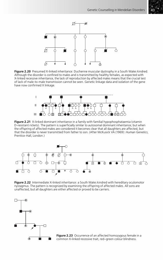
4343Genetic Counselling in Mendelian Disorders
2
Figure 2.20 Presumed X-linked inheritance: Duchenne muscular dystrophy in a South Wales kindred. Although the disorder is confined to males and is transmitted by healthy females, as expected with X-linked recessive inheritance, the lack of reproduction by affected males means that the crucial test of lack of male-to-male transmission cannot be seen. Genetic linkage data and isolation of the gene have now confirmed X linkage.
2 2 26
I
II
III
Figure 2.21 X-linked dominant inheritance in a family with familial hypophosphataemia (vitamin D-resistant rickets). The pattern is superficially similar to autosomal dominant inheritance; but when the offspring of affected males are considered it becomes clear that all daughters are affected, but that the disorder is never transmitted from father to son. (After McKusick VA (1969): Human Genetics,Prentice-Hall, London.)
4 2 2 4
Figure 2.22 Intermediate X-linked inheritance: a South Wales kindred with hereditary oculomotor nystagmus. The pattern is recognized by examining the offspring of affected males. All sons are unaffected, but all daughters are either affected or proved to be carriers.
Figure 2.23 Occurrence of an affected homozygous female in a common X-linked recessive trait, red–green colour blindness.

4444 Practical Genetic Counselling
• Half the daughters of carrier women will themselves be carriers. In the case of an X-linked dominant, half the daughters of affected women will be affected and twice as many females as males will generally be affected.
• Affected homozygous females are exceptional in X-linked recessive disorders, and will occur only when an affected male marries a carrier female.
These simple guidelines will cover most genetic counselling situations and will allow a definite decision to be made in most instances as to whether the disorder is X-linked or not. It can be seen that the situation is essentially similar for X-linked recessive and X-linked dominant disorders, the heterozygous women in the latter group being affected rather than carriers. Apparent anomalies may be the result of chromosome disorders, e.g. occurrence of the disease in a 45,X female (see Fig. 2.18), or of non-paternity. Very few disease-related genes are located on the pairing (pseudo-autosomal) parts of the X and Y chromosomes, giving an autosomal inheritance pattern in families. Some partic-ular problems are shown in the following examples.
The classic X-linked recessive patternFigure 2.19 shows this in a family with haemophilia A.
Disorders where affected males do not reproduceFigure 2.20 shows an example. This may result from the severity of the disease (e.g. Duchenne muscular dystrophy) or from infertility (e.g. Kallmann syndrome). Here the test of male-to-male transmission cannot be applied, so, unless one has other genetic data (e.g. linkage markers, or occurrence in a 45,X female), X linkage is presumed rather than proven. The recognition of gene deletions and an increasing number of specific mutations on the X chromosome, including the examples mentioned, are now provid-ing direct proof in such cases.
X-linked dominant inheritanceFigure 2.21 shows an example. The pattern may at first glance be mistaken for autosomal dominant inheritance, but if offspring of affected males are considered, all sons are unaffected and all daughters are affected. The excess of affected females can also be seen.
‘Intermediate’ X-linked inheritanceThe blurred distinction between dominant and recessive in X-linked disease has already been mentioned. In a few conditions, heterozygotes may show the disease in one branch of a family, but not in another. Figure 2.22 shows an example of this. Although at first sight the pattern appears confusing, the situation is soon clarified if the offspring of affected males are considered – all sons are unaffected, while the females either are affected or are carriers.
X-linked dominant inheritance with lethality in the maleHere the disorder is seen only in the heterozygous females, the affected (hemizygous) males being undetected or appearing as an excess of spontaneous abortions. It is

4545Genetic Counselling in Mendelian Disorders
difficult to prove this situation, but it has now been confirmed for several disorders, including Rett syndrome, focal dermal hypoplasia and incontinentia pigmenti.
Genetic counselling risk estimates require some care. Leaving aside spontaneous abortions, one-third of the offspring of an affected woman will be affected; all the live-born males will be unaffected, as will half of the females. Two-thirds of all offspring will be female. Fetal sexing with termination of female pregnancies may be considered. Where an affected child has been born to healthy parents, this is likely to represent a new mutation and the recurrence risk is likely to be low (most such patients will be female).
Common X-linked genesWhere an X-linked gene is common in a particular population, confusing pedigree patterns may be produced. This may be seen with colour blindness in European popula-tions and with glucose 6-phosphate dehydrogenase deficiency in the Middle East. The marriage of affected males to heterozygous females is not infrequent and will result in homozygous females (see Fig. 2.23), all of whose sons will be affected. A similar pattern can occasionally be seen with rarer disorders when there is consanguinity.
The risk of being a carrier for an X-linked disorder
Methods of carrier detection in X-linked disorders are discussed in Chapter 7, but it is clearly important to estimate the genetic risk of a female relative being a carrier so that information from carrier testing (if any) can be appropriately combined. The estima-tion of these risks is not always easy and is one of the situations where a mathematical approach is needed in genetic counselling. Young’s Introduction to Risk Calculation (see ‘Further reading’) is strongly recommended for this and other areas of risk estimation.
Leaving aside the problems of new mutations, the situation can be approached as follows, using Fig. 2.24 (the pedigree of a family with Duchenne muscular dystrophy) as an example.
1. Obligatory carriers should be identified – these are I-1 and II-2.2. The prior genetic risk of the individual requiring advice, sometimes referred to as
the ‘consultand’ (arrowed), should be estimated. Here it is 1 in 4, since her mother’s prior risk is 1 in 2.
3. Other relevant information must be incorporated. Here the relevant fact is that the consultand has had two normal sons; common sense tells us that this makes it less likely that she is a carrier – the question is, how much less likely? In fact, it can be estimated and combined with other information simply (using what is sometimes termed Bayes’ theorem) by multiplying it with the corresponding prior risk. This is best done by constructing a table, which gives the chances of the two possibilities, as shown in Table 2.3.
The prior risks are clearly 1 in 4 and 3 in 4, respectively. The ‘conditional’ information resulting from the normal sons will give a chance of 1 in 2 for this happening once if she

4646 Practical Genetic Counselling
is a carrier, and 1 in 4 (1/2 � 1/2 � 1/4) for both sons being normal if she is a carrier (three sons would give a risk of 1 in 8, and so on). The corresponding chance of two sons being normal if she is not a carrier is clearly 1 (100 per cent).
The joint risk is then obtained by multiplying the two columns and, if they are placed over the same denominator (giving 1/16 and 12/16), the relative risk can be seen to be 1 to 12 that the consultand is a carrier, or a final risk of 1 in 13.
Care must be taken to relate the ‘conditional’ information to the correct person. Thus, in Fig. 2.25, showing another Duchenne family, the normal sons affect the chances of the mother of the consultand (II-1), being a carrier, and the risk must be worked out for her before proceeding to the daughter (III-1), whose prior risk (1 in 10) is half her mother’s risk (1 in 5) (Table 2.4). If the daughter herself had normal sons, one could proceed as in the previous example but using the new prior risk of 1 in 10 as the starting point.
It can be seen that the use of genetic information in this way can substantially affect the risks given in genetic counselling, and in general with a methodical approach the estimations are not difficult. Incorporation of data from more distant relationships can become complex (computer programs are available to help), but rarely affects the risks to a great extent. It is important to recognize that not only genetic information may be used in this way, but also data on carrier detection, such as may be available in haemophilia and Duchenne muscular dystrophy. Some examples of this are given in Chapter 7.
A final caution should be given: because all these estimates are based on uncer-tainty, they may need to be modified in the light of future events. Thus the birth of an affected child to II-1 in Fig. 2.25 would make her an obligatory carrier and all previous estimates would have to be discarded in the light of this. Likewise, the increasing feasi-bility of direct gene analysis may be a reason for reassessment.
Table 2.3 A working table to combine risk information for the pedigree shown in Fig. 2.24
Consultand a carrier Consultand not a carrier
Prior risk 1/4 3/4
Conditional risk (two normal sons) 1/2 � 1/2 (� 1/4) 1
Joint risk 1/16 3/4 (� 12/16)
Relative risk 1 12
Final risk 1/13 12/13
Figure 2.24 A family with Duchenne muscular dystrophy.
1
1
1
1
2 3
2
2
I
II
III
IV

4747Genetic Counselling in Mendelian Disorders
The isolated case of an X-linked disorder
In autosomal dominant inheritance, new mutations can usually be clearly distinguished from transmitted cases; in autosomal recessive disorders, mutation plays an insignifi-cant part in relation to transmitted genes and can be ignored in genetic counselling. In X-linked recessive disorders, however, it may be extremely difficult, if not impossible, to tell whether an isolated case represents a new mutation or whether the mother is a carrier. Since an accurate distinction is essential for correct genetic counselling, this situation needs careful consideration.
When reliable methods of carrier detection exist, these should be employed. Unfortunately, the variability of gene expression in heterozygotes for X-linked disease makes carrier detection difficult in a proportion of women, even when tests exist. This is clearly seen in haemophilia A and Duchenne muscular dystrophy, where 20–30 per cent of known carriers show results of phenotypic carrier tests falling within normal limits (see Chapter 7).
The example shown in Fig. 2.26 shows the practical aspects of the problem. Three possibilities exist to explain the occurrence of this isolated case of Duchenne muscular dystrophy.
• The case III-1 is the result of a new mutation. In this case none of the numerous female relatives is at significant risk of being a carrier.
• The mother II-1 is a carrier, but is herself the result of a new mutation. In this case, the daughters III-2 and III-3 are at 50 per cent risk of being carriers and their daughters IV-1 and IV-2 have a carrier risk of 25 per cent. However, none of the other female relatives is at risk.
I
II
III
1
1
1
2
2
3
3
Figure 2.25 Carrier risks with an X-linked recessive disorder (Duchenne muscular dystrophy). See Table 2.4 for calculations.
Table 2.4 Risk calculations, X-linked recessive disorder (see Fig. 2.25)
Carrier Not a carrier
Risks for II-1:
Prior risk 1/2 1/2
Conditional risk 1/2 � 1/2 (� 1/4) 1
Joint risk 1/8 1/2 (54/8)
Relative risk 1 4
Final risk 1/5 4/5
Prior risk for III-1 1/10

4848 Practical Genetic Counselling
• The disorder has been transmitted through the mother from the grandmother I-1. In this case, in addition to those already mentioned as being at risk, II-2 and II-3 are at 50 per cent prior risk, with a carrier risk of 25 per cent for their daughters III-5, III-6 and III-8.
Unfortunately, these three situations cannot always be distinguished with certainty by carrier testing, so one needs to approach the problems as follows.
1. The prior risk of II-1 and I-1 being carriers as opposed to III-1 resulting from a new mutation must be estimated. From this, the prior risk of other family members such as II-2 and II-3 can be estimated.
2. Conditional information (from normal sons and carrier testing results) can be tabulated as show in the previous examples.
3. These pieces of information can be combined to give a final risk, as already shown.
The difficult problem is to decide what is the prior risk that this isolated case has resulted from mutation or has been transmitted. It has long been assumed that for an isolated case of an X-linked recessive disorder in which affected males do not reproduce and mutation rates in egg and sperm are equal, the proportions are as follows:
• new mutation (mother not carrier): 1/3;• transmitted case (mother carrier but not grandmother): 1/3;• transmitted case (mother and grandmother carriers): 1/3.
The derivation of this will not be given here, but using this information one can readily assign risks to the various women in the family, as shown in Fig. 2.27. In 2.27(b) the information from the normal sons of II-3 has also been incorporated, and it can be seen that the risks of III-5 and III-6 have been considerably lowered by the normal brothers. Carrier testing results would have produced further information which has been omitted for simplicity.
Unfortunately, there is considerable uncertainty as to whether the formula given above is generally applicable; it assumes equal mutation rates in the sexes, and in some
1
2 31
1
1
2
2
3 4 5 6 7 8
I
II
III
IV
Figure 2.26 An isolated case of a lethal X-linked disorder: Duchenne muscular dystrophy. Is this a new mutation or not? See the text and following figures for the estimation of carrier risks in this difficult situation.

4949Genetic Counselling in Mendelian Disorders
disorders a higher proportion of mothers of isolated cases are carriers than the two-thirds expected, as is also the case for disorders where the affected males reproduce, such as the haemophilias and Becker muscular dystrophy. In the absence of other infor-mation, it is wise to assume that such a woman is a carrier rather than the reverse. For Duchenne dystrophy, the situation may vary according to whether the change is a deletion or a point mutation. Fortunately, molecular tests are increasingly able to dis-tinguish new mutations, although the relatively frequent occurrence of mosaicism, especially in Duchenne dystrophy, still provides problems for genetic counselling in the isolated case, as noted further in Chapter 11.
MITOCHONDRIAL INHERITANCE
While mitochondrial inheritance cannot be described in any sense as mendelian, it seems appropriate to describe it in this chapter, since it provides a characteristic pattern of transmission within families that must be compared with the equally distinctive patterns of mendelian transmission described above. The increasing
Figure 2.27 (a) Carrier risks for women in the family with a lethal X-linked disorder (Duchenne dystrophy) shown in Fig. 2.26, assuming that a third of cases are due to a new mutation. (b) The same pedigree showing the modification of risks from the normal sons of II-2.
2
2
2
1
1
1
1
1/61/6
1/12
2/3
1/3
1/3
1/3
1/61/6
3
3
(a)
4 5 6 7 8
I
II
III
IV
1
21 1/21
1/421/42
1
1 3
3
(b)
4 5 6 7 82
2
I
II
III
IV

5050 Practical Genetic Counselling
identification of specific mitochondrial mutations makes it especially important to recognize this form of inheritance, which has major implications for genetic counselling.
The mitochondria are the principal cell components outside the nucleus to contain DNA, which is present in the form of a small, circular genome that can replicate and which is quite independent of the mechanisms controlling chromosomal DNA. The mitochondrial genome has been sequenced; it determines the proteins of a series of key oxidative enzymes involved in mitochondrial functions, although other such enzymes are produced by genes in the nucleus. Mitochondrial DNA is, for practical purposes, exclusively maternal in origin, with no process involving recombination and a negligi-ble contribution from sperm.
It has long been recognized that any disorder following mitochondrial inheritance should be exclusively maternal in its transmission, as shown in Fig. 2.28. While a number of individual families with differing disorders have in the past been claimed to fit the pattern, one disorder in particular, Leber’s optic atrophy, has consistently done so and been predicted to be mitochondrial in origin, a conclusion now fully confirmed by the identification of mutations. Figure 2.29 shows a typical family with this disorder, which can be used, together with the preceding diagram, to summarize the general risks for family members relevant to genetic counselling, as follows (details specific for Leber’s optic atrophy are given in Chapter 17).
• No transmission occurs to children or other descendants of males, whether they are affected or not. Nor are such descendants at risk of being gene carriers, a situation that contrasts with X linkage, where all daughters of an affected male are obligatory carriers.
• Both sexes may be affected, the precise sex ratio being dependent on the particular disorder and probably determined by other genetic and environmental factors.
Figure 2.28 Mitochondrial inheritance (schematic pedigree): normal mitochondria, °; abnormal mitochondria, •. Since mitochondria are transmitted only through the ovum, not through sperm, affected individuals (shaded) are related through the maternal line. Whether an individual is clinically affected may reflect the proportion of abnormal mitochondria in the relevant tissue.

5151Genetic Counselling in Mendelian Disorders
In Leber’s optic atrophy males are more commonly affected in Europe but not in Japan.
• Females may be symptomless carriers (so may males, but this is irrelevant as they cannot transmit their mitochondria). All daughters of an affected or carrier female are themselves at risk of transmitting the disorder, as well as of becoming affected, while all sons are at risk of becoming affected.
• The precise proportion of offspring, male or female, who will become affected is variable (in Leber’s optic atrophy this is around 50 per cent of sons and 30 per cent of daughters) and not determined by any fixed rule.
It can be seen from the above summary that the recognition of mitochondrial inherit-ance is extremely satisfactory for the genetic counselling of males, since, whether they are affected or not, a confident reassurance can be given that their descendants will not be affected. For females the situation is unsatisfactory in the extreme, since there is some degree of risk to all offspring with none of the sharp demarcations and exclusions that are possible in mendelian inheritance.
A major advance during the past few years has been the identification of specific defects in mitochondrial DNA, both point mutations and large deletions, and the reali-zation that such disorders are much more frequent than previously suspected. Box 2.2 lists some of the more important of these. Not all have a clearly defined phenotype, and the finding that subgroups of such common and heterogeneous disorders such as diabetes and deafness may have an underlying mitochondrial defect is likely to pose logistical problems as to when a molecular defect should be searched for. It should be
Figure 2.29 A family pedigree of a mitochondrial disorder: Leber’s optic atrophy. All the affected individuals are related in the maternal line; no male family members, affected or unaffected, have transmitted the disorder. It can be seen that there are numerous females in the pedigree who are potentially at risk of developing or transmitting the disorder, as well as males who can be reassured that they have no significant risk of transmitting it, whether or not they themselves are or become affected.
3
2
2 2 2
4
Box 2.2 Disorders resulting from mitochondrial mutations
Leber’s optic atrophyMELAS (mitochondrial encephalopathy with lactic acidosis and stroke-like episodes)MERFF (myoclonic epilepsy with ragged red fibres)Kearns–Sayre syndromePearson syndrome (lactic acidosis, pancreatic insufficiency, pancytopenia)Deafness (antibiotic-induced and some forms of progressive nerve deafness)Leigh syndrome (subacute encephalopathy, often with lactic acidosis; some cases)Various poorly classified central nervous system degenerationsDiabetes mellitus (some familial types)

5252 Practical Genetic Counselling
noted, in particular, that there are only genetic implications if the molecular defect is present in the germ line (more likely to be the case if found in DNA from a blood sample), not if the defect is confined to the target organ (e.g. muscle). Most large dele-tions of mitochondrial DNA are sporadic in nature.
It might be thought that once a molecular mitochondrial defect has been identified in a family, analysis of relatives at risk would help to resolve the genetic counselling problems described above. Unfortunately this is not the case at all, since all those genet-ically at risk are likely to show at least some degree of the defect, often showing both mutant and normal mitochondrial DNA (‘heteroplasmy’). We do not know at present whether there is any correlation between the proportion of abnormal mitochondria found (in blood) and the risk of developing or transmitting the disorder, though their absence would suggest a low risk. Tests are also inconclusive in prenatal diagnosis for the same reason, although absence of the mutant line in a prenatal sample also seems likely to indicate a low risk.
Thus the conclusion at present has to be that while the recognition of mitochondrial inheritance by pedigree pattern and molecular analysis is important in identifying genetic risks and in removing risk from descendants in the male line, it is unhelpful in resolving the situation for those known to be at risk, and does not warrant molecular testing of asymptomatic relatives. Looking to the future, the hope must be that effective therapeutic agents enhancing mitochondrial function will be developed that can be applied to those at risk before serious and irreversible clinical problems occur.
A final point to be emphasized is that many mitochondrial enzymes are determined by nuclear genes. Thus a mitochondrial disorder does not necessarily imply mitochon-drial inheritance.
FURTHER READING
General (see also Chapter 1)McKusick VA (1998). Mendelian Inheritance in Man, 12th edn. Baltimore: Johns Hopkins
University Press [an invaluable resource on all aspects of mendelian disorders; a continuously updated on-line computer version (OMIM) is also available at www.ncbi.nlm.nih.gov/omim and is now generally used].
Ostrer H (1998). Non-Mendelian Inheritance in Humans. Oxford: Oxford University Press [despite its title, this book gives a valuable account of the exceptions to mendelian inheritance].
Young ID (2007). Introduction to Risk Calculation in Genetic Counselling, 3rd edn. Oxford: Oxford University Press [a clearly explained and valuable guide to practical situations and to their underlying principles].
Mitochondrial disordersHolt IJ (ed.) (2003). Genetics of Mitochondrial Disease. Oxford: Oxford University Press.Suomalainen A (1997). Mitochondrial DNA and disease, Ann Med 29, 235–46.Wallace DC, Lott MT, Procaccio V (2007). Mitochondrial genes in degenerative diseases, cancer
and aging. In: Rimoin DL, Connor JM, Pyeritz RE, Korf B (eds) Emery and Rimoin’s

5353Genetic Counselling in Mendelian Disorders
Principles and Practice of Medical Genetics, 5th edn, pp. 194–298. Edinburgh: Churchill Livingstone.
MosaicismHall JG (1988). Somatic mosaicism: observations related to clinical genetics, Am J Hum Genet
43, 355–63.
ImprintingClayton-Smith J (2003). Genomic imprinting as a cause of disease, Brit Med J 327, 1121–2.Engel E, Antonorakis S (2002). Genomic Imprinting and Uniparental Disomy in Medicine.
New York: Wiley-Liss.
X inactivationMigeon BR (2007). Females are Mosaics: X Inactivation and Sex Differences in Disease.
New York: Oxford University Press.
Incomplete penetrancePauli RM, Motulsky AG (1981). Risk counselling in autosomal disorders with undetermined
penetrance, J Med Genet 15, 339–45.
AnticipationHarper PS, Harley HG, Reardon W, Shaw DJ (1992). Anticipation in myotonic dystrophy: new
light on an old problem, Am J Hum Genet 51, 1016.Richards K, Sutherland GR (1992). Heritable unstable DNA sequences, Nat Genet 1, 7–9.

Introduction . . . . . . . . . . . . . . . . . . . . . . . . . . . . . . . . . . . . . . . . . . . . . . . . . . . . . . . . . . . . . . . . . . . . 54
The genetic basis of common disorders . . . . . . . . . . . . . . . . . . . . . . . . . . . . . . . . . . . . . . . . . . . 55
General rules for ‘multifactorial’ inheritance . . . . . . . . . . . . . . . . . . . . . . . . . . . . . . . . . . . . . . 58
Empirical risk data . . . . . . . . . . . . . . . . . . . . . . . . . . . . . . . . . . . . . . . . . . . . . . . . . . . . . . . . . . . . . . . 60
The molecular basis of genetic susceptibility . . . . . . . . . . . . . . . . . . . . . . . . . . . . . . . . . . . . . . 61
Further reading . . . . . . . . . . . . . . . . . . . . . . . . . . . . . . . . . . . . . . . . . . . . . . . . . . . . . . . . . . . . . . . . . 62
Common Disorders and Genetic Counselling
CHAPTER 3
INTRODUCTION
The disorders to be considered here, in contrast to most mendelian conditions, will be encountered frequently by those working in all branches of medicine and in primary care. Arbitrarily, a population frequency of greater than 1 in 1000 (for the clinical disease, not for any predisposing genotype) will be taken as a working definition of what is common.
Most common disorders do not follow any of the clear patterns of mendelian inher-itance outlined in the previous chapter. This applies not only to the commoner birth defects (e.g. neural tube defects, most congenital heart disease), but also to most common chronic disorders of later life (e.g. asthma, diabetes, hypertension, coronary heart disease, common cancers). Yet it has long been recognized that, to some degree, these conditions show a familial tendency, which may occasionally be striking. As a result of this, families in which such common disorders occur often seek genetic coun-selling, especially if multiple members have been affected. The recent general increase in awareness of genetic factors, often heightened by media attention, has increased this tendency, notably in the case of cancer.
The fact that many of these disorders are so common, at least by comparison with the mendelian disorders that form the basis of activity for most specialists in medical genetics, creates special challenges. It is quite clear that most genetic counselling for this group must, of necessity, be done by workers in primary care or those involved directly in management of the particular field, since the sheer numbers would swamp the small number of available specialist clinical geneticists and their associated staff. But in practice, this is proving to be much less of a problem than might be expected, partly because the risk figures are often simple and approximate, partly because for most common diseases the demand for genetic counselling is very much less in proportion to their frequency than is the case for single gene disorders. One simple, yet important,

5555Common Disorders and Genetic Counselling
reason for this relative lack of demand is that, while genetic approaches can increase or decrease risks somewhat, they cannot rule out the risk of a condition – as can be done for rare mendelian disorders.
This chapter examines some of these problems in the context of recent develop-ments in attempting to identify the specific genes involved in common disorders. As will be seen, this is one of the most active areas of genetic research, but one where the practical applications of these advances in terms of risk estimation and genetic counsel-ling so far remain extremely limited, as does the use of genetic tests.
THE GENETIC BASIS OF COMMON DISORDERS
The terms ‘multifactorial’ or ‘polygenic’ are frequently used to describe the genetic basis underlying the great majority of common disorders where there is no clear mendelian pattern. Of the two terms, multifactorial is the more appropriate since it recognizes that these disorders are the result of both environmental and genetic factors, and does not prejudge the relative role of either category. ‘Polygenic’ implies that a number of differ-ent genetic loci are involved rather than a single major gene.
The term ‘heritability’ is often encountered in relation to common disorders and refers to the proportion of variation, familial and other, that is determined by genetic factors, regardless of any pattern of inheritance. This can be assessed by a number of approaches, notably the comparison of monozygotic and dizygotic twin pairs where at least one twin has the disorder. Clearly the heritability will be one of the factors influ-encing recurrence risk in a family.
The general principles of common disease genetics were worked out more than 50 years ago by pioneers such as Haldane, Penrose and Edwards, and remain relevant today, but our growing ability to recognize specific genes involved with common disor-ders, described later in this chapter and also in Chapter 5, has allowed two main categories to be defined, as follows:
• common disorders containing a significant mendelian subset, resulting from the action of a single major gene in a family;
• common disorders where a number of genetic (and usually also environmental) factors are involved, each usually being of small or, at most, moderate influence.
From the viewpoint of genetic counselling it is essential, whenever possible, to distin-guish between these two situations, which are often completely different in terms of the risk estimates involved.
Mendelian subsets
This group is of particular importance in genetic counselling because the recognition of a mendelian (usually dominant) subset of cases gives extremely high risks for family members (or very low risk if the particular gene mutation can be excluded). The familial cancers provide a striking example, largely unrecognized until recently. Although very

5656 Practical Genetic Counselling
rare familial cancer syndromes have been recognized for many years (e.g. familial adenomatous polyposis as a cause of colon cancer), the identification of mendelian forms of breast and colon cancer that are not phenotypically distinct from the non-mendelian majority has had a major influence on both research and genetic counselling practice in these disorders, influences now extending to most common cancers (see Chapter 25 for details). It is no longer sufficient to derive overall theoretical risks, as was done in early editions of this book, since the separation of the mendelian forms will affect the recurrence risks for those remaining. This dissection of the multifactorial basis of common diseases is not confined to cancer, but has become relevant to a wide range of conditions where previously unsuspected or undefined mendelian families are proving to account for a significant proportion of recurrent cases. Box 3.1 summarizes the situation for the main common disease groups.
Polygenic disorders
In this second group, where clear mendelian forms are absent or very rare, some of the genetic factors primarily responsible for the genetic influence on the disorders are now also beginning to be identified. Much research originally focused on the HLA region of chromosome 6, long known to be a susceptibility locus for a variety of disorders with an immunological basis and it remains much the strongest and best documented suscepti-bility locus, even after more than a decade of analysing wider genetic markers.
The past 15 years has seen intense research efforts into identifying the other genetic components, utilizing the existence of DNA markers across the whole genome and building on the striking success in mapping genes for mendelian disorders. Unfortunately, this important but difficult procedure was initially often approached with considerable naivety and many of the early claims were not substantiated. In the last few years the approach of genome-wide association studies based on high-throughput sequencing techniques has proved more fruitful in identifying genes and chromosomal regions likely to be involved, but it must be emphasized that these remain mostly at the research stage, that there may be substantial variation between popula-tions, while at present, virtually none is sufficiently strong or reproducible to use in
Box 3.1 Common disorders and mendelian subsets
Mendelian subset frequent (~5% of all cases)Breast cancerColorectal cancerProstate cancer (?)
Mendelian subset not frequent (~1%) but significantCoronary heart disease (familial hypercholesterolaemia)Early-onset Alzheimer’s diseaseMost other cancers (data still uncertain)
Mendelian subset rare or absent (<1%)SchizophreniaBipolar affective disorderAsthmaLate-onset Alzheimer’s diseaseStroke

5757Common Disorders and Genetic Counselling
clinical practice or genetic counselling. It is increasingly likely that, for many common disorders and for most normal characteristics, the genetic component results from a large number of individually small genetic effects, no one gene contributing more than a small part of the susceptibility.
A third type of genetic involvement in common disease requires a mention, although its importance outside the field of common cancers is not clear. This is where a specific gene, relatively rare in the population, has a moderate effect, insufficient to cause much family clustering but still significant in its contribution to the overall causation of the disorder. Examples are seen in some types of cancer and in some congenital malformations, such as Hirschsprung’s disease. So far these intermediate situations seem relatively uncommon.
It must be emphasized strongly that the finding that a particular gene is involved in a common disorder does not convert the disorder (or even a subset of it) into a mende-lian disorder. Of course, the gene itself will show mendelian segregation at the DNA level (it cannot do otherwise), but it is the disease phenotype that is relevant in terms of mendelian inheritance. This may seem an obvious point, but continues to be ignored by many who should know much better!
Perception and the genetic basis of common disease
For clear-cut single gene disorders, there is rarely much doubt in the minds of profes-sionals or the public that one is dealing with a ‘genetic disorder’, where environmental aspects are subsidiary to the inherited factor. For most common disorders, by contrast, the situation is far less clear, even though studies may have clearly identified an impor-tant genetic component. To a considerable extent, the perception of a disorder as ‘genetic’ or ‘environmental’ will depend on how successful workers have been in identi-fying specific factors; new discoveries may result in a radical change in perception – and in demand for genetic counselling.
Consider these two examples:
• Peptic ulcer was regarded for many years as a largely genetic condition, with important familial influences, and with a large body of research on its genetic basis. Once an infective agent was identified, the genetic aspects rapidly became overshadowed and most people would now consider it as having a primarily environmental cause, with relatively little demand for genetic counselling.
• Conversely, in breast cancer, genetic factors were considered of little relevance outside a few striking families until the BRCA genes were identified, which produced a rapid change in perception to this being seen as a mainly ‘genetic’ disorder, despite the fact that these genes are responsible for only a small proportion of cases overall.
In both cases, the underlying facts and balance between genetic and environmental factors have not changed at all, but there has been a major shift in public and medical perception, reflected in the demand for, and uptake of, genetic services.
Since these perceptions and the underlying science can change so rapidly, as has happened with the familial cancers, it can be asked whether a sudden increase in

5858 Practical Genetic Counselling
demand might be seen for other types of common disease. The only field where this seems to be occurring at present is that of cardiovascular disease (see Chapter 19), where the recognition of mendelian forms of both coronary heart disease and arrhythmias is beginning to have a major impact on cardiologists and affected families. By contrast other large groups of disease, such as the major psychoses, asthma and stroke show few high-risk aggregations and create relatively little demand for genetic counselling (see Box 3.1).
A final factor is that the degree to which a condition is seen as genetic may reflect the frequency of the environmental factors involved and these may vary over time or between populations. Thus, for rare infectious disorders, contact with the agent itself will be the determining factor in most cases, while, if the infectious agent is ubiquitous, genetic susceptibility will have most influence.
GENERAL RULES FOR ‘MULTIFACTORIAL’ INHERITANCE
Since research for most common disorders is still at an early stage in resolving the number and nature of the genes involved, it is this second ‘multifactorial’ group where it is helpful to have some general rules that will influence risk prediction and genetic counselling. These are summarized here, but it must be recognized that they will need to be considerably modified when the specific genes involved become clear, or mende-lian subgroups are identified.
The essential distinguishing factor from the mendelian disorders is that a single genetic locus cannot be held responsible for the condition; it is the result of the additive effect or interaction of a number of genetic loci and a number of external factors. The sum of these determines a person’s liability to be affected with the particular disorder, and this liability can be expected to show a more or less ‘normal’ distribution curve in the population, with most people having an intermediate degree of liability and smaller numbers at each end of the distribution curve having unusually low or unu-sually high liability (Fig. 3.1). The last group comprises those who are actually affected, whose liability is above a postulated ‘threshold’ for the disorder.
From this concept, it can be seen that even a person with an unusually high genetic liability may not be affected if environmental factors are favourable, and the converse will also apply. The degree to which liability is determined by genetic factors as opposed to environmental factors is often considered as the heritability of a disorder, although in practice familial but non-genetic factors may often be included in this. The liability of relatives of a patient with the disorder will also be normally distributed in a similar way to that of the general population, but the curve will be shifted towards higher liability because of the increased genetic component.
The ‘threshold model’ is not essential for the operation of multifactorial inherit-ance; for quantitatively distributed variables such as height or blood pressure, the same principles apply. It should also be noted that only a small number of genetic loci (as few as four or five) are required for the discrete patterns of mendelian inheritance to be transformed into a relatively smooth curve of distribution of liability.

5959Common Disorders and Genetic Counselling
Turning to the practical aspects, the following points are relevant (summarized in Box 3.2).
• Increased risk is greatest among closest relatives and decreases rapidly with distance of relationship. It is rare to find a significant increase in risk for relatives more distant than second degree; even here risks are usually small.
• The risk of recurrence will depend on the incidence of the disorder, unlike in mendelian inheritance. A useful approximation where specific figures are not
Number ofindividualswith aparticularliability
First-degree relativesof affected individuals
affected
Liability
General population
Figure 3.1 Liability and genetic risks in multifactorial inheritance. The distribution of liability to the disorder in the general population follows an approximately normal distribution, with individuals exceeding a certain threshold value being affected. First-degree relatives have a similar normal distribution of liability, but the curve is shifted to the right by the increased genetic component, so a greater proportion will exceed the threshold and be affected.
Box 3.2 Multifactorial inheritance: factors increasing risk to relatives
Close relationship to probandHigh heritability of disorderProband of more rarely affected sexSevere or early-onset disease in probandMultiple family members affected
6060 Practical Genetic Counselling
available is that the maximum risk to first-degree relatives is approximately the square-root of the incidence; i.e. where the incidence is 1 in 100, the recurrence risk would be around 1 in 10.
• Dominance and recessiveness do not generally apply. Thus the risk for sibs is comparable to that for offspring. Risks for offspring are not yet available for many serious childhood disorders, but can usually be taken as approximately equivalent to the risks for sibs in the absence of further information, unless there is evidence of a significant proportion of isolated cases being the result of new dominant mutations. In fact the offspring risk might be expected to be lower, since those reproducing are generally less severely affected. (Clearly, it is especially important here not to miss a mendelian subset.)
• Where there is an unequal sex incidence, the risk is higher for relatives of a patient of the sex in which the condition is less common. For example, in pyloric stenosis (which is more common in males) the risk for brothers of a male index case is 3.8 per cent, but for brothers of a female index case it is 9.2 per cent (see Chapter 20). At first sight this may seem unexpected, but when it is considered that girls require a greater genetic liability to develop the disorder than do boys, it can readily be seen that relatives of an unaffected girl will also have a greater genetic liability than where the index case is a boy.
• The risk may be greater when the disorder is more severe. This is well shown in Hirschsprung’s disease, where the risk for sibs of patients with long-segment disease is greater than for those with a short segment affected (see Chapter 20). Again, the greater severity reflects greater liability, part of which will be genetic and thus shared with relatives. For disorders of adult life, early onset in the proband will increase the risk for relatives (as in breast cancer). In part, this effect may result from inclusion of unrecognized mendelian cases in these severe or early-onset groups.
• The risk is increased when multiple family members are affected. This again results from the concentration of genetic liability in the particular family and is in contrast to the mendelian situation where the number of affected family members (leaving aside mutation) is irrelevant. The influence of more distant relatives is less easy to determine, although computer programs have been developed to combine the information. In general, one close (first-degree) relative outweighs several distant ones. Particular care must be taken in such families to be as certain as possible that one is not dealing with a mendelian subtype of the disorder.
EMPIRICAL RISK DATA
This term, already mentioned in Chapter 1, is merely a statement of the fact that someone has actually looked at what happens in a particular situation. Data on the risks to relatives are available for a large number of non-mendelian disorders and, provided the study has been careful and as far as possible unbiased, such information provides the most satisfactory basis for counselling until the genetic basis can be resolved further.

6161Common Disorders and Genetic Counselling
It has to be remembered, however, as stated in Chapter 1, that such risk figures are not universal in their application as are mendelian ratios. In particular:
• Data collected on one population may not be applicable to others where the incidence, and perhaps aetiology, of the disorder in terms of both genetic and environmental factors are different.
• Improved classification of disorders, in particular resolution of heterogeneity, identification of single gene subsets, or identification of specific causative factors, may require radical revision of risk estimates.
• Risks may depend not only on the diagnosis but also on individual factors such as sex, severity of disease and number of affected family members.
Empirical risk figures are given for as many disorders as possible in the specific chapters of this book. Most are based on old, though thorough, studies, so there is a need for new data of this type using revised criteria. In particular, molecular advances have removed or reclassified some cases as mendelian, so it is difficult to know how this may have affected the risk estimates for those cases where no molecular defect can be detected. Unfortunately, empirical risk studies are now regarded as ‘old-fashioned’, so there is a real lack of and need for new data.
THE MOLECULAR BASIS OF GENETIC SUSCEPTIBILITY
The way in which isolation of specific genes has increased our understanding of a disease has already been mentioned. The effects on our understanding of common dis-orders, though, are beginning to be much more profound, especially where single gene subsets are identified, and this forms one of the most exciting areas of current medical research generally. It has to be admitted that epidemiological and other approaches to detecting the environmental components have, for most disorders (with a few excep-tions such as neural tube defects), been disappointing; thus the value of current genetic research is not so much that it shows a greater importance of genetic factors in common disorders but that it is starting to provide knowledge of their pathogenesis, which is equally relevant to the external factors. Colorectal cancer again provides an excellent example, with the series of tumour suppressor genes identified providing defined points on which environmental carcinogens can act. In diabetes, although the evidence remains far from secure, the identification of involvement of different susceptibility genes in types 1 and 2 reinforces the role of different external factors – probably infec-tive in type 1 and nutritional in type 2.
These advances are likely to have major consequences for how we classify diseases and, in the future, how we approach therapy, which may well need different agents according to the underlying genetic changes. This prospect in part explains the high interest and involvement of major pharmaceutical firms in this field.
When it comes to using genetic susceptibility tests in risk prediction and genetic counselling, however, their use is at present of minimal help and potentially of consid-erable harm. Unfortunately, this has not deterred unscrupulous commercial interests

6262 Practical Genetic Counselling
from promoting the use of such tests, increasingly via the internet, in situations where the factual basis is minimal or non-existent. The scientific basis of risk estimates applied to individuals, as opposed to large series, is generally very uncertain, with wide margins of error, complex interactions with other factors and differences between populations. Even the strongest associations (e.g. between HLA B27 and ankylosing spondylitis) are far from definitive (see Chapter 14); although a normal result in a suspected patient may make the condition very unlikely, an ‘abnormal’ result in a healthy relative at risk is still much more likely to be associated with normality than with clinical disease. Most asso-ciations (e.g. ApoE4 and Alzheimer’s disease) are much less strong and consequently of little use in genetic counselling, while, as mentioned earlier, the great majority of asso-ciations with particular genes or polymorphisms either fail to be confirmed, are of uncertain significance, or give only slight risk alteration. As general population screen-ing tools for susceptibility, they are likely to be positively harmful, creating needless worry without being able to resolve it.
Thus the main aims of genetic counselling for common disorders at present remain the identification (and exclusion) of the high-risk genetic subsets, along with the provi-sion of risk estimates based on empirical studies and the general principles given above. With these points in mind, primary clinicians should be able to handle themselves most of the requests that they encounter and be able to refer on the relatively small number where unusual family patterns or clinical features point to a high-risk situation, where a specialist in clinical genetics may be able to be of real additional help. In those fields where mendelian subsets are frequent, a close partnership between relevant specialist (e.g. oncologists, breast or colorectal cancer surgeons and cardiologists) is undoubtedly the best way to ensure that the increased workload and the different levels of risk are handled appropriately. Specialist genetic counsellors are also an increasingly important element in such a system, in which the diagnostic aspects have already been dealt with by others.
FURTHER READING
Academy of Medical Sciences UK (2009). Genome-wide Association Studies: Understanding the Genetics of Common Diseases. London: Academy of Medical Sciences.
King RA, Rotter J, Motulsky AG (eds) (2002). The Genetics of Common Disorders, 2nd edn. Oxford: Oxford University Press [a valuable and comprehensive book giving details on all major topics, as well as on general and theoretical aspects; its often critical approach is especially valuable (e.g. the chapter on evolution of human genetic diseases by Diamond and King)].
Weatherall DJ (2000). The role of nature and nurture in common diseases. Garrod’s legacy. The Harveian Oration. London: Royal College of Physicians.
Wilkie AO (2003). Polygenetic inheritance and genetic susceptibility screening. In: Cooper DN (ed.) Nature Encyclopedia of the Human Genome, pp. 596–601. London: Nature Publishing Group.
Wright A, Hastie N (eds) (2007). Genes and Common Diseases. Cambridge: Cambridge University Press.
Young ID (2007). Introduction to Risk Calculation in Genetic Counselling, 3rd edn. Oxford: Oxford University Press [an extremely useful book for those needing risk estimates].

Introduction . . . . . . . . . . . . . . . . . . . . . . . . . . . . . . . . . . . . . . . . . . . . . . . . . . . . . . . . . . . . . . . . . . . 63
Chromosome terminology . . . . . . . . . . . . . . . . . . . . . . . . . . . . . . . . . . . . . . . . . . . . . . . . . . . . . . . 64
Frequency of chromosome abnormalities in the population . . . . . . . . . . . . . . . . . . . . . . . 67
Trisomies . . . . . . . . . . . . . . . . . . . . . . . . . . . . . . . . . . . . . . . . . . . . . . . . . . . . . . . . . . . . . . . . . . . . 69
Sex chromosome abnormalities . . . . . . . . . . . . . . . . . . . . . . . . . . . . . . . . . . . . . . . . . . . . . . . . . . 71
Chromosome translocations . . . . . . . . . . . . . . . . . . . . . . . . . . . . . . . . . . . . . . . . . . . . . . . . . . . . . 73
Inversions . . . . . . . . . . . . . . . . . . . . . . . . . . . . . . . . . . . . . . . . . . . . . . . . . . . . . . . . . . . . . . . . . . . . . . . 76
Mosaicism . . . . . . . . . . . . . . . . . . . . . . . . . . . . . . . . . . . . . . . . . . . . . . . . . . . . . . . . . . . . . . . . . . . . . 76
Deletions . . . . . . . . . . . . . . . . . . . . . . . . . . . . . . . . . . . . . . . . . . . . . . . . . . . . . . . . . . . . . . . . . . . . . . . 77
Microdeletion syndromes . . . . . . . . . . . . . . . . . . . . . . . . . . . . . . . . . . . . . . . . . . . . . . . . . . . . . . . 77
Uniparental disomy . . . . . . . . . . . . . . . . . . . . . . . . . . . . . . . . . . . . . . . . . . . . . . . . . . . . . . . . . . . . . . 78
Isochromosomes . . . . . . . . . . . . . . . . . . . . . . . . . . . . . . . . . . . . . . . . . . . . . . . . . . . . . . . . . . . . . . . . . 79
Hereditary fragile sites . . . . . . . . . . . . . . . . . . . . . . . . . . . . . . . . . . . . . . . . . . . . . . . . . . . . . . . . . . . 79
Normal variations . . . . . . . . . . . . . . . . . . . . . . . . . . . . . . . . . . . . . . . . . . . . . . . . . . . . . . . . . . . . . . . . 79
Further reading . . . . . . . . . . . . . . . . . . . . . . . . . . . . . . . . . . . . . . . . . . . . . . . . . . . . . . . . . . . . . . . . . . 79
Chromosome Abnormalities
CHAPTER 4
INTRODUCTION
Although chromosomes have been recognized as the structures responsible for inherit-ance for over a century, it was only in 1956 that the normal human chromosome number was found to be 46, not 48 as thought for many years previously. The first chromosome abnormalities were discovered in 1959 and since that time human cytogenetics has formed an important part of medical genetics. Those interested in the history of the field will enjoy TC Hsu’s informative and readable book Human Cytogenetics: an Historical Approach, while my own book First Years of Human Chromosomes takes a more clinical approach, based largely on interviews with the early pioneers (see ‘Further reading’). Over the past half century the field has been greatly extended and changed by a series of technological developments that have not only had diagnostic implications, but have given important insights into how chromosomes actually function. Although this book does not attempt to cover the area of chromosome biology, it is important to recognize that chromosomes are not simply linear aggregations of genes or passive carriers for them, but that they powerfully influence normal and abnormal inheritance in a much wider way.
Increasing understanding of the molecular basis of meiosis and DNA repair, of the function of histones and other protein components of the chromosome, and the

6464 Practical Genetic Counselling
importance of methylation in particular chromosome regions, are a few examples of how cytogenetics has become part of the wider fields of ‘genomics’ and ‘epigenetics’.
Chromosome disorders are relatively common as the cause of serious childhood malformations and rank high in the requests for genetic counselling, but before describ-ing the risks in specific groups, there are two points worthy of mention that the clinician and genetic counsellor should bear in mind from the outset.
• The great majority of disorders following mendelian inheritance, especially those of adult life, show no visible chromosomal abnormality.
• The great majority of chromosomal disorders have an extremely low risk of recurrence in a family, especially where no abnormality is present in a parent.
Thus, not all patients with a genetic disorder need chromosome analysis; to request this uncritically is unnecessary, expensive and will prevent the laboratory from concentrat-ing its resources on samples for which a detailed analysis may be essential. There may even be the danger of a family being given a falsely low genetic risk on the grounds of a normal chromosome result. However, since chromosomal disorders are poorly treata-ble but accurately detectable prenatally, it is essential that situations requiring cytogenetic analysis are recognized and that the small number of cases of chromosome abnormality giving high risks to relatives are clearly distinguished from the much larger number where these risks are low.
During the last few years, the separation between mendelian and chromosomal dis-orders has become less clear with the recognition of small chromosomal deletions in a number of different disorders, including some important malformation syndromes appearing to follow mendelian inheritance. Working from the other direction, molecu-lar geneticists have shown that the mutational event involved in some cases of mendelian disorder is a deletion of genetic material rather than a specific base change. Additional recent developments include the recognition that parent-of-origin effects due to genetic imprinting and uniparental disomy, already mentioned in Chapter 2, are relevant to disorders of whole chromosomes or chromosomal regions.
A series of new technical developments, notably fluorescence in-situ hybridization (FISH), that combine molecular technology with the classical microscopic approach, are now in diagnostic use, helping to resolve chromosome disorders that were previously confusing, as well as identifying others not recognizable by the use of earlier techniques.
The use of genome sequencing and of comparative genome hybridization (CGH) is further advancing the field, to the extent that microscopy could eventually be replaced completely; but for the present, the well tested techniques of classical cytogenetics remain essential for most of the diagnostic situations described in this chapter.
CHROMOSOME TERMINOLOGY
All 23 pairs of human chromosomes can be distinguished microscopically from each other, and much fine detail within each chromosome can also be recognized (Fig. 4.1). An agreed international system of nomenclature (Fig. 4.2), reviewed at regular intervals

6565Chromosome Abnormalities
(most recently 2009), forms the basis of reports from laboratories. Although these reports will usually be accompanied by an explanatory letter, clinicians may be deterred by the terminology itself, so some brief notes are given here.
Take the following example:
46,XY,t(4;22)(q32;q12)
This denotes the fact that the individual has a balanced translocation between the long arms of chromosomes 4 and 22. Several points can be noted:
• The total chromosome number is given first. In this example there is the normal number of 46.
• The sex chromosome constitution comes next. Here the patient is chromosomally male (XY) as opposed to female (XX).
• A translocation is indicated by the letter ‘t’ with details of the chromosomes involved in brackets. In this example, chromosomes 4 and 22 are involved.
• The arms of a chromosome are indicated by the letters ‘p’ for the short arm and ‘q’ for the long arm (‘p’ is for petit, reflecting the strong French influence in early cytogenetics). In our example, the long arms of the two chromosomes are involved in the translocation. The breakpoints are given in the second brackets, indicated by numbers corresponding to band designations at points of exchange, in this case bands q32 and q12 on chromosomes 4 and 22, respectively.
Figure 4.1 The normal human karyotype (male); trypsin–Giemsa preparation to show G-banding pattern. (Kindly provided by Selwyn Roberts and Merle Vaughan.)

6666Practical G
enetic Counselling
4
4
1
1
2
3
3
3
3
2
2
2
2
11
1
14
1
1
2
3
3
3
32
2
2
2
11
1
1 6
5
5
4
4
1
1
2
3
3
3
22
2
11
1
4
1
1
2
3
3
3
2
2
2
11
1
54
1
1
2
3
3
2
2
2
11
1 1
13
3
2211
1
1
23
2
2
1
1
1
1
13
3
2
2
1
1 11
2
321112
3
31
1
2
21
1 1
12
11
54
4
3
3
3
1
1
2
2
2
2
11
1
5
5
4
4
4
3
3
3
1
1
2
2
2
2
1
1
1
6
5
5
4
4
3
32
1
1
2
2
11
1
4
4
3
3
3
2
2
1
1
2
2
1
1
321
32121
4
3
3
32
2
1
1 2
2
2
2
11
1
1
6
5
5
4
4
3
33
2
1
1
2
2
2
2
1
1
1
1
21
654
3
3
1
1
2
2 2
2
2
1
1
1
1
78
6
6
5
5
5
4
4
4
4
4
3
3
3
3
3
3
2
1
2
2
2
2
2
1
1
1
7
1
1
2
16
6
5
5
5
4
4
4
3
3
3
3
3
2
2
2
2
2
11
1
1
78
1
1
5
5
5
4
4
4
3
3
3
3
2
2
2
2
11
1
321
1
16
6
5
5
5
4
4
4
3
3
32
2 2
2
2
2
1
11
1
7
1
1
6
6
5
5
4
4
4
3
3
3
3
2
2
2
2
1
11
1
7
789
2
2
1
1
65
5
4
4
3
3
3
3
4
21
1
1
22
2
1
1
32
2
2
2
4312
11
1
1
13
p
q
p
q
14 15 16 17 18 19 20 21 22 Y X
2 3 4 5 6 7
p
q
p
q
8 9 10 11 12
Figure 4.2 Schematic of the human karyotype using banding techniques, following the international cytogenetic nomenclature (ISCN). The most recent revision (ISCN 2009) should be consulted for details, but no longer contains this simple map.

6767Chromosome Abnormalities
Gain or loss of an entire chromosome is indicated by a ‘�’ or ‘�’. For example, a male infant with Down’s syndrome resulting from trisomy 21 would have the karyotype designation 47,XY, �21.
For gain or loss of part of a chromosome, the terms ‘add’ (addition) or ‘del’ (deletion) are used. An example is 46,XY,del(5)(p15) (deletion of the short arm of chromosome 5), as seen in the cri-du-chat syndrome.
Further details in chromosome reports may deal with mosaicism (the presence of more than one cell line), inversions, ring chromosomes, the identification of particular bands on a chromosome, and the use of FISH and other molecular cytogenetic tech-niques. If in any doubt, the clinician should make personal contact with the cytogenetics laboratory. This will provide a more meaningful idea of the problem and will allow a better assessment of risks by discussion with cytogeneticist colleagues. Most laboratory cytogeneticists will be delighted by the opportunity to learn more specific details from the referring clinician, and may in turn be able to suggest further clinical investigations from their knowledge of the phenotype associated with particular chromosomal defects.
Cytogenetic techniques are not described here but are covered by suggestions in the ‘Further reading’ section. It has already been noted that techniques are evolving rapidly, and that a fusion of molecular and cytogenetic methods is allowing molecular applica-tions to chromosomal disorders, as well as identifying changes in disorders not conventionally thought of as being ‘chromosomal’ in nature. For rapid or preliminary diagnosis, particularly in urgent prenatal situations, the molecular techniques of QF-PCR and array CGH have become part of regular diagnostic practice in the prenatal detection of trisomies.
FREQUENCY OF CHROMOSOME ABNORMALITIES IN THE POPULATION
Four sources of information are available:
• studies of newborn populations;• studies of particular chromosomal disorders;• studies of abortions and stillbirths;• prenatal diagnosis series.
Studies of unselected newborn populations
Table 4.1 summarizes the principal findings, based on studies that revised earlier esti-mates by using banding techniques. It can be seen that 9.2 per 1000 infants (or approaching 1 per cent) had recognizable chromosomal abnormalities, of which around three-quarters had autosomal abnormalities and a quarter had sex chromosome abnor-malities. Although the numbers were insufficient to give accurate data on individual disorders, these newborn figures are a useful reference point with which to compare any increased risk. Table 4.2 gives data collected on the major specific chromosome

6868 Practical Genetic Counselling
disorders; these prevalence data do not exactly correspond with Table 4.1 because of mortality and ascertainment differences.
In Tables 4.3 and 4.4, data on spontaneous abortions are summarized. It can be seen that live-born infants with chromosomal disorders represent only a fraction of those conceived with such abnormalities. Indeed, spontaneous abortion is the rule rather than the exception for most serious autosomal disorders, some of which (e.g. trisomy 16) are extremely common in abortuses, but never reach full term. The incidence of chromosomal abnormalities in abortions declines with increasing gestation, and studies of stillbirths have shown a frequency of around 5 per cent. Identification of a chromosomal abnormality in a stillbirth is of considerable practical importance, espe-cially when multiple malformations are present, because it allows a possible rare mendelian disorder to be ruled out, and may also identify a familial translocation.
Table 4.1 Chromosome abnormalities in unselected newborns
Abnormality Frequency (per 1000 births)
All abnormalities 9.1
Autosomal trisomies 1.4
Balanced autosomal rearrangements 5.2
Unbalanced autosomal abnormalities 0.6
Sex chromosome abnormalities:
in phenotypic males 1.2
in phenotypic females 0.75
Based on Jacobs PA et al. (1992), J Med Genet 29, 103–8.
Table 4.2 Population frequency of specific chromosomal disorders
Per 1000 live births*
Trisomy 21 1.5
Trisomy 18 0.12
Trisomy 13 0.07
XXY (Klinefelter syndrome) 1.5
45,X (Turner syndrome) 0.4
XYY syndrome 1.5
XXX syndrome 0.65
*Births of appropriate sex only for sex chromosome abnormalities.
Table 4.3 Chromosome abnormalities in spontaneous abortions and stillbirths
Percentage
All spontaneous abortions 50
Up to 12 weeks 60
12–20 weeks 20
Stillbirths 5

6969Chromosome Abnormalities
TRISOMIES
Trisomy 21
Most cases of Down’s syndrome, the most important chromosome disorder, result from free trisomy of chromosome 21, and the overall population incidence (in the absence of prenatal diagnosis) is around 1 in 650 live births. It may have increased in recent years in many countries owing to women increasingly delaying pregnancy to a later age. Early and rapid chromosome analysis is important in all clinically suspected cases, since the clinical diagnosis, though usually clear, is not always so and the risk of potential com-plications is considerable.
There is a well recognized (but often misinterpreted) relationship of trisomy 21 with maternal age, as shown in Tables 4.5 and 4.6. Paternal age is of little significance, an observation that fits with the confirmation by molecular studies that over 90 per cent of cases of the major trisomies are of maternal origin. Parental chromosome analysis is not necessary (though often requested) in an isolated and uncomplicated case of trisomy 21.
It can be seen from the two tables that the risk estimate varies according to whether live-birth or amniocentesis data are used. Which is most appropriate will depend on the circumstances.
In making a decision on whether or not to request amniocentesis in a relatively low-risk situation, Table 4.5 is usually most helpful. Where a decision has already been made in favour of testing, Table 4.6 may be more relevant. In any event, it should be made clear which criteria are being used.
It is important to note the following points:
• The incidence in offspring of young women (25 years and under) is very low at 1 in 1000. It may rise again slightly in the youngest mothers.
• The risk does not rise above that of the overall population risk until a maternal age of around 30 years.
• The risk of a Down’s syndrome child reaches 1 per cent at a maternal age of about 40 years and rises steeply thereafter, with a slight fall possible in the few births to women in their late 40s. Increasing use of IVF procedures in older women points to the need for more accurate data for this group.
Table 4.4 Major types of chromosome abnormality in spontaneous abortions
Percentage
Trisomies 52
45,X 18
Triploidy 17
Translocations 2–4

7070 Practical Genetic Counselling
Table 4.5 Risk of Down’s syndrome in relation to maternal age (live births)
Maternal age at delivery (years)
Risk Maternal age at delivery (years)
Risk
15 1:1580 33 1:575
16 1:1570 34 1:475
17 1:1565 35 1:385
18 1:1560 36 1:305
19 1:1540 37 1:240
20 1:1530 38 1:190
21 1:1510 39 1:145
22 1:1480 40 1:110
23 1:1450 41 1:85
24 1:1400 42 1:65
25 1:1350 43 1:49
26 1:1290 44 1:37
27 1:1210 45 1:28
28 1:1120 46 1:21
29 1:1020 47 1:15
30 1:901 48 1:11
31 1:795 49 1:8
32 1:680 50 1:6
Data from Cuckle HS et al. (1987), Br J Obstet Gynaecol 94, 387–402 (combined from eight studies).
Table 4.6 Maternal age and chromosome abnormalities found at amniocentesis (rate per 1000)
Maternal age (years)
Trisomy 21 Trisomy 18 Trisomy 13 XXY All chromosome anomalies
35 3.9 0.5 0.2 0.5 8.7
36 5.0 0.7 0.3 0.6 10.1
37 6.4 1.0 0.4 0.8 12.2
38 8.1 1.4 0.5 1.1 14.8
39 10.4 2.0 0.8 1.4 18.4
40 13.3 2.8 1.1 1.8 23.0
41 16.9 3.9 1.5 2.4 29.0
42 21.6 5.5 2.1 3.1 37.0
43 27.4 7.6 4.1 45.0
44 34.8 5.4 50.0
45 44.2 7.0 62.0
46 55.9 9.1 77.0
47 70.4 11.9 96.0
Based on Ferguson-Smith MA (1983), Br Med Bull 39, 355–64.

7171Chromosome Abnormalities
Recurrent trisomy 21The risk of another affected child being born to a couple who have had one trisomic Down’s syndrome child is increased over the normal risk, but the increase does not show a simple relation to maternal age. Confirmation of a low overall recurrence risk comes from combined amniocentesis data, which show a risk of 1 in 200 (0.5 per cent) for trisomic Down’s syndrome and of 1 per cent for all chromosomal abnormalities in pregnancies where the indication was a previous child with trisomy 21. Because live-birth and amniocentesis data are similar here, an appropriate risk for non-translocation Down’s syndrome recurring is 1 in 200 under the age of 35 years. Above this age the risk appears to be little different from the general population age-specific risks given in Tables 4.5 and 4.6. There is no detectable increase in risk for second-degree or more distant relatives.
Risk for offspring of Down’s syndrome patientsRisks would be expected to be high, but data are scanty since affected males or females rarely reproduce. A risk of around 1 in 3 for offspring of female patients seems likely. There is no satisfactory information for mosaics.
Other viable autosomal trisomies (13, 18 and 22)
Other trisomies are rare (as live births) in comparison with trisomy 21. Recurrence (except for translocation cases) is uncommon but data are few; most age-related recur-rence risk will be for the more common Down’s syndrome, for which the risks given above should be used. Maternal age is also a factor; Table 4.6 gives the age-specific risks for trisomies 18 and 13 at amniocentesis. The live-birth risks are considerably lower because of the frequency of spontaneous abortion, being around one-third the amnio-centesis rate for trisomy 18 and half that for trisomy 13. Since trisomy 13 can be due to a translocation, and both conditions may also be mimicked by non-chromosomal (often autosomal recessive) disorders, cytogenetic analysis is essential in any suspected case.
Non-viable autosomal trisomies (e.g. trisomy 16) are extremely common in sponta-neous abortions and it is questionable whether amniocentesis in a subsequent pregnancy is warranted when such an abnormality is detected. How far risks for a future live-born child are increased by a previous chromosomally abnormal abortion is still uncertain; if the abnormality was a late abortion or stillbirth then it seems wise at present to use the same risk figures as for a live birth.
In triploidy there is a complete additional set of chromosomes. It is frequent as a cause of spontaneous abortion, but usually sporadic. Live birth is exceptional.
SEX CHROMOSOME ABNORMALITIES
Recurrence in a family is exceptional for any of the sex chromosome abnormalities (Table 4.7). Even among the offspring of affected fertile individuals, transmission is rare, for reasons not fully understood.

7272 Practical Genetic Counselling
The 45,X (Turner) syndrome is frequently associated with aortic coarctation and almost invariably with streak gonads, primary amenorrhoea and short stature. It is commonly seen in spontaneous abortions. Despite the apparent mildness of phenotype in most children with Turner syndrome, a wide range of clinical problems may occur in later life, notably cardiovascular disease. Mosaic patients or those with partial deletions of the X chromosome (see page 312) may show streak gonads without the full pheno-typic features, as may isochromosomes (see page 79). During pregnancy, oedema may be reflected in abnormal nuchal thickness on prenatal ultrasound testing.
Hypogonadism is likewise a primary feature of the XXY (Klinefelter) syndrome, but not of the XYY syndrome, where tall stature is the most conspicuous feature, the asso-ciation with behavioural problems being less strong and specific than originally believed.
Long-term information on individuals with sex chromosome abnormalities is now available from studies of infants detected at birth by population studies. In general the phenotypic effects in XXY, XYY and 45,X individuals are mild; in particular, serious mental retardation is exceptional, although there is a slight reduction in mean intelli-gence quotient (IQ), along with an increased incidence of learning disability, often correctable. This may be more marked in XXX individuals. It is important that parents of a newly diagnosed child, as well as couples in whom such a condition has been detected by amniocentesis, are given an accurate picture of the likely situation, rather than one biased by the more serious problems of the minority attending hospital clinics.
Chromosome abnormalities in recurrent abortion and infertility
This topic is covered in Chapter 22.
Table 4.7 The more common sex chromosome disorders: clinical features
Clinical features
Klinefelter syndrome
XYY syndrome
Turner syndrome
Triple X syndrome
Chromosome constitution
XXY XYY 45,X XXX
Phenotypic sex Male Male Female Female
Gonads Atrophic testes Normal Streak ovaries Often normal
Fertile No Yes No Yes
Intelligence Normal/slightly reduced
Usually normal Usually normal Usually reduced
Behavioural problems
May occur May occur Minimal May occur
Other features Hypogonadal features
Tall; severe acne Short; neck webbing; aortic coarctation
Few

7373Chromosome Abnormalities
CHROMOSOME TRANSLOCATIONS
Translocation Down’s syndrome
The great majority of cases of Down’s syndrome have 47 chromosomes owing to trisomy 21, but in about 5 per cent of cases the chromosome number is normal (46) and the extra chromosomal material is translocated on to another chromosome. This type of rearrangement is known as a Robertsonian translocation. Most commonly, the second chromosome involved is chromosome 14, and less commonly chromosome 22, 13 or 15.
The genetic risks in such a situation depend entirely on whether there is an abnor-mality in the parental chromosomes. If the chromosomes are normal, as in 75 per cent of cases, the risk to further offspring is minimal, probably similar to that following a trisomic child at the same maternal age, and under 1 per cent in younger women. If one parent has an abnormal karyotype, the situation is entirely different.
The usual parental abnormality is a balanced Robertsonian translocation, in which the chromosome number is 45, but the total amount of chromosomal material is normal; one chromosome 21 will be absent, and an abnormal chromosome will be seen, composed of the 21 and the chromosome on to which it has been translocated. Banding techniques will allow precise identification.
The possibilities for offspring of such a parent are shown in Fig. 4.3. If segregation were random, one might expect all the categories to occur in equal proportions; but since trisomy 14 or the absence of one chromosome 14 or 21 (monosomy) is lethal and rarely results in an identified pregnancy, the risk of a child with translocation Down’s syndrome should be 1 in 3. In fact it is considerably lower, particularly when the father is carrying the balanced translocation. Table 4.8 summarizes the risks.
Two cautions should be given regarding these risks:
• First, the risk of an abnormality detected at amniocentesis is higher than given in Table 4.8 for live births, probably around 1 in 8 for pregnancies of a woman who is a balanced 14/21 translocation carrier.
21 21 21 21 21
2114
14 14 14 1421 21 14 14 21
2114
1421 21 2114
1414
21 14
Parents
X
Offspring
Balanced translocation carrier Normal spouse
Normal Not viable Not viable Not viable Down’s Balanced translocationcarrier
14
Figure 4.3 Possibilities for offspring in families with translocation Down’s syndrome.

7474 Practical Genetic Counselling
• Second, data have been obtained principally from women who have already had an abnormal child. Where the balanced chromosomal abnormality is detected incidentally, the risk may well be lower.
Data for the rarer 21/22 translocation are much less extensive than for 14/21, but risks are similar. However, in the case of the very rare 21/21 balanced translocation allpregnancies will be abnormal because the only alternatives are the unbalanced trans-location Down’s syndrome and the lethal monosomy. However, most unbalanced 21/21 cases are isochromosomes, with no abnormality in the parents and with minimal recurrence risk.
Once a case of translocation Down’s syndrome has been identified, it is essential to test the parents. If one of them proves chromosomally abnormal, other relatives should also be offered testing in order to identify unsuspected balanced translocation carriers (of either sex), who can then be offered prenatal diagnosis in any future pregnancy. It is rare now for a case of Down’s syndrome not to be karyotyped, but clearly this is espe-cially important in the infants of younger mothers, in whom trisomy is less frequent (although it is much more common than translocation at all ages). A register of both translocations and trisomies will often save unnecessary amniocentesis in relatives at a later date.
Data are available on the likelihood of an unstudied case of Down’s syndrome being the result of an inherited translocation at various maternal ages (Table 4.9). It can be seen that risks for offspring of healthy relatives are extremely small and rarely warrant amniocentesis.
Reciprocal translocations
Down’s syndrome is not the only disorder that may result from autosomal transloca-tions. Some may be responsible for other multiple malformation syndromes, or parental abnormalities may be discovered as a result of chromosomal studies for recurrent abortion or infertility (see Chapter 22).
Table 4.8 Possibilities for offspring in families with translocation Down’s syndrome
Type of translocation Parent carrying balanced translocation
Risk to offspring (%)
14/21 Mother 10
Father 2.5
Neither parent �1
21/22 One parent Data scanty, risks probably as for 14/21 translocation
Neither parent Low (probability �1)
21/21 One parent (either sex) 100
Neither parent Low (probablity �1)

7575Chromosome Abnormalities
The principal form of rearrangement is a reciprocal translocation, where there is an exchange of chromosome material between chromosomes but no change in total chro-mosome number. It can be appreciated that such changes, especially if small, may be difficult to recognize; it may occasionally also be difficult to be sure whether the rearrangement is balanced or unbalanced. FISH and array CGH techniques may be helpful in such situations.
The risk to offspring will depend on whether and how the rearrangement interferes with the normal process of meiosis. The individual situation will need careful study, and it is possible to work out the specific risks from the breakpoints or from diagrams of meiosis involving the abnormal segments, as shown by Gardner and Sutherland and by Young (see ‘Further reading’). It is wise to consult closely with the cytogenetics labora-tory, but the following general points can be made.
• The recurrence risk of an abnormality is very low if neither parent is a balanced carrier for the translocation.
• If the rearrangement in the affected individual appears balanced, it should be questioned whether it is related to the condition concerned or coincidental. Study of the family may help by showing healthy members with the same chromosome ‘abnormality’. Array CGH is helpful in detecting minor imbalance.
• Reciprocal translocations involving certain chromosomal segments – for example the short arm of chromosome 9 and the distal part of the short arms of chromosomes 4 and 5 – have a high risk of an unbalanced defect (possibly as high as 30 per cent). Otherwise, recurrence risks where one parent is a carrier are approximately 20 per cent.
• X-autosome translocations may impair fertility as well as resulting in unbalanced abnormalities in offspring. If the rearrangement has disrupted a specific gene, it may result in a female balanced X-autosome translocation carrier showing an X-linked disease, such as Duchenne dystrophy.
Table 4.9 Figures for the approximate likelihood of a chromosomally unstudied Down’s patient representing an inherited translocation
Age of mother (years)
Likelihood of being inherited translocation (%)
Risk for offspring of a healthy sib (%)
Female Male
�20 2.8 0.14 0.035
20–24 2.7 0.135 0.03
25–29 1.8 0.09 0.02
30–34 1.3 0.065 0.015
35–39 0.4 0.02 0.005
40–44 0.1 0.005 0.001
45–49 0.04 0.002 0.0005
�30 2.3 0.12 0.03
�30 0.5 0.025 0.006
Totals 1.2 0.06 0.15
Based on the data of Albright GG, Hook EB (1980), J Med Genet 17, 273–6.

7676 Practical Genetic Counselling
• Balanced Robertsonian translocations of chromosomes other than 21 (most often 13 and 14) are usually phenotypically normal despite the overall chromosome number being 45. They are relatively common, and are not usually associated with unbalanced chromosome defects in the offspring.
INVERSIONS
Rearrangement of genetic material within a chromosome is increasingly recognized by chromosome banding techniques. When the rearrangement is confined to one arm of a chromosome (paracentric inversion), the risk of abnormality in the offspring is small and possibly due to ascertainment bias (3 per cent has been suggested). However, if the centromere is involved (pericentric inversion) in an autosome, this may cause problems in pairing of chromosomes at meiosis, and gametes with an unbalanced chromosome complement may be formed. This may be discovered in a parent after a child with an unbalanced chromosome abnormality has been born, or it may be an incidental finding.
Once again, figures are scanty as to the risks of abnormality. Where an abnormal child has been born and the inversion is familial, the risk is probably comparable to that seen for translocations and amniocentesis is advisable in future pregnancies. Where it is an incidental discovery, it is much less certain whether there is a significant increase in risk. The situation should be discussed carefully with the cytogenetics laboratory. Pericentric inversion of chromosome 9 is common and is a normal variant.
MOSAICISM
A chromosomal mosaic is an individual whose organs contain more than one chromo-somally distinct line of cells. When one of the cell lines is normal (e.g. 45,X/46,XX Turner mosaicism), the phenotype of the individual will usually be intermediate between the full disorder and normal. Some chromosome disorders are only known in mosaic form (e.g. mosaic trisomy 8), the full condition probably being lethal.
Mosaicism may be undetected in blood chromosome preparations, and skin biopsy should be performed for culture if it is seriously suspected. Mosaicism in placental tissue (as studied in chorionic villus samples) may occur when the fetus is normal (see page 121). Accurate risk figures for the offspring of patients showing chromosomal mosaicism are not available, but prenatal diagnosis for their pregnancies should be offered, even though likely risks of abnormality are small.
Chromosomal mosaicism has been found to underlie some cases of malformation syndrome characterized by striped or whorled skin lesions (see Chapter 16). The increasing recognition by molecular techniques of mosaicism in single gene disorders has already been described in Chapter 2.
Mosaicism should be distinguished from chimaerism, where an individual is composed of cell lines from more than one zygote, which may rarely occur in a twin pregnancy.

7777Chromosome Abnormalities
DELETIONS
Deletions involve visible loss of a part of a chromosome. Phenotypic features may be less severe than when an entire chromosome is lost, so they are seen involving chromo-somes where complete loss or trisomy is incompatible with a full-term pregnancy. Some of the increasing number of abnormalities that give relatively specific clinical features are included in Table 4.10.
Risks of recurrence for sibs will be minimal in the absence of a rearrangement in a parent. Where chromosome studies are normal despite a distinctive phenotype, it is worth considering molecular studies, including in-situ hybridization; such studies have shown deletions in a number of previously undetected cases.
Ring chromosomes are essentially deletions in which the deleted ends of the abnormal chromosome have joined together. Parental chromosomes should be checked, but recurrence is rare if these are normal.
MICRODELETION SYNDROMES
New banding techniques, together with studies on chromosomes in a relatively elon-gated phase, have shown minor but consistent changes in several disorders not previously associated with visible chromosome defects, some of which follow mendelian inherit-ance. These have been confirmed and extended by molecular studies. Table 4.11 lists some of these.
It is important to recognize that not all patients with the clinical disorder show the abnormality, and to ensure that the laboratory knows that it is one of these small and difficult to detect abnormalities that is under suspicion. DNA probes from the specific region are becoming diagnostically useful in these disorders, as is FISH. These tech-niques should be used, if possible, when one of these disorders is strongly suspected.
Table 4.10 Some distinctive cytogenetic syndromes
Chromosome anomaly Syndrome Features
4p� Wolf–Hirschhorn Characteristic facies, frontal bossing, microcephaly, hypospadias
5p� Cri-du-chat Hypertelorism, characteristic cry
Trisomy 8 mosaicism Craniofacial dysmorphism, everted lower lip, marked plantar furrows
12p tetrasomy Pallister–Killian Characteristic facies
18p� Round facies, micrognathia, dental defect
18q� Depressed mid-face, bulbous fingertips, genital anomalies
22q � dic(22) Cat eye Coloboma, anorectal atresia

7878 Practical Genetic Counselling
Deletions of 22q are proving to be frequent in specific forms of congenital heart disease, as discussed in Chapter 19. In Prader–Willi and Angelman syndromes, the same region of chromosome 15 is involved; but because of genomic imprinting, a different pheno-type will result, depending on which parental region is lost, the loss being paternal in Prader–Willi and maternal in Angelman.
For all chromosome microdeletion syndromes, the recurrence risk in sibs will be low if parents are clinically normal and do not show the chromosomal or molecular defect present in the affected child.
UNIPARENTAL DISOMY
It is a fundamental fact of genetics that a child receives one copy of every autosome from each parent, but occasionally this does not happen – when both copies originate from one parent, this is termed uniparental disomy. This may have no harmful effects, in which case it will be unrecognized, but problems may arise if either of the following are true:
• the transmitted copies both carry a mutated gene present in heterozygous state in the parent (this has occurred in rare cases of cystic fibrosis);
• part of the chromosome shows ‘genetic imprinting’ (see Chapter 2), so that two inactivated chromosome regions are transmitted.
Prader–Willi and Angelman syndromes provide the clearest examples of uniparental disomy in conjunction with imprinting, although this is less common than microdele-tions. Recurrence is very unlikely with any form of uniparental disomy.
Now that responsible genes have been identified in the critical region of chromo-some 15, it is becoming possible to determine which are responsible for the phenotype of the two disorders, and to find specific mutations in the minority of cases where no deletion is present.
Table 4.11 Genetic disorders associated with chromosome microdeletions
Chromosome involved Disorder
7q Williams syndrome
8q Langer–Giedion syndrome
11p Wilm’s tumour, aniridia and associated anomalies
13q Retinoblastoma
15q Prader–Willi/Angelman syndrome (see text)
16p Rubinstein–Taybi syndrome
17p Smith–Magenis syndrome
17p Miller–Dieker lissencephaly syndrome
20p Alagille syndrome
22q DiGeorge/Shprintzen syndrome (see Chapter 19)

7979Chromosome Abnormalities
ISOCHROMOSOMES
Isochromosomes are most commonly seen for the X chromosome. The majority result from breakage in the short or long arm so that a symmetrical chromosome consisting of either two long arms, or rarely two short arms, is formed. Patients show varying degrees of the Turner phenotype.
HEREDITARY FRAGILE SITES
Fragile sites occur on several chromosomes, giving the appearance of breakage at a specific point. On the autosomes they are entirely harmless, being seen usually only in the heterozygous state. On the X chromosome, the occurrence of a fragile site near the end of the long arm is generally associated in males with the important form of X-linked mental retardation, the fragile-X syndrome. This is discussed in more detail in Chapter 12.
Fragile sites may be of considerable importance in relation to translocation sites, and to the somatic genetic changes involved in neoplasia, but there is no evidence that carriers of autosomal fragile sites are especially prone to cancer or to other diseases.
NORMAL VARIATIONS
Human chromosomes vary considerably in their morphology and clinicians may be concerned by a report of a minor variant and uncertain of its significance. Since for the most part it is people with a disorder who have their chromosomes examined, these variations are frequently reported in association with clinical problems that later prove unrelated. Studies of the rest of the family may resolve the issue by showing the variant in healthy members; close discussion with the laboratory should avoid problems in most instances. Relatively frequent changes of this kind are especially seen in the hete-rochromatic regions of chromosomes 1, 9 and 16, and variable length of the long arm of the Y chromosome. At a molecular level, array CGH has shown a large amount of normal variation in copy numbers of DNA sequences.
FURTHER READING
Gardner RJM, Sutherland GR (2003). Chromosome Abnormalities and Genetic Counselling, 3rd edn. Oxford: Oxford University Press [a clear and detailed source of recurrence risks and underlying mechanisms in chromosome disorders].
Harper PS (2006). First Years of Human Chromosomes: the Beginnings of Human Cytogenetics. Oxford: Scion [an historical account, based on interviews with those involved].
Hsu TC (1979). Human and Mammalian Cytogenetics: an Historical Perspective. New York: Springer-Verlag.
Rooney DE, Czepulkowski BH (eds) (2001). Human Cytogenetics: a Practical Approach, 3rd edn. Oxford: Oxford University Press.

8080 Practical Genetic Counselling
Schinzel A (2001). Catalogue of Unbalanced Chromosome Aberrations in Man. Berlin: De Gruyter.
Schinzel A (undated). Human Cytogenetics Database. CD-ROM. Oxford University Press.Shaffer LG, Slovak ML, Campbell LJ (eds) (2009). ISCN, an International System for Human
Cytogenetic Nomenclature. Basel: Karger.Slavotinek AM (2008). Novel microdeletion syndromes detected by chromosome microassays.
Hum Genet 124, 1–17.Young ID (2007). Introduction to Risk Calculation in Genetic Counselling, 3rd edn. Oxford:
Oxford University Press.

Introduction . . . . . . . . . . . . . . . . . . . . . . . . . . . . . . . . . . . . . . . . . . . . . . . . . . . . . . . . . . . . . . . . . . . . . . 81
Laboratory aspects . . . . . . . . . . . . . . . . . . . . . . . . . . . . . . . . . . . . . . . . . . . . . . . . . . . . . . . . . . . . . . 81
The human genome project . . . . . . . . . . . . . . . . . . . . . . . . . . . . . . . . . . . . . . . . . . . . . . . . . . . . . 83
The molecular basis of mutation in genetic diseases . . . . . . . . . . . . . . . . . . . . . . . . . . . . . . . 84
Clinical applications of molecular genetics . . . . . . . . . . . . . . . . . . . . . . . . . . . . . . . . . . . . . . . . 85
Further reading . . . . . . . . . . . . . . . . . . . . . . . . . . . . . . . . . . . . . . . . . . . . . . . . . . . . . . . . . . . . . . . . . 90
Molecular Genetics and Genetic Counselling
CHAPTER 5
INTRODUCTION
The possibility of analysing individual genes and detecting specific defects responsible for human genetic disorders has now reached the point where applied molecular genetics is an integral part of medical genetics services; increasingly it is also becoming part of wider diagnosis and medical practice.
Genetic counselling has been affected profoundly by these molecular advances. Not only have they resulted in valuable specific tests that allow carrier detection and prenatal diagnosis where these were previously unreliable or impossible, but we are also gaining insight into the basis of the variability in expression that is characteristic of so many genetic disorders, and which provides some of the most difficult problems of genetic counselling. At an operational level, the establishment of molecular diagnostic services alongside cytogenetics laboratories, and their increasing interrelationship, means that molecular techniques are now an essential part of the way in which we manage the problems of families with genetic disorders.
This chapter touches on some of the more important aspects of molecular genetics relating to genetic counselling, the current status of molecular genetic services, and some of the issues arising in genetic testing.
LABORATORY ASPECTS
It is not essential for those involved in genetic counselling to have a detailed knowledge of laboratory methods of DNA analysis, particularly since the techniques are changing rapidly, but it is helpful for the clinician to have a brief outline of the principal approaches. The book of Strachan and Read (see ‘Further reading’) gives an excellent

8282 Practical Genetic Counselling
and clear account of the field. Anyone involved regularly in genetic counselling should develop close links with their local molecular genetics laboratory.
Isolating DNA from human cells
It is surprisingly easy to isolate pure DNA from human cells. DNA is much more stable than enzymes or most other large molecules, and a series of fairly simple steps allows it to be isolated from any cell type that has a nucleus, including white blood cells, buccal cells, amniotic cells, chorionic tissue and skin fibroblasts. The DNA will be essentially the same whatever its tissue of origin, a considerable advantage over most proteins, which are tissue-specific.
A sample of only 20 mL of whole blood (often less) gives ample DNA for multiple diagnostic procedures. What is more, it is very stable and can be stored for years if necessary. Techniques of DNA amplification, such as the polymerase chain reaction (PCR), can utilize and amplify minute quantities of DNA, allowing material such as mouthwashes, stored filter-paper screening blood samples and hair roots to be used for the detection of specific mutations, although blood remains the choice for most testing.
Polymerase chain reaction and the detection of point mutations
Although in some disorders (e.g. Duchenne dystrophy) a high proportion of mutations are the result of large deletions in DNA, this is the exception and for many conditions the majority of gene defects are the result of point mutations. These can be recognized, once discovered, by constructing DNA primers to cover the short stretch of sequence affected by the mutation. However, obtaining sufficient copies for analysis represented a problem until the PCR technique was devised, which utilizes thermal cycling to produce amplification of the sequence. This has revolutionized the detection of muta-tions by giving a highly sensitive method that can be used on very small samples. Often a ‘multiplex’ system (detecting a number of possible mutations at once) can be used.
Gene and genome sequencing
Automated techniques of DNA sequencing have allowed this approach to become an integral part of molecular diagnostic practice. Sequencing is particularly suitable for smaller genes, and for situations where the precise mutation in a family may not already be known, although it is increasingly becoming standard procedure for a wider range of genes. Against this, however, must be set the possibility that a change in sequence detected might not be causative for the particular disorder. As with all techniques, it requires careful interpretation in an individual context.
Whole genome sequencing of individuals, feasible only since completion of the human genome project, has now become a reality, with rapidly diminishing cost. However, it is far from clear what utility it will have in a clinical context, though cumulatively it will

8383Molecular Genetics and Genetic Counselling
certainly produce valuable research data. Unfortunately, it is being publicized and promoted, so that in future those involved in genetic counselling may be asked to see increasing numbers of individuals who have received large amounts of information of dubious accuracy and uncertain significance, especially in relation to susceptibility to common disorders (see Chapter 3). By contrast, more focused sequencing of specific regions, such as the X chromosome (see Chapter 13), seems likely to prove more valuable.
Similar reservations apply to the clinical use of genome-wide SNPs (single nucle-otide polymorphisms) as risk identifiers for disease. A recent article by Edelman and Eng (see ‘Further reading’) gives a useful and critical assessment of the current situation and concludes that this is of no clinical value at present.
DNA hybridization (Southern blotting)
The types of molecular abnormality detected by this approach are principally those altering the length of a DNA fragment, whether by deletion, by duplication or by altering the point on which a restriction enzyme works. The band corresponding to a fragment may be absent, or its size (as shown by its position) may be altered. Most point mutations in a gene will not be detected.
Southern blotting was the original molecular genetic technique used before PCR or direct sequencing was feasible. It is still needed at times to detect large genomic rear-rangements, but has for most purposes now been superseded by less cumbersome and time-consuming approaches.
THE HUMAN GENOME PROJECT
The sequencing of the human genome, essentially completed by 2003, has been both an epic chapter in the understanding of human biology and the foundation for practical applications in relation to human genetic disorders. In some ways, though, it represents the starting point rather than the end; we still do not understand how most genes work in either health or disease, but we now have the information to begin exploring these questions.
The sequencing of the genome was preceded by an important phase of gene mapping, allowing the ‘positional cloning’ of many important disease genes (e.g. cystic fibrosis, Huntington’s disease), as well as giving direct practical applications in the form of gene markers linked to important inherited disorders. Now that all the genes have been iden-tified (around 23 000, less than originally considered likely), it is possible to note some of the aspects that are of most immediate relevance to genetic counselling and genetic testing.
• Genetic heterogeneity has proved to be the rule rather than the exception, with more than one locus responsible for a clinical disorder in most instances (e.g. adult polycystic kidney disease). Sometimes clinical and other differences can be recognized once it is possible to separate the different genetic forms.

8484 Practical Genetic Counselling
• Multiple phenotypes from a single locus has been a more surprising finding, with some widely different disorders proving to result from mutations in the same gene (e.g. RET oncogene mutations in both multiple endocrine neoplasia and Hirschsprung’s disease).
• Extensive homology of genes and mutations across different species, even those far apart, has been of immense importance in allowing the use of model organisms in research and in understanding developmental processes and defects (see Chapter 6), which show a particularly high degree of sharing across all species.
• There has been recognition of subsets of common disorders following mendelian inheritance within a much larger ‘multifactorial’ group (see Chapter 3).
• With a few exceptions, human genetic diseases seem to be rather randomly scattered around the different chromosomes, with genes involving similar pathways or diseases often widely separated, in contrast to the situation in bacteria. However, there has been considerable conservation during evolution, so that areas of the mouse genome, for example, can be shown to correspond to specific human regions. This is proving important in showing homology between human disorders and the numerous well-studied mouse mutants. The X chromosome shows the highest degree of homology between species.
THE MOLECULAR BASIS OF MUTATION IN GENETIC DISEASES
The different types of mutation that may produce genetic disorders have already been mentioned briefly. Many of these mechanisms were already known from studies on Drosophila and other experimental organisms and from work on well understood mol-ecules such as human haemoglobin. Now that information is becoming available on numerous human genes and diseases, the wide variety of mutations is apparent.
In some disorders, such as cystic fibrosis, a single common mutation may predomi-nate, while in others, dozens or hundreds of mutations may occur with a relatively even frequency. There may be particular ‘hot spots’ in the gene where most mutations occur, as in Duchenne muscular dystrophy; while the actual form of the mutation may range from deletions of varying sizes, duplications or insertions of DNA, to point mutations in different parts of the gene.
The phenotypic effect of the mutation may depend more on its site than on its size, particularly on whether it interrupts the ‘reading frame’ and hence radically alters the amino acid sequence of the protein produced. Mutations may also occur in the control portions of a gene sequence, not only in that part coding for protein.
For most genetic disorders we are only just beginning to gain a clear picture of the correlation (if any) between the type, extent and site of a mutation and its clinical effects. But already this is beginning to be useful in giving an approximate guide to likely prog-nosis, as in myotonic dystrophy, Gaucher’s disease and cystic fibrosis. Computer-based mutation databases (see ‘Further reading’) can help to synthesize this information.

8585Molecular Genetics and Genetic Counselling
CLINICAL APPLICATIONS OF MOLECULAR GENETICS
Most regional medical genetics centres now have molecular genetic diagnostic labora-tories alongside cytogenetic laboratories and clinical genetic services. While the scope and organization of these vary, the close links, formal and informal, between laborato-ries and clinical geneticists around the world mean that it is usually possible for molecular genetic testing to be carried out for a family, once the basic research stage has been validated. Clinical biochemistry and haematology laboratories also often use molecular techniques for inherited disorders such as inborn errors of metabolism and coagulation disorders that would formerly have been studied by protein-based analyses. Increasingly DNA-based tests are now also offered ‘over the counter’ via the internet, usually with little guarantee as to quality.
Thus the onus is now very much on clinicians to be sure that families under their care are aware of what can and cannot be done in terms of molecular prediction, to keep in close touch with their local genetic service, and to ensure that DNA is isolated and banked on those patients with a genetic disorder where information from DNA analysis may be essential in allowing an accurate prediction for relatives in the future.
Previous editions of this book listed those diseases for which molecular analysis was possible. The rapid advances of the past few years have so increased this list that it is now better to assume that molecular analysis is potentially possible for any single gene disorder, to be aware of the minority where it is still not possible, and to check whether recent advances may have altered the situation. There can be few fields of medicine where what is possible is changing so rapidly. The computer databases and internet sites given in the ‘Further reading’ section are probably the best way for anyone to learn the current situation.
Unfortunately there is a growing gap between what has been discovered and what is available in service. Once initial research interest has faded, the disorder may be ‘dropped’ as research workers move on to other fields, while for rare disorders it may prove difficult to set up systematic service funding. There is an urgent need for a sys-tematic network that will cover a wide range of less common genetic disorders on a national or international basis. In the UK this is now being addressed through the UK Genetic Testing Network, and there are comparable initiatives for Europe as a whole and for North America – see the Appendix for website addresses. (Commercial genetic testing websites are not included in this listing, as the value of the tests promoted is often questionable and information inadequate in some cases.)
Mutation analysis
Whether the defect detected is a deletion, a point mutation or some other abnormality, its discovery allows a specific test to be applied to relatives at risk, regardless of pedigree structure. This is particularly helpful in the isolated case, where linked markers often cannot be used. This emphasizes the point already made (but all too often ignored) that DNA should be isolated and stored from such cases (with appropriate consent), to allow future mutational tests that can help family members.

8686 Practical Genetic Counselling
If mutations can be detected in most or all affected individuals, something increas-ingly feasible with gene sequencing, even when there are numerous possible types, this raises the possibility of the test being used as part of primary diagnosis, rather than within established families. This may be extremely helpful in clinically variable disor-ders (e.g. myotonic dystrophy, where detection of the specific expansion can confirm or exclude the diagnosis), or where a particular genetic disorder is part of a range of possible diagnoses. However, it must be borne in mind that the genetic possibilities may not have been explained to patients and relatives (see below).
A further note of caution in interpreting the finding of an apparently specific mutation is the need for firm proof of cause and effect, already mentioned in connec-tion with DNA sequencing. In the early stages of research after a gene is isolated, it may be far from clear whether a particular change is a causative mutation or a harmless normal variation unrelated to the disease state. This emphasizes the need for a clear body of peer-reviewed evidence before a test is accepted as the basis for service molecu-lar diagnosis.
A final point to be emphasized is that the detection of a mutation in a person at risk does not make a diagnosis of a clinical disorder. Many mutations for dominantly inher-ited conditions are proving to have much lower penetrance than was initially thought likely; while the mutation will of course show a mendelian pattern in a family, this does not necessarily mean that the related disorder will. This obvious point is often overlooked and many healthy individuals with a predisposing mutation are falsely (and harmfully) labelled as having a ‘disease’ as a consequence (see ‘Presymptomatic testing’ below).
Linked DNA polymorphisms
There remain many conditions that have been mapped but where the gene itself remains unknown, or where it may be impossible to identify a specific mutation. In this situa-tion, linked DNA markers may be the only possible approach to prediction and may be extremely valuable, especially now that highly polymorphic microsatellite markers are available. However, their use raises numerous possibilities for error and misinterpreta-tion, some of which are considered below. While linked markers are increasingly being replaced by gene-specific tests, it is thus important that their use and limitations are not forgotten.
• What is the recombination rate between marker and disease? This will represent the minimal error rate. Bear in mind that until data are abundant, this figure may have wide confidence limits, and that it may vary between male and female meioses. Now that most markers used are within the gene itself, these error rates are normally very low.
• Is there evidence for genetic heterogeneity in terms of more than one locus? Since most families seen for counselling are relatively small, this is a particularly important point. Again, the answer may not be clear until much information has been collected, as seen in disorders such as adult polycystic kidney disease and tuberous sclerosis, where two loci for each exist, with overlapping phenotypes.

8787Molecular Genetics and Genetic Counselling
• Is the family structure suitable for linkage prediction? This requires consideration before tests are offered. Where parents or affected members are dead or unavailable, it may be impossible to make a prediction. Even when a missing person’s genotype can be inferred, this gives extra scope for error.
Risk estimation using linked markers may thus be extremely difficult, even in an appar-ently simple pedigree. When it is realized that one may also have to incorporate risk data from other tests and more distant pedigree information, it should be clear that the procedure should not be embarked on lightly. Computer programs exist that are helpful and accurate, although they should not be used blindly. It should also be remembered that many individuals and families may have been tested some years previously when no direct mutation analysis was possible, and so be subject to errors that are unlikely using more recent approaches.
Carrier detection
Molecular techniques have revolutionized carrier detection for many genetic disor-ders, as described in more detail in Chapter 7. DNA abnormalities and polymorphisms, in contrast to many protein-based tests, do not show dominance or recessiveness and are thus readily detectable in the heterozygous state. This has its greatest advantage in X-linked disorders, because one avoids the problems produced by X-chromosome inactivation, a process that has made all other attempts at carrier detection in these disorders fraught with uncertainty. Now that accurate molecular approaches are avail-able, we at last have reliable carrier detection for such disorders as the haemophilias and X-linked muscular dystrophies, although previous carrier test results should not be ignored.
Prenatal diagnosis
DNA does not usually alter during embryonic development (with the possible exception of unstable trinucleotide repeat mutations), so a further advantage of the molecular approach is that it can be used in early prenatal diagnosis (see Chapter 8), in particular in first-trimester material from chorion biopsy.
There has already been widespread application of molecular prenatal diagnosis, now normally based on direct mutational detection; the range of disorders involved includes most serious mendelian disorders where the gene has been isolated or accu-rately mapped. Chapter 8 gives further details.
Chorion villus sampling provides a sample that can be analysed directly without the need for cell culture, and the DNA yield of which is generally superior to that of amnio-centesis. Since the risk of an affected pregnancy in prenatal diagnosis of mendelian disorders is usually high, the possible extra miscarriage rate is not usually such a critical factor as it may be in low-risk cytogenetic indications. The potential use of molecular techniques in pre-implantation genetic diagnosis, together with the need for caution, is also discussed in Chapter 8.
8888 Practical Genetic Counselling
Presymptomatic testing
The fact that tests based on DNA are essentially independent of disease onset and other phenotypic features makes it possible to use them in a wide variety of late-onset disor-ders where clinical features of the condition may not be detectable for many years. Although this has obvious advantages where earlier treatment or preventive measures are possible, such situations are few at present. Even where there are such options, as in some of the familial cancers, important issues are raised that need detailed discussion before testing is done.
Depending on the individual disorder, a variable proportion of individuals at risk will decline the offer of testing when fully informed. This proportion ranges from around 10 per cent in familial polyposis, where effective surgical treatment exists, through around 50 per cent in familial breast cancer, where outcome of early treatment is much less clear, to more than 80 per cent in Huntington’s disease, where no effective therapy exists and where the reasons for people wishing to be tested relate more to relief of uncertainty and to reproductive decisions than to medical measures.
There is a general consensus that presymptomatic testing should not be considered as a purely laboratory activity, but that it should be closely linked to genetic counselling, so that fully informed consent can be based on a proper foundation of information, preparation and support. Precisely how this can be ensured will vary according to the disorder; its complexity will reflect the difficulties inherent in the condition and there is no need for an identical model to be applied universally. There is, however, a range of issues that are common to most disorders of late onset, and some to all genetic disor-ders. These include:
• the implications of an abnormal result for future employment and insurance;• family and reproductive issues, including the genetic implications to relatives;• strategies for coping with an abnormal result and likely support from family and
friends;• the more general issue of whether the consequences of all these aspects should lead
to a decision to proceed with testing or to decide against it.
Chapter 28 discusses some of these wider issues.Most experience so far has come from Huntington’s disease (see Chapter 12), which
represents an extreme situation on account of its severity and lack of therapy. A ‘two-stage’ approach, consisting of a preliminary interview, followed by a second interview (at the end of which the sample is taken), has been generally adopted here and found to be necessary, both to allow the complex issues to be fully discussed and to give a ‘pause for reflection’ that permits individuals to withdraw from the testing process should they so wish. Testing protocols for other neurological degenerations have followed a comparable line, as has genetic testing for familial breast cancer. It is probably fair to say that as other specialities begin to appreciate the complexity of the issues, this two-stage approach will prove to be the norm, rather than the exception. It has the additional advantage of allowing two separate samples to be analysed.

8989Molecular Genetics and Genetic Counselling
Diagnostic genetic testing
The term ‘diagnostic’ is often used rather loosely and inaccurately in relation to genetic testing. While it is reasonable to use the term ‘molecular diagnostics’ for this whole field, it must be remembered that most of the analyses relate to determination of genotype, which may or may not provide a definite diagnosis, while many of the indi-viduals tested are healthy individuals, rather than ‘patients’ with symptoms requiring a diagnosis. This distinction may seem pedantic, but there are very real differences in the approach needed to symptomatic and to healthy individuals, which influence greatly any associated genetic counselling.
Increasingly, with specific mutation testing feasible, true molecular genetic diagnosis is now possible when a patient is being investigated whose clinical features suggest a par-ticular genetic disorder. Such testing may make unnecessary other more invasive (or more expensive) tests such as biopsies or electrophysiology, or the different approaches may be used in conjunction or sequentially. In this diagnostic situation, detailed genetic counsel-ling, as practised in relation to presymptomatic testing, may often not be necessary, and a simpler explanation is frequently adequate. Likewise, diagnostic genetic testing is some-thing increasingly requested by a wide range of clinicians as part of their practice, without involvement of a specialist clinical genetics service; again, this is in contrast to presympto-matic testing, where such specialist involvement is usually important.
Before it is concluded, though, that diagnostic genetic testing can be handled just as any other medical test, some important points need to be considered.
• First (but often omitted) is the need to inform the patient that a genetic disorder is under consideration, and that an abnormal result may have a major impact on the wider family. This is especially relevant for those disorders with a small genetic subset, where the individual and family concerned may have no perception that the disorder could be inherited.
• Second, it is essential that any symptoms are related to the disorder being tested for. This is especially so if a family history has been recognized. Thus, to take the example of Huntington’s disease given in Chapter 12, genetic testing for a person at risk whose complaint is headache (unrelated to the disorder) would really be presymptomatic, not diagnostic, testing and should be handled accordingly.
As the practice of genetic testing becomes widespread, many clinicians requesting such tests may not have thought about these important issues.
Genetic testing – the wider process
The above discussion on the applications of genetic testing has illustrated how impor-tant it is for this to be regarded not purely as a laboratory activity (although laboratory quality issues are vital) but as part of a more general process. In some cases the associ-ated aspects are relatively simple and can readily be handled by whoever is initiating the test. In other cases, notably in presymptomatic testing for serious late-onset disorders, the issues can be exceedingly complex and may need the time and skills of a specialist in this area. Chapter 28 discusses some of these aspects further.

9090 Practical Genetic Counselling
Deciding what the particular situation needs is going to be an increasingly impor-tant skill in the training of clinicians and others involved, with implications just as great as deciding on criteria for specialist referral and complex interventions in other fields of medicine.
FURTHER READING
GeneralAcademy of Medical Sciences (UK) (2009). Genome-wide Association Studies: Understanding
the Genetics of Common Diseases. London: Academy of Medical Sciences.Advisory Committee on Genetic Testing (1998). Genetic Testing for Late Onset Disorders.
London: HMSO.Bridge PJ (1997). The Calculation of Genetic Risks: Worked Examples in DNA Diagnostics.
Baltimore: Johns Hopkins University Press.Cooper DN (2003). Encyclopedia of the Human Genome. New York: Wiley.Edelman E, Eng C (2009). A practical guide to interpretation and clinical application of
personal genomic screening. Br Med J 339, 1136–40.European Directory of DNA Diagnostic Laboratories – www.eddnal.com.Eurogenetest – www.eurogentest.org [comprehensive information source on genetic testing in
Europe].GeneTests (International Directory of Genetic Laboratories) – www.genetests.org.Harper PS (1997). What do we mean by genetic testing? J Med Genet 34, 749–52.Holtzman NA, Watson MS (1997). Promoting Safe and Effective Genetic Testing in the United
States. Washington, DC: National Institutes of Health.Jameson LJ (ed.) (1998). Principles of Molecular Medicine. New Jersey: Humana.McKusick VA, Amberger JS (2007). The morbid anatomy of the human genome. In: Rimoin DL,
Connor JM, Pyeritz RE (eds) Emery and Rimoin’s Principles and Practice of Medical Genetics, 5th edn, pp. 191–93. Edinburgh: Churchill Livingstone [this continually updated list of mapped and cloned disease genes can also be accessed through McKusick’s Mendelian Inheritance in Man and OMIM – www.ncbi.nlm.nih.gov/omim].
Scriver CR, Beaudet AL, Sly WS, Valle D (2001). Metabolic and Molecular Bases of Inherited Disease. New York: McGraw-Hill [this and updated CD-ROM and online versions – http://genetics.accessmedicine. com – contain molecular details on many specific disorders].
Strachan T, Read AP (2010). Human Molecular Genetics, 4th edn. London: Garland.Trent RJ (1997). Molecular Medicine: an Introductory Text. Edinburgh: Churchill Livingstone.UK Genetic Testing Network – www.ukgtn.nhs.uk [provides information on availability and
facilities for genetic testing in the UK].
Mutation databasesHuman Gene Mutation Database – www.hgmd.cf.ac.uk. This is a valuable general database of
the range and types of mutation found in genetic disorders.Locus-specific mutation databases. These are increasing in number and often also contain
helpful general information; they can be accessed through the Human Gene Mutation Database (see above). Two valuable examples are Phenylalanine Hydroxylase Mutation Database (for PKU) (www.pahdb.mcgill.ca/) and Cystic Fibrosis Mutation Database (www.genet.sickkids.on.ca/cftr/app). Other disorders covered include breast cancer, haemophilia B, Marfan syndrome and the p53 locus.

Introduction . . . . . . . . . . . . . . . . . . . . . . . . . . . . . . . . . . . . . . . . . . . . . . . . . . . . . . . . . . . . . . . . . . . . 91
Definitions . . . . . . . . . . . . . . . . . . . . . . . . . . . . . . . . . . . . . . . . . . . . . . . . . . . . . . . . . . . . . . . . . . . . 92
The diagnostic approach to the dysmorphic child . . . . . . . . . . . . . . . . . . . . . . . . . . . . . . . . . 93
Syndrome diagnosis and clinical management . . . . . . . . . . . . . . . . . . . . . . . . . . . . . . . . . . . . 94
The aetiological basis of malformation syndromes . . . . . . . . . . . . . . . . . . . . . . . . . . . . . . . . 95
Genetic recurrence risks in malformation syndromes . . . . . . . . . . . . . . . . . . . . . . . . . . . . . . 98
Computerized databases . . . . . . . . . . . . . . . . . . . . . . . . . . . . . . . . . . . . . . . . . . . . . . . . . . . . . . . . 99
Further reading . . . . . . . . . . . . . . . . . . . . . . . . . . . . . . . . . . . . . . . . . . . . . . . . . . . . . . . . . . . . . . . . 100
Dysmorphology and Genetic Syndromes
CHAPTER 6
INTRODUCTION
Congenital malformations represent one of the most frequent and important reasons for seeking genetic counselling, with 2–3 per cent of births in most populations affected by a condition that falls into this category. Until recently, clinicians have had to rely almost entirely on their own skills and experience to make an accurate diagnosis, with laboratory studies helping in only a minority of cases. This is now changing rapidly, and the underlying molecular basis of congenital malformations is one of the most exciting and practically relevant fields of science and medicine.
A range of different laboratory approaches can be used to help genetic counselling (Box 6.1), but the most rapidly growing area is the study of the metabolic and molecular pathways underlying normal development, along with the corresponding gene defects that produce congenital malformations. Advances in this field, including the use of mouse and other models, have now reached the point where a series of developmental pathways can be traced, and defects grouped in families according to the specific type of molecular abnormality.
Box 6.1 Laboratory investigations in diagnosis of congenital malformations
Autopsy (including radiology)Cytogenetics (including FISH and microdeletion studies, where appropriate)Biochemistry (e.g. peroxisomal disorders)Molecular analysis:– known genes– developmental pathways

9292 Practical Genetic Counselling
The situation is now comparable to that reached some years ago for inborn errors of metabolism. Garrod himself foresaw this a century ago and the currently available information has been brought together in a book, Inborn Errors of Development (see ‘Further reading’), which is now the key reference source for the scientific analysis of congenital malformations.
In case any reader should think that these scientific developments make clinical skills redundant, it should be emphasized that the reverse is the case. It is only possible to use the new laboratory approaches effectively if they are clinically guided and targeted, and this relies entirely on a careful and sensitive clinical approach.
The clinical geneticist has a special advantage in the clinical diagnosis of malforma-tion syndromes, partly as the result of providing a service for a large population – often a million or more – enabling considerable experience to be gained of even the rarest dis-orders, which a paediatrician or obstetrician may encounter only a few times during a professional career and a family doctor never encounter. The ‘genetic approach’ of emphasis on accurate and specific diagnosis, and of keeping long-term records and registers that allow comparison of old material with new cases, along with the dysmor-phology groups that have grown up to discuss difficult cases and exchange information, have all placed the clinical geneticist at the centre of this field; but it is others who will be involved first, and who will have to make the principal decisions concerning management.
DEFINITIONS
Confusion of terminology abounds in the study of congenital malformations. The very term dysmorphology is disliked by some, but has the advantage of clearly identifying the field as the study of disordered development, without specifying the causes or limiting the subject to genetic influences. In general, the field covers what are broadly known as congenital abnormalities or birth defects – abnormalities that are apparent at or before the time of birth and where there are recognizable structural anomalies. Thus, most inborn errors of metabolism do not fall into this area, except for those few where visible defects are present at birth, such as maternal phenylketonuria and some of the peroxiso-mal defects. Likewise, many other progressive mendelian disorders are excluded, such as Duchenne muscular dystrophy and Tay–Sachs disease, even though histological or biochemical study may show clear changes before birth.
The term syndrome is used to denote a combination of clinical features occurring together consistently, usually causally related but not necessarily developmental. It is widely used outside the birth defects field, often with variable and inconsistent meanings.
The term malformation is now generally restricted to a specific primary abnormal-ity of development, such as a congenital heart or neural tube defect. A malformation syndrome is the occurrence together as an entity of several such defects as primary events, the cause being unknown in many but clear-cut in some; examples are the occurrence of central nervous system defects, congenital heart disease and cleft palate

9393Dysmorphology and Genetic Syndromes
due to trisomy 13. A malformation sequence occurs when a primary malformation itself determines additional defects, such as foot deformity and hydrocephalus secondary to spina bifida. Some defects in organs already normally developed can be clearly identi-fied as the result of compression, constriction or immobility; such abnormalities are best termed deformities. The term disruption is used when there is major destruction of a structure already formed, such as amputation due to amniotic bands.
Such distinctions of nomenclature may seem pedantic, but agreement and consist-ency are essential if confusion is to be avoided. These basic groupings also largely determine our thoughts and investigations into the causes of such defects, as well as being important for estimation of genetic risks and for the prognosis and management of the affected individual.
Unfortunately, not all syndrome names are based on a scientific approach. It is not uncommon to be faced with referral of a family with a ‘syndrome’ that one has never heard of, only to find that the diagnosis is really something quite familiar masquerad-ing under a strange name.
THE DIAGNOSTIC APPROACH TO THE DYSMORPHIC CHILD
Although many aspects of the diagnostic approach to the dysmorphic child are similar to those already outlined more generally for genetic disorders, there are differences of emphasis. Pregnancy history is of crucial importance, because it may reveal a non-genetic cause: specific drugs or teratogenic infection; mechanical uterine factors causing deformation or disruption (increasingly identifiable with widespread early use of ultrasound); or features such as hydramnios or lack of fetal movements may be a clue to the cause of abnormalities only apparent after delivery.
A full pedigree and information on relevant members is essential, as in any other situ-ation where genetic counselling will be needed, and may show a clear genetic cause in a group where recurrence risk would otherwise be low (e.g. congenital contractures). In examination of the affected infant, careful measurements are essential, while vague terms should be avoided. Precise measurement not only avoids confusion as to whether the characteristic is really abnormal (adequate reference ranges are now available; see ‘Further reading’), but also allows serial evaluations to be made. As in any clinical situ-ation, the extent and direction of the examination will be influenced by pointers given in the history and pedigree. Thus, if a syndrome known regularly to involve the eye is suspected, then a full ophthalmic assessment may need to be undertaken, even in the absence of any obvious eye defect.
A special note should be made of the importance of photography in the investiga-tion of the dysmorphic infant. Not only does it supply an accurate means of documenting structural defects, but it also allows evaluation of serial changes much better than reported descriptions or even measurements can. Most valuable of all is the ability of a good photograph to convey an overall impression of a defect to a group of people who may never have seen the patient. Despite this, photography is unfortunately

9494 Practical Genetic Counselling
often still regarded as a hobby rather than a serious medical investigation. One should be aware of the sensitivity that patients and families may feel about photography but, as with most procedures, people are usually willing to help provided that the trouble is taken to explain why photographs are important and that full consent is obtained and recorded.
Radiography is another technique that is too often neglected, especially in the abnormal stillbirth or fetus. It is remarkable how an investigation with potentially harmful effects on the living is so seldom performed on the dead, to whom it can do no harm, but where a specific diagnosis may be immediately apparent. Lethal bone dyspla-sias and osteogenesis imperfecta are particular examples, but the value of a normal skeletal X-ray of a stillbirth should not be underestimated in excluding particular disorders.
Cytogenetic studies should be undertaken in all dysmorphic infants with multiple defects; skin culture will frequently yield results post mortem and has the further advantage of detecting mosaicism. High-resolution studies detecting small deletions may be appropriate where certain combinations of defect are present; a close link between clinical and laboratory staff will help in determining when such additional studies are needed.
Although biochemical studies currently prove helpful in only a small proportion of dysmorphic infants (e.g. peroxisomal disorders such as Zellweger syndrome, and some lysosomal storage diseases), consideration should also be given to storing tissue for future analysis. Cultured fibroblasts may be saved after cytogenetic analysis, while in fatal cases, skin and liver tissue can be deep-frozen.
Molecular analysis is now of major importance in the diagnosis of malformation syndromes and is increasing rapidly in its applications. The progressive understanding of the molecular basis of malformation syndromes is probably the most exciting area of dysmorphology and is considered later in the chapter. A sample of DNA, or tissue from which it can be isolated, should be kept for future analysis.
Autopsy by an experienced paediatric pathologist is valuable in any infant death or pregnancy loss associated with malformation. It may show unsuspected internal defects (e.g. renal abnormalities) that are related to the dysmorphic features. However, a careful external examination with photographic and radiological records can still be of great value, even if autopsy is not permitted.
Finally, careful literature search, discussion with and presentation to colleagues, and the use of computer databases are all important diagnostic measures, as mentioned later. In particular, the recognition of relatively unusual diagnostic features may provide a useful ‘handle’ that can distinguish a syndrome from other related conditions.
SYNDROME DIAGNOSIS AND CLINICAL MANAGEMENT
The accurate diagnosis of malformations is sometimes disparaged by clinicians as an exercise in classification that contributes little to the welfare of the affected child. Nothing could be further from the truth, and it is surprising how often clinical

9595Dysmorphology and Genetic Syndromes
geneticists have to point out to their paediatric and obstetric colleagues the existence of some major complication, the possibility of which they were unaware of because a precise diagnosis had not been made. Quite apart from affecting the risks of recurrence on which genetic counselling is based, management of the individual case may be criti-cally affected. A few examples among many encountered by the author are given below.
• Holt–Oram syndrome. An atrial septal defect in an infant with limb defects was missed because the possibility of cardiac involvement was not considered.
• Thrombocytopenia with absent radius (TAR) syndrome. Lack of awareness of this disorder led to a diagnosis of Fanconi anaemia, with a prediction of probable mental retardation and high risk for malignancy (both of which are absent in TAR).
• Exomphalos. Prenatal detection by ultrasound led to caesarean section. The infant had multiple other defects due to a major chromosome anomaly; if recognized antenatally, active management would probably not have been undertaken.
Now that highly sophisticated (and often successful) neonatal intensive care and surgery are available, it is all the more important that a full and accurate diagnosis of any genetic syndrome is made rapidly, even prenatally if possible. While laboratory tests, in particular chromosome analysis, are often a vital part of this, the diagnostic skill of a clinical geneticist experienced in dysmorphology may be of even greater value in ensuring the appropriate management of many of these difficult problems.
THE AETIOLOGICAL BASIS OF MALFORMATION SYNDROMES
Many of the important syndromes are described in the individual chapters of Part II of this book and can be located using the index. Here an outline is given of some of the aetiological groups that are increasingly becoming defined. Not only is it important to establish aetiology, where possible, for practical reasons of genetic counselling, but some of these disorders are proving to be of great importance in basic research, particu-larly where specific genes are involved. As the techniques of molecular genetics are applied to dysmorphology, many more syndromes will prove useful as models for disordered developmental processes that can be analysed at the molecular level.
Chromosomal syndromes
While the major chromosomal disorders (see Chapter 4) have been recognized for many years (e.g. trisomy 21, trisomy 13), the past few years have delineated many others that involve parts of specific chromosomes (e.g. 22q� deletion syndrome), or mosaicism for entire chromosomes (e.g. mosaic trisomy 8). Syndromes of this type involving an autosome are usually accompanied by mental retardation, but in many cases the combi-nation of physical abnormalities is sufficiently specific to allow a presumptive diagnosis before the chromosome constitution is known. While most cases are sporadic, it is important to recognize the small proportion where one parent is a balanced carrier.

9696 Practical Genetic Counselling
Microdeletion syndromes
In the microdeletion syndromes, individual components may be the result of deletion of contiguous genes, and the combination of clinical features has been a most valuable clue to the localization and relative ordering of the loci involved. The deletions involved may be visible, especially if high-resolution cytogenetic analysis is used, but in some cases may only be detectable by use of molecular techniques; FISH (see Chapter 4) can be especially valuable where small chromosome rearrangements have occurred. Some members of this group are listed in Table 4.11.
Examples of particular importance are those involving chromosome 11p and giving various combinations of Wilms’ tumour, aniridia, genital defects and mental retarda-tion (the WAGR complex), and the complementary paternal and maternal deletions of 15q resulting in Prader–Willi and Angelman syndromes, respectively. Some cases of these two syndromes have no detectable deletion, resulting from the dysfunction of specific genes in this region rather than from multiple contiguous gene deletion.
A development of considerable importance is the recognition that a significant pro-portion of cases of congenital heart disease result from microdeletions of 22q, with a considerably broader and often more subtle range of features by comparison with the originally recognized categories of DiGeorge syndrome and velocardiofacial syndrome (see Chapter 19).
Since the phenotype in some microdeletions can be relatively mild and compatible with reproduction, study of parents (clinical as well as chromosomal) is of particular importance.
Teratogenic syndromes
In teratogenic syndromes (see Chapter 26), the factor may be infective (e.g. rubella), drug-related (e.g. anticonvulsants) or metabolic (e.g. maternal phenylketonuria). The number of well documented and specific syndromes in the group (e.g. maternal warfarin) remains small. Some may closely mimic genetic syndromes, so it is important not to assume a teratogenic cause without good evidence. Conversely some genetic syn-dromes may easily be mistaken for intra-uterine infections (e.g. recessively inherited microcephaly with calcification of the basal ganglia).
Syndromes due to disordered function of specific genes in development
The possibility of identifying the specific genes responsible for individual malforma-tion syndromes has become one of the most important and rapidly evolving fields of medical genetics, and has immediate practical consequences. It clearly reinforces the need for an accurate clinical classification of dysmorphic syndromes, as well as provid-ing the possibility of early prenatal diagnosis for families. Two main types of advance are in progress: the identification of disease loci by gene mapping and positional cloning, along the lines described in Chapter 5; and the search for defects in specific developmental genes.

9797Dysmorphology and Genetic Syndromes
• The first approach has provided valuable information on the mapping of a number of important malformation syndromes, leading to identification of the gene itself in some cases.
• The second approach is more recent, and has resulted from the isolation of important genes involved in developmental processes of such experimental species as the mouse, Drosophila and even simpler organisms such as the nematode Caenorhabditis elegans and yeast. Notable examples include a series of genes relating to segmentation and various genes involved in cell signalling, allowing the concept of ‘inborn errors of development’ to become firmly established, as discussed earlier in this chapter.
Many of these genes are strongly conserved between species, so that the human counterparts can be isolated and tested for abnormalities in various syndromes. In addition, at a more clinical level, clear homologies exist between a number of mouse mutants and human malformations, so that if a molecular defect is found in the mouse model, it is likely to be relevant also to the human condition. Table 6.1 gives a selection of examples; others will be found in the specific chapters. Interestingly, some of the genes involved in developmental defects are proving to be involved in neoplasia as well (e.g. the RET oncogene in both Hirschsprung’s disease and multiple endocrine neoplasia type 2).
These exciting developments offer the possibility of matching human develop-mental defects with the individual genetic steps in embryonic development. The possibility of constructing transgenic mouse models in addition to those occurring naturally further strengthens this approach. Malformation syndromes can now be seen as the equivalent of inborn errors of metabolism, specific defects that will eventually tell us as much about normal embryonic development as rare inborn errors have told us about metabolic pathways. The publication of Epstein et al.’s Inborn Errors of Development (see ‘Further reading’) gives an authoritative source for defects in different developmental pathways, although rapid change will undoubtedly occur.
Table 6.1 Molecular basis of congenital malformations (specific examples)
Disorder Gene
Apert syndrome Fibroblast growth factor (FGFR 2)
Campomelic dysplasia SOX 9 (SRY-related developmental gene)
Hirschsprung’s disease RET oncogene (also involved in MEN 2)
Holt–Oram syndrome Specific gene, 12q (transcription factor)
Opitz syndrome (X-linked) MID 1 (mid-line developmental gene)
Rubinstein–Taybi syndrome CREB binding protein (16p)
Saethre–Chotzen craniosynostosis TWIST (Drosophila homologue)
Thanatophoric dysplasia Fibroblast growth factor (FGFR 3)
Treacher Collins syndrome Specific gene, 5q (function unknown)
Waardenburg syndrome PAX 3 (mouse homologue)

9898 Practical Genetic Counselling
GENETIC RECURRENCE RISKS IN MALFORMATION SYNDROMES
For many of the more common disorders, empirical recurrence risks exist and are men-tioned in the appropriate chapters in Part II of this book. Frequently, however, such data are inadequate or non-existent, and the clinician is faced with giving genetic counsel-ling without a secure basis on which to estimate the risk of recurrence.
The greatest potential for error in such a situation lies in mistaking a disorder fol-lowing mendelian (particularly autosomal recessive) inheritance for a similar but non-genetic or polygenic condition. In the absence of a positive family history, and especially if full documentation is not available, such a mistake is all too easy. For this reason, lists are given in Boxes 6.2–6.4 of some of the disorders where mendelian inher-itance is probable.
While the potential for recurrence in sibs is greatest for the autosomal recessives (see Box 6.2) and X-linked disorders (see Box 6.4), variability of expression can be a trap with autosomal dominant inheritance, and this group is listed in Box 6.3. As a counter-balance, a list is given of disorders where (for reasons usually unknown) recurrence is rare (Box 6.5).
It must be borne in mind that an increasing range of malformations with specific developmental molecular defects are proving to be new and genetically lethal dominant
Box 6.2 Malformation syndromes following autosomal recessive inheritance
Bardet–Biedl syndromeCampomelic dysplasiaCerebro-oculofacioskeletal (Pena–Shokeir) syndrome (some families)Chondrodysplasia punctata (rhizomelic type)Cockayne syndromeCohen syndromeCryptophthalmos (Fraser) syndromeDubowitz syndromeEllis–van Creveld syndromeFanconi pancytopeniaFryns syndromeHoloprosencephaly (some families)Hydrolethalus syndromeJarcho–Levin syndromeJohanson–Blizzard syndromeLeprechaunism (Donohue syndrome)Marden–Walker syndromeMeckel syndromeMultiple pterygium syndromeNeu–Laxova syndromeOrofaciodigital syndrome type II (Mohr syndrome)Roberts syndromeRothmund–Thompson syndromeSeckel syndromeSeip lipodystrophy syndromeSmith–Lemli–Opitz syndromeThrombocytopenia with absent radius (TAR) syndrome

9999Dysmorphology and Genetic Syndromes
mutations. Chromosomal disorders, craniofacial syndromes and skeletal dysplasias are considered in the relevant chapters.
COMPUTERIZED DATABASES
The recognition and delineation of new syndromes has benefited immensely from the development of computerized databases of known and unknown disorders. Several systems are now available that are regularly updated and are becoming essential tools of the clinical geneticist and others involved with dysmorphic children. These systems not only provide help to the non-expert, but give a framework through which new entities
Box 6.3 Malformation syndromes following autosomal dominant inheritance
Beckwith–Wiedemann syndrome (some families)* (maternally transmitted)Craniosynostoses (including Crouzon’s disease and Saethre–Chotzen syndrome)EEC syndrome (ectrodactyly, ectodermal dysplasia, clefting)Freeman–Sheldon (whistling face) syndrome*Greig cephalopolysyndactylyHolt–Oram syndromeLarsen syndromeMandibulofacial dysostosis (Treacher Collins syndrome)*Multiple lentigines with deafness (LEOPARD) syndromeNail–patella syndromeNoonan syndrome (some families)*Oculodentodigital syndromeOpitz hypertelorism with hypospadias (G) syndrome (some families)Popliteal pterygium syndrome (some families)Rieger syndromeRobinow (fetal face) syndromeStickler syndrome*Townes–Brocks syndromeTrichorhinophalangeal syndrome (type I)Van der Woude syndromeWaardenburg syndrome
*Expression variable and sometimes minimal.
Box 6.4 Malformation syndromes following X-linked inheritance
Aarskog syndrome (faciogenital dysplasia)Börjeson–Forssman–Lehmann syndromeCoffin–Lowry syndromeCorpus callosum agenesis with retinal defects (Aicardi syndrome) (dominant, lethal in male)Ectodermal dysplasia, hypohidroticFG syndrome (Opitz)Focal dermal hypoplasia (dominant, lethal in male)Lenz microphthalmos syndromeMenkes syndromeOrofaciodigital syndrome, type I (dominant, lethal in male)Otopalatodigital syndromeX-linked alpha-thalassaemia mental retardation (ATRX) syndrome

100100 Practical Genetic Counselling
can be worked out. By including information published or presented all over the world, they are especially helpful in rare or atypical disorders; by suggesting various alterna-tive diagnoses and giving key references, they also stimulate the clinician to think in new directions. Molecular details and available tests are now becoming part of these databases.
Two main systems currently exist: the London Dysmorphology Database and POSSUM. Both have illustrations of patients and are regularly updated. It must be emphasized that they do not normally make the diagnosis for the clinicians, but rather give a series of possibilities; they require practice and knowledge for their optimal use.
An additional database of more general information available on the internet is that provided by the National Organization for Rare Disorders (NORD) – see the Appendix.
FURTHER READING
Cohen MM (1997). The Child with Multiple Birth Defects. New York: Oxford University Press.Donnai D, Winter R (1995). Congenital Malformation Syndromes. London: Prentice-Hall.Epstein CJ, Erickson RP, Wynshaw-Boris A (2009). Inborn Errors of Development, 2nd edn. New
York: Oxford University Press [a landmark in the field both practically and scientifically].Gilbert SF, Singer S (2006). Developmental Biology. Sunderland, MA: Sinauer [a beautifully
illustrated guide to basic embryology].Gorlin RJ, Cohen MM, Hennekam RCM (2001). Syndromes of the Head and Neck, 4th edn.
Oxford: Oxford University Press [a comprehensive account of malformation syndromes covering a much wider range than the title implies].
Graham JM (1999). Smith’s Recognizable Patterns of Human Deformation. Philadelphia: WB Saunders.
Box 6.5 Malformation syndromes in which recurrence in siblings is rare (this does not necessarily exclude a genetic basis)
Amniotic bands syndromeCHARGE associationGoldenhar syndromeHemifacial microsomiaKlippel–Feil syndromeKlippel–Trenaunay–Weber syndromede Lange syndrome (see Chapter 14)McCune–Albright (fibrous dysplasia) syndrome (probably somatic mutations)Moebius syndromePoland syndromeProteus syndromeRubinstein–Taybi syndrome (microdeletions or new mutations at 16p13)Russell–Silver syndromeSacral agenesis (caudal regression syndrome)Septo-optic dysplasiaSotos syndromeSturge–Weber syndromeVATER associationWeaver syndrome

101101Dysmorphology and Genetic Syndromes
Hall JG, Allanson JE, Gripp KW, Slavotinek A (2006). Handbook of Normal Physical Measurements. New York: Oxford University Press.
Jones KL (2006). Smith’s Recognizable Patterns of Human Malformation. New York: Elsevier.POSSUM – www.possum.net.au [CD-ROM available from Murdoch Children’s Research
Institute, Melbourne, Australia; pictures of standard syndromes and undiagnosed malformations].
Stevenson RE, Hall JG, Goodman RM (2006). Human Malformations and Related Anomalies. New York: Oxford University Press.
Wiedemann H-R, Kunze J (1997). Clinical Syndromes, 3rd edn. St Louis: Mosby.Winter R (1996). Analyzing human developmental abnormalities. Bioessays 18, 965–71.Winter RM, Baraitser M (2002). The London Dysmorphology Database. Oxford: Oxford
University Press.

Introduction . . . . . . . . . . . . . . . . . . . . . . . . . . . . . . . . . . . . . . . . . . . . . . . . . . . . . . . . . . . . . . . . . . . . . . . . . . 102
What do we mean by ‘carrier’? . . . . . . . . . . . . . . . . . . . . . . . . . . . . . . . . . . . . . . . . . . . . . . . . . . . . . . . 102
Obligatory and possible carriers . . . . . . . . . . . . . . . . . . . . . . . . . . . . . . . . . . . . . . . . . . . . . . 103
Carrier detection in autosomal recessive disease . . . . . . . . . . . . . . . . . . . . . . . . . . . . . . . 104
Carrier detection in autosomal dominant disease . . . . . . . . . . . . . . . . . . . . . . . . . . . . . . . . 107
Carrier detection in X-linked disease . . . . . . . . . . . . . . . . . . . . . . . . . . . . . . . . . . . . . . . . . . 109
Methods of carrier detection . . . . . . . . . . . . . . . . . . . . . . . . . . . . . . . . . . . . . . . . . . . . . . . . . 112
Further reading . . . . . . . . . . . . . . . . . . . . . . . . . . . . . . . . . . . . . . . . . . . . . . . . . . . . . . . . . . . . . . . . 114
Carrier Testing and Genetic Prediction
CHAPTER 7
INTRODUCTION
One of the major tasks in genetic counselling is to identify those individuals who, while apparently healthy themselves, have a high risk of transmitting a genetic disorder. Recognition of a particular mode of inheritance will often allow the risk to be estimated and, in some cases, excluded; but where the risk is high, it may often be impossible to tell with certainty on clinical grounds alone whether or not a particular family member possesses the abnormal gene. For this reason, tests that will identify the correct genotype of a person are of great importance and in many instances form an integral part of the overall process of genetic counselling.
This chapter explores the range and limitations of tests of carrier detection and attempts to show how the information can be used in conjunction with other genetic and clinical data to make as accurate a prediction as possible.
WHAT DO WE MEAN BY ‘CARRIER’?
The term ‘carrier’ is widely used in medicine, and is often applied to those harbouring an infective agent, quite apart from its use in medical genetics. The term may be used by different people with entirely different connotations, and a precise definition is impor-tant if confusion is not to arise. A working definition of the carrier state in inherited disease is as follows:
A carrier is an individual who possesses in heterozygous state the mutated gene determining an inherited disorder, and who is essentially healthy at the time of study.

103103Carrier Testing and Genetic Prediction
From this definition, several important points follow.
• A carrier is a heterozygote, so the term can be applied satisfactorily only to mendelian disorders determined by a single locus. It is logical to talk of a carrier for cystic fibrosis, but not of a carrier for spina bifida, where the genetic determination is poorly understood and non-mendelian. The definition can, however, be stretched to individuals with a balanced chromosomal abnormality such as a translocation, where the inheritance is essentially mendelian (see Chapter 4), and to asymptomatic individuals with a mitochondrial mutation.
• Although a carrier is heterozygous, this does not necessarily imply that the affected individual must be homozygous. This will be the case only in autosomal recessive disorders; in autosomal dominant disorders, almost all individuals with the abnormal gene, whether affected or carriers, will be heterozygous.
• Although the risk of a carrier transmitting the abnormal gene is high (normally 50 per cent), it does not always follow that there is a high risk of having an affected child. The risk may in fact be extremely low and will depend on the mode of inheritance (Table 7.1).
• The fact that a carrier is ‘essentially healthy’ at the time of study does not mean that minor clinical features may not be distinguishable, nor does it mean that the individual will necessarily remain healthy. Those carrying the mutation for Huntington’s disease provide an example of the latter point.
It is clear from these considerations that carrier detection has to be approached in the light of the natural history of the particular genetic disorder and its mode of inheritance.
OBLIGATORY AND POSSIBLE CARRIERS
When the testing of carriers is being considered, it is often not recognized that, in addition to individuals at a higher or lower risk of being a carrier, there are those who, on genetic grounds, must be carriers (Fig. 7.1). Recognition of these ‘obligatory’ carriers is important for several reasons: it may save complex and unnecessary testing procedures, it allows much more definite genetic counselling to be given, and it also provides a refer-ence population against which any new or improved carrier test can be evaluated.
Table 7.1 Genetic risks for carriers of mendelian disorders
Inheritance Risk to offspring of carrier
Autosomal recessive Very low unless the disorder is extremely common, consanguinity is present or the same disorder is in the spouse’s family
Autosomal dominant 50% (risk of overt disease will vary with disorder)
X-linked recessive 50% of male offspring affected; variable expression of disorder possible in female offspring

104104 Practical Genetic Counselling
Obligatory carriers for autosomal recessive disorders include all children and parents of an affected individual (mutation as an alternative is too rare to be a practical problem). In X-linked recessive inheritance, all daughters of an affected male will be obligatory carriers; in autosomal dominant inheritance, obligatory carriers are people who have both a parent and a child affected but who show (or showed when alive) no abnormalities themselves. This situation is commonly seen in Huntington’s disease and other late-onset disorders when an individual has died young of an unrelated cause but is later shown to have transmitted the disorder. It may also occur if the gene concerned is not fully penetrant (see below), which molecular analysis has shown to be a more frequent occurrence than was formerly thought to be the case.
CARRIER DETECTION IN AUTOSOMAL RECESSIVE DISEASE
Autosomal recessive disorders provide by far the largest number of carriers numeri-cally, but are by no means the most important in genetic counselling in terms of giving high risks. We are all likely to be carriers for at least one serious recessive disorder and several lethal ones, quite apart from numerous polymorphisms such as blood groups where heterozygosity is normal and may have been beneficial in the past, if not now.
∗
∗ ∗
∗∗
∗
∗
∗∗
(b)
(c)
(a)
Figure 7.1 Obligatory carriers in mendelian inheritance. Obligatory carriers for the three major modes of inheritance are marked with an asterisk (*). By convention, carriers for autosomal recessive disorders are half shaded, and those for X-linked disorders dotted. (a) Autosomal dominant inheritance: any individual having both an affected parent and affected offspring must be a carrier. (b) Autosomal recessive inheritance: both parents and all offspring of an affected individual are obligatory carriers. (c) X-linked recessive inheritance: obligatory carriers include all daughters of an affected male and all women who have an affected son and at least one other affected male relative.

105105Carrier Testing and Genetic Prediction
From the standpoint of genetic counselling there are three main situations that are encountered:
• an individual is, or is likely to be, a carrier for a rare autosomal recessive disorder;• an individual is, or is likely to be, a carrier for a common autosomal recessive disorder;• the situation is complicated by consanguinity or by marriage to an individual
whose family may be affected by the same disorder.
The importance or lack of importance of the carrier state in these three situations is directly related to the principles of autosomal recessive inheritance, which are discussed in Chapter 2.
Rare autosomal recessive disorders
Rare autosomal recessive disorders are the ones about which advice is most commonly sought, and family members may be greatly worried and alarmed by having been told they may be carriers. Such worry (usually induced by doctors in the first place) is entirely unnecessary, but is often extremely difficult to allay. Some individuals may believe that being a carrier means they have the disease in a mild form, or are in some way not entirely healthy. Others believe that, even though they are healthy themselves, their children will inevitably be affected.
Neither situation is, of course, the case. Heterozygotes for autosomal recessive disor-ders are almost always entirely healthy and will remain so, even when minor distinguishing biochemical or haematological features can be found. Likewise, even though the chance of the sibs of affected individuals (who are those most commonly seeking advice) being carriers is two-thirds, the risk of such carriers having affected children is exceptionally low, in the absence of consanguinity, and for all rare auto-somal recessive disorders a confident reassurance can be given that this is the case. Some family members may request carrier testing for themselves and their spouses, if this is feasible, ‘just to be sure’. In general the author discourages this because, for most rare inborn errors of metabolism where testing is possible, it is likely that the margin of error of the test makes misclassification a real possibility in a situation of low prior risk. Prenatal diagnosis is similarly rarely justified.
The advent of molecular testing for many rare autosomal recessive disorders has increased the likelihood of inappropriate carrier testing. Often it is possible to dis-tinguish the heterozygous carrier within a family having such a disorder, but difficult or impossible to do so in the population as a whole, particularly where a disorder may be due to any one of a large number of mutations. The unsatisfactory situation can thus be created where worry is increased by a sib being shown to be a carrier, without it being possible to resolve the situation by testing the unrelated partner. Even if a specific mutation has been detected in the family, there is no guar-antee that a carrier partner would have the same mutation, so an extensive search for all possible mutations may be needed if one has embarked on this course, often leaving the situation inconclusive, though complete gene sequencing has somewhat improved matters.

106106 Practical Genetic Counselling
To conclude, it can be seen that for most rare autosomal recessive disorders, the time and energy of the person involved in genetic counselling should normally be employed to give a clear explanation of the risks, usually very low, and the lack of importance to one’s health of being a carrier, rather than in indiscriminately recommending what may be difficult, expensive and often unnecessary tests of carrier detection. The limitations and inaccuracy of any testing situation should be clearly understood and explained, before testing is carried out.
Common autosomal recessive disorders
The common autosomal recessive disorders provide a much more important indication for carrier detection (see Table 7.2), although the disorders have to be extremely common to present a significant risk to individual couples. Even for a disorder as rela-tively common as phenylketonuria (1 in 10 000 births), the risk for offspring of a healthy sib is only 1 in 300. Very few of the classic enzyme deficiencies for which carrier testing is available are as common as this, except for special concentrations such as Tay–Sachs disease in Ashkenazi Jewish populations. The haemoglobinopathies and thalassaemias provide the most important group on a worldwide basis, and carrier detection is fortu-nately feasible in most of these; population screening for the carrier state in these disorders is now widespread and is discussed on page 372. Particular countries (e.g. Finland, Quebec) or isolated populations may have high frequencies of other disor-ders, making carrier detection important for them. Laboratories and clinical genetic services both need to ensure that ethnic origins and the pattern of genetic disorders and mutations are taken into account in the analyses and risk estimates undertaken.
When carrier testing for one of the common autosomal recessive disorders is being undertaken, it is logical to test both the family member at risk and the spouse (or pro-spective partner) at the same time. If only one proves to be a carrier, the couple can
Table 7.2 Carrier detection in some common or important autosomal recessive disorders
Disorder Test
Alpha-1-antitrypsin deficiency Alpha-1-antitrypsin electrophoretic typing; DNA analysis
Combined immunodeficiency (one type)
Adenosine deaminase electrophoresis
Congenital adrenal hyperplasia DNA analysis
Cystic fibrosis DNA analysis
Galactosaemia Galactose-1-phosphate uridyl transferase (red cells); DNA analysis
Mucopolysaccharidosis type 1 (Hurler) Alpha-iduronidase (white cells); DNA analysis
Phenylketonuria DNA analysis
Pseudocholinesterase deficiency Pseudocholinesterase level; dibucaine number
Tay–Sachs disease Hexosaminidase A (white cells); DNA analysis
Thalassaemias and other haemoglobinopathies
Red cell morphology; haemoglobin electrophoresis; DNA analysis

107107Carrier Testing and Genetic Prediction
confidently be reassured; if not all mutations are detectable in the testing of an unre-lated partner, this should be pointed out before any testing is embarked upon, as it may make the whole exercise meaningless. Where an individual affected with an autosomal recessive disorder is concerned, it is the testing of the partner that is the critical factor, since the affected person will inevitably transmit one copy of their mutation.
Table 7.2 summarizes the state of carrier detection for some major autosomal reces-sive disorders. DNA mutation analysis has progressively replaced biochemical and haematological tests, though these may still be useful as first-line or screening tests, as in the haemoglobinopathies. Cystic fibrosis is a good example of a disorder where all phenotypic tests for the carrier state proved unreliable but where testing for mutations now allows determination of which close relatives are carriers. This is made easier by the predominance of one common mutation (delta F508) in most north European pop-ulations; excluding the carrier state in an unrelated partner may require testing for a much wider range of less frequent mutations. The implications of this for population screening in cystic fibrosis are discussed in Chapter 27.
Consanguinity (see also Chapter 9) may occasionally produce the need for carrier testing in a rare autosomal recessive disorder where the risks would otherwise be negli-gible; even more rarely, the same disorder may be present in the families of two unrelated partners. The heterozygote can be distinguished in the case of numerous rare inborn errors of metabolism, but it should be noted that there is often considerable overlap between the normal and heterozygote ranges, so that a clear indication of the likely margin of error should be obtained from the laboratory involved. Molecular testing is often easier when consanguinity is present, since if mutation analysis is being used, one is looking for the same mutation in both partners.
CARRIER DETECTION IN AUTOSOMAL DOMINANT DISEASE
For practical purposes all individuals with a dominantly inherited disease are heterozy-gotes, so the carrier state can only exist where the disorder is mild, variable or late in onset. One cannot be a carrier for achondroplasia – one either has it or one does not (gonadal mosaicism can provide occasional exceptions to this; see page 32). Thus, the number of dominantly inherited disorders where carrier detection is applicable is very small compared with autosomal recessive inheritance, but the importance in terms of risks to offspring is much greater.
Table 7.3 lists some of the major autosomal dominant disorders where carrier detec-tion is feasible using clinical or biochemical approaches. These disorders fall into three principal categories.
• The first is disorders that frequently remain asymptomatic and which under normal circumstances might hardly be considered diseases. The acute porphyrias and malignant hyperpyrexia, both drug-aggravated disorders, are examples.
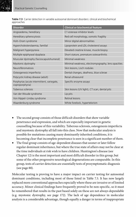
108108 Practical Genetic Counselling
• The second group consists of those difficult disorders that show variable penetrance and expression, and which are especially important in genetic counselling because of this variability. Tuberous sclerosis, osteogenesis imperfecta and myotonic dystrophy all fall into this class. Now that molecular analysis is possible for mutations causing many dominantly inherited conditions, it is becoming clear that incomplete penetrance is seen in a significant number of them.
• The final group consists of age-dependent diseases that sooner or later follow regular dominant inheritance, but where the true state of affairs may not be clear at the time individuals at risk wish to have children. Huntington’s disease (see Chapter 12) is the most important and most difficult disorder in this group, but some of the other progressive neurological degenerations are comparable. In this group, tests of carrier detection are essentially tests of presymptomatic diagnosis (see page 88).
Molecular testing is proving to have a major impact on carrier testing for autosomal dominant conditions, including most of those listed in Table 7.3. It has now largely replaced more conventional approaches especially where these are invasive or of limited accuracy. Minor clinical findings have frequently proved to be non-specific, so it must be remembered that results in the past based solely on these are not always dependable (e.g. myotonic dystrophy, see page 172). The lack of age dependence in molecular analysis is a considerable advantage, though equally a danger in terms of inappropriate
Table 7.3 Carrier detection in variable autosomal dominant disorders: clinical and biochemical approaches
Disorder Clinical or biochemical feature
Angioedema, hereditary C1 esterase inhibitor levels
Hereditary spherocytosis Red cell morphology, osmotic fragility
Holt–Oram syndrome Minor digital abnormalities
Hypercholesterolaemia, familial Lipoprotein and LDL cholesterol assays
Malignant hyperpyrexia Elevated creatine kinase, muscle biopsy
Multiple epiphyseal dysplasia Short stature, premature osteoarthritis
Muscular dystrophy (facioscapulohumeral) Minimal weakness
Myotonic dystrophy Minimal weakness, electromyography; lens opacities
Neurofibromatosis Skin lesions, Lisch nodules
Osteogenesis imperfecta Dental changes, deafness, blue sclerae
Polycystic kidney disease (adult) Renal ultrasound
Porphyrias (acute intermittent, variegate, coproporphyria)
Specific enzyme assays
Tuberous sclerosis Skin lesions (UV light), CT scan, dental pits
Van der Woude syndrome Lip pits
Von Hippel–Lindau syndrome Retinal lesions
Waardenburg syndrome White forelock, hypertelorism

109109Carrier Testing and Genetic Prediction
childhood testing (see page 376). The ease of the procedure from the individual’s view-point, with only a blood sample required, is likewise a double-edged sword, with the danger that samples may arrive in the laboratory without appropriate genetic counsel-ling having been performed.
Gene sequencing techniques have proved particularly valuable in analysis of domi-nantly inherited disorders, especially those where the genes involved are small, since they are able to detect or exclude an abnormality even when this may not be known in a previous family member. It can be assumed that carrier testing is at least potentially feasible for any dominantly inherited disorder whose gene has been mapped or isolated. Whether it is actually possible will depend on the range of mutations, whether there is genetic heterogeneity and, if linked markers rather than mutation analysis are being used, the structure of the particular family pedigree.
In contrast to autosomal recessive disorders, the risk of the offspring of a carrier for an autosomal dominant condition having overt disease at some stage of their life is high. Although in a few cases (e.g. the myopathy underlying malignant hyperpyrexia) the disorder remains constantly subclinical in successive generations, it is common to find more severely affected offspring born to asymptomatic parents. This in part reflects the natural variability of these disorders, but also results from the fact that the carrier parents form the mildest extreme of a range of variability, so that in ‘reverting to the mean’ the children are more likely to be clinically affected. In some instances, as with myotonic dystrophy and Huntington’s disease, there may be parent of origin effects giving severe disease in the offspring as the result of special factors producing anticipation (see pages 30 and 172).
Autosomal dominant disorders of late onset also produce a special difficulty in carrier detection because, in contrast to those with a static course and those with reces-sively inherited disease, the carrier is not just at risk of transmitting the disease but of developing it. Thus to identify an individual as a carrier of Huntington’s disease (apart from those individuals with a subclinical mutation – see Chapter 12) is inevitably to mark out that person as being destined to develop the disease at some time in the future. The whole field of presymptomatic detection for late-onset disorders carries serious ethical and practical consequences (see Chapters 5 and 28). We now have considerable information on the effects of this in Huntington’s disease, which has provided a general model for other late-onset disorders, including the familial cancers, although these differ importantly from Huntington’s disease in having preventable and treatable aspects.
CARRIER DETECTION IN X-LINKED DISEASE
A relatively small number of X-linked recessive disorders provide the most important of all applications of carrier detection. The reason for this is simple: the carriers are generally healthy and so will be likely to reproduce, but in contrast to autosomal recessive inherit-ance, they will be at risk of having affected male offspring regardless of whom they marry. In such a situation, the availability of carrier detection for women at high risk is a major

110110 Practical Genetic Counselling
contribution, and forms such an integral part of genetic counselling that it is often unwise to give a definitive risk estimate until information from testing is available.
The haemophilias and X-linked muscular dystrophies (principally Duchenne dys-trophy) are overwhelmingly the most important disorders that have to be considered in this group, and the approaches to carrier detection and the problems of interpretation are remarkably similar in each, and apply also to most other X-linked disorders. For this reason, although details are given with the individual disorders, they will be used here as examples of carrier detection in X-linked recessive disease generally. Fragile-X syndrome is also a frequent indication for carrier detection, but its unusual genetic features, with normal transmitting males, make it different from most other X-linked disorders in terms of carrier detection.
In both haemophilia and Duchenne dystrophy, a few carriers may be detectable clini-cally, probably as the result of X-chromosome inactivation having randomly resulted in a higher than expected proportion of those X chromosomes bearing the abnormal gene functioning in the particular tissue of importance. Unfortunately, this process will also result in the opposite – namely carriers in whom, principally, the normal X chromosome is functioning, and who will thus be difficult or impossible to detect even by the most sensitive tests. This variability of X-linked carriers is characteristic and must always be borne in mind. A recent review by Ørstavik (see ‘Further reading’) discusses the basis for this in detail. Tests based on DNA are not affected by X inactivation, and this property is one of the most important reasons for their use in X-linked heterozygote detection.
The phenotypic test generally used for Duchenne dystrophy, the elevation of serum creatine kinase levels, is considerably further removed from the basic molecular defect than the assays for factor VIII in haemophilia, with consequently less precision in detect-ing carriers. In both disorders, however, there is a considerable overlap between normal and carrier ranges, which makes it impossible to classify most individuals as ‘normal’ or ‘abnormal’. Instead, a series of likelihood ratios must be used that will give odds for or against the carrier state for any particular result of a carrier test. By using these odds, one can arrive at a much more precise separation of carriers and non-carriers than would otherwise be possible, especially if the results are integrated with other genetic informa-tion, as described in Chapter 2. Although phenotypic tests in these and other X-linked disorders have been progressively replaced by molecular analysis, they retain a place in both Duchenne dystrophy and the haemophilias. In particular, a clearly abnormal result will rarely be outweighed or contradicted by molecular evidence.
The isolated case of an X-linked disorder presents major problems in carrier detec-tion. There is considerable variation between diseases as to the proportion of such cases likely to represent new mutations and, correspondingly, the proportion of mothers who are carriers. The prior risk of such a mother being a carrier will be somewhere between 50 and 100 per cent, depending on the relative mutation rates in the two sexes, and the interpretation of carrier testing will clearly be influenced by this figure. Even the recog-nition of gene deletion in such an isolated case does not always allow exclusion of the carrier state, because some mothers show gonadal mosaicism. This may be a further factor in apparent lack of phenotypic features in carrier women, and seems to be especially frequent in Duchenne dystrophy.

111111Carrier Testing and Genetic Prediction
All daughters of a man with an X-linked recessive disease must be carriers, and so (assuming paternity is correct) tests of carrier detection are irrelevant for such people. Despite this, they are often referred for ‘genetic counselling and carrier detection’ under the misapprehension that a normal result will somehow make a definite carrier less definite! False reassurance is a real danger in such a situation.
Table 7.4 shows some of the X-linked recessive disorders where carrier detection is helpful. The range of approaches is wide and may be morphological, functional,
Table 7.4 Phenotypic changes in carrier detection for X-linked disorders
Disorder Abnormality in carrier
Adrenal leucodystrophy Long-chain fatty acid synthesis
Alport syndrome Microscopic haematuria
Amelogenesis imperfecta Patchy enamel hypoplasia (heterogeneous)
Anhidrotic ectodermal dysplasia Reduced sweat pores, dental defects
Becker muscular dystrophy Serum creatine kinase (less effective than in Duchenne)
Centronuclear myopathy (lethal type) Muscle biopsy changes
Choroideraemia Pigmentary retinal changes
Chronic granulomatous disease Partial NADPH oxidase deficiency; discoid lupus-like skin lesions
Duchenne muscular dystrophy Serum creatine kinase
Fabry’s disease Skin lesions; α-galactosidase assay
Fragile-X mental retardation Variable mental retardation; may show chromosomal fragile site
Glucose-6-phosphate dehydrogenase deficiency
Quantitative enzyme assay and electrophoresis
Haemophilia A Factor VIII assays
Haemophilia B Factor IX assay
Hunter syndrome (MPS II) Enzyme assay on hair bulbs and serum (variable)
Hypogammaglobinaemia (Bruton type) Reduced IgG (some individuals only)
Immune deficiency (SCID) X-inactivation analysis
Lesch–Nyhan syndrome HGPRT assay on hair bulbs
Lowe syndrome Amino aciduria, lens opacities
Ocular albinism Patchy fundal depigmentation
Retinoschisis Cystic retinal changes
Vitamin D-resistant rickets Serum phosphate (may be clinical features)
Wiskott–Aldrich syndrome X-inactivation studies; IgA levels
X-linked congenital cataract Lens opacities
X-linked ichthyosis Corneal opacities, reduced steroid sulphatase
X-linked mental retardation Various non-fragile-X forms; carrier detection rarely feasible
X-linked retinitis pigmentosa Pigmentary changes; abnormality of electroretinogram, more than one locus

112112 Practical Genetic Counselling
biochemical or molecular. The last of these approaches is the definitive one whenever possible. Taken as a group, X-linked disorders are probably the most satisfactory in terms of our ability to detect the carrier state and its applicability in preventing recurrence of the disease within families.
METHODS OF CARRIER DETECTION
The techniques available for detecting the carrier state vary greatly according to the nature of the particular disease and our understanding of its metabolic basis. It is impossible to give all the details here, and available approaches are mentioned as far as possible with individual disorders, but it is worth considering the broad forms of approach and some of the limitations that exist.
Molecular analysisMolecular analysis has become the approach of choice for carrier detection in many, possibly most, mendelian disorders (see Chapter 5 for details), but it should not be assumed that the older technologies are necessarily inferior. Molecular analysis can itself have drawbacks – for example, when any one of several different genes might be involved, or when it may be difficult to be certain whether a particular change is patho-genic or simply a harmless variant. These and other potential problems make it essential that only experienced laboratories, subject to regular quality control and preferably closely linked with clinical genetics services, are used in the molecular analysis of genetic disorders.
Measurement of the primary enzyme or other defectThis remains a very satisfactory approach, where available, and is feasible for numerous inborn errors of metabolism, mostly following autosomal recessive inheritance, as well as for non-enzymatic defects such as haemophilias and various haemoglobinopathies. DNA analysis will often only be required if there is a special need to know the specific mutation involved, such as for prenatal diagnosis. Even in this group, however, the range of results in heterozygotes may show considerable overlap with the normal range, and less commonly with that of the abnormal homozygotes. The appropriate tissue to use will also vary. Serum may be adequate, but more often red or white blood cells or cultured fibroblasts are required and the techniques may be difficult and specialized. X-linked disorders are particularly variable, as already discussed, making DNA analysis preferable where feasible.
Secondary biochemical changesThe value of these will, in general, be related to how close the abnormality is to the primary defect. Important examples are the use of creatine kinase measurement in Duchenne muscular dystrophy, elevation of haemoglobin A levels in beta-thalassaemia, and abnormalities of porphyrin excretion in the acute porphyrias. As our knowledge has increased, such tests have tended to be superseded or used as preliminary screening tests.

113113Carrier Testing and Genetic Prediction
Thus, in phenylketonuria, until the gene was isolated, carrier detection was usually achieved by studying the blood phenylalanine to tyrosine ratio under standardized con-ditions, or by performing a phenylalanine loading test, but this approach is now obsolete.
Cytogenetic studiesThese are of obvious importance in families with a balanced translocation, but new techniques are showing applications in disorders not previously considered chromo-somal, as discussed in Chapter 4. Where a small deletion or translocation is involved (see Table 4.10), the presence or absence of any anomaly in a relative is likely to deter-mine whether the risk is high or low. In the important case of fragile-X mental retardation, molecular analysis for the mutation is of greater value than cytogenetic study in the identification of asymptomatic gene carriers, whether these are females or normal transmitting males (see Chapter 13).
Physiological testsThese are of particular use in those autosomal dominant conditions where we have little biochemical understanding. Examples include the use of electroretinography in detecting the carriers of X-linked retinitis pigmentosa and nerve conduction in Charcot–Marie–Tooth disease. Many are now being superseded by molecular tests (e.g. slit-lamp eye examination and electromyography in myotonic dystrophy).
Microscopic techniquesThese may rely on biopsy, as in Duchenne muscular dystrophy; blood film examina-tion, as with sickle-cell anaemia or thalassaemias; or biomicroscopy, as in slit-lamp examination for the lens opacities of myotonic dystrophy or the Lisch nodules of neu-rofibromatosis. Again, though, specificity of these tests is not always complete, while some are invasive, so their use has diminished as molecular analysis has become possible.
RadiologyThis may show minor skeletal abnormalities in such disorders as osteogenesis imper-fecta, while internal defects may be visible, as in cerebral calcification in tuberous sclerosis and renal abnormalities in polycystic kidney disease. The same cautions over specificity (see above) apply here.
Clinical observationMany carriers may exhibit clinical features that show their genotype and, although the absence of such evidence rarely excludes an individual being a carrier, their presence provides a strong positive indication. Ophthalmic disorders (e.g. choroideraemia and retinitis pigmentosa) are particularly susceptible to carrier detection in this way, as are skin disorders. Female carriers of an X-linked recessive disorder may show a ‘patchy’ appearance, as already noted; again, this is particularly evident in skin, dental and eye disorders where the tissue concerned is open to inspection. Yet again, one must be cautious about the specificity of such minor clinical features.

114114 Practical Genetic Counselling
FURTHER READING
Ørstavik K (2009). X-chromosome inactivation in clinical practice. Hum Genet 126, 363–73.Scriver CR, Beaudet A, Sly WS, Valle D (1996). Metabolic and Molecular Bases of Inherited
Disease. New York: McGraw-Hill [gives details of carrier detection for many specific disorders; see on-line version, chap. 1].
Young ID (2007). Introduction to Risk Calculation in Genetic Counselling. Oxford: Oxford University Press [general approaches to risk estimation are clearly described].

Introduction . . . . . . . . . . . . . . . . . . . . . . . . . . . . . . . . . . . . . . . . . . . . . . . . . . . . . . . . . . . . . . . . . . . 115
Criteria and indications for prenatal diagnosis . . . . . . . . . . . . . . . . . . . . . . . . . . . . . . . . . . . 116
Amniocentesis . . . . . . . . . . . . . . . . . . . . . . . . . . . . . . . . . . . . . . . . . . . . . . . . . . . . . . . . . . . . . . . . . 118
Chorion villus sampling . . . . . . . . . . . . . . . . . . . . . . . . . . . . . . . . . . . . . . . . . . . . . . . . . . . . . . . . . 121
Chromosomal disorders and prenatal diagnosis . . . . . . . . . . . . . . . . . . . . . . . . . . . . . . . . . . 122
Structural malformations . . . . . . . . . . . . . . . . . . . . . . . . . . . . . . . . . . . . . . . . . . . . . . . . . . . . . . . 125
Maternal serum screening for Down’s syndrome . . . . . . . . . . . . . . . . . . . . . . . . . . . . . . . . . 127
X-linked disorders . . . . . . . . . . . . . . . . . . . . . . . . . . . . . . . . . . . . . . . . . . . . . . . . . . . . . . . . . . . . . . 128
Inborn errors of metabolism . . . . . . . . . . . . . . . . . . . . . . . . . . . . . . . . . . . . . . . . . . . . . . . . . . . . 129
Molecular prenatal diagnosis . . . . . . . . . . . . . . . . . . . . . . . . . . . . . . . . . . . . . . . . . . . . . . . . . . . 130
Fetal blood and tissue sampling . . . . . . . . . . . . . . . . . . . . . . . . . . . . . . . . . . . . . . . . . . . . . . . . . 130
Pre-implantation diagnosis . . . . . . . . . . . . . . . . . . . . . . . . . . . . . . . . . . . . . . . . . . . . . . . . . . . . . .131
Fetal DNA analysis of maternal blood . . . . . . . . . . . . . . . . . . . . . . . . . . . . . . . . . . . . . . . . . . . 132
Twins and prenatal diagnosis . . . . . . . . . . . . . . . . . . . . . . . . . . . . . . . . . . . . . . . . . . . . . . . . . . . 132
Maternal aspects of genetic counselling in pregnancy . . . . . . . . . . . . . . . . . . . . . . . . . . . . 133
Further reading . . . . . . . . . . . . . . . . . . . . . . . . . . . . . . . . . . . . . . . . . . . . . . . . . . . . . . . . . . . . . . . . 133
Prenatal Diagnosis and Reproductive Aspects of Medical Genetics
CHAPTER 8
INTRODUCTION
The development of techniques for diagnosing genetic disorders in utero was a major advance in medical genetics, and has so altered the outlook for families at risk of having affected children that it has become one of the main options open to those receiving genetic counselling, especially where there is a high risk of a serious and untreatable disorder. Prenatal diagnostic procedures were initially often developed by, or in close association with, those actively involved in genetic counselling and, perhaps as a result of this, have in general been used appropriately and responsibly. The predominant involvement of obstetricians, together with the diversified nature of the techniques, notably the use of ultrasound and of biochemical screening, have more recently tended to result in prenatal diagnostic procedures being applied as a substitute for genetic counselling, in isolation from the estimation of genetic risks and other relevant factors supporting it.
The author believes strongly that this is unfortunate and potentially harmful, and that prenatal diagnosis, like other clinical and laboratory techniques, must be seen in the context of the entire situation: the risk of a pregnancy being genetically affected, the

116116 Practical Genetic Counselling
other measures such as carrier detection which may define that risk more precisely or even eliminate it, the potential for treatment of the disorder in question and, most importantly, the attitude and wishes of the couple concerned. Whether this is under-taken by non-medical genetic counsellors, clinical geneticists, or by obstetricians themselves is of less importance than that it is done well. In many centres the non-medical genetic counsellor now plays an important role.
Such an approach means that, wherever possible, prenatal diagnosis must be consid-ered, discussed and planned before a pregnancy occurs. To begin this process during pregnancy is highly undesirable (although sometimes inevitable), because not only may procedures have to be hurried, but most pregnant women (and their partners) are not in a state where an objective assessment of the factors for or against prenatal diagnosis and possible termination of pregnancy can easily be undertaken. It is likely that much of the emotional trauma sometimes associated with prenatal diagnosis results from an absence of careful prior planning. The availability of chorion villus sampling (CVS) in the first trimester makes it even more important that the situation be planned carefully in advance of pregnancy.
Most genetic counselling in relation to prenatal diagnosis will inevitably, and rightly, be carried out by obstetricians and those involved in primary care, with special-ists in medical genetics responsible for those cases where the genetic aspects are complex. A welcome development has been the growth of interdisciplinary fetal medicine groups, where obstetrician, radiologist, medical geneticist, non-medical genetic counsellor and other colleagues can meet regularly to discuss specific cases, but there remain too many instances where skilled genetic advice has been sought too late, or not at all.
The widespread use of screening tests in pregnancy now means that most prenatal diagnosis is undertaken where there is no previous risk or expectation of a genetic disorder. The population aspects of this are considered in Chapter 27, but the very real implications that this has for individual families are dealt with later in this chapter.
CRITERIA AND INDICATIONS FOR PRENATAL DIAGNOSIS
When prenatal diagnosis is being considered in genetic counselling, several basic factors must be examined (Box 8.1), but the most important is whether the couple con-cerned actively wish for prenatal diagnosis; all too often it is suggested simply because it may be technically feasible and without adequate information.
Box 8.1 Criteria for prenatal diagnosis
Is the disorder sufficiently severe to warrant termination of the pregnancy?Is treatment absent or unsatisfactory?Is termination of an affected pregnancy acceptable to the couple concerned?Is an accurate prenatal diagnostic test available?Is there a significant genetic risk to the pregnancy?
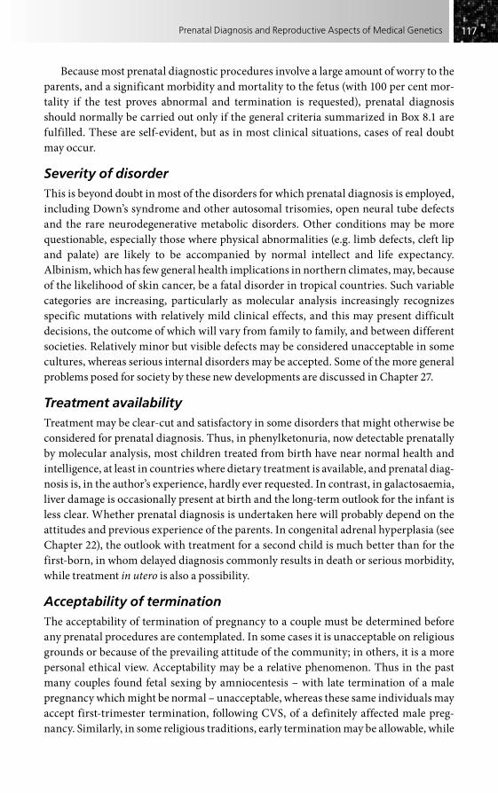
117117Prenatal Diagnosis and Reproductive Aspects of Medical Genetics
Because most prenatal diagnostic procedures involve a large amount of worry to the parents, and a significant morbidity and mortality to the fetus (with 100 per cent mor-tality if the test proves abnormal and termination is requested), prenatal diagnosis should normally be carried out only if the general criteria summarized in Box 8.1 are fulfilled. These are self-evident, but as in most clinical situations, cases of real doubt may occur.
Severity of disorderThis is beyond doubt in most of the disorders for which prenatal diagnosis is employed, including Down’s syndrome and other autosomal trisomies, open neural tube defects and the rare neurodegenerative metabolic disorders. Other conditions may be more questionable, especially those where physical abnormalities (e.g. limb defects, cleft lip and palate) are likely to be accompanied by normal intellect and life expectancy. Albinism, which has few general health implications in northern climates, may, because of the likelihood of skin cancer, be a fatal disorder in tropical countries. Such variable categories are increasing, particularly as molecular analysis increasingly recognizes specific mutations with relatively mild clinical effects, and this may present difficult decisions, the outcome of which will vary from family to family, and between different societies. Relatively minor but visible defects may be considered unacceptable in some cultures, whereas serious internal disorders may be accepted. Some of the more general problems posed for society by these new developments are discussed in Chapter 27.
Treatment availabilityTreatment may be clear-cut and satisfactory in some disorders that might otherwise be considered for prenatal diagnosis. Thus, in phenylketonuria, now detectable prenatally by molecular analysis, most children treated from birth have near normal health and intelligence, at least in countries where dietary treatment is available, and prenatal diag-nosis is, in the author’s experience, hardly ever requested. In contrast, in galactosaemia, liver damage is occasionally present at birth and the long-term outlook for the infant is less clear. Whether prenatal diagnosis is undertaken here will probably depend on the attitudes and previous experience of the parents. In congenital adrenal hyperplasia (see Chapter 22), the outlook with treatment for a second child is much better than for the first-born, in whom delayed diagnosis commonly results in death or serious morbidity, while treatment in utero is also a possibility.
Acceptability of terminationThe acceptability of termination of pregnancy to a couple must be determined before any prenatal procedures are contemplated. In some cases it is unacceptable on religious grounds or because of the prevailing attitude of the community; in others, it is a more personal ethical view. Acceptability may be a relative phenomenon. Thus in the past many couples found fetal sexing by amniocentesis – with late termination of a male pregnancy which might be normal – unacceptable, whereas these same individuals may accept first-trimester termination, following CVS, of a definitely affected male preg-nancy. Similarly, in some religious traditions, early termination may be allowable, while

118118 Practical Genetic Counselling
late termination is forbidden. It is essential to know the attitude of a couple before preg-nancy occurs because this may well affect their decision whether or not to have further children.
Unacceptability of termination should not be considered as automatically ruling out prenatal diagnosis. In rare instances, parents may feel that they will gain by being able to prepare for an affected child, although this is exceptional, especially when the finite risk of prenatal procedures is pointed out. Serious potential ethical problems arise if prenatal diagnosis is undertaken for a late-onset disorder and the pregnancy continues (see Chapter 28).
Feasibility of prenatal diagnosisThe feasibility of diagnosis is something that continues to change rapidly with scien-tific advances, so it cannot be too strongly stressed that the person giving genetic counselling must obtain accurate information on this point before suggesting the pos-sibility to a couple, and must be satisfied that the technique is reliably applicable as a service rather than just as a research procedure. Failure to do this is as reprehensible as submitting a patient to some new surgical procedure without enquiring as to its benefit and mortality. This is especially relevant when using new molecular advances, where the boundary between research discovery and established techniques can be hard to define, especially for very rare disorders, or those where the gene has been recently isolated.
Genetic riskThe final point to be emphasized is that the risk of the disorder occurring in a particular pregnancy must be estimated accurately before prenatal diagnosis is considered; in other words, the consideration of prenatal diagnosis must be an integral part of genetic counselling. All too often the author has seen patients referred for prenatal diagnosis when the risk to the pregnancy has not been properly evaluated, and where the risk on occasion proves to be so low as to make prenatal procedures unwarranted. Even if prenatal diagnosis were free of risk (which it is not), such a slipshod approach cannot be justified. If the clinician involved cannot accurately evaluate the risk (which will not be the case after reading this book, it is hoped), then the advice of a colleague who can do so should be sought.
AMNIOCENTESIS
A variety of techniques exist by which a prenatal diagnosis may be achieved for different disorders. The oldest of these, amniocentesis, the procedure by which a sample of amniotic fluid and its cells is obtained from the pregnant uterus, was first used in 1956 in relation to fetal sexing, but it is still the technique with the widest application, especially for cytogenetic studies on lower-risk pregnancies (Fig. 8.1). It is important, however, to recognize that other approaches exist (Box 8.2), which are rapidly increasing in importance.

119119Prenatal Diagnosis and Reproductive Aspects of Medical Genetics
The amniotic fluid sample should consist of clear fluid, in which are suspended the cells (fetal in origin) that can be cultured for chromosomal, biochemical or molecular studies, or in some situations analysed directly. A bloodstained sample usually indi-cates damage to the placenta; a discoloured fluid may indicate impending or actual fetal death, and is an important factor to note because the subsequent inevitable abortion might otherwise be attributed to the amniocentesis itself. Both these findings reduce the chance of a successful cell culture.
Once obtained, the sample is usually spun immediately and duplicate cell cultures set up. The supernatant fluid can be used for �-fetoprotein (AFP) estimation and, less frequently, for metabolic studies. Most diagnostic studies, however, whether chromo-somal or biochemical, require cultured amniotic fluid cells, and depending on the number required, it may be 8 days to 3 weeks before sufficient are available. It is
Amniocentesis
Assessment of risks – genetic counselling(as far in advance as possible)
Ultrasound (for gestational dating,placental localization, structuralabnormalities and markers)
Amniocentesis (sample taken around week 15 of pregnancy)
Uncultured cellsFISH studiesDNA analysis
Culture(1–3 weeks)
Cultured cellsChromosomes (e.g. Down’s syndrome)Metabolic studies (numerous enzyme defects)DNA analysis
Fluid�-Fetoprotein (neuraltube defects, Down’s syndrome)Other biochemical studies
Figure 8.1 Amniocentesis – the main steps.
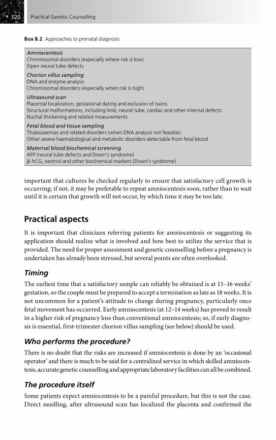
120120 Practical Genetic Counselling
Box 8.2 Approaches to prenatal diagnosis
AmniocentesisChromosomal disorders (especially where risk is low)Open neural tube defects
Chorion villus samplingDNA and enzyme analysisChromosomal disorders (especially when risk is high)
Ultrasound scanPlacental localization, gestational dating and exclusion of twinsStructural malformations, including limb, neural tube, cardiac and other internal defectsNuchal thickening and related measurements
Fetal blood and tissue samplingThalassaemias and related disorders (when DNA analysis not feasible)Other severe haematological and metabolic disorders detectable from fetal blood
Maternal blood biochemical screeningAFP (neural tube defects and Down’s syndrome)-hCG, oestriol and other biochemical markers (Down’s syndrome)
important that cultures be checked regularly to ensure that satisfactory cell growth is occurring; if not, it may be preferable to repeat amniocentesis soon, rather than to wait until it is certain that growth will not occur, by which time it may be too late.
Practical aspects
It is important that clinicians referring patients for amniocentesis or suggesting its application should realize what is involved and how best to utilize the service that is provided. The need for proper assessment and genetic counselling before a pregnancy is undertaken has already been stressed, but several points are often overlooked.
TimingThe earliest time that a satisfactory sample can reliably be obtained is at 15–16 weeks’ gestation, so the couple must be prepared to accept a termination as late as 18 weeks. It is not uncommon for a patient’s attitude to change during pregnancy, particularly once fetal movement has occurred. Early amniocentesis (at 12–14 weeks) has proved to result in a higher risk of pregnancy loss than conventional amniocentesis; so, if early diagno-sis is essential, first-trimester chorion villus sampling (see below) should be used.
Who performs the procedure?There is no doubt that the risks are increased if amniocentesis is done by an ‘occasional operator’ and there is much to be said for a centralized service in which skilled amniocen-tesis, accurate genetic counselling and appropriate laboratory facilities can all be combined.
The procedure itselfSome patients expect amniocentesis to be a painful procedure, but this is not the case. Direct needling, after ultrasound scan has localized the placenta and confirmed the

121121Prenatal Diagnosis and Reproductive Aspects of Medical Genetics
correct gestation, can be done under local anaesthesia without need for hospital admis-sion, although rest afterwards is advisable. It is surprising how many women are referred without a clear picture having been given of what amniocentesis entails, something that is clearly the responsibility of the obstetrician and referring clinician. In many centres, as noted earlier, a fetal medicine group has been set up, with those involved meeting weekly. This allows unexpected findings to be discussed, along with the likely genetic risks and possible diagnosis, something that is especially important in interpreting the findings of ultrasonography in pregnancies not known previously to be at risk.
Results and follow-upThe couple must be warned that a definitive result may take 10 days to 3 weeks (some-times longer for biochemical and DNA studies) and that there is a possibility that a repeat sample may be needed. Results of rapid trisomy testing on uncultured cells should still be regarded as provisional. The possibility of abnormality unrelated to that primarily being looked for should be mentioned (e.g. a sex chromosome anomaly in a pregnancy at risk for Down’s syndrome), and a clear policy must be defined in the event of no definite answer being reached because of culture failure or other reasons.
Follow-up after delivery is important not only to check the correctness of the prediction, but also because the outcome may affect the genetic risk to subsequent pregnancies.
Risks of amniocentesis
Amniocentesis is not without risk to the pregnancy. Approximate figures that can be used in discussion with families, based on a number of large studies, are as follows.
• Abortion. The added risk is around 0.5 per cent, but repeat amniocentesis carries a much higher risk (5–10 per cent), as does the performance of the procedure by an inexperienced person. Each centre should audit its own figures.
• Perinatal problems. These are not significant; an increase in postural orthopaedic deformities was suggested in one early UK study, but this was not confirmed.
• Maternal risks. These are minimal, apart from a possibility of rhesus sensitization (rhesus-negative mothers should receive anti-RhD antibody).
In general, amniocentesis has proved to be a safe and reliable procedure, but it is essen-tial that it be used selectively and appropriately, and that the risks be carefully explained to couples who may be considering it. Each centre should monitor and make available its own risks and failure rate.
CHORION VILLUS SAMPLING
Prenatal diagnosis in the first trimester using fetal tissue obtained from chorionic villi has proved to be a major advance, especially for molecular diagnosis, but has not super-seded amniocentesis for low-risk cytogenetic indications.

122122 Practical Genetic Counselling
Currently, samples are obtained either transabdominally or transcervically at around 1 weeks’ gestation under ultrasound guidance; at this stage chorionic tissue has not yet become localized into a placenta. It is difficult to obtain tissue transcervically after 12 weeks, although transabdominal placental sampling is feasible at a later stage. Maternal tissue is dissected away microscopically, and the remaining villi consist of pure fetal tissue that can be used for a variety of diagnostic approaches, including the following.
• Molecular studies. Uncultured chorionic villi are much the most satisfactory sample for these and can provide a rapid and accurate prenatal diagnosis for any disorder for which mutation analysis or DNA markers are available. There is no evidence that the DNA of this tissue gives significantly different results from other fetal samples.
• Enzyme studies. The normal ranges in this material may differ considerably from amniotic fluid cells, while in X-linked diseases, differential X inactivation may give very low levels in a female heterozygote.
• Chromosome studies. A high frequency of chromosomal mosiacism, not present in the fetus itself, may prove misleading, and is one of the reasons why amniocentesis remains preferable to CVS for most chromosomal indications.
Experience with CVS is now extensive. It is likely that the rate of miscarriage is higher than in amniocentesis (2–3 per cent extra, compared with 1 per cent, and considerably more in inexperienced hands). Reports of an excess of limb defects possibly related to the procedure, especially in biopsies taken at 9 weeks or earlier, have not been con-firmed at 10 weeks or later; but, in general, amniocentesis remains the primary approach for pregnancies at lower risk, with CVS used in higher-risk situations and notably for molecular analyses.
CHROMOSOMAL DISORDERS AND PRENATAL DIAGNOSIS
The recurrence risks for the major types of chromosome disorder are discussed in Chapter 4, and the strength of indication for prenatal diagnosis will depend on the magnitude of this risk as well as on the nature of the disorder and the attitude and expe-rience of the couple concerned in relation to it. The relative risks of amniocentesis and CVS will need careful consideration, especially where the risk of abnormality in the
Box 8.3 Principal chromosomal indications for prenatal diagnosis (see Chapter 4 for detailed risk figures)
One parent carrier of a balanced autosomal translocationAdvanced maternal ageAbnormal Down’s screening testUltrasound findings suggestive of Down’s syndrome or other chromosome disorderPrevious child with autosomal trisomy or similar abnormalityParent mosaic for chromosomal abnormalityChromosomal instability syndromes

123123Prenatal Diagnosis and Reproductive Aspects of Medical Genetics
pregnancy is low. The main chromosomal indications for prenatal diagnosis are consid-ered individually below (see also Box 8.3).
Advanced maternal age
The risk with advanced maternal age is primarily for Down’s syndrome, with a lesser risk of other trisomies. The difference in risk of having a live-born child with an abnormality and that for an abnormality found at amniocentesis has already been emphasized (see Tables 4.5 and 4.6), but there is general agreement that this risk is sufficiently great that all women aged 35 years or over should be informed of the risks of Down’s syndrome and offered amniocentesis. Although the uptake of amniocentesis varies greatly between countries and regions, this often reflects a failure of obstetricians and others to give adequate information, together with geographical variability in the services provided.
The pattern of amniocentesis has been considerably affected by the development of biochemical screening tests for Down’s syndrome in the second trimester, usually a combination of AFP, human chorionic gonadotrophin (hCG) and oestriol assessment (discussed further below and in Chapter 27). As a result of this, it is now possible to modify the age-specific risks so that some older women whose risk is considerably reduced will no longer opt for amniocentesis. Likewise the analysis of fetal DNA in maternal blood as a first-trimester screening test, now beginning to be used as a diag-nostic service, will considerably alter the situation (see page 132).
Translocation Down’s syndrome
The rare, but high-risk, group with translocation Down’s syndrome comprises only 5 per cent of all cases of Down’s syndrome. The risks for the offspring of balanced carriers of the various forms of translocation are discussed in Chapter 4 and are given in Table 4.8.
It is clearly vital that the precise type be established from study of the index case, and that relatives at risk should be investigated using blood before a pregnancy occurs. CVS may be preferred to amniocentesis in a high-risk situation. If the individual is a chro-mosomally normal sib or more distant relative, there is no indication for prenatal tests. Where the parents of a child with translocation Down’s syndrome are both chromo-somally normal, the risk of a further affected child is also low, probably similar to that for trisomic Down’s (see below). Although it may be reasonable to offer prenatal diagno-sis to such couples in a subsequent pregnancy, it should not be undertaken under the false assumption that the risk of Down’s syndrome is high.
Trisomic Down’s syndrome and other autosomal trisomies
All studies agree that the recurrence risk for trisomic syndromes is low, with the excep-tion of very rare families in which there appears to be some special factor causing clustering of chromosomal abnormalities, possibly by predisposing to non-disjunction or as a result of gonadal mosaicism. It is important that the chromosomes of the child, or of
124124 Practical Genetic Counselling
both parents if the child is dead, be studied before another pregnancy is considered, and that the age of the mother is considered in estimating the recurrence risk (see Chapter 4).
In practice, most couples who have had an affected child understandably elect for prenatal diagnosis in a subsequent pregnancy, even in full knowledge of the risk from the procedure and the low probability that the abnormality will recur.
For other relatives of a child with isolated trisomic Down’s syndrome, there is no evidence of an increased risk in their offspring, and this should be made clear at the time of diagnosis of the index case to avoid unnecessary worry. Where the index case is no longer living and no definite chromosomal status has been recognized, it is prefera-ble to undertake an urgent blood karyotype of the at-risk relative rather than perform an unnecessary amniocentesis. Unfortunately, there is still no test – chromosomal or molecular – that will distinguish, prior to conception, the couple particularly likely to have a trisomic child.
Problems with chromosomal prenatal diagnosis
The following problems may complicate prenatal chromosomal diagnosis.
Faulty diagnosis in the index caseDown’s syndrome said to affect an older relative may prove to be an entirely different disorder, and the records (or the affected individual, if available) should be checked wherever possible. A cytogenetic register of all known cases of Down’s syndrome, par-ticularly translocation Down’s syndrome, in a region is helpful.
Chromosomal variants unrelated to clinical abnormalityThese may cause confusion, particularly in investigation of cases of mental retardation, where chromosome studies are commonly performed, and should be particularly sus-pected if healthy family members show the same chromosomal pattern. They may also be discovered in the amniotic cells of the fetus at amniocentesis. In most cases, there should be little doubt as to whether they are pathological; again, help may come from finding the same pattern in a healthy parent. The findings should always be discussed with the cytogenetic laboratory that has actually performed the analysis.
Unrelated or unexpected abnormalitiesThese may be chromosomal (e.g. sex chromosome anomalies or a balanced transloca-tion), or a raised AFP level may suggest a neural tube defect in a pregnancy studied primarily for a chromosomal indication. Such findings may lead to a difficult dilemma, and in general one has to discuss the facts with the couple and respect their decision. It is always easier if the possibility of such a situation arising has been explained prior to the amniocentesis.
MosaicismInterpreting this is often difficult – not only the likely consequences and severity in the child, but also whether it is a genuine reflection of the chromosome state of the fetus.

125125Prenatal Diagnosis and Reproductive Aspects of Medical Genetics
This is especially the case in CVS samples, where mosaicism is often confined to the placenta; amniocentesis may be needed to check this, but close liaison with the labora-tory is wise.
STRUCTURAL MALFORMATIONS
Neural tube defects
A series of factors have combined to reduce the birth frequency of neural tube defects in most populations where these have previously had a high incidence. This is a major achievement which, like rhesus isoimmunization, is in danger of being overlooked now that major defects of this type no longer represent such an important cause of serious abnormality, disability and death in childhood. The principal factors include:
• a natural decline in incidence, especially in high-risk populations, probably related to maternal nutritional status;
• primary prevention in high-risk pregnancies and the wider population by use of folic acid, taken preconceptually and as a food supplement;
• increasing resolution of prenatal ultrasonography;• maternal serum AFP screening.
The first step was the discovery that pregnancies ending in an open neural tube defect had elevated amniotic fluid levels of AFP, which provided an accurate prenatal test for pregnancies known to be at high risk. The subsequent development of ultrasonography provided a means of both confirming the diagnosis and detecting most closed lesions. As ultrasound scanning has become more sensitive and is used more routinely, this has allowed detection in pregnancies not known to be at risk. The first true screening approach came from maternal serum AFP analysis. In areas of high incidence, this remains the main screening test, in conjunction with an ultrasound scan. In areas where the incidence is low, high-resolution ultrasonography alone is often preferred, as the proportion of false-positive raised AFP samples will be higher in these areas.
Amniocentesis is now often not performed where a clearly abnormal pregnancy is detected, since delay will inevitably result. Where there are doubts, this test should be undertaken, together with chromosome analysis on the amniotic fluid if there is any suspicion of a complex malformation syndrome.
In general, these methods of prenatal diagnosis of neural tube defects have proved reliable and free from major problems. However, several points need to be borne in mind.
• There are a number of other causes of a raised serum AFP level (Box 8.4), including normal twin pregnancy. These can often be distinguished by the level of acetylcholinesterase in amniotic fluid, which is specifically raised in neural tube defects.
• Routine obstetric ultrasound scanning is an inadequate substitute for high-resolution ultrasonography performed by an expert, at the optimal gestation (usually around 18 weeks).

126126 Practical Genetic Counselling
• High-risk pregnancies must be considered separately from general population screening and the risks estimated appropriately.
• Any pregnancy terminated should have an expert fetal autopsy, since numerous syndromes associated with neural tube defects exist, some mendelian, others chromosomal (see Chapter 12).
Other structural abnormalities: ultrasound scanning in prenatal diagnosis
Ultrasonography is now a sensitive, valuable and widely available approach to prenatal diagnosis. Box 8.5 summarizes some of the main applications, but a few general points must be made.
• There is an immense difference between the results of expert units specializing in detection of malformations and the results of radiologists, radiographers and obstetricians using ultrasound as a more general tool. This applies to both false positives and false negatives and to the interpretation of any possibly abnormal results. The rapid spread of ultrasonography outside specialist centres can be actively harmful unless there is a comparable spread of expertise, which is often still not the case. Most genetics centres are aware of pregnancies that were terminated because of supposed ultrasonographic abnormality and where the fetus proved to be entirely normal.
• Although ultrasound is itself apparently risk-free, one must also consider the risks of any attendant or consequent investigations resulting from apparently abnormal or uncertain findings. These may be considerable.
• In investigating genetic abnormalities, the prior risk of the situation must be taken into consideration. Ability to detect or exclude an abnormality in the face of a high genetic risk is quite different from doing so when the risk is low or when ultrasound scanning is used as a screening procedure. This obvious fact is still ignored by many of those using the technique.
• Many ultrasonographic ‘abnormalities’ or ‘soft markers’ may be transient and physiological rather than pathological in nature. Others may indicate abnormality in only a proportion of cases (e.g. nuchal oedema in trisomy 21; choroid plexus cysts in trisomy 18).
Box 8.4 Abnormalities other than neural tube defects that may cause a raised amniotic fluid �-fetoprotein level
Spontaneous intrauterine deathOmphalocele and gastroschisisBowel and oesophageal atresiasTurner syndromeCongenital nephrosis (Finnish type)Sacrococcygeal teratomaBladder exstrophyFocal dermal hypoplasia and other skin defectsMeckel syndrome

127127Prenatal Diagnosis and Reproductive Aspects of Medical Genetics
• The best results from ultrasound scanning are likely to be when it is used in conjunction with other appropriate investigations. The balance of value of the various tests will change, but they should be considered together for each particular problem. Likewise, the interpretation of puzzling findings is best carried out in conjunction with other specialities, preferably as part of a fetal medicine group, as mentioned earlier.
The diagnosis of congenital heart disease by ultrasonography provides a good example of how uncertain is the role of the technique. Although severe lesions can be detected in the middle trimester of pregnancy, there is still uncertainty as to how accurately less severe defects can be recognized or excluded, or whether this is a suitable investigation for families with the more common forms of congenital heart defect, especially where the recurrence risk is low.
MATERNAL SERUM SCREENING FOR DOWN’S SYNDROME
Although population maternal serum screening was originally started for neural tube defects in areas of high incidence, as indicated above, the finding that AFP levels were frequently reduced in Down’s syndrome has led to its use in screening antenatally for this disorder, especially since the additional measurement of levels of -hCG, oestriol and pregnancy-associated plasma protein-A (PAPP-A) has given a greater discrimina-tion than AFP assessment alone. An additional screening technique for Down’s syndrome is the fetal nuchal fold thickness as measured by ultrasound scan.
The value of this approach is that – in theory at least – the number of invasive tests such as amniocentesis can be kept constant or even reduced, while the proportion of
Box 8.5 Ultrasound in prenatal diagnosis of structural malformations
General applicationsAccurate gestational datingMultiple pregnancyPlacental localization before amniocentesis or chorion biopsy
Central nervous system abnormalitiesAnencephalySpina bifida (in conjunction with other approaches)Hydrocephalus and hydranencephalyMicrocephaly (see Chapter 12 for limitations)
Skeletal defectsSevere neonatal bone dysplasia (e.g. achondrogenesis)Osteogenesis imperfecta (severe congenital forms only)Limb defects (especially of digits)
Internal abnormalitiesSevere congenital heart defectsRenal agenesisInfantile polycystic kidney diseaseSevere obstructive uropathyOmphalocele and gastroschisisFetal tumours

128128 Practical Genetic Counselling
tests with abnormal results is raised. In practical terms, this can allow mothers of all ages to be offered amniocentesis if the screening test passes an agreed threshold of risk (often 1 in 250), while other women can avoid amniocentesis if their risk following serum screening is reduced to below this level.
In practice, the situation is often less simple. Older women may be reluctant to forego a definitive test (and their obstetricians may fear the legal consequences). The greater completeness of coverage may itself increase the amniocentesis rate, overload-ing the cytogenetics laboratory, while many women (and often their doctors) may be worried by being given a specific risk estimate, however small it is, and request amnio-centesis. An additional problem, as with all screening, is that an abnormal screening result produces severe distress and anxiety in women who have not perceived them-selves to be at high risk, and who may have been given inadequate information about the test. Many genetic clinics have found themselves having to attempt to resolve such traumatic situations, an almost impossible task. There can be no doubt that, for these women at least, screening has caused more harm then good.
The overall issues of population screening are debated in Chapter 27; for maternal serum Down’s syndrome screening, the evidence currently suggests that it will allow the avoidance of Down’s syndrome in a considerably greater proportion (60–70 per cent) of pregnancies than age-related screening alone. How this can be weighed against any adverse effects on women being screened is an open question, for which more evidence, social as well as scientific, is urgently needed as such screening becomes more wide-spread. What is clear is that good practice necessitates the full provision of information to allow informed choices by women and their partners.
X-LINKED DISORDERS
Where a pregnancy is at high risk for an X-linked disorder, fetal sexing offers the possi-bility of determining whether the fetus is indeed at risk. However, in most disorders, direct molecular prenatal diagnosis of an affected male is now possible, and even where it is not, linked DNA markers can often be used. First-trimester fetal sexing using both DNA and cytogenetic methods is now feasible with CVS, and should always accompany metabolic studies, which may give variable and misleading results in X-linked conditions as a result of differential X inactivation.
Where a woman is only a possible carrier, not a definite one, it is vital to estimate the risk before fetal sexing is undertaken and to use methods of carrier detection where applicable (see Chapter 7). The most successful approach to prenatal diagnosis is where no prenatal procedure is required at all, because the carrier state has been excluded. As with any prenatal diagnostic investigation, this should be approached as a planned pro-cedure with the issues for and against resolved, as far as possible, beforehand.
Occasionally, in haemophilia or Becker muscular dystrophy for example, fetal sexing may be requested for a pregnancy with an affected father, with a view to termina-tion of a female fetus, which would inevitably be a carrier. The author has serious misgivings about the wisdom of this, because such daughters would almost always be

129129Prenatal Diagnosis and Reproductive Aspects of Medical Genetics
healthy, and because future advances are likely to allow them the option of direct prenatal diagnosis in their own future children, even for disorders where this is not possible at present. The situation is less clear-cut for disorders where heterozygous females frequently show some clinical effects, such as ornithine transcarbamylase defi-ciency and fragile-X mental retardation.
Prenatal sex selection (usually by ultrasound and sometimes by amniocentesis), solely for parental choice, is currently illegal in most countries, including the UK. However, it is being grossly abused in some parts of the world, notably India, almost always in favour of a male pregnancy. Developments in sperm sorting, still unreliable but likely to become feasible at some point in the future, have raised the issue of whether this should be permitted for non-medical reasons. A recent UK report has recom-mended against this at present.
INBORN ERRORS OF METABOLISM
The development of prenatal diagnostic techniques for a variety of inherited metabolic disorders has been one of the major achievements of human genetics, even if the number of families at risk for such disorders is small in comparison with chromosomal disorders such as Down’s syndrome. So many serious inborn errors are now prenatally detectable that this aspect is one of the first to be raised by any couple who have had an affected child.
Chapter 23 covers some of the more important disorders and virtually all of them are now diagnosable prenatally, by either biochemical or DNA analysis, or a combina-tion of both.
The following points need to be borne in mind when prenatal diagnosis of a meta-bolic disorder is being considered.
• The great majority of these disorders follow autosomal recessive inheritance, so that only sibs of affected patients are at high (1 in 4) risk. Risks for other relatives are rarely high enough to warrant undertaking prenatal diagnosis.
• Most disorders require chorionic villi or cultured amniotic fluid cells for their diagnosis. In general, if a particular enzyme defect can be detected in the cultured skin fibroblast, it can also be detected in cultured amniotic fluid cells or villus material, but normal ranges may differ considerably.
• Large numbers of cells are often required, making direct analysis of chorionic villi especially suitable. The need for cell culture with amniocentesis may add delay and uncertainty, which should be explained beforehand. A late termination may be unacceptable to a couple who would accept termination in early pregnancy.
• Many of the enzymatic techniques are exceptionally specialized and can be undertaken only by a few laboratories. Careful advance planning is essential because cells may have to be sent long distances. Fortunately, a remarkable degree of cooperation nationally and internationally exists between laboratories involved in this work, although careful distinction is needed between a centre that is interested in a disorder and one that has proven experience in its prenatal diagnosis.

130130 Practical Genetic Counselling
DNA analysis also requires great familiarity with the genes concerned, but there is a higher degree of ‘common technology’ between the different disorders.
• Wherever possible, samples from the affected individual should be studied alongside those at risk. If the affected child is likely to die, every effort should be made to reach a precise enzymatic or molecular diagnosis beforehand, and to arrange for storage of DNA, cultured cells or postmortem material to be used at a later date. Failure to do this may result in serious problems in relation to a future pregnancy.
A few inherited metabolic diseases can be diagnosed directly from amniotic fluid itself, although the use of cultured cells is usually desirable as a back-up. These include the organic acidurias (propionic and methylmalonic aciduria), mucopolysaccharidoses, the 21-hydroxylase form of congenital adrenal hyperplasia, and congenital nephrosis (diagnosed by elevated amniotic AFP levels).
MOLECULAR PRENATAL DIAGNOSIS
The impact of molecular genetics has been felt in the area of prenatal diagnosis more than in most other branches of medical genetics. There are many reasons for this, including the independence of the techniques from gestational timing and tissue spe-cificity, the lack of variability in heterozygotes, and the suitability of first- trimester or even pre-implantation samples. Most of all, molecular analysis has extended the possi-bility of prenatal diagnosis to numerous serious disorders, often dominantly inherited, where lack of specific biochemical knowledge previously precluded any attempts at prenatal diagnosis. In any inherited disorder for which the specific gene has been isolated, it can be assumed that prenatal diagnosis is potentially feasible.
The range of molecular approaches available is the same as already indicated in Chapter 5, and also outlined in relation to carrier detection in Chapter 7. Where a mutation has been identified in an affected relative, search for this will avoid the necessity for additional tests. Where this is not possible, a wider search for mutations may be needed and this may have limitations, though whole gene sequencing has reduced these. If linked genetic markers are being used, intragenic markers will be reliable provided that other family members can be typed; for recessive disorders a previous affected child is usually the critical sample. The importance of storing a sample from such key individuals for the future must yet again be emphasized.
If linked DNA markers outside the gene are used, a situation much less frequent now than formerly, the risk of error due to recombination must be carefully estimated and discussed before embarking on prenatal testing.
FETAL BLOOD AND TISSUE SAMPLING
Direct umbilical cord sampling under ultrasonographic control has been used in the past to obtain pure fetal blood, but DNA techniques have made it unnecessary in most

131131Prenatal Diagnosis and Reproductive Aspects of Medical Genetics
situations where it was used before – notably in the haemoglobinopathies and haemophilias.
Fetal skin biopsy has proved both reliable and safe for a variety of severe conditions, including lethal and dystrophic epidermolysis bullosa, ichthyosiform erythroderma, Sjogren–Larsson syndrome and severe oculocutaneous albinism; there is usually no visible scar. As with fetal blood sampling, though, molecular analysis using DNA from a chorion villus sample has progressively replaced this approach.
PRE-IMPLANTATION DIAGNOSIS
For many couples at high risk of transmitting a serious genetic disorder, this could potentially offer the possibility of avoiding termination of an established pregnancy. It is now possible to undertake molecular analysis of cells from an early embryo produced by in-vitro fertilization, and to implant only those embryos shown to have an unaf-fected genotype.
In principle this should allow pre-implantation diagnosis to be offered for any disorder where a specific molecular or chromosomal defect can be identified. In practice there are major reservations, which means that pre-implantation diagnosis must still be considered as a research procedure in most situations until much more evidence is available. These reservations include:
• inadequate numbers studied to give meaningful rates of error or failure to obtain a result;
• absence of long-term data on potential harmful effects of the procedure;• lack of prior experience in identifying the defect for most individual disorders;• limited success rate for IVF in general.
In view of these serious limitations, it is most unfortunate that some centres offer pre-implantation diagnosis without making it clear to referring clinicians or to families what prior experience they have had with individual disorders, what prepar-atory research they have undertaken, or whether what they are offering is research or service. Even more disturbing is the fact that pre-implantation diagnosis is not always being offered in the context of full genetic counselling. Given the complex genetic situations commonly involved, expert information on the wider genetic aspects is essential if families are to make an informed choice between the possible options open to them. There is a clear need for effective regulation in this field, espe-cially since it is often run as a commercial venture, rather than as part of established universal health services. Such regulation is present in some European countries (including the UK to a limited extent), but is largely absent in the rest of the world. Until uniformly high standards are produced, it is important that all those involved in genetic counselling maintain a critical and fully informed attitude to pre-implan-tation diagnosis, have detailed knowledge of the services in their region or country, and try to ensure that families embarking on the procedure are similarly well informed.

132132 Practical Genetic Counselling
FETAL DNA ANALYSIS OF MATERNAL BLOOD
This is another field that is beginning to come into clinical use, though considerable research is still needed. It has long been known that a small number of fetal cells are present in maternal blood but more recently free fetal DNA has also been shown to be present; the development of this technique provides a non-invasive approach to prenatal diagnosis. Initially used in relation to Rh haemolytic disease, current research is con-centrating on the use of this as part of a primary test for Down’s syndrome and other aneuploidies, and for fetal sexing. A recent report (2009) from an expert working group of the UK Public Health Genetics Foundation (www.phgfoundation.org) is a valuable information source on good practice in its future use, and on its limitations.
TWINS AND PRENATAL DIAGNOSIS
The discovery of a twin pregnancy poses obvious practical problems in undertaking amniocentesis, but it also alters the genetic risk figures that would normally be given for a singleton pregnancy. The problems involved in estimating the modified risks are summarized in Table 8.1. It is assumed here that a third of twin pairs are monozygous.
In general, the risks are considerably higher than for a singleton pregnancy, and they are altered if information is available on one twin from amniocentesis. The figures do not include the increased risk of malformations known to be associated with
Table 8.1 Genetic risks in twin pregnancies
Chromosome abnormality
Neural tube defect
X-linked (fetal sexing only)
X-linked (specific diagnostic test)
Autosomal recessive
Before amniocentesis
Risk for singleton pregnancy
Y* 125
12
14
14
Risk of at least one twin being affected
53Y
225
23
38
38
Risk of both twins being affected
~13 Y < 1
10013
18
18
Twin A normal; risk of twin B being abnormal
23 Y
125
13
16
16
After amniocentesis (one sac successfully tested)
Twin A abnormal; risk of twin B being normal
23 –
23 Y
910
13
12
12
Twin A abnormal; risk of twin B being abnormal
13 +
23 Y
110
23
12
12
*Risk (Y) will vary with maternal age, etc.After Hunter AGW, Cox DM (1985), Clin Genet 16, 34–42.

133133Prenatal Diagnosis and Reproductive Aspects of Medical Genetics
monozygotic twin pregnancies (see page 147). Clearly, the couple concerned should be informed of these altered risks, and the same is true even when prenatal diagnosis is not being considered.
A further point to be noted is that the normal range of maternal serum AFP con-centrations is raised in twin pregnancies. Twin percentiles are now available for assessing AFP levels; it should be noted that all twin pregnancies ending in a neural tube defect had a serum AFP concentration at least 75 times the normal singleton median value.
Techniques of selective abortion are possible when only one of a twin pair is found to be abnormal, but twin pregnancies remain a difficult field of management. Multiple births are also increasingly seen following IVF procedures.
MATERNAL ASPECTS OF GENETIC COUNSELLING IN PREGNANCY
Although prenatal diagnosis is principally concerned with procedures involving the fetus, genetic counselling in pregnancy also needs to look at aspects of health involving the mother, as well as the psychosocial issues. In many dominantly inherited disorders, the mother will be affected by the disorder, which may pose direct risks to her health in pregnancy or give obstetric complications. Prognosis of an affected parent is a further relevant point in relation to any decisions. Examples of the range of problems needing consideration are given in Box 8.6.
Box 8.6 Genetic disorders in pregnancy and maternal health
Maternal health adversely affected by pregnancyCystic fibrosis (respiratory decompensation)Homocystinuria, sickle-cell disease (thrombotic episodes)
Obstetric complications increasedAchondroplasia (pelvic disproportion, uterine myomas)Ehlers–Danlos syndrome (premature rupture of membranes)Myotonic dystrophy (postpartum haemorrhage, anaesthetic problems)Steroid sulphatase deficiency (delayed labour, increased risk of stillbirth)
FURTHER READING
Abramsky L, Chapple J (2003). Prenatal Diagnosis: the Human Side, 2nd edn. London: Chapman & Hall.
Brock DJH, Rodeck CH, Ferguson-Smith MA (eds) (1992). Prenatal Diagnosis and Screening. Edinburgh: Churchill Livingstone.
Clarke A (1997). Prenatal genetic screening: paradigms and perspectives. In: Harper PS, Clarke A (eds) Genetics, Society and Clinical Practice, pp. 119–140. Oxford: BIOS.
De Crespigny L, Dredge R (1991). Which Tests for My Unborn Baby? Oxford: Oxford University Press [a clearly written guide for the educated and articulate parent-to-be].

134134 Practical Genetic Counselling
Gardner RJM, Sutherland GR (2003). Chromosome Abnormalities and Genetic Counselling. Oxford: Oxford University Press [a valuable source for all aspects of chromosomal prenatal diagnosis].
Milunsky A, Milunsky JM (eds) (2010). Genetic Disorders and the Fetus, 6th edn. Baltimore: Johns Hopkins University Press.
Prenatal Diagnosis (Wiley InterScience), a journal that is a valuable source of recent advances.Online Metabolic and Molecular Bases of Inherited Disease (OMMBID) – http://ommbid.com
[a regularly updated version of Scriver et al. (2001), Metabolic and Molecular Bases of Inherited Disease, McGraw-Hill, New York].
Simpson JL, Golbus MS (1998). Genetics in Obstetrics and Gynaecology. Philadelphia: WB Saunders.
Twining P, McHugo J, Pilling D (1999). Textbook of Fetal Abnormalities. Edinburgh: Churchill Livingstone.
Wright CF, Chitty LS (2009). Cell free DNA and RNA in maternal blood: implications for safer antenatal testing. Br Med J 339, 161–4.

Consanguinity . . . . . . . . . . . . . . . . . . . . . . . . . . . . . . . . . . . . . . . . . . . . . . . . . . . . . . . . . . . . . . . . . 135
Paternity and relationship testing . . . . . . . . . . . . . . . . . . . . . . . . . . . . . . . . . . . . . . . . . . . . . . . 144
Twins . . . . . . . . . . . . . . . . . . . . . . . . . . . . . . . . . . . . . . . . . . . . . . . . . . . . . . . . . . . . . . . . . . . . . . . . . . 145
Legal problems in medical genetics . . . . . . . . . . . . . . . . . . . . . . . . . . . . . . . . . . . . . . . . . . . . . . . 147
Further reading . . . . . . . . . . . . . . . . . . . . . . . . . . . . . . . . . . . . . . . . . . . . . . . . . . . . . . . . . . . . . . . . 148
Special Issues in Genetic Counselling
CHAPTER 9
CONSANGUINITY
Consanguinity, or marriage between close relatives, is a common and important issue in genetic counselling. Where an inherited disorder is present in the family, consan-guinity may significantly influence the risks. Even without a known disorder, couples who are closely related may be concerned about the risks to their offspring.
There are three aspects of consanguinity that need to be considered in relation to genetic counselling:
• What is the exact relationship between the two individuals?• How is the risk of a genetic disorder in the family influenced by the occurrence of
consanguinity?• How likely is it that any harmful gene might be handed by both members of the
couple to a child, i.e. that the child is homozygous by descent for that gene?
An attempt is made here to answer these questions in simple terms, avoiding a complex mathematical approach. These same questions are of more general interest to popula-tion geneticists, and detailed accounts of the subject can be found in genetics textbooks (see ‘Further reading’ in Chapter 1).
Genetic relationships
An accurate idea of how individuals are related is important in all genetic counselling, regardless of the presence or absence of consanguinity, but when a marriage between close relatives is being considered it becomes essential. Table 9.1 summarizes the main categories of relationship, and it is always important to construct a precise pedigree pattern rather than relying on verbal descriptions, which may be confusing. Table 9.2 gives examples.

136136 Practical Genetic Counselling
Table 9.1 Degrees of relationship
Degree Proportion of genes shared
First-degree: 1/2
– Sibs
– Dizygotic twins
– Parents
– Children
Second-degree: 1/4
– Half-sibs
– Uncles, aunts
– Nephews, nieces
– Double first cousins
Third-degree: 1/8
– First cousins
– Half-uncles, aunts
– Half-nephews, nieces
Table 9.2 Patterns of relationship
Relationship (between shaded individuals)
Degree of relationship
Proportion of genes shared
Chance of homozygosity by descent (F)
Monozygotic twins – 1 –
Dizygotic twins First 1/2 1/4
Sibs First 1/2 1/4
Parent–child First 1/2 –
Uncle (aunt)– (niece) nephew
Second 1/4 1/8
Half-sibs Second 1/4 1/8
Double first cousins Second 1/4 1/8
First cousins Third 1/8 1/16
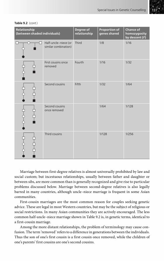
137137Special Issues in Genetic Counselling
Marriage between first-degree relatives is almost universally prohibited by law and social custom; but incestuous relationships, usually between father and daughter or between sibs, are more common than is generally recognized and give rise to particular problems discussed below. Marriage between second-degree relatives is also legally barred in many countries, although uncle–niece marriage is frequent in some Asian communities.
First-cousin marriages are the most common reason for couples seeking genetic advice. These are legal in most Western countries, but may be the subject of religious or social restrictions. In many Asian communities they are actively encouraged. The less common half-uncle–niece marriage shown in Table 9.2 is, in genetic terms, identical to a first-cousin marriage.
Among the more distant relationships, the problem of terminology may cause con-fusion. The term ‘removed’ refers to a difference in generations between the individuals. Thus the son of one’s first cousin is a first cousin once removed, while the children of one’s parents’ first cousins are one’s second cousins.
Table 9.2 (cont.)
Relationship (between shaded individuals)
Degree of relationship
Proportion of genes shared
Chance of homozygosity by descent (F)
Half-uncle–niece (or similar combination)
Third 1/8 1/16
First cousins once removed
Fourth 1/16 1/32
Second cousins Fifth 1/32 1/64
Second cousins once removed
1/64 1/128
Third cousins 1/128 1/256

138138 Practical Genetic Counselling
It should be noted that the ‘degrees’ of relationship shown in Table 9.1 are those used in genetic terminology, as well as in English canon and common law, but not in civil law, where a different approach is used. Thus, uncle and niece, who would generally be con-sidered second-degree relatives, would be termed ‘third-degree relatives’ in civil law.
Some of the legal aspects of consanguineous marriages are given in Table 9.3. It is helpful in genetic counselling to be aware of these because many couples have unspoken fears about the legality of a relationship as well as the genetic risks. Clearly the situation will vary between countries, and in the USA between individual states. The situation in the USA provides some remarkable inconsistencies, although some have been amended. First-cousin marriages are illegal in over half the states, while half-niece or half-nephew marriages are prohibited in only a quarter of states. Some states prohibit marriages between first cousins once removed. On the other hand, 11 states allow marriages between half-sibs. Some continental European countries have recently relaxed or removed restrictions on marriage between even the most closely related individuals.
Quite apart from these complexities, there are a number of restrictions on marriages between unrelated individuals. Thus, the wife of an uncle may be considered as a legal aunt even though no genetic relationship exists. In addition, different religions may have specific prohibitions over and above the legal requirement.
Taking all these facts together, it can be seen that there are numerous legal as well as genetic pitfalls confronting those involved in genetic counselling for consanguineous marriages. If there is any doubt, the couple should obtain legal advice and this aspect should be clarified before the genetic risks are discussed in detail.
Social aspects of consanguinity
Working in the field of medical genetics, it is easy to regard consanguinity as a ‘problem’ and something to be discouraged. In fact, it is a preferred pattern of marriage over large parts of the world, something that would not have occurred without good reasons. These are obvious to members of such populations, but need to be emphasized. Quite apart from legal and property rights, a consanguineous marriage often greatly strengthens the
Table 9.3 Legal restrictions on marriage (UK and USA)
Marriage UK USA
Full sibs Illegal Illegal (all states)
Parent–child Illegal Illegal
Grandparent–grandchild Illegal Illegal
Half-sibs Illegal Illegal in 42 states
Uncle (aunt)–niece (nephew) Illegal Illegal
Half-uncle–niece (or half-aunt–nephew) Illegal Illegal in 18 states
First cousins Legal Illegal in 30 states
First cousins once removed Legal Illegal in 7 states
Double first cousin Legal Illegal in N. Carolina

139139Special Issues in Genetic Counselling
position of a woman, who is much less likely to be ill-treated if her husband’s family are bound by ties of kinship. These considerations apply just as much to immigrant groups in Western countries, who are even more dependent on family links to survive and prosper. Consanguinity is a beneficial pattern for many millions of couples, though one with a genetic price to pay in terms of autosomal recessive disorders.
Risks of consanguinity with a specific genetic disorder in the family
Where a specific genetic disorder occurs in a consanguineous marriage, this may have serious implications for the extended family, who will have to weigh up the social benefits against the genetic risks. It is clearly important that the extended family as well as the immediate family have information on the precise risks involved, as outlined below. Such risks decline rapidly with distance of relationship.
In general, consanguinity will have no effect on risks if the disorder is X-linked recessive or autosomal dominant, unless both partners of the couple actually have the condition or carry the gene concerned. Autosomal recessive inheritance provides the main problem; it is likely that risks are also increased for polygenic disorders, even though this is difficult to estimate.
The essential question to be asked for an autosomal recessive disorder is this: what is the chance that the harmful mutant gene will have passed down both sides of the family simultaneously and appear in homozygous state in the child? The situation is best illus-trated by the example shown in Fig. 9.1. Here, a man having a sister with a rare autosomal recessive disorder has married his first cousin. We can ignore the very small added risk of new mutation or the mutant gene being present by chance.
• The chance of the husband carrying the mutant gene is 2 in 3 (not 2 in 4, as is commonly thought, because the homozygous affected category has been excluded). The chance of a child receiving the gene from him is thus 1 in 3.
• The chance of the wife (first cousin) also being a carrier must now be estimated. Both parents of the husband must be carriers and one of the common paternal grandparents also must be, so the chance of their daughter being a carrier is 1 in 2 and for their grand-daughter (the wife in the couple being considered) it is 1 in 4. The chance of the mutant gene being transmitted to her child will be 1 in 8. The risk of the child receiving the gene from both sides simultaneously (i.e. of having the disorder) is simply the product: 1/3 � 1/8 � 1/24.
A second example is given in Fig. 9.2. It is a little more complex, but the approach is the same. Every generation added to the path diminishes the risk by one-half.
Figure 9.3 summarizes some of the general risks in this situation. This information could be important for genetic counsellors and others working in isolated areas with little access to specialist advice, as is the case in many parts of the world where consan-guinity is the norm. Indeed, there is no reason why community or religious leaders should not use such a table for the commonest situations, since they are often involved when marriages are being arranged. A simple computer program, based on those

140140 Practical Genetic Counselling
already widely used in population genetics, could also be readily produced. The figures show clearly how substituting a more distant consanguineous marriage for a closer one could give an acceptably low risk, and thus allow the social benefits of consanguinity to be retained. If the situation is simply presented in terms of consanguinity being a problem, rather than of a graded risk, considerable social damage could result that might well outweigh the potential harm from a genetic disorder.
General risks of consanguinity
Estimates of risk for the offspring of consanguineous marriages when no disorder is known in the family are based on two approaches:
• information on the probable number of deleterious recessive genes carried by healthy individuals in the population;
• surveys of the outcome of pregnancies from consanguineous marriages.
Several studies have suggested that everyone carries at least one gene for a harmful recessive disorder, and probably at least two for lethal conditions that would result in a spontaneous abortion or stillbirth. Using this as a basis, risks can easily be estimated if one knows the relationship of the couple concerned.
Final risk 1/128
1/4
1/2
1/2
1/41/32
1/4
1/4
1/8
1/16
1
Figure 9.2 Genetic risks in a consanguineous marriage (first cousins once removed). The chance of a child being homozygous for (i.e. affected by) the rare autosomal recessive disorder present in the husband’s niece is 1 in 128.
1
Final risk 1/24
1/21/2
1/2
1/4
1/31/8
2/3
Figure 9.1 Genetic risks in a consanguineous marriage (see text for explanation).

141141Special Issues in Genetic Counselling
The simplest approach is to trace the fate of such a harmful mutant gene from the common ancestor or ancestors to the offspring at risk, in a manner similar to that described in the previous section. Figure 9.4 shows this for a first-cousin marriage, where it is clear that the risk of one harmful recessive gene reaching the offspring by both sides simultaneously is 1 in 64. Because the other common ancestor has to be con-sidered as well, the total risk is 1 in 32. If the two lethal genes carried by each individual are considered in the same way, the risk is 1 in 16. This latter estimate is identical to the risk of any gene being homozygous by descent in the offspring, a figure known as the coefficient of consanguinity (F). This forms the generally used yardstick for measuring closeness of relationship between two individuals (see Table 9.1) and can also be used in connection with populations as well as individuals, as discussed below.
Studies of the actual empirical risks to the offspring of consanguineous marriages, summarized in Table 9.4, show considerable variation between populations.
It is also difficult to separate an increase in specific genetic disorders from problems with a large environmental contribution. In an extensive early study in Japan, the off-spring of first-cousin marriages followed over a 10-year period showed a 3 per cent increase in mortality over those with unrelated parents, but only a small increase in severe malformations (1.7 vs. 1.0 per cent). The offspring of marriages between first cousins once removed and between second cousins showed no increase in
A 1/21/1 1/1
1/2
1/4
1/8
1/16
1/4 2/3
1/3
1/6
2/3
1/3
1/6
1/8
1/16
A � D* � 1/16A � F � 1/12A � H* � 1/32E � F � 1/24E � H* � 1/64E � K � 1/48E � L* � 1/128F � H � 1/24F � K � 1/18F � L � 1/48I � F � 1/48I � J � 1/96I � N � 1/192
I � H* � 1/128I � L* � 1/256I � P* � 1/512J � K � 1/36J � H � 1/48J � L � 1/96J � P � 1/192M � N � 1/384M � L* � 1/512
N � O � 1/144N � L � 1/192N � P � 1/384
N � K � 1/72M � P* � 1/1024
B C D
H
L
P
E F
J
N
G
K
O
I
M
Figure 9.3 Genetic risks in consanguineous marriages where a known autosomal recessive disorder is present in the family (sex of individuals not specified for simplicity). The diagram indicates the main categories of marriage for which genetic risk estimates may be sought. In each case, the risk of the disorder will be 1/4 � the product of the two individual carrier risks; for example, for E � L, the risk is 1/4 � 1/4 � 1/8 � 1/132. Selected examples are summarized above. Note: Some of the relationships are not consanguineous, even though genetic risks are increased. These are indicated by an asterisk (*).

142142 Practical Genetic Counselling
malformation rate. The data from different studies are well summarized in Vogel and Motulsky’s book (see ‘Further reading’).
Studies of the offspring of incestuous matings have confirmed the high predicted risk to offspring, and show a risk of around 1 in 3 of childhood death or severe abnor-mality. In addition, there appears to be an increased risk of mental retardation without physical abnormality, so that only about half of the children may be fully normal. This poses a special problem because such children are commonly placed for adoption, and adoption agencies and potential adoptive parents will want to know how great is the risk of an undetected serious recessive disorder. It seems likely that around three-quarters of such disorders will express themselves in the first 6 months of life, so it is probably reasonable to wait until around this age before finalizing an adoption placement. It is also worth actively testing for the more common autosomal recessive disorders such as cystic fibrosis and phenylketonuria.
There is no clear evidence for a significant effect of consanguinity on intelligence in first-cousin or more distantly related marriages.
Occasionally there may be a need to resolve uncertainty as to whether a child is indeed the offspring of an incestuous mating. In this situation, information can be obtained from the proportion of DNA polymorphisms for which the individual is homozygous.
Table 9.4 Observed increase in severe abnormalities and mortality among offspring of consanguineous parents
Parental relationship Increase (%)
Incestuous matings 30
First cousins 3
First cousins once removed and second cousins 1
1 1
1
1/21/2
1/4
1/81/8
1/64
1/81/8 1/8
1/81/64
1/4
1/21/2
1/21/2
1/41/4 1/4
1/4
(a) (b)
Figure 9.4 (a) First cousin marriage: the chance of a harmful gene in a common ancestor being homozygous by descent in the child is 1 in 64. Because there are two common ancestors, the risk will be twice this (total risk 1 in 32). (b) Marriage between half first cousins: the situation is as for full first cousins but there is only one common ancestor to be considered (total risk 1 in 64).

143143Special Issues in Genetic Counselling
In summary, consanguinity without known genetic disease in the family appears to cause an increase in mortality and malformation rate which is extremely marked in the children of incestuous matings, but which is of little significance when the relationship is more distant than that of first cousins. First-cousin marriages, the most common counselling problem, seem to have an added risk of about 3 per cent, so that a total risk of 5 per cent for abnormality or death in early childhood, about double the general pop-ulation risk, is a reasonable, though approximate, guide. It is possible but not certain that the risk is less for populations with a long tradition of cousin marriage; it is only recently that genetic disorders are being fully recognized and accurately diagnosed in these populations. By contrast, some immigrant groups of Asian origin in the UK show an unusually high frequency of recessively inherited disorders, some extremely rare. This may well reflect increased consanguinity due to isolation and restriction of marriage partners.
Multiple consanguinity
Individuals may be related to each other in more than one way. This causes difficulty in drawing the pedigree as well as in calculating the precise degree of relationship. The simplest approach is to deal with each mode of relationship separately, work out the coefficient of consanguinity for each, and then add them.
More complex situations may need the help of a colleague expert in population genetics, but this method should be sufficient for most counselling situations. Figure 9.5 gives an example. The couple are first cousins by one set of parents, but also second cousins by the other set. Their coefficient of consanguinity (F) is thus that for first cousins (1/16) plus that for second cousins (1/64), giving a total of 5/64. The risk of a serious recessive disorder, assuming one harmful recessive gene per person, would be about half this, i.e. 5/128 or about 4 per cent.
Alternatively, the route of a harmful gene can be plotted as in Fig. 9.5, taking the ‘inner loop’ (first cousin) first and then the outer (second cousin) loop. The two
1
1
1/2 1/2
1/21/2
1/4
1/41/4
1/81/8
1/4
1/81/8 1/16
1/16
(a) (b)
Figure 9.5 Estimation of risks with multiple consanguinity. One harmful recessive allele is assumed for each individual. (a) Risk is 2 � 1/8 � 1/8 � 1/32. (b) Risk is 2 � 1/16 � 1/16 � 1/128. Total risk is 1/32 � 1/128 (about 4 per cent).

144144 Practical Genetic Counselling
pathways give risks of 1/32 and 1/128, respectively (each path must be gone over twice for the two common ancestors), giving a combined risk of 5/128, as did the first approach.
Inbred populations
It is possible for a couple to be closely related simply because they are both members of an inbred population which has many of its genes in common. Such populations, par-ticularly when isolated or derived from a small founding population, such as the Amish of North America, are often notable for the rare recessively inherited diseases occurring in them, and for many of them estimates of the coefficient of consanguinity (F) for the population as a whole are available. Even in the most inbred, the level of consanguinity rarely approaches the first-cousin level.
In some cases, the frequency of carriers of a harmful gene in the population may be known, and this may be used in counselling. Thus, for a healthy man whose sister had Tay–Sachs disease, the risk of marrying a carrier would be 1 in 20 if he married someone of Ashkenazi Jewish descent, but only 1 in 400 if he married a non-Jewish person.
In the absence of such information, the risks are similar to those where the partners are known relatives with a particular coefficient of consanguinity. Thus a couple from the highly inbred Canadian Hutterite community (F � 0.03 or 1/67) would be predicted to have a risk of recessively inherited disorders in the offspring similar to that of second cousins (F � 1/64). Practical proof of these risks is rarely available, however.
Where a known consanguineous marriage occurs in an already inbred population, the two contributions must be added. Thus the risk of homozygosity by descent (F) for a first-cousin marriage in a Welsh gypsy population would be 1/16 (F for a first-cousin marriage) plus 1/50 (F for the whole population), a total of 1/12. The risk for a serious recessive disorder would be half this, i.e. 1/24 (as discussed above).
PATERNITY AND RELATIONSHIP TESTING
The uncertainty of paternity is a subject that has always concerned people, more for legal and social reasons than for any connections with genetic disease. Indeed, some societies in the past have not recognized the existence of paternity at all, while only in comparatively recent times has it been accepted that the paternal and maternal con-tributions to the child are approximately equal. The possibility of non-paternity must always be considered when trying to explain a puzzling pedigree pattern; non-maternity, by contrast, is an exceptionally rare problem – it is seen chiefly in possible confused identity of infants in maternity hospitals and in instances where a woman claims a kidnapped baby to be her own.
The testing of paternity depends almost entirely on the use of genetic polymor-phisms. Clearly, the more polymorphic a system is, the greater the chance of it distinguishing between two individuals, which is the object of the exercise. This is now possible using the technique of ‘DNA fingerprinting’, resulting from the discovery by

145145Special Issues in Genetic Counselling
Jeffreys and colleagues of hypervariable mini-satellite DNA sequences giving a pattern unique to each individual, apart from identical twins (Fig. 9.6).
The difference between individuals results from the existence of a variable number tandem repeat polymorphism (see Chapter 5), which can detect a considerable number of genetic loci, and which can be thought of as a large number of separate polymor-phisms combined into a single test. The end result is a large number (30–40) of distinct bands, half of which come from the father, and half from the mother. Provided that one parent and the child are known, the bands inherited from the other parent can be deter-mined and matched against any proposed candidate for paternity.
This technique is in widespread use for paternity testing (in animals as well as in humans), for immigration procedures involving kinship, and in forensic investigations such as rape and murder cases. The growth of forensic DNA databases has serious potential for abuse, however, particularly if disease-related or behavioural-related geno-types were to be used. Regulatory bodies need to monitor this area closely.
Modification of the techniques and development of locus-specific markers have increased stability and reliability, so that this is now an exceptionally powerful and legally acceptable tool for use in a wide variety of medicolegal situations. Nevertheless, it is not infallible, like any technique, and can be misinterpreted and misused. Practical and ethical guidelines for paternity testing are provided by the report given in the ‘Further reading’ section. There are important issues of consent involved. The same approach can also be used, though with less certainty, in establishing or excluding less close relationships.
TWINS
Studies of twins have played an important part in human genetics, but most of the avail-able data are neither suitable nor relevant to genetic counselling. Only a few of the more important areas are discussed here; some further reading is given at the end of this
Figure 9.6 Use of the hypervariable mini-satellite DNA approach in paternity testing (‘genetic fingerprinting’). The arrows indicate bands present in the child but not in the mother, which should thus be present in the biological father. The putative father can here be seen to lack these and is readily excluded. (Example kindly provided by Professor Alec Jeffreys, University of Leicester.)

146146 Practical Genetic Counselling
chapter. Twinning in relation to prenatal diagnosis is discussed in Chapter 8. Multiple births are frequently seen as the result of IVF procedures.
Determination of zygosity
Determination of zygosity may be simple; for example, twins of unlike sex must be dizygotic. Triplets and other multiple pregnancies resulting from fertility drugs are likewise usually not monozygotic. In a like-sex twin pair, the most reliable clinical information comes from whether the twins consider themselves identical and are confused by others. This has been shown to correspond closely with detailed genotyp-ing. Placentation is not a reliable guide. DNA fingerprinting now gives a definitive method of confirming zygosity.
Risks of monozygotic twin pairs
For a fully penetrant mendelian disease, there should be complete concordance – both twins should either have or not have the disorder. There is often remarkable similarity in clinical features and age at onset.
For dominantly inherited disorders, an affected pair of monozygotic twins with normal parents is compatible with a new mutation (unlike dizygotic twins) and there is no increased risk of the disease in subsequent children of the normal parents, so it is important to confirm that the twins are indeed monozygous.
For non-mendelian disorders, the risk to a co-twin will vary with the degree of genetic causation of the disease, being very high where genetic factors are predominant (as seen also in normal facial features and dermatoglyphics).
Risks of dizygotic twin pairs
Since dizygotic twins are genetically no more alike than sibs, risks for mendelian disor-ders are essentially the same as for sibs. For non-mendelian conditions, the shared intrauterine (and to some extent postnatal) environment gives a somewhat higher risk than that seen in sibs, but few accurate figures are available. In some instances concord-ance may be less than expected. For example, in neural tube defects, affected co-twins are exceptional and it is possible that such pregnancies are lost, a point of relevance when one twin is found to be abnormal in prenatal diagnosis.
Twinning in families
Monozygotic twins are rarely familial and have a rather constant incidence of around 4 in 1000 pregnancies. Dizygotic twinning, by contrast, shows marked geographical variation and frequently is around 6 in 1000 in European and white American popula-tions, but it occurs in about 1 per cent of pregnancies in American blacks, and up to 4 per cent of pregnancies in parts of Nigeria. The recurrence risk for dizygotic twins is about 1.7 per cent for Europeans.

147147Special Issues in Genetic Counselling
Malformations in twins
Monozygotic (but not dizygotic) twins have an increased overall malformation rate of around 5 per cent, twice that for a singleton pregnancy, a fact of considerably greater importance in genetic counselling than the twinning itself. The increase is for a variety of structural malformations, including neural tube defects and congenital heart disease.
Conjoined twins
The phenomenon of conjoined twins can be regarded as an extreme example of monozygotic twinning and is most unlikely to recur in a family. Ultrasonography should now give early warning of the problem. A partially developed co-twin may cause confusion on ultrasound scan.
Twin risks in common disorders
Twins have played an important role in determining the relative importance of heredity and environment in common non-mendelian disorders. The critical point is not the absolute concordance in twins but the difference between monozygotic and dizygotic twin concordance rates. Unaffected monozygotic co-twins are of special importance; thus the demonstration that the increased risk of schizophrenia in the children of such co-twins is similar to that in the children of the affected monozygotic twins provides strong evidence for a primary genetic basis of the disorder.
Estimates for the risk of monozygotic co-twins are available for a number of common disorders and are mentioned in the relevant chapters; however, it is not always certain that these data are representative.
A final point of caution concerning monozygotic twins relates to DNA testing. Should such a twin request presymptomatic testing (e.g. for Huntington’s disease), the result would automatically apply also to the co-twin. Obviously testing in such a situa-tion needs handling with the greatest care.
LEGAL PROBLEMS IN MEDICAL GENETICS
Few areas of medical practice can avoid serious consideration of the legal aspects; medical genetics, involving many controversial and relatively new situations, might be expected to be especially vulnerable. Yet it is surprising (in the UK anyway) how rarely serious medicolegal problems do arise. The author suspects that this probably speaks more for the tolerance and goodwill of our patients than for the skills of the medical profession. The fact that in medical genetics we spend a great deal of time with our patients is also relevant, as is the practice of providing families with a written letter.
Obviously there are areas that are bound to be difficult in legal terms, such as artifi-cial insemination and in-vitro fertilization (discussed in Chapter 10), the identification of relatively mild disorders such as sex chromosome defects in pregnancy, and the

148148 Practical Genetic Counselling
demand for fetal sexing for non-medical reasons. However, these are not the issues likely to involve the clinician with the law. Recent discussions with the various medical defence unions in Britain leave no doubt that the main problem is that of ‘negligence’, as in other areas of medical practice.
Negligence in this respect is not likely to mean giving an imprecise recurrence risk in genetic counselling, but rather giving no information at all when it could have been given, by a colleague if not by oneself. Thus the obstetrician or other clinician who fails to give genetic counselling, or to inform the patient that particular tests are available, is indeed likely to be held negligent if this is felt to fall short of what might reasonably be expected from a person in that position, while a specialist in medical genetics might be expected to have provided information of greater detail and accuracy. The laboratory is more likely to face problems from misidentification of samples than from the misinter-pretation of results, though both are important. It is important to note that a screening test, with a significant false positive and false negative rate, may be regarded by the family as a definitive test, unless this is carefully explained.
In the USA the situation is probably not radically different from Europe, although the climate is much more litigious.
Among the broader legal issues that need consideration, most link closely with the social and ethical problems discussed in the final section of this book. Few are unique to genetics, but genetic situations often highlight them or bring them up in new form.
Consent
This is probably the most important general issue. It arises when genetic testing is being undertaken, especially in the case of children and others unable to consent fully, in the scope and nature of research studies on genetic disorders, the long-term storage of samples, and in the use and transfer of laboratory or clinical information for third parties, such as insurance companies or employers. Since genetic information is often relevant and necessary for family members, particular issues arise as to how far consent is necessary within the context of genetic counselling for family members. This and related areas raise both ethical and legal issues. In general, there is little legal precedent, so that it is unlikely that a person following ethical guidelines and accepted standards of good practice will be exposed to unexpected legal challenges. However, recent episodes arising outside medical genetics in which good practice has not been followed have resulted in a sharp change of attitudes, making it essential that practice in all areas involving consent be clarified.
FURTHER READING
Baird PA, McGillivray R (1982). Children of incest. Pediatrics 101, 854–7.Bittles AH (2003). Consanguineous marriage and childhood health. Dev Med Child Neurol 45,
571–6.Krawczak M, Schmidtke J (1994). DNA Fingerprinting. Oxford: BIOS.

149149Special Issues in Genetic Counselling
McGillivray R, Campbell DM, Thompson B (1988). Twinning and Twins. New York: Wiley.Milunsky A, Annas GJ (1985). Genetics and the Law III. New York: Plenum.Modell B, Darr A (2002). Genetic counselling and customary consanguineous marriage.
Nat Rev Genet 3, 225–9.Vogel F, Motulsky AG (1996). Diagnosis of zygosity. In: Human Genetics: Problems and
Approaches, appendix 5. Berlin: Springer.

Introduction . . . . . . . . . . . . . . . . . . . . . . . . . . . . . . . . . . . . . . . . . . . . . . . . . . . . . . . . . . . . . . . . . . . . . . . 150
The setting . . . . . . . . . . . . . . . . . . . . . . . . . . . . . . . . . . . . . . . . . . . . . . . . . . . . . . . . . . . . . . . . . . . . . . 151
Equipment . . . . . . . . . . . . . . . . . . . . . . . . . . . . . . . . . . . . . . . . . . . . . . . . . . . . . . . . . . . . . . . . . . . . . . 151
Pedigrees and records . . . . . . . . . . . . . . . . . . . . . . . . . . . . . . . . . . . . . . . . . . . . . . . . . . . . . . . . . . . . . . . . . . . 151
The genetic counselling service . . . . . . . . . . . . . . . . . . . . . . . . . . . . . . . . . . . . . . . . . . . . . . . . . 152
Counselling after stillbirth and termination of pregnancy . . . . . . . . . . . . . . . . . . . . . . . . 155
Information sources: genetics and the internet . . . . . . . . . . . . . . . . . . . . . . . . . . . . . . . . . . 155
The back-up to genetic counselling . . . . . . . . . . . . . . . . . . . . . . . . . . . . . . . . . . . . . . . . . . . 156
Genetic registers . . . . . . . . . . . . . . . . . . . . . . . . . . . . . . . . . . . . . . . . . . . . . . . . . . . . . . . . . . . . . . . 160
Treatment for genetic diseases . . . . . . . . . . . . . . . . . . . . . . . . . . . . . . . . . . . . . . . . . . . . . . . . . . . . . 162
Further reading . . . . . . . . . . . . . . . . . . . . . . . . . . . . . . . . . . . . . . . . . . . . . . . . . . . . . . . . . . . . . . . . 164
The Genetic Counselling Clinic
CHAPTER 10
INTRODUCTION
A primary aim of this book from its beginning 30 years ago has been to encourage clini-cians to regard genetic counselling as an integral part of the management of patients and their families, and to dispel the view that all genetic problems must of necessity be referred to a specialist clinic. Nevertheless, the organizational aspects of genetic coun-selling require careful consideration, whoever is doing it, and the following notes are intended to cover some of the practical aspects needing close attention if a satisfactory and efficient service is to be provided for patients.
Adequate time is essential if genetic counselling is to be at all worthwhile, and it may be argued that the main advantage of a patient or family being seen in a specialist genetic clinic is the greater amount of time that a medical geneticist or non-medical genetic counsellor is likely to be able to devote to the problem, compared with a busy clinician. All too often patients complain not that their doctor advised them incorrectly, but that he or she was too busy to answer their questions adequately or even to appreciate that the problem existed.
Clearly the first duty of any professional interested in a patient’s genetic problems is to ensure that this time is provided. The author finds an hour to be the usual time required to establish what are the main issues, take full pedigree details, undertake examination of the patient and discuss the genetic risks and possible measures of support. A follow-up visit is frequently required to interpret the results of investigations or records that have been obtained on relatives, and half an hour is usually allowed for this. Investigation of an extended family may take considerably longer and it is wise to attempt this in stages. The

151151The Genetic Counselling Clinic
arrival of a complete kindred of people can create consternation in a clinic, while the conflicting views of several branches of a family are also best dealt with separately.
THE SETTING
The necessity of adequate time usually means that a specific session is best set aside for genetic counselling, whether this be in a medical genetics centre, a family doctor’s office or surgery, or in a hospital clinician’s outpatient department. Whatever the precise location, several needs must be borne in mind.
Quiet and freedom from disturbance are essential. The design of many hospital out-patient departments makes this almost impossible, while the rumpus associated with most children’s clinics is also a distraction. The absence of a telephone is an advantage (they can usually be unplugged), and the coming and going of well-meaning nurses or other clinical staff must be discouraged. The author, until he was fortunate enough to have his own genetic counselling area, found it necessary to place a sign on the door, reading ‘Genetic Counselling: Do Not Disturb’.
The number of people present must be small if patients are not to be inhibited in dis-cussing personal details – usually not more than one medical colleague and a genetic counsellor or co-worker. A single student could perhaps be added, but pressure to accommodate large student or postgraduate groups should be resisted. One-way screens are sometimes advocated to avoid this problem, but the author dislikes them.
If the clinic is held in a paediatric setting, there must be facilities for examining adults as well. The unexpected emergence of a large, undressed adult in the midst of a paediatric clinic may cause anxiety. Legal requirements now exist in the UK regarding clinical facilities for children.
EQUIPMENT
Very little equipment is needed. It is a pleasure in these days of expensive technology and centralization to be able to provide a high-class service to people in relatively remote areas without the need for cumbersome equipment or excessive cost to the community (or to the patient). A full set of examination equipment is essential, for many patients sent for counselling will prove to be really in need of diagnosis. Facilities for taking blood and for radiography (especially skeletal X-rays) are essential, as is a good camera or access to a medical illustration department. Blood samples for most laboratory tests, including cytogenetic, biochemical and DNA studies, can be taken at a distance if necessary, and brought back or even mailed to the central laboratory for analysis.
PEDIGREES AND RECORDS
The number of families seen increases with a remarkable rapidity, and unless a clear and simple system is decided on from the outset, the clinician will soon be unable to

152152 Practical Genetic Counselling
find relevant information. The keeping of systematic records is also the basis for much of the research on inherited disorders, and should allow for follow-up and long-term preventive measures, where appropriate.
A clear and detailed pedigree is the foundation of good genetic counselling and a simple lined sheet is useful to keep it neat and orderly. The temptation to construct a rough pedigree for later improvement should be resisted – time and energy are rarely enough for this to happen and with practice a clear pedigree can be produced quickly using the method and symbols described in Chapter 1. Relevant information can be recorded at the foot of the page using letters to identify the particular individuals; Fig. 1.2 on page 7 shows typical examples on the author’s pedigree chart.
Other medical details will also need recording and in the author’s clinic the policy has always been to write these, or put a copy, in the regular hospital case-record, where a copy of all correspondence is also placed, unless it contains details that might adversely affect other individuals. A copy of all notes and letters, along with the pedigree, is kept in the medical genetics department, where it is immediately available for consultation, in contrast to the main hospital case-record, which may be in use elsewhere or missing.
An easily workable index system is vital, especially in regions like Wales where many unrelated people share the same few surnames. It can be embarrassing to discover that the Mrs Davies whom one thought had a child with Duchenne dystrophy is, in fact, a different Mrs Davies who is at risk for Huntington’s disease. Computerized systems are now almost universal in medical genetics clinics and other medical records, as well as in registers for specific disorders. Some of these allow integration of clinical genetics records with molecular and cytogenetic laboratory information. A clear diagnostic index system is also essential.
THE GENETIC COUNSELLING SERVICE
Much genetic counselling, especially for common disorders, will be provided by clini-cians who are not specialists in medical genetics. There will always be patients who require a specialist referral, however, and at present the awareness of genetic problems and consequent need for genetic counselling is still growing faster than the ability of general clinicians to provide it. Many countries, including the UK, have evolved a system of regional genetic counselling centres to provide a specialist service and, since resources are limited and potential demand very large, it is important to consider how these are best utilized by doctors and other referring personnel.
In the UK, there is a genetic counselling and clinical genetics centre in every health-care region, usually based in the medical teaching centre of the region concerned and commonly staffed by, or closely associated with, an academic medical genetics unit. There is usually a close link with cytogenetic, molecular and other laboratory services. Hospital regions in Britain commonly serve a population of two to five million people, and vary in the size of the area. The service of the author and his colleagues covers Wales, a country that functions as equivalent in size to a single region within the UK National Health Service (NHS), with a population of three million and a maximum
153153The Genetic Counselling Clinic
distance of 300 kilometres from the teaching centre. Here, as in some other parts of the UK, a series of regular district-based clinics has been set up to serve more distant parts of the region, which are serviced from the main unit in the medical teaching centre, originally on a ‘hub and spoke’ basis, but now with two sub-centres. In addition to this service, a number of specialist clinics have been set up in the main centre, often jointly with relevant clinicians to deal with particular problems such as Marfan syndrome, muscular dystrophies and neurogenetic disorders.
The genetic counselling service in the UK has evolved historically within the frame-work of the NHS and thus differs in many ways from the pattern seen in much of continental Europe and the USA. It may well prove to be one of the major and lasting achievements of the NHS that the development of medical genetics has been able to take place relatively painlessly inside the service, without the necessity for charging of fees to patients or relatives. This applies equally to the provision of cytogenetic, molecular and prenatal diagnostic services. Whether the existing structure of services will prove adequate to cope with the steady increase in demand remains to be seen, and will depend partly on how clinical geneticists, genetic counsellors and clinicians in hospital practice and primary care can develop appropriate mutual roles in providing genetic services, as well as on whether the government is prepared to support the service by adequate funding. The pressure from an earlier administration in the UK to commer-cialize services and to disband or fragment regional services and planning mechanisms seriously harmed the development of medical genetics services and similar problems have occurred in other countries. While the UK outlook is now more encouraging, con-siderable support and commitment from those planning and administering health services are needed to restore the situation.
Most clinical genetics services around the world are led by medically qualified staff with specialist training in genetics. Many countries, including the USA and the UK, have a carefully constructed and monitored programme of training and accreditation for those specializing in the field. The rapid development of such allied areas as prenatal diagnosis, screening and early therapy, together with the delineation of numerous clini-cally recognizable syndromes, has added to the importance of a medical training, even though much of the underlying research responsible for these advances has been made by non-medical scientists.
The author believes strongly that most genetic counselling should preferably be undertaken by people who are medically trained, largely for the reason that it is often quite impossible to separate the actual counselling from the associated aspects of clinical diagnosis. On numerous occasions, what is referred as an apparently straight-forward problem of risk estimation and counselling produces a completely unexpected diagnostic problem, which a non-medical person would not only be unable to solve, but might well fail to recognize. Thus, in planning for the future, the author is in no doubt that training in both medicine and genetics is desirable for those intending to devote all or much of their time to genetic counselling. It may be argued that to train such indi-viduals is expensive; this is true, but it is preferable to have a relatively small number of well trained people and for them to be used selectively. This view returns to the under-lying theme of this book, that most genetic counselling is, and will continue to be, done

154154 Practical Genetic Counselling
by regular clinicians as a part of the overall management of patients under their care, while the medical geneticist is principally involved in those families where the situation is less simple, and in educating clinical colleagues.
This should not be taken as implying that non-medically trained staff are not impor-tant in genetic counselling – quite the opposite is the case. Any well-developed clinical genetics service needs to rely heavily on members of its team who are non-medical, and this trend is increasing. Provided that training of such staff is thorough, that their remit is clear and that links and support within the team are satisfactory, this mixed structure of a clinical genetics service should add to its quality and effectiveness, as has been the experi-ence in many other fields of medicine. An increasing number of referrals for genetic counselling are proving able to be satisfactorily handled by genetic counsellors without the direct involvement of a medically trained clinical geneticist. It has been interesting to see how these changes have evolved over the past 30 years and that, in the UK at least, this has occurred harmoniously and with mutual respect between those involved.
At present, non-medical co-workers in genetic counselling tend to follow two main streams. The first, most common in America, is the ‘genetic counsellor’ model, with extensive graduate training in basic genetics and in psychological aspects. In recent years this has steadily increased in the UK also, with two established graduate pro-grammes. In the second approach, the ‘genetics nurse specialist’, training in genetics and genetic counselling, follows a more general nursing background. In both cases there are important roles that can complement that of the medically trained clinical geneticist. In the UK, there has been rapid development of specific training pro-grammes, career structures and professional bodies, which have united both models. In much of continental Europe, by contrast, a medical qualification remains essential.
A home visit prior to the clinic appointment can be most valuable, although geogra-phy and resources may not always mean this is feasible. This will frequently clarify and reassure those referred as to the purpose of the clinic; it is surprising how often this will not have been explained and they may have fears and concerns about it. A home visit will usually dispel these and will often also identify sensitive issues that might have been difficult to raise for the first time in a hospital setting, or might have resulted in non-attendance. Likewise, such a visit may uncover the true needs and concerns, which may not be those stated in a referral letter.
A further valuable role of a home visit (though this can also often be achieved by an unhurried phone call) is to determine which records or family details are needed before a clinic appointment is scheduled. Advance preparation of this kind can avoid much wasted time subsequently. Construction of a pedigree is often done at this time, although in the author’s view this also forms an important part of the actual genetic counselling process and is something that should not always be delegated. Taking the family details gives a valuable opportunity to ‘break the ice’ and form a relationship with a family being seen for the first time. Much can be learned about their fears and worries, their general attitude to the disorder in the family, how well they are likely to understand risk figures, and whether there are disagreements and tensions within the family. On several occasions it has become clear during this preliminary process that someone coming primarily for counselling is affected by the disorder. Huntington’s

155155The Genetic Counselling Clinic
disease is an example of such a condition, where a period of quiet observation during history-taking may give much more information than a formal examination.
Non-medical staff have further valuable roles to play in the genetic counselling clinic. A trained genetics nurse or counsellor, involved with the family from the point of first referral, will often be able to detect problems that family members have not spoken about, and ensure that they have actually understood what the person giving genetic counselling thinks they have; the two often prove surprisingly different. In many instances, practical support may need to be arranged; this may prove as valuable as the genetic counselling itself.
The help of such staff is also invaluable in contacting the extended family at home, taking samples from relatives, and obtaining additional information left incomplete at the time of a clinic visit. It is difficult to provide more than a very limited genetic coun-selling service without the availability of such non-medical personnel, and the author has been especially fortunate in having such colleagues working with him.
COUNSELLING AFTER STILLBIRTH AND TERMINATION OF PREGNANCY
The need for parental support and for the natural process of grieving to occur after neonatal death or stillbirth is well recognized, although all too often still ignored by doctors. The need is perhaps particularly great after a malformed infant has been born, and insensitive or inadequate management at this stage can seriously affect the parents’ attitude to subsequent pregnancies and to genetic counselling. Frequently, a mother may not have seen her baby and may have exaggerated ideas about the abnormalities; paediatricians now feel that she should see and hold the baby in most circumstances.
With the increasing use of prenatal diagnosis and termination for genetic disorders, it is becoming clear that parents need corresponding help in dealing with the combina-tion of grief and guilt that is inevitable in this situation. The best way to achieve this will vary from family to family, but a sensitive counsellor, not necessarily a genetic counsel-lor, will probably be able to help most couples to express and to come to terms with their feelings. It seems likely that this more open approach is preferable to attempting to minimize the entire episode. In the past, this activity has often been left to staff of a medical genetics unit because no-one else recognized the need. Fortunately, obstetric departments are increasingly prepared to take on this role themselves, which is appro-priate unless there are specifically genetic aspects involved.
INFORMATION SOURCES: GENETICS AND THE INTERNET
The revolution in information technology has been both a blessing and a problem for the field of genetic counselling. Professionals in medical genetics were among the earliest to develop and use computerized databases, not just in terms of genetic registers (see page 160) but also such information resources as McKusick’s Mendelian Inheritance in Man (and its on-line successor OMIM); syndrome databases such as the London

156156 Practical Genetic Counselling
Dysmorphology Database and POSSUM (see page 99) and computer-based pedigree drawing and risk estimation programs. Most of these are interactive in nature and are thus of most value to those who are already broadly familiar with the field and have a clear idea as to what information they want.
A further category of useful information for professionals is represented by a rapidly increasing number of databases on the internet, including data on human disease muta-tions, both general and locus-specific; directories of laboratory genetic services (e.g. Helix; European Directory of Genetics Laboratories); and more general genetic disease information (e.g. GeneReviews). These systems are especially valuable for small or isolated units and those in less developed countries. In theory, they should make books like this redundant! On the other hand, they can create a problem of having too much information; digesting and assessing it can be as much work now as obtaining it was in the past. The Appendix has a list of helpful internet-based and other electronic information resources.
The greatest change in the past few years has been in the amount of information available directly to patients and families. There have always been determined individ-uals who have consulted textbooks and founded lay societies. Now many of these societies publish valuable information for both families and professionals, while most also have websites, allowing direct access by those affected and at risk, or who are merely interested. More informal groups of patients and others are also developing and can form a valuable link for those who do not wish to become involved personally with specific genetic disease societies.
This rapid development, while undoubtedly beneficial on the whole, can have its drawbacks, apart from the embarrassment of the professional faced with a sheaf of internet information when first meeting a new family. Some of the information encoun-tered may be inappropriate or distressing; for example, a couple searching for information because of a relatively distant history of neural tube defects were immedi-ately confronted with a colour photograph of an anencephalic fetus. Others may be confused or misled by detailed information that would have been better presented as part of a professional consultation. The international nature of the internet, while extremely helpful for those living or working in remote areas or less developed coun-tries, can often mean that recommendations cannot be implemented or may be unsuitable. Finally, it must be remembered that there is no data privacy on the internet; professionals and patients alike should avoid giving identifying details of other family members, while no computer with internet access should be used for storing confiden-tial patient-related information unless there is an effective firewall.
Whatever one’s views, though, those involved in genetic counselling, whether as specialists in medical genetics or in their own disease field, now have to adapt to dealing with a far more informed and articulate clientele than has been the case until recently.
THE BACK-UP TO GENETIC COUNSELLING
Genetic counselling does not take place in a vacuum. The topics of carrier detection and prenatal diagnosis have already been discussed, but there are a number of other

157157The Genetic Counselling Clinic
practical aspects that arise in connection with genetic counselling and these are dealt with here.
Contraception
Ready access to a family planning clinic is essential for any clinician involved in genetic counselling, and it is always wise to enquire tactfully about contraception at an early stage, particularly if the results of investigations are going to take some weeks or months before definitive counselling can be given. It is surprising how often couples aware of the genetic risk and not intending to have children nevertheless take no active measures to prevent pregnancy. The author has more than once had the unhappy experience of seeing such a couple on a follow-up visit, to inform them of a high risk of a serious disorder, only to find that the woman had become pregnant in the meantime. Such an event may not always be as accidental as it seems. Although some religious bodies still restrict or prohibit contraception many of their adherents decide differently, so it is a topic that should not be avoided just because of the family’s religious affiliation.
Sterilization
Sterilization is often preferable to long-term contraception where a couple has made a definite decision not to have further children. This may apply even to young couples. Before sterilization is undertaken, however, careful consideration must be given to the following points.
• What is the precise genetic risk to offspring? It is not uncommon for sterilization to be requested ‘on genetic grounds’ when the risks of transmitting a disorder are minimal (e.g. sibs of a patient with an autosomal recessive disorder).
• Is there an alternative, such as carrier detection or prenatal diagnosis, that could reduce or avoid the risk?
• Is it likely that advances in knowledge will change the situation in the next few years?• Do the couple really agree that sterilization is the best course, and which partner
should undergo it?
In general, it is logical to sterilize the affected or at-risk individual in the case of a dominant disease; for autosomal recessive disorders there is no genetic preference. Many couples choose vasectomy on the grounds of simplicity and lower risk. In some instances the unaffected member of a couple insists on being sterilized; the motivation in such circumstances can be complex. In the case of a fatal disorder, the possibility that the healthy spouse may remarry and wish to have children must be faced. Divorce and remarriage, frequent in many communities, are also relevant for recessive disorders where the genetic risks in a new marriage will be very low.
Sterilization of the mentally handicapped patient is a difficult and emotive issue. Genetic risks to offspring are often confused with more general questions concerning whether a child could be satisfactorily reared by such parents. Despite arguments over the rights of disabled people to reproduce, sterilization seems a reasonable course if the

158158 Practical Genetic Counselling
retardation is more than mild, the genetic risks are high and the risk of pregnancy is con-siderable. The right of a child to be born, where possible, into a family able to give care does not seem to have received sufficient emphasis in discussion of this issue. As with most ethical problems, the right course is often clear, provided that each case is considered on its own merits, and the situation is not allowed to degenerate into general polemics.
Gamete donation and in-vitro fertilization
This is now technically possible for either sperm or egg, although the latter requires the use of in-vitro fertilization (see below). Sperm donation (AID) is simpler. The main genetic indications are:
• autosomal dominant disorders where one partner is affected or at risk of becoming so (e.g. Huntington’s disease);
• rare autosomal recessive disorders, in particular where prenatal diagnosis is not feasible or termination of pregnancy not acceptable to the couple. Here a mutated gene must be contributed by each parent and an unrelated donor is most unlikely to carry the same mutation.
One should not lose sight of the fact that there may also be implications for the donor. If a child is born with a recessively inherited disorder, this will imply that the donor is a gene carrier, while if donor screening tests are undertaken for such common recessive disorders as cystic fibrosis, there will be comparable implications. It is not always clear what information is given on such aspects to donors in advance, nor what support is provided if such a result is obtained.
In-vitro fertilizationIn-vitro fertilization (IVF) is now well established as an option for some forms of infer-tility. It offers the prospect of using an ovum from an unrelated donor to avoid serious autosomal dominant disorders in the female line and X-linked recessive conditions such as Duchenne muscular dystrophy. Because (in contrast to AID) potential ovum donors may be sisters or other relatives, great care will be needed to avoid using indi-viduals who might be gene carriers. It is perhaps surprising that this approach has not been more frequently used by couples with a genetic problem. This may reflect the increasing feasibility and acceptability of first-trimester prenatal diagnosis, as well as the expense, uncertainty and low success rate, though improving, still limiting IVF. The topic of pre-implantation genetic diagnosis is discussed in Chapter 8.
Adoption
The question of adoption in relation to genetic counselling arises in two main situations:
• adoption is being considered as one of the options open to a couple at risk of transmitting a genetic disorder;
• a child being placed for adoption has a family history of a genetic disorder and the adoption agency wishes to know how great the risk is before finalizing the placement.

159159The Genetic Counselling Clinic
In the past it was possible to recommend adoption as a possible course of action for couples not wishing to take the risk of having a natural child with a genetic disorder. With increasing use of abortion for social indications and with most single mothers retaining their children, the number of available children is now small, and couples with a family history of genetic disease will find themselves competing with many healthy but infertile couples for a few children. In these circumstances, couples are often discour-aged even from considering adoption, but the author feels that this is wrong and that, if a couple want to adopt, they should attempt to do so. They should realize that considerable determination is needed, since a large amount of ‘red tape’ and bureaucratic inertia may be encountered that will discourage the faint-hearted. The following advice may help.
• Apply early, since a long wait may be inevitable.• If one agency states that their list is closed try others, in another region if necessary.
A serious lack of communication exists between agencies and areas.• Be prepared to be inspected and questioned, and to fill out a large number of forms.• If barriers of religion are raised, work through a local authority or other non-
denominational agency.• Be persistent (without being aggressive) if delays occur.• Consider the adoption of an older child, or one for whom some other reason has
made adoption difficult.
One group of people who may have particular difficulty in adopting are those where one partner is at high risk of actually developing a serious genetic disorder, such as Huntington’s disease. Here a decision must be made in each case based on a careful evaluation of the size of the risk, the chance that the disease will develop while the child is being brought up, and the nature of the disorder. For a condition with such serious consequences as Huntington’s disease, few adoption agencies will feel able to accept a couple at high risk, except perhaps for placing an older child who would be likely to be grown up in the event of the disorder developing in the adoptive parent.
Adoption and the child at riskAdvice is commonly sought from adoption agencies when a child to be placed for adoption has a family history of a serious disorder. Sadly this advice is rarely sought before the birth of the child, with the result that unnecessary delay may occur, with resulting uncertainty and harm to the infant, natural mother and adoptive parents. The estimation of risks is no different from that in other genetic counselling situations, although unavailability (or uncertainty) of the father may cause difficulty. A more dif-ficult problem is where the child results from an incestuous mating (see Chapter 9), where the risks are high for a variety of recessively inherited disorders, not all of which are detectable in infancy. In this situation it may be wise to defer placement for a few months until the major part of the risk can be excluded.
Where a high risk of a late-onset disorder does exist, many infants are excluded from adoption. This seems unfortunate, because from the child’s viewpoint a family would be likely to deal with problems that arise better than an institution would. It is also often overlooked that there are many highly motivated couples who are prepared to

160160 Practical Genetic Counselling
adopt or foster children with even very severe disabilities, provided they are fully in the picture as to what they are taking on; Down’s syndrome is a good example. Very few children should be considered ‘unsuitable for adoption’.
The development of presymptomatic tests for late-onset genetic disorders has led to occasional requests for children being placed for adoption to be tested (e.g. for Huntington’s disease). This raises serious ethical issues (see Chapter 28) and it seems unjustified to undertake such testing unless there are clear childhood medical reasons for doing so.
GENETIC REGISTERS
The keeping of accurate and complete records is an essential (although often neglected) part of all branches of clinical medicine, but the long-term and preventive nature of medical genetics makes this an especially important aspect. Much of the information given in genetic counselling may only be fully used many years later. The sister of a boy with Duchenne muscular dystrophy or the child of a patient with familial polyposis coli may have been too young to be given any information at the time of the initial family study; so unless careful records are kept, investigations may have to be repeated. Likewise, it is of great help to know that a person with vague neurological symptoms and a family history of possible Huntington’s disease is in fact a member of a kindred in which the diagnosis has been fully established.
A genetic register is something more than an accurate records system. The term ‘register’ implies that the approach is systematic and at least aiming at completeness, and that the information is actively maintained and updated. Genetic registers may be of several types and vary in complexity, although they will now generally be computer-based. Most are specific to a particular disorder or group of disorders.
Management-related registers
These are likely to be of greatest interest to the practising clinician with a special interest in a particular group of inherited disorders, and the registers often involve both a clini-cian and a clinical geneticist. Here the genetic aspects are only part of the objective, and a register may be of considerable help in overall management and surveillance, as well as in genetic counselling. The familial cancers, notably familial colorectal cancer, provide an excellent example of the value of such a register, especially since the advent of molecu-lar testing now means that many individuals can be effectively excluded from risk at an early age. Such registers are also valuable in audit and applied research, although this is not their primary aim.
Contact and genetic counselling registers
Here the specific aim is to allow genetic counselling and associated measures such as carrier detection to be offered in inherited disorders where there may be numerous

161161The Genetic Counselling Clinic
family members at risk who might be unaware of the risks. The most suitable disorders for such a register (Box 10.1) are the late-onset dominant disorders such as Huntington’s disease, or X-linked disorders such as Duchenne and Becker muscular dystrophies and fragile-X syndrome. Here an accurate knowledge of affected and at-risk individuals in a region is likely to be of considerable help in ensuring that genetic counselling is provided early rather than when an affected or potentially affected child has already been born. By contrast, autosomal recessive disorders, the common polygenic conditions and chromosomal abnormalities (apart from translocations) are not suitable for this type of register because the risks are either low or confined to immediate family members, who are likely to be aware of them already.
Particular issues with genetic registers
How can the register be kept specific and limited?It is all too easy for the scope of a register to expand until it is out of control. It is better to confine the register to a small number of well-defined conditions and to deal with them thoroughly.
How can the quality of the information be maintained?An inaccurate or out-of-date register is worse than useless. Accepting data at face value from outside sources is dangerous, and the only person likely to have sufficient sus-tained enthusiasm to check the information thoroughly is the person actually responsible for maintaining the register. It must also be recognized that updating a register entails a great deal of work and that the expense in time is thus significant.
ConfidentialityConfidentiality is important in all conditions, but in a disorder such as Huntington’s disease it is of the utmost significance. It is essential that no information be given out without the permission of the individuals concerned, that the register is kept securely,
Box 10.1 Disorders worth considering for a genetic register
X-linked recessiveDuchenne muscular dystrophyBecker muscular dystrophyHaemophilia (A and B)Fragile-X and other forms of X-linked mental retardationOther serious rare X-linked disorders
Autosomal dominantPolyposis coli (and other inherited cancer syndromes)Polycystic kidney diseaseHuntington’s diseaseRetinitis pigmentosa (also X-linked form)Myotonic dystrophyMarfan syndrome
ChromosomalTranslocation Down’s syndrome (and other translocations)

162162 Practical Genetic Counselling
and that no identifying details are put on any computerized system that involves transfer of information outside the genetics unit.
Permission to place the individual on the register in the first place is also necessary if the register is anything more than a regular records system. In most countries, any com-puterized register is now subject to data protection legislation, while hospitals and other medical services will have codes of practice that must be carefully followed. The author’s experience, gained chiefly from a register of muscular dystrophies and Huntington’s disease, is that most individuals are happy to participate provided that they can have a personal relationship with those running the register. If it is under bureaucratic control or any information is divulged without permission, this cooperation would almost certainly be lost.
TREATMENT FOR GENETIC DISEASES
In comparison with other types of disease, such as major infections, nutritional defi-ciencies or even cancers, most genetic disorders have been relatively unresponsive to treatment or to primary preventive measures. It is this, rather than any absolute increase, that has resulted in their increasing prominence in chronic disorders of child-hood and of later life. It would be wrong, however, to give the impression that treatment is not an area of importance to those working with genetic disorders, or that those affected can be offered little of value. The wide range of increasingly effective approaches is summarized in Table 10.1.
Table 10.1 Approaches to therapy of genetic diseases (the order of the table reflects closeness to the primary defect, not degree of proven effectiveness)
Approach Example
Replacement of defective gene (‘gene therapy’) Inherited immune deficiencies
Replacement of deficient enzyme Gaucher’s disease
Other gene product replacement Type 1 diabetes mellitus (insulin), haemophilias (factors VIII, IX)
Drug treatment based on molecular defect Current trials for Huntington’s disease, tuberculous sclerosis
Dietary modification Phenylketonuria
Galactosaemia
Other medical therapy Hyperuricaemias (allopurinol)
Wilson’s disease (penicillamine)
Organ transplantation Familial amyloidosis (liver transplant)
Polycystic kidney disease (renal transplant)
Corrective surgical approaches Arthrogryposes, limb defects, etc. (numerous orthopaedic measures)
Early detection and surveillance for avoidable complications
Cardiac arrhythmias in myotonic dystrophy

163163The Genetic Counselling Clinic
Avoidance of serious morbidity or death by early detection of those at risk is now of major importance in many genetic disorders; the familial cancers and myotonic dystro-phy are but two examples.
The importance of supportive measures, even when not curative, has already been stressed in this chapter and in Chapter 1. For many structural conditions, corrective surgical and physical procedures may be valuable secondary measures. Increasingly, though, possibilities are developing for specific therapy for disorders where, until recently, there has been no real prospect of altering the course of the disease. In numerous genetic disorders, large-scale clinical trials are now being organized, based on new understanding of the underlying pathology. Many patients welcome participat-ing in these, even though they give no firm promise of benefit, and the actual process of involvement may itself be therapeutic. At the same time, patients must be warned against false hope often raised by the media, or by information circulating informally through lay groups. No attempt to give details of these new advances is made here, though accounts can be found in many of the books cited in Chapter 1.
Clearly, the possibility of effective treatment will be of critical importance in genetic counselling, since it will determine the attitude of family members to the disease, as well as whether they are likely to take up options such as prenatal diagnosis, as already mentioned for phenylketonuria. It has to be borne in mind that the physician’s concept of ‘successful treatment’ may not be the same as that of the patient or family member, especially if painful, stressful and prolonged therapeutic measures are involved. Treatment may also change from generation to generation; for example, many patients with adult polycystic kidney disease in past generations died from renal failure, while now a combination of dialysis and transplantation offers a much improved outlook. Even within a single sibship, prognosis may alter as a result of early detection and treat-ment; for example, in congenital adrenal hyperplasia, the first affected sib may die undetected or suffer from serious metabolic problems, while these can be largely antici-pated and prevented in subsequent sibs.
Gene therapy deserves a mention, especially in view of the widespread media pub-licity. It is now possible, when the gene responsible for an inherited disorder has been isolated, to insert a normal copy of it into the DNA of the genetically abnormal cell, and for it to function normally under certain conditions in experimental animals. Clearly there are major practical problems in such an approach, including inserting the gene into the relevant cell types, ensuring that it functions adequately (but not too much), and being as certain as possible that any virus vector used in the process does not itself cause harmful effects (immediate or long-term).
At present, gene therapy remains at the stage of basic research for all genetic dis-orders (with the possible exception of some rare immune deficiencies). The slow deterioration and variability of many genetic conditions will often make it very difficult to evaluate the success or otherwise of any therapeutic approach. Long-term safety issues remain a major concern. It is much more likely that it will enter clinical practice for serious non-genetic disorders (e.g. cancer) and it is unfortunate that some workers in the field have promoted the view that it is close to becoming a widespread form of treatment for inherited diseases.

164164 Practical Genetic Counselling
A final point that should be noted is that gene therapy as now envisaged will only affect somatic cells and not the germ line, so that there should be no implications for future generations. There is a consensus that any measures to alter the germ line would, in the present state of our knowledge, be unethical.
FURTHER READING
See the Appendix for internet addresses and for information on lay societies.Clinical Genetics Committee of the Royal College of Physicians of London (1998). Clinical
Genetic Services: Activity, Outcome, Effectiveness and Quality. London: RCP.Dean JC, Fitzpatrick DR, Farndon PA, Kingston H, Cusine D (2000). Genetic registers in clinical
practice: a survey of UK clinical geneticists. J Med Genet 37, 636–40.Dubowitz V (2002). Therapeutic possibilities in muscular dystrophy: the hope versus the hype.
Neuromusc Disord 12, 113–16.Royal College of Physicians (1998). Clinical Genetics Services into the 21st Century. London: RCP.

Genetic Counselling: Specific Organ Systems
PART I I

This page intentionally left blank

Muscular dystrophies . . . . . . . . . . . . . . . . . . . . . . . . . . . . . . . . . . . . . . . . . . . . . . . . . . . . . . . . . . . 167
Congenital myopathies . . . . . . . . . . . . . . . . . . . . . . . . . . . . . . . . . . . . . . . . . . . . . . . . . . . . . . . . . 171
Metabolic myopathies . . . . . . . . . . . . . . . . . . . . . . . . . . . . . . . . . . . . . . . . . . . . . . . . . . . . . . . . . . 171
Myotonic dystrophy . . . . . . . . . . . . . . . . . . . . . . . . . . . . . . . . . . . . . . . . . . . . . . . . . . . . . . . . . . . . 172
Hyperpyrexic myopathy . . . . . . . . . . . . . . . . . . . . . . . . . . . . . . . . . . . . . . . . . . . . . . . . . . . . . . . . . . . . . 174
Myasthenia gravis . . . . . . . . . . . . . . . . . . . . . . . . . . . . . . . . . . . . . . . . . . . . . . . . . . . . . . . . . . . . . . 174
Spinal muscular atrophies . . . . . . . . . . . . . . . . . . . . . . . . . . . . . . . . . . . . . . . . . . . . . . . . . . . . 175
Motor neurone disease . . . . . . . . . . . . . . . . . . . . . . . . . . . . . . . . . . . . . . . . . . . . . . . . . . . . . . . . . 176
Charcot–Marie–Tooth disease . . . . . . . . . . . . . . . . . . . . . . . . . . . . . . . . . . . . . . . . . . . . . . . . 176
Other hereditary neuropathies . . . . . . . . . . . . . . . . . . . . . . . . . . . . . . . . . . . . . . . . . . . . . . . 177
Further reading . . . . . . . . . . . . . . . . . . . . . . . . . . . . . . . . . . . . . . . . . . . . . . . . . . . . . . . . . . . . . . . . 178
Neuromuscular Disorders
CHAPTER 11
MUSCULAR DYSTROPHIES
A high proportion of neuromuscular disorders have a mendelian basis. Partly for this reason, and partly because of the severe chronic disability produced by many of them, they represent a major category of genetic counselling requests and referrals. The muscular dystrophies, a large group of progressive, primary muscle disorders, form a particularly important problem, made more complex by their heterogeneity. On account of the prominence of the X-linked Duchenne muscular dystrophy, though, many lay people (and some doctors!) still have the impression that all muscular dystrophy affects boys, but is carried by girls. The first task in genetic counselling is thus to establish the precise diagnosis beyond all reasonable doubt, something that is now normally possible using a combination of clinical, molecular and histological approaches.
Table 11.1 lists the major categories of muscular dystrophy and their inheritance. Reviews listed under ‘Further reading’ give details of the genetic and molecular aspects, knowledge of which has been entirely transformed by recent developments. In many types, DNA analysis of blood is the primary confirmatory test, while in others this needs to follow immunohistochemical studies of specific muscle proteins.
Clinical management issues (e.g. respiratory and cardiac complications) are impor-tant and complex in the muscular dystrophies. It is still frequent for families seen for genetic counselling to be having no expert clinical support, so ensuring that this is provided is an essential part of any consultation.

168168 Practical Genetic Counselling
Duchenne muscular dystrophy
Duchenne muscular dystrophy (DMD) is one of the major problems in genetic counsel-ling for paediatricians and neurologists, as well as for clinical geneticists. Its X-linked recessive mode of inheritance means that numerous female relatives may be at risk of being carriers, even with a single case in the family. It is essential that they be advised accurately and that all available information is used correctly in determining carrier status. At present this is still not always the case, though in countries such as the UK, genetic registers and the systematic testing of potential female carriers have minimized this problem. Lack of this, or misinformation arising from inaccurate risk estimation, may lead to disastrous results for the family concerned.
This condition has seen important progress over recent years and has served as a prototype for other genetic disorders. The gene, located on the short arm of the X chromosome, was isolated by the positional cloning approach (see Chapter 5); it is one of the largest known human genes (around two million base-pairs). The size of the gene makes recombination within it frequent, so that even DNA markers within the gene can have a significant error rate of up to 5 per cent. However, a high proportion of cases (around two-thirds) result from large partial gene deletions, so that specific prenatal diagnosis and, in most cases, carrier detection are now feasible, provided that DNA from an affected male in the family is available to determine the nature of the mutation.
The protein product of the DMD locus, dystrophin, was entirely unknown before isolation of the gene. It is largely muscle-specific, with an alternative form present in brain. It is usually absent in typical cases of DMD, in contrast to Becker muscular dys-trophy (see below), where it is present but altered in structure or amount, and other dystrophies where it is generally normal. Immunological assays on muscle biopsy
Table 11.1 Major progressive muscular dystrophies
Type Molecular basis
X-linked DuchenneBecker
Emery–Dreifuss
Dystrophin (usually absent)Dystrophin (usually present but altered)Emerin (protein function unknown)
Autosomal recessive Early-onset ‘Duchenne-like’ girdle
Limb girdle (late-onset types)
Congenital muscular dystrophy
17q; 13q. Adhalin/�-sarcoglycan; �-sarcoglycanVery heterogeneous (different sarcoglycans; calpain 3)9q; 6q (merosin/laminin �-2 chain)
Autosomal dominant FacioscapulohumeralDistalEmery–DreifussOculopharyngealAdult-onset limb girdleMyotonic dystrophy type 1Myotonic dystrophy type 2
4q deletions (gene unknown)Heterogeneous (titin, dysferlin)Lamin A/C14q (trinucleotide repeat mutation)5q19q expanded trinucleotide repeatSeparate expanded repeat on chromosome 3

169169Neuromuscular Disorders
sections now form an important part of the investigation of suspected DMD; properly stored frozen muscle may be used for this.
Prenatal diagnosis of DMD by first-trimester DNA analysis is now feasible in the great majority of cases. Every effort should be made to establish the precise molecular defect or genotype of an affected family member in advance of prenatal diagnosis being required, along with the risk that the mother is indeed a carrier. A ‘multiplex’ PCR-based approach allows prenatal detection of most gene deletion cases, but those due to point mutation or gene duplication are a greater problem, requiring more difficult forms of DNA or RNA analysis. Linked markers are still required in some non-deletion cases.
Carrier detection remains a considerable problem in some female relatives and has been less completely resolved by molecular advances than might have been expected. It thus remains important to start from basic principles of pedigree analysis and to use all available information in risk estimation, including creatine kinase information, rather than relying on DNA results in isolation. This particularly applies to relatives of an isolated case, and where a specific mutation has not been identified.
The general problems of calculating risks for a lethal X-linked disorder are discussed in Chapters 2 and 7. A woman with two affected sons or with one affected son and another affected close male relative should be considered as an obligatory carrier, with a 50 per cent risk of further sons being affected and of daughters being carriers. Detailed examples of risk estimation in DMD can be found in Chapter 2 (see page 45), and in the books of Young and Emery (see ‘Further reading’).
Population screeningThe exceptionally high levels of serum creatine kinase seen in presymptomatic cases of DMD leave no doubt that the great majority of affected males (not carriers) can be detected soon after birth. Cord blood values are too variable for this, but population screening of males can be undertaken using the newborn samples collected on filter paper for phenylke-tonuria testing. There is still debate as to whether this approach is justified as a service at present, for a number of reasons. There is no effective treatment; thus the justification would be to offer genetic counselling to families at risk of having a second affected child. Whether the distress likely to be caused in these families (and in the families whose tests prove to be false positives) is outweighed by the resulting benefits needs careful considera-tion. It must also be remembered that around two-thirds of cases (the first in a family) will not be avoidable, however effective the newborn screening programme may be.
Becker muscular dystrophy
While we now know that Becker muscular dystrophy (late-onset X-linked dystrophy) is determined by mutations at the same locus as DMD, it is valuable to consider this form separately, because the clinical and genetic problems are somewhat different. Onset and course vary greatly from family to family and asymptomatic males must be checked carefully, even in adult life, before being pronounced unaffected. Isolated male cases can be distinguished from other muscular dystrophies by the use of DNA analysis on blood and dystrophin analysis of muscle.

170170 Practical Genetic Counselling
Most patients with Becker muscular dystrophy (BMD) develop symptoms in adoles-cence and remain ambulant into adult life, though with increasing disability. This creates a totally different genetic counselling situation to DMD, since many patients will reproduce, and all their daughters will be obligatory carriers. Since no offspring will be affected, the genetic risks may well be forgotten or ignored by the time grand-children at risk are born. Levels of creatine kinase (CK) in carriers are more frequently normal than in DMD; obligatory carriers may be erroneously reassured on the basis of such inappropriate testing.
Other progressive muscular dystrophies
The main forms are listed in Table 11.1. In investigating most of this group, analysis of muscle by immunohistochemistry is essential in distinguishing the different types. Mutation analysis is often only helpful once the particular protein involved has been determined.
Emery–Dreifuss muscular dystrophyThis is usually X-linked, determined by a specific gene for the protein ‘emerin’, allowing specific mutation testing in some families. Affected males and some carrier females are at risk of serious cardiac conduction defects, even though weakness is often mild. Some clinically similar families follow autosomal dominant inheritance and the disorder is due to defects in the gene for the muscle protein lamin A/C.
Facioscapulohumeral dystrophyThis is a variable autosomal dominant disorder that is often mild but is seriously disabling in 15 per cent of cases. Although the gene is 95 per cent penetrant by early adult life, careful examination to exclude minimal signs is essential in counselling asymptomatic family members. Serum CK levels are often normal in mild or presymp-tomatic cases. The gene is on distal 4q, with a specific though variable deletion of a repetitive sequence in most affected individuals, which allows presymptomatic detec-tion or exclusion, as well as prenatal diagnosis, although this is requested by only a minority of families.
Autosomal limb girdle dystrophiesMolecular studies have greatly helped to resolve this confusing and heterogeneous group. Most are recessively inherited and due to defects in molecular components of the muscle membrane and contractile complex. The severe and extremely rare (except in North Africa) autosomal recessive dystrophy clinically resembling DMD should be considered, along with X-chromosome abnormalities, in any apparent case of DMD in a girl. More common are the later childhood-onset types of autosomal recessive limb girdle dystro-phy, although in most populations this group is considerably less common than Becker dystrophy. It is essential to avoid confusion between female cases and manifesting DMD carriers.

171171Neuromuscular Disorders
Finally, occasional families with limb girdle dystrophy, often following a benign course with adult onset, show autosomal dominant inheritance.
CONGENITAL MYOPATHIES
Unless the pattern of inheritance within a particular family is clear-cut, genetic counsel-ling for the heterogeneous group of congenital myopathies (Table 11.2) should not be undertaken without an accurate muscle biopsy and, where possible, molecular diagnosis from an expert centre. Variation in severity within families may be marked, and both autosomal dominant and recessive inheritance have been recorded for the main types. The X-linked form of lethal centronuclear (myotubular) myopathy is more common than previously thought as a cause of neonatal death and is easily confused with congenital myotonic dystrophy; muscle biopsy and molecular analysis can distinguish the two. The term ‘congenital muscular dystrophy’ should be reserved for those specific forms that are progressive, as well as having congenital onset; they may also show CNS involvement.
METABOLIC MYOPATHIES
Specific metabolic defects are being found for an increasing number of myopathies, as shown in Table 11.3. This emphasizes the need for full biochemical and ultrastructural investigation of any obscure case of muscle disease before genetic counselling is given. The relevant individual chapters of the textbooks given in ‘Further reading’ provide details on the different forms.
Mitochondrial myopathies, which may present as muscle disorders or as central nervous system degenerations with wider system involvement, may be determined by nuclear genes and follow mendelian inheritance, or may be maternally transmitted following the general principles of mitochondrial inheritance (see Chapter 2).
Table 11.2 Congenital myopathies
Type Inheritance Gene location or defect
Nemaline myopathy Autosomal recessive (severe congenital)Autosomal dominant (milder type)
Nebulin�-tropomyosin (1q)
Centronuclear (myotubular) myopathy
X-linked recessive lethal neonatal type (see text); rarely autosomal dominant or autosomal recessive
Myotubularin
Central core disease Autosomal dominant Ryanodine receptor (19q)
Congenital fibre type disproportion
Uncertain (probably not a distinct entity) –
Congenital muscular dystrophy
Autosomal recessive (heterogeneous) Merosin, fukutin
Congenital myotonic dystrophy
Autosomal dominant (maternally transmitted)
See text

172172 Practical Genetic Counselling
MYOTONIC DYSTROPHY
Myotonic dystrophy is an autosomal dominant disorder, for long a special interest of the author. It ranks second only to Duchenne dystrophy as a major genetic counselling problem in inherited muscle disease and is the most common muscular dystrophy of adult life. Its extreme clinical variability adds special difficulty.
Definitely affected people have a 50 per cent risk for affected offspring. Affected women, even if mildly affected, have a considerable risk (Table 11.4) that an affected child will have the severe congenital form of the disease, which may result in neonatal death or severe respiratory problems after birth and severe physical and mental handicap in survivors. The risk is especially high where a woman has already had such an affected child and very low when no neuromuscular abnormalities are present. The risk of this form in the offspring of affected males is minimal, although childhood onset may occur.
Both genetic counselling and our general understanding of myotonic dystrophy have been entirely transformed as a result of the identification of the gene and specific mutation on chromosome 19. An unstable trinucleotide repeat sequence is involved, with expansion in a specific CTG repeat found in virtually all cases so far studied. The expansion correlates broadly with severity of phenotype, and tends to increase from generation to generation, thus explaining the progressively earlier onset and greater severity (anticipation) characteristic of this disorder. The gene itself codes for a previ-ously unknown protein, which shows protein kinase activity. Unlike DMD, there does not appear to be absence of the protein in patients – the mutation is in an untranslated part of the gene and its effects are related to its size. An effect on the function of other genes at the RNA level is involved, and the cell biology of the disorder is complex.
Individuals carrying a minimal change in the gene usually show little or no muscle disease, but they may have cataract. All patients may have originated from a few, even a single, ancestral mutations, which may have been relatively stable for many generations, progressive instability resulting once expansion had passed a critical point. As with Huntington’s disease and other trinucleotide repeat disorders, asymptomatic individu-als with borderline expansions form a reservoir of gene carriers who may transmit clinically significant disease to their offspring.
Table 11.3 Metabolic myopathies
Type Inheritance
Mitochondrial myopathies (heterogeneous)
Infantile cytochrome oxidase deficiency Autosomal recessive
Late onset with ophthalmoplegia Autosomal dominant (some families)
Hypermetabolic (Luft type) Sporadic or mitochondrial
With retinal and other changes (Kearns–Sayre type) Uncertain; usually sporadic
Carnitine palmityl transferase deficiency Autosomal recessive
McArdle syndrome (glycogenosis type V) Autosomal recessive
Other glycogenoses (except type VIII) Autosomal recessive

173173Neuromuscular Disorders
Although these molecular advances should not become an excuse for omitting a careful clinical assessment, asymptomatic relatives at risk can now be confirmed or excluded as having the mutation by specific molecular analysis, while this is also useful in situations of uncertain diagnosis. Electromyography and eye examination for lens opaci-ties should now be regarded as a means of assessing clinical, rather than genetic, status. Prenatal diagnosis is also feasible by molecular analysis. Testing of samples from extended relatives in the older generation or from children should not be done without careful con-sideration of the implications, since we remain uncertain of the closeness of correlation of mutation with phenotype and of the risk for instability in family branches where overt disease has not occurred. The reason why congenitally affected cases are exclusively maternally transmitted appears to be that expansions over a certain size cannot be trans-mitted by sperm. The varying risks of severe disease according to the clinical status of the mother (see Table 11.4) can now in part be logically explained by the size of the mother’s DNA expansion and its likelihood for further progression in the child.
Presymptomatic testing should be done with full genetic counselling, as for other disorders, and should be avoided in healthy children in whom clinical examination is normal. Because the disorder is largely penetrant by adult life, the risk of an abnormal result in a clinically normal person is only around 10 per cent.
Type 2 myotonic dystrophyA small number of myotonic dystrophy patients (around 1 per cent in the UK but con-siderably higher in Germany) fail to show the expected chromosome 19 mutation and it is now known that a separate gene (on chromosome 3) is involved, also with an unstable mutation (CCTG) causing the disease. This type 2 myotonic dystrophy is the same as the disorder previously known as proximal myotonic myopathy (PROMM) and shows more proximal weakness, no childhood form, but some similar systemic features to the classical ‘type 1’ form. Inheritance is autosomal dominant.
Other myotonic syndromes
Other myotonic syndromes are rare in comparison with myotonic dystrophy, which must be carefully excluded (Table 11.5). Myotonia congenita (Thomsen’s disease) is heterogeneous and at least half the cases are recessively inherited, despite the promi-nence of some large dominantly inherited families. The two types show clinical as well as genetic differences. Because new mutations for this benign condition are likely to be rare, it is wise to give a 1 in 4 risk for further children born to healthy parents of an isolated
Table 11.4 Risks (approximate) of a congenitally or severely affected child with myotonic dystrophy in relation to clinical status of the mother*
Maternal status Risk (%)
Established neuromuscular disease 10–30
Neuromuscular disease minimal or absent �5
Risk after birth of one congenitally affected child 40
*All groups have the same chance (50%) of a child being genetically unaffected.

174174 Practical Genetic Counselling
case. Correspondingly, the risk of such an isolated case in the absence of a molecular diagnosis transmitting the condition is small. Careful neurophysiological tests by an expert are important in distinguishing the different disorders in this group.
The molecular advances in myotonic dystrophy have been equalled by corresponding progress in the non-progressive myotonias, but instead of a previously unknown gene and protein, the defects have proved to be those predicted by physiological and pharmacologi-cal studies. Following cloning of the adult muscle sodium and chloride channel genes, the former (on chromosome 17) has been shown to be responsible for both paramyotonia and myotonic periodic paralysis, while different mutations in the chloride channel gene on chromosome 7 occur in the dominant and recessive forms of myotonia congenita.
HYPERPYREXIC MYOPATHY
Hyperpyrexic myopathy (malignant hyperthermia) is a particularly important domi-nantly inherited disorder to recognize in those at risk. It gives a subclinical myopathy (often with muscle hypertrophy and raised CK levels), and also profound contracture with hyperthermia following anaesthesia. Detection of carriers at risk remains unsatis-factory. The previously used in-vitro contractural test on muscle biopsy is invasive, has proved unreliable, and should be abandoned. In some families the condition is due to defects in the ryanodine receptor, but the condition is heterogeneous, which currently limits the applicability of molecular diagnosis.
MYASTHENIA GRAVIS
In the usual adult form of myasthenia gravis associated with antibodies to the acetyl-choline receptor, genetic risks are extremely low. In one series of over 400 patients, one
Table 11.5 The myotonic disorders
Syndrome Inheritance Mutation or gene involved
Myotonic dystrophy (type 1) Autosomal dominant Expanded CTG repeat
Progressive myotonic myopathy (PROMM, type 2 myotonic dystrophy)
Autosomal dominant Expanded CCTG repeat
Myotonia congenita
Thomsen’s diseaseRecessive type
Autosomal dominantAutosomal recessive
Skeletal muscle Cl� channelSkeletal muscle Cl� channel
Paramyotonia congenita Autosomal dominant Skeletal muscle Na� channel
Periodic paralysis
Normo/hyperkalaemic (adynamia episodica)Hypokalaemic
Autosomal dominantAutosomal dominant
Na� channel Ca�� channel
Chondrodystrophic myotonia (Schwartz–Jampel syndrome)
Autosomal recessive Perlecan (HSPG2)

175175Neuromuscular Disorders
pair of affected sibs was the only familial case. A risk of around 1 per cent for myasthe-nia gravis is appropriate for first-degree relatives, along with an increase in autoimmune thyroid disease. An association has been found between myasthenia gravis and the antigens HLA-DRw3 and HLA-B8. This is a good illustration of the fact that such asso-ciations do not necessarily imply high risks to family members. Transient congenital myasthenia gravis is seen in about 20 per cent of the offspring of affected mothers, but, unlike the corresponding situation in myotonic dystrophy, rarely has permanent effects. The only group showing a clear genetic basis is that comprising the rare forms with onset in infancy, the most frequent due to acetyl choline receptor deficiency, which are mostly autosomal recessive; antibodies are absent.
SPINAL MUSCULAR ATROPHIES
Proximal spinal muscular atrophy
The spinal muscular atrophies are a group of anterior horn cell disorders that require careful distinction from primary myopathies on clinical, electromyographic, histologi-cal and molecular grounds. The great majority of all childhood proximal types follow autosomal recessive inheritance, so that sporadic cases should be counselled as such in terms of risk for sibs, even if it is difficult to assign them to a particular type. All the major forms appear to be allelic, with deletions in a specific gene on chromosome 5q (SMN1) that now allow prenatal diagnosis; the adjacent SMN2 gene may also be involved, with variable combinations of deletion, resulting in differing severity and age at onset, in over 95 per cent of cases.
In type I, or severe infantile spinal muscular atrophy (Werdnig–Hoffman disease), onset is at or shortly after birth, with death invariably before 2 years and usually before 18 months. Severity in other affected sibs is closely correlated, so that couples taking the 1 in 4 risk of recurrence can be told that the risk of a severely disabled but surviving child is minimal and that a sib apparently healthy at 6 months will almost certainly remain normal, something that can be confirmed by molecular analysis. DNA analysis or banking on the first affected child is vital to allow subsequent prenatal diagnosis.
Type II, or intermediate spinal muscular atrophy, includes cases with childhood onset, but survival beyond 2 years. The majority are severely disabled during child-hood, and this may occur in sibs of mild cases. Variability between sibs is greater than for the infantile type, but apparently healthy sibs will have passed through 90 per cent of their risk by the age of 12 years. The same molecular analysis can be used in prediction as for type I.
In type III, or benign spinal muscular atrophy (Kugelberg–Welander disease), most cases follow autosomal recessive inheritance, and are determined by the same 5q locus as types I and II. A few isolated cases may be non-genetic or represent new dominant mutations. Offspring of affected individuals will usually be normal, but where an isolated case is of adult onset, the possibility of dominant inheritance gives a risk to offspring of around 1 in 20.

176176 Practical Genetic Counselling
Other types of spinal muscular atrophy
Distal spinal muscular atrophy is more likely to be confused clinically with Charcot–Marie–Tooth disease (see below) than with the types described above, which are proximal in distribution. Inheritance is usually autosomal dominant, even in child-hood-onset cases; the course is often very slow. It is important to explain to families the very different prognosis and inheritance from the proximal spinal muscular atrophies. A number of cases are difficult to categorize, and since isolated cases may rarely be new dominant mutations, a risk for offspring of around 5 per cent should be given for such atypical spinal muscular atrophies until they are better understood.
Some forms of lethal arthrogryposis are due to spinal muscular atrophies with onset in utero. Some are due to defects in SMN1, but one type is X-linked.
MOTOR NEURONE DISEASE
Motor neurone disease is generally sporadic. Risks to first-degree relatives in two unse-lected series were under 1 per cent, although these studies need extending in the light of molecular advances. Rare familial types have been recorded, following autosomal dominant inheritance, and due to mutations in the superoxide dismutase gene on chro-mosome 21 in some families; familial clustering also occurs in specific geographical areas (e.g. Guam). Kennedy’s disease or X-linked bulbospinal atrophy, more common than previously recognized and usually following a more prolonged course, must also be distinguished. This disorder is due to a mutational defect (a CAG repeat) in the androgen receptor, which can be recognized in the isolated case by mutation analysis and clinically by features of mild androgen deficiency.
CHARCOT–MARIE–TOOTH DISEASE
The majority of cases of Charcot–Marie–Tooth disease (also termed peroneal muscular atrophy and hereditary motor-sensory neuropathy) are autosomal dominant. Cases in families previously thought to be recessive probably mostly result from germ-line mosai-cism. Nerve conduction studies allow two main groups to be distinguished, although there is overlap phenotypically. The nomenclature and classification still remain fluid, but are becoming clearer as the different specific molecular defects are identified.
Type I (delayed nerve conduction)
In this group, usually dominantly inherited, demyelination is the underlying problem. A specific and unusual molecular defect has been found in many families with type I, with duplication of a small region of chromosome 17p detectable by DNA probes, although not usually visible microscopically, so that affected individuals have three functioning copies of the segment.

177177Neuromuscular Disorders
In most families with apparently normal parents but multiple affected children, the disease can now be shown to result from mosaicism in a parent. The specific gene involved is that for peripheral myelin protein (PMP22). This defect provides a specific diagnostic and presymptomatic test, applicable even to isolated cases. A few families show no abnormality of, or linkage to, chromosome 17, and some severe cases are due to changes in a separate myelin gene (P0); interestingly, deletions of the PMP22 gene produce a different clinical disorder, ‘hereditary liability to pressure palsies’.
The X-linked form of Charcot–Marie–Tooth disease must be borne in mind when considering an isolated male case or a pedigree suggestive of X-linkage. Molecular defects in connexin 32 have been found in this type. Nerve conduction is often variable in this form, and usually normal in heterozygous females, who may be variably affected clinically. Friedreich’s ataxia (autosomal recessive, see Chapter 12) may also show prominent neuropathic features.
Type II (normal nerve conduction)
Type II disease, with an underlying pathology of axonal degeneration, is clinically similar to type I, though often milder. This group is also very similar to the distal spinal muscular atrophies. Asymptomatic relatives need careful examination to exclude minimal features before they can be given a low risk. In most families with multiple affected sibs but normal parents, inheritance is probably still autosomal dominant but with either mosaicism or undetected parental disease. Molecular studies are showing considerable heterogeneity, so DNA testing is feasible at present only in those families where a specific defect has been identified.
OTHER HEREDITARY NEUROPATHIES
Now that specific molecular tests are becoming available, it is increasingly possible to distinguish other hereditary neuropathies with greater confidence, and to identify isolated cases with a specific genetic basis. The group of autosomal dominant familial amyloidoses (often characterized by central nervous system, cardiac and other systemic involvement as well as neuropathy) have been shown to result mainly from defects in transthyretin or gelsolin, with different mutations resulting in different phenotypes, often showing marked geographical variation. Recent evidence suggests that liver transplantation may reverse or prevent progression of the disorder. It should be noted that most cases of primary amyloidosis are not familial, but no clear figure exists to put the proportion that is heritable into perspective.
Familial dysautonomia (autosomal recessive) has highly specific features and is most frequent in those of Ashkenazi Jewish origin, while neuropathy may form an important pattern of such metabolic disorders as Refsum’s disease and the porphyrias (see Chapter 23).
Rare forms of pure hereditary sensory neuropathy (usually autosomal recessive) may result in disabling trophic changes.

178178 Practical Genetic Counselling
The diagnosis of Moebius syndrome of multiple cranial nerve palsies is frequently misapplied to children with other myopathies (e.g. myotonic and FSH dystrophies) or anterior horn cell disorders presenting with facial and ocular palsies. If these can be excluded, the recurrence risk is probably low (1–2 per cent).
FURTHER READING
General (see also the list in Chapter 12.)Emery AEH (ed.) (1998). Neuromuscular Disorders: Clinical and Molecular Genetics. Chichester:
Wiley.Engel AG, Banker BQ (eds) (2004). Myology, 3rd edn. New York: McGraw-Hill [this
comprehensive book contains a series of chapters on all the principal inherited muscle diseases].
Karpati G, Hilton-Jones D, Griggs RC (eds) (2001). Disorders of Voluntary Muscle, 7th edn. Cambridge: Cambridge University Press [another excellent and comprehensive book].
Young ID (2007). Introduction to Risk Calculation in Genetic Counselling, 3rd edn. Oxford: Oxford University Press.
Muscular dystrophiesBushby K (2001). The limb girdle muscular dystrophies. Eur J Paediatr Neurol 5, 213–14.Coelho T (1996). Familial amyloid polyneuropathy: new developments in genetics and
treatment. Curr Opin Neurol 9, 355–9.Emery AEH (2003). Duchenne Muscular Dystrophy, 3rd edn. Oxford: Oxford University Press.Emery AEH, Emery MLH. (1995). The History of a Genetic Disease: Duchenne Muscular
Dystrophy or Meryon’s Disease. Oxford: Oxford University Press.Harper PS (2001). Myotonic Dystrophy, 3rd edn. London: WB Saunders.Harper PS (2009). Myotonic Dystrophy: the Facts, 2nd edn. Oxford: Oxford University Press [a
book for patients and families].
Neuropathies and spinal muscular atrophiesMurakami T, Garcia CA, Reiter LT, Lupski JR (1996). Charcot–Marie–Tooth disease and related
inherited neuropathies. Medicine (Baltimore) 75, 233–50.Siddique T, Nijhawan D, Hentati A (1996). Molecular genetic basis of familial ALS. Neurology
47, S27–34.Zerres K, Wirth B, Rudnik-Schoneborn S (1997). Spinal muscular atrophy: clinical and genetic
correlations. Neuromusc Disord 7, 202–7.

Introduction . . . . . . . . . . . . . . . . . . . . . . . . . . . . . . . . . . . . . . . . . . . . . . . . . . . . . . . . . . . . . . . . . . . 179
Huntington’s disease . . . . . . . . . . . . . . . . . . . . . . . . . . . . . . . . . . . . . . . . . . . . . . . . . . . . . . . . . . . 180
Parkinson’s disease . . . . . . . . . . . . . . . . . . . . . . . . . . . . . . . . . . . . . . . . . . . . . . . . . . . . . . . . . . . . . 185
Other involuntary movement disorders . . . . . . . . . . . . . . . . . . . . . . . . . . . . . . . . . . . . . . . . . 186
Hereditary ataxias . . . . . . . . . . . . . . . . . . . . . . . . . . . . . . . . . . . . . . . . . . . . . . . . . . . . . . . . . . . . . 187
Hereditary spastic paraplegia . . . . . . . . . . . . . . . . . . . . . . . . . . . . . . . . . . . . . . . . . . . . . . . . . . . 188
Other inherited degenerations with dementia . . . . . . . . . . . . . . . . . . . . . . . . . . . . . . . . . . . 188
Multiple sclerosis . . . . . . . . . . . . . . . . . . . . . . . . . . . . . . . . . . . . . . . . . . . . . . . . . . . . . . . . . . . . . . 188
Syringomyelia . . . . . . . . . . . . . . . . . . . . . . . . . . . . . . . . . . . . . . . . . . . . . . . . . . . . . . . . . . . . . . . . . . 188
Neurofibromatosis . . . . . . . . . . . . . . . . . . . . . . . . . . . . . . . . . . . . . . . . . . . . . . . . . . . . . . . . . . . . . 188
Von Hippel–Lindau syndrome . . . . . . . . . . . . . . . . . . . . . . . . . . . . . . . . . . . . . . . . . . . . . . . . . . 189
Tuberous sclerosis . . . . . . . . . . . . . . . . . . . . . . . . . . . . . . . . . . . . . . . . . . . . . . . . . . . . . . . . . . . . . . 189
Epilepsy . . . . . . . . . . . . . . . . . . . . . . . . . . . . . . . . . . . . . . . . . . . . . . . . . . . . . . . . . . . . . . . . . . . . . . . 190
Narcolepsy . . . . . . . . . . . . . . . . . . . . . . . . . . . . . . . . . . . . . . . . . . . . . . . . . . . . . . . . . . . . . . . . . . . . . 192
Cerebral aneurysms and stroke . . . . . . . . . . . . . . . . . . . . . . . . . . . . . . . . . . . . . . . . . . . . . . . . . 192
Migraine . . . . . . . . . . . . . . . . . . . . . . . . . . . . . . . . . . . . . . . . . . . . . . . . . . . . . . . . . . . . . . . . . . . . . . 193
Cerebral palsy . . . . . . . . . . . . . . . . . . . . . . . . . . . . . . . . . . . . . . . . . . . . . . . . . . . . . . . . . . . . . . . . . 193
Neural tube defects . . . . . . . . . . . . . . . . . . . . . . . . . . . . . . . . . . . . . . . . . . . . . . . . . . . . . . . . . . . . 194
Hydrocephalus . . . . . . . . . . . . . . . . . . . . . . . . . . . . . . . . . . . . . . . . . . . . . . . . . . . . . . . . . . . . . . . . . 196
Other structural CNS malformations . . . . . . . . . . . . . . . . . . . . . . . . . . . . . . . . . . . . . . . . . . . . . 197
Degenerative metabolic disorders of the CNS . . . . . . . . . . . . . . . . . . . . . . . . . . . . . . . . . . . . 198
Further reading . . . . . . . . . . . . . . . . . . . . . . . . . . . . . . . . . . . . . . . . . . . . . . . . . . . . . . . . . . . . . . . . 199
Central Nervous System Disorders
CHAPTER 12
INTRODUCTION
Disorders of the nervous system, both central and peripheral, make up a remarkably high proportion of genetic counselling referrals. This partly reflects the large number of conditions that follow mendelian inheritance, but is also likely to be due to the severe burden resulting from many neurological disorders and the consequent concern of relatives to avoid recurrence, if possible. Awareness of the genetic aspects has been heightened by isolation of many of the genes responsible, a field in which inherited neu-rological disorders have led the way, and which is rapidly increasing our understanding of many conditions in terms of their molecular basis. The widespread availability of molecular analysis, allowing accurate diagnosis and prediction, makes it particularly important that families in which an inherited neurological disorder has occurred have

180180 Practical Genetic Counselling
access to accurate genetic counselling. Increasingly, neurologists are playing an active role in this, handling most of the requests for molecular diagnostic tests themselves, with clinical genetics centres more concerned with the extended family and with complex situations such as presymptomatic testing.
HUNTINGTON’S DISEASE
Huntington’s disease (HD), due to an expanded and unstable trinucleotide (CAG) repeat, represents one of the most difficult genetic counselling problems among the mendelian disorders of adult life. The severe burden imposed on families by the disease and by fear of it, the present inadequacy of preventive and therapeutic measures (although current research on transgenic animals and cell implantation gives reason for optimism), and the very real possibility that hasty or insensitive genetic counselling or inappropriate testing may do more harm than good, all add to the difficulties. The advent of accurate molecular presymptomatic testing has provided a particular chal-lenge in ensuring appropriate genetic counselling and support, and Huntington’s disease has become a model for presymptomatic testing in serious late-onset disorders overall.
Huntington’s disease, although regularly autosomal dominant in its inheritance, is late and very variable in its onset, and most of the difficulties arise from this. To advise affected individuals or their spouses that children have a 50 per cent risk is arithmetically simple, but most individuals requiring advice are the healthy offspring themselves.
Although molecular testing can now resolve whether a relative at risk carries the mutation, most people (70–80 per cent) decide against this, and in any event it is vital that decisions regarding testing and other aspects are made in the context of an accurate general risk estimate. Fortunately, the careful use of all available genetic information and knowledge of the distribution of ages at onset can be of considerable help in this, as shown in Table 12.1. Although drawn up for a particular population, this table is of general application.
Figure 12.1 shows the more general risk curves for first-degree relatives, corre-sponding to the data shown in Table 12.1.
Tabular data for second-degree relatives are summarized in Table 12.2. This infor-mation is especially valuable where a parent at risk has died relatively young but apparently healthy. Risk estimates for limited periods at different ages are also available (see Table 12.2) and may be useful in situations relating to employment or fostering and adoption.
Figure 12.2 gives a practical example of how the use of age-modified risks can alter the situation for a second-degree relative.
A difficult question, not confined to Huntington’s disease, is to what extent ages at onset are correlated within families. The variation within a family and our recent understanding of the unstable nature of the mutation are such that, unless information is available from many members, it is better to rely on the overall curve. However, sibs of

181181Central Nervous System Disorders
Table 12.1 Risk for a healthy subject at 50 per cent prior risk of Huntington’s disease (HD) carrying the HD gene at different ages
Age (years) Risk (%)
20.0 49.6
22.5 49.3
25.0 49.0
27.5 48.4
30.0 47.6
32.5 46.6
35.0 45.5
37.5 44.2
40.0 42.5
42.5 40.3
45.0 37.8
47.5 34.8
50.0 31.5
52.5 27.8
55.0 24.8
57.5 22.1
60.0 18.7
62.5 15.2
65.0 12.8
67.5 10.8
70.0 6.2
72.5 4.6
From Harper PS, Newcombe RG (1992), J Med Genet 29, 239–42.
Age (years)
P
(b)
(a)
1.0
0.9
0.8
0.7
0.6
0.5
0.4
0.3
0.2
0.1
00 10 20 30 40 50 60 70
Figure 12.1 Probabilities (a) that an individual possessing the gene for Huntington’s disease will have developed the disorder by a certain age, and (b) that the healthy child of an affected parent has the Huntington’s disease gene at a particular age. (Based on Harper PS, Walker DA, Tyler A et al. (1979): Lancet ii, 346–9, with additional data provided by Dr Robert Newcombe.)
juvenile cases have passed through about half their risk by the age of 25 years and virtu-ally all of it by the age of 40 years.
Juvenile HD is rare, but often unrecognized in its early stages. It is almost always paternally transmitted (the opposite to myotonic dystrophy) and results from large expansions (commonly 60–100 repeats) in the HD gene. It often presents atypically, with general neurodegeneration and rigidity rather than chorea. Familial chorea occur-ring in childhood is unlikely to be due to HD, and other conditions such as benign hereditary chorea or one of the dystonias (see below) should be considered. Molecular testing must be handled with great caution if juvenile HD is suspected, and other possible causes ruled out first; otherwise presymptomatic detection of a mutation unre-lated to the symptoms may inadvertently result.

182182 Practical Genetic Counselling
Isolated cases
Most isolated cases of HD prove, on careful investigation of living family members and records of previous generations, not to be isolated at all. In some cases, early death of parents and lack of records make it impossible to exclude transmission of the disease, or paternity may be in doubt; but often a parent (usually the father) may have had a gene expansion in the borderline range giving no symptoms, but expanding in the next gener-ation to give full HD. Most cases of progressive adult-onset chorea prove to be HD and onset at the age of over 60 has proved to be commoner than previously thought. Diagnostic molecular testing can now resolve the situation for most atypical or isolated cases, although autopsy should also be performed on such individuals to confirm the diagnosis, as well as on apparently unaffected relatives dying of other causes in order to
Table 12.2 Risk estimates (percentages) for second-degree relatives of a patient with Huntington’s disease (HD): the residual risk of a healthy second-degree relative carrying the HD gene at various combinations of age for the individual and the intervening parent
Age of second-degree relative (years)
Age of parent (years)
20– 25– 30– 35– 40– 45– 50– 55– 60– 65– 70–
70– 1.6 1.5 1.5 1.4 1.2 1.0 0.8 0.6 0.4 0.3 0.1
65– 3.8 3.7 3.5 3.3 3.0 2.5 1.9 1.5 1.0 0.7 0.3
60– 5.6 5.4 5.2 4.9 4.3 3.7 2.8 2.2 1.5 1.0 0.4
55– 8.5 8.3 7.9 7.4 6.7 5.6 4.4 3.4 2.3 1.6 0.7
50– 11.2 10.9 10.5 9.8 8.9 7.5 5.8 4.6 3.1 2.1 0.9
45– 14.9 14.6 14.0 13.2 11.9 10.1 7.9 6.2 4.2 2.9 1.3
40– 18.1 17.8 17.0 16.1 14.6 12.5 9.8 7.7 5.3 3.7 1.6
35– 20.6 20.2 19.4 18.4 16.7 14.3 11.3 9.0 6.1 4.3 1.8
30– 22.2 21.8 20.9 19.9 18.1 15.5 12.3 9.8 6.7 4.7 2.0
25– 23.5 23.1 22.2 21.0 19.2 16.5 13.1 10.4 7.2 5.1 2.2
20– 24.2 23.7 22.8 21.7 19.7 17.0 13.6 10.8 7.4 5.2 2.3
From Harper PS, Newcombe RG (1992), J Med Genet 29, 239–42.Where the individual at risk has an affected grandparent, but the intervening parent is healthy, the risk can be found in the table from the age of the individual and the age of the parent at risk.
60
2525
Prior risk 1 in 4Modified risk � 1 in 4
Prior risk 1 in 4Modified risk 1 in 14
�30 years
Figure 12.2 Use of age-modified risks for a second-degree relative in Huntington’s disease.

183183Central Nervous System Disorders
exclude it. It is possible, however, for early clinically evident HD to show no neuropatho-logical changes at all unless special cell counting techniques are used.
Associated aspects
Calculating the risks is perhaps the least difficult task in the genetic counselling of families with Huntington’s disease. It is the range of more general problems that provides the challenge.
In the past, ignorance of the genetic nature of the disorder was a major problem in relation to genetic counselling of those at risk for Huntington’s disease, and while there is no specific legal or professional duty to inform the wider family, the author believes that all adults at high risk have a right to know that they are at risk, preferably before having children. Some parents protect their children from the knowledge well into adult life, with long-term consequences that are often disastrous for them, resulting in deep family discord. An increasingly open attitude to the disease, partly due to active lay groups, seems to be decreasing this problem, but increasing mobility of the population may be having an opposite effect in decreasing awareness of the family background.
When the information should be given will vary from family to family, but the best time for the subject to be raised initially is probably the early to mid-teens, or before this if the child asks questions or is an early maturer. In this way, further, more detailed dis-cussion can occur when relationships and family are planned, without this coming as a disruptive shock at this time.
It cannot be too strongly stressed that individuals at high risk for HD need support to help them cope with the information that they have been given. This is particularly the case when the disease is newly diagnosed in a family, or when the individuals concerned are being seen as part of an extended family investigation rather than having actively sought advice themselves. Wherever possible, the initial information should come through a responsible family member or sympathetic family doctor, with the genetic clinic providing the opportunity for a fuller and more independent discussion. Where this is not feasible (all too often), the clinician giving advice, whether neurologist or geneticist, must ensure that support is provided. There is no substitute for an experienced genetic counsellor or comparable professional in this respect; any success the author may have had in this field is due largely to the availability of such colleagues.
The confidentiality of records and registers, while important for all disorders, is a special problem in Huntington’s disease, as the information may be exceedingly damaging to prospects for careers or insurance. Such information must be severely restricted to clinicians directly involved with the family; access by others should not be allowed without the specific written consent of the individual concerned. This need for consent should include enquiries from other centres about living relatives.
Reproductive options have increased since prenatal exclusion testing and specific prenatal diagnosis have become feasible (see below), although these are used only by a minority of couples. For those who decide not to reproduce, it is important to find alter-natives. Adoption is usually ruled out by the severity of the disorder, although adoption or fostering of an older child may be possible, based on a low risk of the disorder developing

184184 Practical Genetic Counselling
before such a child has become independent. Gamete donation (see Chapter 8) could potentially be a valuable option, but has not been widely used so far. Pre-implantation diagnosis, as yet only established in one or two centres, also is beginning to prove useful.
Genetic testing for HD
Although genetic testing using linked markers was possible for almost a decade before the HD gene itself was isolated, the discovery of its specific molecular basis has greatly affected the practical aspects of testing, as well as increasing our understanding of the disease and its possible treatment.
Huntington’s disease results from the expansion of a trinucleotide repeat sequence in a specific gene. Most normal individuals have fewer than 30 repeats, while HD is associated with 39 repeats or more. Age at onset correlates (though only approximately) with repeat length, juvenile cases having the largest repeats (often 60–100) while late-onset cases may be close to the borderline. The instability of the expanded repeat gives a tendency for the mutation to increase and the disease onset to become earlier in succes-sive generations (anticipation), especially when transmitted by males.
The repeat sequence (CAG) appears as polyglutamine in the protein (huntingtin) produced by the HD gene, and it is now clear that this has a direct effect on cellular pathology, which can be reproduced in transgenic mice and which is similar to that of other CAG repeat disorders such as the ataxias. These major advances in understanding should lead to therapeutic possibilities, and active clinical trials are under way.
The practical genetic consequences of these major advances are as follows.
• Essentially all cases of HD show the same mutational basis, so both diagnostic and presymptomatic testing can be done by a single test, regardless of geographical origin or family structure.
• Presence of the mutation correlates very closely with neuropathology of HD. It is wise to ensure that either the mutation or typical neuropathology is present in an affected family member, but testing no longer depends on this.
• The number of repeats is too variable to be of use in predicting onset in an individual (except in the juvenile range).
• Prenatal testing is feasible (though not commonly requested). Pre-implantation testing is becoming available in one or two centres worldwide (see Chapter 8).
Specific mutation testing rapidly replaced indirect testing by linked markers, and there has now been extensive experience of it (over 5000 presymptomatic tests in the UK). Some of the main points are summarized below.
• Presymptomatic testing for HD should only be done within a framework of adequate preparation, information and support. Almost all centres use two separate interviews before giving results.
• Important information needing to be given, and thought through, includes implications of an abnormal test result (on individuals and their family members, for insurance, employment etc.); information on the disorder itself (not all those
185185Central Nervous System Disorders
being tested will have had personal experience of HD); sources of support; and approaches to coping with an abnormal result.
• Serious adverse reactions have so far been few and seem to reflect the nature of the individual rather than whether the result was abnormal or normal. This is probably because testing has been done cautiously, with ample opportunity for those who wish to change their mind before testing is done, rather than only realizing the consequences afterwards.
• The reasons for requesting testing vary, but commonly involve the wish to resolve uncertainty and to remove risk from children, whether already born or planned for the future.
• It is generally agreed that presymptomatic testing of young children, requested by parents, should not be carried out, the view being that individuals have the right to decide for themselves, as adults. Requests from adolescents are few, and these need sensitive discussion before any decision is made.
• It has become generally accepted that presymptomatic testing for HD is something that should be done by clinical geneticists, in contrast to diagnostic testing of symptomatic individuals, which is becoming part of the practice of neurologists, psychiatrists and other involved clinicians. It is important that the difference between these two categories of genetic testing be fully appreciated (see Chapter 5), as there are major practical considerations involved.
One issue in presymptomatic testing that has arisen since specific mutation analysis became possible is the testing of those at 25 per cent risk; that is, individuals whose parent is at risk but healthy. If the mutation is detected in the younger generation, then this will imply that the parent also carries it, and will probably be much closer to onset of the disease. How should one respond to such a request if the parent does not wish to be tested themselves, or does not know about their offspring’s request? Fortunately, data from both the UK and elsewhere show that such difficult scenarios are rare, probably because the situation is resolved by sensitive genetic counselling. In this area, and many others, HD not only highlights an issue which is relevant to presymptomatic testing in general, but also shows how important it is that presymptomatic testing is not isolated from the general genetic counselling process or regarded as an activity purely for the laboratory.
The points noted above are equally relevant to the increasing number of other late-onset dominantly inherited neurological disorders where presymptomatic testing has become feasible.
PARKINSON’S DISEASE
Parkinsonian features may occur in more general brain degenerations, such as fronto-temporal and Lewy body dementias (see Chapter 13).
Most cases of Parkinson’s disease are primary rather than secondary to arterioscle-rosis or encephalitis, and a prevalence of around 1 in 10 000 is seen in most European

186186 Practical Genetic Counselling
countries. Most are sporadic, or at least non-mendelian, but a few large early-onset dominant families have been recorded and some of these have shown mutations in the α-synuclein gene on chromosome 4q.
One study has shown that the risk to sibs varies with age at onset in the proband, being around 1 in 12 when age of onset is under 45 years; 1 in 20 when it is 45–55 years; and under 1 in 50 when it is over 65 years. It would seem reasonable to use the above figures in the absence of other affected family members, but they may be an overesti-mate, since another study has shown no difference between sibs and controls, while two twin studies have shown low concordance in both monozygotic and dizygotic twins, although the rate is higher if new functional imaging studies are used. The very rare juvenile Parkinson’s disease, in which molecular defects have also been identified in a gene termed ‘parkin’, is autosomal recessive in inheritance.
OTHER INVOLUNTARY MOVEMENT DISORDERS
Essential tremor is a common and benign disorder, inherited as a late-onset autosomal dominant, and is important mainly as a condition to be distinguished from other movement disorders, particularly Parkinson’s disease. At the other extreme of life, there are several benign dominantly inherited tremors of head and chin beginning in infancy.
Primary torsion dystonia follows autosomal dominant inheritance in most cases, though with incomplete penetrance in some gene carriers and minimal expression in others; the disorder is more common in Ashkenazi Jews. A single mutation in a specific gene on chromosome 9 has been found to be responsible for almost all typical cases, Jewish and non-Jewish, including most sporadic early-onset cases. Late-onset dystonia is poorly understood and most cases may not be genetic. Dystonia may also form part of other more general degenerative brain disorders. Dopa-responsive dystonia, due to a specific molecular defect, is an important, treatable form to recognize, even though it is very rare.
Hereditary benign chorea is characterized by non-progressive chorea present from infancy, without mental retardation. Inheritance is autosomal dominant, with reduced penetrance in females. It is most important not to confuse this benign condition with Huntington’s disease. A separate locus and specific gene have been identified in some families.
Familial paroxysmal choreoathetosis shows intermittent symptoms, as its name implies. It is autosomal dominant.
Chorea-acanthocytosis (neuroacanthocytosis), described mainly from Japan, is autosomal recessive and can be distinguished by the red blood cell changes.
DRPLA (dentato-rubro-pallido-luysian atrophy) shows a variable combination of chorea, myoclonus and ataxia and is due, like Huntigton’s disease, to a specific expanded CAG repeat. It also is relatively common in Japan.
A variety of other hereditary movement disorders exist, including a number of rare striatonigral degenerations (mostly dominant), while metabolic disorders such as

187187Central Nervous System Disorders
Wilson’s disease (autosomal recessive) and Lesch–Nyhan (X-linked recessive) syndrome must be considered.
HEREDITARY ATAXIAS
Genetic heterogeneity and confused classification of the hereditary ataxias have now started to resolve with molecular advances. Careful family documentation and clinical assessment allow correct genetic counselling to be given in most cases, even when the diagnosis remains uncertain. The following broad framework is a starting point.
• Hereditary ataxia may form part of a generalized syndrome. Over 50 such syndromes have been identified, many from a single family.
• Classic Friedreich’s ataxia, with absent reflexes due to demyelinating neuropathy, cardiac involvement and early onset, is autosomal recessive, and the risk for offspring of affected individuals is minimal in the absence of consanguinity. The gene has been isolated and the mutation found to be a trinucleotide repeat expansion, but this is not in the coding region of the gene, in contrast to the dominant ataxias. Prenatal diagnosis is now feasible.
• Other ataxias with congenital or childhood onset, a very heterogenous group, are generally autosomal recessive, including congenital cerebellar ataxia with aplasia of the vermis (Joubert syndrome) and ataxia telangiectasia. Metabolic causes must be excluded and MRI of the cerebellum is important to detect or exclude structural defects.
• Late-onset ataxia accompanied by upper motor neurone signs (spinocerebellar ataxia) is a heterogenous group and has been found to result from trinucleotide repeat (CAG) expansions in specific genes. The repeat appears in the protein as polyglutamine (as in HD) and is probably directly responsible for the disease pathology. The group is now classified by its specific numbered genetic types, all autosomal dominant, and this largely replaces the previous names (e.g. type III for Machado–Joseph disease). The phenotype of the different forms overlaps extensively; all show a relationship between clinical phenotype and size of repeat expansion, with anticipation present (see page 30) in most. In addition to molecular diagnosis, presymptomatic testing is now increasingly feasible and a comparable protocol to that used for HD is advisable.
• X-linked cerebellar ataxia is very rare, but well documented. Cases are too infrequent to affect counselling for isolated male cases of ataxia. However, it has recently been recognized that a proportion of fragile-X pre-mutation carriers may develop a progressive, late-onset neurodegeneration with ataxia, something that has wide implications for genetic counselling in fragile-X syndrome (see Chapter 13).
A small number of patients will fail to fit into any of the above groups, even after molecu-lar studies. If the particular pattern of inheritance in the family is characteristic, this should be the basis for counselling. For isolated cases, it is wise to assume autosomal dominant inheritance for adult cases unless there is evidence to the contrary.

188188 Practical Genetic Counselling
HEREDITARY SPASTIC PARAPLEGIA
Hereditary spastic paraplegia is often very benign in its course. Autosomal dominant inheritance is usual, but X-linked and, rarely, autosomal recessive inheritance has occurred in several families. Numerous different loci have been identified for the dominant types, with one specific gene (‘spastin’) on chromosome 2 particularly involved. It is impossible to exclude a high risk for offspring of an isolated case, unless there is a known molecular defect, and parents should always be examined carefully because manifestations can be very mild, especially in females. Confusion with anoxic cerebral palsy and the dystonias is still frequent.
OTHER INHERITED DEGENERATIONS WITH DEMENTIA
These are considered in Chapter 13, but the close parallels with Huntington’s disease in terms of presymptomatic testing issues deserve note here.
MULTIPLE SCLEROSIS
Familial occurrence in multiple sclerosis is well documented, but uncommon. The risk to first-degree relatives is low (around 2–3 per cent from birth and only around 1 per cent by age 30); even monozygotic twin pairs are usually discordant (risk around 30 per cent). In the rare families in which two first-degree relatives (usually parent and child) are affected, risks are higher (around 10 per cent for subsequent children). It is likely that the major aetiological factors will prove to be immunological, but that a specific genotype may be required for susceptibility. An association with specific HLA antigens exists, but is too weak to be used in genetic counselling. The same applies to the other susceptility loci now beginning to be detected by genome-wide association studies. For familial cases presenting with optic neuropathy, care must be taken to exclude Leber’s optic atrophy and related mitochondrial disorders.
SYRINGOMYELIA
Recurrence of syringomyelia in a family is unusual.
NEUROFIBROMATOSIS
Several forms of neurofibromatosis can be distinguished, the most common being type I (von Recklinghausen’s disease). Inheritance is autosomal dominant, with new mutations representing one-quarter to one-half of all cases. Careful examination of apparently healthy family members is essential before pronouncing them to be

189189Central Nervous System Disorders
unaffected. The presence of six or more pigmented spots over 1.5 cm in diameter and of the characteristic appearance is an indication that the gene is present. Young children frequently show only inconspicuous signs of the disorder, but careful examination can usually exclude the disorder by the age of 1 year, and exclude it with confidence by the age of 5 years. Lisch nodules (harmless hamartomatous lesions in the iris) are a helpful confirmatory clinical sign.
There is no evidence that particular families with neurofibromatosis are free from serious effects. Most surveys have shown that around one-third of affected individuals have one or more serious problems (including optic glioma, various cerebral and spinal tumours, and mild or moderate mental retardation), while two-thirds are mildly affected. The gene for von Recklinghausen’s disease has been isolated and shown to produce an important protein (neurofibromin) regulating cell division and differentia-tion. A wide variety of different mutations can occur in this large gene, limiting diagnostic usefulness. In practice the diagnosis is usually a clinical one.
Type 2 (bilateral acoustic or central) neurofibromatosis is a separate, much rarer dominantly inherited abnormality, which also produces multiple Schwannomas and meningiomas, with characteristic lens opacities, but has relatively few skin lesions. The gene has been isolated, allowing genetic testing in families for this serious condition.
VON HIPPEL–LINDAU SYNDROME
Von Hippel–Lindau syndrome is characterized by haemangiomatous cysts and tumours of the retina, brain (especially cerebellum), kidney and other viscera. Inheritance is autosomal dominant. Penetrance is age-dependent, so apparently unaffected members require periodic medical review and cannot be completely reassured until late in adult life. Renal ultrasonography, MRI brain scan and ophthalmological assessment are essential. The need for recurrent assessment of both patients and relatives makes this disorder (like other familial tumour syndromes) especially suitable for a genetic register. The gene is located on chromosome 3, and has been isolated. Mutations can now be detected in around 75 per cent of cases, so in such families genetic testing of rel-atives can allow those without the mutation to avoid the need for ongoing surveillance.
TUBEROUS SCLEROSIS
Tuberous sclerosis follows autosomal dominant inheritance, but causes considerable problems in genetic counselling because of its variability. The classic skin lesions (multiple depigmented patches, adenoma sebaceum, shagreen patches and subungual fibromas) may be the only abnormalities present in some patients, while others may develop epilepsy or may be severely retarded from cerebral involvement. Computed tomographic (CT) scan or magnetic resonance imaging (MRI) are particularly helpful in showing intracerebral lesions; many patients have renal cysts, while affected infants may have cardiac tumours. The birth of a severely affected child to a mildly affected

190190 Practical Genetic Counselling
parent cannot be excluded, and since individuals who reproduce are less severely affected than average, their offspring are likely to be more severely affected than themselves.
About 60 per cent of cases appear to represent new mutations, in which case the risks of recurrence in future children of normal parents will be small, although the pos-sibility of mosaicism leaves a small residual risk (around 3 per cent). Careful study of such parents, including examination of the skin under ultraviolet light for depigmented patches, and, if doubt exists, brain CT or MRI scans and renal ultrasonography, should be made before concluding that the case is likely to represent a new mutation. These tests are now being progressively replaced by specific molecular testing.
Two specific tumour suppressor genes on chromosomes 9 and 16 have been identi-fied, allowing molecular prediction in many cases. The chromosome 16 form may be more likely to be associated with mental handicap. Some patients also have involvement of the adjacent polycystic kidney disease gene (see page 297) and these may have serious renal disease.
Glioma
Although the conditions described above and others may be associated with the devel-opment of gliomas, the risk to relatives of an isolated case is minimal, whether occurring in adult life or in childhood.
EPILEPSY
About one person in 20 has an epileptic attack at some time, while the prevalence of recurrent epileptic attacks is around 1 per cent in North America and is probably similar in the UK.
Seizures may be secondary to a variety of environmental or hereditary disorders, in which case the primary cause is the determining factor for genetic risks. Some forms of primary epilepsy have a major genetic contribution and several of the genes have now been mapped and isolated, particularly for some of the rare mendelian types that are proving to be due to ion-channel mutations. So far, specific genes have not been recog-nized in common primary epilepsy.
Simple absence epilepsy
When accompanied by the typical ‘spike and wave’ electroencephalographic (EEG) pattern, this type of epilepsy is probably controlled by a major gene following autosomal dominant inheritance. Close to 50 per cent of first-degree relatives show the EEG defect when studied in adolescence, but penetrance is much reduced in both early childhood and adult life. Not all those with an abnormal EEG have clinical attacks; the empirical risk for sibs is around 6 per cent.

191191Central Nervous System Disorders
Febrile convulsions
These are extremely common in the general population (2–7 per cent in various studies). The risk to sibs is increased threefold (8–29 per cent), the highest figures coming from Japan.
Benign neonatal convulsions
Although most cases are sporadic, a clear autosomal dominant form is relatively frequent, with the gene isolated on chromosome 20. Convulsions cease after infancy in around 90 per cent of patients.
Intractable infantile convulsions (infantile spasms)
These may occur with or without encephalopathy, at times with devastating outcome, but are rarely familial (recurrence risk for sibs around 2 per cent), provided that under-lying conditions such as tuberous sclerosis or metabolic disorders have been ruled out. This is a group that requires expert diagnosis. MRI and EEG are especially important, in showing features both likely to be environmental and those characteristics of some of the rare genetic causes. A few very rare types respond to specific treatment (e.g. biotini-dase deficiency, pyridoxine sensitive seizures; both autosomal recessive). The molecular basis of this group and the overlapping one of myoclonic epilepsies (see below) is now beginning to be sorted out.
Myoclonic epilepsy
Rare progressive cases may form part of general neurodegenerative disorders, usually autosomal recessive; one gene (‘cystatin B’) for a specific form (Unverricht–Lundborg epilepsy) has been isolated on chromosome 21. The possibility of an underlying mito-chondrial disorder (see Chapter 2) should also be considered as a cause in some families with encephalopathy. A benign form of recurrent childhood myoclonic epilepsy is auto-somal recessive and is HLA-linked.
Partial benign epilepsy of childhood
Partial benign epilepsy of childhood is a variable disorder that may follow an autosomal dominant pattern. The prognosis is good with appropriate treatment.
Primary idiopathic generalized epilepsy
Numerous surveys of primary idiopathic epilepsy have been carried out, with diverging results, probably depending on the severity of disease in the patients studied and on the social attitudes at the time. Most of the studies were done 20–40 years ago and, although they included massive numbers, the earlier ones did not have the benefit of EEG classification.

192192 Practical Genetic Counselling
More recent studies have shown both higher population frequencies and higher risks to relatives, partly as a result of using the cumulative incidence of epilepsy rather than the prevalence (see ‘Further reading’).
The risks given in Table 12.3 reflect these higher estimates for both relatives and controls and represent the cumulative risk of epilepsy up to the age of 20 years. The relationship between risk to sibs and age at onset in the proband should be noted. In giving the risks for offspring, the possible teratogenic effects of antiepileptic drugs must be remembered. These are likely to be as important as, or more important than, the actual genetic risks (see Chapter 26). It is possible that current pharmacogenetic research may prove helpful in improving control by antiepileptic drugs and in mini-mizing effects.
NARCOLEPSY
Individuals with narcolepsy show a marked excess of the HLA haplotype DR2. Some families show a pattern suggestive of autosomal dominant inheritance, but a study of unselected patients has shown only 1 per cent of first-degree relatives of an isolated case to be affected, much less than the figure of 14 per cent for offspring found in a previous study – providing a good example of the limitations of genetic susceptibility testing. Somnolence may be confused with epileptic absences and can also result from other genetic disorders (e.g. myotonic dystrophy).
CEREBRAL ANEURYSMS AND STROKE
Occasional family clusters of cerebral aneurysm suggestive of autosomal dominant inheritance have been recorded, but in general these aneurysms are rarely familial (they
Table 12.3 Genetic risks in idiopathic epilepsy
Individual affected Cumulative risk of clinical epilepsy up to age 20 years (febrile convulsions excluded) (%)
Monozygotic twin 60 (approx.)
Dizygotic twin 10 (approx.)
Sib
Onset � 10 years 6
Onset � 25 years 1–2
Overall 2.5
Parent 4
Parent and sib 10 (approx.)
Both parents 15 (approx.)
General population (variable) 1

193193Central Nervous System Disorders
are, however, associated with polycystic kidney disease). The same is true for most cerebral angiomas (provided that von Hippel–Lindau syndrome has been excluded). The possibility of a connective tissue disorder (e.g. Ehlers–Danlos syndrome type IV) should be considered. Autosomal dominant microaneurysms with cerebral haemorrhage have also been associated with mutations in the �-amyloid precursor protein gene; they may also present as dementia (see Chapter 13).
Table 12.4 lists some of the more important mendelian causes of stroke; all are rare and make up only 1 per cent of all stroke cases. The genetic susceptibility for the great majority of stroke cases is likely, as with most common diseases, to involve numerous genes, each of small individual effect.
MIGRAINE
This exceedingly common condition is frequently familial, but specific genes have not yet been identified in common migraine. However, a specific ion-channel gene on chro-mosome 19 is now known to underlie the rare familial hemiplegic migraine and intermittent ataxia in some families, with a dominant inheritance pattern.
CEREBRAL PALSY
A diagnosis of cerebral palsy should be mistrusted. All too often it merely camouflages ignorance of a variety of neurological disorders, some of which are genetic. The question
Table 12.4 Single gene causes of stroke and related vascular disorders
Cause Inheritance
Connective tissue disorders
Pseudoxanthoma elasticum AR or AD
Ehlers–Danlos syndrome (type IV) AD
Marfan syndrome AD
Metabolic disorders
Sickle cell disease AR
Homocystinuria AR
Fabry’s disease XR
Thrombotic factor disorders Various (see Chapter 24)
Others
Hereditary haemorrhagic telangiectasia AD
CADASIL AD
Moya Moya disease Uncertain (most sporadic)
AD, autosomal dominant; AR, autosomal recessive; XR, X-linked recessive.Based on Markus H (2003) – see ‘Further reading’.

194194 Practical Genetic Counselling
to be asked first is whether sufficient evidence of perinatal anoxia, prematurity or other factors exists to explain the observed clinical problems.
• If the answer is ‘yes’, then genetic risks are likely to be small, although it must be remembered that an underlying genetic disorder could compromise neonatal respiratory function.
• If the answer is ‘no’, then one should ask whether sufficient investigation has been done to identify any specific primary neurological disorder such as familial specific paraplegia (see page 188).
The problem lies less with newly diagnosed patients, carefully studied in a good centre, than with those families in which a relative in a previous generation has been labelled as having ‘cerebral palsy’ with little or no investigation. It may be necessary to reassess the original patient if accurate genetic counselling is to be given.
One early but thorough British study showed an overall recurrence risk of only 1 per cent, but as environmental factors are progressively reduced, it is likely that genetic risks for remaining cases have increased. Several subgroups have been noted to have a higher risk, notably congenital ataxia and symmetrical tetraplegia occurring without definite external cause. In both of these conditions, the recurrence risk is about 10–12 per cent for sibs, which includes a number of recessive disorders such as Joubert syndrome (see earlier) and disequilibrium syndrome. The athetoid type, formerly associated strongly with kernicterus due to rhesus haemolyic disease, may also have a largely genetic basis when no external factors exist, in which case a similar recurrence risk is appropriate. We do not yet have adequate figures for offspring risk in any of the groups.
NEURAL TUBE DEFECTS
Despite much work and many hypotheses, the aetiology of neural tube defects remains incompletely understood. Their incidence varies greatly even within restricted geo-graphical areas, and it is recognized that a high proportion of affected fetuses are lost as spontaneous abortions. Nutritional factors, notably folic acid, form an important environmental component; genetic factors may prove to involve folate metabolism (e.g. the folate receptor gene, MTHFR). Segmental developmental genes (see Chapter 6) do not seem to play a major role outside specific syndromes and no clearly mendelian subset has been defined.
Neural tube defects may occur as part of chromosomal and other severe malforma-tion syndromes, including the recessively inherited Meckel syndrome (see below). There is an increased frequency in association with congenital heart disease, diaphrag-matic aplasia and oesophageal atresia.
All studies agree that anencephaly and spina bifida are closely related genetically and in pathogenesis. It is essential that this is indicated to families seen for genetic counselling, because a high risk of recurrence of the invariably fatal anencephaly is acceptable to some, whereas a surviving but handicapped child with spina bifida

195195Central Nervous System Disorders
might not be. In general, the recurrence risk is equally distributed for anencephaly and spina bifida, regardless of which condition the index case had. The possibility that low sacral lesions form a separate group is still debatable.
The recurrence risks for neural tube defects are summarized in Table 12.5. The sex of the index case or individual at risk does not appear to alter the risks greatly. A detectable increase in risk is not seen for a relationship more distant than first cousins. The 5 per cent risk for sibs given in early editions of this book is probably now an overestimate in the light of the marked fall in incidence in recent years. Where accurate recent incidence data are available, a risk of 10 times the incidence is a reasonable one; that is, 2.5–3 per cent for most of North America and Europe.
The situation for families at risk has been completely changed by the widespread use of prenatal diagnosis, based originally on amniotic fluid α-fetoprotein (AFP) and ace-tylcholinesterase assessments (see Chapter 8), now largely superseded by high-resolution ultrasound scan. These will detect virtually all subsequent cases of anencephaly and at least 95 per cent of cases of spina bifida, those undetected being covered defects or small open ones. Thus, the risk of an undetected neural tube defect in the offspring of a couple with an affected child is extremely low.
The use of maternal serum AFP assay as a screening test for all pregnancies in the detection of neural tube defects has been adopted in many areas of high incidence, while high-resolution ultrasonography is now comparably sensitive provided that the operator is experienced (not always the case). As discussed in Chapter 8, it is now possible to detect almost all cases of anencephaly and over 90 per cent of cases of open spina bifida in this way, although the organization and social aspects of such a screen-ing approach are considerable (see Chapter 27).
Data are now available for the offspring of patients affected with spina bifida, and show a risk of around 3–4 per cent regardless of which parent is affected. Amniocentesis and careful ultrasound screening should be offered for such pregnancies. No increase in other abnormalities has been noted.
The primary prevention of neural tube defects has been greatly helped by the finding that preconceptional folic acid supplementation reduces the recurrence risk for women with one affected child to a maximum of 1 per cent. General population supplementation
Table 12.5 Anencephaly and spina bifida: approximate recurrence risks (per cent) in relation to population incidence
Individual affected
Population incidence
1/200 1/500 1/1000
One sib 5 3 2
Two sibs 12 10 10
One second-degree relative
2 1 1 (uncle/aunt or half-sib)
One third-degree relative
1 0.5–1 0.5
One parent 4 4 4

196196 Practical Genetic Counselling
of flour and other foodstuffs is now widespread. It seems likely that reduction of popula-tion incidence as a result of such measures is greatest in areas of high incidence, where defective nutrition has been particularly marked.
Spina bifida occulta
Spina bifida occulta is a term applied both to individuals with spinal dysraphism, showing a significant spinal defect, usually lumbosacral and often associated with a pigmented or hairy patch of skin, and to individuals with radiological absence of one or two vertebral arches, without a visible lesion, usually discovered incidentally fol-lowing radiography for backache or other unrelated symptoms (sometimes termed ‘uncomplicated spina bifida’). The first group shows an increased incidence of overt neural tube defects in their offspring and sibs, with a risk similar to that for overt spina bifida, and it is reasonable to offer them prenatal testing. The second group, amounting to around 5 per cent of the general population, shows no evidence of any increased risk and it is unfortunate that the term ‘spina bifida’ is used at all here, as women aware that they have this variant may be seriously alarmed at the possibility of clinical spina bifida occurring in their children. Amniocentesis is not justified in this situation.
HYDROCEPHALUS
Hydrocephalus frequently accompanies spina bifida, and a careful check should be made before assuming that hydrocephalus is an isolated and primary phenomenon. The great majority of families do not follow a mendelian pattern; an X-linked type with aqueduct stenosis exists but is extremely rare, and counselling as for an X-linked trait should only be given if the pedigree pattern is clearly X-linked or if the other character-istic features of this type are present. Mutations have been shown in this form in the LCAM gene, involving a neural cell adhesion protein. The general recurrence risk for sibs of an isolated case of hydrocephalus is 1–2 per cent, 4–5 per cent for male sibs of an isolated male case, and around 8 per cent where two sibs are affected. Where an isolated male case is due to aqueduct stenosis, the risk to male sibs has been shown to be somewhat higher, possibly 5–10 per cent, although the author suspects that this is an overestimate.
Ultrasound scanning is now able to detect some cases of hydrocephalus in early pregnancy, especially early developing types with spina bifida, and the severe hydra-nencephaly, for which the recurrence risk is similar to hydrocephalus, unless syndromal. In other cases, though, hydrocephalus may not develop until later pregnancy. The Walker–Warburg syndrome, with associated retinal changes, is a rare autosomal reces-sive cause of hydrocephalus, as is the hydrolethalus syndrome. The combination of hydrocephalus with hypoplasia of the cerebellar vermis (Dandy–Walker syndrome) may be part of several more general disorders, but otherwise recurrence risks are similar to those in isolated hydrocephalus.

197197Central Nervous System Disorders
OTHER STRUCTURAL CNS MALFORMATIONS
Encephalocele
Encephalocele should probably be regarded as part of the anencephaly–spina bifida complex and risks given as such. An important association to recognize is the autosomal recessive Meckel syndrome, in which encephalocele and hypoplasia of the olfactory lobes are accompanied by a variety of other malformations, notably cleft lip or palate, polydactyly, renal cystic disease and eye defects (coloboma, cataract, microphthalmos).
Microcephaly
This diagnosis is best restricted to individuals whose head circumference is more than two standard deviations below the mean. The obvious but poorly resolved heterogene-ity present in microcephaly, together with its severe consequences and high overall recurrence risk, makes this a particularly difficult area for genetic counselling. The book by Baraitser listed in ‘Further reading’ (see especially pp. 17–18) provides some valuable cautionary notes, although the identification of specific genes for some of the recessive types is beginning to unravel some of the complexity.
Microcephaly may result from a variety of intrauterine factors, including congenital infections, teratogens and maternal phenylketonuria. It may also be part of many genetic malformation syndromes, including the more severe autosomal trisomies and deletions, and is a striking feature of the autosomal recessive Seckel syndrome. Occasional mild dominantly inherited families have been recorded. Isolated severe microcephaly with a normal facial structure is often inherited as an autosomal recessive; the overall recur-rence risk has been 10–20 per cent in different studies. Several specific genes have now been identified. Where no specific cause can be found, a recurrence risk of 10–15 per cent is appropriate, but this is 25 per cent if consanguinity is present.
Ultrasound monitoring should be seriously considered in a pregnancy at risk for the severe forms, but recognition is not always possible until late in pregnancy.
Macrocephaly
Macrocephaly (to be distinguished from hydrocephalus) may be part of a neurological disorder (e.g. neurofibromatosis, various cerebral degenerations), or part of a more general growth disorder (e.g. Sotos syndrome), or an isolated feature usually not associ-ated with serious consequences, and commonly autosomal dominant. Fragile-X syndrome should be excluded.
Holoprosencephaly
In holoprosencephaly there is a variable failure of development of the forebrain with associated facial features. It is usually lethal, especially the extreme forms of cebocephaly and cyclopia. More minor forms may occur though; an external

198198 Practical Genetic Counselling
indicator may be the presence of fused central incisor teeth. The condition may be isolated or it may be part of trisomy 13. Apart from Meckel syndrome (see above), cases of autosomal recessive inheritance have been reported, as well as occasional dominant inheritance with a very mildly affected parent, so caution, chromosome analysis and thorough pathology are needed. The frequency is considerably increased in maternal diabetes. The recurrence risk after an isolated non-syndromal case is relatively low (4–5 per cent). The human counterpart of a specific Drosophila develop-mental gene (SHH) on chromosome 7q shows mutations in some dominantly inherited cases.
Lissencephaly and related neuronal migration defects
This group contains various disorders affecting cerebral gyral development, including microdeletions of chromosome 17p (Miller–Dieker syndrome), classical ‘type 1’ lissen-cephaly involving specific genes in the same 17p region, and an important X-linked recessive type. It is important to base a specific diagnosis on a combination of neurora-diological, molecular and clinical criteria. The wider Walker–Warburg syndrome is also characterized by lissencephaly. Sib recurrence risk is around 7 per cent when specific causes have been carefully excluded. Older diagnoses in this heterogenous group should be reassessed, as a more specific and accurate diagnosis with molecular confirmation may well affect genetic counselling.
Agenesis of the corpus callosum
This may occur as part of a more general cerebral maldevelopment or it may be isolated. Most isolated cases have been sporadic, although occasional families following an apparently X-linked recessive pattern have been recorded. Aicardi syndrome, recorded only in females, and possibly an X-linked dominant lethal in males, is associated with infantile spasms and retinopathy. Several syndromes with limb defects (acrocallosal, Neu–Laxova) are autosomal recessive.
Septo-optic dysplasia, with variable optic nerve and pituitary involvement, is gener-ally sporadic.
DEGENERATIVE METABOLIC DISORDERS OF THE CNS
Where degenerative metabolic disorders of the central nervous system are part of systemic metabolic conditions, these are considered in Chapter 23. An important group of disorders is largely confined to the nervous system (Table 12.6). Most are mendelian, making the recognition of a specific biochemical diagnosis, and increasingly the use of molecular techniques, of great importance in relation to genetic counselling. Frozen tissue or stored cultured cells are often crucial where the only affected individuals are dead. Isolation of the principal gene for infantile Batten’s disease on chromosome 16p (CLN3), with one particular mutation predominant in several populations, has been

199199Central Nervous System Disorders
a development of especial importance in view of the lack of any previously known metabolic defect.
FURTHER READING
GeneralAicardi J (1999). Diseases of the Nervous System in Childhood, 3rd edn. London: MacKeith Press.Baraitser M (1997). The Genetics of Neurological Disorders. Oxford: Oxford University Press.Ptacek LJ (1997). Channelopathies: ion channel disorders of muscle as a paradigm for
paroxysmal disorders of the nervous system. Neuromusc Disord 7, 250–5.Rosenburg RN, DiMauro S, Paulson HL, Ptacek L, Nestler EJ (eds) (2007). Molecular and
Genetic Basis of Neurologic and Psychiatric Disease. New York: Lippincott, Williams & Wilkins.
Wells RD, Warren ST (eds) (1998). Genetic Instabilities and Hereditary Neurological Diseases. London: Academic Press.
NeurodegenerationsBates G, Harper PS, Jones AL (eds) (2002). Huntington’s Disease, 3rd edn. Oxford: Oxford
University Press.Hammans SR (1996). The inherited ataxias and the new genetics. J Neurol Neurosurg Psychiatry
61, 327–32.Jarman PR, Warner TT (1998). The dystonias. J Med Genet 35, 314–18.
Table 12.6 Degenerative metabolic disorders of the central nervous system (see also Chapter 23)
Disorder Enzyme or molecular defect Inheritance
Leucodystrophies
Metachromatic leucodystrophy Arylsulphatase A AR
Adrenoleucodystrophy Long-chain fatty acid defect XR
Pelizaeus–Merzbacher disease �-myelin protein XR
Krabbe’s disease �-galactosidase AR
Neuronal storage disease
Gangliosidoses (including Tay–Sachs, Sandhoff’s diseases)
See Chapter 23 All AR
Batten’s disease (infantile neuronal lipofuscinosis)
Main gene now isolated (see text) AR
Kuf’s disease (adult lipofuscinosis) Unknown AD
Canavan’s disease Aspartoacylase AR
Others
Menkes disease Copper metabolism XR
Neuroaxonal dystrophy (previously known as Hallervorden–Spatz disease)
Pantothenate kinase (PANK2) AR
Leigh’s encephalopathy (heterogeneous) Pyruvate metabolism (some cases) AR, also mitochondrial
AD, autosomal dominant; AR, autosomal recessive; XR, X-linked recessive.

200200 Practical Genetic Counselling
Klockgether T, Dichgans J (1997). The genetic basis of hereditary ataxia. Prog Brain Res 114, 569–76.
EpilepsiesBerkovic SF, Scheffer IE (1997). Epilepsies with single gene inheritance. Brain Dev 19, 13–18.Callenbach PM, Brouwer OF (1997). Hereditary epilepsy syndromes. Clin Neurol Neurosurg 99,
159–71.Gardiner RM (2000). Impact of our understanding of the genetic aetiology of epilepsy. J Neurol
247, 327–34.
OtherFinnell RH, Mitchell LE (2007). Neural tube defects. In: Rimoin DL, Connor JM, Pyeritz RE,
Korf B (eds). Emery and Rimoin’s Principles and Practice of Medical Genetics, 5th edn, pp. 2648–60. Edinburgh: Churchill Livingstone.
Gomez MR, Sampson JR, Whittemore VH (eds) (1998). Tuberous Sclerosis Complex. Oxford: Oxford University Press.
Huson SM, Hughes RAC (1994). The Neurofibromatoses. London: Chapman & Hall.Markus H (ed.) (2003). Stroke Genetics. Oxford: Oxford University Press.

Introduction . . . . . . . . . . . . . . . . . . . . . . . . . . . . . . . . . . . . . . . . . . . . . . . . . . . . . . . . . . . . . . . . . . . 201
The dementias . . . . . . . . . . . . . . . . . . . . . . . . . . . . . . . . . . . . . . . . . . . . . . . . . . . . . . . . . . . . . . . . . 201
Mental retardation . . . . . . . . . . . . . . . . . . . . . . . . . . . . . . . . . . . . . . . . . . . . . . . . . . . . . . . . . . . . . 204
Behavioural disorders . . . . . . . . . . . . . . . . . . . . . . . . . . . . . . . . . . . . . . . . . . . . . . . . . . . . . . . . . . 211
Major psychiatric illness . . . . . . . . . . . . . . . . . . . . . . . . . . . . . . . . . . . . . . . . . . . . . . . . . . . . . . . . . 213
Further reading . . . . . . . . . . . . . . . . . . . . . . . . . . . . . . . . . . . . . . . . . . . . . . . . . . . . . . . . . . . . . . . . 215
Disorders of Mental Function
CHAPTER 13
INTRODUCTION
Psychiatric genetics has a long history of experimental studies, but until recently these impinged relatively little upon genetic counselling for mental illness, nor was there significant contact between clinical geneticists and workers in psychiatric genetics. The past few years have seen a striking convergence, largely because it is becoming possible to identify some of the specific genes involved in key processes, but also because investigators have developed rigorous diagnostic criteria that can help in the analysis of family data.
Four main groups of disorders are covered in this chapter: the dementias, mental retardation, behavioural disorders and major psychiatric illnesses. Some of these might, with justification, be placed in the preceding chapter as neurological disorders – their location in this book has been determined more by the groups of professionals most involved than by biological factors. Until now the detection of specific genes involved, particularly for the common mental disorders, has been a slow process and, perhaps as a result, genetic counselling referrals remain few, but there is a need for psychiatrists and associated staff to become familiar with how to identify and handle well the genetic and family aspects of this difficult and sensitive group of disorders.
THE DEMENTIAS
It is now recognized that most of the major dementias have a significant familial and genetic component (Table 13.1). At present, dementias following strict mende-lian inheritance are few (Huntington’s disease has already been considered in

202202 Practical Genetic Counselling
Chapter 12), but susceptibility loci are beginning to be identified in some of the non-mendelian forms.
Alzheimer’s disease
Alzheimer’s disease is the most common dementia of old age, and increased survival makes it a major problem for society as well as for individual families. Certain diagnosis is only possible at autopsy and rests upon the demonstration of large numbers of senile plaques and neurofibrillary tangles in the brains of sufferers. A specific locus on chro-mosome 21 was suggested by the occurrence of Alzheimer’s disease in most older Down’s syndrome patients, and by the location of the gene encoding �-amyloid precur-sor protein (the major constituent of the senile plaques) on this chromosome. Specific mutations have now been identified in this gene in a few families showing dominant inheritance of early-onset Alzheimer’s disease, but in rather more such families the disease is determined by mutations in the presenilin 1 gene on chromosome 14, and in a second presenilin gene on chromosome 1.
Families where the disease shows clear autosomal dominant inheritance are rare (less than 1 per cent of all cases) and onset is usually early (� 65 years). For these rare mendelian families, a small minority even of early-onset cases, presymptomatic testing may be feasible if a specific mutation can be identified in an affected individual. The issues involved in such testing and the type of protocol involved are closely comparable to those for Huntington’s disease.
For the great majority of Alzheimer cases not showing mendelian inheritance, which includes almost all those of late onset, genetic risks to relatives are not greatly
Table 13.1 Major dementias with a significant genetic component
Disorder Inheritance Gene or chromosome involved
Huntington’s disease AD Chromosome 4p, unstable CAG repeat
Alzheimer’s disease
Rare dominant early-onset (�65 years) form
AD Beta amyloid precursor protein (APP), chromosome 21; also chromosomes 1 and 14 (presenilin loci)
Later-onset cases Unclear Apolipoprotein E susceptibility locus (see text)
Pick’s disease (frontotemporal dementia)
Unclear Tau protein (occasional families)
Amyloid cerebrovascular disorders AD Mutations on chromosomes 21 and 9 (some families)
Familial multi-infarct dementia (CADASIL)
AD Human homologue of Drosophila‘Notch’ gene
Creutzfeldt–Jakob disease (and related spongiform encephalopathies)
AD (when mendelian)
Prion mutations (chromosome 20) in some familial cases
Mitochondrial encephalopathies Mitochondrial or sporadic
Various mitochondrial DNA mutations
AD, autosomal dominant.

203203Disorders of Mental Function
increased (Table 13.2). They are strongly age-dependent, as also shown by a Canadian study of a series including all non-mendelian cases, which found a risk to first-degree relatives of 2 per cent at age 75, rising to 4 per cent by age 80. Put in a different way, the chance of such a relative not having Alzheimer’s disease (if they live to this age) is around 95 per cent.
The term Lewy body dementia is now often used to separate a subgroup of Alzheimer patients with somewhat different clinical features (e.g. a fluctuating course) and particular brain changes. Genetic aspects have not so far been clearly resolved.
The accidental finding that common normal variants of the lipoprotein gene ApoE showed an association with Alzheimer’s disease led to suggestions that this marker might be used in prediction. Further evidence shows that the association is less strong than first thought – a two- to threefold increase for those with one ApoE 4 allele and a 10-fold increase for the rare homozygotes for this. A series of consensus meetings in both the USA and the UK has firmly concluded that this should not be used in either prediction or diagnosis. The need for caution is further strengthened by the finding that the associa-tion may actually be reversed in some populations so that the E4 allele could actually be protective in these.
The whole ApoE story shows how complex such interactions are likely to be in multifactorial disorders (see Chapter 3) and how unwise it is to rush into using genetic susceptibility factors in prediction. Unfortunately this has not deterred the promotion of such use by some commercial ventures.
Other familial dementias
While all of these are rare, they represent an important group because of the mendelian inheritance (usually dominant) and high risks to relatives. Increasingly, detectable specific molecular defects allow presymptomatic testing, which should follow a proce-dure comparable to that for Huntington’s disease.
Familial multi-infarct dementia (‘CADASIL’)This rare, but probably under-recognized dominantly inherited disorder, is due to mutations in a specific gene on chromosome 19q, homologous to the Drosophila gene known as ‘Notch’. As with Alzheimer’s disease, it represents only a very small fraction of all vascular dementia cases.
Table 13.2 Genetic risks in Alzheimer’s disease
Risk of dementia before age 75 years (%)
Sib affected (onset � 65 years) 2
Sib affected (onset � 65 years) 4–12
Sib and parent affected (onset � 65 years) 4–5
Sib and parent affected (onset � 65 years) 16–22
Based on Bundey S (1992), Genetics and Neurology, Churchill Livingstone, Edinburgh.

204204 Practical Genetic Counselling
Frontotemporal dementia (Pick’s disease)This is much less clearly defined and understood in both clinical and pathological terms than is Alzheimer’s disease, and genetic risks are poorly defined. The term frontotempo-ral dementia is now more generally used. As with Alzheimer’s disease, only a small subset is likely to follow mendelian dominant inheritance, but in some of these muta-tions have been recognized in the tau gene on chromosome 17.
Prion dementias and Creutzfeldt–Jakob diseaseThe group of disorders comprising the prion dementias and Creutzfeldt–Jakob disease (CJD) is now recognized to be more common and to have a broader and more variable phenotype than was first thought. The late-onset but rapidly progressive dementia known as CJD disease is familial in around 15 per cent of cases, showing clear auto-somal dominant inheritance in a small proportion of these. Mutations in the prion protein gene on chromosome 20 have been shown to be present in most of these rare families, as well as in some families in which cerebellar involvement predominates with a more protracted course (Gerstmann–Straussler syndrome). An unusual phenotype known as fatal familial insomnia has also proved to be due to a specific prion mutation. Other cases may be of a particular susceptible genotype, but no significant increase in risk to relatives of an isolated case exists unless a prion mutation is present.
Analogy with scrapie in sheep (and human kuru) had suggested that human Creutzfeldt–Jakob disease might be mostly dietary in origin, a matter of considerable concern in relation to the recent UK epidemic of bovine spongiform encephalopathy (BSE). There is now clear evidence for this being responsible for the (so far) small number of ‘new variant’ CJD cases, but it seems unlikely that there is any dietary basis for CJD overall.
Testing for prion mutations is clearly important in the context of an unexplained familial dementia, but may have serious consequences. Widespread testing of all dementias for these mutations, as with those for Alzheimer’s disease, would identify high genetic risks for families of some cases not thought to be familial, while testing of samples from relatives should be done only if specifically requested, and only then within a counselling framework comparable to that developed for Huntington’s disease. Testing of healthy children is clearly unethical and relatives at risk should not be tested as part of a research study without specific consent.
MENTAL RETARDATION
Mental retardation has traditionally been divided into two principal categories:
• moderate or severe mental retardation (IQ 50 or less) – prevalence about 3 per 1000;• mild mental retardation (IQ 50–70) – prevalence about 30 per 1000.
All IQ levels above 70 are generally considered as part of the normal range. The impor-tance of this division stems from the fact that mild mental retardation behaves genetically as the lower end of a normal distribution, so that the IQ levels in sibs or

205205Disorders of Mental Function
offspring are closely influenced by those of the parents (see below). By contrast, in severe mental retardation, parental intelligence is usually normal and a sharp disconti-nuity is seen between family members who are affected and the normal members, with little increase in mild retardation in between. It is also in severe mental retardation that specific causes are most likely to be found, whose accurate recognition is essential for genetic counselling.
Specific causes of mental retardation
The number of specific recognized disorders of which mental retardation is an integral or major component is exceedingly large and is growing steadily, a fact that makes it important to reassess individuals who have not received thorough recent investigation. Some of the disorders have been found to have a definite aetiological basis, which may be biochemical, chromosomal or environmental; in most cases the underlying cause remains unknown, but the occurrence of a constant series of physical abnormalities may allow the delineation of a clinical syndrome.
One major group of specific disorders needing to be recognized is that following mendelian inheritance, for it is here that the risks of recurrence in sibs are highest, par-ticularly the autosomal recessive and X-linked recessive disorders. Box 13.1 lists some of the major causes; many are considered in more detail in other chapters.
Among the non-mendelian causes of mental retardation (Box 13.2), chromosomal disorders are particularly important to recognize (see Chapter 4). Almost all unbal-anced autosomal disorders are associated with mental retardation; in older patients said previously to have been chromosomally normal, it is worth investigating further with the more sensitive banding techniques. Imprinting defects (e.g. Angelman syndrome) may need to be considered and may also provide a high-risk subgroup if there is a parental defect. This area is a good example of the value of ‘targeted’ molecular and cytogenetic investigation, with detailed studies reserved for situations where expert clinical opinion makes them likely to be fruitful. Techniques of studying chromosome telomeres for submicroscopic rearrangements are proving of particular importance, identifying defects in around 5 per cent of cases previously undiagnosed. Molecular analysis, in-situ hybridization and recently genome sequencing are identifying small, invisible deletions in some cases. Unless a parent also has a chromosomal rearrange-ment, the recurrence risk for such a disorder will be low, in particular those with syndromal features.
If an environmental cause can be identified, recurrence is unlikely, provided that the harmful agent is not still operating. Care must be taken not to attribute mental retardation falsely to perinatal anoxia or other factors that may be the result of the underlying disorder rather than its cause.
The less constant association of mental retardation with a large number of specific physical disorders is of extreme importance in genetic counselling, since many couples who would accept the risk of physical handicap in an affected child are unwilling to accept the additional risk of mental handicap. Unfortunately, bias of ascertainment or reporting often makes the frequency of mental retardation in genetic disorders difficult

206206 Practical Genetic Counselling
to assess. Duchenne and myotonic dystrophies are examples where there is a true association, while in other situations where the association has been suggested (e.g. histidinaemia, X-linked ectodermal dysplasia) it is now considered unlikely.
Severe non-specific mental retardation
Despite the most careful study, many severely mentally retarded children have no clear underlying causative factor or associated syndrome, and there is often no relevant pedigree information. Here one is forced to use the older general empiric recurrence
Box 13.1 Mendelian disorders causing or frequently associated with mental retardation
Autosomal dominantApert syndromeMandibulofacial dysostosis (not constant)Myotonic dystrophy (particularly early-onset and congenital cases)Neurofibromatosis type I (not constant)Tuberous sclerosis
Autosomal recessiveAtaxia telangiectasiaBardet–Biedl syndromeCanavan’s disease (spongy degeneration of white matter)Carpenter’s acrocephalopolysyndactylyGalactosaemiaHomocystinuriaMarinesco–Sjögren syndromeMicrocephaly (some forms)Mucopolysaccharidoses (types I, III)Neurolipidoses (including Tay–Sachs, Gaucher’s, metachromatic leucodystrophy and numerous others)PhenylketonuriaSeckel syndromeSjögren–Larsson syndromeWilson’s diseaseXeroderma pigmentosum (some forms)
X-linkedAlpha-thalassaemia X-linked mental retardation syndromeAdrenoleucodystrophyCerebral sclerosis with Addison’s diseaseCerebral sclerosis, Pelizaeus–Merzbacher typeCoffin–Lowry syndromeDuchenne muscular dystrophy (not constant)Fragile-X syndromeHunter syndrome (MPS II)Incontinentia pigmentia (male lethal, X-linked dominant) some casesLesch–Nyhan syndromeLowe (oculocerebrorenal) syndromeMenkes syndromeNorrie’s diseaseOrofaciodigital syndrome type I (male-lethal, X-linked dominant) some casesRett syndrome (male-lethal, X-linked dominant)X-linked aqueduct stenosis with associated anomalies, MASA syndromeX-linked ‘non-specific’ mental retardation without fragile-X

207207Disorders of Mental Function
risks (see Genetics and Neurology by Bundey, listed in ‘Further reading’, for details), even though many such cases are likely to prove in the future to have their own specific basis. Fortunately, a number of studies have been carried out with broadly similar results; the overall recurrence risk to sibs appears to be a little under 3 per cent (i.e. about 10 times the population risk). The somewhat higher risk to male sibs in some series probably results from a generally greater susceptibility of males as well as from inclusion of X-linked families (see below).
Table 13.3 gives approximate risks suitable for counselling. These figures assume that a careful search has been made for possible mendelian causes, notably the fragile-X syndrome.
Exclusion of this disorder is essential, even in an isolated case of either sex, in view of its high frequency. Additional family information may modify the risk estimate. Thus consanguinity in the parents increases the likelihood of autosomal recessive inherit-ance, giving an empiric risk of 1 in 7 for sibs of such cases.
Where two affected sibs exist, a risk of close to 1 in 4 to future sibs is appropriate, regardless of sex, unless a pattern suggesting X linkage is present in previous generations.
Box 13.2 Non-mendelian and chromosomal syndromes associated with mental retardation
Major chromosomal abnormalities (see Chapter 4)Down’s syndromeOther autosomal abnormalities (numerous)XXY (Klinefelter) syndrome (variable)XXX syndrome (and other multiple X)XXXY syndromeHypomelanosis of Ito (mosaicism in some cases)
Chromosome microdeletions (not in all cases) (see Chapter 4)Prader–Willi syndromeAngelman syndromeRubinstein–Taybi syndrome (molecular deletions of locus on chromosome 16p)Williams’ (infantile hypercalcaemia) syndrome.
No consistent chromosomal defect detectable at present (some may prove to result from new dominant mutations)de Lange syndromeNoonan syndromeSturge–Weber syndromeHallerman–Streiff syndromeHydrocephalus and hydranencephalus
Environmental factors (see Chapter 26)Congenital infections (rubella, cytomegalovirus, Toxoplasma)Teratogens (alcohol, phenytoin)AnoxiaBrain damage associated with prematurityIntrauterine growth retardation syndromesMaternal phenylketonuriaCongenital hypothyroidism (heterogeneous)Trauma (non-accidental)Lead poisoning

208208 Practical Genetic Counselling
Risks to offspring of affected individuals are not a significant problem in severe mental retardation; no estimate of risk can usually be deduced from the rare examples of reproduction in such cases. The risk to second-degree relatives – the offspring of healthy individuals who have a mentally retarded sib or sibs – is a considerable worry to families. This is especially difficult to resolve where a healthy woman has a retarded brother or brothers, a situation in which the possibility of X-linked inheritance must be seriously considered. If a maternally related affected male is also present in a previous generation, this is strong support for X linkage. Where the affected individuals are females, or females and males, an X-linked recessive basis is less likely, and risks for sec-ond-degree relatives are small. Third-degree relatives are unlikely to be at significant risk unless the family pattern is clearly X-linked.
Fragile-X mental retardation
Fragile-X syndrome is a frequent cause of inherited mental retardation, with a fre-quency of around 1 in 5000 males (less than earlier estimates). Its recognition is crucial to genetic counselling and to the prevention of recurrence in mental retardation as a whole. The delineation of its clinical and genetic basis and the recognition of the under-lying molecular defect represent advances of the greatest importance.
At a clinical level, the retardation varies from mild to severe in males and mild to moderate in females. Accelerated growth, characteristic facies and, in postpubertal males, macro-orchidism, are all valuable clinical signs. The visible chromosome fragile site consists of a constriction near the end of the long arm of the X chromosome (Xq27), with partial detachment of the distal portion.
The defect in this disorder is now known to be the result of expansion in a specific DNA triplet repeat (CGG) in a gene whose function is still largely unknown. The size of the expansion correlates with the degree of mental retardation and is usually progressive from one generation to the next. Thus males with a minimal expansion are mentally normal, but will transmit the mutation to their daughters, who will all be carriers,
Table 13.3 Genetic risks in severe ‘non-specific’ mental retardation (IQ 50 or less)
Affected Individual at risk Risk
Isolated case, male or female Sib (both sexes)Male sibFemale sib
1 in 351 in 251 in 50
Two sibs, regardless of sex Sib of either sex 1 in 4
Isolated case, male or female, parents consanguineous
Sib of either sex 1 in 7
Affected male with affected maternal uncle Male sibFemale sib
1 in 2 (X linkage probable)Low
One affected parent (either sex) Child of either sex 1 in 10
One affected parent, affected child Child of either sex 1 in 5
Two affected parents Child of either sex 1 in 2

209209Disorders of Mental Function
though again clinically normal. Their offspring have a high risk of mental retardation (Table 13.4), owing to expansion of the DNA sequence, although some of their daugh-ters will also be clinically normal carriers, and some sons will also be carriers. For those female carriers who have affected brothers, the sons inheriting the mutation (50 per cent) will almost all be retarded, as will half of those daughters who are heterozygous for the mutation.
This previously puzzling pattern of inheritance, particularly the normal male trans-mitters and the anticipation in successive generations, is satisfactorily explained by the expansion of the unstable sequence. At a practical level, it is now possible not only to give a more accurate range of risks, but also to offer specific molecular tests for carrier detection and prenatal diagnosis and for detecting the minimal mutation in chromosomally normal individuals. Cytogenetic analysis remains a useful primary diagnostic test in retarded patients, but has been superseded by molecular testing for carrier detection. It should be remembered that there are rare cases of fragile-X mental retardation due to different fragile sites (Frax E and F), where molecular tests for the usual form (Frax A) will be normal.
The possibility now exists technically of using these advances in the general screen-ing of mentally handicapped individuals, or even of the general population, raising important ethical issues, especially since many heterozygous females are only mildly retarded or in the normal range. Until further experience has been obtained, it seems preferable to concentrate on the complete identification of affected individuals and the offering of counselling and appropriate testing to their relatives.
A recently recognized complexity is that a proportion of male pre-mutation carriers develop a late-onset neurodegeneration with ataxia, while some female pre-mutation carriers develop premature ovarian failure. This is still further reason against wide-spread screening for fragile-X and must be carefully considered, even when undertaking testing of relatives.
Table 13.4 Fragile-X mental retardation
Clinical features Risk to children Cytogenetic results
DNA results
Index cases (male or female)
Mental retardation and typical facies
Rarely reproduce Fragile site present, frequent
Large DNA expansion
Normal transmitting male
Normal (but see text)
All daughters carriers, sons normal
Fragile site not usually seen
Small expansion pre-mutation
Carrier daughters of normal transmitting male
Normal (but see text)
Around 75% of affected sons and one-third affected daughters retarded
Fragile site absent or occasional
Small to moderate expansion (heterozygous)
Carrier sister of affected male
Variable; one-third retarded
All affected sons and half affected daughters retarded
Fragile site usually present
Moderate to large expansion (heterozygous)

210210 Practical Genetic Counselling
Other forms of X-linked mental retardation
Apart from a number of well-defined syndromes (see Box 13.1), there are numerous forms of non-specific X-linked mental retardation that are distinct from the fragile-X disorder. Fortunately, molecular and cytogenetic studies now allow this distinction to be made with confidence. Collectively, they are likely to account for around a third of the cases of X-linked mental retardation. Gene mapping suggests as many as 50 differ-ent loci, some of which have begun to be isolated. With the complete X chromosome DNA sequence now available, this is proving to be one of the most important applica-tions of detailed sequencing, and may well become of practical diagnostic importance in the near future.
Rett syndrome
Rett syndrome is a clinically distinctive, but until recently poorly understood, disorder, almost exclusively affecting females and almost always sporadic in occurrence. It is characterized by deterioration of mental function after normal early development, with autistic behaviour and subsequently stereotypical hand movements; the course often levels out for some years. Other neurodegenerative disorders must be carefully excluded before the diagnosis is made. It may be confused clinically with Angelman syndrome.
The condition is determined by an X-linked dominant gene, MECP2, lethal in the male, but the recurrence risk is very low, probably because most mutations occur during spermatogenesis. A few males with X-linked mental handicap and neonatal encepha-lopathy are now proving to show mutations in this gene as well.
Other distinct groups in ‘non-specific’ mental retardation
In addition to families showing X-linked inheritance, there are other groups with sufficient distinguishing features to give risk figures different from those in Table 13.3. A particularly high risk is that of symmetrical spasticity with mental retardation; here the recurrence in sibs seems to be around 10 per cent. By contrast, the recurrence risk for other forms of cerebral palsy associated with mental retarda-tion is low, although this should be restricted to cases with clear evidence of anoxia (see page 194).
Another important and mostly low-risk subgroup is that of intractable convulsions in infancy (infantile spasms; see Chapter 12). A similarly low risk has been found for mental retardation associated with slight microcephaly. This contrasts with the auto-somal recessive inheritance of the specific type of severe microcephaly with normal facial structure (see Chapter 12).
Mild mental retardation
It has already been stated that, in contrast to severe mental retardation, mild mental retardation behaves as part of the normal distribution of intelligence, as a polygenic

211211Disorders of Mental Function
trait. One or both parents are commonly mildly retarded and the intelligence of future children will be distributed between the mid-parental and general population mean. Correspondingly, the risk of an intelligent couple having a further mildly retarded child is low.
Nevertheless, a careful search should be made for specific causes that may underlie mild mental retardation and which, if found, may radically alter the genetic risks. The heterozygous state for the fragile-X syndrome should be considered in females, espe-cially if there is a history of more severely retarded male relatives.
Normal intelligence
When faced with an enquiry about the inheritance of normal intelligence, the initial reaction of the physician, daily seeing patients with inherited causes of severe mental and physical handicap, is to tell parents to be content with the fact that their child is normal. Nevertheless, intelligence is undeniably an attribute of the highest importance and is not so completely under the control of the environment as some would wish to believe. The following general comments may be helpful if answering questions about families.
• The mating pattern is highly assortative for intelligence; intelligent people tend to marry each other, as do the less intelligent.
• On average, the intelligence of a child is likely to be intermediate between that of the parents (the mid-parental point) and the general population mean, with a considerable scatter around this.
• It is possible for the intelligence of a child to be outside this range; the greater the deviation, the less likely it will be.
• Too much reliance should not be placed on the results of single IQ tests, especially in early childhood. (One of the author’s patients with Marfan syndrome, initially investigated in infancy for ‘mental retardation’, later studied astrophysics after winning scholarships to three separate universities!)
Current research attempts to identify specific genes involved in normal IQ do not seem, in the author’s view, to have given proper consideration to the important societal issues involved and should only be pursued with public awareness and support. Perhaps fortu-nately, it seems likely that any genes involved will contribute only very small amounts individually to the variance, making it improbable that genetic testing will prove mean-ingful at an individual level. This may not, though, deter parental pressure for such testing if and when such genes are identified.
BEHAVIOURAL DISORDERS
Under the heading of behavioural disorders are grouped various abnormalities that cannot be regarded as diseases, but which may result in considerable functional

212212 Practical Genetic Counselling
disability. While long recognized as being frequently familial, studies to localize the genes are in progress, in relation both to the disorders and to normal function.
This, like the genetics of IQ, is a highly sensitive area, something that not all research workers in the field appear to appreciate. The social stigma often involved and the tendency for entrenched attitudes regarding the role of environmental and genetic factors add to the difficulties. At present, genetic counselling is rarely requested, but this could change if genetic tests become feasible, something that will present society, as well as professionals, with difficult issues. Any suggestions that specific genes identified in this group should be used for prediction or diagnosis should be viewed critically and with the greatest caution. This applies also to possible forensic applications. Perhaps fortunately, the genetic basis of common behavioural disorders is proving to be much more complex than previously anticipated and no genetic tests seem likely for the immediate future.
A further complexity relevant to genetic counselling is the increasing tendency to ‘medicalize’ the field by giving diagnostic labels to phenotypes that probably have a largely social basis. This dilutes any core of true medical disorders and makes family analyses more difficult.
Dyslexia (specific reading disability)
A careful family study suggests that, if minor degrees are included, dyslexia may often be transmitted as a variable autosomal dominant trait. No clear molecular basis has yet been identified outside occasional unusual families.
Autism
This puzzling and undoubtedly heterogeneous condition requires considerable care in genetic counselling and should be regarded more as a label than a diagnosis. The term seems to be used popularly to cover a wide and poorly defined range of phenotypes. The term Asperger syndrome may be used to cover the milder cases. It is essential to exclude specific developmental disorders, including minor chromosome anomalies, Rett syndrome and fragile-X syndrome which can present with autistic features. Even though there is no clear mendelian pattern, there is a considerable familial component, as judged from twin and family studies, with a significant (around 5 per cent) recur-rence risk for sibs of isolated cases. When onset is abrupt and there are no associated dysmorphic features, a higher sib risk has been suggested, especially for males.
Tourette syndrome
Tourette syndrome, characterized by a childhood onset of motor and vocal tics, often with behavioural abnormalities, usually runs a fluctuating course and may be deter-mined by a major autosomal dominant gene in some families. The fact that this has not yet been localized, despite extensive study, may reflect inadequate definition of the phe-notype and over-diagnosis of the condition.
213213Disorders of Mental Function
Alcoholism
Distinguishing environmental and genetic components of the familial tendency to alcoholism is particularly difficult. There is no consensus as to whether a separate genetic subtype exists or whether inherited biochemical variation in alcohol metabo-lism is relevant.
MAJOR PSYCHIATRIC ILLNESS
Two disorders, schizophrenia and affective disorder (which includes manic–depressive illness or bipolar affective disorder and unipolar disorder with episodes of depression only), account for a considerable proportion of all serious mental illness. They are not only extremely disabling and distressing conditions, often occurring in young people, but also result in a major burden for the community in terms of long-term care. A con-siderable body of genetic research has been developed over several decades, utilizing carefully planned twin and adoption studies as well as family data. Diagnostic criteria have been standardized to overcome inevitable difficulties of interpretation, while quantitative genetic analysis has been developed to circumvent the lack of any clear mendelian inheritance pattern.
The result of this painstaking research is that for both schizophrenia and manic–depressive illness, a strong familial and genetic basis is clear, but a mendelian basis is not. Biochemical and neurophysiological studies have not provided any clear primary factors either, so it is not surprising that psychiatric geneticists turned to DNA markers as a way of finding specific genetic loci. Unfortunately, early claims of success for both disorders needed to be withdrawn, a salutary lesson for all concerned that molecular methods are no panacea and should be approached (like other new technologies) critically and with scepticism. Large collaborative studies in progress stand a good chance in time of identi-fying relevant specific genes, some of which may be neurodevelopmental in nature. Current evidence, at least for schizophrenia, suggests a considerable number of small genetic effects, rather than a few major genes, making it unlikely that the old empirical risks will be superseded by molecular approaches in the foreseeable future.
Schizophrenia
When strict diagnostic criteria are used, the risk of anyone developing schizophrenia during their lifetime is close to 1 per cent.
The seriousness of the disease and its high prevalence make schizophrenia a major problem, although requests for genetic counselling are relatively rare. Numerous surveys have been done on the risks to relatives, and Table 13.5 gives approximate risk figures. It can be seen that the risks are considerable for all first-degree relatives. Several additional points need to be considered in counselling.
• Much of the genetic risk, especially for schizophrenia, is for the early part of life, so that the remaining risk for older relatives is considerably less than the figures given here.

214214 Practical Genetic Counselling
• In addition to the occurrence of classic schizophrenia in relatives, there is an increased frequency of less florid psychiatric states of doubtful classification (so-called schizophrenia-spectrum disorders).
• There is a slight increase of schizophrenia in the sibs of patients with other types of psychosis; but in general, schizophrenia appears to be genetically distinct, particularly when diagnostic difficulties are allowed for. On the other hand, some recently identified susceptibility loci appear to be common to both schizophrenia and affective psychosis.
• Care must be taken to exclude other primary disorders that may present with features suggestive of schizophrenia and that may follow mendelian inheritance, such as homocystinuria and Huntington’s disease.
Adoption studies in a number of countries have shown an incidence in adoptive children close to that predicted from their natural parents rather than their adoptive parents. This not only suggests that the familial nature of the disorder is predominantly genetic in origin, but is also of direct importance in advising potential adoptive parents. Of particular interest is the observation that, where a monozygotic co-twin is unaf-fected, the risk for offspring is as high as in offspring of the affected twin, whereas this is not the case for offspring of unaffected dizygotic co-twins.
Affective psychoses
Affective psychoses, like schizophrenia, represent a major problem in the community. The expectation of developing a major manic–depressive psychosis in a person’s lifetime is around 1 per cent, but if milder depressive states are included, the figure may be as high as 5 per cent. There seems to be a genetic distinction among affective disorders between those that are unipolar (characterized by recurrent episodes of depression only) and those that are bipolar (characterized by alternating mania and depression – manic–depressive disorder – or more rarely by hypomania and mania alone). The risk
Table 13.5 Genetic risks (approximate) in schizophrenia and manic-depressive illness
Affected relative Risk (%)
Schizophrenia Manic-depressive illness
No close family history (general population risk) 1 2–3
Sib 9 13
Parent 13 15
Sib and one parent 15 20
Both parents 45 50
Second-degree relative 3 5
Monozygotic twin 40 70
Dizygotic twin 10 20
First cousin 1–2 2–3

215215Disorders of Mental Function
to relatives also appears to be higher where the proband has early onset than when the onset is later (over 40 years).
Table 13.5 summarizes the main risk categories. It should be stressed that different surveys have given a wide range of estimates. Where age at onset in the proband is known, the risk to first-degree relatives should probably be increased to 20 per cent when onset was under 40 years, and reduced to 10 per cent when onset was over 40 years.
Adoption studies, as in schizophrenia, have shown risks for adoptive children cor-responding to their natural parents rather than their adoptive parents.
No specific major genes have yet been isolated, but genome-wide association studies are beginning to point to some susceptility loci.
FURTHER READING
Baker HF, Ridley RM (1996). Prion Diseases. New Jersey: Humana Press.Baraitser M (1997). The Genetics of Neurological Disorders. Oxford: Oxford University Press.Bundey S (1992). Genetics and Neurology. Edinburgh: Churchill Livingstone [the sections on
mental handicap and its underlying causes are still valuable].Fisch GS (ed.) (2002). Genetics and Genomics of Neurobehavioral Disorders. Totowa, NJ:
Humana Press.Hagerman RJ, Hagerman PJ (eds) (2002). Fragile X Syndrome: Diagnosis, Treatment and
Research. Baltimore: Johns Hopkins University Press.Hagerman PJ, Hagerman RJ (2004). The fragile-X premutation: a maturing perspective. Am J
Hum Genet 74, 805–16.McGuffin P, Owen MJ, Farmer AE (1995). Genetic basis of schizophrenia. Lancet 346, 678–82.McGuffin P, Owen MJ, Gottesman II (2002). Psychiatric Genetics and Genomics. Oxford:
Oxford University Press.Nuffield Council on Bioethics (1998). Mental Disorders and Genetics: the Ethical Context.
London: NCB.Nuffield Council on Bioethics (2002). Genetics and Human Behaviour: the Ethical Context.
London: NCB.O’Donovan Mc, Owen MJ (eds) (2009). Special Issue: Neurological psychiatric diseases and
traits. Hum Genet 126, 1–232.Piven J (1997). The biological basis of autism. Curr Opin Neurobiol 7, 708–12.Plomin R, Defries JC, McClearn GE, Rutter M (1997). Behavioral Genetics. New York: Freeman.
Primary bone dysplasias . . . . . . . . . . . . . . . . . . . . . . . . . . . . . . . . . . . . . . . . . . . . . . . . . . . . . . . . 216
Osteopetrosis . . . . . . . . . . . . . . . . . . . . . . . . . . . . . . . . . . . . . . . . . . . . . . . . . . . . . . . . . . . . . . . . . . . . . . 220
Multiple exostoses (diaphyseal aclasis) . . . . . . . . . . . . . . . . . . . . . . . . . . . . . . . . . . . . . . . . . . 220
Limb defects . . . . . . . . . . . . . . . . . . . . . . . . . . . . . . . . . . . . . . . . . . . . . . . . . . . . . . . . . . . . . . . . . . . . . 221
Connective tissue disorders . . . . . . . . . . . . . . . . . . . . . . . . . . . . . . . . . . . . . . . . . . . . . . . . . . . . . 223
Arthritis and arthropathies . . . . . . . . . . . . . . . . . . . . . . . . . . . . . . . . . . . . . . . . . . . . . . . . . . . . . . . . 227
Various skeletal syndromes . . . . . . . . . . . . . . . . . . . . . . . . . . . . . . . . . . . . . . . . . . . . . . . . . . . . . . . . 231
Further reading . . . . . . . . . . . . . . . . . . . . . . . . . . . . . . . . . . . . . . . . . . . . . . . . . . . . . . . . . . . . . . . . 233
Disorders of Bone and Connective Tissue
CHAPTER 14
PRIMARY BONE DYSPLASIAS
Genetic counselling in the confusing group of primary bone dysplasias requires special care. Full radiographic and clinical assessments are essential for a firm diagnosis to be reached; even so, some cases remain undiagnosed. In such a situation, one must be guided by the pedigree pattern of the individual family. Most types follow mendelian inheritance, but for an isolated case it is often impossible to distinguish between a new dominant mutation and autosomal recessive inheritance. Few clinicians see many cases of bone dysplasia, so it is unreasonable to expect familiarity with every type. Pooling experience is of great help; the author has had fruitful associations with bone dysplasia groups in different centres involving radiologists, orthopaedic surgeons and paediatri-cians, as well as geneticists, and there is no doubt that the discussion of problem cases at such meetings has allowed accurate diagnosis and genetic counselling that would not have been possible otherwise.
A special effort should be made to obtain photographic and X-ray evidence on all skeletal dysplasias, particularly stillbirths. It is both surprising and frustrating how often such vital diagnostic information is not obtained or, in the case of radiographs, has been destroyed by hospitals. Computerized imaging techniques should help to preserve them. Even old family photographs can be of great value in the case of members no longer living. New X-ray investigations should be requested sparingly and selec-tively, with appropriate gonadal shielding to minimize the mutational load involved. Geneticists have a special responsibility to set an example in this.
Individuals with different (or even the same) forms of dwarfism commonly marry each other, with confusing results in the offspring. Interaction of the genes is to be expected only if the disorders are allelic. The birth of a child of normal stature to such a

217217Disorders of Bone and Connective Tissue
couple may be unexpected and even pose a difficult problem for them. Many such couples are anxious to adopt children who are also of short stature, and this should be facilitated. Obstetric difficulties in women owing to a small pelvis must not be forgotten.
Ultrasonographic prenatal diagnosis is feasible for a number of the severe neonatal bone dysplasias, especially those with limb shortening, although it can be misleading if used in situations of low prior risk. Specific genes have now been identified for many of the major forms of bone dysplasia, notably those due to collagen mutations, but also mutations in important developmental genes.
Box 14.1 summarizes the inheritance of some of the major types of bone dysplasia and indicates specific gene defects where these have been recognized. More informa-tion is available in the ‘Further reading’ section; a valuable general review is given by Unger et al. (2007), while various atlases, (notably that of Spranger et al. (2003), show the radiological and clinical features. The book by Gorlin et al. (2001) is especially valuable for disorders with craniofacial involvement. Some specific disorders are mentioned below.
Achondroplasia
A diagnosis of achondroplasia should never be accepted without checking. True achon-droplasia rarely causes problems in neonatal life, although it can be recognized at birth; most fatal cases of ‘achondroplasia’ are other dysplasias. Inheritance is invariably auto-somal dominant, but around 80 per cent of cases are new mutations, with minimal recurrence risk for future sibs (around 1 in 500 in one study), probably due to gonadal mosaicism. Homozygous achondroplasia occurs in 1 in 4 of the children of two achon-droplastic parents and is lethal soon after birth. Another 1 in 4 of the children of such couples are normal.
There are a number of important management issues in achondroplasia, both in childhood and adult life, especially those relating to anaesthesia, spinal instability and spinal stenosis, that need to be understood by families and by professionals.
The milder disorder, hypochondroplasia, is allelic to achondroplasia; ‘compounds’ have been recorded when one parent has achondroplasia and the other hypochondro-plasia. Although most patients with hypochondroplasia have few physical problems and are mentally entirely normal, a small number of cases with mental retardation have been reported.
A specific molecular defect in one of the fibroblast growth factor receptor genes (FGFR3) has now been identified in achondroplasia that allows diagnostic confirma-tion. The same receptor (but a different mutation) is now known to be involved in hypochondroplasia and in the severe thanatophoric dysplasia.
Pseudoachondroplasia
Most cases of pseudoachondroplasia are autosomal dominant. Multiple affected sibs with normal parents may more often be the result of mosaicism than of
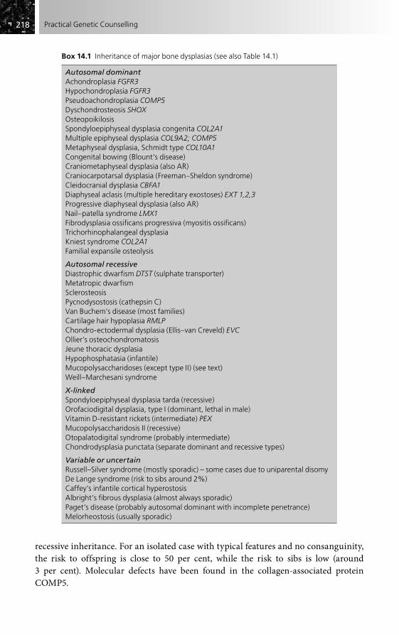
218218 Practical Genetic Counselling
recessive inheritance. For an isolated case with typical features and no consanguinity, the risk to offspring is close to 50 per cent, while the risk to sibs is low (around 3 per cent). Molecular defects have been found in the collagen-associated protein COMP5.
Box 14.1 Inheritance of major bone dysplasias (see also Table 14.1)
Autosomal dominantAchondroplasia FGFR3Hypochondroplasia FGFR3Pseudoachondroplasia COMP5Dyschondrosteosis SHOXOsteopoikilosisSpondyloepiphyseal dysplasia congenita COL2A1Multiple epiphyseal dysplasia COL9A2; COMP5Metaphyseal dysplasia, Schmidt type COL10A1Congenital bowing (Blount’s disease)Craniometaphyseal dysplasia (also AR)Craniocarpotarsal dysplasia (Freeman–Sheldon syndrome)Cleidocranial dysplasia CBFA1Diaphyseal aclasis (multiple hereditary exostoses) EXT 1,2,3Progressive diaphyseal dysplasia (also AR)Nail–patella syndrome LMX1Fibrodysplasia ossificans progressiva (myositis ossificans)Trichorhinophalangeal dysplasiaKniest syndrome COL2A1Familial expansile osteolysis
Autosomal recessiveDiastrophic dwarfism DTST (sulphate transporter)Metatropic dwarfismSclerosteosisPycnodysostosis (cathepsin C)Van Buchem’s disease (most families)Cartilage hair hypoplasia RMLPChondro-ectodermal dysplasia (Ellis–van Creveld) EVCOllier’s osteochondromatosisJeune thoracic dysplasiaHypophosphatasia (infantile)Mucopolysaccharidoses (except type II) (see text)Weill–Marchesani syndrome
X-linkedSpondyloepiphyseal dysplasia tarda (recessive)Orofaciodigital dysplasia, type I (dominant, lethal in male)Vitamin D-resistant rickets (intermediate) PEXMucopolysaccharidosis II (recessive)Otopalatodigital syndrome (probably intermediate)Chondrodysplasia punctata (separate dominant and recessive types)
Variable or uncertainRussell–Silver syndrome (mostly sporadic) – some cases due to uniparental disomyDe Lange syndrome (risk to sibs around 2%)Caffey’s infantile cortical hyperostosisAlbright’s fibrous dysplasia (almost always sporadic)Paget’s disease (probably autosomal dominant with incomplete penetrance)Melorheostosis (usually sporadic)

219219Disorders of Bone and Connective Tissue
Spondyloepiphyseal and spondylometaphyseal dysplasias
The group of spondyloepiphyseal and spondylometaphyseal dysplasias is particularly variable clinically. Most are autosomal dominant, and isolated cases should be consid-ered as dominant, since cases of affected sibs with normal parents, previously thought to be recessive, have transmitted the disorder, probably representing gonadal mosai-cism. Defects in type II collagen have been found in some families. The X-linked ‘tarda’ form should be clearly recognizable, even in an isolated case, by the characteristic radi-ographic appearance of the spine, with a central ‘hump’ of bone and relatively normal distal limb bones.
The severe spondyloepiphyseal dysplasia congenita is frequently confused with Morquio syndrome (mucopolysaccharidosis IV) and usually follows autosomal dominant inheritance. Retinal detachment is an important complication.
Dyschondrosteosis and Madelung deformity
Most cases of Madelung deformity of the wrist are part of the mild but generalized dys-plasia, dyschondrosteosis, following autosomal dominant inheritance, and due to a specific developmental gene mutation (SHOX). A severe dysplasia may result from homozygosity in the children of two affected parents.
Multiple epiphyseal dysplasia
Mildly affected individuals may have only moderately reduced stature and the pattern of autosomal dominant inheritance characteristic of most families may be missed as a result. This disorder must be excluded when giving genetic advice to patients with bilat-eral Perthes’ disease. Two specific gene loci have been identified, those for collagen type 9 and the glycoprotein COMP5.
Chondrodysplasia punctata (Conradi syndrome)
Stippled bone epiphyses may be seen in early life on bone X-rays in a variety of condi-tions, so care must be taken before accepting the specific diagnosis of Conradi syndrome. In this, cataract, mental retardation and ichthyosis may all occur. Both auto-somal dominant and recessive forms exist (the latter more severe and due to a peroxisomal defect); X-linked inheritance, both dominant and recessive, occurs occa-sionally. A phenocopy is also produced by maternal warfarin ingestion in early pregnancy and must be excluded before a genetic basis is assumed.
Lethal newborn dysplasias
The major causes of lethal newborn dysplasia are listed in Table 14.1. Some are invaria-bly fatal, others not so. Sensitive ultrasound scanning is now of real help in prenatal

220220 Practical Genetic Counselling
diagnosis for this group; serial measurements are of particular value. Radiographs should always be taken of a stillbirth suspected of falling in this group because specific diagnosis may otherwise be impossible. Osteogenesis imperfecta congenita can be easily mistaken for a lethal newborn dysplasia. Although a number of disorders in this group follow autosomal recessive inheritance, the most common, thanatophoric dwarfism, is usually sporadic and shows mutations in the fibroblast growth factor gene FGFR3 (also involved in achondroplasia). Heterogeneity or mosaicism may well be responsible for the few recurrent cases that have occurred.
OSTEOPETROSIS
A number of conditions are characterized by increased bone density, including pycnodys-ostosis, sclerosteosis and van Buchem’s disease, but true osteopetrosis exists in two forms:
• a mild form, often asymptomatic, following autosomal dominant inheritance;• a severe childhood form with bone marrow involvement, which is autosomal
recessive – a specific gene has been identified for this type.
MULTIPLE EXOSTOSES (DIAPHYSEAL ACLASIS)
The inheritance of multiple exostoses follows a classical autosomal dominant pattern, although some individuals have only a few lesions that may not be symptomatic. It may form part of the Langer–Giedion syndrome (microdeletion of chromosome 8q), with
Table 14.1 Frequently lethal newborn bone dysplasias
Type Inheritance
Thanatophoric dwarfism Usually sporadic; new dominant mutations in FGFR3
AchondrogenesisType 1 Autosomal recessive; defects in DTSTType 2 New dominant mutation in type II collagen
Ellis–van Creveld syndrome Autosomal recessive
Thoracic dysplasia (Jeune) Autosomal recessive
Majewski and other short rib polydactyly syndromes
Autosomal recessive
Chondrodysplasia punctata Usually autosomal recessive when severe (but see text)
Metatropic dwarfism Autosomal recessive
Hypophosphatasia (severe type) Autosomal recessive
Osteogenesis imperfecta congenita Usually autosomal dominant (but see page 224)
Campomelic dysplasia Mostly new dominant mutations; chromosome analysis needed (some females are XY); mutations of SOX9 gene in some families
Spondylothoracic dysplasia (Jarcho–Levin) Autosomal recessive, but heterogeneous; mutations in DLL3

221221Disorders of Bone and Connective Tissue
mental retardation and features of trichophalangeal dysplasia. Specific genes on chro-mosomes 8 and 11 have now been isolated. By contrast, enchondromatosis (Ollier’s disease) is rarely transmitted to children and is of uncertain inheritance.
LIMB DEFECTS
It is impossible to deal with all the different types here, so the books listed under ‘Further reading’ should be consulted for details. A high proportion of bilateral abnor-malities follow mendelian inheritance; many form part of more general syndromes, some of which are summarized in Table 14.2. Unilateral defects, by contrast, are usually
Table 14.2 Syndromes involving limb defects
Syndrome Inheritance
Polydactyly
Trisomy 13 Usually sporadic
Ellis–van Creveld syndrome AR
Jeune thoracic dystrophy AR
Bardet–Biedl syndrome AR
Meckel syndrome AR
Carpenter syndrome AR
Greig syndrome AD
Acrocallosal syndrome AR
Syndactyly
Poland syndrome Usually sporadic
Apert syndrome AD (see Chapter 15)
Other craniosynostosis syndromes Mostly AD
Orofaciodigital syndrome XD (male lethal) most common (see Chapter 15)
Brachydactyly
Albright’s hereditary osteodystrophy AD
Turner syndrome Usually sporadic
Limb reduction defects
Thalidomide embryopathy Non-genetic
Roberts syndrome AR
Thrombocytopenia–absent radius syndrome AR
Fanconi pancytopenia AR
Amniotic bands Usually sporadic
Holt–Oram syndrome AD
VATER syndrome Uncertain
Ectrodactyly
EEC syndrome AD
AD, autosomal dominant; AR, autosomal recessive; XD, X-linked dominant.

222222 Practical Genetic Counselling
non-genetic. Where the recurrence risk is high, sensitive ultrasonography allows prenatal diagnosis of the more severe defects (see Chapter 8).
Limb defects are currently receiving much study to identify the specific develop-mental genes involved, something which is becoming feasible by a combination of molecular techniques and the recognition of homologous mouse models. (See Chapter 6 and its ‘Further reading’ section.)
Polydactyly
Isolated postaxial polydactyly is a harmless but common (especially in African popula-tions) autosomal dominant condition, showing incomplete penetrance. Preaxial polydactyly is more frequently associated with wider syndromes. Important conditions with polydactyly include trisomy 13 and a number of autosomal recessive conditions including Meckel, Ellis–van Creveld and Jeune and Bardet–Biedl syndromes.
Syndactyly
Poland syndrome of unilateral syndactyly and pectoral muscle aplasia is an important form to recognize because it appears to be non-genetic in the great majority of cases, possibly vascular in aetiology. Bilateral isolated syndactyly of hands and/or feet has several forms, all autosomal dominant. Important syndromes include the orofaciodigital syndrome type I (X-linked dominant, lethal in the male) and the acrocephalosyndactylies (see Chapter 15). Greig syndrome, cephalopoly–syndactyly (autosomal dominant), combines syndactyly with polydactyly and skull abnormalities. The extreme fusion defect of the lower limbs, sirenomelia, is sporadic.
Brachydactyly
Various distinct types of isolated brachydactyly exist, clinical types now increasingly able to be correlated with specific mutational defects. Inheritance is generally auto-somal dominant. Syndrome associations include Albright’s hereditary osteodystrophy or pseudohypoparathyroidism (variable autosomal dominant with genetic imprinting), and Turner syndrome.
Ectrodactyly (split hand or lobster-claw defect)
Most isolated bilateral cases of ectrodactyly follow autosomal dominant inheritance. A number of families exist in which multiple affected sibs born to healthy parents have gone on to have affected children themselves. Autosomal recessive inheritance and lack of penetrance seem unsatisfactory explanations and it is likely that germinal mosaicism is operating (see Chapter 2). This means that affected individuals have a high risk of producing affected children, even if the family pattern does appear to be autosomal recessive. An important syndrome to recognize is the EEC (ectrodactyly, ectodermal dysplasia, cleft lip and palate) syndrome, also autosomal dominant.

223223Disorders of Bone and Connective Tissue
Limb reduction defects
Limb reduction defects may be extremely difficult to distinguish from each other. Some, in particular asymmetrical amputation defects associated with ‘amniotic con-striction bands’, are likely to be non-genetic. Other asymmetrical defects may be associated with oesophageal, anal, cardiac, renal and vertebral abnormalities (the VATER association); again, the recurrence risk is low. Thalidomide was previously a major cause, but no other definite drug-induced defects of this type are known. The severe symmetrical limb reduction disorder, Roberts syndrome, which shows a charac-teristic abnormality of chromosomal division, is autosomal recessive. Limb changes in Holt–Oram syndrome (autosomal dominant) can be very similar to those caused by thalidomide, and may occur without accompanying cardiac defect. The specific gene has now been isolated.
Fanconi pancytopenia and the syndrome of thrombocytopenia with absent radius (TAR) – both autosomal recessive – are important generalized syndromes to recognize and to distinguish from each other. The thumb is usually involved in the former, but preserved in the latter.
Limb assymetry and hypertrophy
Assymetry (with dwarfing) is characteristic of the Russell–Silver syndrome, due to genetic imprinting on chromosome 7. Hypertrophy can result from various vascular and lymphatic disorders (see Chapter 20). Recurrence is rare in all of these causes.
CONNECTIVE TISSUE DISORDERS
Collagen has always been recognized as fundamental to inherited connective tissue disorders, but it is only recently, with identification of the numerous and often tissue-specific forms of collagen and their genes, that individual diseases can be matched up with particular collagen defects (Table 14.3).
Osteogenesis imperfecta
Many cases of osteogenesis imperfecta are now recognized as defects in type I collagen, whose helix is determined by two separate genetic loci. Although there may be some correlation between phenotype and the type and site of mutation, this is not clear-cut. Unfortunately, this, together with the very wide range of mutations, means that molec-ular analysis is not regularly available in diagnosis, which remains clinically and radiologically based.
Type I (previously ‘tarda’)The great majority of ‘classic’ non-lethal cases are type I, which varies greatly within and between families and follows autosomal dominant inheritance. Sclerae are usually

224224 Practical Genetic Counselling
blue, and deafness and osteoporosis may occur in later life, while the number of frac-tures is extremely variable. Mutations may be found in either of the two type I collagen loci, but molecular analysis is not normally needed for accurate genetic counselling. The occasional occurrence of germinal mosaicism means that there is a small risk (around 1 per cent) for sibs of an apparently new mutation case.
Type IIAlthough the terms ‘perinatal lethal’ and ‘congenita’ have been applied to this group, they are not fully accurate. Several subtypes can be distinguished by careful radiologi-cal examination.
• Type IIa. This causes stillbirth or is lethal in the neonatal period, with limb shortening and multiple intrauterine fractures; X-ray shows beaded ribs. Recurrence risk is very low (under 2 per cent) and most cases are due to new dominant mutations in type I collagen, some of which can be recognized by molecular studies.
• Types IIb, IIc. In these rare types neither long bones nor ribs are thickened. Although mostly due to new dominant mutations, these groups contain recessively inherited forms and the empirical recurrence risks are higher (probably around 10 per cent, but 25 per cent if consanguinity is present). Careful discussion with an expert radiologist is needed.
Where no X-ray evidence is available in a perinatally lethal case, the empirical recur-rence risk in a thorough survey was found to be around 4 per cent. Recessive forms are
Table 14.3 Molecular basis of bone dysplasias and connective tissue disorders
Molecule Principal site Disorders
Collagen I (A1 and 2) Bone Osteogenesis imperfecta (various types)
Ehlers–Danlos syndrome (some types)
Collagen II (A1) Cartilage Spondyloepiphyseal dysplasias
Stickler syndrome
Kniest dysplasia
Premature osteoarthrosis
Achondrogenesis type 2
Collagen III Vascular Ehlers–Danlos syndrome, arteriopathic type (IV)
Familial aneurysms(?)
Collagen IV Basement membrane Alport syndrome (see Chapter 21)
Collagen VII Skin (see Chapter 16) Epidermolysis bullosa (one dystrophic form)
Collagen IX Multiple epiphyseal dysplasia (some families); pseudoachondroplasia (some families)
Fibrillin Vascular; eye Marfan syndrome
COMP5 glycoprotein Cartilage Multiple epiphyseal dysplasia (some families); pseudoachondroplasia (some families)

225225Disorders of Bone and Connective Tissue
definitely a minority in all subgroups; molecular evidence shows that some recurring cases may be the result of germinal mosaicism.
Type III (severe deforming type)Type III may be difficult to separate from type II in the newborn period, and the dis-tinction may prove artificial. The empirical recurrence risk is around 7 per cent.
Type IVThe term ‘type IV’ is sometimes applied to severe dominantly inherited forms, but it is doubtful whether this is really separate from type I.
Marfan syndrome
Marfan syndrome is an important disorder now known to be due to deficiency of the connective tissue protein fibrillin, determined by a gene on chromosome 15. This disorder tends to be overdiagnosed in tall individuals of slender habitus but with no cardinal signs, especially rapidly growing adolescents. There is rarely doubt, however, when the presence or absence of the various major features is considered as a whole.
Inheritance is autosomal dominant; the occurrence of major aortic complications is unpredictable and many patients live a relatively normal life until aortic surgery is needed or a sudden demise occurs, although others may have severe early orthopaedic and cardiac problems. Around 15 per cent of all patients appear to be new mutations. Penetrance is probably full but apparently healthy family members should be carefully checked (including slit-lamp examination for minor degree of lens dislocation).
The isolation of the gene, and recognition of specific mutations in some cases, now gives the possibility of definitive molecular exclusion or confirmation, at least in larger families, as well as prenatal diagnosis if requested. The primary diagnosis remains clinical, however, and if a composite scoring system is used, most cases can be clearly confirmed or excluded. Recent striking advances in both surgery and medical treat-ment make early diagnosis and expert management of particular importance.
Other disorders to be distinguished include homocystinuria (autosomal recessive), patients with isolated lens dislocation due to spherophakia, who by chance are tall and thin, and neuromuscular disorders giving a ‘marfanoid’ habitus. A separate dominant syndrome of arachnodactyly with contractures but no internal complications (Beal syndrome) has also been described and appears to be due to a separate form of fibrillin deficiency involving a locus on chromosome 5. The Loeys–Dietz syndrome, in which aneurysms are prominent, results from defects in the allied TGFR beta genes.
Ehlers–Danlos syndrome
The Ehlers–Danlos group of disorders in which hypermobility of skin and joints, skin fragility and bruising, and rarer vascular, visceral and ocular complications are the main features is extremely heterogeneous. It is important not to confuse the group with the much commoner and essentially harmless benign familial joint hypermobility
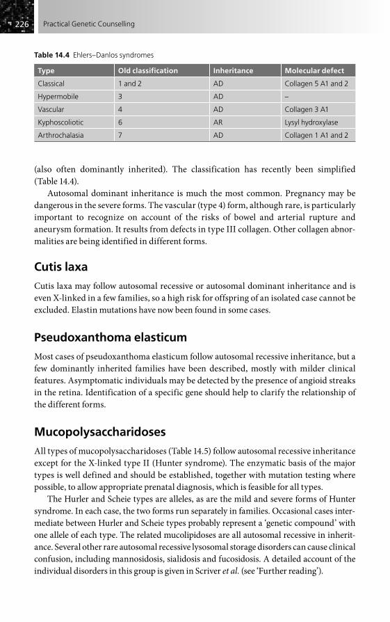
226226 Practical Genetic Counselling
(also often dominantly inherited). The classification has recently been simplified (Table 14.4).
Autosomal dominant inheritance is much the most common. Pregnancy may be dangerous in the severe forms. The vascular (type 4) form, although rare, is particularly important to recognize on account of the risks of bowel and arterial rupture and aneurysm formation. It results from defects in type III collagen. Other collagen abnor-malities are being identified in different forms.
Cutis laxa
Cutis laxa may follow autosomal recessive or autosomal dominant inheritance and is even X-linked in a few families, so a high risk for offspring of an isolated case cannot be excluded. Elastin mutations have now been found in some cases.
Pseudoxanthoma elasticum
Most cases of pseudoxanthoma elasticum follow autosomal recessive inheritance, but a few dominantly inherited families have been described, mostly with milder clinical features. Asymptomatic individuals may be detected by the presence of angioid streaks in the retina. Identification of a specific gene should help to clarify the relationship of the different forms.
Mucopolysaccharidoses
All types of mucopolysaccharidoses (Table 14.5) follow autosomal recessive inheritance except for the X-linked type II (Hunter syndrome). The enzymatic basis of the major types is well defined and should be established, together with mutation testing where possible, to allow appropriate prenatal diagnosis, which is feasible for all types.
The Hurler and Scheie types are alleles, as are the mild and severe forms of Hunter syndrome. In each case, the two forms run separately in families. Occasional cases inter-mediate between Hurler and Scheie types probably represent a ‘genetic compound’ with one allele of each type. The related mucolipidoses are all autosomal recessive in inherit-ance. Several other rare autosomal recessive lysosomal storage disorders can cause clinical confusion, including mannosidosis, sialidosis and fucosidosis. A detailed account of the individual disorders in this group is given in Scriver et al. (see ‘Further reading’).
Table 14.4 Ehlers–Danlos syndromes
Type Old classification Inheritance Molecular defect
Classical 1 and 2 AD Collagen 5 A1 and 2
Hypermobile 3 AD –
Vascular 4 AD Collagen 3 A1
Kyphoscoliotic 6 AR Lysyl hydroxylase
Arthrochalasia 7 AD Collagen 1 A1 and 2
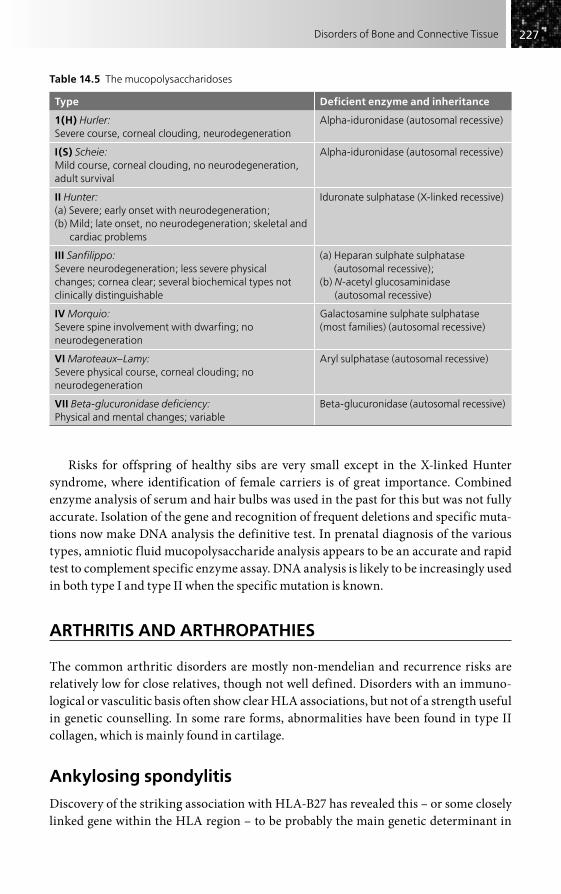
227227Disorders of Bone and Connective Tissue
Risks for offspring of healthy sibs are very small except in the X-linked Hunter syndrome, where identification of female carriers is of great importance. Combined enzyme analysis of serum and hair bulbs was used in the past for this but was not fully accurate. Isolation of the gene and recognition of frequent deletions and specific muta-tions now make DNA analysis the definitive test. In prenatal diagnosis of the various types, amniotic fluid mucopolysaccharide analysis appears to be an accurate and rapid test to complement specific enzyme assay. DNA analysis is likely to be increasingly used in both type I and type II when the specific mutation is known.
ARTHRITIS AND ARTHROPATHIES
The common arthritic disorders are mostly non-mendelian and recurrence risks are relatively low for close relatives, though not well defined. Disorders with an immuno-logical or vasculitic basis often show clear HLA associations, but not of a strength useful in genetic counselling. In some rare forms, abnormalities have been found in type II collagen, which is mainly found in cartilage.
Ankylosing spondylitis
Discovery of the striking association with HLA-B27 has revealed this – or some closely linked gene within the HLA region – to be probably the main genetic determinant in
Table 14.5 The mucopolysaccharidoses
Type Deficient enzyme and inheritance
1(H) Hurler:Severe course, corneal clouding, neurodegeneration
Alpha-iduronidase (autosomal recessive)
I(S) Scheie:Mild course, corneal clouding, no neurodegeneration, adult survival
Alpha-iduronidase (autosomal recessive)
II Hunter:(a) Severe; early onset with neurodegeneration; (b) Mild; late onset, no neurodegeneration; skeletal and
cardiac problems
Iduronate sulphatase (X-linked recessive)
III Sanfilippo:Severe neurodegeneration; less severe physical changes; cornea clear; several biochemical types not clinically distinguishable
(a) Heparan sulphate sulphatase (autosomal recessive);
(b) N-acetyl glucosaminidase (autosomal recessive)
IV Morquio:Severe spine involvement with dwarfing; no neurodegeneration
Galactosamine sulphate sulphatase (most families) (autosomal recessive)
VI Maroteaux–Lamy:Severe physical course, corneal clouding; no neurodegeneration
Aryl sulphatase (autosomal recessive)
VII Beta-glucuronidase deficiency:Physical and mental changes; variable
Beta-glucuronidase (autosomal recessive)

228228 Practical Genetic Counselling
susceptibility, although genetic risks are not high. One family study showed a risk of 5 per cent (7 per cent for males and 2 per cent for females) for clinical disease in first-degree relatives of patients with ankylosing spondylitis. Sixteen per cent showed radiological sacroiliitis. The chance of a B27 child of a B27 patient developing clinical ankylosing spondylitis is 9 per cent, compared with a risk of less than 1 per cent for offspring without this antigen.
The clinical or genetic applications of HLA-B27 testing are actually of very limited value (see Chapter 3 and ‘Further reading’ to this chapter), since most B27-positive indi-viduals, whether or not there is a family history of ankylosing spondylitis, will not develop clinical features of the disorder, though a B27-negative result will make the diagnosis or future development of the disorder very unlikely. These limitations are a good example of why ‘susceptibility testing’ for common diseases is generally of little help in genetic counselling.
Rheumatoid arthritis
This common disorder (around 1 per cent in most populations) rarely follows a mende-lian pattern and the risks of clinical rheumatoid arthritis to relatives are not high; they appear to be around 3–5 per cent for first-degree relatives, although the incidence of radiological abnormalities is considerably higher. As might be expected with multifac-torial inheritance (see Chapter 3), the risk appears to be higher for relatives of very severe cases (as high as 10 per cent) and minimal for relatives of mild seronegative cases. The antigen HLA-DRw4 is found in rheumatoid arthritis patients with twice the normal frequency, and is six times more common in familial cases, suggesting that this may be a major genetic determinant in the disorder. The occurrence of positive tests for rheumatoid factor is associated with HLA-DRw3.
A distinct, progressive ‘pseudorheumatoid chondrodysplasia’ has been recognized which is probably autosomal recessive in inheritance. Generalized contractures and painful swelling can cause confusion with juvenile rheumatoid arthritis. The hand deformities of Freeman–Sheldon syndrome and mucopolysaccharidoses may also be misdiagnosed as arthritis.
Systemic lupus erythematosus
Association has been found with specific HLA haplotypes and several other genes involved in immune function. There is strong concordance between monozygotic twins (25–75 per cent), but the empirical risk for first-degree relatives (and for dizygotic twins) is around 3 per cent for systemic lupus erythematosus. As with many autoimmune disor-ders, there appears to be an increased incidence of other autoimmune conditions in relatives, suggesting that a dominantly inherited general predisposition to autoimmunity may be involved.
Systemic lupus may produce important intrauterine effects, with congenital heart block occurring in some infants born to affected mothers, while the ‘lupus anticoagu-lant factor’ (not always associated with overt systemic lupus) may be responsible for recurrent abortion in a small proportion of such cases.
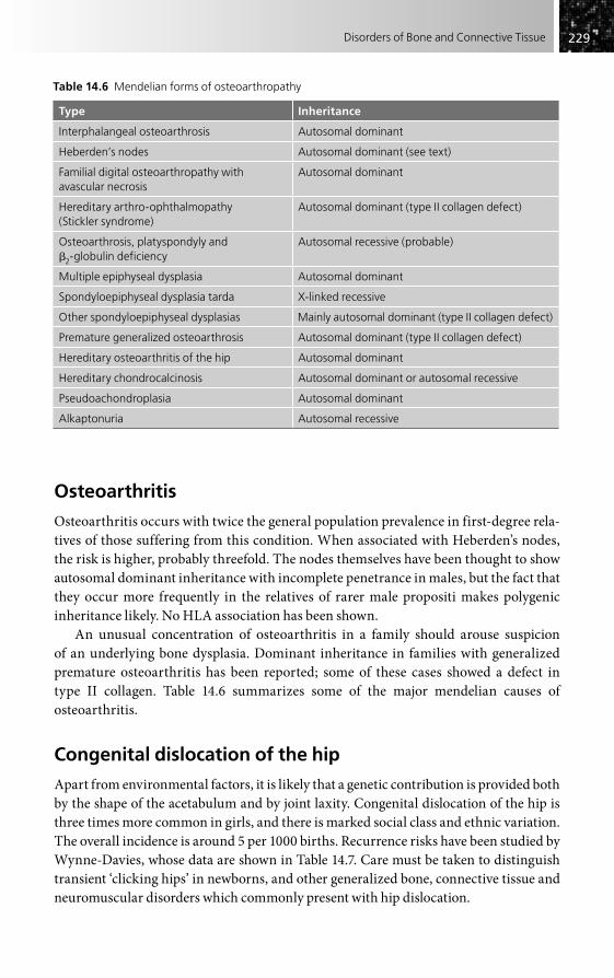
229229Disorders of Bone and Connective Tissue
Osteoarthritis
Osteoarthritis occurs with twice the general population prevalence in first-degree rela-tives of those suffering from this condition. When associated with Heberden’s nodes, the risk is higher, probably threefold. The nodes themselves have been thought to show autosomal dominant inheritance with incomplete penetrance in males, but the fact that they occur more frequently in the relatives of rarer male propositi makes polygenic inheritance likely. No HLA association has been shown.
An unusual concentration of osteoarthritis in a family should arouse suspicion of an underlying bone dysplasia. Dominant inheritance in families with generalized premature osteoarthritis has been reported; some of these cases showed a defect in type II collagen. Table 14.6 summarizes some of the major mendelian causes of osteoarthritis.
Congenital dislocation of the hip
Apart from environmental factors, it is likely that a genetic contribution is provided both by the shape of the acetabulum and by joint laxity. Congenital dislocation of the hip is three times more common in girls, and there is marked social class and ethnic variation. The overall incidence is around 5 per 1000 births. Recurrence risks have been studied by Wynne-Davies, whose data are shown in Table 14.7. Care must be taken to distinguish transient ‘clicking hips’ in newborns, and other generalized bone, connective tissue and neuromuscular disorders which commonly present with hip dislocation.
Table 14.6 Mendelian forms of osteoarthropathy
Type Inheritance
Interphalangeal osteoarthrosis Autosomal dominant
Heberden’s nodes Autosomal dominant (see text)
Familial digital osteoarthropathy with avascular necrosis
Autosomal dominant
Hereditary arthro-ophthalmopathy (Stickler syndrome)
Autosomal dominant (type II collagen defect)
Osteoarthrosis, platyspondyly and �2-globulin deficiency
Autosomal recessive (probable)
Multiple epiphyseal dysplasia Autosomal dominant
Spondyloepiphyseal dysplasia tarda X-linked recessive
Other spondyloepiphyseal dysplasias Mainly autosomal dominant (type II collagen defect)
Premature generalized osteoarthrosis Autosomal dominant (type II collagen defect)
Hereditary osteoarthritis of the hip Autosomal dominant
Hereditary chondrocalcinosis Autosomal dominant or autosomal recessive
Pseudoachondroplasia Autosomal dominant
Alkaptonuria Autosomal recessive

230230 Practical Genetic Counselling
Perthes’ disease
True Perthes’ disease carries a low recurrence risk, under 1 per cent in sibs and around 3 per cent in children of affected patients. The risks do not appear to be higher in the relatives of patients with bilateral hip disease. Risks for second-degree and third-degree relatives are minimal. Familial concentrations should arouse suspicion that some other disorder, such as multiple epiphyseal dysplasia, may be present.
Arthrogryposis (congenital contractures)
There is so much heterogeneity in the group of arthrogryposes that the possible causes cannot be listed here – see the review by Hall (2003) for details. Every effort should be directed to making a primary diagnosis, with a careful pregnancy history (to exclude envi-ronmental factors) and family history (relevant neurological or skeletal disorders). Full examination and investigations to determine whether the contractures are part of a general syndrome, or have a neurogenic or myopathic basis, are also essential. If, after this, no specific cause has been found and the case is isolated, the recurrence risk is likely to be low.
Two forms that appear to be specific are distal arthrogryposis (autosomal dominant, often very mild) and amyoplasia, with symmetrical contractures of all four limbs (usually sporadic). Where a parent is affected, even mildly, autosomal dominant inher-itance is likely. It should be noted that the prognosis with treatment is generally good when the condition is a secondary deformity.
Talipes
As with arthrogryposis, there are many primary causes of talipes, in particular neuro-logical defects, which must be excluded. Idiopathic talipes equinovarus occurs in around 1 in 1000 births in the UK, with a male predominance of 3:1. The risk for sibs is around 3 per cent overall, around 10 times the population frequency. The risk for sibs of a male patient is lower (2 per cent) than for sibs of a female patient (5 per cent), as expected on the basis of polygenic inheritance. Full data on risks to offspring of patients are not yet available. Wynne-Davies suggested that the risk may be as high as 25 per cent for further offspring of an affected parent with an affected child. Other forms of talipes (calcaneovalgus and metatarsus varus) appear to run separately in families from equi-novarus, and may carry slightly higher sib recurrence risks (4–5 per cent).
Table 14.7 Recurrence risks for congenital dislocation of the hip
Individual affected Individual at risk Risk (%)
Overall Males Females
One sib Sibs 6 1 11
One parent Children 12 6 17
One parent, one child Children 36
Second-degree relative Nephews, nieces 1
After Wynne-Davies R (1985), J Med Genet 2, 227–32.

231231Disorders of Bone and Connective Tissue
Idiopathic scoliosis
Idiopathic scoliosis may be infantile or adolescent.
• The incidence of infantile idiopathic scoliosis is around 1.3 per 1000 births in the UK, and much lower in North America. Satisfactory risk figures for sibs are not available.
• The incidence of the adolescent form is around 0.3 per 1000 births in boys and 4 per 1000 births in girls. The overall risk for first-degree relatives is around 5–7 per cent for major defects, but data are insufficient to split by sex and type of relatives.
Dupuytren’s contracture
When no primary cause is apparent, the inheritance of Dupuytren’s contracture is thought to be autosomal dominant, but if this is so, one would expect severe forms due to homozygosity for such a common condition, which have not been recorded.
Hereditary digital clubbing
Hereditary digital clubbing is a common and harmless autosomal dominant trait, fre-quently confused by doctors with acquired clubbing of more serious import. Patients usually correctly recognize its hereditary nature.
VARIOUS SKELETAL SYNDROMES
Nail–patella syndrome
Nail–patella syndrome (autosomal dominant) may present with talipes or hip disloca-tion in addition to the dysplastic nails and absent patellae. Renal involvement may occur in later life. Linkage with the ABO blood group system was the first autosomal linkage to be discovered on chromosome 9. A specific gene has now been isolated.
Craniocarpotarsal (Freeman–Sheldon or ‘whistling face’) syndrome
The hand deformity superficially resembles severe rheumatoid disease, but the lack of X-ray changes and characteristic pinched face should enable recognition of the syndrome. Inheritance is usually autosomal dominant, but the birth of affected sibs to normal consanguineous parents suggests a recessive form as well.
Rubinstein–Taybi syndrome
Broad thumbs and great toes, short stature, moderate to severe mental retardation and characteristic facies are the principal features of Rubinstein–Taybi syndrome.

232232 Practical Genetic Counselling
Recurrence in a family is rare, but patients do not usually reproduce. Molecular studies have recently identified mutations in the CREB binding protein gene on chromosome 16p, so most cases probably represent new dominant mutations.
Larsen syndrome
A combination of multiple joint dislocation with an unusual facies (and often cleft palate), this syndrome was originally thought to be autosomal recessive, but parent–child trans-mission has now been reported so the situation is not clear. Heterogeneity is likely.
Stickler syndrome (hereditary arthro-ophthalmopathy)
Stickler syndrome is a variable autosomal dominant disorder, with characteristic flat-tened facies, cleft palate, severe myopia with frequent retinal detachment and early osteoarthritis. It has been shown to be due to a defect in type II collagen.
Klippel–Feil syndrome
Many causes of a short neck find their way into this category, but Klippel–Feil syndrome remains heterogeneous even after their removal. Where the case is an isolated one, the risk for sibs is probably low, although minor degrees of cervical vertebral fusion may be more frequent in relatives. The risk for children is significant since some cases are dom-inantly inherited; no precise figure exists. Congenital heart disease is a common accompaniment in patients. The specific association with severe deafness (Wildervanck syndrome) appears to be confined to girls. Careful clinical assessment and full skeletal survey should allow future delineation of specific entities within this group.
De Lange syndrome
Low birthweight, dwarfism, mental retardation, characteristic facies with synophrys and a variety of limb defects are the principal features of this syndrome. A variety of chromosomal defects has been found and a specific gene recently identified that identi-fies mutations in almost half of cases. Most cases are sporadic and the risk to sibs is around 2 per cent.
Popliteal pterygium syndrome
The multiple pterygia are associated with cleft lip or palate, cryptorchidism and often syndactyly. Inheritance may be either autosomal dominant or recessive (usually the latter in the severe infantile form).
Sacral agenesis
Almost all cases of sacral agenesis are sporadic, but there seems to be a specific relation-ship to maternal diabetes mellitus. There does not appear to be an association with neural tube defects (see Chapter 12).

233233Disorders of Bone and Connective Tissue
FURTHER READING
GeneralBeighton P (ed.) (1993). McKusick’s Heritable Disorders of Connective Tissue. St Louis: Mosby.Gorlin RJ, Cohen M, Hennekam RCM (eds) (2001). Syndromes of the Head and Neck. Oxford:
Oxford University Press.Royce PM, Steinmann B (1993). Connective Tissue and its Heritable Disorders. New York:
Wiley-Liss.Scriver CK, Beaudet AL, Sly WS, Valle D (eds) (2001). The Metabolic and Molecular Bases of
Inherited Disease. New York: McGraw-Hill.Spranger J (1992). International classification of the osteochondrodysplasias. Eur J Paediatr 151,
407–15.Young ID (2002). Genetics for Orthopedic Surgeons. London: Remedica.
Bone dysplasiasHorton WA (1995). Molecular genetics of the human chondrodysplasias. Eur J Hum Genet 3,
357–73.Olsen BR (1995). Mutations in collagen genes resulting in metaphyseal and epiphyseal
dysplasias. Bone 17, 45S–49S.Rimoin DL, Lachman RS (1993). Genetic diseases of the osseous skeleton. In: Beighton P (ed.)
McKusick’s Heritable Disorders of Connective Tissue, pp. 557–689. St Louis: Mosby.Spranger J, Brill PJ, Poznanski A (2003). Bone Dysplasias: an Atlas of Genetic Disorders of
Skeletal Development. Oxford: Oxford University Press.Unger S, Lachman RS, Rimoin DL (2007). Chondrodysplasias. In: Rimoin DL, Connor JM,
Pyeritz RE, Korf BR (eds) Emery and Rimoin’s Principles and Practice of Medical Genetics, 5th edn, pp. 3709–753. Edinburgh: Churchill-Livingstone.
Connective tissue disordersGray JR, Davies SJ (1996). Marfan syndrome. J Med Genet 33, 403–8.Pope M (2004). Molecular abnormalities of collagen and connective tissue. In: Isenberg D,
Maddison P, Woo P et al. (eds) Oxford Textbook of Rheumatology, 3rd edn, chap. 3.2. Oxford: Oxford University Press.
Pope FM, Burrows NP (1997). Ehlers–Danlos syndrome has varied molecular mechanisms. J Med Genet 34, 400–10.
Tsipouras P, Devereux RB (1993). Marfan syndrome: genetic basis and clinical manifestations. Semin Dermatol 12, 219–28.
OthersCran JT, Husby G (1995). HLA-B27 and spondyloarthropathy: value for early diagnosis? J Med
Genet 32, 497–501.Hall JR (2007). Arthrogryposes (multiple congenital contractures). In: Rimoin DL, Connor JM,
Pyeritz RE, Korf B (eds) Emery and Rimoin’s Principles and Practice of Medical Genetics, 5th edn, pp. 3785–857. Edinburgh: Churchill Livingstone.
Rubin LA, Amos CI, Wade JA et al. (1994). Investigating the genetic basis for ankylosing spondylitis. Arthritis Rheum 37, 1212–20.

Introduction . . . . . . . . . . . . . . . . . . . . . . . . . . . . . . . . . . . . . . . . . . . . . . . . . . . . . . . . . . . . . . . . . . . 234
The teeth . . . . . . . . . . . . . . . . . . . . . . . . . . . . . . . . . . . . . . . . . . . . . . . . . . . . . . . . . . . . . . . . . . . . . . 234
Cleft lip and palate . . . . . . . . . . . . . . . . . . . . . . . . . . . . . . . . . . . . . . . . . . . . . . . . . . . . . . . . . . . . . . . 235
Other oral disorders . . . . . . . . . . . . . . . . . . . . . . . . . . . . . . . . . . . . . . . . . . . . . . . . . . . . . . . . . . . . 237
Craniofacial disorders . . . . . . . . . . . . . . . . . . . . . . . . . . . . . . . . . . . . . . . . . . . . . . . . . . . . . . . 238
Further reading . . . . . . . . . . . . . . . . . . . . . . . . . . . . . . . . . . . . . . . . . . . . . . . . . . . . . . . . . . . . . 241
Oral and Craniofacial Disorders
CHAPTER 15
INTRODUCTION
For most clinicians, craniofacial abnormality is a confusing area, on the borderline between medicine and dentistry, yet overlapping broadly into other fields. The plastic or maxillofacial surgeon is the person who sees most of the facial disorders, and there is no doubt that genetic counselling is an integral part of management of these patients. Even minor facial anomalies can cause great distress, and accurate information regard-ing possible risks to offspring will provide considerable relief from worry for such people.
The amount of information available regarding the inheritance of these disorders is considerable. A number of medical geneticists who began their careers as dentists have provided some thorough reviews of the subject (see ‘Further reading’).
THE TEETH
Hypodontia and anodontia
Hypodontia, or lack of one or a few permanent teeth, is extremely common (5–10 per cent in most surveys) and is often inherited as a variable autosomal dominant trait. It may be the only significant finding in female heterozygotes for X-linked hypohidrotic ectodermal dysplasia (see Chapter 16), where incisors may also be peg-shaped. Complete anodontia is commonly associated with this disorder in males, but can also occur in other ectodermal dysplasia syndromes, in type I orofaciodigital syndrome (X-linked dominant) and with iris dysplasia in Rieger syndrome (autosomal dominant). A single central incisor tooth may be associated with midline abnormalities such as holoprosencephaly.
235235Oral and Craniofacial Disorders
Enamel defects
Enamel defects are seen in a number of generalized genetic disorders; defects occurring in isolation are termed amelogenesis imperfecta. Classifications have tended to be based on the apparent phenotype, either hypoplasia (a reduction in thickness of the enamel) or hypomineralization (a reduction in the degree of calcification of the enamel), the latter often subdivided into hypocalcification and hypomineralization according to the severity of the defect. In all probability, both hypoplasia and hypomineralization occur together in the majority of cases.
Autosomal dominant, autosomal recessive and X-linked modes of inheritance are recognized. In the X-linked forms, characterized by vertical bands of normal and abnormal enamel in heterozygous females, there is evidence of genetic heterogeneity. One locus is the gene coding for amelogenin (the main structural protein of enamel) synthesis in the Xp22 region; another locus lies at the opposite end of the X chromo-some in the Xq22–q28 region.
Enamel pits are a characteristic finding in the permanent teeth in tuberous sclerosis.
Dentine defects
The most common of the defects of dentine is dentinogenesis imperfecta. This may occur in isolation, inherited in an autosomal dominant pattern, or in the various forms of osteogenesis imperfecta. The teeth are opalescent with an amber or grey colour. The teeth may be subject to attrition and chipping, most probably due to frac-tures within the dentine. Dentinogenesis imperfecta occurring alone is determined by a gene on the long arm of chromosome 4. When associated with osteogenesis imperfecta, there may be more variation in the severity of involvement, with some teeth being clinically normal, although radiographically and histologically they may show abnormalities.
CLEFT LIP AND PALATE
Before genetic counselling is given, a careful examination must be made to exclude the numerous syndromal associations with clefting. In some of these (e.g. chromosomal trisomies), the other defects are obvious, while in others, e.g. the Van der Woude (lip pits) syndrome (gene isolated on 1q), they may be inconspicuous and even absent in some family members. Maternal teratogens (notably anticonvulsants) must also be con-sidered. The most important syndromes to recognize are those that follow mendelian inheritance; Box 15.1 lists some of the major ones, and fuller lists are given in the books listed in the ‘Further reading’ section. Cleft lip and palate also occur with other malfor-mations in a non-specific manner, and are more common than one might expect. If a careful search for a specific syndrome proves negative, one is forced to use the empirical risks for the abnormalities in isolation.

236236 Practical Genetic Counselling
Numerous studies have shown that cleft palate alone runs in most families (apart from Van der Woude syndrome) separately from cleft lip with or without cleft palate. Table 15.1 summarizes the overall risks. As expected with polygenic inheritance, the presence of other affected family members considerably raises the risks. The population incidence of cleft lip (with or without cleft palate) is 1 in 500–1000, compared with around 1 in 2500 for isolated cleft palate. A small number of undoubtedly X-linked families have been docu-mented with cleft palate and ankyloglossia. While of great interest for isolating the gene involved, they do not affect the general genetic recurrence risk.
One point to note from Table 15.1 is that the risk for sibs of a patient with cleft lip with or without cleft palate is lower when it can be definitely established that no other relatives are affected (2.2 per cent) than is the overall risk to sibs (4 per cent) found by surveys, which will include some families with other affected relatives of varying close-ness. Therefore, this higher risk should be used when family information is unavailable or unreliable. Some data are now available on the influence of severity (Table 15.2) and, as expected, there is a higher risk when the abnormality is bilateral and a lower risk when there is only cleft lip. It is likely that cleft lip and palate are examples of polygenic
Box 15.1 Some major syndromes associated with cleft lip and/or palate
Autosomal dominantVan der Woude syndrome (lip pits with cleft lip/palate)EEC syndrome (ectrodactyly, ectodermal dysplasia and clefting)Hereditary arthro-ophthalmopathy (Stickler syndrome)Larsen syndrome (originally thought to be recessive)Retinal detachment, myopia and cleft palate (Marshall syndrome)Spondyloepiphyseal dysplasia congenita
Autosomal recessiveChondrodysplasia punctata (Conradi syndrome)Diastrophic dysplasiaSmith–Lemli–Opitz syndromeMeckel syndromeOrofaciodigital syndrome, type IIFryns syndrome (with diaphragmatic hernia, limb and facial anomalies)Roberts syndrome
X-linkedOrofaciodigital syndrome, type I (dominant, lethal in male)Otopalatodigital syndromeIsolated X-linked cleft palate with ankyloglossia
ChromosomalTrisomy 13Trisomy 18Chromosome 18 deletionsVarious other autosomal abnormalitiesVelocardiofacial (Shprintzen) syndrome (22q deletion)
Non-mendelianPierre Robin sequenceClefting with congenital heart diseaseDe Lange syndrome

237237Oral and Craniofacial Disorders
inheritance; so far, apart from the mendelian and syndromal forms, no major suscepti-bility genes have been identified.
Median cleft lip should be regarded as genetically separate from the usual types, and may have its own specific syndromal associations (e.g. Ellis–van Creveld syndrome). Lateral and oblique cleft lip and mandibular clefting are likewise due to distinct processes.
Pierre Robin sequence (cleft palate with mandibular hypoplasia)
Mandibular hypoplasia, with or without cleft palate and with resulting respiratory obstruction from the tongue, may be part of a variety of skeletal or muscular syn-dromes, some mendelian (e.g. Stickler syndrome, congenital myotonic dystrophy). In the absence of these, the risk of recurrence is low. The prognosis for patients with careful treatment is good.
OTHER ORAL DISORDERS
Recurrent aphthous ulcers of the mouth
Around 20–25 per cent of the general population suffer from this minor, but tiresome, complaint to some degree, and around 40 per cent of first-degree relatives appear to be affected.
Table 15.1 Cleft lip and palate: genetic risks in the absence of a defined syndrome or mendelian pattern
Relationship to index case Cleft lip � palate (%) Isolated cleft palate (%)
Sibs (overall risk) 4.0 1.8
Sib (no other affected members) 2.2
Sib (two affected sibs) 10 8
Sib and affected parent 10
Children 4.3 3
Second-degree relatives 0.6
Third-degree relatives 0.3
General population 0.1 0.04
Table 15.2 Genetic risks in cleft lip/palate: effect of severity
Anomaly Risk to sibs (%)
Bilateral cleft lip and palate 5.7
Unilateral cleft lip and palate 4.2
Unilateral cleft lip alone 2.5
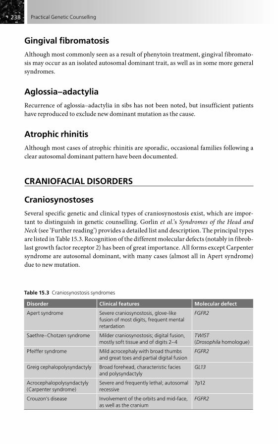
238238 Practical Genetic Counselling
Gingival fibromatosis
Although most commonly seen as a result of phenytoin treatment, gingival fibromato-sis may occur as an isolated autosomal dominant trait, as well as in some more general syndromes.
Aglossia–adactylia
Recurrence of aglossia–adactylia in sibs has not been noted, but insufficient patients have reproduced to exclude new dominant mutation as the cause.
Atrophic rhinitis
Although most cases of atrophic rhinitis are sporadic, occasional families following a clear autosomal dominant pattern have been documented.
CRANIOFACIAL DISORDERS
Craniosynostoses
Several specific genetic and clinical types of craniosynostosis exist, which are impor-tant to distinguish in genetic counselling. Gorlin et al.’s Syndromes of the Head and Neck (see ‘Further reading’) provides a detailed list and description. The principal types are listed in Table 15.3. Recognition of the different molecular defects (notably in fibrob-last growth factor receptor 2) has been of great importance. All forms except Carpenter syndrome are autosomal dominant, with many cases (almost all in Apert syndrome) due to new mutation.
Table 15.3 Craniosynostosis syndromes
Disorder Clinical features Molecular defect
Apert syndrome Severe craniosynostosis, glove-like fusion of most digits, frequent mental retardation
FGFR2
Saethre–Chotzen syndrome Milder craniosynostosis; digital fusion, mostly soft tissue and of digits 2–4
TWIST(Drosophila homologue)
Pfeiffer syndrome Mild acrocephaly with broad thumbs and great toes and partial digital fusion
FGFR2
Greig cephalopolysyndactyly Broad forehead, characteristic facies and polysyndactyly
GL13
Acrocephalopolysyndactyly (Carpenter syndrome)
Severe and frequently lethal; autosomal recessive
7p12
Crouzon’s disease Involvement of the orbits and mid-face, as well as the cranium
FGFR2

239239Oral and Craniofacial Disorders
Isolated craniosynostosis
Most cases of isolated craniosynostosis are sporadic, regardless of which sutures are involved, and probably represent secondary deformations. Risks for sibs where parents are normal are around 5 per cent for coronal and 1 per cent for sagittal suture fusion, although these will need reassessment in the light of molecular studies. If multiple family members are affected, it is wise to assume that one is dealing with a mendelian type and to consider DNA analysis. Some such families have shown mutations in FGFR2 or HOX 8 genes.
Mandibulofacial dysostosis (Treacher Collins syndrome)
Severity of the facial abnormality in mandibulofacial dysostosis varies greatly but inheritance is autosomal dominant, with a specific gene isolated on chromosome 5. Deafness is a common feature in addition to external ear defects; mental retardation is said to occur but is possibly an artefact of ascertainment. Potential parents will tend to be more mildly affected than average and must be warned that an affected child could be considerably more severely affected. High-resolution ultrasonogra-phy can detect severe cases. A separate syndrome of mandibulofacial dysostosis with preaxial limb defects (Nager syndrome) usually follows autosomal recessive inheritance.
Nasal defects
The nose may be characteristically involved in a wide range of craniofacial conditions. Nasal hypoplasia is a feature of Binder syndrome (usually sporadic) and Stickler syndrome (see Chapter 14), while trichorinophalangeal syndrome is characterized by a bulbous nose. The very large normal range and ethnic variation in nasal shape must be taken into account.
Other craniofacial syndromes
Some members of this very extensive group are covered in other chapters and listed in the boxes in Chapter 6. A precise diagnosis is often difficult; Gorlin et al. (2001) provide a detailed and comprehensive guide. Only a few of the most important are mentioned here.
Hallermann–Streiff syndrome (oculomandibulofacial syndrome; Francois dyscephalic syndrome)Congenital cataracts, short stature, beaked nose with micrognathia and characteristic facies are all features of the Hallermann–Streiff syndrome. Inheritance is probably autosomal dominant, with most patients being new mutations, but few patients have reproduced. The risk of recurrence in sibs is minimal.

240240 Practical Genetic Counselling
Goldenhar syndrome (oculoauriculovertebral dysplasia)Goldenhar syndrome must be distinguished from the superficially similar Treacher Collins syndrome. The external ear defects are more marked, mental retardation is common, and epibulbar dermoid cyst of the eye is characteristic. Most cases are sporadic, and the recurrence risk where parents are normal is low.
Hemifacial microsomiaUnilateral hypoplasia of most facial structures is the characteristic feature of hemifacial microsomia. Recurrence is exceptional.
Sturge–Weber syndromeSturge–Weber syndrome must be distinguished from other angiomatous malforma-tions of the face. The involvement of the ophthalmic trigeminal area and extension to the deep tissue of skull and meninges is characteristic, as is congenital glaucoma. No causative factors are known, but the condition is almost always sporadic.
Orofaciodigital syndromesCharacteristic features of the most common form (type I) are clefting of the jaw and tongue, with digital abnormalities (usually syndactyly) and sometimes mental retarda-tion. Renal cysts may occur and almost all cases are female, suggesting X-linked dominant inheritance lethal in the male (see Chapter 2). A risk of 50 per cent for female offspring of an affected individual should be given. An extremely rare form (type 2 or Mohr syndrome) following autosomal recessive inheritance, and which is clinically dis-tinguishable, has been described; further heterogeneity is likely in the group.
Oculodentodigital syndromeThe narrow alae nasi and small eyes are characteristic of the oculodentodigital syndrome, combined with mild digital curvature and fusion, and dental enamel hypo-plasia. Inheritance is autosomal dominant.
Dubowitz syndromeThe autosomal recessive Dubowitz syndrome combines microcephaly and mental retardation with a small face, shallow supraorbital ridges and severe eczema.
Frontonasal dysplasia and median cleft face syndromeAlmost all cases of frontonasal dysplasia and median cleft face syndrome have been sporadic, but affected individuals rarely reproduce.
Aarskog syndrome (faciogenital dysplasia)Aarskog syndrome is an X-linked recessive disorder combining hypertelorism with digital and spinal abnormalities and characteristic scrotal shape. The Opitz syndrome of hypertelorism and hypospadias is somewhat similar, but follows either autosomal dominant or X-linked inheritance.

241241Oral and Craniofacial Disorders
FURTHER READING
Cohen MM, Maclean RE (2000). Craniosynostosis. Oxford: Oxford University Press.Gorlin RJ, Cohen MM, Hennekam RJM (2001). Syndromes of the Head and Neck, 4th edn.
New York: Oxford University Press [a comprehensive and well illustrated volume that is the definitive source for both clinical and genetic information, both on well recognized disorders and on incompletely delineated syndromes].
Reardon W, Winter RM (1995). The molecular pathology of syndromic craniosynostosis. Mol Med Today 1, 432–7.

Introduction . . . . . . . . . . . . . . . . . . . . . . . . . . . . . . . . . . . . . . . . . . . . . . . . . . . . . . . . . . . . . . . . . . . 242
Skin pigmentation and its disorders . . . . . . . . . . . . . . . . . . . . . . . . . . . . . . . . . . . . . . . . . . . . . 244
Psoriasis . . . . . . . . . . . . . . . . . . . . . . . . . . . . . . . . . . . . . . . . . . . . . . . . . . . . . . . . . . . . . . . . . . . . . . . 246
Atopic eczema . . . . . . . . . . . . . . . . . . . . . . . . . . . . . . . . . . . . . . . . . . . . . . . . . . . . . . . . . . . . . . . . . 246
The ichthyoses . . . . . . . . . . . . . . . . . . . . . . . . . . . . . . . . . . . . . . . . . . . . . . . . . . . . . . . . . . . . . . . . . 247
Palmoplantar hyperkeratosis (tylosis) . . . . . . . . . . . . . . . . . . . . . . . . . . . . . . . . . . . . . . . . . . . . 248
Epidermolysis bullosa . . . . . . . . . . . . . . . . . . . . . . . . . . . . . . . . . . . . . . . . . . . . . . . . . . . . . . . . . . . 248
Ectodermal dysplasias . . . . . . . . . . . . . . . . . . . . . . . . . . . . . . . . . . . . . . . . . . . . . . . . . . . . . . . . . . 249
Pigmented naevi . . . . . . . . . . . . . . . . . . . . . . . . . . . . . . . . . . . . . . . . . . . . . . . . . . . . . . . . . . . . . . . 249
Baldness . . . . . . . . . . . . . . . . . . . . . . . . . . . . . . . . . . . . . . . . . . . . . . . . . . . . . . . . . . . . . . . . . . . . . . . 250
Acanthosis nigricans . . . . . . . . . . . . . . . . . . . . . . . . . . . . . . . . . . . . . . . . . . . . . . . . . . . . . . . . . . . . 250
Skin tumours . . . . . . . . . . . . . . . . . . . . . . . . . . . . . . . . . . . . . . . . . . . . . . . . . . . . . . . . . . . . . . . . . . . 250
Further reading . . . . . . . . . . . . . . . . . . . . . . . . . . . . . . . . . . . . . . . . . . . . . . . . . . . . . . . . . . . . . . . . 252
The Skin
CHAPTER 16
INTRODUCTION
A high proportion of disorders affecting the skin and its appendages follow mendelian inheritance, and because they are readily available for inspection they are easier than most to document in families. Since skin disorders are rarely fatal and interfere rela-tively little with reproduction, it is often possible to identify with confidence what mode of inheritance is operating, even if one is ignorant of the precise pathology or aetiology of the condition. It must be remembered, however, that skin lesions may be the external marker for more serious internal or generalized disease, and also that their cosmetic effect may be considered much more serious by the patient than by the physician.
For the small but significant number of lethal or seriously disabling skin disorders of infancy, including the epidermolysis bullosa and congenital ichthyosis groups, prenatal diagnosis by fetal skin biopsy is now a proven technique, which has had no serious risks in skilled hands. It is increasingly superseded, though, by specific molecu-lar analysis of a chorion villus sample (see Chapter 8) where a specific mutation has been identified.
Most of the mendelian disorders in this chapter are simply tabulated here, without any attempt to describe them (Table 16.1), although molecular information is included where this is available. Useful sources are given at the end of the chapter. As the major genes involved in skin biology (e.g. keratins, collagens) are progressively isolated, it is becoming possible to identify more of the mendelian skin disorders as due to specific

243243The Skin
Table 16.1 Skin disorders following mendelian inheritance
Disorder Molecular defect or gene localization
Autosomal dominant
Acanthosis nigricans
Acrokeratosis verruciformis
Angioneurotic oedema C1 esterase inhibitor
Basal cell naevus syndrome 9q (PATCH homologue)
Blue rubber bleb naevus
Cutis laxa (also autosomal recessive)
Cylindromatosis (turban tumours)
Ectodermal dysplasia, hidrotic types
Epidermolysis bullosa (most families) Keratin and collagen defects
Epithelioma, multiple self-healing 9q
Erythrokeratodermia variabilis
Hailey–Hailey disease (benign familial pemphigus) ATP 2C2
Ichthyosis hystrix
Ichthyosis vulgaris Filaggrin
Keratosis follicularis (Darier’s disease) ATP 2A2
Koilonychia, hereditary
Mastocytosis, familial
Monilethrix Specific hair keratin genes
Nail–patella syndrome 9q (gene isolated)
Neurofibromatosis (von Recklinghausen) type I 17q (gene isolated)
Neurofibromatosis type 2 22 (gene isolated)
Pachyonychia congenita Keratin defects
Palmoplantar hyperkeratosis (tylosis) Various keratin defects
Porokeratosis of Mibelli
Porphyria (all types except congenital erythropoietic) Specific enzyme and gene defects
Steatocystoma multiplex Specific keratin defects
Hereditary haemorrhagic telangiectasia Specific vascular growth factors
Trichorhino phalangeal syndrome, type 1 TRPS (may also be part of microdeletion syndrome)
Tuberous sclerosis Specific tumour suppressor genes on 9q and 16p
Autosomal recessive
Acrodermatitis enteropathica
Albinism, oculocutaneous (tyrosinase negative and positive)
Tyrosinase (genes isolated for both forms)
Ataxia telangiectasia Specific DNA repair gene isolated
Bloom syndrome
Chediak–Higashi syndrome
(Continued)
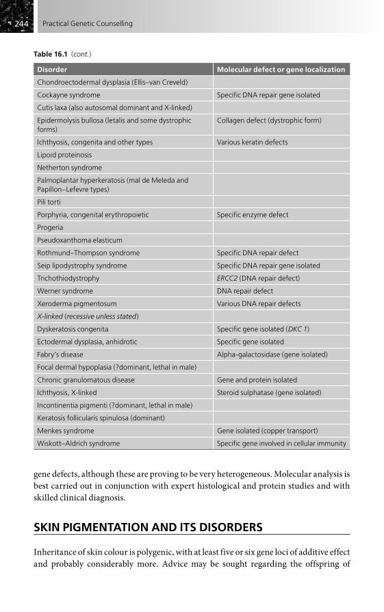
244244 Practical Genetic Counselling
gene defects, although these are proving to be very heterogeneous. Molecular analysis is best carried out in conjunction with expert histological and protein studies and with skilled clinical diagnosis.
SKIN PIGMENTATION AND ITS DISORDERS
Inheritance of skin colour is polygenic, with at least five or six gene loci of additive effect and probably considerably more. Advice may be sought regarding the offspring of
Table 16.1 (cont.)
Disorder Molecular defect or gene localization
Chondroectodermal dysplasia (Ellis–van Creveld)
Cockayne syndrome Specific DNA repair gene isolated
Cutis laxa (also autosomal dominant and X-linked)
Epidermolysis bullosa (letalis and some dystrophic forms)
Collagen defect (dystrophic form)
Ichthyosis, congenita and other types Various keratin defects
Lipoid proteinosis
Netherton syndrome
Palmoplantar hyperkeratosis (mal de Meleda and Papillon–Lefevre types)
Pili torti
Porphyria, congenital erythropoietic Specific enzyme defect
Progeria
Pseudoxanthoma elasticum
Rothmund–Thompson syndrome Specific DNA repair defect
Seip lipodystrophy syndrome Specific DNA repair gene isolated
Trichothiodystrophy ERCC2 (DNA repair defect)
Werner syndrome DNA repair defect
Xeroderma pigmentosum Various DNA repair defects
X-linked (recessive unless stated)
Dyskeratosis congenita Specific gene isolated (DKC 1)
Ectodermal dysplasia, anhidrotic Specific gene isolated
Fabry’s disease Alpha-galactosidase (gene isolated)
Focal dermal hypoplasia (?dominant, lethal in male)
Chronic granulomatous disease Gene and protein isolated
Ichthyosis, X-linked Steroid sulphatase (gene isolated)
Incontinentia pigmenti (?dominant, lethal in male)
Keratosis follicularis spinulosa (dominant)
Menkes syndrome Gene isolated (copper transport)
Wiskott–Aldrich syndrome Specific gene involved in cellular immunity

245245The Skin
interracial marriages or adoptions, and questions on this subject may merely be the focusing point of a considerable amount of stress, ignorance and latent prejudice. The attitude of other family members such as in-laws or grandparents may be frankly hostile, and it may in fact be not so much skin colour as other racial characteristics, such as hair or facial features, that are the main concern.
In general, children are likely to show skin colour intermediate between that of the parents. Where both parents are of mixed race, this will still apply, but here the likeli-hood is greater that a child could be either darker or lighter than both parents as a result of inheriting a particular selection of pigment-determining genes.
Light-skinned individuals of mixed race married to a white person may enquire as to whether a child or subsequent descendant might have extremely dark skin colour or African features – in other words, might be a clearly black person who perhaps would not be accepted in the white community into which he or she is born. This is very unlikely, but again it cannot be excluded that the degree of pigmentation, though probably not of other features, might exceed that of the darker parent, especially if the ‘white’ partner is relatively dark-skinned. Where the partner is blond and light-skinned, this possibility can be discounted.
Where mixed-race origin is known or is a possibility, caution must be advised in predicting later appearance from features present in early infancy. Skin colour may darken significantly and African-type hair may not be apparent for some months after birth. Reed, in his 1955 book Counseling in Medical Genetics, dealt in detail with these various features and it is of interest that inheritance of skin colour was the most common reason for seeking genetic counselling at his clinic. Although the climate of opinion has significantly altered since that time, and interracial marriages and adop-tions are now frequent and more accepted, one should not underestimate the significance for families of what may, to the physician, appear trivial features.
Several of the genes involved in skin colour have been identified; important and dif-ficult societal questions will arise as to how or whether these should be used as genetic tests for this normal characteristic. It would be wise for society to consider these issues now, rather than wait until misuse of such tests occurs.
Albinism
Generalized oculocutaneous albinism is autosomal recessive. Two main types exist:
• a severe form (tyrosinase negative) with total lack of pigment throughout life, and in which mutations in the tyrosinase gene have now been identified;
• a milder tyrosinase-positive form for which the gene has also been cloned (the p gene, corresponding to ‘pink eye’ in the mouse).
In the latter type, pigmentation of hair and iris gradually increases and mild cases may easily be missed. The forms are non-allelic, marriages between albinos of different type resulting in all normal offspring. A very rare type, associated with a bleeding diathesis (Hermansky–Pudlak syndrome), is also autosomal recessive. Prenatal detection of severe oculocutaneous albinism by fetal skin biopsy may be relevant in tropical
246246 Practical Genetic Counselling
countries where the morbidity is high. DNA analysis will be simpler as mutations become recognized. It also seems likely that some of the commoner genetic variants of pigmentation will be relevant to skin cancer susceptibility.
Ocular albinism (see Chapter 17) is X-linked, the gene being mapped to distal Xp.
Vitiligo
Vitiligo is commonly associated with a variety of autoimmune endocrine disturbances, and like them often follows a variable autosomal dominant pattern.
Piebaldism
Piebaldism commonly follows autosomal dominant inheritance and has been shown to result from specific mutations in the c-KIT oncogene. It may form part of the more gen-eralized Waardenburg syndrome, which is also autosomal dominant in inheritance and determined by the developmental gene PAX3. Isolated white forelock may also occur as a dominantly inherited trait.
PSORIASIS
Psoriasis is a common and variable disorder (prevalence around 1–2 per cent) which is frequently familial. Families apparently following all major types of mendelian inherit-ance have been reported, but it is likely that most of these represent extreme examples of a disorder that is polygenically determined. Susceptibility loci have been identified in the HLA region of chromosome 6, and others have been claimed, but so far none is con-sistent or strong enough to give any practical genetic test (see Chapter 3).
The risk for first-degree relatives of an isolated case is at least 10 per cent, and probably double this where there are two affected first-degree relatives. Where the disorder appears to follow an autosomal dominant pattern, it is probably wise to give a risk approaching 50 per cent for offspring of an affected member, but it is doubtful whether unaffected members of such pedigrees are completely free from the risk of transmitting it.
The children of two psoriatic patients also have a risk of around 50 per cent of being affected, but there does not seem to be a specially severe form in such children, as might have been expected if homozygosity at a single gene locus were operating.
ATOPIC ECZEMA
Atopic eczema is an extremely common problem, often associated with asthma and other allergic phenomena, and is probably determined by one or more autosomal dominant genes of rather variable expression. The genetics of atopy is discussed in Chapter 19 in relation to asthma and remains confused; a locus on chromosome 11 has
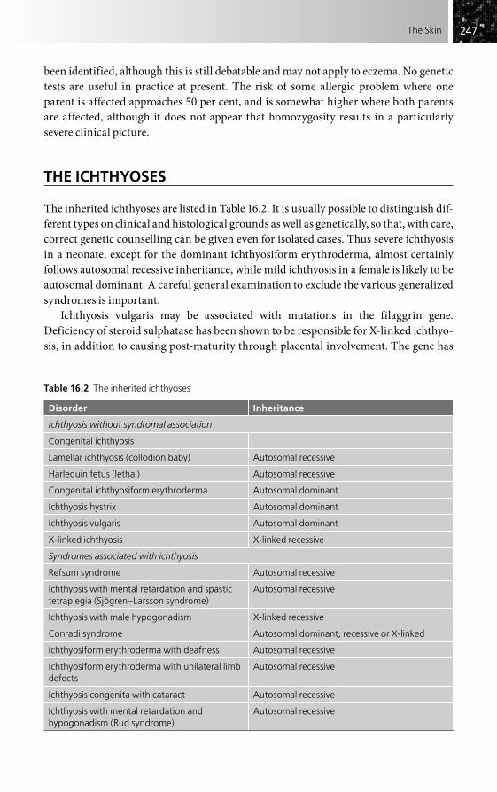
247247The Skin
been identified, although this is still debatable and may not apply to eczema. No genetic tests are useful in practice at present. The risk of some allergic problem where one parent is affected approaches 50 per cent, and is somewhat higher where both parents are affected, although it does not appear that homozygosity results in a particularly severe clinical picture.
THE ICHTHYOSES
The inherited ichthyoses are listed in Table 16.2. It is usually possible to distinguish dif-ferent types on clinical and histological grounds as well as genetically, so that, with care, correct genetic counselling can be given even for isolated cases. Thus severe ichthyosis in a neonate, except for the dominant ichthyosiform erythroderma, almost certainly follows autosomal recessive inheritance, while mild ichthyosis in a female is likely to be autosomal dominant. A careful general examination to exclude the various generalized syndromes is important.
Ichthyosis vulgaris may be associated with mutations in the filaggrin gene. Deficiency of steroid sulphatase has been shown to be responsible for X-linked ichthyo-sis, in addition to causing post-maturity through placental involvement. The gene has
Table 16.2 The inherited ichthyoses
Disorder Inheritance
Ichthyosis without syndromal association
Congenital ichthyosis
Lamellar ichthyosis (collodion baby) Autosomal recessive
Harlequin fetus (lethal) Autosomal recessive
Congenital ichthyosiform erythroderma Autosomal dominant
Ichthyosis hystrix Autosomal dominant
Ichthyosis vulgaris Autosomal dominant
X-linked ichthyosis X-linked recessive
Syndromes associated with ichthyosis
Refsum syndrome Autosomal recessive
Ichthyosis with mental retardation and spastic tetraplegia (Sjögren–Larsson syndrome)
Autosomal recessive
Ichthyosis with male hypogonadism X-linked recessive
Conradi syndrome Autosomal dominant, recessive or X-linked
Ichthyosiform erythroderma with deafness Autosomal recessive
Ichthyosiform erythroderma with unilateral limb defects
Autosomal recessive
Ichthyosis congenita with cataract Autosomal recessive
Ichthyosis with mental retardation and hypogonadism (Rud syndrome)
Autosomal recessive

248248 Practical Genetic Counselling
been cloned and a remarkably high proportion of cases (over 90 per cent) found to result from gene deletion, reflecting its localization near the pseudoautosomal region. Several X-linked syndromes with ichthyosis are proving to be due to contiguous gene deletion syndromes in this region, analogous to those around the Duchenne locus.
PALMOPLANTAR HYPERKERATOSIS (TYLOSIS)
Most cases of palmoplantar hyperkeratosis (tylosis) follow autosomal dominant inher-itance, and isolated cases thus have a high risk of transmitting the disorder. A variety of specific keratin gene defects have been identified (Table 16.3). The rare form known as ‘mal de Meleda’ is autosomal recessive and can occur outside the Adriatic area. The remarkable families with dominantly inherited oesophageal cancer and tylosis have late childhood onset of the skin disorder which appears not to result from a keratin defect. These cases are, however, exceptional, and families with tylosis from early child-hood and no history of oesophageal cancer in the family should not be worried unnecessarily by having this possibility raised.
EPIDERMOLYSIS BULLOSA
Epidermolysis bullosa is another exceptionally heterogeneous group of disorders in which genetic differences are supported by the clinical and histological features and where expert clinical and laboratory diagnosis is particularly essential. Thus the neonatal letalis form (now partly treatable by steroids) is autosomal recessive, while the mild simplex types, without scarring, are autosomal dominant; at least one form results from a molecular defect in keratin. The dystrophic forms with scarring may follow either pattern, but most severe cases are autosomal recessive. A defect in type 7 collagen has been found in most families. Junctional forms are due to defects in the basement
Table 16.3 Hereditary disorders of keratin
Disorder Keratin type
Epidermolysis bullosa simplex K5, K14
Epidermolytic hyperkeratosis K1, K10
Acral type (ichthyosis bullosa of Siemens) K2e
Palmoplantar keratodermia (hyperkeratosis)
Epidermolytic type K9
Non-epidermolytic type K1, K16
Pachyonychia congenita K6, K16, K17
Steatocystoma multiplex K17
Monilethrix (hair defect) Hb1, Hb6 (hair-specific keratins)
Courtesy of Dr Paul Bowden, Cardiff.
249249The Skin
membrane zone. Prenatal diagnosis of the letalis and dystrophic forms by fetoscopic skin biopsy is possible, but DNA analysis on chorion biopsy is preferable where the molecular defect is known.
ECTODERMAL DYSPLASIAS
Hypohidrotic (anhidrotic) ectodermal dysplasia
Hypohidrotic ectodermal dysplasia is the most common of the ectodermal dysplasias. It is X-linked recessive, with variable expression in female carriers, who may show dental anomalies as well as a reduced sweat pore count and a patchy distribution of the sweating pattern on starch-iodine testing. Isolation of the gene has made carrier detec-tion and prenatal diagnosis feasible in many families.
Other types
Other types of ectodermal dysplasia are autosomal. Both dominant and recessive types have been described, as well as a number of syndromes, including EEC syndrome (autosomal dominant, see Chapter 14) and chondroectodermal dysplasia (autosomal recessive).
PIGMENTED NAEVI
When present in a particular site, pigmented naevi commonly follow autosomal dominant inheritance. Multiple pigmented naevi are a feature of Turner syndrome and of the dominantly inherited syndrome of multiple naevi with nerve deafness (LEOPARD syndrome). The autosomal dominant dysplastic naevus syndrome is espe-cially important to recognize in view of the risk of melanomas developing. The skin lesions of neurofibromatosis (see Chapter 11) and tuberous sclerosis must be distin-guished, as must lesions overlying a spina bifida.
Cavernous haemangiomas
Cavernous haemangiomas of the facial region are usually sporadic, as is the trigeminal area flat vascular naevus of the Sturge–Weber syndrome, and the limb angiomas asso-ciated with hypertrophy (Klippel–Trenaunay–Weber syndrome). Somatic mutations have been confirmed in vascular endothelial growth factors in some types. Haemangiomatous and lymphangiomatous lesions of the limbs may be associated with hypertrophy and are usually sporadic, although familial cases are described (see Chapter 25). Rare instances of autosomal dominant inheritance in association with Wilms’ tumour have also been recorded. Other specific types of naevus following

250250 Practical Genetic Counselling
autosomal dominant inheritance are naevus flammeus of the nape of the neck and the ‘blue rubber bleb’ multiple naevi.
Segmental pigmentary disorders
Several unusual disorders of skin pigmentation have been described that follow a patchy or whorled distribution corresponding to Blaschko’s lines, and which may be accompanied by mental retardation or other systemic features. Hypomelanosis of Ito, incontinentia pigmenti and focal dermal hypoplasia (Goltz syndrome) are the most clearly defined. Chromosomal mosaicism has been found to underlie many cases previ-ously classed as hypomelanosis of Ito, and should be sought in blood and skin biopsies. Recurrence risk is low unless a parent shows mosaicism. An X-linked dominant (male lethal) gene may be responsible for most cases of incontinentia pigmenti, and the patchy skin distribution may be related to the pattern of X-chromosome inactivation.
BALDNESS
Severe, early male baldness is probably due to autosomal dominant inheritance, with expression of the gene limited to the male unless it is present in homozygous state. The author has never received a genetic counselling request for this innocuous condition, but a specific gene has recently been identified for a form of total alopecia, so it will be interesting to see if this changes the situation. Premature balding is a feature of myotonic dystrophy. Hair loss or scarcity may also result from a variety of ectodermal dysplasias and specific hair disorders (e.g. monilethrix, pili torti). Alopecia areata is often associ-ated with autoimmune endocrine disorders.
Isolation of the gene responsible for red hair colour (usually autosomal recessive) is more relevant to population genetic studies than to genetic counselling, but it is begin-ning to be used in forensic situations, raising important issues regarding the use of wider genetic approaches in this area.
ACANTHOSIS NIGRICANS
Primary acanthosis nigricans starts early in life and follows autosomal dominant inher-itance. Acanthosis nigricans may also accompany a variety of other genetic disorders. Onset in later life is commonly an indication of acquired visceral malignancy. The inherited type has no such association.
SKIN TUMOURS
A remarkable number of the rare skin-related tumours follow mendelian inheritance, mostly autosomal dominant, and appear in the lists in Table 16.1 and Table 25.1

251251The Skin
(see page 336). The recessively inherited disorders of DNA repair also frequently present with skin manifestations.
Xeroderma pigmentosum
Xeroderma pigmentosum (autosomal recessive) is now detectable prenatally as a repair defect of ultraviolet-induced DNA damage. Since several separate types exist, it is important for the cultured cells of the affected sib to be studied before embarking on prenatal diagnosis. Specific DNA repair genes have now been isolated and may simplify prediction.
Kaposi sarcoma
Most cases of Kaposi sarcoma are now associated with human immunodeficiency virus (HIV) infection, but for many years a small but well-documented number of families with multiple cases have been recognized. Whether these are truly genetic needs reassessment.
Malignant melanoma
Most cases of malignant melanoma appear to be non-genetic and largely related to UV radiation, but autosomal dominant inheritance occurs in a small subset of families. One type is known as familial dysplastic naevus syndrome. Transplacental passage of malig-nant cells is also recorded. Several different specific tumour suppressor genes (notably CDKNZA and CDK4) have been found to be involved in the dominantly inherited families. Genetic testing is beginning to be helpful for such families, especially in high-exposure countries like Australia.
Basal cell naevus (Gorlin) syndrome
Basal cell naevus syndrome is a dominantly inherited disorder which may be recog-nized from skeletal abnormalities, especially jaw cysts, before tumours appear. There may be also an increased risk of cerebral tumours. The gene responsible on chromo-some 9 has proved to be a homologue of the Drosophila gene ‘Patch’, involved in cell signalling, and the same gene may prove to underlie the rare and remarkable familial self-healing epithelioma known principally from western Scotland, which is localized to the same region. Isolated basal cell tumours are not known to be genetic.
Congenital fibromatosis
Congenital fibromatosis is a rare disorder in which multiple spindle-cell fibromatous tumours occur and commonly mature spontaneously. The condition may be fatal if gut tumours occur, but is usually benign. Although autosomal recessive inheritance has been claimed, autosomal dominant inheritance with incomplete penetrance (especially

252252 Practical Genetic Counselling
in older individuals) is more probable. Careful search for small lesions in apparently unaffected parents is important.
FURTHER READING
GeneralBale SJ (2000). Genetics for Dermatologists. London: Remedica [a useful, compact guide with
clinical and molecular information].Moss C, Savin J (1995). Dermatology and the New Genetics. Oxford: Blackwell.Sybert VP (1997). Genetic Skin Disorders. New York: Oxford University Press [an excellent book,
profusely illustrated and with molecular details, and an authoritative reference source for all those disorders that are mentioned only briefly (or not at all) in the present chapter (new edition expected 2010)].
Specific disordersArin MJ (2009). The molecular basis of human keratin disorders. Hum Genet 125, 355–73.Bale SJ, DiGiovanna JJ (1997). Genetic approaches to understanding the keratinopathies.
Adv Dermatol 12, 99–113.Eady RAJ, Dunhall MGS (1994). Epidermolysis bullosa: hereditary skin fragility diseases as
paradigms in cell biology. Arch Dermatol Res 287, 2–9.Kasparian NA, Meiser B, Burton PN, Simpson JM, Mann GJ (2009). Genetic testing for
melanoma risk: a prospective study of uptake and outcomes among Australian families. Genet Med 11, 265–78.
Paller AS (1996). The genetic basis of hereditary blistering disorders. Curr Opin Pediatr 8, 367–71.
Spitz JL (2004). Genodermatoses: a Clinical Guide to Genetic Skin Disorders. Philadelphia: Lippincott Williams & Wilkins.
Spritz RA (1994). Molecular genetics of oculocutaneous albinism. Hum Mol Genet 3, 1469–75.Spritz RA, Hearing VJ (1995). Genetic disorders of pigmentation. In: Harris H, Hirschhorn K
(eds) Advances in Human Genetics, pp. 1–45. St Louis: Mosby.

Introduction . . . . . . . . . . . . . . . . . . . . . . . . . . . . . . . . . . . . . . . . . . . . . . . . . . . . . . . . . . . . . . . . . . . 253
Choroidoretinal degenerations . . . . . . . . . . . . . . . . . . . . . . . . . . . . . . . . . . . . . . . . . . . . . . . . . 255
Nystagmus . . . . . . . . . . . . . . . . . . . . . . . . . . . . . . . . . . . . . . . . . . . . . . . . . . . . . . . . . . . . . . . . . 256
Colour vision . . . . . . . . . . . . . . . . . . . . . . . . . . . . . . . . . . . . . . . . . . . . . . . . . . . . . . . . . . . . . . . . . . . 257
Leber’s optic atrophy . . . . . . . . . . . . . . . . . . . . . . . . . . . . . . . . . . . . . . . . . . . . . . . . . . . . . . . . . . . 257
Corneal dystrophies . . . . . . . . . . . . . . . . . . . . . . . . . . . . . . . . . . . . . . . . . . . . . . . . . . . . . . . . . . . . 258
Retinal detachment . . . . . . . . . . . . . . . . . . . . . . . . . . . . . . . . . . . . . . . . . . . . . . . . . . . . . . . . . . . . 258
Retinoblastoma . . . . . . . . . . . . . . . . . . . . . . . . . . . . . . . . . . . . . . . . . . . . . . . . . . . . . . . . . . . . . . . . 258
Cataract . . . . . . . . . . . . . . . . . . . . . . . . . . . . . . . . . . . . . . . . . . . . . . . . . . . . . . . . . . . . . . . . . . . . . . . 259
Glaucoma . . . . . . . . . . . . . . . . . . . . . . . . . . . . . . . . . . . . . . . . . . . . . . . . . . . . . . . . . . . . . . . . . . 260
Refractive errors . . . . . . . . . . . . . . . . . . . . . . . . . . . . . . . . . . . . . . . . . . . . . . . . . . . . . . . . . . . . . . . 261
Heterochromia of the iris . . . . . . . . . . . . . . . . . . . . . . . . . . . . . . . . . . . . . . . . . . . . . . . . . . . . . . . 261
Eye colour . . . . . . . . . . . . . . . . . . . . . . . . . . . . . . . . . . . . . . . . . . . . . . . . . . . . . . . . . . . . . . . . . . 262
Strabismus . . . . . . . . . . . . . . . . . . . . . . . . . . . . . . . . . . . . . . . . . . . . . . . . . . . . . . . . . . . . . . . . . . . . . 262
Hereditary ptosis . . . . . . . . . . . . . . . . . . . . . . . . . . . . . . . . . . . . . . . . . . . . . . . . . . . . . . . 262
Developmental eye defects . . . . . . . . . . . . . . . . . . . . . . . . . . . . . . . . . . . . . . . . . . . . . . . . . . . . . 262
Further reading . . . . . . . . . . . . . . . . . . . . . . . . . . . . . . . . . . . . . . . . . . . . . . . . . . . . . . . . . . . . . . . . 264
The Eye
CHAPTER 17
INTRODUCTION
The study of inherited eye disorders formed a major part of early work on the mende-lian basis of genetic disease, largely because their non-lethal nature led to large families with clear inheritance patterns, but also because many generalized genetic disorders have ocular manifestations important in diagnosis. We are now seeing a renaissance as the individual genes are isolated; there are even future possibilities for localized gene therapy. These rapid developments make it especially important that patients and family members be given accurate and up-to-date genetic information.
Patients with congenital or childhood blindness frequently marry each other, with complex results, although in contrast to congenital deafness it is usually possible to dis-tinguish clinically the precise genetic type of each parent’s disorder. The author has found that a clinic at a school for visually impaired children, run jointly with an oph-thalmologist, has been of great help to school leavers and their parents. Such schools serve a wide geographical area and allow many patients to be seen who might have been missed through the regular genetic counselling service. Such a system also minimizes the risk of erroneous diagnosis, which a non-specialist is in no position to query alone.

254254 Practical Genetic Counselling
The increasing trend for visually impaired children to be educated in regular schools is making provision of this type of service more difficult.
Since this chapter cannot possibly list, let alone discuss, all the hereditary ophthal-mic disorders, a selective approach has been adopted, aimed to help paediatricians and other clinicians who encounter hereditary eye disorders. It is not primarily intended for ophthalmologists, although they may perhaps find some parts helpful. Around 3–4 per 1000 children in developed countries have some form of severe visual handicap and at least half of these are genetic in origin. Mendelian disorders are also prominent in pro-gressive blindness of later onset, while genetic factors are now being identified in such common problems as glaucoma, cataracts and age-related macular degeneration.
A remarkable number of X-linked disorders affecting the eye are known, and these are listed separately (Table 17.1) because they produce special problems in genetic coun-selling. The carrier state can be recognized in a number of these and they provide direct evidence for mosaicism due to X-chromosome inactivation in the female. Patchy mor-phological changes can be seen in a number of these carriers, which allow diagnosis of the carrier state in the absence of biochemical tests. Molecular analysis can now often provide accurate tests based on the direct detection of mutations, especially for this X-linked group, but also for some autosomal disorders.
Table 17.1 X-linked eye disorders
Disorder Changes in heterozygote
Ocular albinism* Patchy fundal depigmentation, translucency of iris
Oculocutaneous albinism with deafness Partial hearing loss
X-linked congenital cataract Sutural lens opacities
Choroideraemia* Retinal pigmentary changes (sometimes symptomatic)
Abnormal electroretinogram
Colour blindnessDeutan*Protan*Incomplete achromatopsia
Minor defects in colour vision
Iris hypoplasia with glaucoma
X-linked macular dystrophy
Megalocornea (also rarely dominant) Corneal diameter increased
Microphthalmos with multiple anomalies (Lenz syndrome)
Congenital stationary night blindness with myopia
Norrie’s disease (pseudoglioma)* Some cases due to gene deletion
Hereditary oculomotor nystagmus Variable; may be fully affected
Oculocerebrorenal (Lowe’s) syndrome* Mild lens opacities
X-linked retinitis pigmentosa* Patchy retinal pigmentary and electroretinographic changes
Retinoschisis*
*Indicates gene cloned.
255255The Eye
CHOROIDORETINAL DEGENERATIONS
A great variety of types exist, characterized by particular features of fundal appear-ance, by differences in severity and progression, and by different responses to various types of electrodiagnostic investigation. It is most unwise for someone who is not an ophthalmologist to venture into diagnosis, but a valuable contribution can be made by documenting the pedigree pattern and by carefully searching for any associated syndromic features. This information can then be combined with a specific ophthal-mic diagnosis to allow accurate genetic counselling. Two broad groups can be distinguished:
• those mainly affecting peripheral vision (e.g. the retinitis pigmentosa group);• those principally involving central vision (e.g. the macular dystrophies).
Retinitis pigmentosa
Retinitis pigmentosa is the most common of the retinal degenerations. This group of disorders may follow all three main modes of mendelian inheritance, autosomal reces-sive forms being the most common (about 50 per cent), with autosomal dominant inheritance accounting for around 25 per cent of families. X-linked cases account for around 15 per cent of the total, but for about 50 per cent of isolated male cases. Marked variation in course occurs in different families, suggesting further heterogeneity. Currently around 40 different loci are known.
Carriers of the X-linked form may often (but not always) show visible pigmentary disturbance and an abnormal electroretinogram, a useful distinguishing point from the other forms of inheritance in an isolated male case. The carriers for autosomal recessive forms do not generally show abnormalities, so distinction from a new dominant mutation is often impossible. Clinical features are too variable to help much, while an additional problem is incomplete penetrance of the gene in around 10 per cent of heterozygotes for the dominant form. The empirical risk for an affected child being born to a parent who is an isolated case is around 1 in 8. Should a child indeed be affected, the risk to subsequent offspring would be 1 in 2.
Molecular developments in retinitis pigmentosa are now becoming of practical help in those families where a defect is known, with mutations in rhodopsin and peripherin (key molecules involved in retinal function) occurring in a significant proportion of autosomal dominant families; at least three X-linked loci exist, making prediction dif-ficult outside the occasional very large family. Retinitis pigmentosa currently provides a good example of the all-too-frequent situation in genetic testing, of research advances not yet becoming regularly available as a service. Given that there appear to be at least 75 different loci involved, this is hardly surprising.
Choroideraemia is a specific X-linked disorder that may be confused with retinitis pigmentosa, especially in its early stages. Its recognition is particularly important since the gene has been isolated, allowing accurate presymptomatic and carrier detection. Retinoschisis is yet another X-linked disorder, with retinal degeneration associated with a characteristic splitting of the retina, where specific molecular analysis is possible.

256256 Practical Genetic Counselling
A number of syndromes with retinitis pigmentosa exist, including the Bardet–Biedl syndrome (with polydactyly, hypogonadism and mental retardation), Hallgren syndrome (with deafness, ataxia and mental disturbance) and Usher syndrome (two types, one with profound nerve deafness), all of which are autosomal recessive.
Macular dystrophies
The macular dystrophies are a heterogeneous group, selectively involving central vision in contrast to the early peripheral involvement in retinitis pigmentosa. This is readily distinguished by an experienced ophthalmologist, but separating the different forms may be very difficult, causing problems in genetic counselling, especially for the isolated case.
Many late-onset macular dystrophies, as well as the early-onset Best’s macular degen-eration (gene isolated on chromosome 11q), follow an autosomal dominant pattern, while the juvenile Stargardt form is autosomal recessive and due to mutations in a specific ion transport gene on chromosome 1. Another rare but treatable recessive type is gyrate atrophy associated with a metabolic defect in ornithine aminotransferase. Cone dystro-phies are a further heterogeneous group, associated with deterioration in colour vision. An important recent development has been the finding that some families with dominant macular degeneration in old age are heterozygous for mutations in the Stargardt gene.
Since we have no clear idea yet as to the penetrance of this heterozygous mutation or whether this is a generally valid association, testing is not helpful clinically at present. Most cases of age-related macular degeneration are not mendelian, and smoking is an important risk factor, as may be a variant of complement factor H.
Leber’s congenital amaurosis
This must not to be confused with Leber’s optic atrophy (see later). Leber’s congenital amaurosis is a primary retinal disorder that is one of the most common causes of child-hood blindness, and is autosomal recessive in inheritance. The condition can be detected in early infancy by electroretinogram. Occasional families with associated cerebral and renal degeneration are known, but do not overlap with the isolated form. Mutations in a retinal homeobox gene (CRX) have been identified, while a separate retinal epithelium protein gene (RPE65) is also involved.
Congenital stationary night blindness
This is usually autosomal dominant or X-linked dominant. An X-linked recessive form with myopia also exists.
NYSTAGMUS
A clear primary diagnosis for nystagmus is essential, because the causes may be neuro-logical or vestibular as well as ocular. Even when the nystagmus is primary, there are a

257257The Eye
number of causes. Probably the most important ones to recognize are the various types of albinism, congenital stationary night blindness (see above) and the X-linked heredi-tary oculomotor nystagmus, which shows very variable manifestation in females (see Fig. 2.22 on page 43).
COLOUR VISION
The common forms of colour blindness, whether protan or deutan in type, are uni-formly X-linked recessive in inheritance and occur in about 8 per cent of males.
Because of this high frequency, matings of affected males and carrier females are not uncommon, with a 50 per cent risk of children of either sex being affected. Around 0.4 per cent of women have colour blindness. The genes for red and green colour vision have now been cloned, allowing molecular analysis of colour vision defects.
The rare total colour blindness (monochromatism) is autosomal recessive in inher-itance, while the even rarer ‘blue cone’ type is X-linked. All three disorders should be distinguished from the progressive cone dystrophies.
LEBER’S OPTIC ATROPHY
This must not be confused with Leber’s congenital amaurosis (see above). Leber’s optic atrophy follows classical mitochondrial inheritance (see Chapter 2), but other genetic and environmental factors modify the primary pattern. The main empirical risks are as follows.
• Males are affected more often than females (85 per cent), in Europe, but not in Japan.
• Males never transmit the disease to descendants of either sex, not even to grandchildren or subsequent generations.
• Where a female is affected or has an affected son, the risk to subsequent sons is 1 in 2, but all her daughters appear to be either carriers (80 per cent) or affected (20 per cent), unlike X linkage.
Mitochondrial DNA analysis can now identify specific germ-line mutations and is especially valuable in cases without a clear genetic pattern. However, as discussed in Chapter 2, the identification of mitochondrial inheritance is of the greatest help in con-firming the diagnosis and giving the general pattern of risk, but is singularly unhelpful for the female carrier, since it gives no indication as to whether she will herself become affected, or the risk to particular offspring. An exception is for offspring of a woman who is heteroplasmic for the mutation, who has both normal and mutant DNA; if these offspring show no or very low mutant DNA themselves, their risk of developing the disorder is low. It is hoped that some preventive therapy that enhances or spares mito-chondrial function may be developed that will help those at risk. Avoiding risk factors such as smoking is important.

258258 Practical Genetic Counselling
Other forms of hereditary optic atrophy
Several forms exist following both autosomal dominant and recessive patterns. Those of adult life are mostly dominant and usually show a slowly progressive course, unlike the subacute onset of Leber’s optic atrophy. The Wolfram or DIDMOAD syndrome (diabetes insipidus, diabetes mellitus, optic atrophy and deafness), generally considered to follow autosomal recessive inheritance, has now been shown to result from mito-chondrial mutations in some families, so caution is needed in giving risks.
CORNEAL DYSTROPHIES
Numerous types of corneal dystrophy exist. The slit-lamp appearance is often very char-acteristic (to the expert), and unless a clear pedigree pattern is seen, it is wise to be guided by ophthalmological opinion. Most types are mendelian. Corneal clouding and opacifi-cation may be a helpful diagnostic feature in various generalized diseases, notably the mucopolysaccharidoses, but also in some lipoprotein disorders and cystinosis.
Some dominant forms of corneal dystrophy have recently been shown to result from molecular defects in different forms of keratin, such as K3, K12, kerato-epithelin and type 8 collagen (see also Chapter 16).
RETINAL DETACHMENT
Retinal detachment is commonly associated with severe myopia, and a significant risk to relatives is only likely when they also have severe myopia. Occasional dominantly inherited families are documented, with one locus mapped. Several other genetic syndromes may be accompanied by retinal detachment, including type II collagen defects such as severe spondyloepiphyseal dysplasia and Stickler syndrome, as well as the related, but distinct, condition known as Wagner’s retinopathy, all of which are dominantly inherited.
RETINOBLASTOMA
Retinoblastoma provides an extremely important and difficult area for genetic counsel-ling. All bilateral cases appear to be hereditary, compared with only about 15 per cent of unilateral cases. Further, only 90 per cent of those with the gene develop tumours, and there are instances where the disorder seems to have been suppressed in an entire branch of a kindred. Occasionally, spontaneous disappearance of a tumour may leave a retinal scar as the only feature, so parents of an isolated case should always be examined carefully. Survivors have an increased risk (around 10 per cent) of other neoplasms, notably osteosarcoma, in later life.
Most cases of retinoblastoma are not associated with other malformations, but abnormalities of chromosome 13q may be accompanied by retinoblastoma, a finding that led to localization of the gene on this chromosome.

259259The Eye
The gene has been cloned and molecular analysis can be used to provide accurate prediction for those at risk. Tumour DNA analysis is important in unilateral cases. Retinoblastoma provided the first full vindication of the ‘two hit’ mutation theory of cancer (see Chapter 25), and tumour DNA analysis shows that the same locus is indeed involved in both germinal and somatic mutations. The lack of penetrance can now be readily explained by absence of the necessary somatic mutation; while the genetic pre-disposition is dominantly inherited, the developing retinal tissue must be homozygous for the defect if a tumour is to occur. Equally, a germ-line mutation must have been inherited for more than a single tumour to occur in an individual.
The empirical risk estimates given in Table 17.2 need to be reassessed in the light of molecular developments, but remain useful where affected relatives are dead or when DNA analysis is not possible. It should be noted that the risks for some categories have been reduced by comparison with earlier editions of this book, in the light of further evidence.
Norrie’s disease (pseudoglioma)
In the past, Norrie’s disease was frequently confused with retinoblastoma. The frequent occurrence of mental retardation makes genetic counselling for this X-linked recessive disease of considerable importance. The gene has been identified and molecular analysis is relevant to carrier identification and prenatal diagnosis.
CATARACT
In cases of both congenital and adult-onset cataract, the nature and location of the opacities, especially in the early stages, can be of help in distinguishing the specific types.
Table 17.2 Genetic risks to offspring in retinoblastoma
Risk (%)
Unilateral
Affected, with affected parent or sib 45
Unaffected, parent and sib or two sibs affected 5
Affected, no other affected relatives 1
Unaffected, one affected child 1
Unaffected, one affected sib 1
Bilateral
Affected, other family members affected 45
Affected, no other affected family members 45
Unaffected, one affected child 2
Unaffected, parent affected 5
Based on Fuhrmann W, Vogel F (1992), Genetic Counselling: a Guide for the Practising Physician, Springer, Berlin; also on Draper GJ et al. (1992), Br J Cancer 66, 211–219.

260260 Practical Genetic Counselling
Congenital cataract
Numerous types of congenital cataract exist, with all forms of inheritance recorded. The incidence is around 1 in 250 births. Environmental causes (e.g. rubella) and metabolic and other primary disorders (e.g. galactosaemia and hypoparathy-roidism) must be excluded, and syndromal associations (e.g. Conradi’s disease) sought.
Because most genetic forms without a metabolic cause follow dominant inheritance, the risk for offspring of an affected person is not far short of 50 per cent. The risks for sibs of an isolated case is probably 10 per cent or less, but more accurate figures are needed.
A number of specific genes, some involving lens crystallin proteins, have been isolated, but are not yet in regular diagnostic use.
Cataracts in later life
Primary disorders, both mendelian (e.g. myotonic dystrophy) and non-mendelian (e.g. diabetes), must be excluded. Most families showing a clear-cut aggregation appear to follow autosomal dominant inheritance.
Lens dislocation
Lens dislocation is a feature of the Marfan and Marchesani syndromes and of homocystinuria, but may occur as an isolated abnormality due to an abnormally small and spherical lens (spherophakia), usually following autosomal dominant inheritance. The author has seen tall, thin members of one such family persistently misdiagnosed as having Marfan syndrome, with much unnecessary worry caused. In some families at least, the same fibrillin locus on chromosome 15 is involved as in Marfan syndrome.
GLAUCOMA
Glaucoma can be a part of a surprisingly large number of genetic syndromes and should be checked for in any ocular assessment of the patient with syndromes involving the eye, since it may be a treatable aspect in an individual without specific ocular com-plaints. Major advances are occurring in isolation of genes involved with primary glaucoma (see ‘Further reading’ for a review by Sarfarazi), although it is not yet clear how these findings will affect genetic risks for those without a clear mendelian inherit-ance pattern.
Primary closed-angle glaucomaPrimary closed-angle glaucoma seems to be determined largely by anatomical orbital factors, particularly shallowness of the anterior chamber; 12 per cent of sibs were found to be clinically affected in one study.
261261The Eye
Primary open-angle glaucomaPrimary open-angle glaucoma is common in the general population and is found in 1 in 200 elderly people. Studies of sibs have shown between 5 and 16 per cent to be affected; 10 per cent is probably an appropriate risk for clinically significant glaucoma. The risks for children have been lower, but extremely variable. Because the children studied were always much younger than the sibs, it seems likely that the lifetime risk will approach the 10 per cent seen for sibs. The proportion of families that has mendelian basis is uncertain, but some large families following autosomal dominant inheritance exist, some adult-onset, others juvenile, and have been mapped to specific chromosomes. Mutation in several differ-ent genes, showing variable penetrance, have been shown to occur in some juvenile families.
Congenital glaucoma
Congenital glaucoma may be associated with other generalized ocular problems (e.g. Sturge–Weber syndrome). When it is primary, a proportion of families appears to follow autosomal recessive inheritance, but isolated cases are much too common for this mode to explain all cases. The risk to sibs after a single affected child is around 10 per cent; after two affected sibs, a 25 per cent risk should be advised. Risks to children of affected individuals are uncertain. Assuming a mixture of recessive and polygenic forms, a risk of 5 per cent seems appropriate until data are available. A specific cyto-chrome P450 gene on chromosome 2p has now been shown to be responsible for some recessively inherited families, but other genetic loci also exist.
REFRACTIVE ERRORS
Twin studies show a very close concordance between monozygotic twin pairs, suggest-ing a high degree of genetic determinance. Individual pedigrees showing all types of mendelian inheritance have been produced for each of the major types of refractive error, but are of little help in deriving general risks for relatives. Studies of unselected families show high correlations for refractive values between both sibs and parents and offspring, suggesting that a polygenic basis is present with genes of additive effect and little dominance or recessivity. The same situation applies to disorders of corneal shape such as astigmatism, keratoconus and cornea plana.
Some regular syndromes of refractive error exist, including myopia and night blind-ness, which are usually X-linked recessive. Refractive errors may also accompany other primary mendelian disorders, such as myopia in Marfan syndrome and some skeletal dysplasias. In isolated cases of severe myopia, a risk of 4–5 per cent for similar severe eye problems in the children has been suggested.
HETEROCHROMIA OF THE IRIS
Heterochromia is frequently an isolated and harmless trait, often autosomal dominant in inheritance. The most important cause of heterochromia to recognize
262262 Practical Genetic Counselling
is Waardenburg syndrome (see Chapter 18), in which piebaldness and deafness are major features. Variation in expression of this autosomal dominant disorder is considerable.
EYE COLOUR
In the early medical genetics literature, eye colour was given as one of the most common reasons for requesting genetic counselling, but this rarely seems to be the case now. It is possible that such enquiries were really aimed at establishing paternity. In fact, while brown eye colour in general behaves as dominant to light blue eye colour, the genetic control is considerably more complex than this, and exceptions are sufficiently frequent for this trait not to be used as evidence for or against paternity.
STRABISMUS
Strabismus is a frequent feature of many generalized neuromuscular disorders, which may follow mendelian inheritance (see Chapter 11). Duane syndrome is due to aberrant inner-vation of ocular muscles; it is generally sporadic, but a few families follow autosomal dominant inheritance. Isolated strabismus, whether classified as convergent or divergent, fits a polygenic pattern. Variation between studies results, in part, from the extent to which minor deviations are classed as abnormal. From the viewpoint of counselling, it seems that where parents are normal and one child is affected, the risk for subsequent children is around 15 per cent. Where one parent is also affected, the risk is around 40 per cent.
HEREDITARY PTOSIS
Hereditary ptosis is usually autosomal dominant and may persist unchanged through life. Care must be taken to distinguish more general neuromuscular causes (see Chapter 11), such as myotonic dystrophy, oculopharyngeal muscular dystrophy, myasthenic syndromes and the mitochondrial myopathies.
DEVELOPMENTAL EYE DEFECTS
This field provides an excellent example of the value of comparative genetic studies, genes being strongly conserved between species, with an increasing number of muta-tions recognized in human disorders.
Microphthalmos and anophthalmos
Microphthalmos and anophthalmos constitute an extremely heterogeneous group. Unilateral cases are frequently non-genetic, but cannot be securely distinguished from

263263The Eye
genetic forms. Rubella, toxoplasmosis and fetal alcohol syndrome are causes to be excluded for bilateral disease. Mental retardation is frequently associated, and microph-thalmos is a feature of severe chromosomal defects as well as mendelian syndromes. The X-linked Lenz syndrome of microphthalmos with cataract, mental retardation and digital and genitourinary abnormalities must be considered. Microphthalmos with coloboma is usually autosomal dominant (in the absence of known external causes). Complete bilateral anophthalmos is generally autosomal recessive. Cryptophthalmos, with absent palpebral fissures, may be part of the above disorders, or may occur with relatively normal eye development, usually following autosomal recessive inheritance. Some cases are part of the more general Fraser syndrome (autosomal recessive), where renal agenesis and laryngeal atresia may be major features, and where a specific developmental gene defect is now known.
Cyclops
Almost all cases of this lethal malformation, an extreme form of holoprosencephaly (see Chapter 12), have been sporadic. Chromosomal abnormalities have been found in some cases.
Coloboma and aniridia
Both bilateral coloboma of the iris and the more severe aniridia usually follow auto-somal dominant inheritance; colobomas may form part of more extensive ocular disorders. Because colobomas may vary considerably in extent, a thorough ophthalmic examination of both parents and patient is needed. The rare syndrome of ocular coloboma with anal atresia (cat-eye syndrome) follows an autosomal dominant pattern but is associated with an extra chromosome 22 fragment.
Aniridia may be associated with Wilms’ tumour, mental retardation and genital defects, but such cases are generally sporadic, most dominant families being deter-mined by a gene on chromosome 2. A small deletion on the short arm of chromosome 11 is seen in some cases. Detailed molecular analysis of the region has shown that a series of overlapping deletions is responsible for the various syndrome conditions, anal-ogous to those seen in other microdeletion syndromes.
Mutations in the gene PAX6 and some other developmental genes may occur in aniridia and other anterior chamber abnormalities.
Corneal and anterior chamber abnormalities
These are heterogeneous and at times syndromal (e.g. Rieger syndrome, Peter abnor-mality). They are rare and require expert diagnosis, as well as checking of the parents for minor defects. Molecular abnormalities have been found in some cases, though less consistently than in aniridia.

264264 Practical Genetic Counselling
FURTHER READING
Deeb SS, Motulsky AG (1996). Molecular genetics of human color vision. Behav Genet 26, 195–207.
Hackett SE (1997). Leber’s hereditary optic neuropathy: a genetic disorder of the eye. Insight 22, 94–6.
Rennie WA (ed.) (1986). Goldberg’s Genetic and Metabolic Eye Disease. Boston: Little, Brown.Sarfarazi M (1997). Recent advances in molecular genetics of glaucomas. Hum Mol Genet 6,
1667–77.Smith BJ, O’Brien JM (1996). The genetics of retinoblastoma and current diagnostic testing.
J Pediatr Ophthalmol Strabismus 33, 120–3.Sullivan LS, Daiger SP (1996). Inherited retinal degeneration: exceptional genetic and clinical
heterogeneity. Mol Med Today 2, 380–6.Taylor D (1990). Pediatric Ophthalmology. Oxford: Blackwell.Traboulsi EI (1998). Genetic Diseases of the Eye. Oxford: Oxford University Press.Traboulsi EI (2005). Compendium of Inherited Disorders and the Eye. New York: Oxford
University Press.Wright AF, Jay B (eds) (1994). Molecular Genetics of Inherited Eye Disorders. Chur: Harwood.
Introduction . . . . . . . . . . . . . . . . . . . . . . . . . . . . . . . . . . . . . . . . . . . . . . . . . . . . . . . . . . . . . . . . . . . 265
Severe congenital sensorineural deafness . . . . . . . . . . . . . . . . . . . . . . . . . . . . . . . . . . . . . . . 266
Mild-to-moderate deafness . . . . . . . . . . . . . . . . . . . . . . . . . . . . . . . . . . . . . . . . . . . . . . . . . . . . . 268
Deafness as part of syndromes . . . . . . . . . . . . . . . . . . . . . . . . . . . . . . . . . . . . . . . . . . . . . . . . . . 269
The external ear . . . . . . . . . . . . . . . . . . . . . . . . . . . . . . . . . . . . . . . . . . . . . . . . . . . . . . . . . . . . . . . 270
Ménière’s disease . . . . . . . . . . . . . . . . . . . . . . . . . . . . . . . . . . . . . . . . . . . . . . . . . . . . . . . . . . . . . . 270
Further reading . . . . . . . . . . . . . . . . . . . . . . . . . . . . . . . . . . . . . . . . . . . . . . . . . . . . . . . . . . . . . . . . 270
Deafness
CHAPTER 18
INTRODUCTION
At least 50 per cent of cases of congenital and childhood deafness may be genetically determined. In the case of non-syndromic deafness, a precise clinical diagnosis may be impossible owing to phenotypic overlap with deafness of environmental origin. Careful attention to family history and detailed audiological evaluation, not just of the proband but also of other family members, may help to resolve the question of aetiology in appar-ently isolated cases. Close consultation with audiological colleagues and others is needed if errors are to be avoided.
Recognition of the specific genes involved in deafness has had a major impact on our understanding of this field and is also now playing a significant role in diagnosis and genetic counselling. For non-syndromic congenital sensorineural deafness, the loci and mutations involved vary considerably between different populations and it is important to know the distribution of molecular defects in the particular population, if possible.
Two groups, in particular, may request genetic counselling: parents of a severely affected child wishing to have further children, and young adults with deafness, who frequently marry partners similarly affected.
Even without access to specialized audiological testing, it is often possible to assess the genetic situation accurately if the following points are borne in mind.
• What does the pattern of inheritance in the particular family suggest?• Is the hearing loss severe congenital deafness, or some milder form?• If hearing loss is milder, is it static or progressive?• Is there an identifiable syndrome involving other systems?

266266 Practical Genetic Counselling
Genetic counselling for the profoundly deaf is a service that requires a radically different approach from that in most other fields. The process of communication will usually require an intermediary, unless one has special experience with sign language or other forms of communication. Attitudes to deafness within the community of the profoundly deaf may well be quite different from those of doctors or of normally hearing patients. Genetic coun-selling may be perceived by some as a threat, unless it is sensitively and appropriately integrated into the overall educational provision for young adults and adolescents.
SEVERE CONGENITAL SENSORINEURAL DEAFNESS
The incidence has been estimated to be around 1 in 1000 births. Care must be taken to exclude external factors such as mild congenital rubella or cytomegalovirus infection.
In the absence of clear evidence of environmental aetiology, there is no doubt that a high proportion of cases result from autosomal recessive inheritance. It is difficult to decide exactly what this proportion is, because the different types are at present clinically indistinguishable. This is changing now that specific genes and mutations have been identified in a significant proportion of cases, but one often remains dependent on older, though well founded, data.
Most studies have suggested that 40–50 per cent of the cases are autosomal recessive, with around 10 per cent due to autosomal dominant inheritance and most of the rest due to unknown or undetected environmental (or at least non-mendelian) factors. This would suggest that the risk of deafness in sibs of an isolated case is about 1 in 10. Where consanguinity exists, autosomal recessive inheritance is even more likely, and a 1 in 4 risk should be given. Likewise, should a couple have a second affected child, autosomal recessive inheritance is almost certain. Table 18.1 summarizes the various risks.
The risk for offspring of healthy sibs and other family members is often asked about. This is extremely low (well under 1 per cent) in the absence of consanguinity or of deafness in the family of the other partner.
The risk for offspring of an affected individual who is an isolated case and married to a normal person is low, but not negligible (around 5 per cent). This risk probably results from an inclusion of unrecognized new dominant mutations with the much larger number following recessive inheritance, unless there is consanguinity or other factors suggesting that a recessive gene may have been transmitted by the healthy
Table 18.1 Genetic risks in profound childhood deafness of unknown cause
Affected relative Risk
One child only; environmental factors carefully excluded 1 in 10
One child only; consanguinity present 1 in 4
Two affected children 1 in 4
One parent � one child 1 in 2
One parent only 1 in 20
Parent � sib(s) of parent only 1 in 100
Sib(s) of parent; parent unaffected � 1 in 100

267267Deafness
parent. Although severe dominantly inherited congenital deafness is rare in compari-son to recessive forms, and is more variable in severity, it nevertheless accounts for the majority of two-generation families. In families with two affected sibs and healthy parents with normal audiograms, where recessive inheritance is almost certain, the risk for offspring of the affected individuals is low (around 1 per cent).
X-linked recessive inheritance is well documented as a mode of inheritance in severe congenital deafness but is not frequent enough to affect the risks for isolated male cases or single sibships containing only affected males. Distinctive findings may be present on cochlear CT scan.
Marriage between two individuals with severe congenital deafness is common, and the offspring of such marriages provide clear evidence for the existence of several non-allelic recessive genes. If all cases were due to the same gene, or to different alleles at the same locus, one would expect all children to be affected. In fact, deaf children occur only in around 15 per cent of marriages between affected individuals and the risk of a pregnancy resulting in a deaf child is only around 10 per cent; in around 80 per cent all children are unaffected, being heterozygous at each of the two loci involved. Such an outcome may come as a surprise, not without problems, to deaf couples. In only 5 per cent of marriages are all children affected; in the other 10 per cent, some children, but not all, prove to be affected, probably representing the situation where one of the partners has a dominant form of deafness.
Figure 18.1 shows the various possibilities, while Table 18.2 summarizes the risks for individual couples. Genetic testing, now increasingly available in a service setting, is proving especially useful for this group.
Parents Offspring
Both parents with same recessive form: offspring all affected
Parents with different recessive forms: offspring all unaffected
One parent with dominant form, onewith recessive: half offspring affected
Figure 18.1 Possibilities for marriages between congenitally deaf individuals.

268268 Practical Genetic Counselling
It is important to recognize that the risk for subsequent children of a couple may well be markedly altered by whether their first child proves to be affected or not (see Table 18.2), and this should be stressed when genetic counselling is given initially. Most couples are able to understand that the initial risk estimate is a provisional one, being made up of high-risk and low-risk elements that cannot be distinguished until the couple have actually had a child. A deaf couple whose first child is also deaf have at least a 50 per cent chance of this recurring in the next pregnancy.
Numerous different genes may cause severe non-syndromic deafness. A precise molecular diagnosis is particularly helpful for genetic counselling when both parents are deaf, but not all tests are available outside a research setting. The principal gene responsi-ble for non-syndromic congenital sensorineural deafness is connexin 26; mutations in this are responsible for around one-quarter of cases, although the proportion and type of mutations vary considerably between populations. A range of other genes, including other ‘connexin’ genes, may also be involved in some cases; genes causing syndromal types may also be responsible. Mitochondrial mutations are more often involved in less severe or later-onset deafness, but should be considered if there is a maternally inherited family pattern, or a history of exposure to aminoglycoside antibiotics.
MILD-TO-MODERATE DEAFNESS
Partial nerve deafness includes numerous genetic forms of deafness whose effect is confined to the ear but where hearing loss is not sufficient to present as congenital deaf-mutism. Some forms are present from birth and are static; others are later in onset and progressive, while detailed audiological testing may show loss of particular frequencies. The classification of this group is currently in a state of flux and it is uncertain to what extent clinical, audiological and genetic characteristics can be matched, so expert advice should be sought.
Molecular analysis is now beginning to identify some of the specific genes, notably those on the X chromosome, and is likely to become helpful in resolving the heterogeneity.
Several factors are especially relevant to genetic counselling in this group.
• A considerably higher proportion of cases results from autosomal dominant inheritance than occurs with severe congenital deafness.
Table 18.2 Risks for children when both parents have profound childhood deafness (risk for next child)
Number of children already born
0 One unaffected One affected Two affected
Parents related � 1/2 1 in 10 All All
Parents unrelated but from same minority ethnic group
� 1/2 1 in 10 All All
Parents unrelated, not from same minority ethnic group
1 in 10 1 in 20 � 1/2 All

269269Deafness
• Variability within a family can be considerable, so careful testing is required before an individual is pronounced normal.
• Isolated cases are extremely difficult to distinguish from non-genetic forms of hearing loss.
• X-linked deafness of various types is especially important to recognize in view of the risks to the extended family.
• It seems likely that mitochondrial mutations may be involved in predisposition in some families, and also in drug-induced deafness.
Otosclerosis
Otosclerosis is the most common disorder in this group and can be recognized by its progressive course and mixed conductive and neural pattern. It follows autosomal dominant inheritance with rather incomplete penetrance (around 40 per cent).
DEAFNESS AS PART OF SYNDROMES
The number of syndromes associated with deafness is exceedingly large. Previous editions of this book listed some of the more common syndromes, but readers are now advised to consult Toriello et al. or Tewfik and der Kaloustian (see ‘Further reading’) for details. Both publications contain valuable summary tables as well as full individual descriptions.
Hearing problems are most important to recognize in any syndrome since they may be remediable, as well as causing avoidable educational problems that may be mistaken for mental retardation.
Three syndromes (all autosomal recessive) that are of particular importance are mentioned here since they are considerably more common than the others and have serious consequences if overlooked.
Pendred syndromeThis disorder has probably been considerably under-diagnosed. Variable but generally severe nerve deafness occurs with goitre; early thyroxine treatment is important but many patients are euthyroid. Vestibular imaging may show characteristic changes. The gene responsible is involved in ion transport.
Jervell and Lange–Nielsen syndromeSevere nerve deafness is accompanied by abnormal cardiac conduction. Sudden death may occur (see page 278 for molecular details).
Usher syndromeNerve deafness and retinitis pigmentosa are the defining features, but several forms, all autosomal recessive, are now recognized. In type I the deafness is severe, with

270270 Practical Genetic Counselling
vestibular involvement, while this is absent and the deafness moderate in type II. Molecular defects in myosin VII have been found in type 1b.
THE EXTERNAL EAR
Several syndromes, both dominantly and recessively inherited, have been described in which deafness (usually conductive) has been associated with abnormal shape of the external ear as the only visible feature.
External ear malformation is also striking in such craniofacial disorders as Goldenhar syndrome and mandibulofacial dysostosis. Environmental causes include rubella and thalidomide embryopathies, and – usually only in fatal cases – the ‘Potter facies’ resulting from oligohydramnios secondary to renal agenesis or other causes. Lesser degrees of abnormality form part of the characteristic facies of many genetic syndromes.
Isolated external ear abnormalities, particularly when unilateral, carry a low recurrence risk, but a careful examination should be carried out for minor audiological or branchial arch defects on both sides. The books of Toriello et al. and Tewfik and der Kaloustian (see ‘Further reading’) contain abundant information on this aspect. The latter contains an extensive summary table of congenital anomalies and syndromes involving the ear. The frequent associations with renal abnormalities are especially worth considering.
MÉNIÈRE’S DISEASE
A few familial aggregations of Ménière’s disease have been recorded, but one can safely say that the risks to family members of a single case of this common disorder are low.
FURTHER READING
Fischel-Ghodsian N (1998). Mitochondrial mutations and hearing loss: paradigm for mitochondrial mutations. Am J Hum Genet 62, 15–19.
Keats BJB, Popper AN, Fay PR (eds) (2002). Springer Handbook of Auditory Research: Genetics and Auditory Disorders. Berlin: Springer-Verlag.
Petit C (1996). Genes responsible for human hearing loss: symphony of a thousand. Nat Genet 14, 385–91.
Reardon W (1998). Connexin 26 gene mutation and autosomal recessive deafness. Lancet 351, 383–4.
Steel K, Brown SD (1996). Genetics of deafness. Curr Opin Neurobiol 6, 520–5.Tewfik TL, der Kaloustian VM (1997). Congenital Anomalies of the Ear, Nose and Throat.
New York: Oxford University Press.Toriello HV, Reardon W, Gorlin RJ (2004). Hereditary Hearing Loss and its Syndromes.
New York: Oxford University Press.
Internet sourceHereditary Hearing Loss Homepage – http://webh01.ua.ac.be/hhh.

CARDIOVASCULAR DISORDERS . . . . . . . . . . . . . . . . . . . . . . . . . . . . . . . . . . . . . . . . . . . . . . . . . 271
Congenital heart disease . . . . . . . . . . . . . . . . . . . . . . . . . . . . . . . . . . . . . . . . . . . . . . . . . . . . . . . 272
Cardiomyopathies . . . . . . . . . . . . . . . . . . . . . . . . . . . . . . . . . . . . . . . . . . . . . . . . . . . . . . . . . . . . . . 277
Coronary heart disease . . . . . . . . . . . . . . . . . . . . . . . . . . . . . . . . . . . . . . . . . . . . . . . . . . . . . . . . . 278
Aneurysms and related vascular lesions . . . . . . . . . . . . . . . . . . . . . . . . . . . . . . . . . . . . . . . . . . 281
Hypertension . . . . . . . . . . . . . . . . . . . . . . . . . . . . . . . . . . . . . . . . . . . . . . . . . . . . . . . . . . . . . . . . . . 282
Lymphatic disorders . . . . . . . . . . . . . . . . . . . . . . . . . . . . . . . . . . . . . . . . . . . . . . . . . . . . . . . . . . . . 282
Rheumatic fever . . . . . . . . . . . . . . . . . . . . . . . . . . . . . . . . . . . . . . . . . . . . . . . . . . . . . . . . . . . . . . . . 282
RESPIRATORY DISORDERS . . . . . . . . . . . . . . . . . . . . . . . . . . . . . . . . . . . . . . . . . . . . . . . . . . . . . . 283
Cystic fibrosis. . . . . . . . . . . . . . . . . . . . . . . . . . . . . . . . . . . . . . . . . . . . . . . . . . . . . . . . . . . . . . . . . . . 283
Microcystic disease of the lung . . . . . . . . . . . . . . . . . . . . . . . . . . . . . . . . . . . . . . . . . . . . . . . . . . 284
Kartagener syndrome . . . . . . . . . . . . . . . . . . . . . . . . . . . . . . . . . . . . . . . . . . . . . . . . . . . . . . . . . . 284
Asthma and atopy . . . . . . . . . . . . . . . . . . . . . . . . . . . . . . . . . . . . . . . . . . . . . . . . . . . . . . . . . . . . . . 284
Emphysema and chronic obstructive pulmonary disease . . . . . . . . . . . . . . . . . . . . . . . . . . 285
Sarcoidosis . . . . . . . . . . . . . . . . . . . . . . . . . . . . . . . . . . . . . . . . . . . . . . . . . . . . . . . . . . . . . . . . . . . . . 285
Lung cancer . . . . . . . . . . . . . . . . . . . . . . . . . . . . . . . . . . . . . . . . . . . . . . . . . . . . . . . . . . . . . . . . . . . . 285
Congenital laryngeal and tracheal defects . . . . . . . . . . . . . . . . . . . . . . . . . . . . . . . . . . . . . . . 285
Further reading . . . . . . . . . . . . . . . . . . . . . . . . . . . . . . . . . . . . . . . . . . . . . . . . . . . . . . . . . . . . . . . . 285
Cardiovascular and Respiratory Disorders
CHAPTER 19
CARDIOVASCULAR DISORDERS
Cardiovascular disorders pose a large number of genetic counselling challenges, but fortunately they have been well studied from the viewpoint of recurrence risks and underlying aetiology. Congenital heart disease is the largest group for which advice is asked, usually regarding the risks for further affected children, but increasingly con-cerning the offspring of a successfully treated patient. Our knowledge of the genetic basis of congenital heart disease is rapidly increasing through the study of the genes involved in heart development, while the previously confused area of childhood cardio-myopathy has also developed a molecular clarification.
Major advances have occurred in the understanding of many types of arrhythmia, but coronary heart disease of later life is proving a much more complex area to resolve. While large-scale genetic–epidemiological studies are beginning to dissect the different genetic and environmental risk components, these are not yet at the stage of affecting risk estimation and genetic counselling. Nor is it clear that this will change rapidly

272272 Practical Genetic Counselling
(see also Chapter 3), since a series of complex interactions seems likely that makes attempts at individual risk prediction very uncertain and probably unhelpful, outside those families with a clear mendelian pattern, such as familial hypercholesterolaemia.
Genetic heart disease is now becoming a field, alongside familial neurological disease and cancer, where clinical specialists in the area are incorporating genetic advances in their regular diagnostic practice. This is mainly because of the numerous rare mendelian forms being recognized – in contrast to common polygenic cardiovas-cular disease, where genetic testing still has no significant role to play.
CONGENITAL HEART DISEASE
Clinically significant congenital heart disease occurs in around 1 in 200 births (nearer to 1 in 100 if minor cases picked up by investigation are included). Although around 90 per cent of cases are not obviously familial, it seems increasingly likely that impor-tant genetic factors are involved in most cases and specific genes and chromosome regions are starting to be identified, notably chromosome 22q. Thus, the first task in genetic counselling of families is to ensure that a high-risk form has been excluded, particularly if abnormalities additional to the cardiac lesion are present.
Table 19.1 lists some of the more important mendelian disorders characterized by cardiac involvement that must be distinguished. There are a number of syndromes that
Table 19.1 Heart disease in mendelian disorders
Disorder Main extracardiac features
Usual heart defects Inheritance
Holt–Oram syndrome Upper limb defects, especially digits and radius
Atrial septal defect Autosomal dominant
Ellis–van Creveld syndrome
Dwarfism, midline cleft lip, polydactyly
Ventricular septal defect
Autosomal recessive
Noonan syndrome ‘Turner-like’ phenotype
Pulmonary stenosis Variable autosomal dominant
Williams syndrome Facial features, mental handicap
Supravalvular aortic stenosis
Microdeletions around elastin gene; most cases sporadic
Marfan syndrome Skeletal abnormalities, lens dislocation
Mitral valve prolapse, dilated aortic root
Autosomal dominant
Tuberous sclerosis See Chapter 12 Intracardiac tumours Autosomal dominant
LEOPARD syndrome Nerve deafness, lentigenes
Conduction defects, pulmonary stenosis
Autosomal dominant
Kartagener syndrome Situs inversus (see page 284)
Dextrocardia Autosomal recessive
Jervell and Lange-Nielsen syndrome
Nerve deafness Congenital conduction defects
Autosomal recessive
Friedreich’s ataxia See Chapter 12 Cardiomyopathy Autosomal recessive

273273Cardiovascular and Respiratory Disorders
do not always follow mendelian inheritance (Table 19.2). Congenital heart disease is also prominent in chromosomal disorders, particularly the autosomal trisomies and Turner syndrome; around 15 per cent of congenital heart disease is due to chromosomal defects. An important discovery has been that chromosome 22q microdeletions may be responsible for some cardiac syndromes not previously showing a visible cytogenetic defect, possibly around 3 per cent of all congenital heart defects.
Among the identified environmental causes, rubella is still the most important, but congenital heart defects are produced by almost all the less specific teratogens and envi-ronmental factors (see Chapter 26), which should be carefully enquired after, even though it may not be possible to prove cause and effect in an individual case. Lithium is specifically associated with Ebstein’s anomaly. The offspring of diabetic women also appear to form a high-risk group (see Chapter 20). Monozygous twinning is itself a risk factor for congenital heart disease, such twins having a threefold increase in risk (around 1.5 per cent). Some increase in incidence of congenital heart disease with maternal age has also been shown (excluding Down’s syndrome) and a risk of around 1 per cent – twice that for the general population – is appropriate for women aged 40 years or more.
Genetic advice is most frequently sought for future sibs of an affected child, or sometimes for more distant relatives; risks other than for first-degree relatives are low in the absence of multiple cases or an identified mendelian basis. Information is now becoming available for the offspring of affected individuals and there is increasing evidence that risks are higher for offspring of affected females than of males.
Table 19.2 Non-mendelian syndromes with congenital heart disease
Disorder Non-cardiac features
Cardiac lesion Recurrence risk
Asplenia and polysplenia syndromes
Complex lateralized defects
Dextrocardia and other complex defects
3–5%
VATER association Oesophageal, anal, vertebral and radial limb defects
Variable 1%
Klippel–Feil syndrome Fusion and reduction of cervical vertebrae
Cardiac involvement (commonly septal defects) in at least 1/4
Low
DiGeorge syndrome Absent thymus and parathyroids
Commonly conotruncal defects; due to 22q microdeletions
Recurrence risk low for sibs if parents normal (see page 276)
Velocardiofacial (Shprintzen) syndrome
Cleft palate, facial anomalies
As for DiGeorge syndrome
As for DiGeorge syndrome
Goldenhar syndrome Eye, ear and facial abnormalities
Various defects in 1/2 Low
CHARGE association Ocular coloboma, choanal atresia, ear and genital defects
Commonly conotruncal
Low
Kabuki syndrome Characteristic facies Coarctation Low

274274 Practical Genetic Counselling
Overall risks are summarized in Table 19.3 but, whenever possible, a specific ana-tomical diagnosis should be used as the basis for risk estimates. Data for a number of the more common defects, excluding syndromes, are given in Table 19.4. The data are given for sib risk only, since the figures for offspring in different studies have given widely divergent results and are based on small numbers when broken down into individual types. Where recurrence does occur, the defect is the same as previously in only about half the cases. This is relevant to counselling because it may mean that a sib of a proband with a correctable defect may have a fatal or untreatable lesion, or vice versa.
The increasing resolution of cardiac ultrasound imaging can now identify many structural defects in the later part of pregnancy, but prenatal diagnosis before 18 weeks’
Table 19.3 Overall risks in congenital heart disease (for use when details of specific disorder are uncertain or unavailable)
Risk (%)
Population incidence 0.5
Sib of isolated case 2–3
Half-sibs or other second-degree relatives 1–2
Offspring of isolated case:
Father 2–3
Mother 5–6
Two affected sibs (or sib and parent) 10
More than two affected first-degree relatives 50 (approx.)
Table 19.4 Approximate genetic risks for sibs of isolated cases of congenital heart disease
Defect Risk (%)
Ventricular septal defect 3
Atrial septal defect 2.5
Patent ductus arteriosus 3
Fallot’s tetralogy 2.5
Arteriovenous canal defect 2.5
Pulmonary stenosis 2
Aortic stenosis 2
Coarctation of the aorta 2
Transposition of great vessels 1.5
Hypoplastic left heart 3
Pulmonary atresia* 1
Common truncus 1
Tricuspid atresia* 1
Ebstein’s anomaly* 1
*Provisional: based on limited data.Based on multiple studies collated by Nora JJ, Berg K, Nora AH (1991), Cardiovascular Diseases: Genetics, Epidemiology and Prevention, Oxford University Press, Oxford.

275275Cardiovascular and Respiratory Disorders
gestation is reliably feasible only for the more severe defects. Most published data derive from testing in referral centres where many of the pregnancies monitored are already known to be at high risk, so the proven value of ultrasonography in cardiac prenatal screening generally remains limited outside the high-risk situation.
Risks to more distant relatives
Data are inadequate but the excess risk for second-degree relatives of an isolated case of congenital heart disease is certainly under 1 per cent and it is doubtful whether third-degree relatives have a significantly raised risk. Families are not infrequently encountered where there are several affected members, none of whom is a first-degree relative. The possibility of a variable dominantly inherited form should be seriously considered here.
Multiple cases
Family clusters of congenital heart disease are not uncommon. Their occurrence should prompt a careful search for a mendelian or chromosomal syndrome or teratogenic factor. After two affected children, the risk of congenital heart disease in future sibs is approximately trebled, regardless of whether the affected individuals have the same heart defect or not. This gives risks ranging from 5 per cent for the rare defects to 10 per cent for a common abnormality such as ventricular septal defect (VSD). A similar risk would be likely for future children where an affected parent has an affected child, although data to confirm this are not yet available. Numbers are insufficient to give individual estimates for specific defects. The occurrence of more distant affected rela-tives does not greatly raise the risks given in Table 19.4. In the occasional families with more than two affected first-degree relatives, risks are likely to approach 50 per cent, even though the factors underlying such occurrences are not understood.
Atrial septal defect
Occasional families following autosomal dominant inheritance exist (one form has been mapped to chromosome 1), as well as dominant syndromal associations such as the Holt–Oram syndrome and atrial septal defect (ASD) with atrioventricular conduc-tion defects. These families are too rare to affect the general recurrence risks, but should be borne in mind when familial clusters of three or more patients are encountered, which should probably be counselled as mendelian. Minimal hand defects should be checked for in such families, to rule out Holt–Oram syndrome, for which the gene has now been isolated and which has proved to be the human counterpart of a previously known mouse developmental gene (TBX5).
Ventricular septal defect
The figures given in Table 19.4 apply to severe VSDs, mostly those patients requiring surgery. It is doubtful whether the risks are as high for relatives of patients with asymp-tomatic or transient defects.

276276 Practical Genetic Counselling
Patent ductus arteriosus
Congenital rubella must be excluded. The recurrence risk of patent ductus arteriosus varies considerably between series, but 3 per cent seems a reasonable figure for sibs. The similar overall risk to offspring seems to mask a higher maternal risk of around 4 per cent. A rare, dominantly inherited form with dysmorphic abnormalities exists (Char syndrome).
Conotruncal defects
This is the group where a careful clinical family and molecular search is most likely to show an underlying chromosome 22 deletion (see below). Few such cases are truly non-syndromal, but the features can be subtle.
Dextrocardia with asplenia
Absence of the spleen, or the presence of multiple spleens, is an important point to note at autopsy in congenital heart disease, because a combination of defects involving left-sided visceral structures is seen with asplenia, and a corresponding series involving right-sided ones with polysplenia. The recurrence risk overall is probably comparable to other types of congenital heart disease, but there is an X-linked isomerism form for which a gene (ZIC3) has been isolated. An important group of developmental defects involving laterality and isomerism is starting to be defined, with the heart prominently involved. Total situs inversus also provides an association with dextrocardia, as in Kartagener syndrome (see page 284).
Endocardial fibroelastosis
Endocardial fibroelastosis may be secondary to acquired myocarditis or may accom-pany other congenital heart defects; idiopathic fibroelastosis should be accepted as the diagnosis only with autopsy evidence. A thorough study from Toronto found a recur-rence risk of 3.8 per cent in sibs, rather higher than expected from the incidence of the disorder. It is possible that a small subgroup follows autosomal recessive inheritance, but if it exists, it cannot be distinguished from the majority at present.
Syndromes with congenital heart disease
These are numerous and not all show a clear inheritance pattern. The following deserve special note.
Noonan syndromeThe characteristic facies with ptosis and low-set ears, as well as a different cardiac defect from that of Turner syndrome (commonly pulmonary stenosis but also hypertrophic cardiomyopathy), should allow clinical recognition of this relatively common disorder

277277Cardiovascular and Respiratory Disorders
in either sex. Autosomal dominant inheritance occurs but the risk to sibs of an isolated case with entirely normal parents is low. Mutations in the gene PTPN II have been found in about 50 per cent of patients. This gene is also involved in the LEOPARD syndrome of lentigines, deafness and pulmonary stenosis.
22q deletion syndromeLoss of chromosome material from the proximal part of 22q, involving a number of genes, is now known to be an important syndromal cause of congenital heart disease, particularly conotruncal defects (around 30 per cent of all cases). It combines condi-tions formerly considered as distinct, such as DiGeorge syndrome (with thymic and parathyroid hypoplasia) and velocardiofacial (Shprintzen) syndrome. Facial features are characteristic but often subtle, and some family members have no heart defect. The precise combination of features probably reflects which genes are involved. Molecular diagnostic FISH testing may show a defect when chromosomes appear normal. The recurrence risk is small if parental tests are normal, but it is close to 50 per cent where a parent has the defect.
Williams syndromeThis combination of supravalvular aortic stenosis with a characteristic facies, mental handicap and variable hypercalcaemia is now known to be a microdeletion syndrome of chromosome 7p, with loss of several genes, including that for elastin, which provides a valuable molecular test. Recurrence is rare if parents are clinically normal and show no elastin defect. Isolated dominantly inherited supravalvular aortic stenosis involves the same region.
CARDIOMYOPATHIES
Cardiomyopathies can occur in childhood or adult life; they may form part of primary mendelian disorders such as type 2 glycogenosis, Duchenne, Emery–Dreifuss and myotonic dystrophy, mitochondrial myopathies and Friedreich’s ataxia, as well as specific syndromes. Much adult cardiomyopathy is secondary to external factors such as alcohol.
Molecular studies have now defined a range of defects in contractile proteins and ion channels, mostly dominant but with significantly reduced penetrance. These advances now require studies of the natural history of the different forms before genetic testing becomes widespread as part of management of family members.
Hypertrophic cardiomyopathy
Hypertrophic cardiomyopathy in older children and young adults is not rare (around 1 in 500 prevalence) and appears to be inherited as a variable autosomal dominant. Penetrance is almost complete if the family is studied by echocardiography, but only around 25 per cent clinically; that is, there is a risk of 1 in 8 for clinical disease in

278278 Practical Genetic Counselling
first-degree relatives. Specific mutations at the myosin beta-heavy chain locus have been identified in some families, while others result from defects in tropomyosin and troponin. This may help in the genetic counselling of dominantly inherited families, but the ethical issues of such testing in isolated cases and their asymptomatic relatives are of major importance and need careful consideration. Whether and when to test children is an equally problematic issue, since as yet we have little information on the degree or timing of clinical risk to those carrying such mutations, which appears to vary with the specific gene and mutation involved. Suggestions for the general screen-ing of athletes seem even more unwise. Caution is strongly urged until the situation becomes clearer.
Dilated cardiomyopathy
This is rarer than hypertrophic cardiomyopathy (around 1 in 2000 births). Most child-hood cases have no clear genetic basis or are part of more general metabolic disorders, but specific X-linked and dominant forms are recognized (at least six gene loci mapped), although gene identification has progressed less far than in hypertrophic cardiomyopa-thy. The X-linked Barth syndrome also shows skeletal myopathy and general metabolic changes. Dystrophin-related cardiomyopathies (also X-linked) are another rare group.
Congenital cardiac conduction defects
These are important causes of unexpected sudden death. A major recent advance in the long QT syndrome has been the identification of specific sodium or potassium ion-channel defects for the different forms. All the specific types follow autosomal dominant inheritance, but penetrance is incomplete, so a normal ECG is not a guarantee of being genetically unaffected. The form with deafness (Jervell and Lange–Nielsen syndrome) is now known to be due to homozygosity, each parent carrying a mutation (though not necessarily symptomatic). Occasional dominantly inherited families with Wolff–Parkinson–White (WPW) syndrome have been recorded, but most cases are sporadic. As with hypertrophic cardiomyopathy, great caution should be exercised when consid-ering testing healthy relatives, especially children. Familial heart block may very rarely occur congenitally, but an important dominantly inherited form exists with onset in adult life and no early clinical or other detectable abnormalities.
Although genetic arrhythmias may account for some cases of unexplained sudden deaths in childhood or in young adults outside known families, it is not yet clear whether genetic tests have significant value when autopsy has shown a normal heart, and there is no justification for their ‘routine’ use in such a situation.
CORONARY HEART DISEASE
The epidemic increase of coronary heart disease in recent decades, now declining in much of North America and Europe, is likely to have environmental causes, but

279279Cardiovascular and Respiratory Disorders
susceptibility has a strong genetic basis, particularly when onset is early. Insurance companies have long recognized this, basing their conclusions on extensive actuarial data, but little practical use has yet been made of our knowledge in terms of information to relatives and the application of preventive measures.
The risk of death from ischaemic heart disease in relatives of patients with ischae-mic heart disease has been known for many years (Table 19.5) and has not been altered by recent studies. As expected from the higher incidence in males, the increase in rela-tives is greater where the index patient is female (see Chapter 3), although the absolute risk figure is greater for male relatives than for females. One-third of monozygotic twin pairs show concordance for coronary heart disease.
Two particular genetic influences have been identified:
• the major gene of familial hypercholesterolaemia and other genetic loci affecting lipid levels;
• other genetic factors independent of lipid status.
Familial hypercholesterolaemia
This should be suspected whenever a familial aggregation of early coronary heart disease occurs. Many patients will show xanthomas or other cholesterol deposits, but vascular disease may be the only clinical feature. It is most important to recognize that the great majority of lipid abnormalities found in patients with vascular disease are sec-ondary to dietary factors or other disorders and that familial hypercholesterolaemia should be diagnosed only when these have been excluded. Even when only primary cases with typical lipoprotein abnormalities are considered, it seems likely that the majority are multifactorial in origin, rather than following simple mendelian inherit-ance. Classic familial hypercholesterolaemia, accounting for around 10–20 per cent of early coronary heart disease, is an autosomal dominant disorder, with a heterozygote prevalence estimated to be around 1 in 400–1000, although in some populations (e.g. South Africans of Dutch origin) the prevalence may be as high as 1 in 100. Affected homozygotes with severe childhood disease are well recognized but extremely rare.
The risk of offspring of a heterozygous patient inheriting the gene is 50 per cent, but the risk of heart disease, which is the relevant factor, is considerably lower than this, particularly in females (Table 19.6). The risk to offspring of an isolated case showing this lipid abnormality is also considerably lower than where an established dominant pattern exists in the family, since a proportion of the non-genetic lipid defects are indistinguishable from it, unless a specific mutation has been demonstrated.
Table 19.5 Risks of death from ischaemic heart disease between the ages of 35 and 55 years in first-degree relatives of index patients with ischaemic heart disease
Male index case Female index case
Male first-degree relative 1 in 12 (�5) 1 in 10 (�6.5)
Female first-degree relative 1 in 36 (�2.5) 1 in 12 (�7)
From Slack J, Evans KA (1966), J Med Genet 3, 239–57.

280280 Practical Genetic Counselling
The disorder can be reliably detected in early childhood by lipoprotein electrophore-sis, and possibly in cord blood by analysis of low-density lipoprotein (LDL) cholesterol. It is still uncertain to what extent early childhood therapy modifies the course of the disorder, or whether newborn testing would be justified. While childhood testing of those at high genetic risk is important, there is certainly no case for population-level cholesterol screening in childhood. By contrast, it is now clear that therapy with statins results in a major reduction in vascular complications and mortality.
The basic defect is an LDL receptor deficiency, or in some families an alteration of the receptor. The gene itself, located on chromosome 19, has been fully characterized, and a variety of mutational defects found in patients. DNA polymorphisms can be used to predict risks in families without an identified mutation.
At a practical level, familial hypercholesterolaemia provides a lamentable example of a neglected opportunity for primary prevention of a common and treatable genetic disorder, despite clear evidence of benefit shown by systemic cascade testing of family members from studies in the Netherlands and the UK. The diagnosis is often missed; families are rarely studied systematically, and even more rarely given proper informa-tion and management, while no system of genetic registers exists in most areas. This is probably because the disorder is too frequent to be handled at a regional level by clinical geneticists, while other clinicians and those in primary care are not set up for such a process. The disorder should be a useful indicator as to the success (or otherwise) of moving genetics into regular clinical practice. Fortunately, specific programmes of family testing and cascade testing by mutation analysis (not whole population screen-ing) are at last beginning to address the problem in the UK.
Other genetic influences in coronary heart disease
Apart from familial hypercholesterolaemia, genetic variations exist in a variety of other lipoproteins and their corresponding DNA polymorphisms – for example apoAI and AII, apoB, ApoE, Lp(a) – which show correlations with atherosclerosis at a population level, and which may well be important risk determinants. Unlike familial hypercholes-terolaemia, their relevance within individual families is not yet well understood and there is no proven case for testing asymptomatic relatives. This may change as we learn more about the mendelian patterns involved and the precise levels of risk. The general issue of cholesterol and related lipid screening is even more questionable.
Table 19.6 Risks of heart disease in familial hypercholesterolaemia
Age (years) Percentage with ischaemic heart disease
Males Females
30 5 0
30–39 24 0
40–49 51 12
50–59 85 57
60–69 100 74

281281Cardiovascular and Respiratory Disorders
Familial aggregations of early coronary heart disease are quite often encountered in clinical practice (although rarely referred to genetics clinics), where there is no detecta-ble lipid abnormality or any other primary cause that can be found. Despite extensive genome-wide searches and investigation of possible candidate gene loci (e.g. homocys-tine and ACE pathways), no clear major risk factors have been conclusively identified and certainly none relevant at present to genetic counselling and testing. Some rare causes of premature vascular disease are listed in Table 19.7. They account for only a small proportion of the total problem, but as their molecular basis is now largely known, this may give important clues as to factors involved in more common vascular degenerations.
Venous thrombosis is considered in Chapter 24.
ANEURYSMS AND RELATED VASCULAR LESIONS
• Aortic aneurysms and dissection are a feature of Marfan syndrome (see Chapter 14). Large vessel rupture and other serious vascular incidents are also a feature of several of the other primary connective tissue disorders (see Chapter 14), notably those involving type III collagen, such as type IV (vascular) Ehlers–Danlos syndrome and pseudoxanthoma elasticum. Primary cystic medial necrosis may also follow dominant inheritance.
• Abdominal aneurysms have been found to involve deficiencies of type III collagen, although it seems unlikely that this deficiency follows a mendelian pattern and such tests should not at present be used as a basis for risk estimation. Population screening using ultrasound of older individuals has been suggested for early detection, but there is no evidence at present supporting the use of this in relatives.
• Cerebral aneurysms and stroke are considered in Chapter 12.• Hereditary haemorrhagic telangiectasia (autosomal dominant) may give serious
circulatory problems from pulmonary arteriovenous shunts, as well as the more obvious cutaneous telangiectases and nosebleeds. Two specific genes involved in endothelial growth regulation have been identified.
Table 19.7 Some rare genetic causes of premature vascular disease
Disorder Inheritance
Pseudoxanthoma elasticum Autosomal recessive (sometimes autosomal dominant)
Homocystinuria Autosomal recessive
Progeria Autosomal recessive
Cockayne syndrome Autosomal recessive
Werner syndrome Autosomal recessive
Menkes syndrome X-linked recessive

282282 Practical Genetic Counselling
• Cavernous haemangiomas are considered in Chapter 16.• Familial glomus tumours provide an unusual example of autosomal dominant
inheritance modified by genetic imprinting (see page 29), which results in the disorder being expressed only when paternally transmitted.
HYPERTENSION
With rare exceptions, this behaves as a classical polygenic disorder, with around a threefold risk increase for individuals with two or more affected relatives. Despite early reports of angiotensin-converting enzyme (ACE) variants being a risk factor, this has not been confirmed, nor has any other specific locus been found to be involved.
Rare, early-onset, dominantly inherited forms of hypertension are seen in the disor-ders of aldosterone metabolism, Liddle and Gordon syndromes.
Primary pulmonary hypertension is a very rare condition, dominantly inherited in some families, and mapped to chromosome 2q.
LYMPHATIC DISORDERS
Mild lymphoedema of hands and feet may be seen in both Turner and Noonan syn-dromes. Prenatal onset of oedema may be responsible for a number of dysmorphic features. The most common and most severe form of lymphoedema, Milroy’s disease, follows autosomal dominant inheritance and can usually be recognized at birth. It is now known to result from defects in the gene for vascular endothelial growth factor (VEGFR3). A milder and later-onset form (Meige’s disease) is more variable in inherit-ance. It is uncertain at present whether the pattern is autosomal recessive or autosomal dominant with reduced penetrance.
Two specific autosomal dominant syndromes are lymphoedema with yellow nails and lymphoedema with distichiasis (double eyelashes).
RHEUMATIC FEVER
The recognition of a streptococcal basis for rheumatic fever and its dramatic decline in Western populations should not obscure the fact that susceptibility is strongly influenced by inheritance. Early studies (see pp. 49–52 of Nora et al., 1991) showed a risk of around 10 per cent in sibs and offspring of an affected person developing the condition at some later stage of life, and double this risk when a parent and a sib were affected. The risks are now likely to represent susceptibility rather than actual disease, but may still be appropriate for the many parts of the world where the disorder remains common.

283283Cardiovascular and Respiratory Disorders
RESPIRATORY DISORDERS
Two important pulmonary disorders, cystic fibrosis and �1-antitrypsin deficiency (the latter discussed in Chapter 20), follow autosomal recessive inheritance and in both there have been major advances relevant to genetic counselling. There is no convincing evidence that the heterozygotes in either disorder are more prone to lung or other diseases.
CYSTIC FIBROSIS
Cystic fibrosis is the most common serious autosomal recessive disorder in northern Europe, where the frequency of the disease is around 1 in 2500 and the carrier frequency is around 1 in 25.
The remarkable recent advances in our understanding of cystic fibrosis (CF) have sprung almost entirely from genetic studies, the gene being the first to be isolated by the positional cloning approach without the help of chromosome rearrangements. Characterization of the protein (the CF transmembrane regulator) promises to lead to therapeutic progress too, but genetic tests are currently based on the gene itself. A single mutation, a deletion of three base-pairs (delta F508), is responsible for 60–80 per cent of CF mutations in northern European populations, although it shows a steady decline towards southern Europe and is rare in most non-European populations. Numerous rarer mutations make up the remainder, although some are commoner in particular population groups.
Genetic counselling in CF can now be extremely accurate with use of appropriate mutational tests, making it essential to analyse the mutations present in the particular family, especially any affected individual. Where a couple have had an affected child, with a 1 in 4 risk for future children, early prenatal diagnosis based on DNA analysis can detect or exclude the disorder in most cases; closely linked markers may still be needed if specific mutations cannot be identified.
Carrier detection within families is also now feasible in most cases where the affected individual is available for molecular analysis. Parents and children of a CF patient will be obligatory carriers, but the risk for offspring of sibs (in the absence of consanguinity) is low at 1 in 150, and even lower in more distant relatives. In fact, for sibs and for parents of a CF child who subsequently marry other partners, it will be the carrier status of the unrelated partner that will be the major determinant of whether there is risk to a child. Testing for this has been unsatisfactory until very recently and is still not without problems, as indicated below.
Mutational testing for delta F508 of a person with no family history of CF (in northern Europe) will detect around 70 per cent of carriers, leaving a residual risk of being a carrier of a little over 1 per cent for a person with a normal result. In most popu-lations, the testing for three or four further mutations is likely to bring detection close to 90 per cent, leaving a residual risk of under 1 in 200, or 1 in 800 for a child being

284284 Practical Genetic Counselling
affected if the partner is a known carrier. These low levels of risk are usually adequate for genetic counselling of CF families and those married into them. Most molecular genetics laboratories will have a battery of tests for the most frequent mutations in the particular populations, which can be supplemented by testing for rare mutations if required. Increasingly, full sequencing of the gene is being used. For population screen-ing, the issues are more complex, as discussed in Chapter 27. In southern Europe, the mutational spectrum is different, as it is in non-Caucasian populations as well, while in Jewish populations some specific mutations are prominent.
The different CF mutations show varying degrees of correlation with severity of lung and gastrointestinal problems. Some with minimal systemic effects are being dis-covered, including isolated absence of the vas deferens (many CF males are infertile because of this; see Chapter 22).
MICROCYSTIC DISEASE OF THE LUNG
Microcystic disease of the lung is a rare disorder that also follows autosomal recessive inheritance.
KARTAGENER SYNDROME
Kartagener syndrome is characterized by bronchiectasis, recurrent sinusitis, dextrocar-dia and other heart defects, and often asplenia, and has traditionally been considered autosomal recessive. Recent work has shown this disorder to be part of a more extensive group of defects of ciliary structure and function, often accompanied by male infertil-ity. The finding, a recurrence risk in sibs of 13 per cent but no transmission to children, would fit well with autosomal recessive inheritance and penetrance of 50 per cent.
ASTHMA AND ATOPY
The familial tendency for asthma has been recognized for many years, especially in relation to a general atopic sensitivity, including eczema (see page 246). The common-ness of the condition makes it difficult to disentangle a clear genetic basis, since relatives from both parental lines are commonly affected but it is unlikely to follow mendelian inheritance. The risk of intrinsic asthma, with no atopic tendency, is around 5 per cent for first-degree relatives, but for atopic cases the figures are higher, especially if the risk for any form of atopy is considered. Some studies have shown higher risks if the mother is the affected parent, but this is not yet sufficiently established to be incorporated into genetic counselling risks.
The identification of specific genes involved in asthma and atopy remains a contro-versial area, with conflicting results on earlier findings of a locus on chromosome 11q. There are no genetic tests helpful for counselling at present.

285285Cardiovascular and Respiratory Disorders
EMPHYSEMA AND CHRONIC OBSTRUCTIVE PULMONARY DISEASE
Even when smoking and other environmental variables have been allowed for, and rare genetic causes ruled out, there is a familial aggregation for this, but no specific genes have yet been identified despite extensive genome searches.
Genetic causes include �1-antitrypsin deficiency (but not its heterozygous state) (see Chapter 20), CF and ciliary dyskinesia syndromes, discussed separately, as well as connective tissue disorders such as pseudoxanthoma elasticum and cutis laxa.
SARCOIDOSIS
Sarcoidosis is occasionally familial, but affected members are usually related through the maternal line. Satisfactory risk figures do not appear to exist.
LUNG CANCER
Lung cancer has long been recognized to have a genetic predisposition, interacting with environmental factors, which include uranium mining, radon and asbestos exposure, as well as smoking. These environmental factors themselves interact. Small-cell lung carcinoma is associated with somatic chromosome changes involving chromosome 3, but whether this is also involved in genetic susceptibility is still unknown. No clear mendelian form has been recognized, possibly because it is submerged by smoking as an environmental factor, but susceptibility loci are being actively sought and should help in understanding the pathogenesis.
CONGENITAL LARYNGEAL AND TRACHEAL DEFECTS
A variety of these have been described, including laryngomalacia, congenital subglottic stenosis, laryngeal atresia and laryngeal clefts. They may be isolated or syndromal. Congenital vocal cord paralysis may also be part of a wider neurological disorder. Very little information is available on genetic risks or specific inheritance patterns, but a useful general review and summary table of syndromes is provided in Tewfik and der Kaloustian (see ‘Further reading’).
FURTHER READING
Burn J, Goodship J (2007). Congenital heart disease. In: Rimoin DL, Connor JM, Pyeritz RE, Korf BR (eds) Emery and Rimoin’s Principles and Practice of Medical Genetics, 5th edn, pp. 1083–159. Edinburgh: Churchill Livingstone.

286286 Practical Genetic Counselling
Caulfield M, Bouloux PM, Munroe P (1997). Progress in determining the genes for hypertension, insulin resistance and dyslipidemia. Ann NY Acad Sci 20, 110–17.
Knollmann BC, Roden DM (2008). A genetic framework for improving arrhythmia therapy. Nature 451, 929–36.
Kumar D (ed.) (2010). Principles and Practice of Clinical Cardiovascular Genetics. New York: Oxford University Press.
Marian AJ (2000). Genetics for Cardiologists. London: Remedica [a compact and useful source of information].
Nora JJ, Berg K, Nora AH (1991). Cardiovascular Diseases. Genetics, Epidemiology and Prevention. Oxford: Oxford University Press.
Silverman EK (2005). Respiratory Genetics. London: Hodder.Tewfik TL, der Kaloustian VM (1997). Congenital Anomalies of the Ear, Nose and Throat.
New York: Oxford University Press.

Introduction . . . . . . . . . . . . . . . . . . . . . . . . . . . . . . . . . . . . . . . . . . . . . . . . . . . . . . . . . . . . . . . . . . . 287
Oesophageal atresia . . . . . . . . . . . . . . . . . . . . . . . . . . . . . . . . . . . . . . . . . . . . . . . . . . . . . . . . . . . . . . 288
Diaphragmatic hernia . . . . . . . . . . . . . . . . . . . . . . . . . . . . . . . . . . . . . . . . . . . . . . . . . . . . . . . . . . 288
Infantile pyloric stenosis . . . . . . . . . . . . . . . . . . . . . . . . . . . . . . . . . . . . . . . . . . . . . . . . . . . . . . . . 288
Omphalocele and gastroschisis . . . . . . . . . . . . . . . . . . . . . . . . . . . . . . . . . . . . . . . . . . . . . . . . . . . 289
Bowel atresias and malrotations . . . . . . . . . . . . . . . . . . . . . . . . . . . . . . . . . . . . . . . . . . . . . . . . 289
Peptic ulcer . . . . . . . . . . . . . . . . . . . . . . . . . . . . . . . . . . . . . . . . . . . . . . . . . . . . . . . . . . . . . . . . . . . . 289
Atrophic gastritis and pernicious anaemia . . . . . . . . . . . . . . . . . . . . . . . . . . . . . . . . . . . . . . . 290
Coeliac disease . . . . . . . . . . . . . . . . . . . . . . . . . . . . . . . . . . . . . . . . . . . . . . . . . . . . . . . . . . . . . . . . . 290
Gastrointestinal enzyme defects . . . . . . . . . . . . . . . . . . . . . . . . . . . . . . . . . . . . . . . . . . . . . . . . 290
Hereditary pancreatitis . . . . . . . . . . . . . . . . . . . . . . . . . . . . . . . . . . . . . . . . . . . . . . . . . . . . . . . . . 291
Cystic fibrosis . . . . . . . . . . . . . . . . . . . . . . . . . . . . . . . . . . . . . . . . . . . . . . . . . . . . . . . . . . . . . . . . . . 291
Gallstones . . . . . . . . . . . . . . . . . . . . . . . . . . . . . . . . . . . . . . . . . . . . . . . . . . . . . . . . . . . . . . . . . . . . . 291
Intussusception . . . . . . . . . . . . . . . . . . . . . . . . . . . . . . . . . . . . . . . . . . . . . . . . . . . . . . . . . . . . . . . . 291
Inflammatory bowel disease . . . . . . . . . . . . . . . . . . . . . . . . . . . . . . . . . . . . . . . . . . . . . . . . . . . . 291
Familial adenomatous polyposis and colon cancer . . . . . . . . . . . . . . . . . . . . . . . . . . . . . . . . 292
Hirschsprung’s disease . . . . . . . . . . . . . . . . . . . . . . . . . . . . . . . . . . . . . . . . . . . . . . . . . . . . . . . . . . 292
Anal atresia . . . . . . . . . . . . . . . . . . . . . . . . . . . . . . . . . . . . . . . . . . . . . . . . . . . . . . . . . . . . . . . . . . . . 293
Genetic liver disease . . . . . . . . . . . . . . . . . . . . . . . . . . . . . . . . . . . . . . . . . . . . . . . . . . . . . . . . . . . . . . 293
Further reading . . . . . . . . . . . . . . . . . . . . . . . . . . . . . . . . . . . . . . . . . . . . . . . . . . . . . . . . . . . . . . . . 295
The Gastrointestinal Tract
CHAPTER 20
INTRODUCTION
Relatively few gastrointestinal disorders follow clear mendelian inheritance, and so genetic counselling is more dependent on empirical risks than is the case for some other systems. Most data of this type have been collected from western European or American populations, so figures must be applied with caution in other areas, particu-larly where the incidence of the disorder is known to differ significantly. They are also mostly old data, and likely to be altered by changes in disease incidence and by the recognition of mendelian subsets.
Gastrointestinal disorders of early life have also proved more treatable than most groups, so that individuals with previously fatal disorders now reproduce. Data for risks to offspring of affected individuals are so far available for only a few conditions, such as pyloric stenosis, and in preliminary form for Hirschsprung disease and oesophageal atresia.
Cancers of the gastrointestinal tract are discussed in Chapter 25.

288288 Practical Genetic Counselling
OESOPHAGEAL ATRESIA
Most cases of oesophageal atresia are combined with tracheo-oesophageal fistula. As many as 55 per cent of cases have been found to be associated with other malformations, notably rectal and duodenal atresia, diaphragmatic hernia, hypoplasia of the radius and renal agenesis, thus forming part of the VATER complex. These syndromic cases are progres-sively becoming better defined, in both clinical and now molecular terms (see ‘Further reading’). Around 10 per cent of cases are associated with chromosome abnormalities. Despite this, the recurrence risk to sibs in a large series of 345 patients was very low (one affected sib), so that the presence of associated defects (unless part of a specific syndrome) should not be grounds for giving a higher risk. Risk figures for offspring of treated patients are now available, and also appear to be low, probably not more than 1 per cent.
DIAPHRAGMATIC HERNIA
Although a few instances of affected sibs, most with complete aplasia of the diaphragm, have been reported, the overall risk is extremely low, probably no greater than 1 per cent, even though half the cases in one study had associated defects, most commonly of the nervous system. Diaphragmatic hypoplasia is seen in some primary myopathies (e.g. congenital myotonic dystrophy), and in some specific malformations (e.g. Pallister–Killian syndrome, tetrasomy 12p and in trisomies 13 and 18).
Hiatus hernia, whether in infancy or adult life, does not seem to show any notable increase in incidence in relatives.
INFANTILE PYLORIC STENOSIS
Pyloric stenosis follows the pattern expected for polygenic inheritance, with risks diminishing rapidly outside first-degree relatives, and with relatives of index patients of the more rarely affected sex (female) having a higher risk (Table 20.1).
Table 20.1 Risks to relatives of patients with infantile pyloric stenosis
Relative Male index patients Female index patients
Risk (%) Increase Risk (%) Increase
Brothers 3.8 �8 9.2 �18
Sisters 2.7 �27 3.8 �38
Sons 5.5 �11 18.9 �38
Daughters 2.4 �24 7.0 �70
Nephews 2.3 �4.6 4.7 �9.4
Nieces 0.4 �3.6 – –
Male first cousins 0.9 �1.9 0.7 �1.3
Female first cousins 0.2 �2.3 0.3 �2.6

289289The Gastrointestinal Tract
The overall population incidence for the UK is around 3 per 1000 births (5 per 1000 male births and 1 per 1000 female births). Data on risks for families with more than one affected individual are not available, but such families are not rare and may provide the clue to specific genes involved.
OMPHALOCELE AND GASTROSCHISIS
Beckwith syndrome of exomphalos, macroglossia, general somatic overgrowth and hypoglycaemia is an important and treatable cause to exclude. Abnormal features may diminish during childhood and are often inconspicuous by adult life. There is an increased risk of various embryonal tumours. Autosomal dominant inheritance is usual, but paternal transmission is rare, owing to genetic imprinting. Microdeletions on chromosome 11p involving the insulin growth factor IGF-2 are responsible for some cases, while others result from loss of the maternal copy due to uniparental disomy (see page 29).
Isolated omphalocele has a low (under 1 per cent) recurrence risk in sibs, although a few clusters have been reported. It is a cause of raised amniotic fluid �-fetoprotein levels requiring distinction from neural tube defects (see Chapter 8); ultrasonography provides help in this situation. This is most commonly an incidental finding in a preg-nancy tested for other reasons.
It is most important that such cases detected by �-fetoprotein measurement or ultrasound screening be carefully assessed antenatally and postnatally to detect any associated syndrome or chromosome disorder.
BOWEL ATRESIAS AND MALROTATIONS
Most cases are sporadic, possibly resulting from intrauterine vascular occlusions. Familial aggregations are extremely rare – meconium ileus from cystic fibrosis must be distinguished from true atresia. Duodenal atresia occurs with increased frequency in Down’s syndrome. Bowel duplications and enterogenous cysts are usually sporadic. Some familial cases of jejunal atresia (‘apple peel syndrome’) have been reported, but are uncommon. ‘Echogenic bowel’ is an ultrasound finding (not in itself an abnormal-ity) that may, in a proportion of cases, indicate cystic fibrosis or other pathology, although the outcome is normal in the majority.
PEPTIC ULCER
Genetic studies are difficult in a disorder that in the past was common (around 4 per cent of adult males and 2 per cent of adult females in the UK, though it has declined sharply) and where symptoms are often ill-defined. A detailed account of the genetic factors and associations is provided by Rotter et al. (2002, see ‘Further reading’).
290290 Practical Genetic Counselling
Most surveys have shown around a threefold increase in risk in both sibs and offspring. Childhood cases more commonly have an affected relative. The empirical risk for first-degree relations is around 10 per cent, possibly higher if adjusted to a lifetime risk.
Now that infective agents (Helicobacter pylori) and high serum pepsinogen levels are established as relevant factors in peptic ulcer, this may help to identify specific genes involved. Paradoxically, though, these advances have altered the perception of the disorder to being ‘less genetic’, and in the author’s experience genetic counselling requests are now rare.
Numerous associations exist between peptic ulcer and other primary disorders, some of them mendelian, such as multiple endocrine neoplasia type I. These should be borne in mind, especially when striking familial aggregations are encountered.
ATROPHIC GASTRITIS AND PERNICIOUS ANAEMIA
One-quarter of first-degree relatives of patients with atrophic gastritis have histological evidence of the disorder, and a similar proportion show parietal cell antibodies. It seems likely that an autosomal dominant gene controlling production of autoantibodies is involved. There is extensive overlap with other autoimmune disorders and autoanti-bodies within families.
COELIAC DISEASE
The risks to relatives depend on the thoroughness with which they are investigated and the criteria for diagnosis, but around 10 per cent would seem the soundest estimate for first-degree relatives based on several series using jejunal biopsy. Risks to second-degree or more distant relatives appear small, not exceeding 1 per cent for overt disease.
Coeliac disease is strongly associated with proteins of the major histocompatibility complex and it is likely that the disease-associated haplotype contains genes coding for specific HLA products which may be necessary for the disease to develop. However, HLA testing is of little help in risk prediction for relatives. A wider genome search for other loci involved is in progress.
GASTROINTESTINAL ENZYME DEFECTS
Gastrointestinal enzyme defects all follow autosomal recessive inheritance and include:
• disaccharidase deficiencies (maltase, sucrase, lactase) – partial lactase deficiency after infancy is normal in most Asian and African populations;
• pancreatic enzyme deficiencies (trypsinogen, enterokinase, lipase);• acrodermatitis enteropathica (possibly due to a defect in zinc metabolism);

291291The Gastrointestinal Tract
• congenital chloride diarrhoea (due to mutations in an ion-transport gene related and adjacent to that for cystic fibrosis on chromosome 7).
HEREDITARY PANCREATITIS
Inheritance is autosomal dominant with penetrance around 80 per cent. Hereditary pancreatitis is a rare but important cause of recurrent abdominal pain in childhood and young adults and may predispose to later pancreatic cancer. It is now known to result from mutations in the cationic trypsinogen gene. Genetic testing should be used with caution in view of the great variability of the condition. Type V hyperlipoproteinaemia (autosomal recessive) may also cause recurrent pancreatitis.
CYSTIC FIBROSIS
Cystic fibrosis is discussed in Chapter 19. The diagnostic use of DNA analysis as well as the sweat test in cases of meconium ileus now allows most of such cases where CF is the cause to be recognized.
GALLSTONES
These are only simply inherited when secondary to disorders causing excess haemolysis (e.g. hereditary spherocytosis) or disturbed lipid metabolism.
INTUSSUSCEPTION
A risk of 1 in 40 for sibs of childhood cases has been shown, compared with 1 in 750 for the general population. Cystic fibrosis and the polyposes may be underlying causes.
INFLAMMATORY BOWEL DISEASE
Overlap within families has been shown between ulcerative colitis and Crohn’s disease. Recurrence risks are low in both disorders, with a risk of around 3 per cent for first-degree relatives (possibly around 5 per cent if adjusted to lifetime risks). The risks in families of Jewish origin may be somewhat higher.
Recently a susceptibility locus for Crohn’s disease (but surprisingly not for ulcera-tive colitis) has been identified, with mutations in the gene NOD2 increasing the risk, especially in the infrequent homozygotes. Use of this in practice needs to await more information.

292292 Practical Genetic Counselling
A rare, lethal form of enterocolitis in infancy has been described that apparently follows autosomal recessive inheritance. It may well prove to have a metabolic basis.
FAMILIAL ADENOMATOUS POLYPOSIS AND COLON CANCER
These are discussed in Chapter 25.
HIRSCHSPRUNG’S DISEASE
The risks depend on the sex of the index patient and relative, and on the length of the aganglionic segment, as is to be expected in multifactorial inheritance. The male to female ratio in patients is around 3:1, and long-segment involvement makes up 13 per cent of all cases. The overall population incidence is about 1 in 5000 births (0.02 per cent).
Table 20.2 combines the results of three major family studies. Risks for offspring of affected patients with short-segment disease are now becoming available and are low, probably not over 2 per cent. Risks for second-degree and third-degree relatives are imprecise but are low.
An important development has been the finding of defects in the RET oncogene in about half of cases, allowing risks to be defined better in these families. Other genes are also involved but the RET oncogene appears to be the major determinant, making Hirschsprung’s disease one of rather few disorders that are intermediate between men-delian and truly polygenic inheritance. However, all the mutations show low penetrance. It is of great interest that the same gene is involved in multiple endocrine neoplasia type 2, and that both disorders relate to neural crest tissue. The risk estimates in Table 20.2
Table 20.2 Genetic risks to sibs in Hirschsprung’s disease
Brothers (%) Sisters (%) Offspring (both sexes)
All lengths of aganglionic segment
Male index patient 5.3 2.3 2.0
Female index patient 11.3 13.6 2.0
Short aganglionic segment
Male index patient 4.7 0.6 2.0
Female index patient 8.1 2.9 2.0
Long aganglionic segment
Male index patient 16.1 11.1 High (about 50%)
Female index patient 18.2 9.1 High (about 50%)
Based on Passarge E (1972), Birth Defects 8, 63–7; also see Amiel and Lyonnet’s review (2001), J Med Genet38, 729–39.

293293The Gastrointestinal Tract
will need redefining in the light of more systematic molecular studies, but so far do not seem to require significant revision. Molecular analysis is not currently helpful in terms of counselling individual families.
ANAL ATRESIA
Anal atresia is seen in various syndromes, notably with ocular coloboma in association with an extra chromosome 22 fragment (see Chapter 4) and with vertebral and radial limb defects (see Chapter 14). There is no evidence of a high recurrence risk to sibs either in the VATER syndrome or in isolated anal atresia.
GENETIC LIVER DISEASE
Liver involvement occurs in numerous metabolic disorders, including lipidoses, muco-polysaccharidoses, glycogenoses and galactosaemia. A few require separate mention.
Wilson’s disease
This autosomal recessive disorder may present to neurologists or to haematologists as well as with liver disease and is treatable if recognized early. Prenatal diagnosis and carrier detection are both possible now that the gene (on chromosome 13) has been isolated.
Haemochromatosis
This condition, characterized by excessive iron storage in a variety of organs, includ-ing liver, pancreas and heart, has traditionally been considered as following autosomal recessive inheritance, but the genetic component should really be considered more as a susceptibility locus than as a true mendelian disorder. It is largely sex-limited to males. Transmission of the full disease by an affected person is rare, but sibs have a higher risk (clinical onset delayed and reduced in females). Although earlier studies suggested this might be as high as 1 in 4, this is likely to be a considerable overesti-mate in the light of recent research (see below). A close HLA linkage indicating an important and common gene controlling iron storage in this region led to isolation of the gene itself (HFE). As with cystic fibrosis, one particular mutation (C282Y) seems to be particularly common in northern European populations, with 80–90 per cent of patients homozygous for it.
Application of the findings, however, is complex, and pressure for widespread testing and screening has been both premature and inappropriate. Particularly relevant is the dis-parity between the high frequency of homozygosity for the main mutation (around 1 in 150–200) and the frequency of the disease, as measured by clinical and pathological studies (around 1 in 5000–6000), 30 times less. Even allowing for underdiagnosis, this can only

294294 Practical Genetic Counselling
mean that most homozygotes are, and remain, clinically unaffected, rendering it both unnecessary and unwise for them to be converted into patients and treated by venesection.
Thorough studies in both Britain and the USA have now confirmed that as few as 1 per cent of homozygotes for the predisposing mutation may develop clinically signifi-cant iron overload, although the figure could well be higher for close relatives on account of other shared genes. Heterozygotes (10 per cent of the population in northern Europe) are at no significant risk and it is most important that they are not falsely worried. Until considerably more is known about the relationship between genotype and disorder, in both sexes, genetic testing should be focused on high-risk individuals – sibs, and possibly offspring in particular circumstances. A further com-plexity is that a considerable proportion of patients with porphyria cutanea tarda are also homozygous for the main haemochromatosis mutation.
Alpha-1-antitrypsin deficiency
This relatively frequent (1 in 2000–4000) autosomal recessive disorder, with the gene isolated on chromosome 14q, is an important cause of neonatal hepatitis and cirrhosis. Some adults develop emphysema, accounting for around 1 per cent of all cases, while others remain healthy. A Swedish newborn screening survey has shown 17 per cent with liver problems as infants, two-thirds of these recovering, the others progressive. This variable prognosis and lack of specific therapy (apart from avoidance of both active and passive smoking, and early treatment of chest infections in childhood and adult life) make the disorder unsuitable for population newborn screening. Families tend to be concordant for mild or severe disease. The disorder is rare in African origin and Oriental populations. Heterozygotes are healthy, with no good evidence of increased risk of either lung or liver disease. Unfortunately they are frequently misinformed and unnecessarily worried as the result of genetic testing, and this may increase with the promotion of DNA-based genotyping as a ‘diagnostic’ test. Prenatal diagnosis is feasible by molecular analysis but has been rarely used.
Hyperbilirubinaemias
The hyperbilirubinaemias are a heterogeneous group, including the following conditions:
• Gilbert’s syndrome – probably autosomal dominant, a harmless condition that is so common (around 5 per cent of the population) it should be considered a normal variant rather than a disease;
• Dubin–Johnson syndrome – variable autosomal dominant, rarely causes significant symptoms;
• defective bilirubin conjugation (Crigler–Najjar) – Arias type 1 probably autosomal recessive, Arias type 2 autosomal dominant;
• benign recurrent cholestasis – autosomal recessive;• fatal progressive cholestasis (Byler’s disease) – autosomal recessive.
295295The Gastrointestinal Tract
Biliary atresia
Biliary atresia may occur in chromosomal trisomies or be the result of intrauterine viral hepatitis. Most cases are of unknown cause, and recurrence seems rare in sibs regard-less of cause, although no satisfactory figures exist.
Alagille syndrome (arteriohepatic dysplasia)
Alagille syndrome shows a combination of bile duct hypoplasia with facial anomalies and cardiac defects (commonly peripheral pulmonary stenosis). Microdeletions of chromosome 20 are found in some cases and have led to isolation of a specific gene homologous to the Drosophila ‘Jagged’ gene. Recurrence risk is low if the defect is present in the child but not in a parent. Parents showing partial expression may transmit the disorder.
Polycystic disease of the liver and congenital hepatic fibrosis
A variety of forms can be recognized pathologically, depending on the predominance of fibrosis or cystic change and the degree of renal involvement (see Chapter 21). All appear to follow autosomal recessive inheritance. Adult polycystic kidney disease (auto-somal dominant) rarely has more than a few hepatic cysts in early life. A separate entity of adult polycystic liver disease without renal involvement may exist, but is rare compared with polycystic kidney disease, so that offspring of patients may have a risk of renal as well as hepatic involvement.
Adult chronic liver disease
In patients with an autoimmune cause, a strong association with other autoimmune disorders and the presence of various autoantibodies suggest the action of a dominant gene affecting immune responses. A few striking familial aggregations of cirrhosis and hepatoma have been found to result from the hepatitis virus and to have been mater-nally transmitted. Susceptibility to persistence of the hepatitis virus may itself follow autosomal recessive inheritance.
FURTHER READING
GeneralKing KA, Rotter JI, Motulsky AG (2002). Genetic Basis of Common Diseases. Oxford: Oxford
University Press [the chapter on peptic ulcer is particularly worth consulting].Scriver CK, Beaudet AL, Sly WS, Valle D (2001). The Metabolic and Molecular Bases of Inherited
Disease. New York: McGraw-Hill [see the chapters on specific disorders of carbohydrate metabolism].

296296 Practical Genetic Counselling
SpecificAmiel J, Lyonnet S (2001). Hirschsprung disease, associated syndromes, and genetics: a review.
J Med Genet 38, 729–39.Beutler E, Felitti VJ, Kiziol JA et al. (2002). Penetrance of 845 G→A (C282Y) HFE hereditary
hemochromatosis mutation in the USA. Lancet 359, 211–18.Cox DW (1997). Review. Molecular approaches to inherited liver disease: focus on Wilson
disease. J Gastroenterol Hepatol 12, S251–5.de Paepe A, Dock H, Lechat MF (1993). The epidemiology of tracheo-oesophageal fistula and
oesophageal atresia in Europe. Arch Dis Child 147, 1203–11.Ellis I, Lerch MM, Whitcomb DC (2001). Genetic testing for hereditary pancreatitis: guidelines
for indications, counselling, consent and privacy issues. Pancreatology 1, 405–18.Eng C (1996). The RET proto-oncogene in multiple endocrine neoplasia type 2 and
Hirschsprung’s disease. N Engl J Med 335, 943–51.McCune CA, Ravine D, Worwood M et al. (2003). Screening for hereditary haemochromatosis
in families and beyond. Lancet 362, 1897–8.Mitchell E, Risch N (1993). The genetics of infantile hypertrophic pyloric stenosis: a reanalysis.
Am J Dis Child 147, 1203–11.Taylor KD, Rotter JI, Yang H (2007). Inflammatory bowel disease. In: Rimoin DL, Connor JM,
Pyeritz RE, Korf BR (eds) Emery and Rimoin’s Principles and Practice of Medical Genetics, 5th edn, pp. 1549–82. Edinburgh: Churchill Livingstone.
Shaw-Smith C (2005). Oesophageal atresia, tracheo-oesophageal fistula, and the VACTERL association: review of genetics and epidemiology. J Med Genet 43, 545–54.

Polycystic kidney disease . . . . . . . . . . . . . . . . . . . . . . . . . . . . . . . . . . . . . . . . . . . . . . . . . . . . . 297
Other hereditary nephropathies . . . . . . . . . . . . . . . . . . . . . . . . . . . . . . . . . . . . . . . . . . . . . . . . 300
Urinary tract malformations . . . . . . . . . . . . . . . . . . . . . . . . . . . . . . . . . . . . . . . . . . . . . . . . . . . . 301
Renal stones . . . . . . . . . . . . . . . . . . . . . . . . . . . . . . . . . . . . . . . . . . . . . . . . . . . . . . . . . . . . . . . . . . . 303
Renal tumours . . . . . . . . . . . . . . . . . . . . . . . . . . . . . . . . . . . . . . . . . . . . . . . . . . . . . . . . . . . . . . . . . 304
Further reading . . . . . . . . . . . . . . . . . . . . . . . . . . . . . . . . . . . . . . . . . . . . . . . . . . . . . . . . . . . . . . . . 304
Renal and Urinary Tract Disease
CHAPTER 21
POLYCYSTIC KIDNEY DISEASE
Renal cystic disease may occur in a number of generalized syndromes (Box 21.1). It may also be secondary to obstructive anomalies in utero and to end-stage renal disease in later life. Thus, the question to be asked is not just whether the case is one of renal cystic disease, but also what is the cause.
Once the various other causes have been excluded (not always an easy task), primary polycystic disease most commonly falls into two distinct groups:
• adult polycystic kidney disease – autosomal dominant;• infantile polycystic disease – variable but often following autosomal recessive
inheritance.
Adult polycystic kidney disease
Autosomal dominant polycystic kidney disease is one of the most common mendelian genetic disorders in adulthood (incidence around 1 in 1000), but it is extremely variable. Occasionally, newborn infants present with massive renal enlargement, while mild cases may be found incidentally at autopsy in old age. Genetic counselling of young, apparently healthy, adult family members presents problems. All such individuals should have a careful examination, urinalysis and renal ultrasound scan before being told that they are unlikely to be a carrier.
Apparently isolated cases should not be accepted as new mutations unless both parents are alive and have been shown to be normal.
Studies of adult polycystic kidney disease suggest that high-resolution ultrasonogra-phy will detect almost all carriers of the principal form (PKD1) over 19 years old, the risks of carrying the altered gene being under 5 per cent for those at 50% risk who are normal by

298298 Practical Genetic Counselling
these tests. There is a strong case for a genetic register for all families in a region, to ensure full ascertainment of families, with information for relatives and access to testing of rela-tives at risk. A systematic study from Australia has shown that early detection of the disorder in young adults can recognize and often avoid treatable complications, such as those due to hypertension or infection. Intracranial aneurysms are an important cause of mortality. Liver cysts are frequent but not usually of clinical significance.
Both the major genes for adult polycystic kidney disease have been isolated. The gene for the commonest and more severe type (PKD1) is located on chromosome 16 immedi-ately next to the gene for tuberous sclerosis (TSC2), and some of the renal cystic disease observed in that disorder is due to both genes being involved. Molecular tests are still not feasible for most cases of pure PKD1, so linked markers and renal ultrasound remain the mainstay of family testing. The PKD2 gene, on chromosome 14, is associated with milder disease and later onset, and has probably been under-diagnosed in the past.
The demand for prenatal diagnosis is currently small. It is debatable whether young children should be tested in the absence of clinical indications, whether by ultrasound scan (which can be regarded as a ‘genetic’ test in terms of its implications) or DNA markers. Certainly this should not be done without careful consideration of the issues (see page 376), advice that is still too often ignored.
Autosomal recessive (infantile) polycystic kidney disease
Infantile polycystic disease is a disorder of both the kidneys and liver. The renal cysts are a manifestation of collecting duct ectasia and are accompanied by hepatic fibrosis and biliary dysgenesis.
The predominant presentation is in infancy. More recently, however, onset at later ages and survival into adulthood have been recognized. Generally, sibs show close concordance of onset and severity.
Box 21.1 Genetic disorders associated with renal cystic disease
Autosomal dominantTuberous sclerosisVon Hippel–Lindau diseaseBeckwith–Wiedemann syndrome
Autosomal recessiveMeckel syndromeCerebrohepatorenal (Zellweger) syndromeBardet–Biedl syndromeShort-rib polydactyly syndromesFryns syndrome
X-linkedOrofaciodigital syndrome type 1
ChromosomalTrisomy 13, 18Turner syndromeTriploidy

299299Renal and Urinary Tract Disease
The histological appearance is distinct from that of adult polycystic disease presenting in infancy. Thus it is vital for an accurate autopsy to be done on probands who die, to avoid confusion with early childhood cases of this form and to exclude non-mendelian causes, particularly cystic renal dysplasia. In isolated cases with onset after the neonatal period, ultrasound examination of the liver, spleen and pancreas may be helpful. Increased hepatic echogeneity or dilated biliary ducts are characteristic findings. Although parents are usually clinically normal, both parents should be studied to exclude an asymptomatic case of the dominantly inherited form. Prenatal diagnosis by ultrasonography is now feasible in the more severe types, but false-negative and false-positive results have occurred.
Genetic heterogeneity was originally thought likely, but most cases now appear to be due to a single gene locus on chromosome 6p, with closely linked DNA markers that can be used in prenatal diagnosis. In addition, some severe cases result from deletion of the PKD1 gene (adult cases result from alteration, not loss of gene function).
Cystic dysplasia (multicystic kidney disease)
Usually sporadic in occurrence and commonly secondary in aetiology to obstruction or other problems, cystic dysplasia may produce cystic changes and renal enlargement that may be clinically confused with inherited polycystic disease. While high-resolution ultrasonography is making diagnosis easier, careful histological review by an experi-enced pathologist is wise if there is any doubt.
Medullary cystic disease (juvenile nephronophthisis, microcystic disease)
Medullary cystic disease is a heterogeneous group defined by characteristic clinical and pathological features. The kidneys are small and sonographically echogenic. Despite the emphasis on cysts, they appear to contribute relatively little to the functional renal abnor-mality. Bernstein suggests that these disorders are therefore better regarded as hereditary tubulointerstitial nephritis with three principal subcategories:
• medullary cystic disease with either sporadic or dominant inheritance and a predominantly adult onset;
• familial juvenile nephronophthisis with autosomal recessive inheritance and an onset predominantly in children (the gene, on chromosome 2q, has been cloned);
• renal–retinal dysplasia with recessive inheritance and retinal degeneration (a comparable renal lesion is seen in Jeune asphyxiating thoracic dysplasia and Bardet–Biedl syndrome).
These types should be distinguished from the benign medullary sponge kidney, where no clear familial tendency has been shown.
Small cortical cysts occur in the rapidly fatal, autosomal recessive Zellweger (cerebrohepatorenal) syndrome. Bernstein provides a useful summary of the many other syndromes associated with renal cortical cysts, although much of the information will require reassessment in the light of molecular advances.
300300 Practical Genetic Counselling
OTHER HEREDITARY NEPHROPATHIES
Congenital nephrosis
Congenital nephrosis comprises a heterogeneous group. In the rare Finnish type (auto-somal recessive), prenatal diagnosis is feasible from a greatly raised concentration of amniotic fluid �-fetoprotein (AFP; presumably derived from fetal urine). Specific mutation analysis is also now feasible.
Idiopathic (minimal-change) childhood nephrotic syndrome
A careful family study has shown a 6 per cent risk to sibs.
IgA nephritis
There is a familial component to IgA nephritis, an immune-mediated disorder that accounts for up to a quarter of primary glomerulonephritis cases in adults. There are some familial clusters of the disease and there is a reported increase in the frequency of HLA-Bw35 or HLA-DR4, or both.
Amyloidosis
Renal involvement may occur in association with some of the primary amyloid neuropathies (due to defects in transthyretin; see Chapter 11) and with familial Mediterranean fever (autosomal recessive).
Alport syndrome (hereditary nephropathy with nerve deafness)
Once other hereditary nephropathies have been excluded, true Alport syndrome is almost always X-linked, with variable expression in females and deafness a variable feature even in affected males. Microscopic haematuria is probably the most accurate clinical detector of heterozygous females. Renal electron microscopic detection of extensive thickening and splitting of the glomerular basement membrane helps to distinguish true Alport syndrome from various other hereditary nephropathies, most of which are dominantly inherited when onset is in adult life. The gene for one of the chains of type IV collagen is now known to be responsible, with specific mutations detected, and accurate carrier detection and prenatal diagnosis are possible. Molecular analysis is also helpful in the rare autosomal Alport families, also due to type IV collagen defects but involving different chains; it has largely replaced renal biopsy in the distinction from other nephropathies.
Benign familial haematuria
The haematuria, usually recurrent and painless, sometimes gross or microscopic, is not associated with deafness, ocular defect, hypertension or renal impairment. Moderate

301301Renal and Urinary Tract Disease
basement membrane thinning is sometimes found. Transmission is usually consistent with autosomal dominant inheritance when analysis is done on all available family members. Molecular defects in one of the type IV collagen genes on chromosome 2q have recently been detected.
Fabry’s disease
Fabry’s disease is X-linked with partial manifestation in some females. Cardiac involve-ment, characteristic skin lesions and painful neuropathy are other predominant features, in addition to nephropathy. Isolation of both the gene and the enzyme involved (�-galactosidase) allows carrier detection and prenatal diagnosis in most cases.
Cystinosis (cystine storage disease)
Progressive renal failure is one of the main features of cystinosis, which is an autosomal recessive disorder. Positional cloning has isolated a specific gene responsible for a lyso-somal membrane protein. The condition must not be confused with the renal transport defect cystinuria (see below).
URINARY TRACT MALFORMATIONS
The review of Gruskin et al. (see ‘Further reading’) gives details of this complex field. Many chromosome abnormalities and other general malformations are associated with structural urinary tract abnormalities, associations that are important to detect in order to avoid preventable obstruction or infection. Ultrasonography is playing an increasing role in the prenatal diagnosis of urinary tract malformations, but it needs expert interpretation. In the case of some obstructive uropathies, it has been used in conjunction with intrauterine insertion of shunts, although the results so far have been inconclusive.
Renal agenesis
The estimates of the recurrence risk in sibs range from 3 to 8 per cent for bilateral renal agenesis, which may have associated congenital abnormalities. There may be some additional risk for unilateral agenesis (often undetected without ultrasound). The defect may be part of more general syndromes, including the autosomal recessive Fraser (cryp-tophthalmos) syndrome, for which a specific developmental gene has recently been isolated.
Hydronephrosis
Most bilateral cases of hydronephrosis are secondary to obstruction or other disorders. Unilateral hydronephrosis apparently following autosomal dominant inheritance has been recorded, but most cases are not obviously familial.

302302 Practical Genetic Counselling
Horseshoe kidney
Horseshoe kidney is rarely familial as an isolated defect. It is frequent in Turner syndrome.
Bladder exstrophy
Recurrence of bladder exstrophy in sibs is very rare (below 1 per cent). Prenatal diagno-sis may result from a raised serum AFP level detected by a screening test, as well as from ultrasound scan.
Smith–Lemli–Opitz syndrome
This autosomal recessive multisystem disorder is due to a primary defect in cholesterol biosynthesis. Hypospadias, other genital defects and a variety of structural urinary tract abnormalities are prominent features, together with mental retardation, charac-teristic facies and cardiac involvement.
Prune-belly syndrome (abdominal muscle deficiency, megaureter, megacystis, undescended testis)
Almost all cases of prune-belly syndrome are male. Recurrence in sibs is rare (below 1 per cent) and several discordant monozygotic twin pairs are known. An increasing body of opinion is of the view that the syndrome is a consequence of early urethral obstruction and that the recurrence risk is dependent on the specific cause of the urethral obstruction.
Urethral valves
Satisfactory risk figures are not available, but the risk is certainly small in sibs.
Hypospadias
Incidence of hypospadias is at least 1 in 1000 males. The recurrence risk in male sibs is around 10 per cent, and in children of affected males the risk is similar. The risk for offspring of female sibs of patients is uncertain. Care must be taken to distinguish various intersexual states and mendelian syndromes such as the autosomal recessive Smith–Lemli–Opitz syndrome, the separate Opitz syndrome (autosomal dominant or X-linked), and the X-linked Aarskog syndrome, as well as deletions in the region of the Wilms’ tumour gene region of chromosome 11. Association with maternal hormone therapy has been suggested and mutations in the androgen receptor have recently been found to be responsible for some cases.
Cryptorchidism
The recurrence risk in sibs is around 10 per cent. Underlying primary causes must be borne in mind.

303303Renal and Urinary Tract Disease
Vesicoureteric reflux
A family study of vesicoureteric reflux has shown a risk of around 10 per cent to sibs (about 10 times the population frequency), which is relevant to the early detection and prevention of renal scarring. A similar proportion of parents were affected. In some families, a single dominant gene may be acting; certainly the genetic contribution to reflux is strong.
Enuresis
This common problem is frequently familial and appears to follow an autosomal dominant pattern in some families, with a locus on chromosome 13q suggested.
RENAL STONES
Most cases of renal stones, whether calcium, urate or associated with infection, are not mendelian in inheritance. There appears to be an increased risk for close relatives but no precise figures are available. Numerous rare metabolic causes exist for renal stones; some of those due to renal transport defects are listed in Table 21.1.
Isolated hyperparathyroidism is rarely familial. In most families showing dominant inheritance, renal stones are due to familial hypocalcaemic hypercalciuria or are part of multiple endocrine neoplasia type 1 (see Chapter 22). Urate stones are frequent in the X-linked Lesch–Nyhan and related hyperuricaemic syndromes.
Cystinuria (see Chapter 23) is autosomal recessive in inheritance, so sibs of affected children deserve careful screening for this relatively common (about 1 in 7000) and readily treatable disorder.
Table 21.1 Inherited renal transport defects
Disorder Inheritance
Cystinuria (See Chapter 23) Autosomal recessive
Xanthinuria Autosomal recessive
Renal glycosuria Autosomal recessive
Hartnup disease Autosomal recessive
Dibasic aminoaciduria Autosomal recessive
Fanconi syndrome (usually secondary to cystinosis)
Autosomal recessive
Lowe syndrome X-linked recessive
Familial hypophosphataemia X-linked (variable female expression)
Nephrogenic diabetes insipidus X-linked (variable female expression)
Renal tubular acidosis Several types; most commonly autosomal dominant (when familial); occasionally X-linked
Familial hypocalcaemic hypercalciuria Autosomal dominant

304304 Practical Genetic Counselling
Renal transport disorders
Table 21.1 lists some of the inherited renal transport disorders. As expected with inborn errors of metabolism, most are autosomal recessive, but the three X-linked conditions are all variable in the heterozygous female. Most carriers of Lowe (oculocerebrorenal) syndrome are detectable by lens opacities, while heterozygotes for familial hypophos-phataemia may be short in stature and have low serum phosphate levels. Renal tubular acidosis is heterogeneous; many cases are sporadic, but numerous families with auto-somal dominant inheritance exist, as well as rarer autosomal recessive and X-linked forms, so careful classification of the precise tubular defect is important.
RENAL TUMOURS
Wilms’ tumour and the related syndrome complex are discussed on page 344. An impor-tant mendelian cause of adult renal cell carcinoma is von Hippel–Lindau disease (see Chapter 25), which is determined by a specific tumour suppressor gene on chromosome 3. Tuberous sclerosis is another cause, although angiomyolipoma is the more usual renal lesion. In the absence of associated disorders, occasional families with renal cell carci-noma following dominant inheritance have been reported, but most cases are sporadic.
FURTHER READING
Flinter F, Maher ER, Saggar-Malik A (2003). Genetics of Renal Disease. Oxford: Oxford University Press.
Griffin MD, Torres VE, Kumar R (1997). Cystic kidney diseases. Curr Opin Nephrol Hypertens 6, 276–83.
Gruskin D, Kanil E, Rimoin DL (2002). Congenital disorders of the urinary tract. In: Rimoin DL, Connor JM, Pyeritz RE, Korf BR (eds) Principles and Practice of Medical Genetics, pp. 1659–92. Edinburgh: Churchill Livingstone.
Lifton R, Somlo S, Giebisch G, Seldin D (eds) (2009). Genetic Diseases of the Kidney, pp. 199–212. San Diego, CA: Elsevier.
Maher ER, Yates RW (1991). Familial renal cell carcinoma: clinical and molecular genetic aspects. Br J Cancer 63, 176–179.
Ravine D, Walker RG, Gibson RN (1994). Phenotype and genotype heterogeneity in autosomal dominant polycystic kidney disease. Lancet 340, 1330–3.
Tryggvason K (ed.) (1996). Molecular Pathology and Genetics of Alport Syndrome. Basel: Karger.

Diabetes mellitus . . . . . . . . . . . . . . . . . . . . . . . . . . . . . . . . . . . . . . . . . . . . . . . . . . . . . . . . . . . . . . . . . 305
Pituitary gland disorders . . . . . . . . . . . . . . . . . . . . . . . . . . . . . . . . . . . . . . . . . . . . . . . . . . . . . . . . . . 307
Thyroid gland disorders . . . . . . . . . . . . . . . . . . . . . . . . . . . . . . . . . . . . . . . . . . . . . . . . . . . . . . . . . . . . . . 309
Parathyroid gland disorders . . . . . . . . . . . . . . . . . . . . . . . . . . . . . . . . . . . . . . . . . . . . . . . . . . . . . 310
Multiple endocrine neoplasia . . . . . . . . . . . . . . . . . . . . . . . . . . . . . . . . . . . . . . . . . . . . . . . . . . . 310
Congenital adrenal hyperplasia (adrenogenital syndrome) . . . . . . . . . . . . . . . . . . . . . . . . . 311
Hypogonadism and allied states . . . . . . . . . . . . . . . . . . . . . . . . . . . . . . . . . . . . . . . . . . . . . . 311
Infertility . . . . . . . . . . . . . . . . . . . . . . . . . . . . . . . . . . . . . . . . . . . . . . . . . . . . . . . . . . . . . . . . . . . . . . 312
Recurrent abortions . . . . . . . . . . . . . . . . . . . . . . . . . . . . . . . . . . . . . . . . . . . . . . . . . . . . . . . . . . . . 314
Other reproductive problems . . . . . . . . . . . . . . . . . . . . . . . . . . . . . . . . . . . . . . . . . . . . . . . . . . . 315
Further reading . . . . . . . . . . . . . . . . . . . . . . . . . . . . . . . . . . . . . . . . . . . . . . . . . . . . . . . . . . . . . . . . 315
Endocrine and Reproductive Disorders
CHAPTER 22
DIABETES MELLITUS
The genetics of diabetes mellitus provides a paradigm for the role of genetic factors in common disorders of complex determination. The following summary is an oversimplifi-cation of a difficult subject, but several recent reviews are available that give more detail.
Diabetes can be divided broadly into three main groups:
• type I (insulin-dependent or juvenile) diabetes;• type II (non-insulin-dependent or maturity-onset) diabetes;• diabetes of varying types associated with specific primary genetic disorders
(Table 22.1).
Neonatal diabetes is extremely rare and usually sporadic. Transient neonatal diabetes is the result of an imprinting defect on chromosome 6.
Type I diabetes
This affects around 1 in 300 individuals in most of Europe and a moderate familial tendency has long been recognized. Approximate risks to different categories of relative are given in Table 22.2.
Autoimmune destruction of the insulin-producing beta cells of the pancreas, associ-ated with T-cell dysfunction, results in low or absent insulin production. Viral infections may be an important environmental trigger. The HLA region of chromosome 6 is one of

306306 Practical Genetic Counselling
the principal determinants of genetic susceptibility. Sibs with an identical HLA haplo-type have around double the usual sib risk, while those with a completely different haplotype have only a 1 per cent risk. It is uncertain how useful such modest risk modifi-cations are in genetic counselling. The HLA-DQ region appears to be particularly important, with some alleles giving susceptibility and other alleles conferring protec-tion, the findings applying in a number of races. These associations have now been confirmed by studies at the DNA level.
Outside the HLA system, a series of other susceptibility loci have been proposed, including the region of the insulin gene on chromosome 11. Despite much research, no associations have proved to be sufficiently consistent or strong to alter the empiric estimates in Table 22.2.
Table 22.1 Specific genetic disorders frequently associated with diabetes
Disorder Type of diabetes Inheritance
Cystic fibrosis I AR (see Chapter 19)
Haemochromatosis II AR (but see Chapter 20)
Myotonic dystrophy II AD (trinucleotide repeat, see Chapter 11)
Seip syndrome (lipoatrophic diabetes) II AR
Kobberling partial lipodystrophy II X-linked
Alstrom syndrome (with deafness, retinal degeneration
II AR (see Chapter 18)
Werner syndrome II AR
Prader–Willi syndrome II 15q microdeletion
Bardet–Biedl syndrome II AR
DIDMOAD (Wolfram) syndrome (diabetes insipidus, diabetes mellitus and optic atrophy)
I AR or mitochondrial
Friedreich’s ataxia Variable AR (trinucleotide repeat, see Chapter 12)
Leprechaunism (Donohue syndrome) II AR (insulin receptor defect)
Ataxia telangiectasia II AR (see Chapter 25)
AR, autosomal recessive; AD; autosomal dominant.
Table 22.2 Approximate genetic risks in diabetes mellitus
Risk category Type I Type II
General population 1 in 300 Very variable (commonly 1–5%)
Sib of isolated case 1 in 14 1 in 10
Sib, no shared HLA haplotype 1 in 100 –
Sib, two or more shared HLA haplotypes 1 in 6 –
Sib and another first-degree relative affected 1 in 6 1 in 5
Offspring of isolated case 1 in 25 1 in 10
Monozygous co-twin affected 1 in 3 1 in 2

307307Endocrine and Reproductive Disorders
Type II diabetes
This is an exceptionally common and increasing disorder that shows extreme geographi-cal variation, probably owing to genetic as well as environmental factors. It is proving to be a major problem in developing countries as they increasingly adopt ‘Western’ dietary habits. Attempts to replace Neel’s ‘thrifty genotype’ hypothesis by a ‘thrifty phenotype’ based on intrauterine nutrition to explain the increase shown by certain populations have not, in the author’s view, been convincing. Insulin levels are initially increased and insulin resistance is present, but the specific genetic determinants are currently even less clear than in type I diabetes. Risks to relatives are high (see Table 22.2), especially for monozy-gotic twins, but are mainly for the same form of diabetes, not for severe type I disease.
With insulin resistance a feature of the disorder, molecular defects in the insulin molecule or its receptor might have been expected, but these have been found only in a few individuals. The rare MODY (maturity-onset diabetes in youth) form with juvenile onset, which appears to follow autosomal dominant inheritance (although with incom-plete penetrance), has been linked to the glucokinase gene locus in some families, with specific mutations found in this gene, and more frequently in the hepatocyte nuclear factor-1 alpha (HNF-1�) gene, as well as more rarely in others. This appears to make up about 1 per cent of all type II diabetes. Other susceptibility loci have so far proved too inconsistent to use in practice. Mitochondrial mutations have also been found in some families showing maternal transmission.
The risk of developing diabetes is not the only factor to be considered in giving genetic counselling to diabetic families. The offspring of a diabetic mother face special hazards, although these appear to be declining markedly with better diabetic control during pregnancy. The perinatal mortality has been shown to correlate with the severity of maternal diabetes – one large early study gave an overall perinatal mortality of 20 per cent, rising to almost 40 per cent in the most severely affected group. Although risks have declined since these data were collected, they remain far from negligible.
There is also an increase in the incidence of congenital malformations in the off-spring of the diabetic mother, with a threefold excess over the general population. Recent Scandinavian data show a clear relation to control of the diabetes, with a low risk where this is well controlled. There is no detectable increase in malformations when the mother has preclinical or gestational diabetes.
A number of specific malformations seem to occur particularly in the offspring of diabetic mothers, including sacral agenesis, proximal femoral deficiency and related caudal regression syndromes. The recurrence risk of these is small in relation to the other malformations, which do not follow any specific pattern.
PITUITARY GLAND DISORDERS
Although the pituitary gland may be involved in a variety of generalized syndromes – see the review by Potter et al. (‘Further reading’) for a full list – most of the genetic counselling problems arise in relation to specific hormonal deficiencies, many of which
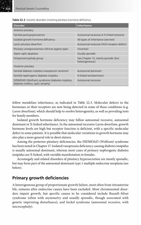
308308 Practical Genetic Counselling
follow mendelian inheritance, as indicated in Table 22.3. Molecular defects in the hormones or their receptors are now being detected in some of these conditions (e.g. Laron dwarfism), which should help to resolve heterogeneity, as well as providing tests for family members.
Isolated growth hormone deficiency may follow autosomal recessive, autosomal dominant or X-linked inheritance. In the autosomal recessive Laron dwarfism, growth hormone levels are high but receptor function is deficient, with a specific molecular defect in some patients. It is possible that molecular variations in growth hormone may also play a more general role in short stature.
Among the posterior pituitary deficiencies, the DIDMOAD (Wolfram) syndrome has been noted in Chapter 17. Isolated vasopressin deficiency causing diabetes insipidus is usually autosomal dominant, whereas most cases of primary nephrogenic diabetes insipidus are X-linked, with variable manifestation in females.
Acromegaly and related disorders of pituitary hypersecretion are mostly sporadic, but may form part of the autosomal dominant type 1 multiple endocrine neoplasia (see below).
Primary growth deficiencies
A heterogeneous group of proportionate growth failure, most often from intrauterine life, remains after endocrine causes have been excluded. Most chromosomal disor-ders impair growth, but specific causes to be considered include Russell–Silver syndrome (often with asymmetry and usually sporadic, though associated with genetic imprinting disturbance), and Seckel syndrome (autosomal recessive, with microcephaly).
Table 22.3 Genetic disorders involving pituitary hormone deficiency
Disorder Inheritance
Anterior pituitary
Familial panhypopituitarism Autosomal recessive or X-linked recessive
Isolated growth hormone deficiency All types of inheritance (see text)
Laron pituitary dwarfism Autosomal recessive (HGH receptor defect)
Pituitary unresponsiveness (African pygmy type) Uncertain
Septo-optic dysplasia Usually sporadic
Holoprosencephaly group See Chapter 12; mainly sporadic (but heterogeneous)
Posterior pituitary
Familial diabetes insipidus (vasopressin sensitive) Autosomal dominant
Familial nephrogenic diabetes insipidus X-linked semidominant
DIDMOAD (Wolfram) syndrome (diabetes insipidus, diabetes mellitus, optic atrophy)
Autosomal recessive

309309Endocrine and Reproductive Disorders
Overgrowth syndromes
These are likewise mostly sporadic and include Sotos syndrome (often due to new mutations), Weaver syndrome, and Beckwith–Wiedemann syndrome, commonly with macroglossia and omphalocoele.
THYROID GLAND DISORDERS
Congenital hypothyroidism
Most cases of congenital hypothyroidism are due to failure of thyroid gland develop-ment and are sporadic. Occasional occurrence in sibs may indicate recessively inherited forms that cannot at present be distinguished. Recessively inherited cases of thyroid-stimulating hormone deficiency have also been recorded. Newborn screening is now universal in developed countries and has led to the recognition of hypothyroidism being part of several unusual malformation syndromes. No satisfactory data on off-spring risks exist at present.
The presence of a goitre in a non-endemic region indicates that an inborn error of thyroxine synthesis is likely. The various types all follow autosomal recessive inherit-ance, including Pendred syndrome, in which defective iodine organification is associated with nerve deafness (see Chapter 18) and due to defects in a specific ion transport gene.
Absence of thyroxine-binding globulin may be X-linked recessive or autosomal dominant. It is usually harmless, but may be confused with hypothyroidism biochemically.
Autoimmune thyroid disease
Autoimmune thyroid disease may frequently form part of a broader autoimmune disorder, with other endocrine glands and different systems affected (e.g. myasthenia gravis, pernicious anaemia) – something that must be borne in mind when investigat-ing families. An autosomal dominant susceptibility gene is likely to be involved in many such families.
Both Graves’ disease and Hashimoto’s thyroiditis show strong familial aggregation, with both disorders commonly seen in the same family. A common autoimmune basis is now recognized, association with antigens HLA-Dw3 and HLA-B5 being seen in addition to other abnormalities. About 50 per cent of monozygotic twins are concordant (compared with 5 per cent of dizygotic twins). Clinical thyroid disease in other relatives is much less frequent than the incidence of thyroid antibodies. The lifetime risk probably does not exceed 10 per cent, except in a small number of families where a pattern strongly suggestive of autosomal dominant inheritance is seen. Transient neonatal hyperthy-roidism may occur in infants of affected mothers with Graves’ disease.
A distinct, recessively inherited, autoimmune polyglandular syndrome (APECED), which involves parathyroid and pancreas as well as thyroid, is due to mutations in a specific gene isolated on chromosome 21.

310310 Practical Genetic Counselling
PARATHYROID GLAND DISORDERS
Most, but not all, familial cases of hyperparathyroidism are part of multiple endocrine neoplasia type 1 (see below), but the great majority of parathyroid tumours are non-mendelian, with a low recurrence risk for relatives; the same applies to Cushing’s syndrome and to pituitary tumours.
A dominantly inherited, benign entity of hypercalcaemia with hypocalciuria has been recognized that can mimic hyperparathyroidism.
Hypoparathyroidism is commonly sporadic, but a rare X-linked recessive type has been recognized, as has an autosomal recessive syndrome with adrenal failure and candidiasis. Absent parathyroid glands are a feature of DiGeorge syndrome, part of the broader microdeletion syndrome involving chromosome 22q (see Chapter 19).
Albright’s hereditary osteodystrophy (encompassing pseudohypoparathyroidism and pseudopseudohypoparathyroidism) is now understood in molecular terms as being due to receptor defects in the adenyl cyclase system. Formerly considered to be X-linked, it is now recognized as autosomal dominant, with most fully expressed cases being maternally transmitted, probably due to an imprinting effect.
MULTIPLE ENDOCRINE NEOPLASIA
Multiple endocrine neoplasia comprises two major types:
• Type 1 (MEN 1). Parathyroid, anterior pituitary and pancreatic endocrine tumours are the most frequent. Inheritance is autosomal dominant, with the gene on chromosome 11 isolated.
• Type 2 (MEN 2). Medullary carcinoma of the thyroid may occur alone or coexist with phaeochromocytoma and with mucosal neuromas (type 2b). Some patients may be ‘marfanoid’ in appearance. Inheritance is usually autosomal dominant. The RET oncogene has proved to be the gene responsible, with a small number of specific mutations responsible for most cases of both 2a and 2b types.
Both major forms are important examples of high-risk neoplastic disorders, where recognition of the mendelian inheritance and the existence of a genetic register can prevent fatal disease in relatives, quite apart from the importance of genetic counselling (see Chapter 25).
Phaeochromocytoma may form part of other tumour syndromes than MEN2, such as Von Hippel–Lindau disease and familial paraganglioma.
Obesity
A few cases of severe childhood obesity have syndromal associations (e.g. Prader–Willi and Bardet–Biedl syndromes), but most is not clearly genetic (a very few cases of auto-somal recessive leptin deficiency have been recorded).
311311Endocrine and Reproductive Disorders
CONGENITAL ADRENAL HYPERPLASIA (ADRENOGENITAL SYNDROME)
At least eight types of congenital adrenal hyperplasia exist, resulting from different dis-orders of steroid hormone biosynthesis; all follow autosomal recessive inheritance. The most important type, 21-hydroxylase deficiency, is closely linked to the HLA system. The gene has been isolated and a variety of deletions and other mutations identified, correlating with phenotype to some extent and allowing accurate prenatal diagnosis and carrier detection.
Direct prenatal diagnosis from amniotic fluid is also possible, and could allow prenatal treatment to minimize virilization, as well as neonatal treatment to avoid a salt-losing crisis. Whether termination of an affected pregnancy is required may be strongly influenced by the severity of effects, particularly virilization of a female, in the first affected child – some families opting for first-trimester prenatal diagnosis and termination, and others for steroid therapy.
HYPOGONADISM AND ALLIED STATES
Recent advances in understanding the molecular basis of sex determination have considerable relevance to the classification and pathogenesis of the different forms of hypogonadism and intersex. The principal points of relevance are as follows:
• A specific gene on the Y chromosome (Sry) has been identified that commits the undifferentiated gonad to become a testis. If this is absent or defective, the gonadal phenotype will be female.
• Male phenotype is dependent on the presence of and sensitivity to androgen, which is itself dependent on a differentiated testis.
• Two functioning X chromosomes are needed for formation of a fully developed ovary with ova.
• Other hormonal influences (e.g. adrenal steroid precursors) may modify genital development and result in ambiguous genitalia.
Genetic counselling in this area must rest on a well-defined clinical and endocrinologi-cal diagnosis, with cytogenetic and (increasingly) molecular analysis as appropriate. Recognition of X-linked and autosomal recessive disorders, along with translocations involving X and Y chromosome material, is especially important, as it is in these groups where recurrence in the family is most likely.
The following broad groups are important to distinguish for genetic counselling.
Primary sex chromosome disordersIn particular these include XXY (Klinefelter) and 45,X (Turner) syndromes, which are discussed in Chapter 4. Recurrence in a family is exceptional, except where there is mosaicism.

312312 Practical Genetic Counselling
Disorders where chromosomal and phenotypic sex do not correspond, including XX males and XY femalesThis complex group is becoming resolved with isolation of the specific genes on the Y and X chromosomes involved with sex determination. A careful endocrine, cytogenetic and molecular assessment is essential before genetic counselling can be given. The main disorders include XX males and XY females.
• XX males. These are usually sporadic and resulting from Y chromosome material translocated on to an X chromosome and often detectable by molecular tests.
• XY females. The most frequent cause is the androgen insensitivity syndrome (formerly known as testicular feminization) where a normal female phenotype is associated with presence of a testis. This diagnosis may not be suspected until failure of menarche or absent pubic and axillary hair is investigated. Partial androgen insensitivity may result in ambiguous genitalia at birth. In XY gonadal dysgenesis, due to molecular defects in the Y-linked sex-determining Sry gene, or to corresponding X-linked defects, there may be a normal phenotype or varying degrees of intersex. The dysgenetic gonad is at risk of malignant change and should be removed. Both these disorders may follow X-linked recessive inheritance with phenotypic and chromosomally normal female relatives at risk of having an affected XY daughter.
• Ambiguous genitalia. This may result from some of the conditions mentioned above (e.g. congenital adrenal hyperplasia, sex chromosome disorders), from partial androgen insensitivity, from disorders involving the Sry or Wilms’ tumour genes (e.g. camptomelic dysplasia, Drash syndrome). It will require careful endocrine, chromosome and clinical assessment to be sure of the cause.
Genetic counselling in all these conditions needs to be especially sensitive to the true sexual identity of those affected and to link closely with overall management. In general, it will be the phenotypic and psychological sexual identity that is more relevant than chromosomal sex. Great care must be taken not to confuse or upset affected individuals by inappropriately assuming that sex chromosomal status must necessarily determine actual sexual identity.
Hypogonadism due to specific genetic disordersHere the recurrence risk will be that of the underlying disorder, while the hypogonad-ism itself may be of gonadal, pituitary or hypothalamic origin (Box 22.1).
INFERTILITY
There are two important questions that need to be asked (but frequently are not) by those attempting to investigate, counsel and treat infertility:
• Is the infertility one aspect of a genetic disorder that might be transmitted?• Will correction of infertility give an increased risk of malformations in the
offspring?

313313Endocrine and Reproductive Disorders
The genetic causes of both female and male infertility are numerous and in part overlap with those of hypogonadism already mentioned (see Box 22.1). As with these, it is only X-linked or autosomal recessive disorders that are of practical importance for genetic counselling, because only in these will unaffected people be at risk of having an affected child.
Premature ovarian failure has been shown to result in some cases from X-autosome translocation involving a critical region of the long arm, but it may also be associated with pre-mutations in the fragile-X (FMR) gene.
Disorders of sperm production include primary sex chromosome disorders such as XXY (Klinefelter) syndrome, other disorders affecting the testis (e.g. myotonic dystro-phy), balanced chromosomal translocations causing abnormalities of chromosome pairing in meiosis, clinical defects affecting sperm motility, and a variety of poorly defined biochemical disorders. Molecular deletions of the Y chromosome involving the azoospermic factor AZF are a recently recognized case. Blockage or aplasia of the vas deferens causes infertility in most male cases of cystic fibrosis. A mild form of this may be responsible for some apparently primary cases of such obstruction.
The question of increased risk to offspring arises principally in those patients in whom apparent infertility is really a reflection of early unrecognized fetal loss as a result of abnormal gamete production. The most important group to detect is where one parent carries a balanced translocation, where the risk of an unbalanced chromosome abnormality in a pregnancy that goes to term is considerable, especially when the defect is carried by a female (see Chapter 4). This problem is closely related to that of recurrent abortion, considered below.
Artificial insemination by donor (AID) and in-vitro fertilization are mentioned in Chapter 10 as increasingly used modes of treating infertility. There has now been suffi-cient experience with AID to make it clear that there is no increase in abnormalities. The same is probably true of in-vitro fertilization from preliminary studies of children
Box 22.1 Genetic causes of infertility and hypogonadism
Phenotypic maleSex chromosome anomalies and reversalY chromosome microdeletionsAndrogen insensitivity (partial)Kennedy’s disease (spinobulbar muscular atrophy)Kallmann syndromePituitary hormone defects (various)Dysmorphic syndromes with hypogonadism (e.g. Bardet–Biedel, Prader–Willi)Myotonic dystrophyCystic fibrosisPrimary ciliary dyskinesia
Phenotypic femaleSex chromosome anomalies and reversalAndrogen insensitivity (complete)Fragile-X pre-mutation (premature ovarian failure)Congenital adrenal hyperplasia (some types)Pituitary hormone defects (various)Sry gene defects (Y chromosome)Galactosaemia (premature ovarian failure)
314314 Practical Genetic Counselling
conceived in this way, but there are doubts remaining about the safety of intracytoplas-mic sperm injection (ICSI).
RECURRENT ABORTIONS
Most women with a history of recurrent abortions will be under the care of a gynaecolo-gist, who will have already searched for a gynaecological cause and will have excluded most serious maternal disorders. Rhesus haemolytic disease is now usually prevented as a cause of later pregnancy loss. Factors such as lupus anticoagulant antigen or maternal thrombotic disorders may be relevant in a small proportion, but numerous proposed factors and corresponding remedies have failed to be confirmed and the cause of most recurrent abortions remains unknown.
The main question to be answered is not so much whether another abortion will occur, but what are the chances of having a healthy child, together with the risk that a pregnancy reaching term will result in an abnormality. The question of amniocentesis may also arise: clearly, one does not want to expose a pregnancy to any added risk of abortion unless there is a likelihood of an abnormality detectable by this.
There will be many women in whom careful search reveals no genetic or other factors involved, but with care considerable help can be given by the following measures.
• Pathological examination of the abortus, where possible, may identify major structural malformations.
• Cytogenetic study of the parents is especially important where a translocation is a possibility and should always be done. In general the finding of a chromosome abnormality in the abortus but not in a parent is not likely to be relevant or to affect the genetic risks (see Chapter 4).
• Search for possible lethal mendelian causes. Examples are consanguinity, which increases the risk of autosomal recessive lethals; X-linked dominant disorders lethal in the male; and myotonic dystrophy, which gives heavy fetal loss in the offspring of mildly affected women.
The most important group to detect are the autosomal translocations, where one parent is a balanced translocation carrier. As stated in Chapter 4, these carry a significant risk of an abnormal live-born offspring, probably around 12 per cent where the carrier is female, but nearer 5 per cent where the male is the carrier, the precise risk depending on the type of translocation. Amniocentesis or chorion biopsy is clearly indicated in any such pregnancy, and there is no evidence that it is accompanied by a greater risk of abortion in such a situation. A study by Franssen et al. (see ‘Further reading’) has shown that couples where a parent has a balanced chromosome rearrangement have as high a chance of eventually having a healthy child as a control group.
It is important for couples to realize that spontaneous abortion is an exceedingly common event, occurring in at least 1 in 8 recognized pregnancies. Thus 1 in 64 women might be expected to have two consecutive abortions on the grounds of chance alone, and unless there are other reasons, it is probably not worth undertaking investigations

315315Endocrine and Reproductive Disorders
unless they have had at least three spontaneous abortions, when around 5 per cent of couples will be found to carry a balanced translocation.
OTHER REPRODUCTIVE PROBLEMS
Hydatidiform moles
Hydatidiform moles are masses of trophoblastic tissue that are rarely familial but show unusual chromosomal features. The complete hydatidiform mole has a normal chro-mosome number that is of entirely paternal origin, while the partial mole is a complete triploid, with two paternal complements. These structures represent perhaps the most extreme examples of genomic imprinting (see page 29). A 1 per cent recurrence risk in future pregnancies is likely for the complete mole and may represent persistence of the original mole with the risk of malignant change; the recurrence risk is probably less than this for the partial form, which is not associated with malignancy.
Sacrococcygeal teratomas are now usually detected by ultrasound and are mostly sporadic.
Toxaemia of pregnancy (pre-eclampsia)
Toxaemia of pregnancy may be associated with genetic disorders affecting placental development (e.g. trisomy 18), but in the absence of a specific cause it is frequently familial, possibly determined by a common autosomal recessive gene giving toxaemia when both mother and fetus are homozygous for this. Recurrence risks are around 20 per cent for a pregnancy in a woman whose own mother had toxaemia in the pregnancy leading to that woman’s birth; and about 15 per cent where a sister has had toxaemia in pregnancy, figures rather less than suggested in earlier studies (the population risk is around 10 per cent).
FURTHER READING
Arngrimson R (2002). Preeclampsia. In: Rimoin DL, Connor JM, Pyeritz RE, Korf BR (eds) Emery and Rimoin’s Principles and Practice of Medical Genetics, 4th edn, pp. 1496–518. Edinburgh: Churchill Livingstone.
Elliot DJ, Cooke HJ (1997). The molecular genetics of male infertility. Bioessays 19, 801–9.Franssen MTM, Korevaar JC, van der Veen F, Leschot NJ (2006). Reproductive outcome after
chromosome analysis in couples with two or more miscarriages: case control study. Br Med J 332, 759–62.
Froguel P (1997). Genetics of type 1 insulin-dependent diabetes mellitus. Horm Res 48, 55–7.Gardner RJM, Sutherland GR (2003). Chromosome Abnormalities and Genetic Counselling.
Oxford: Oxford University Press [has helpful sections on reproductive failure, intersex and sex chromosome anomalies].
Jameson JL (1996). Inherited disorders of the gonadotrophin hormones. Mol Cell Endocrinol 125, 143–9.

316316 Practical Genetic Counselling
Kahn CR, Vicent D, Doria A (1996). Genetics of non-insulin-dependent (type-II) diabetes mellitus. Annu Rev Med 47, 509–31.
Layman LC (2002). Human gene mutations causing infertility. J Med Genet 39, 153–61.Medeiros-Neto G, Stanbury JB (1994). Inherited Disorders of the Thyroid System. London: CRC
Press.Newfield RS, New MI (1997). 21-Hydroxylase deficiency. Ann NY Acad Sci 816, 219–29.Potter A, Phillips JA, Rimoin DL (2007). Genetic disorders of the pituitary gland. In: Rimoin DL,
Connor JM, Pyeritz RE, Korf BR (eds) Emery and Rimoin’s Principles and Practice of Medical Genetics, 5th edn, pp. 2135–82. Edinburgh: Churchill Livingstone.
Raffel LJ, Goodarzi MO, Rotter JI (2007). Diabetes mellitus. In: Rimoin DL, Connor JM, Pyeritz RE, Korf BR (eds) Emery and Rimoin’s Principles and Practice of Medical Genetics, 5th edn, pp. 1980–2022. Edinburgh: Churchill Livingstone.
Rotig A, Bonnefont JP, Munnich A (1996). Mitochondrial diabetes mellitus. Diabetes Metab 22, 291–8.
Scriver CK, Beaudet AL, Sly WS, Valle D (eds) (2001). Metabolic and Molecular Bases of Inherited Disease. New York: McGraw-Hill. Web based version at www.ommbid.com [a series of chapters give valuable details on individual disorders].

Introduction . . . . . . . . . . . . . . . . . . . . . . . . . . . . . . . . . . . . . . . . . . . . . . . . . . . . . . . . . . . . . . . . . . . .317
Amino acid disorders . . . . . . . . . . . . . . . . . . . . . . . . . . . . . . . . . . . . . . . . . . . . . . . . . . . . . . . . . . . .319
Bartter’s syndrome . . . . . . . . . . . . . . . . . . . . . . . . . . . . . . . . . . . . . . . . . . . . . . . . . . . . . . . . . . . . . 320
Galactosaemia . . . . . . . . . . . . . . . . . . . . . . . . . . . . . . . . . . . . . . . . . . . . . . . . . . . . . . . . . . . . . . . . . 320
Sphingolipidoses . . . . . . . . . . . . . . . . . . . . . . . . . . . . . . . . . . . . . . . . . . . . . . . . . . . . . . . . . . . . . . . 321
Glycogen storage diseases . . . . . . . . . . . . . . . . . . . . . . . . . . . . . . . . . . . . . . . . . . . . . . . . . . . . . . 321
Hyperlipidaemias . . . . . . . . . . . . . . . . . . . . . . . . . . . . . . . . . . . . . . . . . . . . . . . . . . . . . . . . . . . . . . 322
The porphyrias . . . . . . . . . . . . . . . . . . . . . . . . . . . . . . . . . . . . . . . . . . . . . . . . . . . . . . . . . . . . . . . . . 322
Pseudocholinesterase deficiency . . . . . . . . . . . . . . . . . . . . . . . . . . . . . . . . . . . . . . . . . . . . . . . . 322
Pharmacogenetics . . . . . . . . . . . . . . . . . . . . . . . . . . . . . . . . . . . . . . . . . . . . . . . . . . . . . . . . . . . . . . 323
Fatty acid metabolic defects and sudden infant death . . . . . . . . . . . . . . . . . . . . . . . . . . . . 323
Further reading . . . . . . . . . . . . . . . . . . . . . . . . . . . . . . . . . . . . . . . . . . . . . . . . . . . . . . . . . . . . . . . . 324
Inborn Errors of Metabolism
CHAPTER 23
INTRODUCTION
Since most mendelian disorders eventually prove to be the result of a deficient or defec-tive specific gene product, there is no absolute distinction between inborn errors of metabolism and other genetic disorders. Indeed, the concept of ‘inborn errors of devel-opment’ has now become a reality, as discussed in Chapter 6 (and as foreseen by Garrod himself). For this chapter it seems wise to restrict the term to those conditions where some form of metabolic basis, usually enzymatic, has been clearly identified, but each year more diseases are added to the group. In many cases, the discovery of a specific metabolic basis radically changes the concept of a disease. Thus, Tay–Sachs disease is no longer thought of as purely a brain degeneration but as a generalized metabolic disorder, and the preventive measures of carrier detection and prenatal diagnosis involve biochemical and genetic techniques far removed from those generally associ-ated with neurology. Xeroderma pigmentosum and allied disorders of DNA repair are further examples of disorders entering the inborn error category, while the positional cloning approach has been responsible for isolation of the genes for important meta-bolic disorders such as Batten’s disease, Menkes syndrome and Lowe syndrome, whose primary protein defect was previously unknown.
The development of specific tests for direct identification of gene mutations (see Chapter 5) is causing rapid changes in our understanding of many inherited metabolic diseases. In particular, heterogeneity in the types of mutation involved is becoming
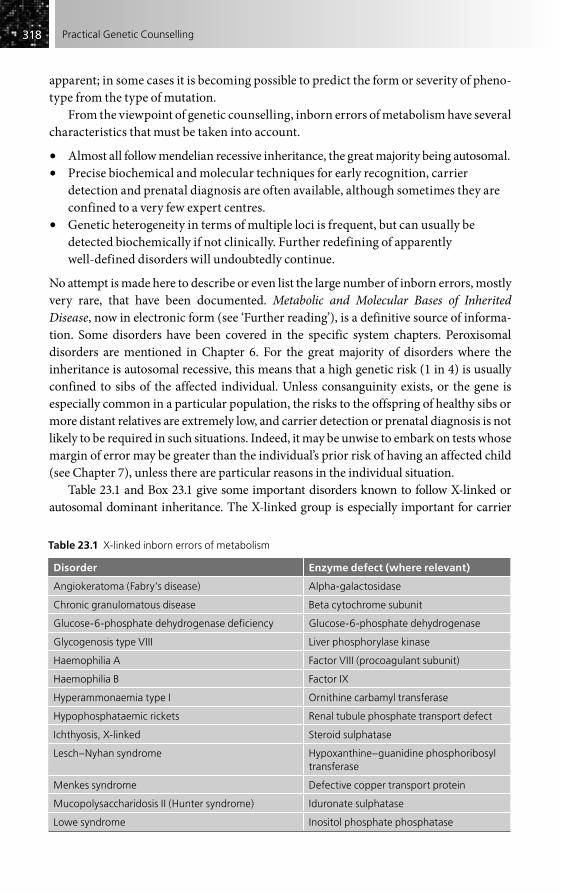
318318 Practical Genetic Counselling
apparent; in some cases it is becoming possible to predict the form or severity of pheno-type from the type of mutation.
From the viewpoint of genetic counselling, inborn errors of metabolism have several characteristics that must be taken into account.
• Almost all follow mendelian recessive inheritance, the great majority being autosomal.• Precise biochemical and molecular techniques for early recognition, carrier
detection and prenatal diagnosis are often available, although sometimes they are confined to a very few expert centres.
• Genetic heterogeneity in terms of multiple loci is frequent, but can usually be detected biochemically if not clinically. Further redefining of apparently well-defined disorders will undoubtedly continue.
No attempt is made here to describe or even list the large number of inborn errors, mostly very rare, that have been documented. Metabolic and Molecular Bases of Inherited Disease, now in electronic form (see ‘Further reading’), is a definitive source of informa-tion. Some disorders have been covered in the specific system chapters. Peroxisomal disorders are mentioned in Chapter 6. For the great majority of disorders where the inheritance is autosomal recessive, this means that a high genetic risk (1 in 4) is usually confined to sibs of the affected individual. Unless consanguinity exists, or the gene is especially common in a particular population, the risks to the offspring of healthy sibs or more distant relatives are extremely low, and carrier detection or prenatal diagnosis is not likely to be required in such situations. Indeed, it may be unwise to embark on tests whose margin of error may be greater than the individual’s prior risk of having an affected child (see Chapter 7), unless there are particular reasons in the individual situation.
Table 23.1 and Box 23.1 give some important disorders known to follow X-linked or autosomal dominant inheritance. The X-linked group is especially important for carrier
Table 23.1 X-linked inborn errors of metabolism
Disorder Enzyme defect (where relevant)
Angiokeratoma (Fabry’s disease) Alpha-galactosidase
Chronic granulomatous disease Beta cytochrome subunit
Glucose-6-phosphate dehydrogenase deficiency Glucose-6-phosphate dehydrogenase
Glycogenosis type VIII Liver phosphorylase kinase
Haemophilia A Factor VIII (procoagulant subunit)
Haemophilia B Factor IX
Hyperammonaemia type I Ornithine carbamyl transferase
Hypophosphataemic rickets Renal tubule phosphate transport defect
Ichthyosis, X-linked Steroid sulphatase
Lesch–Nyhan syndrome Hypoxanthine–guanidine phosphoribosyl transferase
Menkes syndrome Defective copper transport protein
Mucopolysaccharidosis II (Hunter syndrome) Iduronate sulphatase
Lowe syndrome Inositol phosphate phosphatase
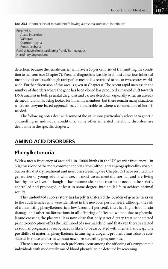
319319Inborn Errors of Metabolism
detection, because the female carrier will have a 50 per cent risk of transmitting the condi-tion to her sons (see Chapter 7). Prenatal diagnosis is feasible in almost all serious inherited metabolic disorders, although rarity often means it is restricted to one or two centres world-wide. Further discussion of this area is given in Chapter 8. The recent rapid increase in the number of disorders where the gene has been cloned has produced a marked shift towards DNA analysis in both prenatal diagnosis and carrier detection, especially when an already defined mutation is being looked for in family members; but there remain many situations where an enzyme-based approach may be preferable or where a combination of both is needed.
The following notes deal with some of the situations particularly relevant to genetic counselling in individual conditions. Some other inherited metabolic disorders are dealt with in the specific chapters.
AMINO ACID DISORDERS
Phenylketonuria
With a mean frequency of around 1 in 10 000 births in the UK (carrier frequency 1 in 50), this is one of the more common inborn errors, although it is geographically variable. Successful dietary treatment and newborn screening (see Chapter 27) have resulted in a generation of young adults who are, in most cases, mentally normal and are living healthy, active lives, although it has become clear that treatment needs to be strictly controlled and prolonged, at least in some degree, into adult life to achieve optimal results.
This undoubted success story has largely transferred the burden of genetic risks on to the adult females who were identified in the newborn period. Here, although the risk of transmitting phenylketonuria is low (around 1 per cent), there is a high risk of brain damage and other malformations in all offspring of affected women due to phenyla-lanine crossing the placenta. It is now clear that only strict dietary treatment started prior to conception offers the likelihood of a normal child, and that even therapy started as soon as pregnancy is recognized is likely to be associated with mental handicap. The possibility of maternal phenylketonuria causing teratogenic problems must also be con-sidered in those countries without newborn screening programmes.
There is no evidence that such problems occur among the offspring of asymptomatic individuals with moderately raised blood phenylalanine detected by screening.
Box 23.1 Inborn errors of metabolism following autosomal dominant inheritance
Porphyrias:Acute intermittentVariegateCoproporphyriaProtoporphyria
Familial hypercholesterolaemia (rarely homozygous)Hereditary angioedema

320320 Practical Genetic Counselling
Prenatal diagnosis is feasible but rarely requested except for the very rare and usually fatal form of phenylketonuria, due to dihydropteridine reductase deficiency, which does not respond to the usual dietary treatment and where the gene has now been cloned. In the classic form due to phenylalanine hydroxylase deficiency, the enzyme is confined to the liver. Different mutations are proving to be correlated with severity and need for dietary control. Carrier detection is feasible by DNA analysis of polymorphisms within the gene or by specific mutation analysis, but prenatal diagnosis is finding significant application only in countries without effective dietary treatment. It should be borne in mind that the great majority of treated phenylketonuria patients are now entirely normal.
Histidinaemia
Most cases of histidinaemia appear to be asymptomatic and the original association thought to exist with speech problems and mental retardation seems doubtful. There is no evidence of a significant maternal effect in the heterozygous offspring of affected women.
Cystinuria
Renal calculi are the only significant clinical features in cystinuria. Heterozygotes in one of the two main types may excrete small amounts of amino acids in the urine, but are symptomless and must not be confused with the affected homozygotes, where large quantities of cystine and other dibasic amino acids are excreted. The disorder is quite distinct from cystinosis (also autosomal recessive), which is a generalized storage disease of cystine, with much more serious clinical effects, including renal failure. (See Chapter 21 for other causes of renal calculi.)
BARTTER’S SYNDROME
This disorder of electrolyte metabolism, causing hypertension and hypokalaemia, is proving to be heterogeneous, with several different ion-channel genes responsible in different families.
GALACTOSAEMIA
Recognition of galactosaemia, a rapidly fatal disorder, is important not only because effective treatment exists, but also to allow immediate diagnosis from cord blood in a subsequent pregnancy. Distinction of classic galactosaemia must also be made from the form due to galactokinase deficiency in which cataract is the only abnormality, and from harmless enzyme variants that may be picked up by screening programmes.
Prenatal diagnosis is feasible but not widely requested in view of the relatively good outcome of treatment in most cases, although long-term prognosis is less clear than for

321321Inborn Errors of Metabolism
well-treated phenylketonuria. The various options and their consequences must be fully discussed with the couple concerned before a decision is made. Pituitary–ovarian dysfunction may result in infertility in treated women.
SPHINGOLIPIDOSES
Specific lysosomal enzyme defects have been identified for most disorders in the severe and clinically confusing group of sphingolipidoses, all autosomal recessive, and prenatal diagnosis is feasible in these. Carrier detection is of only major significance for Tay–Sachs disease, where the gene is at high frequency in Ashkenazi Jewish popula-tions. Screening for adult carriers, with prenatal diagnosis offered to couples who both carry the gene, has been successfully applied in some American Jewish communities, in which 1 in 30 individuals are carriers. A comparable approach is now beginning to be applied in Finland and in Quebec, where the genes for some other specific metabolic neurodegenerations are at high frequency.
Other important sphingolipidoses, all autosomal recessive in inheritance, include Gaucher’s disease, Niemann–Pick disease, metachromatic leucodystrophy and general-ized gangliosidosis. Batten’s disease has had no specific enzyme defect known until positional cloning identified one form on chromosome 16, with one particularly frequent mutation, allowing, for the first time, prenatal diagnosis for some families with this devastating disorder (see also page 198).
Fabry’s disease is X-linked, with minor signs and biochemical changes often detect-able in female carriers. The �-galactosidase gene responsible has been cloned. The mucopolysaccharidoses are mentioned in Chapter 14.
The importance of obtaining a precise enzymatic (and where possible DNA) diag-nosis in inborn errors of metabolism has already been stressed, but it is probably more important in the group of lysosomal enzyme deficiencies than in any other, since clinical differentiation is often extremely difficult and the prognosis poor, but prenatal diagnosis is feasible if one knows which enzyme is defective (see Chapter 8). Bone marrow transplantation is proving effective in some members of this group, while in Gaucher’s disease, direct enzyme replacement therapy is feasible.
GLYCOGEN STORAGE DISEASES
All members of the heterogeneous group of glycogen storage diseases are autosomal recessive in inheritance, apart from the exceedingly rare type VIII (X-linked recessive). Type II (Pompe’s disease), caused by lysosomal acid maltase deficiency, can be prena-tally diagnosed and exists in two distinct forms:
• an infantile type with severe cardiomyopathy and cerebral involvement;• a later neuromuscular type that may mimic a muscular dystrophy.

322322 Practical Genetic Counselling
The classic type I (von Gierke) glucose-6-phosphatase deficiency has the enzyme confined to the liver, but identification of the gene should now permit prenatal diagnosis.
HYPERLIPIDAEMIAS
The classification of the hyperlipidaemias is still in a state of flux, but is beginning to be clarified by recognition of specific molecular defects. Most hyperlipidaemias, even with multiple affected family members, are likely to be secondary to diet and other external factors. Type I hyperlipidaemia (hyperchylomicronaemia) and type V are autosomal recessive. The relatively common type II (familial hypercholesterolaemia), especially important because of its association with early coronary heart disease, is discussed in Chapter 19.
THE PORPHYRIAS
The acute porphyrias form the most striking exception to the rule of recessive inherit-ance for most inborn errors due to enzyme defects. Acute intermittent porphyria, porphyria variegata, hereditary coproporphyria and protoporphyria all follow autosomal dominant inheritance. Careful investigation of urine and faecal porphyrins, enzyme studies and, where possible, molecular analysis are needed to exclude subclinical disease. Now that specific enzyme and gene defects are known, prenatal diagnosis may be possible but is rarely requested. The severe congenital erythropoietic porphyria follows autosomal recessive inheritance. Porphyria cutanea tarda, much the most common of the group, is usually sporadic, with low recurrence risk for family members. It appears to result from a combination of homozygosity for mutation in the iron storage haemochromatosis sus-ceptibility gene with adverse environmental factors such as alcoholic liver damage. A rare familial type can be distinguished enzymatically.
PSEUDOCHOLINESTERASE DEFICIENCY
Pseudocholinesterase deficiency – an important cause of apnoea following muscle relaxants (notably suxamethonium) – follows autosomal recessive inheritance. Sibs are thus the principal relatives at risk, although, because the gene is relatively common, it may also be worth testing the parents. Heterozygotes (4 per cent of the population) are not at significant risk of clinical problems, and in testing relatives it is most important not to confuse them with affected homozygotes. Since heterozy-gotes commonly show a moderate reduction in pseudocholinesterase level, the dibucaine number, which measures the degree of inhibition of the enzyme by dibu-caine, should be measured. This will be under 25 in affected homozygotes, 50–70 in heterozygotes, and over 75 for normal homozygotes. Other rare genetic variants

323323Inborn Errors of Metabolism
exist; their nomenclature is most confusing, but the subject is clearly discussed by Whittaker (see ‘Further reading’). Molecular analysis has not yet come into regular clinical use.
PHARMACOGENETICS
Inherited differences in metabolism of, and response to, drugs has been recognized for over 50 years (e.g. gene loci such as those for cholinesterase (see above) and acetylation), but the possibility of finding simple DNA-based tests that will predict response or avoid serious adverse reactions has attracted recent attention. It is not clear at present whether such inherited variation might also be predictive for disease risk, nor whether it will really lead to individually tailored therapy. The term ‘pharmacogenomics’, identical in meaning to pharmacogenetics, is now often used for this DNA-based approach, along with the even more misleading term ‘personalized medicine’.
As with all common disease genetics, the underlying basis seems likely to require a considerable amount of more basic research before it reaches practical applications, including genetic counselling.
FATTY ACID METABOLIC DEFECTS AND SUDDEN INFANT DEATH
Several previously unrecognized defects in fatty acid and organic acid metabolism (e.g. MCAD deficiency) have been shown to be responsible for some cases (probably around 1 per cent) of unexplained sudden infant death, and for some cases of what was formerly termed Reye’s syndrome, with rapidly developing hypoglycaemia and encephalopathy. The proportion that are genetic has probably risen since the removal of likely environmental factors such as aspirin in infancy. The forensic implications in this field are exceptionally serious and provide a striking example of how essential it is to use and integrate all available information in a statistically appropriate manner before attempting to estimate the chance that an underlying metabolic defect may be responsible for a particular case. Since effective therapy appears possible by dietary modification, recognition of a possible metabolic basis in such situations is most important; autosomal recessive inheritance seems likely and prenatal diagnosis theo-retically possible.
The frequency of most such disorders appears to be very low in the general popula-tion, although the predominance of one specific MCAD mutation, with frequency of homozygotes 1 in 18 000, makes molecular diagnosis of this condition feasible. Population newborn screening using tandem mass spectrometry is also now techni-cally feasible, but it is not yet clear that it will be beneficial. Unfortunately, pressure groups have led to wider tandem mass spectrometry screening being introduced on a population basis in some countries before such evidence is available and this will need to be critically assessed.

324324 Practical Genetic Counselling
FURTHER READING
Scriver CR, Beaudet AL, Sly W, Valle D (eds) (2001). Metabolic and Molecular Bases of Inherited Disease. New York: McGraw-Hill [the definitive source of information on all inborn errors of metabolism and related metabolic disorders, which is now on-line and continually updated (www.ommbid.com)].
Public Health Genetics Unit (2003). My Very Own Medicine. What Must I Know? Information Policy for Pharmacogenetics. Cambridge: Public Health Genetics Unit.
Whittaker M (1986). Cholinesterase. Basel: Karger.

Disorders of haemoglobin structure and synthesis . . . . . . . . . . . . . . . . . . . . . . . . . . . . . . 325
Other red blood cell disorders . . . . . . . . . . . . . . . . . . . . . . . . . . . . . . . . . . . . . . . . . . . . . . . . . 326
White blood cells and platelets . . . . . . . . . . . . . . . . . . . . . . . . . . . . . . . . . . . . . . . . . . . . . . . . . . . . . 328
Immune deficiency disease . . . . . . . . . . . . . . . . . . . . . . . . . . . . . . . . . . . . . . . . . . . . . . . . . . . 328
Genetic aspects of infectious disease . . . . . . . . . . . . . . . . . . . . . . . . . . . . . . . . . . . . . . . . . . . . 330
Haemophilia . . . . . . . . . . . . . . . . . . . . . . . . . . . . . . . . . . . . . . . . . . . . . . . . . . . . . . . . . . . . . . . . . . . 330
Other coagulation disorders . . . . . . . . . . . . . . . . . . . . . . . . . . . . . . . . . . . . . . . . . . . . . . . . . . . . 332
Further reading . . . . . . . . . . . . . . . . . . . . . . . . . . . . . . . . . . . . . . . . . . . . . . . . . . . . . . . . . . . . . . . . 333
Disorders of Blood and Immune Function
CHAPTER 24
DISORDERS OF HAEMOGLOBIN STRUCTURE AND SYNTHESIS
The large group of haemoglobin disorders, of great importance in many parts of the world, contains perhaps the best understood disorders (in molecular terms) that exist. Most are autosomal recessive in inheritance, and only a few points will be mentioned here. The works listed in the ‘Further reading’ section, particularly those by Weatherall (1991, 1997), give full details.
Sickle-cell disease
Sickle-cell disease, caused by a glutamic acid to valine mutation at a specific site in beta-globin, is exceptionally common in some regions, and the heterozygote frequency approaches 1 in 8 in some parts of Africa. Thus, carrier testing is of great importance, but fortunately it is readily feasible using a sickling test screen on the red cells, with hae-moglobin electrophoresis as a confirmatory measure. Only couples who are both carriers will be at risk of having an affected child. Heterozygotes are essentially healthy and have partial protection against malaria in endemic areas. It is important for them not to be given the erroneous impression that they have a mild form of the disease. Severity in homozygotes can vary greatly according to geographical region.
Prenatal diagnosis, originally using fetal blood but now normally based on DNA analysis of chorionic villi or amniotic cells, has been feasible for some years but has not found wide application, in part because of the variation in clinical severity. The early screening programmes for carriers also met with little success, partly because of their hasty and ill-judged introduction, partly because of the stigmatization of carriers that

326326 Practical Genetic Counselling
resulted. Recent, more sensitively approached programmes are proving more accepta-ble, as is newborn screening in high-risk groups or, in some instances, whole populations, to allow early treatment of complications.
Numerous other betaglobin chain abnormalities are known, some of which, such as haemoglobin C, may be encountered in combination with haemoglobin S. This may result in complex genetic situations, emphasizing the need for expert haematological and molecular diagnosis to be available to those undertaking genetic counselling for haemoglobinopathies.
Thalassaemias
Thalassaemias, characterized by a failure of globin chain synthesis owing to a variety of underlying causes, are another disease group that is exceedingly common in some regions of the world, as well as in immigrant populations in Europe and America. All the thalassaemias are recessively inherited, and various compounds with different abnormal alleles may occur.
Beta-thalassaemia major is an important problem in parts of the Mediterranean, the Middle East and Asia, while in South-East Asia forms of alpha-thalassaemia make a large contribution to intrauterine and neonatal deaths. Carrier detection of most forms is feasible, and so is prenatal diagnosis, particularly for beta-thalassaemia major, for which there is now considerable experience. Molecular techniques had their first diag-nostic applications in this group, and identified a wealth of different defects at the DNA level (see Weatherall, 1997). The advent of first-trimester prenatal diagnosis based on DNA makes it particularly important to establish the precise molecular nature of the disorder.
In planning prenatal diagnosis, it is important to recognize that the great majority of cases in any particular population will result from a few specific mutations and that the programme must be adjusted to this, and to the cultural background of the popula-tion involved. Deletions may be frequent in some situations (especially for alpha-thalassaemias), while DNA polymorphisms, especially when used in combina-tion as a haplotype, may still be used for cases of beta-thalassaemia where the specific mutation is unknown. Carrier detection and prenatal diagnosis have been taken up on a large scale by many populations, notably in Mediterranean countries. Population and screening aspects are discussed further in Chapter 27.
The uncommon but important alpha-thalassaemia mental retardation syndromes, due to defects on chromosome 16 and the X chromosome, are mentioned in Chapter 13.
OTHER RED BLOOD CELL DISORDERS
Hereditary spherocytosis and elliptocytosis
Hereditary spherocytosis is a disorder of the red cell membrane which usually follows autosomal dominant inheritance, but haemolysis is often mild, requiring red cell fragility tests to be sure that an individual is not affected. Numerous other causes of

327327Disorders of Blood and Immune Function
spherocytosis must be excluded before this diagnosis is made. Specific forms of heredi-tary spherocytosis have been identified as due to defects in several different red cell membrane proteins, including spectrin and ankyrin.
Hereditary elliptocytosis is also autosomal dominant. Several forms exist, several due to defects in glycophorin C and other membrane proteins.
Glucose-6-phosphate dehydrogenase deficiency
This important red-cell enzyme defect is particularly common in parts of the Middle East, the Mediterranean and South-East Asia and in people of African descent, but is not unknown in others. Numerous enzyme variants exist, with varying loss of activity which determines the haemolytic severity of the disease. The disorder is X-linked reces-sive, but alterations in the gene are so common in some areas (e.g. the Arabian peninsula) that homozygous affected females are frequent. Carrier detection is often feasible, but this depends on the type of the abnormality.
Other red-cell enzyme defects
Other red-cell enzyme defects are mostly autosomal recessive, with the exception of phosphoglycerate kinase deficiency, which is X-linked. Some are confined to the red cell, others have generalized clinical effects (e.g. triose phosphate isomerase deficiency). Prenatal diagnosis from fetal blood is a possibility for this group, but as the genes are cloned, DNA analysis has become preferable.
Sideroblastic anaemia
Most cases, especially in later life, are acquired, but an X-linked recessive form, though rare, is important to recognize.
Pernicious anaemia
Pernicious anaemia has already been mentioned in connection with atrophic gastritis (see Chapter 20). Congenital vitamin B12 deficiency is an exceptionally rare disorder caused by intrinsic factor deficiency, following autosomal recessive inheritance.
Rhesus incompatibility
The prevention of haemolytic disease of the newborn due to rhesus incompatibility has been so successful that there is a danger of overlooking the problem completely. It cer-tainly ranks as one of the major contributions of genetics to medicine, largely thanks to the work of Cyril Clarke and his colleagues in Liverpool.
Although the genetics of the rhesus system is complex (its molecular basis has now been established) and is not discussed here, in essence the problem arises when a homozygous rhesus-negative woman with a rhesus-positive partner (heterozygous or homozygous) develops antibodies that will react with the red cells of a rhesus-positive

328328 Practical Genetic Counselling
fetus. Sensitization may be the result of a previous pregnancy or abortion, prenatal diagnostic procedures or transfusion, and is now usually prevented by giving anti-RhD antibody at the appropriate time.
Once sensitization has occurred, any rhesus-positive fetus will be at risk. This relates to 50 per cent of pregnancies where the father is heterozygous, and 100 per cent where he is homozygous for the RhD antigen.
Hydrops fetalis
The control of rhesus haemolytic disease means that other causes must now be sought for hydrops. These are often genetic and include various haemoglobinopathies, red-cell enzyme defects and congenital heart defects (some associated with Down’s syndrome and other chromosome abnormalities), as well as non-genetic causes such as fetal infections.
Other blood group systems
In the past, these were relevant to paternity and zygosity testing (see Chapter 9). Most blood group systems do not cause regular clinical problems, although haemolytic disease of the newborn may occur, particularly with the ABO and Kell systems. Most blood group antigens are co-dominant, expressing themselves without interfering with the action of other alleles that may be present. The specific genes have now mostly been identified.
A variety of disease associations have been described with the ABO blood group system, but are too weak to be of use in genetic counselling. The Duffy blood group is associated with malaria susceptibility. Similarly, although blood groups have been useful genetic markers in the study of genetic linkage, it is rare to be able to apply this form of information in risk prediction. DNA markers have superseded blood groups in these areas.
WHITE BLOOD CELLS AND PLATELETS
A number of rare genetic disorders of white blood cells and platelets exist and informa-tion is summarized in Table 24.1. There is some overlap with the immune deficiency disorders considered below.
A number of syndromal associations with skeletal dysplasias also exist and should be carefully looked for.
Leukaemias are considered in Chapter 25.
IMMUNE DEFICIENCY DISEASE
Numerous forms of immune deficiency exist, mostly mendelian in inheritance (Table 24.2). The X-linked types, probably involving at least five distinct loci, are parti-cularly important to recognize in view of the high risk to offspring of female carriers.

329329Disorders of Blood and Immune Function
Some carriers can be recognized by lowered immunoglobulin levels in their blood, but levels are frequently normal, so molecular analysis is preferable if the defect has been defined specifically. Study of the clonal origin of lymphocytes may allow more defini-tive recognition of the carrier state. Fetal blood sampling will often allow prenatal detection of both immune deficiencies and blood cell disorders but is progressively being replaced by molecular analysis of chorion villus samples, as the specific genes are identified. Bone marrow transplantation is proving valuable in some situations, but the possible role of gene therapy is still uncertain in terms of long-term safety.
Table 24.2 Immunological deficiency disorders
Disorder Inheritance
Hypogammaglobulinaemia:
Bruton type X-linked recessive
Swiss type Autosomal recessive and X-linked recessive
Combined immunodeficiency due to:
Adenosine deaminase deficiency Autosomal recessive
Nucleoside phosphorylase deficiency Autosomal recessive
Other types X-linked and autosomal recessive
Pure thymic dysplasia Autosomal recessive
Thymic and parathyroid aplasia (DiGeorge syndrome)
Small chromosome 22 deletion; most cases sporadic (see Chapter 19)
Ataxia telangiectasia Autosomal recessive
Wiskott–Aldrich syndrome X-linked recessive
Chronic granulomatous disease X-linked recessive (rarely autosomal recessive)
Complement factor deficiencies (various types) Autosomal recessive
Hereditary angioedema (C1 inhibitor) Autosomal dominant
Table 24.1 Hereditary disorders of blood cell production
Disorder Inheritance
Blackfan–Diamond red cell hypoplasia Autosomal recessive
Fanconi pancytopenia Autosomal recessive
Infantile hereditary agranulocytosis Autosomal recessive
Cyclic neutropenia Autosomal recessive or dominant
Chediak–Higashi syndrome Autosomal recessive
Chronic granulomatous disease X-linked recessive (rarely autosomal recessive)
Hereditary isolated thrombocytopenia X-linked recessive (may be autosomal recessive or autosomal dominant)
Thrombocytopenia with absent radius (TAR) syndrome
Autosomal recessive
Familial lymphohistiocytosis Autosomal recessive

330330 Practical Genetic Counselling
The autosomal recessive, severe combined immunodeficiency due to adenosine deaminase deficiency can be recognized prenatally in chorionic villi or cultured amniotic cells. X-linked chronic granulomatous disease is located close to the gene for Duchenne muscular dystrophy at Xp21 and may be associated with it due to deletions in the region.
Disorders of the complement system form a sequence of recessively inherited defects, some characterized by immune deficiency, others of which are symptomless. An exception is the dominantly inherited C1 esterase inhibitor deficiency, responsible for hereditary angioedema. In view of the potentially lethal laryngeal problems and the success of preventive and acute therapy, it is important for all close relatives of a patient with this disorder to be carefully checked for the deficiency.
GENETIC ASPECTS OF INFECTIOUS DISEASE
Susceptibility and resistance to major infectious diseases have a strong genetic basis, something that has been recognized for many years, especially when the environ-mental agent was close to universal (e.g. tuberculosis, rheumatic fever). The underlying basis is beginning to be understood and may involve both rare genetic changes in the immune system as well as more common population variants (e.g. sus-ceptibility to meningitis).
So far these developments have not impacted on genetic counselling, but this could well change. It should be remembered also that, in the past, major infections are likely to have played an important role in the population distribution of a number of common polymorphisms, not just those related to malaria where the relationship can still be seen today.
HAEMOPHILIA
Haemophilia represents a major genetic counselling problem, particularly since most haemophilic males now reach adult life with only moderate disability, and frequently reproduce. Both major forms of haemophilia are X-linked; haemophilia A results from a deficiency of factor VIII, while haemophilia B (Christmas disease) results from a defi-ciency of factor IX. It is now recognized that factor VIII occurs in association with its carrier molecule, von Willebrand factor (VWF). Factor VIII is encoded by the X-linked locus that is defective in haemophilia A, while von Willebrand factor is encoded by an autosomal locus, defective in the disorder von Willebrand’s disease (VWD).
Occasional forms of ‘autosomal haemophilia A’ occur owing to defects in the factor VIII-binding site of VWF.
Genetic advice for men affected with haemophilia (A or B) is straightforward, although mistakes are often made. As with any X-linked recessive disorder, all sons will be healthy, as will their descendants; all daughters will be carriers. It is unnecessary and often misleading for such daughters to have tests of carrier detection, because whatever

331331Disorders of Blood and Immune Function
results these give, the daughter of an affected male must be a carrier. A daughter can be fully affected (though expression in heterozygotes is variable) only if her father is affected and her mother is a carrier (an exceptionally rare event), or if there is a sex chromosome abnormality, such as Turner (45,X) syndrome.
Risks for the offspring of definite carriers are also clear, there being a 50 per cent risk of sons being affected and of daughters being carriers (see Chapter 2). The main problems in genetic counselling lie in determining how great is the chance of women at risk being carriers, and this is of particular importance if decisions are to be made regarding fetal sexing and prenatal diagnosis. The risk of being a carrier will depend on:
• the prior genetic risk• other genetic information• the results of carrier detection studies.
The general approach to the subject for X-linked diseases is discussed in Chapters 2 and 7. Although specific molecular defects can now identify most carriers, it remains extremely important to identify the genetic risks appropriately from pedigree informa-tion if molecular investigations are to be interpreted accurately.
The factor VIII gene, located at Xq28, is a large gene that shows relatively few polymorphisms and is deleted in only 4 per cent of affected males, but an inversion within the gene is responsible for around 40 per cent of severe cases. Point mutations differ widely between families, so prediction is still sometimes dependent on DNA polymorphisms. The factor IX gene, located more proximally at Xq26, is more informa-tive. In both disorders, the identification of a mutation in one family member will usually allow much simpler prediction for any relative, so it is important for this infor-mation to be recorded and to be available. The concept of a ‘mutational register’ has been established for both haemophilia A and B.
The shift to first-trimester DNA testing makes it imperative for carrier status to be established before a pregnancy occurs. This anticipatory approach is greatly enhanced if there is close communication regarding testing and counselling between the haemo-philia and genetics services within a region.
The use of immunological and coagulation assays is now largely ancillary to DNA analysis in carrier testing, but should not be ignored. In prenatal diagnosis, DNA analysis from chorion biopsy samples is now the approach of choice, but where the family is not informative or presents too late, fetal blood sampling and analysis of the appropriate factor is still a reliable test.
These striking advances in the haemophilias have tragically been overshadowed by the catastrophe of HIV infection and AIDS in these families. Not only must HIV infection be especially considered in the handling of samples, but it has to be recognized that this disaster has radically affected attitudes to haemophilia, both within families and in the community at large. Many families now opt for prenatal diagnosis and termi-nation who would not have done so before, and the stigmatization of families that has occurred gives reason for profound concern in relation to the attitudes of society towards those with presymptomatic tests proving positive for other serious disorders.

332332 Practical Genetic Counselling
OTHER COAGULATION DISORDERS
Von Willebrand’s disease, determined by the autosomal portion of the factor VIII molecule, is usually mild in heterozygotes, although a very rare, severe homozygous form also exists. Deficiencies of numerous other coagulation factors have been recog-nized (Table 24.3). All are autosomal recessive. A variety of forms of thrombocytopenia have also been identified, showing various modes of inheritance, but an X-linked reces-sive form is the best recognized. Prenatal diagnosis from a fetal blood sample or by molecular analysis is becoming possible for most of these disorders.
An increased risk of venous thrombosis is now known to result in some families from coagulation factor alterations. In some instances (e.g. factor V), deficiency may cause bleeding, whereas other alterations may predispose to thrombosis. Table 24.3 lists some of this group. In general, the thrombotic tendency (which may be very variable) is shown by heterozygotes; that is, the inheritance is autosomal dominant.
The relatively common population ‘Leiden’ variant of factor V deserves note here since it occurs at a frequency of around 5 per cent of northern European populations. Those carrying it have a two- to threefold increase in thrombotic risk, which may be higher in relation to oral contraceptive use. This should be regarded as a susceptibility risk factor, rather than a genetic disorder, and it is not clear that widespread screening or testing of family members is of value, since the risk of venous thrombosis for hetero-zygotes is less than 0.1 per cent a year.
Unfortunately, because results on patients are reported often as genotypes, inappro-priate testing of relatives has become widespread, usually without adequate information, so that genetic counselling referrals are mainly occupied with attempting to reduce problems already created.
Table 24.3 Inherited coagulation disorders
Deficiency/disorder Inheritance
Deficiency resulting mainly in bleeding
Factor VIII (haemophilia A) X-linked recessive
Factor IX (haemophilia B) X-linked recessive
Von Willebrand’s disease See text
Other coagulation factor deficiencies (V, VII, X, XI, XII, XIII) Autosomal recessive
Deficiency resulting mainly in thrombosis
Antithrombin III deficiency Autosomal dominant
Protein C deficiency Autosomal dominant
Protein S deficiency Autosomal dominant
Factor V (Leiden variant) See text
Plasminogen deficiency Autosomal dominant
Dysfibrinogenaemias Autosomal dominant

333333Disorders of Blood and Immune Function
FURTHER READING
Arya R, Layton DM, Bellingham AJ (1995). Hereditary red cell enzymopathies. Blood Rev 9, 165–75.
Beutler E (1996). G6PD: population genetics and clinical manifestations. Blood Rev 10, 45–52.Bloom AL, Thomas DP (1993). Haemostasis and Thrombosis. Edinburgh: Churchill Livingstone.Bossi D, Russo M (1996). Hemolytic anemias due to disorders of red cell membrane skeleton.
Mol Aspects Med 17, 171–88.Cooper DN, Krawczak M (1997). Venous Thrombosis: From Genes to Clinical Medicine. Oxford:
BIOS.Ginsburg D (2007). Hemophilias and other disorders of hemostasis. In: Rimoin DL, Connor JM,
Pyeritz RE, Korf BR (eds) Emery and Rimoin’s Principles and Practice of Medical Genetics, pp. 1699–726. Edinburgh: Churchill Livingstone.
Old J (2007). Hemoglobinopathies and thalassaemias. In: Rimoin DL, Connor JM, Pyeritz RE, Korf BR (eds) Emery and Rimoin’s Principles and Practice of Medical Genetics, pp. 1638–74. Edinburgh: Churchill Livingstone.
Serjeant GR (1997). Sickle-cell disease. Lancet 350, 725–30.Tuddenham EGD, Cooper DN (1994). Molecular Genetics of Haemostasis and its Inherited
Disorders. Oxford: Oxford University Press.Weatherall DJ (1991). The New Genetics and Clinical Practice. Oxford: Oxford University Press.Weatherall DJ (1997). The thalassaemias. Br Med J 314, 1675–8.

Introduction . . . . . . . . . . . . . . . . . . . . . . . . . . . . . . . . . . . . . . . . . . . . . . . . . . . . . . . . . . . . . . . . . . . 334
The molecular genetic basis of cancer . . . . . . . . . . . . . . . . . . . . . . . . . . . . . . . . . . . . . . . . . . . 335
Tumour syndromes following mendelian inheritance . . . . . . . . . . . . . . . . . . . . . . . . . . . . 337
Genetic counselling and the common cancers . . . . . . . . . . . . . . . . . . . . . . . . . . . . . . . . . . . 338
Genetic syndromes predisposing to malignancy . . . . . . . . . . . . . . . . . . . . . . . . . . . . . . . . . 343
Embryonal and childhood cancer . . . . . . . . . . . . . . . . . . . . . . . . . . . . . . . . . . . . . . . . . . . . . . . 344
Leukaemias and related disorders . . . . . . . . . . . . . . . . . . . . . . . . . . . . . . . . . . . . . . . . . . . . . . 346
Further reading . . . . . . . . . . . . . . . . . . . . . . . . . . . . . . . . . . . . . . . . . . . . . . . . . . . . . . . . . . . . . . . .347
Genetic Risks in Cancer
CHAPTER 25
INTRODUCTION
Cancer genetics now forms a major part of genetic services, a striking contrast to little more than a decade ago when there was little practical help that could be offered to most families at high risk for cancer. The change has largely resulted from recognition of the fact that genetic risks for cancer are rather sharply partitioned. The great majority (�95 per cent) of cases show only weak inherited influences, while a small proportion (�5 per cent) show mendelian inheritance, often with very high risks.
Alongside this has come the possibility of recognizing many of the high-risk forms through identifying the genes and mutations involved, allowing specialist genetic services, including molecular testing, to be concentrated on these, rather than spread diffusely across a much larger number that mostly do not need such applications. Again, the end result is a further partitioning into:
• those where high risk is excluded (the majority);• the smaller number of individuals known or likely to carry mutations conferring
high cancer risk, who can benefit from surveillance and early therapy.
These advances have become a paradigm for common disorders showing a mendelian or high-risk subset, and genetic services in some other fields (e.g. cardiac disorders) are beginning to reflect this. The advances also create two considerable challenges.
• How can one accurately distinguish those families with a high-risk genetic situation from the great majority for whom risks are low?
• How can one ensure that scarce resources and specialist services are appropriately targeted at those who need them?
335335Genetic Risks in Cancer
In terms of the first challenge, there are helpful clinical and genetic pointers for identi-fying high-risk situations. The first is to recognize the numerous rare clinical syndromes that show mendelian inheritance by careful clinical and pedigree assessment. Second, other risk factors can be used, such as young age at onset and occurrence of multiple tumours, as well as the presence of close relatives. These can be turned into useful guidelines, as in the ‘Amsterdam criteria’ for familial colorectal cancer (see later). The properties of the tumour itself may give indications (e.g. microsatellite instability in colorectal cancer).
The second challenge, of providing a systematic service to patients and families appropriate to the level of risk, is also beginning to be met, at least in those countries with planned rather than consumer-led health services. For this, a partnership between specialist genetics services, the relevant clinical specialities, including oncologists and surgeons and, most importantly, primary care workers is essential, with guidelines for their relevant roles. Non-medical genetic counsellors are now an important part of this service structure, especially in linking with the others involved and ensuring that the extended families are fully informed and counselled, often through a systematic genetic register. Such programmes are now well established for familial breast and colorectal cancer and are beginning to follow for other forms as their genetic basis becomes better understood and molecular analysis becomes possible.
Genetic counselling problems in cancer fall into several major groups, listed below, which will be considered in turn (although many of the individual disorders are covered in other chapters):
• familial tumour syndromes following mendelian (usually but not always autosomal dominant) inheritance;
• common cancers of later life and their genetic subsets;• genetic disorders giving a general predisposition to malignancy (commonly
autosomal recessive);• embryonal and childhood cancer.
Before these groups are considered, it is essential to give a brief background to some of the key scientific advances that have so radically affected possibilities for genetic counselling.
THE MOLECULAR GENETIC BASIS OF CANCER
It has long been recognized that chromosomal changes are seen in many cancers, and that while not completely specific in most cases, they are not random. Molecular analysis has now given added precision to these changes, with the result that a number of tumour types can be associated with somatic cell changes in specific chromosomal regions. Loss of heterozygosity is the most important finding, as measured by compar-ing the DNA of the tumour with DNA of blood from the same individual.
In parallel with this work, genetic linkage studies on rare mendelian tumours have shown that the same loci (or at least the same chromosome regions) are often involved

336336 Practical Genetic Counselling
as in the tumours themselves, indicating a direct relationship between the familial and non-familial forms of the same tumour type.
This research has validated a long-standing concept, the ‘two-hit hypothesis’. This proposes that for a tumour to occur, the gene defect must be homozygous. Normally this will be the result of two independent somatic mutations, but if an individual has inherited one defect germinally, only a single somatic event will be required to initiate a tumour. This readily explains why, in inherited forms of cancer, the tumours tend to be earlier and multiple. Retinoblastoma and Wilms’ tumour provided the initial examples of these changes being directly identified. This concept also explains the previously puzzling examples of ‘lack of penetrance’ in retinoblastoma (see Chapter 17).
The genes involved in this process are mostly tumour suppressor genes, with an important normal function in growth, development and cell signalling pathways, which is progressively becoming better understood. Most of the disorders listed in Table 25.1 are due to inherited defects in tumour suppressor genes. The ‘second hit’ or somatic event is often a more extensive loss of the relevant chromosome region in the cell.
A second category of genes involved in cancer is that of the oncogenes, whose activation may predispose to tumour formation. The structure of these oncogenes is homologous to that of particular RNA retroviral sequences. Their normal counter-parts, the cellular or proto-oncogenes, present in all individuals, are proving to be the site of somatic point mutations or chromosomal rearrangements characteristic of a number of tumour types, especially leukaemias and lymphomas. Thus the 9/22
Table 25.1 Familial tumour syndromes following autosomal dominant inheritance
Disorder Gene locus Gene identified
Retinoblastoma (inherited and syndromal) 13q �
Wilms’ tumour (syndromal form) 11p �
Neurofibromatosis (type 1) 17q �
Bilateral acoustic (type 2) neurofibromatosis 22q �
Von Hippel–Lindau syndrome 3q �
Basal cell naevus syndrome 9q � (PATCH)
Familial glomus tumours 14q � (imprinting; only affected if paternally transmitted)
Familial melanoma 1,19 � (some types)
Multiple self-healing keratoacanthoma 9q � (? also PATCH)
Multiple endocrine neoplasia type 1 11q �
Multiple endocrine neoplasia type 2 10 � (RET oncogene)
Li–Fraumeni syndrome 17p � (P53)
Oesophageal cancer with tylosis 17q –
Polyposis coli 5q �
Ovarian dysgerminoma 17q –
Cowden disease 10q � (PTEN)

337337Genetic Risks in Cancer
translocation characteristic of chronic myeloid leukaemia (the ‘Philadelphia’ chromo-some) occurs at the site of the c-abl oncogene locus on chromosome 9.
Numerous oncogene loci are now recognized, and those so far characterized are proving to represent important growth factors or receptors involved in cell regulation. These loci may also be important in early development and some (e.g. the RET oncogene) are proving to be involved in developmental malformations as well as tumours, linking the two important areas of cancer and developmental genetics.
TUMOUR SYNDROMES FOLLOWING MENDELIAN INHERITANCE
Although individually rare, the number of mendelian tumours is considerable (see Table 25.1) and there is little doubt that many cases are missed from lack of careful his-tory-taking in what initially may appear to be an ordinary ‘common or garden variety’ of non-familial neoplasm. It can be seen that almost all these conditions follow auto-somal dominant inheritance and the recognition of specific loci for a number of them has been one of the major contributions of molecular genetics. Not only is it now possible to provide practical tests for relatives at risk, but, as outlined above, these loci are also proving to be involved in the somatic mutations producing common non-inherited tumours of the same organs. More than any other group of genetic disorders, the familial cancers are proving the value of systematic genetic register systems. The combination of early ascertainment, molecular diagnosis and effective therapy provides direct benefits to the health of those who are affected or at risk, as well as allowing risks to be reduced or excluded for many family members. Familial adenoma-tous polyposis (FAP) provides an excellent example of this, and is likely to form a prototype of how genetic services can be delivered in the rare dominantly inherited cancers.
Familial adenomatous polyposis
This is an important autosomal dominant disorder that shows around 90 per cent pen-etrance for polyps by early adult life and carries a very high risk of colonic malignancy if left untreated. It is thus a prime candidate for a genetic register, which can facilitate the direct management of those at risk and those affected, as well as allowing genetic coun-selling. The gene (APC on chromosome 5q) has been cloned, with mutations detectable in some patients and accurate prediction from either mutation analysis or intragenic polymorphisms possible for almost all families. DNA analysis allows avoidance of invasive procedures in those shown to be at low risk, while giving extra motivation for these procedures where the risk is confirmed to be high. The occurrence of bony and soft tissue desmoid tumours, previously considered to be a separate disorder (Gardner syndrome), is now known to be part of regular polyposis coli, as is the occasional association with brain tumours (Turcot syndrome); a rare but fatal complication is hepatoblastoma in infancy.

338338 Practical Genetic Counselling
The occurrence of eye lesions representing congenital hypertrophy of the retinal pigment epithelium (CHRPE) is a further systemic feature, but it is too inconsistent for use in clinical or presymptomatic detection.
Familial aspects of colorectal cancer without polyposis are considered on page 339.
Other forms of polyposis
Other forms of polyposis include the following.
• Peutz–Jeghers syndrome (autosomal dominant). This is characterized externally by circumoral pigment spots, polyps and neoplasms, which may occur throughout the gastrointestinal tract. The risk of malignancy is lower than in polyposis coli but is considerable and less preventable owing to the wider distribution of lesions. The gene, on chromosome 19p, has been isolated.
• Discrete solitary colonic polyps. A few families following autosomal dominant inheritance have been reported.
• Juvenile polyposis. This appears to be histologically distinct and to follow autosomal dominant inheritance. Whether the genetic locus is the same as for FAP is not yet clear.
• Recessively inherited attenuated polyposis and colorectal cancer due to defects in the DNA repair gene MYH. This recently recognized disorder is of considerable significance, since it means that one can no longer assume that all familial colorectal cancers and polyposis are dominantly inherited. A number of apparently isolated cases are proving to be due to this.
GENETIC COUNSELLING AND THE COMMON CANCERS
Until recently it was considered that genetic factors in most common cancers were rela-tively subsidiary to environmental influences, such as diet and viral factors. Family studies had shown some risk increase to close relatives, but these were rather non-specific and genetic counselling requests were few, reflecting the public perception that most cancer was not genetic in nature. The striking change in recent years has resulted from two related advances.
• It has been recognized that, while most cases do indeed probably not involve major heritable factors, an important minority of cases follows mendelian inheritance. Although this minority is only around 5 per cent of all cases, where this can be determined accurately, for common conditions this makes up a large number of families at high risk by comparison with most other inherited disorders, considerably exceeding all the rare tumour syndromes mentioned above.
• The specific genes involved are progressively being identified, most notably for colorectal and breast cancer, discussed below. This has not only produced practical implications for genetic counselling and testing, but has radically altered the general attitude to genetic services of those with a family history of cancer.

339339Genetic Risks in Cancer
These developments make it extremely important for clinicians and genetic counsellors to recognize the familial subsets among the much larger number of cancers likely to be largely non-genetic. Some relevant factors applying generally to different forms of cancer that might suggest a mendelian basis include:
• any evidence of one of the rare tumour syndromes (listed in Table 25.1);• particularly young age at onset;• multiple cancers in a single individual;• strong family history of a single form of cancer.
Awareness of these risk factors will not only prevent many high genetic risk cases being overlooked, but will also help to avoid indiscriminate referral or genetic testing of those (the great majority) who, while worried, do not have a significantly increased risk.
Colorectal cancer
This has provided a most important development in terms of both practical applica-tions and increase in our fundamental understanding of the disease. It has long been recognized that, apart from FAP (see above), there are rare families with dominantly inherited colorectal cancer without polyps (hereditary non-polyposis colon cancer, HNPCC). Also previously recognized were dominantly inherited families where colon cancer occurred along with other adenocarcinomas, such as stomach, pancreas and breast – a condition variably termed adenocarcinomatosis, cancer family syndrome or Lynch syndrome. It is now clear that these are essentially the same disorder, probably best considered as HNPCC, provided that the less frequent occurrence of these other tumours is not forgotten.
The discovery of the genes involved stemmed from the recognition that the tumours themselves show genetic instability of a characteristic type, also seen in a series of genetic defects in yeast and bacteria. When the human equivalents of these genes were isolated, they were found to map to precisely the same chromosome regions as the loci defined by studying HNPCC families, and were rapidly found to show specific muta-tions. Four gene loci are involved (the phenotypes appear similar), and molecular testing is now able to provide presymptomatic testing where a defect has been identi-fied in an affected member. As with FAP, this allows regular clinical surveillance to be specifically targeted at those who are definite gene carriers, rather than all those at risk. It should be emphasized that this is only appropriate for those fulfilling strict criteria for this category and not for the great majority of colorectal cancers. Wider testing is less likely to be helpful, particularly in the absence of a confirmed gene defect in an affected family member. The use of microsatellite instability testing on tumour samples in conjunction with mutation analysis is likely to help in the recognition of mendelian cases.
The discovery of a recessively inherited form of colorectal cancer, usually with a small number of polyps (see page 338), means that genetic risks and genetic counselling will need reassessing where the condition is present in a sibship rather than in two or more generations.

340340 Practical Genetic Counselling
In giving genetic risks for non-mendelian colorectal cancer, one has to rely on the older empirical data, as given in Table 25.2. Computer programs have been developed to individualize risk estimates, as for breast cancer, but it must be remembered that there will be a considerable number of patients with mendelian cancer whose family history does not suggest this, while the true risks for most patients may be smaller than current data suggest once all mendelian cases have been recognized and excluded from the calculations.
Oesophageal cancer
The risk to relatives is not obviously increased, at least in western Europe. The situation may be different in areas of high incidence such as central Asia and parts of Africa, where striking family clusters have been reported. Environmental, particularly dietary, factors are probably responsible for the major differences in incidence. The remarkable, but exceedingly rare, families with oesophageal cancer and tylosis (hyperkeratosis of palms and soles; see also Chapter 16) following autosomal dominant inheritance should be borne in mind if a family cluster is found in an area of low incidence. The gene locus is on chromosome 17 (apparently not one of the keratin genes) and it will be of great interest to see whether this is also involved in the somatic mutations of common oesophageal cancers.
Gastric cancer
As with oesophageal cancer, there are considerable geographical variations in inci-dence, in which genetic factors may play a part (e.g. in the Welsh and Japanese). Unlike colorectal cancer, clear mendelian forms are exceptional, although there may be an increased risk in such conditions as HNPCC (see above). A few familial cases, usually of diffuse gastric cancer, result from rare mutations in alpha-cadheirin. A modest two- to fourfold increase has been shown in first-degree relatives of patients with gastric cancer as a whole.
Table 25.2 Genetic risks in colorectal cancer
Lifetime risks of colorectal cancer for relatives (excluding HNPCC and FAP)
Risk % (approx.)
Population risk 1 in 50 2
One first-degree relative affected 1 in 17 6
One first- and one second-degree relative affected 1 in 12 8
One first-degree relative with onset under 45 affected 1 in 10 10
Two first-degree relatives affected 1 in 6 15
Clear dominant inheritance pattern; first-degree relative affected
1 in 2 50
Based on Houlston RS et al. (1990), Br Med J 301, 366–8.

341341Genetic Risks in Cancer
Breast cancer
Around 5 per cent of breast cancers are now recognized as being the result of specific mutations in major genes, and as following autosomal dominant inheritance. Apart from family history, factors pointing to this group include young age at onset and bilateral breast involvement. Two specific tumour suppressor genes (BRCA1 and BRCA2) have been isolated. Their large size and the wide range of different mutations make clinical use complex, as have unwarranted and currently disputed attempts to restrict use on commercial grounds, but the advances have had a powerful effect on public awareness of the genetic aspects of breast cancer.
Breast cancer is a common disorder (lifetime risk for women is 1 in 10), and there-fore multiple affected members may occur by chance. Table 25.3 summarizes the overall risks, which are strongly age-related, in terms of both the affected patient and the indi-vidual at risk. If the family history is typical of autosomal dominant inheritance, or if a specific mutation has been identified, then risk estimates should be given accordingly.
Several points need consideration for BRCA1 and BRCA2 gene carriers. First, pene-trance is not complete, so a woman who is a definite gene carrier has only an 85–90 per cent chance of developing breast cancer. In each case males only rarely show clinical features, but male BRCA1 carriers have an increased risk of prostate cancer, while BRCA2 is associated with male breast cancer (lifetime risk around 6 per cent). Clearly it is important for male relatives to recognize that they are as likely to transmit the disorder to their daughters as are female gene carriers. BRCA1 is also associated with a marked increase in the risk of ovarian cancer (dependent on the site of the mutation in the gene), and by a lesser increase in risk for a variety of other cancers. One specific mutation is especially frequent in people of Ashkenazi Jewish origin.
Most women concerned by a family history of breast cancer will prove to have little increased risk, and genetic testing in such low-risk situations will give only a low yield of abnormal results. A normal result in such a situation will have little meaning and may well lead to false reassurance, given the high general population incidence.
Likewise, the benefits for those women detected as being BRCA1 or BRCA2 gene carriers are less clear-cut in terms of reducing mortality than is the case in colorectal
Table 25.3 Genetic risks in breast cancer
Lifetime risk % (approx.) Increase relative to population
General population* 1 in 12 8 1
First-degree relative with diagnosis at over 55
1 in 8 12 1.6
First-degree relative with diagnosis at under 55
1 in 6 18 2.3
First-degree relative with diagnosis under 45
1 in 3 30 3.8
First-degree relative with bilateral breast cancer
1 in 2 50 6.4
*North Europe, North America.

342342 Practical Genetic Counselling
cancer, as evidence on the value of early (or prophylactic) surgery is still limited, while there have been suggestions that these individuals may be unduly radiosensitive and thus have their breast cancer risk increased by frequent mammography. All of these factors support a cautious approach to the use of genetic tests in breast cancer until more evidence becomes available. The same applies to the consideration of preventive measures such as prophylactic mastectomy and oophorectomy.
The prevalence of breast cancer and the high level of media attention given recently to genetic aspects have created serious logistical problems in providing genetic counsel-ling for women concerned because of their family history. The large numbers and the fact that most are at low risk make it both impractical and inappropriate for specialist genetic services to be involved in all instances, but those in primary care are often unhappy to give risk estimates, while surgical family history clinics are more suited to give reassurance that a person is currently unaffected than to advise on genetic aspects. Current UK policy has developed towards a ‘tripartite’ system of management. Genetics centres, increasingly containing a specialist cancer geneticist and often with involve-ment of a non-medical genetic counsellor, are concentrating on the small, high-risk group where presymptomatic genetic testing is most relevant; the intermediate group is being handled by staff in specialist breast clinics, usually having links with clinical genetics services and also often involving genetic counsellors; while attempts are being made to educate those in primary care regarding cancer genetics and give them suffi-cient information to allow the majority of concerned but low-risk individuals to be dealt with at this level. The success of this approach makes it likely that it will prove to be a prototype for handling the genetic aspects of other common cancers (e.g. colorectal) and be more widely adopted by genetic services for common disorders.
Presymptomatic genetic testing in breast cancer, like presymptomatic genetic testing in general for late-onset disorders, needs to be undertaken within a framework of full information, preparation and consent, and not purely as a laboratory procedure (see Chapter 5). Identification of a specific mutation in an affected relative is important whenever possible. It should be borne in mind that, as with other serious late-onset disorders where treatment is unsatisfactory, not all those at risk wish to be tested; only 50 per cent of high-risk women taking part in one research study wished to know their result, even when they knew it was already available.
Other common cancers
After the impressive genetic developments in colorectal and breast cancer, it is not sur-prising that both clinical and basic research workers have been looking closely at other common cancers to see if comparable mendelian subsets exist. The current situation is fluid, but the evidence for most common cancers suggests that it will be possible to identify families with a mendelian pattern, but that these will mostly be fewer in number and less easy to define. From the viewpoint of clinicians asked about genetic risks, accurate documentation of family history is the most important task. If this looks clearly mendelian, then it is probably wise to consider the risks high, especially if the affected members show the same specific tumour type, if age at onset is unusually young, or if there are multiple tumour types or tumour sites in a single individual.

343343Genetic Risks in Cancer
Ovarian cancer represents a particular problem, partly because BRCA1 gene carriers are at increased risk of this, especially with some specific mutations, but also because methods of screening and prevention are of uncertain value. Other gene loci are likely to be identified in high-risk families without breast cancer. The proportion of definitely mendelian cases seems to be very low; for other families the empirical risk for first-degree relations is around 5 per cent with one affected relative, and 7 per cent with two (population risk around 1.5 per cent).
Prostate cancer has until recently been a neglected field of research in comparison with breast cancer. Problems are created by the late age at onset, making recognition of a mendelian pattern difficult, and by the high proportion of tumours that do not progress significantly. Male BRCA1 gene carriers are at increased risk for relatively early-onset prostate cancer and monitoring for prostate specific antigen (PSA) levels may be helpful. A number of other reported susceptibility loci have not been confirmed.
Uterine endometrial cancer can occur as part of HNPCC, but a separate locus on chromosome 10q seems to account for a few rare dominant families. Cervical cancer, like lung cancer, is mainly due to external agents, in this case viral ones.
For those attempting to ensure that an efficient system of service delivery is devel-oped for cancer genetics, it is perhaps fortunate that scientific discoveries in this field have been sequential rather than simultaneous. The evolution of a system such as that outlined for breast cancer, using the combined resources of primary care, cancer clini-cians and geneticists, should allow genetic counselling and testing guidelines to be drawn up that can be applied to the entire area of common cancers. This should help to avoid wasteful use of scarce resources and needless worry for many patients and their relatives. It should also be useful when future population screening programmes for cancer (see Chapter 27) begin to incorporate genetic approaches, as is beginning to happen in relation to breast cancer screening.
GENETIC SYNDROMES PREDISPOSING TO MALIGNANCY
In contrast to the dominantly inherited specific tumour syndromes, the majority of mendelian disorders showing a generalized tendency to malignancy, especially in early life, follow autosomal recessive inheritance (Table 25.4). Some of these have already been shown to be inborn errors of DNA repair and it is likely that others will prove to have a comparable basis. Others are immune deficiencies (see page 328).
The syndromes in this group are progressively proving amenable to prenatal diag-nosis as more specific molecular tests are becoming available. This is already feasible for xeroderma pigmentosum, for which several of the underlying DNA repair genes have been isolated, and for ataxia telangiectasia. The earlier use of cytogenetic studies on fetal blood samples is being superseded by DNA analysis.
A potentially important early suggestion was that heterozygotes for these rare reces-sive disorders might be at increased risk of developing malignancy. The evidence for this remains uncertain, particularly in relation to ataxia telangiectasia and breast cancer.

344344 Practical Genetic Counselling
The recessively inherited form of colorectal cancer with polyps has also proved to be a DNA repair defect (mutations in the MYH gene).
EMBRYONAL AND CHILDHOOD CANCER
When known genetic syndromes are excluded, the overall risk of malignancy in child-hood is around 1 in 600. The risk of malignancy occurring in sibs is approximately doubled (1 in 300), with most cases concordant for the same neoplasm. The relative increases in risk, divided into the major groups of leukaemias, lymphomas and other malignancies, are given in Table 25.5, although for some of these the familial factors may prove to be environmental rather than genetic.
It would seem reasonable to use this estimate for the various rare forms of child-hood cancer where individual data are not yet available, where no other cases of childhood cancer have occurred and where a clear genetic basis for the neoplasm is not known to exist. Cytogenetic and molecular analysis of the tumours is becoming increasingly important in defining, distinguishing and planning treatment of these disorders. Retinoblastoma is considered more fully in Chapter 17.
Wilms’ tumour (nephroblastoma)
The incidence of Wilms’ tumour is 1 in 10 000. Survival into adult life has only recently become frequent, but it is now clear that the situation is comparable to that seen in retinoblastoma (see page 258) and that a proportion of cases follow mendelian dominant inheritance, including most of those with bilateral tumours. Unfortunately it is often impossible to separate this group from the other cases and, as with retino-blastoma, penetrance is incomplete (around 60 per cent). Empirical risks for relatives of patients with Wilms’ tumour are given in Table 25.6. Other estimates of the sib risk for isolated cases are much lower than the 5 per cent estimate given here; an upper limit
Table 25.4 Mendelian syndromes predisposing to malignancy
Syndrome Inheritance Type of neoplasm
Xeroderma pigmentosum Autosomal recessive Various skin tumours
Fanconi pancytopenia Autosomal recessive Leukaemias
Ataxia telangiectasia Autosomal recessive Leukaemias and carcinomas
Bloom syndrome Autosomal recessive Leukaemias
Chediak–Higashi syndrome Autosomal recessive Lymphomas
Werner syndrome Autosomal recessive Various
Dyskeratosis congenita X-linked recessive Pharyngeal and oesophageal cancer
Wiskott–Aldrich syndrome X-linked recessive Leukaemias, lymphomas
Lymphoproliferative disease X-linked recessive Lymphomas

345345Genetic Risks in Cancer
Table 25.5 Increase in risk for sibs of a proband with childhood cancer
Sib Proband
Leukaemia Lymphoma Other malignancy
Leukaemia �2.3 �2.3 �1.3
Lymphoma �2.9 �5.4 �0.7
Other malignancy �1.2 �0.6 �2.7
Totals �1.7 �1.7 �2.0
After Draper GJ, Heaf MM, Wilson LMK (1977), J Med Genet 14, 81–90.
of 1 per cent seems likely for cases where high-risk factors have been carefully excluded. These risk estimates will require reassessment as molecular studies become more precise.
In addition, Wilms’ tumour may occur in syndromal association with aniridia, genital defects and mental retardation, as well as with hemihypertrophy and Beckwith–Wiedemann syndrome. A series of overlapping microdeletions, involving a tumour suppressor gene on the short arm of chromosome 11, is responsible for these syndromal cases. Molecular studies have shown that, as with retinoblastoma, the tumour may be homozygous for a defect present in heterozygous state throughout the body; that is, the tumour-predisposing gene is ‘recessive’ even though the trait is ‘dominant’ in its trans-mission. Most cases of familial but non-syndromal Wilms’ tumour are determined by a separate locus.
Neuroblastoma
In contrast to Wilms’ tumour, the great majority of cases of neuroblastoma are sporadic, but poorer survival means that adequate data are not available for the offspring of affected patients. In the few two-generation families known, the parent has usually had spontaneous maturation of the tumour. The risk for further sibs of an isolated case is unlikely to exceed 1 per cent and is probably nearer the 1 in 300 risk found overall for sibs in childhood cancer. Where two sibs, or a parent and child, are affected the risk for further sibs is much greater, probably that of an incompletely penetrant dominant gene, about 30 per cent, as for Wilms’ tumour. No specific gene locus is yet definite, although chromosome rearrangements suggest chromosome 1 is involved. The case for regular
Table 25.6 Risks for relatives in Wilms’ tumour
Affected member Risk for subsequent children (%)
Parent with bilateral tumours 30
Parent with unilateral tumour; affected relative 30
Parent unaffected: two affected children 30
Parent with unilateral tumour 10
Sib with bilateral tumours 10
Sib with unilateral tumour; no chromosome defect or associated malformations
1

346346 Practical Genetic Counselling
screening of sibs, or for general population screening of infants using urine, has not yet been proved.
Li–Fraumeni syndrome
This rare, dominantly inherited multiple tumour syndrome, due to defects in the key tumour suppressor gene P53, gives a high risk of sarcoma, brain tumour, breast cancer and various other tumours, most with childhood or early adult onset.
LEUKAEMIAS AND RELATED DISORDERS
The great majority of cases of all types of leukaemia do not seem to have a significant hereditary basis, but specific oncogenes and somatic mutations are involved.
Acute leukaemia
Acute leukaemia is most commonly seen in childhood, where it accounts for a major proportion of all malignancies. The risks for sibs have been given above and amount only to a doubling of risk for leukaemia with a 1 in 300 chance of childhood malignancy overall. Monozygotic twins appear to carry a risk of around 25 per cent for concordance of childhood leukaemia, a finding that might seem to suggest a strong genetic influ-ence, but probably results from shared circulation of precursor cells. Whatever the case, the high risk has important implications for the careful surveillance and early therapy of such co-twins.
The various chromosomal abnormalities described in blood cells appear to be the result of somatic genetic changes, some involving particular oncogenes. The well-recognized relationship between leukaemias and irradiation has led to concern about the possible epidemiology and association of childhood leukaemia clusters with radia-tion sources such as nuclear power stations. It was suggested that such cases may be related to occupational exposure of the father, implying that a germ-line rather than somatic mutational mechanism might be operating, but this now seems unlikely.
Leukaemia may also be a complication of a number of primary genetic disorders, including immune deficiencies, DNA repair defects and Down’s syndrome. No data are yet available for the offspring of the increasing number of survivors. Risks of leukaemia are likely to be small, but an increase in other abnormalities as a result of therapy cannot be excluded.
Chronic myeloid leukaemia
Chronic myeloid leukaemia carries little risk to relatives, although the ‘Philadelphia’ chromosome abnormality – a partial deletion of chromosome 22 resulting from translocation of part of it on to chromosome 9 – is a constant finding in most cases. This is a somatic event, not affecting the germ line, and involves the c-abl locus on chromosome 9.

347347Genetic Risks in Cancer
Chronic lymphatic leukaemia
Chronic lymphatic leukaemia rarely recurs in a family, but a small number of multigen-eration families makes it possible that a dominantly inherited form exists among the much more common non-genetic cases.
Lymphomas
As with leukaemias, most cases of lymphoma are sporadic; clustering is suggestive of an infective or immunosuppressive agent and may well not be genetic. Burkitt’s lymphoma shows a characteristic translocation at the site of the immunoglobulin genes on chro-mosome 14. The same primary genetic diseases as predispose to leukaemias (except for Down’s syndrome) may also be responsible for lymphomas, and the same reservations about the offspring of ‘cured’ patients apply. Lymphomas may also occur in the X-linked immune deficiencies, notably in X-linked lymphoproliferative disease.
Study of the sibs of childhood lymphoma cases shows a fivefold increase in risk for lymphomas, but the overall risk of childhood malignancy is still only around 1 in 300.
Histiocytosis
Histiocytosis comprises a confused and heterogeneous group including several mende-lian disorders presenting in childhood – adult cases appear to be non-genetic. The group includes familial erythrophagocytic lymphohistiocytosis (autosomal recessive), which is rapidly progressive and fatal, as well as familial reticuloendotheliosis with eosinophilia (Omenn syndrome, autosomal recessive).
FURTHER READING
Campeau PM, Foulkes WD, Tischkowitz MD (2008). Hereditary breast cancer: new genetic developments, new therapeutic avenues. Hum Genet 124, 31–42.
Eeles RA, Easton DF, Ponder BAJ, Eng C (eds) (2004). Genetic Predisposition to Cancer, 2nd edn. London: Arnold.
Hodgson SV, Eng V, Maher ER (2007). A Practical Guide to Human Cancer Genetics, 3rd edn. Cambridge: Cambridge University Press.
Kasparian NA, Meiser B, Burton PN, Simpson JM, Mann GJ (2009). Genetic testing for melanoma risk: a prospective cohort study of uptake and outcomes among Australian families. Genet Med 11, 265–78.
Lalloo F (2002). Genetics for Oncologists. London: Remedica.Mitelman F. Catalog of Chromosome Aberrations in Cancer. Now online as Mitelman Database
of Chromosome Aberations in Cancer – http://cgap.nci.nih.gov/chromosomes/Mitelman.Morrison PJ, Hodgson SV, Haites NE (2003). Familial Breast and Ovarian Cancer: Genetics,
Screening and Management. Cambridge: Cambridge University Press.Phillips RKS, Spigelman AD, Thomson JPS (1994). Familial Adenomatous Polyposis. London:
Arnold.Sampson JR, Dolwani S, Jones S et al. (2003). Autosomal recessive colorectal adenomatous
polyposis due to inherited mutations of MYH. Lancet 362, 39–41.Vogelstein B, Kinzler KW (1998). The Genetic Basis of Human Cancer. New York: McGraw-Hill.

Introduction . . . . . . . . . . . . . . . . . . . . . . . . . . . . . . . . . . . . . . . . . . . . . . . . . . . . . . . . . . . . . . . . . . . 348
Congenital infections . . . . . . . . . . . . . . . . . . . . . . . . . . . . . . . . . . . . . . . . . . . . . . . . . . . . . . . . . 348
Drugs and malformations . . . . . . . . . . . . . . . . . . . . . . . . . . . . . . . . . . . . . . . . . . . . . . . . . . . . . . 351
Genetic effects of radiation . . . . . . . . . . . . . . . . . . . . . . . . . . . . . . . . . . . . . . . . . . . . . . . . . . . . 354
Further reading . . . . . . . . . . . . . . . . . . . . . . . . . . . . . . . . . . . . . . . . . . . . . . . . . . . . . . . . . . . . . . . . 357
Environmental Hazards
CHAPTER 26
INTRODUCTION
At first sight, the subject of environmental hazards might seem to bear little relation to genetic counselling, but in practice there are several reasons why environmental agents and their risks need consideration.
• First, they may come into the differential diagnosis of malformation syndromes. For example, congenital rubella must be considered among the possible causes of congenital cataract, and the recurrence risks will be greatly affected if such an agent can be confirmed or firmly excluded. Congenital infections share a number of features, including growth retardation, microcephaly and liver involvement, that may easily be confused with a primary developmental or metabolic cause.
• Second, many agents causing fetal damage in pregnancy may also cause harmful mutations; radiation is a prime example.
• Third, enquiry may be made as to whether cytogenetic or prenatal diagnostic tests may be of help in confirming or excluding fetal damage.
Three groups of agents are briefly discussed here: congenital infections damaging the fetus, drugs believed to be teratogenic, and radiation and other potential mutagenic agents. The role of genetic factors in susceptibility to infectious diseases is also consid-ered briefly.
CONGENITAL INFECTIONS
Table 26.1 lists the major types of congenital infection. Of these, congenital syphilis is rarely seen now in Western populations; overwhelmingly the most important is con-genital rubella. Systematic immunization is reducing the frequency of this in some populations, but it remains a major problem worldwide.

349349Environmental Hazards
Congenital rubella
The principal malformations seen in congenital rubella include cataracts, nerve deafness, congenital heart defects (commonly patent ductus arteriosus) and micro-cephaly with mental retardation. Since congenital rubella may occur in the absence of overt maternal infection, it is a condition that must be considered seriously in the differ-ential diagnosis of any syndrome where these abnormalities occur.
The risk to a subsequent pregnancy after a child with congenital rubella has been born is negligible. The critical information usually required is the risk to a current pregnancy in which the mother has developed or has been exposed to the infection. When the mother is already known to have immunity, on the basis of immunization or previous serological tests, the risk to the fetus is exceedingly low. When this infor-mation is not available, it is extremely difficult to obtain rapid direct evidence for fetal infection or lack of it. Tests on amniotic f luid are not reliable, but preliminary evidence suggests that IgM-specific antibodies on a fetal blood sample may be an accurate indicator of infection. Techniques for detecting viral antigens or nucleic acids are available, but these have not yet been fully evaluated for the diagnosis of congenital infection.
General information that can currently be used to predict risks is that infection in the first month of pregnancy carries an extremely high risk of abnormality (around 60 per cent), which falls to about 25 per cent for infection in the second month and about 8 per cent in the third month. Risks are small for serious abnormality after infection in the second trimester, and negligible after this. Indications for termination of pregnancy are clearly strong in the early stages, but the decision may be difficult around the third month, or if dates are uncertain. More specific tests will be of great help. Careful exami-nation of an apparently normal infant at risk (especially audiometry) is important to exclude minor degrees of damage.
Cytomegalovirus infection
Microcephaly with mental retardation, cerebral palsy, chorioretinitis, deafness, hepat-osplenomegaly and purpuric rash are common features of cytomegalovirus infection.
Table 26.1 Congenital infections
Agent Common defects
Rubella Cataract, deafness, congenital heart disease
Cytomegalovirus Microcephaly, chorioretinitis, hepatosplenomegaly
Varicella Microcephaly, chorioretinitis, scarring limb defects
Hepatitis virus Biliary atresia(?), hepatic damage
HIV (AIDS) Immune deficiency; possibly minor dysmorphic features
Other viruses See text
Toxoplasma Chorioretinitis, microcephaly, hepatosplenomegaly
Syphilis Facial and other bony abnormalities, keratitis
350350 Practical Genetic Counselling
MRI brain scan has helpful distinguishing features, although only around 5 per cent of cases have serious neurological problems. Maternal infection is often asymptomatic, and no preventive measures – apart from avoidance of known or potential sources of infection – are available. Childcare and healthcare workers may be at increased risk of acquiring CMV infection. Good hygiene, particularly washing hands after changing nappies, regular cleaning of toys and surfaces, and care with food preparation, may reduce this risk.
Varicella zoster virus
Varicella in pregnancy poses a risk to the fetus. If acquired during the first 5 months of pregnancy, maternal varicella can occasionally result in the congenital varicella syndrome. Varicella-induced defects include areas of skin scarring with a clear der-matomal distribution, hypoplasia of the bone and muscle of a limb, microcephaly, mental retardation, cataract, microphthalmia and chorioretinitis.
Parvovirus B19
Infections are common in childhood. Infection in pregnancy, particularly the second trimester, may not be apparent clinically but may present as hydrops fetalis with severe anaemia, congestive heart failure, generalized oedema and fetal death. A prospective study of symptomatic and asymptomatic maternal infection found an overall fetal loss rate of 16 per cent. There is no convincing evidence that B19 is teratogenic. Animal parvoviruses are not known to be transmissible to humans.
HIV infection
Congenital HIV infection is a serious problem among the offspring of women carrying the virus. It seems unlikely that it produces significant dysmorphic features in affected infants, but it may need consideration in the context of immune deficiencies and of unusual infections. Antiviral drugs are not known to be teratogenic.
Other viral infections
Although there have been many suggested associations, evidence for the teratogenicity of other viruses is not well established. It is possible that hepatitis virus may be involved in at least some cases of biliary atresia. Maternal transmission of this virus, especially hepatitis C, gives a high risk for chronic liver disease, including liver cancer in later life in susceptible individuals. Congenital herpes simplex virus infection is extremely rare but may cause fetal death, mainly after primary infection in the first trimester.
Influenza virus has been claimed to be responsible for some of the cyclical peaks of malformations such as neural tube defects. Live viral vaccines, while obviously unde-sirable in pregnancy, have in fact only occasionally produced any evidence of fetal damage.

351351Environmental Hazards
Toxoplasmosis
Chorioretinitis (which may be progressive and not always present at birth), central nervous system involvement with convulsions, hepatosplenomegaly, pneumonia, myo-carditis and rash are the main features of Toxoplasma gondii infection. The incidence of severe fetal infection falls from 75 per cent associated with first-trimester maternal infection to under 5 per cent when the woman is exposed to T. gondii in the third tri-mester. Children born infected but symptom-free will develop retinal disease in over 85 per cent of cases. Late development of mental retardation and hearing defects can occur. Maternal infection is often asymptomatic and is usually from domestic animals, cats in particular.
DRUGS AND MALFORMATIONS
Since the epidemic of limb defects due to thalidomide, not only has there been stringent testing of new drugs for teratogenicity, but many studies have investigated possible associations. In fact, the number of specific malformation syndromes clearly related to individual drugs is extremely small (Box 26.1). Much more difficult to assess are situa-tions in which a commonly used drug (e.g. an anticonvulsant) appears to be associated with an increased incidence of certain malformations, and where the type of malforma-tion is either variable or commonly seen in the absence of the agent. It is likely that most of the associations still to be discovered are in this latter group, where proof of a causal relationship is exceedingly difficult to obtain.
Despite the small number of firm teratogenic syndromes due to drugs, it is clearly prudent for all drugs that are not strictly essential to be avoided in pregnancy, and indeed avoided by all women who are at risk of conceiving. Avoidance of cigarette smoking and the taking of a nutritious, balanced diet with folate supplementation are additional common-sense factors that are desirable. General advice of this type can often be given as part of more general pre-conception advice because most couples
Box 26.1 Drugs with teratogenic effect
DefiniteThalidomideWarfarinAlcoholRetinoidsAminopterin and methotrexate
ProbableAnticonvulsants (in particular phenytoin, trimethadione, valproate)Lithium
PossibleSex hormonesAntiemeticsAnaesthetic gasesIndustrial chemicals

352352 Practical Genetic Counselling
known to be at increased risk for abnormality in the offspring will be anxious to do anything possible to reduce this risk (see Chapter 27).
Thalidomide
A generation of children with thalidomide-induced limb defects and other abnormali-ties has now grown up, particularly in continental Europe, but also in the UK. Those used to seeing malformations in younger children may not think of thalidomide as a possible cause; a useful clinical review by Smithells and Newman (1992; see ‘Further reading’) gives the range of features.
Where the relationship with thalidomide is clear-cut, there should, of course, be no increased risk of abnormalities in the offspring of these patients, but it seems likely that some dominantly inherited limb reduction defects, including Holt–Oram syndrome, may in the past have mistakenly been attributed to thalidomide, in which case affected children may well be born. The recessively inherited pseudothalidomide or Roberts’ syndrome should not be confused if full radiographs are available.
Warfarin
Warfarin has been clearly associated with a syndrome identical to the severe form of chondrodysplasia punctata. Although occurring only in a small proportion of exposed pregnancies, there is a high fetal and perinatal loss overall, so it seems clear that warfarin and related anticoagulants are undesirable in women who are pregnant or at risk of becoming so. In one case known to the author, the pregnancy resulted from stopping oral contraceptives which had produced a venous thrombosis, warfarin then being used for therapy in a double iatrogenic misfortune.
Alcohol
There seems no doubt as to the existence of a syndrome of abnormal facies, reduced somatic and brain growth, mental retardation and congenital heart disease in children of mothers with a high alcohol intake during pregnancy. There is, however, real doubt as to how common this fetal alcohol syndrome disorder is and whether lesser degrees of alcohol consumption, as opposed to ‘binge drinking’, are teratogenic.
Recent studies suggest that the full syndrome occurs only when alcohol intake exceeds 80 g a day, but that low birthweight is seen at lower intake levels, with an effect from very early in the pregnancy.
Antiepileptic drugs
Chapter 12 has more information on epilepsy (see page 190). There is now little doubt that there is an overall increase in the incidence of malformations in the offspring of epileptic mothers (around 6–7 per cent compared with 2–3 per cent in controls), and that this is related to therapy rather than to the epilepsy per se. The spectrum of defects

353353Environmental Hazards
is broad, including congenital heart disease, clefting and neural tube defects. The risks may be dose-related.
It is difficult to blame or to exonerate specific drugs since multiple therapy is frequent, and probably more teratogenic, but it is essential that all epileptic women of child-bearing age receiving therapy be told of the potential risks, as well as the parents of young girls who may remain on drugs initially prescribed in childhood. Before embarking on child-bearing, there should be a reassessment of the need for therapy and, if possible, a trial period on no drugs or a minimal dose, with careful blood level measurements. Neurologists may protest that this is unnecessary, but they do not gen-erally see the resulting problems. The author’s personal view is that this is probably the major avoidable source of teratogenic agents at present.
Specific syndromes related to antiepileptic drugs exist but are much less common than the overall effects.
• Phenytoin. This drug is associated with a moderately specific syndrome of low birthweight, mental retardation, unusual facies with hypertelorism, congenital heart defects and hypoplastic digits, which appear to occur rarely. There is an overall increase in the incidence of cleft lip and palate and the overall malformation rate is doubled in comparison with the general population.
• Trimethadione. After trimethadione administration there appears to be an occasional specific combination of congenital heart defects, genitourinary abnormalities, unusual facies with V-shaped configuration of eyebrows, and mental retardation, with a considerable increase (possibly as high as 20 per cent) of congenital heart disease in isolation.
• Valproate. An increased incidence of neural tube defects is likely with the use of sodium valproate, and a characteristic craniofacial appearance has been suggested. Overall the malformation rate is increased around fourfold (6.6 vs. 1.6 per cent for controls in one study).
• Lithium. This is a frequently used agent in affective disorders. It has now been convincingly associated with the occurrence of congenital heart disease, in particular Ebstein’s anomaly.
Vitamin A analogues
A relatively characteristic pattern of defects with some similarities to DiGeorge syndrome is associated with the administration of vitamin A analogues in early preg-nancy, and with vitamin A excess for other reasons. Agents such as isotretinoin and etretinate are used increasingly for various skin disorders, while an increasing use of high-dose vitamin A supplements in some areas has caused concern.
Sex hormones
The use of female sex hormones in early pregnancy for prevention of threatened abortion and for treatment of infertility is now rare, partly due to concern regarding fetal abnormalities. Doubt still exists as to whether conception while on oral

354354 Practical Genetic Counselling
contraceptives is harmful. An increased malformation rate has been seen following ovulation-inducing drugs, but whether this is a direct effect, or related to the frequently associated twinning or the underlying cause of the infertility, is unknown.
An important albeit rare example of delayed teratogenicity has been the occurrence of vaginal adenocarcinoma in the daughters of women treated with diethyl-stilboestrol during pregnancy because of threatened abortion.
Immunosuppressive and cytotoxic drugs
Increasing numbers of women are reproducing while taking immunosuppressive or cytotoxic drugs for previously lethal diseases or following renal transplantation. So far, few obvious abnormalities have been found in pregnancies going to term. Aminopterin and its derivative methotrexate have been associated with specific craniofacial anoma-lies, mental retardation and limb reduction defects. Problems in this group are perhaps more likely to arise from their mutagenic properties (see below).
Industrial and other chemicals
Despite widespread and reasonable concern, actual evidence for human teratogenic effects is scanty. Claims for increased abnormalities after deliberate mass spraying or industrial accidents involving the herbicide 2,4,5-T are circumstantial, as is the case for other chemicals such as hair dyes. An effect of anaesthetic gases inhaled by pregnant operating theatre staff and anaesthetists seems more soundly based; an increase in spontaneous abortions and in a variety of common malformations was seen, rather than any particular combination.
GENETIC EFFECTS OF RADIATION
The genetic effects of radiation are of much concern in view of the very real possibility of a localized accident involving a civil or military nuclear installation. While the genetic effects of nuclear conflict would be overshadowed by the scale of the immediate catas-trophe, the consequences of an isolated disaster or near-disaster should be anticipated. Although reactions to such an event are likely to be largely based on fear, the Chernobyl disaster, which exposed large parts of Europe to radioactive contamination and which resulted in large numbers of probably unnecessary terminations of pregnancy, has emphasized how ill-prepared are the radiation protection services of most countries.
The great majority of individuals exposed are likely to receive a low or even insig-nificant dose of irradiation, but it may be difficult to be certain of this at the time. Provided that the dose is approximately known, information on the possible genetic effects can be given with a reasonable degree of confidence, because of the very large amount of work that has been done on the topic.
Much of the biological information available on the effects of radiation relates to population effects (see below), but first the risks for a particular conception or

355355Environmental Hazards
pregnancy must be dealt with. Two separate situations must be considered, which are often confused by those requesting information:
• mutagenic effects, resulting in damage to germ cells before fertilization;• teratogenesis, i.e. damage to the developing embryo.
Mutagenesis must be considered separately for the two parents, because the method of germ cell formation in each sex is entirely different. In males, animal experiments have shown two major classes of abnormality:
• major chromosomal abnormalities, occurring mainly in the offspring conceived a few days or weeks after irradiation;
• an increased incidence of point mutations, persisting in offspring conceived long after irradiation has been given.
For the human male, some direct evidence is now available from the study of human sperm chromosomes. The incidence of abnormalities more than doubles after radio-therapy and changes may persist for at least 3 years. One can probably draw the following conclusions regarding risks to offspring.
• Diagnostic and similar low-level irradiation does not significantly increase the risk to an individual offspring.
• Conception in the few months after therapeutic or other high-dose irradiation (especially of the gonads) is unwise. Amniocentesis is advisable should pregnancy occur in order to detect chromosomal defects. The same probably applies when a man is, or has recently been, taking cytotoxic drugs or other known chemical mutagens.
• A variable period of infertility is common after gonadal irradiation (and with cytotoxic drugs), but should not be relied upon.
• Long-term risks for a pregnancy conceived many months or years after irradiation are small, and result mainly from increased dominant mutations, which are unlikely to be detected by amniocentesis. However, the incidence of such abnormalities is low (not more than 1 per cent).
In women the oocyte is especially radiation-sensitive around the time of fertilization. Outside this period, the risk is likely to be similar to or less than that for males. Diagnostic radiation is unlikely to be a significant risk factor for future children of an individual woman, although unnecessary exposure clearly should be avoided to prevent even a small population increase in point mutations and chromosome defects.
Irradiation during pregnancy is a somewhat different problem and is the most common cause of referral for genetic counselling in this context. Such irradiation is almost always diagnostic, with a dose of 0.01 Gray (Gy) or less; it is usually inadvertent and given in the earliest weeks of pregnancy before a pregnancy has been recognized. It is not uncommon for such irradiation to be given during the course of investigation for infertility, and the combination of a wanted pregnancy with the feelings of guilt shared by patient and doctor that radiation exposure should have occurred may produce considerable anxiety.

356356 Practical Genetic Counselling
Until recently, termination of pregnancy has been frequently advised in such a situ-ation, but it is likely that the risks to the fetus have been considerably overestimated and are in fact very small for diagnostic levels of radiation. A valuable review by Mole (see ‘Further reading’) concludes that after 0.01 Gy (the upper limit for most diagnostic radi-ation) the total added risk to the fetus is unlikely to exceed 1 in 1000, this risk being partly for malformations, partly for mental retardation and possibly childhood cancer. In such circumstances, neither termination nor amniocentesis seems warranted, although it is probably wise to stress the relatively frequent occurrence of abnormalities in the general population, because there is a real danger that any such occurrence will be attributed to the radiation.
Information on the risks of heavy doses of radiation (see UNSCEAR reports in ‘Further reading’) has come mainly from follow-up of Japanese atomic bomb casualties. At levels over 0.6 Gy there is a clear and dose-related increase in mental retardation and microcephaly in such exposed pregnancies (10 per cent at 2 Gy). Because amniocentesis or other prenatal procedures are unlikely to exclude this, there is a strong indication for termination of pregnancy. Such an exposure is rare in peacetime and would probably be associated with severe maternal illness anyway if therapeutic irradiation were involved. No clear increase in the incidence of malformations or other genetic disorders in children conceived after exposure has been shown, despite much concern and publicity.
Leukaemia and other malignancies involving somatic cells currently seem to be a greater short-term hazard, but the hidden load of recessive mutations will take many generations to show itself and may ultimately prove to be the more serious aspect. Neel (1993) provides interesting background information and more recent evaluation of this important study. Evidence of mutations from direct studies of DNA is now beginning to be available and so far supports the older studies.
Similarly, there is no clear evidence to support any increase in malformations or genetic disorders in the offspring of military personnel exposed to nuclear test explo-sions, despite considerable publicity.
Irradiation and the population
The generally low risks to future offspring and to radiation-exposed individuals them-selves have been emphasized here, as most relevant for genetic counselling. The population and long-term effects stand in contrast to this, but are difficult to consider because they are so spread out in both space and time. Even a slight increase in back-ground radiation will be likely to cause a significant increase in both point mutations and chromosome disorders, although in the case of recessive mutations, the full effects will not be noticed for many generations. Thus the exposure of a population of 1 million to an increased level of radiation of 1 Gy per generation has been estimated as likely to result in an extra 2000 genetic disorders per million births in the first generation; the eventual total, including the effect on subsequent generations, would be considerably higher. Because these cases are indistinguishable from ‘naturally’ arising mutations, they tend to be overlooked; were such an occurrence to be concentrated in a single town it would be considered a disaster.

357357Environmental Hazards
At present, most gonadal radiation received over the background level comes from medical diagnostic X-rays. Responsibility for this is shared by all physicians, who should help to reduce this load to the minimum necessary. In the author’s view, the pro-fession has an equal social duty to help to ensure that this load is not further increased by the avoidable exposure from other sources, notably nuclear weapons or accidents in the future. The widespread nuclear contamination resulting from the disaster at Chernobyl, the preceeding nuclear disaster in the Urals region of Russia, and a series of lesser accidents are examples of the potential danger of nuclear energy, and have resulted in greatly increased awareness of the problems in the population as a whole. Whether effective action will be taken is a different matter.
FURTHER READING
Congenital infectionsBerrebi A, Kobuch WE, Bessieres MH et al. (1994). Termination of pregnancy for maternal
toxoplasmosis. Lancet 344, 36–9.Friedman JM, Polifka JE (1994). Teratogenic Effects of Drugs: a Resource for Clinicians.
Baltimore, MD: Johns Hopkins University Press.Greenough A, Osborne J, Sutherland S (eds) (1992). Congenital Perinatal and Neonatal
Infections. Edinburgh: Churchill Livingstone.
TeratogensBriggs GG, Freeman KK, Yaffe SJ (1990). Drugs in Pregnancy and Lactation: a Reference Guide to
Fetal and Neonatal Risk. Baltimore, MD: Williams & Wilkins.Sever JM, Brent RL (1986). Teratogen Update: Environmentally Induced Birth Defects. New York:
Alan Liss.Shepard TH (1995). Catalog of Teratogenic Agents, 8th edn. Baltimore, MD: Johns Hopkins
University Press [an electronic database, TERIS, derived from this work is available on subscription and provides data on over 1300 agents].
RadiationDoll R (1993). Epidemiological evidence of effects of small doses of ionising radiation with a
note on the causation of clusters of childhood leukaemia. J Radiol Prot 13, 233–41.International Atomic Energy Authority (1991). International Chernobyl Project: an Overview.
Vienna: United Nations.Mole H (1979). Radiation effects on prenatal development and their radiological significance.
Br J Radiol 52, 89–101.Neel JV (1993). Physician to the Gene Pool. New York: Wiley.Searle A (1987). Radiation: the genetic risk. Trends Genet 3, 152–7.Smithells RW, Newman CGH (1992). Recognition of thalidomide defects. J Med Genet 29,
716–23.UNSCEAR report (1982). Ionizing Radiation: Sources and Biological Effects. Annex 1: Genetic
Effects of Radiation. New York: United Nations.

This page intentionally left blank

Genetic Counselling: the Wider Picture
PART I I I

This page intentionally left blank

Introduction . . . . . . . . . . . . . . . . . . . . . . . . . . . . . . . . . . . . . . . . . . . . . . . . . . . . . . . . . . . 361
Wider aspects of genetic counselling. . . . . . . . . . . . . . . . . . . . . . . . . . . . . . . . . . . . . . . 363
Population screening for genetic disorders . . . . . . . . . . . . . . . . . . . . . . . . . . . . . . . . . . 368
Conclusion . . . . . . . . . . . . . . . . . . . . . . . . . . . . . . . . . . . . . . . . . . . . . . . . . . . . . . . . . . . . 373
Further reading . . . . . . . . . . . . . . . . . . . . . . . . . . . . . . . . . . . . . . . . . . . . . . . . . . . . . . . . 373
Population Aspects of Genetic Counselling and Genetic Screening
CHAPTER 27
INTRODUCTION
The primary aim of this book is to provide information that will help particular families in which a genetic disorder exists or is at risk of occurring. Throughout the book, the family – at times nuclear, at times extended – has been the unit under consideration, and it is hoped that both the general discussions and the more specific information in the various chapters will have helped readers deal with many of the problems they are likely to meet, as well as alerting them to some of the pitfalls and unsolved problems that exist.
Most people, however, will not be fully satisfied dealing with these individual problems in isolation, but will wish to place them in the more general context of the disorder overall – and to know how far they can relate these individual instances of genetic counselling to the wider prevention of inherited disorders in the population they serve.
These wider population aspects have so far received very little emphasis in this book, in part because the author’s view is that the primary duty of a physician is to individuals and their immediate families, and in part because the general aspects can usually only be approached through study of the specific conditions. However, it would be entirely wrong to suggest, as is sometimes done, that these wider aspects are not the concern of those involved with genetic disorders. In the author’s opinion, to take such a view would be as short-sighted as it would have been for the nineteenth-century physician to insist that only the individual case of typhoid fever was his concern, not the broader epidemiology or prevention of the disease.
The increasing application of prenatal and other screening programmes has highlighted the differences in approach and potential for conf lict between population-based and individual family approaches and has in some instances

362362 Practical Genetic Counselling
resulted in considerable disagreement. The debate has been helpful in clarifying both the issues and the thoughts of those involved. The author’s principal conclu-sions are as follows.
• Genetic counselling is an activity whose goals and processes relate to individuals and families, and will thus inevitably and appropriately vary according to the nature, attitudes and wishes of different families, as well as the society in which they live.
• Success or failure in genetic counselling (extremely difficult to evaluate) must be gauged by the extent to which it has helped the problems of individual families, not by any effect on the frequency of a genetic disorder or by a particular type of outcome (e.g. termination of affected pregnancies).
• Prevention or avoidance of a genetic disorder in an individual family is a valid goal provided that it is also the goal of the family. For most severe genetic disorders, this will commonly be the case, but with milder or more variable conditions it may well not be so in a variable proportion of families.
• Population prevention of serious genetic disorders is also a valid goal, provided that it does not conf lict with the individual aims of families, and provided that it is clearly recognized as something separate from genetic counselling. The potential for conf lict will depend partly on the nature of the disorder, but also on the degree to which those introducing any population-based programme are aware of, and sensitive to, the wishes and needs of individual families.
For those responsible for the delivery and development of genetic services to particu-lar regions and populations, it is clearly important to know the extent to which a genetic counselling service is reaching the families that need it, and whether it is having beneficial effects. It is also important to be aware of changes in the overall frequency of the commoner disorders. Equally, though, it is essential to resist pressure to establish or extend programmes on ‘economic’ grounds, where the primary aims could be of a ‘public health’ or ‘eugenic’ nature in terms of reducing disease frequency. There may be a considerable temptation to use economic argu-ments when attempting to obtain funding for programmes, but the author’s strong view is that any possible short-term benefit from such an approach would be greatly outweighed by longer-term loss of trust and respect for the field of medical genetics by families and professionals alike. Increasingly, medical geneticists and other professionals involved in genetic counselling are to be found arguing against population-based programmes that are inappropriate or not fully considered.
The wider population aspects considered in the rest of this chapter fall into two main groups:
• extensions of ‘family-based’ services to ensure equity of and full access to services – the wider aspects of genetic counselling;
• population screening for genetic disorders.
363363Population Aspects of Genetic Counselling and Genetic Screening
WIDER ASPECTS OF GENETIC COUNSELLING
Whom does genetic counselling reach?
Anyone regularly involved in genetic counselling will be under no illusions that their advice is reaching all the individuals who wish for or need it. In general, one will be seeing a small segment of the community which is both sufficiently motivated and suf-ficiently articulate to ask for referral or to ask the type of question that makes their own doctor either answer them or arrange for referral. Inevitably, this means that the less privileged, generally with the greatest need, are less well served. Even in a system like the UK’s National Health Service, where no direct payment is required, the same situa-tion applies, and at present it seems likely that genetic counselling is still only dealing with the ‘tip of the iceberg’, even in those situations where it could have a profound effect on people’s options and decisions.
Improved awareness of genetic problems among medical and paramedical staff in primary care and hospital-based specialities will undoubtedly help, and is one of the principal aims of this book. This alone, however, is unlikely to have more than a minor effect unless there is a comparable change in the awareness and motivation of the popu-lation as a whole. Genetic counselling given to those who do not wish for it or who are unaware of the underlying problem is often an unrewarding procedure for professional and patient alike.
The author’s view is that genetic counselling will have its full impact only when an awareness of its importance and availability is built into the general education of young people, especially around school-leaving age, and when the population is much more scientifically literate in general. At a time when small families are the rule, it seems essential for those having children to be given every opportunity to ensure that their children will be healthy and to avoid known risk factors if they so wish. At present, public awareness of the subject, largely the result of television and increasingly the internet, is often focused on problems and diagnostic techniques during pregnancy itself – quite the worst time for a rational appreciation of the situation. As a result, there is a real danger at present of increasing the general level of anxiety without any corre-sponding increase in the overall level of people’s knowledge.
Fortunately, a shift of emphasis is occurring to the preconceptual phase, with genetic counselling increasingly requested prior to reproduction. This permits a much more balanced and objective approach to the problems involved.
A logical extension of these ideas is the development of ‘preconception clinics’ where couples planning a pregnancy or school-leavers considering their future families could receive information on a wide range of subjects relating to the health and well-being of a future child. Genetic aspects could be included along with other factors such as diet (notably folic acid), avoidance of smoking and medications, and the need for rubella immunization. Rather than formal genetic counselling being directly associated with such a venture, it should be reserved for the small minority where a clear problem is identified by the taking of a simple family history.

364364 Practical Genetic Counselling
A well-organized and motivated family practice would make an excellent setting for a preconception clinic and could allow contact with the less educated and motivated parts of the population who, as already mentioned, are the least likely to request advice although the most in need. If our health services are indeed to be ‘primary care led’, this seems an important and appropriate field in which primary care can indeed take the lead. So far, largely due to the work load in primary care, there are still few signs of this happening on a wide scale and this remains an unmet challenge for all those involved.
The prevention of genetic disease
Even with the most complete ascertainment and cooperation from the population, the prevention of inherited disorders has to rest on the basic facts of the genetic situation. Thus, in such dominantly inherited disorders as the familial cancers, where the propor-tion of cases due to new mutation is low, the prospect for prevention is ultimately good, certainly for preventing disease in those genetically at risk, and possibly also (but much less certainly) for reducing the numbers born with the harmful mutation. For many other more severe and early-onset conditions, such as those many congenital malfor-mations where almost all cases are new mutations, or even in a more common disorder such as achondroplasia (where mutations account for 80 per cent of cases), genetic counselling and application of genetic tests will have little impact on the population incidence of the disorder, since most cases will arise ‘out of the blue’ into a family that is not known to be at risk.
In earlier editions of this book, Huntington’s disease was cited as an example of a dominant disorder with very few new mutations, where a genetic register and system-atic genetic counselling might be expected to result in a marked long-term decline in disease prevalence. Recognition of the molecular basis has shown that, for this and other trinucleotide repeat disorders, such a suggestion is an over-simplification. The unstable nature of the mutation means that most apparent new mutations are arising from a pool of healthy individuals carrying ‘intermediate alleles’, which can expand and cause overt disease in later generations. This does not in any way invalidate the sys-tematic approach to genetic counselling for such conditions, nor the value of genetic registers, but it reinforces the need for careful consideration of what the goals for such efforts should be.
The same lack of population effects will apply to most rare autosomal recessive disorders, since the overwhelming majority of the abnormal genes are in healthy heterozygotes, who will not be aware of this unless they marry someone carrying the same harmful gene and have an affected child. Only the small number of second or sub-sequent cases in a sibship are likely to be preventable unless population screening of heterozygotes is feasible and appropriate, as discussed below.
X-linked disorders seem at first sight to be a suitable area for prevention in the popu-lation, and certainly the testing of the extended family for carrier status is probably one of the most valuable parts of genetic counselling. Even here, however, new mutations may account for a considerable proportion of cases, and efforts at prevention have to be seen in perspective. However, the experience of centres that have maintained a genetic

365365Population Aspects of Genetic Counselling and Genetic Screening
register of Duchenne muscular dystrophy patients and carriers has shown a progressive decrease in recurrent cases within a family, alongside an increase in healthy births to couples at risk, while new mutation cases are still as common as before.
For chromosome disorders, the benefits of the ‘extended genetic counselling’ approach are of great value where a translocation is identified that could be carried in balanced form by healthy relatives (see Chapter 4). Prevention of the great majority of serious chromosome disorders (including Down’s syndrome) is only feasible through prenatal screening programmes (see below), implying the acceptance of termination of affected pregnancies.
Prevention of disease in those at risk
While the reduction of serious genetic disorders by prenatal diagnosis and termination of pregnancy can be a valid goal when treatment is absent or unsatisfactory, there are numerous situations where the application of genetic approaches may result in the avoidance of serious morbidity or death for those who are at risk or affected, as dis-cussed in Chapter 10. A combination of systematic ascertainment, maintenance of a genetic register and the offer of genetic testing to those at risk is forming the basis of many preventive programmes. Examples that have already been discussed include the familial cancers, drug-induced disease (e.g. the porphyrias and malignant hyperpy-rexia), and other chronic diseases such as adult polycystic kidney disease, where control of blood pressure and infection is facilitated by early detection. The most common disorder in this category, familial hypercholesterolaemia, has been relatively ignored until very recently (see Chapter 19), reflecting perhaps inadequate awareness of genetic approaches by the clinicians and epidemiologists involved.
Common, ‘multifactorial’ disorders such as diabetes, hypertension and certain cancers are often raised as examples where the widespread identification of those at increased genetic susceptibility might be of benefit through allowing subsequent pre-vention of disease (see also Chapter 3). ‘Changes in lifestyle’ and ‘targeted drug therapy’ are arguments given in favour of this, but it has to be said that, at present, the evidence of benefit is almost wholly lacking. ‘Lifestyles’, whether in relation to diet or other aspects, are often remarkably resistant to change unless this involves the entire popula-tion, while selective drug therapy based on genetic differences is equally speculative at present, however attractive to pharmaceutical manufacturers.
Will genetic counselling increase the load of deleterious genes?
The pessimist predicting harmful effects of genetic advances is likely to be as wrong as the optimist who hopes to ‘wipe out genetic disease’, if generalizations are made. We have already seen that genetic counselling may result in a reduction in frequency of serious disease in certain dominantly inherited disorders, but that with other disorders there is little effect. Influences increasing gene frequency are likely to be equally diverse.

366366 Practical Genetic Counselling
Successful treatment of previously fatal or disabling dominant disorders might cer-tainly allow a rapid rise in frequency if accompanied by unrestrained reproduction, although it would be the treatment, rather than genetic counselling, that would tend to produce this. If treatment of a disorder really proves successful, then the problem might be considered to have disappeared anyway.
X-linked disorders where fetal sexing is employed without a direct prenatal diagno-sis might probably have provided the major example where genetic measures could increase the population frequency. By allowing female carriers to have daughters (half of whom will themselves carry the gene) without the risk of having an affected son, it is likely that a steady (though at present undefined) increase would be seen. Direct prenatal diagnosis of affected males, now feasible for most X-linked disorders, reduces this trend because, by avoiding abortion of healthy male fetuses, fewer pregnancies are needed to reach the desired family size.
A more important potential source of increase in deleterious genes might be expected to arise with the numerous polygenic malformations which in the past were generally fatal but for which treatment (usually surgical) now allows a near-normal lifespan and fertility. Congenital heart disease, pyloric stenosis, Hirschsprung’s disease and hydrocephalus are but a few examples. Although the risks for offspring of such individuals are relatively low (usually under 5 per cent), there is no doubt that reproduc-tion of such individuals will produce a slow but eventually appreciable rise in the overall level of genetic liability in the population. Again, genetic counselling is not likely to be a significant factor in any such change.
Marriage between affected individuals
Marriage between affected individuals is common in some groups of disorders, such as congenital deafness and blindness, and dwarfism. It may well be increasing as a result of the social activities of ‘disease-specific’ lay societies. The genetic risks for couples in particular situations have already been discussed, but worry is sometimes expressed as to the overall effects of such assortative matings on the population level of the particu-lar harmful genes.
In fact, such effects are negligible in the case of rare mendelian disorders and usually also for the more common polygenic disorders (e.g. diabetes). The general effect is a redistribution of affected children so that more are likely to be born to affected parents and fewer to unaffected parents. Thus, although genetic counselling is of great impor-tance for these high-risk couples, their reproduction will have little overall effect on the population frequency of the disease or the genes.
Inbreeding and outbreeding
Many inbred populations are characterized by high levels of autosomal recessive disorders and, where this is the case, there is no doubt that outbreeding would greatly reduce the frequency of the disease. Thus, a marked increase in the proportion of mar-riages between Ashkenazi Jews and gentiles would sharply decrease the incidence of

367367Population Aspects of Genetic Counselling and Genetic Screening
Tay–Sachs disease, especially common in the former. The gene frequency would not be decreased, but a greater proportion of the genes would be present in healthy heterozygotes.
Conversely, fragmentation and isolation of populations combined with inbreeding, as seen in some immigrant minority populations, are likely to increase the incidence of autosomal recessive disorders, even when the parent population does not have a partic-ularly high frequency of deleterious genes. Again, it is not the gene frequency but the frequency of affected homozygotes that is increased. Prolonged inbreeding over many generations might in theory actually ‘breed out’ harmful recessive genes by progres-sively eliminating them as homozygotes. However, this is not a helpful course to recommend prospectively and there is no evidence that it has actually occurred in inbred populations. While the precise effects on gene and phenotype frequencies are thoroughly analysed in a number of books on population genetics, the moral for the clinician is to be wary of generalizations, and to realize that in the great majority of situations the advice given to individual couples may have a profound effect on them and their offspring, but will rarely alter the population structure to a significant extent.
The social aspects of consanguineous marriage are discussed in Chapter 9.
Cultural aspects of genetic counselling
Genetic counselling cannot be carried out in a vacuum, and the way it is practised inevitably reflects and interacts with the attitudes and structure of the society in which one works. In most Western countries, this society is now heterogeneous, containing a variety of ethnic, cultural and religious groups whose views on medicine, genetics and life in general may differ considerably. The author, trained in a ‘Western’ milieu like most medical geneticists, has become increasingly aware that the approaches to genetic counselling outlined in this book cannot always be applied easily to families coming from different societies.
A comparable recognition has led to the involvement of workers, including non-medical genetic counsellors, from specific minority groups, especially when population programmes are involved, as with the haemoglobin disorders. The issue of consanguin-ity, already discussed, is another major area of societal difference, while the importance of using a language familiar to those seeking advice is a further obvious, though often overlooked, point.
Some differences are difficult to reconcile with accepted concepts of genetic coun-selling, but need to be recognized and perhaps confronted. The attitude to women in many more traditional societies may result in stigma and even divorce if a particular genetic defect is found to come from the female partner, even if this is only implied or construed. One may have to be extremely careful in divulging information to a husband or male relative. Not infrequently, a woman may understand little English, and infor-mation filtered through the husband may be different from that given originally. In other communities (including much of the author’s own South Wales population), men are traditionally peripheral to reproductive decisions, women frequently attending with their own mother, while the husband is left at work, or parking the car!

368368 Practical Genetic Counselling
The traditional pattern of the extended family often runs contrary to the concept of privacy regarded as so important in modern Western society. In such a situation everyone in a family may want to know the result of someone’s test, while requests for testing (including decisions on prenatal diagnosis) may be determined by older patriarchs or matriarchs, who may well not have been seen by those giving genetic counselling. Marriages may be arranged in childhood, leading to requests for testing on children that would otherwise have been postponed until adult life.
These are only a few of the complex issues about which anyone giving genetic coun-selling needs to be aware. However, awareness does not necessarily mean that genetic counselling should always try to fit in with the accepted practices of society. Thus in eastern Europe, genetic counselling was traditionally directive, and colleagues in these countries frequently assured the author that this was what people expected and wanted. He long suspected that this was largely a reflection of people being faced with authori-tarian rules and attitudes in all fields of life, and it has been interesting to see that recent changes in these countries have been accompanied by a trend towards non-directive genetic counselling.
POPULATION SCREENING FOR GENETIC DISORDERS
The term ‘screening’ is one that is used widely but often misleadingly in relation to various aspects of medical genetics. It should be confined to those programmes that aim to identify genetic disorders or gene carriers by studying broad groups, either entire populations or large subgroups (e.g. pregnant older women, or the mentally handi-capped), rather than specific individuals or families. Thus genetic testing for Huntington’s disease, or indeed most genetic testing, is not a screening activity.
Screening for genetic disorders may in principle be a clinical activity or involve any form of testing, not just those using genetic technologies. For this reason, the term ‘genetic screening’ needs to be used cautiously and specifically, since DNA-based tests can also be used to screen for non-genetic disorders (e.g. viral infections), while most primary screening tests for genetic disorders do not use genetic technology.
Despite this, screening for genetic disorders does raise important issues over and above the more general ones common to all forms of screening. It is most important that these are not ignored when programmes are being planned and implemented, and there is a real danger of this happening since most of those involved with screening have an epidemiology and public health medicine background and are largely unfamiliar with genetic issues, while most geneticists are equally unfamiliar with epidemiology. Points of fundamental importance include the aim of the screening programme: is it primarily to eliminate a disorder or to help individual families and to allow choice? The impact on other family members must be taken into account, while the issues arising from detection of healthy carriers in a programme primarily designed to detect affected homozygotes must also be considered. Most important of all is that, in a screening pro-gramme, those tested will mostly have no personal experience of the particular disorder and will not usually have actively requested the test, while the numbers involved may
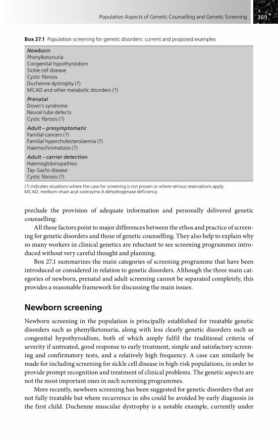
369369Population Aspects of Genetic Counselling and Genetic Screening
preclude the provision of adequate information and personally delivered genetic counselling.
All these factors point to major differences between the ethos and practice of screen-ing for genetic disorders and those of genetic counselling. They also help to explain why so many workers in clinical genetics are reluctant to see screening programmes intro-duced without very careful thought and planning.
Box 27.1 summarizes the main categories of screening programme that have been introduced or considered in relation to genetic disorders. Although the three main cat-egories of newborn, prenatal and adult screening cannot be separated completely, this provides a reasonable framework for discussing the main issues.
Newborn screening
Newborn screening in the population is principally established for treatable genetic disorders such as phenylketonuria, along with less clearly genetic disorders such as congenital hypothyroidism, both of which amply fulfil the traditional criteria of severity if untreated, good response to early treatment, simple and satisfactory screen-ing and confirmatory tests, and a relatively high frequency. A case can similarly be made for including screening for sickle cell disease in high-risk populations, in order to provide prompt recognition and treatment of clinical problems. The genetic aspects are not the most important ones in such screening programmes.
More recently, newborn screening has been suggested for genetic disorders that are not fully treatable but where recurrence in sibs could be avoided by early diagnosis in the first child. Duchenne muscular dystrophy is a notable example, currently under
Box 27.1 Population screening for genetic disorders: current and proposed examples
NewbornPhenylketonuriaCongenital hypothyroidismSickle cell diseaseCystic fibrosisDuchenne dystrophy (?)MCAD and other metabolic disorders (?)
PrenatalDown’s syndromeNeural tube defectsCystic fibrosis (?)
Adult – presymptomaticFamilial cancers (?)Familial hypercholesterolaemia (?)Haemochromatosis (?)
Adult – carrier detectionHaemoglobinopathiesTay–Sachs diseaseCystic fibrosis (?)
(?) indicates situations where the case for screening is not proven or where serious reservations apply.MCAD, medium-chain acyl-coenzyme A dehydrogenase deficiency.

370370 Practical Genetic Counselling
evaluation (see Chapter 11), while in cystic fibrosis a case can be made on grounds of therapy as well as prevention. It is clear that to justify screening for disorders on genetic grounds requires a detailed analysis of the effects on families in addition to the criteria already mentioned.
A current development causing considerable debate is the use of tandem mass spec-trometry for the detection of MCAD (medium-chain acyl-coenzyme A dehydrogenase deficiency), responsible for some cases of sudden infant death, and other metabolic dis-orders. The problem here lies with the detection of a range of often poorly defined conditions, as well as the one for which the programme is primarily designed.
Prenatal screening for genetic disorders
There has been a succession of developments in screening for genetic disorders in preg-nancy that have collectively had a major impact on the way in which pregnancy is managed and perceived. Whether these have been a success or not depends largely on what the appropriate aims and outcomes of such programmes have been considered to be. If the primary aim is to reduce the birth frequency of a serious disorder, then some have certainly been successful. If, on the other hand, the aim is to give maximum choice, information and support to women and their families, then the outcomes have been much more questionable, and in most cases the relevant issues have still not been adequately examined.
Almost all prenatal screening programmes have assumed the termination of an affected pregnancy, with one notable exception – rhesus haemolytic disease detection and immunization – which stands alone as an example of successful true prevention of a genetic disorder by prenatal screening.
Down’s syndrome – the commonest single cause of mental handicap – has been the focus of prenatal screening for many years (see Chapters 4 and 8). Initially based on increased maternal age and amniocentesis, and now utilizing maternal serum for the main preliminary screening test, with the strong likelihood of fetal DNA analysis from maternal blood becoming the approach of choice in the near future, this has certainly reduced the birth frequency of Down’s syndrome in many populations. There have been some concerns about inequity of availability and information to women concerning this service, but equally there have been problems in relation to quality of information, pressure to be tested, proper consent and choice, and subsequent support. These latter issues were inadequately considered in the setting up of most programmes and are only now receiving proper evaluation, reflecting the fact that the primary aims have not so much been orientated to the choice and benefit of individual women as to the ‘public health’ goal of reducing the birth frequency of Down’s syndrome in the population.
Exactly the same concerns apply to screening for neural tube defects using serum �-fetoprotein (see Chapter 8) and to the more general screening for structural malfor-mations using ultrasound. The lack of prior information and experience concerning a disorder can cause particular difficulties when coming at such a sensitive time as pregnancy.

371371Population Aspects of Genetic Counselling and Genetic Screening
So far, most prenatal screening approaches have not used genetic technology, but this could change in future (e.g. maternal blood screening for fetal DNA in trisomy 21). Suggestions that cystic fibrosis and fragile-X syndrome might be screened for prena-tally have not given sufficient weight to the incidental detection of carriers or to other family aspects. It is probably fair to say that the entire development of prenatal genetic screening has been unbalanced, based almost exclusively on the aim of eradicating par-ticular disorders and giving inadequate consideration to the wishes and potential effects of the programme on individuals. It is important that this field be radically and critically reassessed from a wider perspective. This is now beginning to be done, but genetic screening remains very much a ‘technology driven’ field.
Genetic screening in adults
Population screening in later life for genetic disorders is infrequent at present. Indeed, the potential for identifying one common and serious genetic disorder, familial hyper-cholesterolaemia, has been largely ignored until recently, and often confused with more general cholesterol screening. It could be argued that the high frequency and treatable nature of familial hypercholesterolaemia make it perhaps the only subgroup of disor-ders with raised cholesterol levels that everyone would agree is worth detecting. Fortunately, carefully planned programmes based on both lipid and DNA analysis, and following the approach of ‘cascade screening’ (see below), are now at last under way in several countries.
Detection of the genetic subset of common cancers is an area under considerable discussion in relation to screening, although at present this is entirely confined to family-based testing for the high-risk mutations. The term ‘screening’ is especially open to confusion here, since those at high genetic risk may be offered screening for overt disease (e.g. mammography, ovarian ultrasound) while non-genetic screening may be the first line in general population detection (e.g. cervical cancer screening). At present, it seems unlikely that detection of heritable mutations giving a high cancer risk will be used as a screening tool, although it has been proposed where a single specific mutation occurs at high frequency (e.g. for BRCA1 in Ashkenazi Jewish people).
A disorder for which screening was suggested is haemochromatosis, where recogni-tion of a genotype giving increased susceptibility is now feasible (see Chapter 20). This provides an excellent example of the potential pitfalls and lack of clear thinking that abound in the screening field. In contrast to the testing of high-risk sibs, the great majority of individuals detected would not be likely to develop disease and would not require treatment, while even larger numbers of heterozygotes would be detected where issues would arise as to what (if anything) they should be told.
This example shows how screening for an apparently definite mendelian disorder can merge into the more general area of susceptibility testing (Chapter 3), where at present even family-based testing is rarely helpful and wider screening is most likely to be positively harmful.
Screening of particular subgroups, rather than of entire populations, may be appro-priate for certain genetic disorders. A case can be made for this in screening the mentally

372372 Practical Genetic Counselling
handicapped for fragile-X syndrome, allowing a high genetic risk situation to be recognized that may give options for families avoiding recurrence of the disorder. Clear information and proper consent, with access to fuller genetic counselling, are essential factors that must be built into any such programmes (and properly resourced), which should be carefully distinguished from more general population and prenatal screening.
The term ‘cascade screening’ deserves a note here. This refers to the systematic testing of extended families to detect the abnormal genotype or disorder. For some dis-orders this may allow the detection of more such individuals than would be the case with more general population screening, and without some of the problems mentioned above. Familial hypercholesterolaemia is providing an example of its value, and it has also been used in carrier screening for cystic fibrosis (see below).
Carrier screening for autosomal recessive disorders
Most of our experience with carrier screening has come from work on the haemoglobin disorders, in particular beta-thalassaemia, which represents a serious and extremely common problem in many Mediterranean and Asian countries. It is notable that carrier screening only became widely accepted once the option of prenatal diagnosis was avail-able, in particular first-trimester DNA diagnosis. The remarkable degree of acceptance of carrier screening with prenatal diagnosis in widely different countries, many with a traditional society not previously accepting termination, is an indication of how screen-ing programmes can be successful, provided that they are carried out in sympathy with the attitudes of ordinary people. Such programmes must be accompanied by education and full information and must have a tangible result to offer – in this case the possibility of having healthy children without the risk of a severely disabling disease.
A comparably successful programme has been the introduction of carrier screening for Tay–Sachs disease in people of Ashkenazi Jewish origin. By contrast, sickle-cell carrier screening was not successful in early programmes, partly because of the more variable nature of the disorder, partly because of the defects in the necessary educa-tional and information aspects of the programmes.
The identification of the gene for cystic fibrosis, the most common autosomal reces-sive disorder in northern Europe, has given the possibility of DNA-based screening for the carrier state. Over 90 per cent of mutations can be identified in countries such as Britain and North America. A series of pilot projects has been carried out in Britain and the USA to assess the feasibility and desirability of screening. Aspects studied include the timing and location of carrier screening (antenatal clinic and primary care settings); the psychological effects on those screened; the attitudes of professionals and families; and the costs of testing and counselling.
The results of these pilot programmes have been interesting and not entirely expected. Essentially they show a high degree of compliance with a thorough and sensi-tively handled screening programme, but only a low uptake when the initiative is left to individuals or when they are given a time interval to make a decision. The launch of ‘over the counter’ carrier testing for cystic fibrosis carrier status, introduced to meet the

373373Population Aspects of Genetic Counselling and Genetic Screening
supposed ‘unmet demand’ for this, has likewise had minimal uptake. Thus the conclu-sion must be that such carrier testing is a low priority for most people and there is not a strong case for carrier screening outside families at risk, whether during pregnancy or before, becoming an established part of health services.
The approach of ‘cascade screening’, noted above, has also been used in cystic fibrosis and some other autosomal recessive disorders. It has the advantage that a much higher proportion of individuals tested will be carriers and that many of them will be aware of the disorder because of its occurrence in the family.
Population screening for a wider series of recessively inherited disorders is begin-ning to be introduced in some countries, notably Finland, where population bottlenecks have resulted in high frequency for a number of serious disease-causing genes. This approach will provide a challenge for ensuring satisfactory information and genetic counselling and could cause major problems if such multiple tests are offered as part of a ‘laboratory driven’ or commercial initiative.
CONCLUSION
When genetic counselling is dealing with the individual family, it is often capable of being precise and helpful, and of profoundly affecting the decisions of individual couples. This is rarely true at the population level, and here the clinician should be scep-tical about whether genetic counselling is having any significant effect, either beneficial or adverse, except in a few specific situations. This is perhaps fortunate, for it means that there is rarely any ethical conflict for either physician or patient between the course that is most beneficial for an individual or a family and that which is beneficial for society as a whole.
Finally, it should be borne in mind that variation is the basis of life and of human evolution, and that genetic characteristics today considered harmful may not always remain so. The ‘thrifty genotype’ of the diabetic patient may once have been associated with advantageous factors and may yet be again in a world with shrinking food resources. The phenylketonuric genotype, recently genetically lethal, is now almost of neutral effect, at least for males, and the advent of successful treatment will undoubt-edly ameliorate many other genetic diseases.
FURTHER READING
Bekker H, Modell M, Denniss G et al. (1993). Carrier screening for cystic fibrosis in primary care: supply push or demand pull? Br Med J 306, 1584–6.
Clarke A (1997). Newborn screening. In: Harper PS, Clarke A (eds) Genetics, Society and Clinical Practice, pp. 107–18. Oxford: BIOS.
Clarke A, Parsons E (eds) (1997). Culture, Kinship and Genes: Towards Cross-cultural Genetics. Basingstoke: St Martin’s Press.
Darr A (1999). Access to Genetic Services by Minority Ethnic Populations. London: Genetic Interest Group.

374374 Practical Genetic Counselling
Middleton A, Hewison J, Mueller RF (1998). Attitudes of deaf adults towards genetic testing for hereditary deafness. Am J Hum Genet 63, 1175–80.
Mitchell J, Cupua A, Clow C, Scriver CR (1996). Twenty-year outcome analysis of genetic screening programs for Tay–Sachs and beta-thalassaemia disease carriers in high schools. Am J Hum Genet 59, 793–8.
Modell B, Darr A (2002). Genetic counselling and customary consanguineous marriage. Nat Rev Genet 3, 225–9.
Nuffield Council on Bioethics (1993). Genetic Screening: Ethical Issues. London: Nuffield Council.
Super M, Schwarz MJ, Malone G et al. (1994). Active cascade screening for carriers of the cystic fibrosis gene. Br Med J 308, 1462–8.
Zlotogora J (2009). Population programs for the detection of couples at risk for severe monogenetic diseases. Hum Genet 126, 247–53.

Introduction . . . . . . . . . . . . . . . . . . . . . . . . . . . . . . . . . . . . . . . . . . . . . . . . . . . . . . . . . . . . . . . . . . . 375
Genetic testing issues . . . . . . . . . . . . . . . . . . . . . . . . . . . . . . . . . . . . . . . . . . . . . . . . . . . . . . . . . . . 376
Aims and outcome measures in genetic counselling . . . . . . . . . . . . . . . . . . . . . . . . . . . . . . . 379
Privacy and confidentiality . . . . . . . . . . . . . . . . . . . . . . . . . . . . . . . . . . . . . . . . . . . . . . . . . . . . . . 380
Eugenics and the abuse of genetics . . . . . . . . . . . . . . . . . . . . . . . . . . . . . . . . . . . . . . . . . . . . . . 381
Conclusion . . . . . . . . . . . . . . . . . . . . . . . . . . . . . . . . . . . . . . . . . . . . . . . . . . . . . . . . . . . . . . . . . . . . . 382
Further reading . . . . . . . . . . . . . . . . . . . . . . . . . . . . . . . . . . . . . . . . . . . . . . . . . . . . . . . . . . . . . . . . 383
Genetics and Society
CHAPTER 28
INTRODUCTION
The previous chapter examined some of the ways by which new genetic advances can be delivered more generally to the population, rather than just to those who are already aware of their own need. It also addressed the advances that may occur in genetic screen-ing and some of the problems. In this final chapter, the broader issues involving society itself are considered.
These new advances are having powerful effects on our social attitudes, widely dis-seminated through media publicity and often resulting in concern and controversy. This process is also affecting how genetics as a whole, including medical genetics, is perceived by the general population, a perception that is often ambivalent, even at times antagonis-tic, and which will have powerful effects on whether and how people use medical genetics services.
It may be argued that these wider issues are not directly relevant to genetic counsel-ling, but this is not the case. The services we try to offer, at both individual family and population levels, are strongly influenced by the societal background and attitudes of the community involved, and need to be adapted to those particular factors. One need look no further than such topics as prenatal diagnosis and consanguineous marriage to see how this has affected the development of genetic services in different populations. Many of these issues have already been raised in the specific chapters of this book, but it is useful to bring them together here. No attempt is made to cover general ethical and phil-osophical topics, but rather those that impinge directly on clinical genetic practice, whether carried out by a specialist or by other clinicians in their particular field. It is important to recognize that as genetic applications become increasingly used by all clini-cians in their own particular fields, they may be less aware of these issues, and the associated practical pitfalls, than are specialists in medical genetics.

376376 Practical Genetic Counselling
GENETIC TESTING ISSUES
Although genetic testing has been in practice for many years in the form of chromosome and protein analyses, the large-scale development of DNA-based testing and its progres-sive use outside specialist centres have created important issues that are largely new in practice, if not in concept. Some of these are raised here and have already stimulated con-siderable thinking and debate among workers in medical genetics, a healthy sign and one that is now spreading to those involved from more general fields.
What do we mean by genetic testing?
The author has offered the following working definition:
Genetic testing is the analysis of a specific gene, its product or function, or other DNA and chromosome analysis, to detect or exclude an alteration likely to be associ-ated with a genetic disorder.
Although most genetic testing is DNA-based, it is the aim of the test, rather than the technology, that is important. Thus phenotypic or protein-based tests such as electrophys-iology in Charcot–Marie–Tooth disease or haemoglobin analysis in sickle-cell disease can be regarded in principle as genetic tests when used to detect asymptomatic gene carriers (see Chapter 7), while DNA-based tests used for diagnosing infectious diseases are clearly not. Despite this lack of absolute distinction, though, it is principally the analysis of specific genes, especially in healthy individuals, that we are concerned with.
Presymptomatic testing for late-onset disorders
This important topic has been largely covered in Chapter 5, but here the broader societal impact needs consideration. Experience from Huntington’s disease and familial cancers has shown the need for the cautious handling of such testing, with proper consent, infor-mation and support. The perception of those tested and found to have an abnormal result that they frequently consider themselves already ‘affected’ is often mirrored by profes-sionals and society in general, and may powerfully affect society’s attitude towards those carrying genes for serious late-onset disorders.
As such presymptomatic testing becomes more widespread, a population of asympto-matic gene carriers destined to develop serious genetic disorders has developed. The situation is not unlike that for HIV carriers, and the potential for stigmatization and discrimination is comparable, whether this be for employment, insurance or general relationships. One could argue that the way in which society treats such individuals is an indicator of its maturity and humanity.
Genetic testing of children
The age independence of DNA testing makes it possible to test for a disorder at any age, regardless of the usual time of onset. To test young children for a late-onset disorder that

377377Genetics and Society
is not likely to have effects until adult life, and which is untreatable, poses serious problems. The author first encountered this in relation to Huntington’s disease and was surprised to receive requests to test children from parents and social workers. Testing was refused on the grounds that it would not benefit the child and would remove any later choice, and subsequently guidelines from both the UK and USA have supported the view that testing for late-onset disorders should normally be postponed until the indi-vidual can give full consent. Clearly, the situation may vary according to whether treatment exists or if the condition may also have childhood onset; individual circum-stances need careful consideration, but the general principle remains valid.
Different issues arise when adolescents themselves request genetic testing (although the two situations are often confused). Again, current guidelines suggest postponement of testing until an individual becomes an adult, but the topic will need full discussion with the individual, and the maturity of the adolescent will be an important factor – often a request for testing is a mask for a more general need for information and support, making it all the more important that sensitive discussion precedes any decision on actual testing. In the UK, decisions on genetic testing and the privacy of results need to be considered in the light of the 1985 ‘Gillick’ case, which considers individuals over 16 years old to be competent in relation to contraception and other reproductive issues, and for them and professionals to have the right to withhold information from parents, if necessary.
Most paediatricians, and other clinicians, have given little thought to this important topic until recently but, in the UK at least, there has been a marked shift in views towards those outlined above, already held by most clinical geneticists.
Use of research samples
Most research on genetic disorders requires samples from patients and many workers have large series of stored DNA samples from both affected and at-risk family members. When the gene is discovered for the particular disease, what should one do – or not do – with these samples?
Using the samples from affected individuals does not pose serious problems, but to test samples from those at risk carries grave consequences, since it is likely that some will show the mutation. The research worker is then in the serious situation of having impor-tant information on people without their knowledge, often not knowing whether they would have wished to be tested or what they originally gave consent for. Such samples should not have been used in the first place; if the person requests testing, it is better to take a separate sample specifically for this. Comparable issues can arise when genetic tests are incorporated into a more general research study, identifying individuals with a genetic basis among a larger group (e.g. studies of cancer).
Insurance and genetic testing
The past 15 years have seen a continuing battle between the insurance industry, unwilling to accept any regulation and insisting that genetic tests on healthy individuals are

378378 Practical Genetic Counselling
‘no different from other medical information’, and professionals in genetics, supported by a series of independent government reports in several countries, who have pointed out the dangers of individuals being forced to disclose information on genetic tests that they have had for entirely different purposes. Detailed actuarial studies have now confirmed the author’s view that, at least for life insurance, the industry would suffer little from not demanding such results except for very large sums. A moratorium has now been agreed in the UK, while the field is analysed objectively, and legislation restricting use of this information in insurance is now in place in the USA and some European countries. The adverse publicity that this issue generated for the insurance industry is an indication of how society is becoming reluctant to see new advances used to harm, rather than to benefit, its members.
‘Over-the-counter’ genetic testing
The traditional model of laboratory and other investigations ordered by and after con-sultation with a clinician or other professional has served medical genetics well but, as in other fields, is coming under increasing challenges. This is partly because of increas-ingly ‘consumerist’ attitudes to medicine, especially in America, but also because the internet now provides a means of promoting and delivering such ‘over-the-counter’ services without being bound by the regulations and restrictions of specific countries. The great majority of tests promoted in this way are of minimal or no clinical value, but they are increasingly being accompanied by misleading or frankly erroneous informa-tion, for purely commercial ends. If the same approach should spread to prenatal tests, such as those based on analysis of free fetal DNA, the consequences could be much more serious.
These rapid, current and potential future changes require a critical rethinking of where professionals involved in genetic counselling stand in relation to them, which is itself desirable in such a rapidly evolving area. Over-the-counter testing is already widely accepted in such areas as pregnancy and paternity testing and in one sense it can be seen as an extension of the outward diffusion of genetic testing from specialist genet-icists to more general clinical workers that is now largely established in many parts of medicine.
At present it is far from clear what real value and degree of uptake such over-the-counter testing has or is likely to have. There is certainly intense media publicity, which has currently shifted its focus from individual disorder tests to whole genome profiling and sequencing. This publicity has to a considerable extent been generated by the uncriti-cal enthusiasm of basic genetics researchers (many of whom should know better) for widespread testing, together with corresponding commercial pressures from companies who wish to see a return on their money. These two, often overlapping, groups have gen-erated a degree of ‘hype’ that risks bringing more reputable and justified genetic testing into disrepute.
Fortunately, a brake on this unhelpful development is provided in those countries (mainly European at present) fortunate enough to have a universal system of healthcare, by regulatory mechanisms that not only discourage ‘direct-to-consumer’ approaches,

379379Genetics and Society
but which fund only those tests for which there is a satisfactory evidence base of both scientific and clinical effectiveness. In such countries, testing for most of the serious genetic disorders covered by this book is already available without charge, as is the pro-fessional advice, which most families regard as helpful rather than as a barrier to genetic testing.
At present, it is in fact difficult to know whether there is indeed any significant demand for, as opposed to promotion of, these direct-to-consumer services. Data on uptake are normally unavailable, and the main guide to future trends is likely to be the number of genetic counselling referrals prompted by anxiety or confusion resulting from such testing. My own impression, based in practice in the UK, is that such referrals are uncommon, suggesting that actual use of direct-to-consumer tests remains minimal, at least in relation to serious genetic disorders, contrasting with what one might conclude from media publicity. Much more frequent are referrals resulting from the use of genetic tests by poorly informed clinicians who are not aware of the complexities and sensitive issues in medical genetics.
Regulation of genetic testing
Until recently, there have been few regulations, advisory or mandatory, in the field of genetic testing. Fortunately this has now changed, less in terms of the law than in the establishment of broadly based advisory bodies who can consider major issues, both practical and ethical, and whose recommendations are likely to be as influential as the passing of actual laws, in such a rapidly changing field as medical genetics.
The UK now has a well established advisory committee, the Human Genetics Commission, which deals with broad social and ethical issues in genetics and has already produced statements and reports on genetic testing and insurance, genetic privacy, over-the-counter testing and genetic testing for late-onset disorders. It is a broad-based body with only a minority of expert professionals.
One of the few areas in the UK that is legally controlled is that of pre-implantation genetic diagnosis and in-vitro fertilization.
In some of the USA, state specific laws have been passed limiting the use of genetic testing, particularly in relation to insurance, but it is not clear whether such laws have had significant effects over and above those resulting from more advisory recommendations.
AIMS AND OUTCOME MEASURES IN GENETIC COUNSELLING
Any professional activity, especially if it is being supported by public funding, needs to be able to produce clear goals as to what it is trying to achieve, and objective evidence that it is being successful in meeting those goals. In relation to genetic counselling, the whole area of aims and outcome measures is extremely complex and it is as well to
380380 Practical Genetic Counselling
recognize that there are no simple answers. It is somewhat easier to indicate which aims and outcome measures are inappropriate, especially since some of these have already been, and continue to be, used.
Thus the reduction or elimination of genetic disorders is not (at least in the view of the author and most professionals in the field) an aim of genetic counselling, even though it could, under some circumstances, be a valid aim of a broader population programme. Correspondingly, outcome measures such as number of pregnancies terminated or esti-mated financial savings from abnormal births avoided are not appropriate for genetic counselling, nor indeed for medical genetics services as a whole.
Recent reports (see ‘Further reading’) have tried to take initial steps in providing satisfactory alternatives. Such areas as understanding of information given, reproduc-tive plans and behaviour, client satisfaction and quality of process are all relevant, although all also have drawbacks. Perhaps the most important point is that much thought is now going into the difficult process of defining satisfactory aims and outcomes.
PRIVACY AND CONFIDENTIALITY
Genetic counselling raises some extremely difficult issues in this important area, some-thing that is inevitable since the process so often depends on having accurate information on family members and since its implications may extend far beyond those individuals seeking advice.
It should be stressed that, in practice, most difficulties can be resolved provided that time and trouble are taken to ask people’s permission, to explain why information is being requested and, in the case of risks to the extended family, to ascertain as far as possible whether they wish for information to be shared.
Given such a careful and sensitive approach, most family members will prove helpful and reasonable, although the process may take a considerable time. The temptation to ‘cut corners’ and assume consent when it has not been specifically given must be strongly resisted. Written permission is increasingly the rule, and is a valuable safeguard if problems arise at a later stage.
In the very few situations where relatives still refuse to give access to their records or test results after appropriate approaches have been made, this usually reflects family conflict or sometimes fear and denial of a genetic disorder. It has been suggested that the need of relatives may in some circumstances override the basic principles of confidential-ity, but the author’s view is that such situations are exceptional and must be based on clear evidence of the harm that would result if the information were not available. Against such abandonment of confidentiality must be set the effects of a general loss of trust if indi-viduals were to feel that their personal genetic data could be divulged in situations other than dire need.
Much more common, in the author’s experience, are requests for information from third parties which can and should be firmly denied unless specific permission is given. These include requests from insurers, employers, social services, adoption agencies and

381381Genetics and Society
various other bodies. There is no case for ‘public health interest’ disclosure of informa-tion as may be present for infectious diseases.
At a practical level, difficulty may arise when one is seeing several members of a family separately and is unaware of what information they have shared between them-selves. It is best to assume that nothing has been shared and to keep information compartmentalized unless one has clear permission to the contrary. Sometimes it can be preferable for a colleague to see the other part of the family if it becomes too complicated to keep things separate.
General issues of privacy, confidentiality and consent have become of increasing concern in medicine overall and the consequences of ignoring this have been shown in a series of high-profile cases involving pathology (see Chapter 10). Legislation is increas-ingly being introduced (e.g. the ‘Gillick’ ruling mentioned above in relation to childhood testing), but in the practical situations involving genetic counselling one is currently rarely in serious difficulty if one follows the broad lines indicated above.
EUGENICS AND THE ABUSE OF GENETICS
Everyone working in medicine, whether clinician or scientist, likes to believe that their own field is making a special contribution to human health and well-being. Those of us in medical genetics are no exception, and indeed the advances of recent years have helped families in ways that were unthought of only a generation ago. There is a darker side, though, which needs to be recognized, not ignored. This relates to what is generally known as eugenics, in which the principles of genetics (as understood in the past) were applied to attempting to change the genetic make-up of the population in general, rather than to individual families.
Eugenics had its origins in nineteenth-century Britain, but later became wide-spread in other European countries and in the USA. It was closely bound up with the social systems of the time, notably the rigidities of class and the deprivation of much of the population. It was convenient to find a biological explanation in terms of genetic inferiority for these deep-seated problems, with solutions that justified the mainte-nance of the social status quo. The newly emerging patterns of quantitative and mendelian inheritance were enthusiastically applied not just to clearly inherited disor-ders but to a whole series of diseases and characteristics that today would be regarded as heterogeneous and complex in their basis, such as mental impairment, epilepsy and criminality.
These and other studies, enthusiastic but largely uncritical in nature, provided the scientific justification for a series of coercive measures, including the segregation, institutionalization and (particularly in the USA) sterilization of mentally impaired people and other groups. Restrictive legislation on proposed immigrants was another aspect in the USA and Australia. While politicians were responsible for the imple-mentation of these measures, they were enthusiastically promoted and supported by many scientists (notably Charles Davenport, director of the Cold Spring Harbor Laboratory).

382382 Practical Genetic Counselling
The lowest point in the history of eugenics was reached in Nazi Germany, where in 1933 a national eugenics law was one of the first measures introduced, and where com-pulsory sterilization of those with mental illness or impairment, along with people suffering from disorders such as Huntington’s disease, was followed by active killing of such individuals, together with those with mental handicap and with congenital malformations. Again it is tempting to regard this terrible chapter in history as an aberra-tion and to blame the politicians, but this would be wrong. The entire basis for these policies was provided by scientists, including some of the most eminent geneticists of the time, and by clinicians, notably psychiatrists. It should not be forgotten that Josef Mengele was himself a human geneticist.
Eugenics is now a largely discredited field, but it has recently, to the great concern of many people, re-emerged in China, where a frankly eugenic law has been introduced under the guise of ‘maternal and child health’. Fortunately, continuing opposition among many professionals seems at present to have resulted in the application of this being quietly abandoned. If serious abuses of new genetic developments are not to occur again, everyone – especially those working in medical genetics – must be aware of the past and its potential threat to the future. The increase in the power of technology, especially in computing and molecular genetics, makes it especially necessary to ensure that safe-guards are introduced as these techniques are applied.
CONCLUSION
The range and depth of the many societal issues arising in genetic counselling, including those outlined above, may seem daunting for the professionals involved and it is easy to feel at times that one’s own concepts of ‘good practice’ are likely to be submerged beneath an avalanche of inappropriate or even harmful developments. My own view, however, shaped over a period of 40 years in the field, is a more optimistic one. It is remarkable to see how issues recognized and views held by the relatively small number of practising clinical geneticists and genetic counsellors have gradually permeated not only the medical genetics community, but also the work of clinicians and laboratory colleagues more generally, until they have eventually become standard good practice.
Working in the field of medical genetics, and being involved in genetic counselling in one’s own particular area, offers remarkable opportunities for being in the forefront of, and helping to shape, new patterns of medical practice. It provides challenges, too, in trying to ensure that one’s own practice can set an example to others, and in one’s efforts to educate colleagues more generally to adopt comparable standards. Medical genetics in general, and genetic counselling in particular, is now an activity that extends far beyond the boundaries of medical genetics as a speciality, and how families are best helped is increasingly dependent on those whose main background and training lie outside this field. This book has been written in the hope that such workers can recog-nize the facts, problems and issues that arise in relation to genetic disorders, and can use them wisely and sensitively in genetic counselling to help the families with whom they are involved.

383383Genetics and Society
FURTHER READING
Adams MB (1990). The Wellborn Science. Oxford: Oxford University Press.Advisory Committee on Genetic Testing (1997). Code of Practice and Guidance on Human Genetic
Testing Services Supplied Direct to the Public. London: UK Health Departments [see Appendix for internet address].
Advisory Committee on Genetic Testing (1998). Genetic Testing for Late-onset Disorders. London: UK Health Departments.
American Society of Human Genetics, Social Issues Subcommittee on Familial Disclosure (1998). ASHG Statement. Professional disclosure of familial genetic information. Am J Hum Genet 62, 474–83.
Anderson RM (ed.) (1997). Human genetics: uncertainties and the financial implications ahead. Phil Trans R Soc B 352, 1035–114 [a valuable symposium report on genetic testing and insurance].
Clinical Genetics Society Working Party (1994). Report on the genetic testing of children. J Med Genet 31, 785–97.
Farndon P, Douglas F (2006). Consent and Confidentiality in Genetic Practice: Guidance on Genetic Testing and Sharing Genetic Information. London: Joint Committee on Medical Genetics.
Harper PS, Clarke AJ (1997). Genetics, Society and Clinical Practice. Oxford: BIOS [topics covered include genetic testing of children, insurance, genetic privacy, ethical aspects of genetic screening, China’s ‘eugenics’ law and the abuse of genetics in Nazi Germany].
Holtzman NA, Watson MS (1997). Promoting safe and effective genetic testing in the United States. Washington, DC: National Institutes of Health.
Human Fertilisation and Embryology Authority (2003). Sex Selection: Options for Regulation and Human Fertilisation: Options for Regulation. London: HFEA.
Human Genetics Commission (2002). Inside Information: Balancing Interests in the Use of Personal Genetic Data. London: Department of Health [especially the conclusions and recommendations].
Human Genetics Commission (2003). Genes Direct: Ensuring the Effective Oversight of Genetic Tests Delivered Direct to the Public. London: Department of Health.
Human Genetics Commission (2007). More Genes Direct: a Report on Developments in the Availability, Marketing and Regulation of Genetic Tests Supplied Directly to the Public. London: Department of Health.
Kevles DJ (1986). In the Name of Eugenics. New York: Knopf.Mao X (1994). China’s misconception of eugenics. Nature 367, 1–2.Marteau T, Richards M (eds) (1996). The Troubled Helix: Social and Psychological Implications of
the New Genetics. Cambridge: Cambridge University Press.Müller-Hill B (1998). Murderous Science. New York: Cold Spring Harbor Laboratory Press
[re-issue of the English translation of this important and disturbing book, even more relevant now than when first published (and should be compulsory reading for all those involved in medical genetics)].
National Information Resource on Ethics and Human Genetics – http://genethx.georgetown.edu.Nuffield Council on Bioethics (1993). Genetic Screening: Ethical Issues. London: Nuffield Council
on Bioethics.Parker M, Lucassen A (2003). Concern for families and individuals in clinical genetics. J Med
Ethics 29, 70–3.

This page intentionally left blank

Appendix: Useful Information in Connection with Genetic Counselling
Various items of information which it is hoped will be of practical use are listed below. Since this type of information is particularly liable to change, any amended or supplementary details will be welcomed. The internet now provides a wealth of information (see also Chapter 10) available directly to patients and families but often very useful for professionals.
REGIONAL GENETIC CENTRES
All parts of the UK now have specialist genetic counselling centres, usually working in close association with genetics laboratories. These are not listed here, partly to save space, but also because any interested clinician should find out personally the best channels of referral. Most regional services provide a network of genetics clinics outside teaching hospitals, reducing the need for families to travel, while still giving access to centralized laboratory services. Most other European countries, Australia and Canada have a com-parable service network.
In the USA, the number of centres involved in genetic counselling is much greater and many do not have a clearly defined and secure service basis.
LAY SOCIETIES INVOLVED WITH INHERITED DISEASES
Few genetic disorders now seem to be without their own society, and these organizations often produce helpful information for patients and their families (and for their doctors), increasingly web-based, which may be extremely useful in conjunction with genetic counselling. Many families find the support given by such lay groups valuable, while raising money for research and pressing for adequate services are also valuable functions. On the other hand, it has to be remembered that many families do not wish to be associ-ated with lay groups, and it is important that clinicians involved in providing genetic counselling do not become so closely identified with such groups that they fail to provide an equal service to those families not involved with them.
The number of such societies is now too large to list comprehensively, and addresses change frequently. Fortunately, in both the UK and USA ‘umbrella’ groups have been formed to coordinate the activities of the different societies, and in particular to lobby
Regional genetic centres . . . . . . . . . . . . . . . . . . . . . . . . . . . . . . . . . . . . . . . . . . . . . . . . . . . . . . . . 385
Lay societies involved with inherited diseases . . . . . . . . . . . . . . . . . . . . . . . . . . . . . . . . . . . . . 385
Internet addresses . . . . . . . . . . . . . . . . . . . . . . . . . . . . . . . . . . . . . . . . . . . . . . . . . . . . . . . . . . . . . . 386

386386 Appendix
governments and other bodies for improved services, a role that is effectively per-formed by these groups and which is much more appropriately undertaken by them than by doctors or scientists. Some addresses are included in the list of websites given below.
INTERNET ADDRESSES
The list given below is a sample of useful sources of information and includes some of these given earlier in specific chapters. It is increasing very rapidly and it is often not apparent from the title how much clinically useful information the site contains. Frequently one will encounter cross-links to other addresses that may prove more useful than the one originally intended. The best course is to try out the different websites and this list may be a useful starting point.
Most genetics journals are also now available electronically, while official reports are often now available on the internet. As well as specific disease organizations, many medical genetics units and university institutes have their own website, giving a useful picture of services and research interests.
American College of Medical Genetics www.acmg.net
American Journal of Human Genetics www.cell.com/AJHG/
American Society of Gene Therapy www.asgt.org
American Society of Human Genetics www.ashg.org/
British Society for Human Genetics www.bshg.org.uk
CLIMB (Children Living with Inherited Metabolic Diseases)
www.climb.org.uk
Contact a Family (UK) www.cafamily.org.uk
Dutch Genetic Alliance (VSOP) www.vsop.nl
European Directory of DNA Laboratories www.eddnal.com
European Society of Human Genetics www.eshg.org
EURORDIS www.eurordis.org
Linked to NORD, this gives information on
European initiatives on rare disorders.
Linked to Orphanet (www.orpha.net)
European Directory of DNA Diagnostic Laboratories
www.eddnal.com
Eurogenetest www.eurogentest.org
Comprehensive information source on genetic testing in Europe
European Genetics Alliances’ Network (EGAN)
www.egan.eu
Geneclinics www.geneclinics.org

387387Appendix
GeneTests (International Directory of Genetic Laboratories)
www.genetests.org
Genetic Alliance UK www.geneticalliance.org.uk
Genetic Alliance (formerly Alliance of Genetic Support Groups) USA
www.geneticalliance.org
Human Fertilisation and Embryology Authority
www.hfea.gov.uk
Hereditary Hearing Loss homepage http://webh01.ua.ac.be/hhh
Human Gene Mutation Database www.hgmd.cf.ac.uk
Valuable general database of the range and types of mutation found in genetic disorders, based on the book by Cooper and Krawczak
Human Genetics Commission www.hgc.gov.uk
Human Genome Organization www.hugo-international.org
Human Genome Project Information www.ornl.gov/sci/techresources/Human_Genome/project/about.shtml
Mitelman Database of Chromosome Aberations in Cancer
http://cgap.nci.nih.gov/Chromosomes/Mitelman
National Information Resource on Ethics and Human Genetics
http://genethx.georgetown.edu
National Organization for Rare Disorders (NORD), PO Box 8923, New Fairfield, CT 06812, USA
www.rarediseases.org
National Society of Genetic Counselors, Inc. (in the USA)
www.nsgc.org
OMMBID (Online Metabolic and Molecular Bases of Inherited Disease)
http://ommbid.com
Continually updated version of Scriver et al. (2001)
OMIM (Online Mendelian Inheritance in Man)
www.ncbi.nlm.nih.gov/omim
Continually updated version of McKusick’s reference book (CD-ROM also available). An essential companion to all working in the field
Rare Disease UK (RDUK) www.raredisease.org.uk
National alliance for people with rare diseases and all who support them
Research Program on Ethical, Legal and Social Implications of Human Genome Project
www.genome.gov/10001618
TERIS http://depts.washington.edu/terisweb/teris/index.html
Electronic database derived from Shepard TH (1995), Catalog of Teratogenic Agents, 8th edn, Baltimore, MD, Johns Hopkins University Press. Available on subscription and provides data on over 1300 agents

388388 Appendix
UK Genetic Testing Network www.ukgtn.nhs.uk
Provides information on availability and facilities for genetic testing in the UK
UK National Screening Committee www.screening.nhs.uk
UK Health Department Genetics information and documents
www.dh.gov.uk/en/Publichealth/Scientificdevelopmentgeneticsandbioethics/Genetics/index.htm
Wiley Encyclopedia of Life Science (2006) http://www.els.net

GlossaryThe following list covers only those genetic terms used in this book that may be unfamiliar. A fuller list can be found in King RC, Stansfield WD, Mulligan PK (2006), A Dictionary of Genetics, Oxford University Press, Oxford.
allele One of several alternative forms of a geneamniocentesis The procedure of taking a sample of amniotic fluid from the
pregnant uterusaneuploidy The occurrence of an additional or missing chromosome to
give an unbalanced chromosome complementanticipation The occurrence of a genetic disorder at earlier age of onset
and/or at greater severity in successive generationsautosomal Determined by a gene on one of the chromosomes other
than the sex chromosomesBarr body The sex chromatin body visible beneath the nuclear
membrane of a cell from a female, representing the inactive X chromosome
candidate gene A gene suspected as being the gene mutated in a given disordercDNA (complementary DNA)
A DNA sequence corresponding to the messenger RNA produced by a gene, and lacking the non-coding regions (introns) of the gene
Caenorhabditis elegans A simple nematode worm, used as a model organism, whose genome has been completely sequenced
centromere The portion of the chromosome joining the two chromatids and separating the long and short arms of the chromosome
chimaera An individual containing cells of more than a single genetic origin
chorion biopsy (chorionic villus sampling)
The sampling of tissue from the chorionic membrane of the embryo
chromatid A single DNA strand of a dividing chromosome, joined to its ‘sister chromatid’ at the centromere
chromatin The proteins and other materials composing the structure of the chromosomes in conjunction with the DNA itself
chromosome A structure within the nucleus (normally numbering 46 in humans), consisting of a long molecule of double-stranded DNA, bearing a linear arrangement of genes, which is associated with nucleoproteins and RNA; it condenses to become visible under the microscope at cell division and it is able to reproduce its molecular structure with great fidelity through successive cell divisions and in transmission from generation to generation

390390 Glossary
clone An identical copy of a DNA sequence, cell or whole organism; ‘to clone’ also means to isolate and replicate a specific DNA sequence or gene
comparative genomic hybridization (CGH)
A method for detecting increased or decreased copies of a gene sequence, either constitutional or somatic
congenital Present at birthconsanguinity Mating between close relativesconsultand The individual (not always affected) through whom a family
with a genetic disorder comes to clinical attentioncytogenetics The study of chromosomes, their structure, function and
abnormalitiesdeletion Loss of genetic material (may be applied to a chromosome or
a gene)disomy, uniparental The transmission of both copies of a chromosome from a
single parent, instead of one from each parentdizygotic twins Twins originating from two fertilized eggsDNA Deoxyribonucleic acid, the molecule whose sequence and
replication determine the genetic information present in genes and chromosomes
dominant A characteristic or disorder expressed in the heterozygote, i.e. requiring only one altered copy to show itself
Drosophila The fruit fly, a classical experimental organism for genetic studies, whose genome has been completely sequenced
duplication (as applied to a gene or portion of chromosome)
Presence of an additional copy of part of a chromosome or of a gene
dysmorphology The study of malformation syndromesempirical risks Risk estimates based on those actually observed, rather than
on general principlesepigenetic Genetic changes due to processes involving chromatin and
methylation, among others, rather than to DNA sequence changes
eugenics The use of genetic measures to attempt to alter and improve the genetic nature of a whole population
exon The segments of a gene whose sequence is expressed by formation of messenger RNA and (usually) protein
FISH Fluorescence in-situ hybridization of DNA, allowing microscopic detection of abnormalities
fragile site An uncoiled site on a chromosome giving an apparent break (as in fragile X syndrome)
gamete Egg or spermgene The unit of inheritance, consisting of a DNA sequence
coding for a specific protein or component of a protein, arranged in a linear manner on chromosomes

391391Glossary
genome The entire genetic material of a cell or organismgenomics Study of the genetic material overallgenotype The genetic constitution of an individual (either overall or
referring to a specific gene locus)germ line The cell lineages resulting in eggs or spermgonadal (germ line) mosaicism
The occurrence of more than one genetic constitution in the precursor cells of eggs or sperm
haploid Having only a single set of genes (as in sperm or egg)haplotype A series of alleles found at linked loci on a single
chromosomehemizygous The presence of a gene in only a single copy, e.g. X-linked
genes in males, but also autosomal genes where one copy is deleted
heritability The proportion of variance of a characteristic due to genetic rather than environmental factors
heterogeneity (genetic) The occurrence of a single phenotype due to mutation of more than one gene (usually implies more than one genetic locus)
heteroplasmy Presence of two or more different forms of mitochondrial DNA
heterozygote An individual with two different alleles at a particular locusHLA system The major histocompatibility region, determined by a
complex of genetic loci on chromosome 6homozygote An individual with identical alleles at a particular locusimprinting (genomic/genetic)
The differential expression of a genetic characteristic or disease depending on parent of origin, often related to differences in methylation
in-situ hybridization The technique of applying molecular probes to a chromosome spread or section
interphase The stage of the nucleus between cell divisionsintron The DNA sequences in a gene that are not converted into
messenger RNA, and which separate the coding regions (exons)
inversion The turning round of a chromosomal segment with consequent alteration of its fine structure and sometimes its function
isochromosome An abnormal chromosome composed of two identical arms
karyotype The chromosome constitution as displayed by a microscopic preparation of dividing chromosomes photographed and arranged in homologous pairs
linkage, genetic The occurrence of two genetic loci close enough on the same chromosome to interfere with independent assortment at cell division
locus The specific site of a gene on a chromosome

392392 Glossary
Lyon hypothesis The principle of inactivation of one of the two X chromosomes in normal female cells (first proposed by Dr Mary Lyon)
lysosome An intracellular body containing important enzymes involved in the breakdown of cell components
meiosis The process of cell division leading to formation of eggs and sperm, with halving of the chromosome number
mendelian Following the patterns of inheritance proposed originally by Gregor Mendel
microdeletion A small or invisible loss of genetic material on a chromosome; often detectable by FISH techniques
mitochondrial inheritance
Exclusively maternal inheritance determined by DNA in the mitochondria, cytoplasmic bodies that also contain key enzymes
mitosis The process of cell division of somatic cells, in which the daughter cells are normally genetically identical to the parent
monosomy The occurrence of only a single member of a chromosome pairmonozygotic twins Twins derived from a single fertilized eggmosaicism The occurrence in an individual of more than one genetic
constitution, arising after fertilization (cf. chimaera)mutation/mutant The change from the normal to an altered form of a
particular gene/the individual who has undergone such a change
non-disjunction Failure of the normal separation of chromosomes at cell division, giving an unbalanced chromosome number (as in trisomy 21)
oncogene A gene involved in cell proliferation whose abnormal activation or over-expansion is important in turning a normal cell into a tumorous cell
p The short arm of a chromosomepenetrance The proportion of individuals with a particular genetic
constitution who show its effectpericentric inversion An inversion of genetic material around the centromere of
a chromosomephenotype The visible expression of the action of a particular gene; the
clinical picture resulting from a genetic disorderpolygenic/multifactorial Determined by multiple genes and usually also by non-genetic
factorspolymerase chain reaction (PCR)
The amplification of DNA using a specific technique, which allows analysis of minute original amounts of DNA
polymorphism Frequent hereditary variations at a genetic locuspositional cloning Gene isolation based on the mapping position of that gene
rather than on its nature or functionpre-mutation A clinically insignificant change in a gene that predisposes
to a subsequent full mutation

393393Glossary
proband The affected individual through whom a family with a genetic disorder is ascertained (also called propositus)
probe A labelled DNA sequence that can be used to identify a corresponding sequence by hybridization
propositus, proposita Proband (see above)q The long arm of a chromosomerecessive A characteristic or disorder only expressed when both alleles
at a genetic locus are alteredrecombination The separation of alleles that are close together on the same
chromosome by crossing over of homologous chromosomes at meiosis
restriction enzyme A group of bacterial enzymes that cleaves the DNA chain at sequence-specific sites
RFLP Restriction fragment length polymorphism: inherited variation in DNA detected by the cutting of DNA at different points by a restriction enzyme
ring chromosome A chromosome in which breakage close to the ends has been followed by rejoining to form a ring
RNA Ribonucleic acid: the nucleic acid for which DNA forms a template before protein is produced
Robertsonian translocation
The formation of a single abnormal chromosome by the joining of two chromosomes by their short arms
somatic Involving the body cells rather than the germ lineSouthern blotting Transfer of DNA fragments from a electrophoretic gel to a
membrane prior to DNA hybridizationstem cell A cell capable of reproducing and giving rise to different cell
types in its descendantssyndrome A combination of clinical features forming a recognizable entitytelomere The terminal region of the chromosome armsteratogen An agent that can cause developmental abnormalities in the
embryotranslocation Transfer and exchange of genetic material between different
chromosomes, usually not members of the same pairtrinucleotide repeat A repeated sequence of three bases (e.g. CAG) expanded and
unstable in a group of genetic disorderstriploid Having three rather than two complete sets of chromosomes
(usually lethal in humans)trisomy The presence of three copies of a specific chromosomeX inactivation Inactivation of one of the two X chromosomes in a normal
female cell (see Lyon hypothesis)Y chromosome The small chromosome possessed by males only, whose
principal function is involvement in sex determinationzygote The fertilized egg

This page intentionally left blank

IndexAarskog syndrome (faciogenital dysplasia)
240, 302 abdominal muscle deficiency (prune-belly
syndrome) 302 abortion recurrent 72, 313 spontaneous 314–15 acanthosis nigricans 250 achondrogenesis 220 achondroplasia 217 homozygosity 33 acrocephalosyndactylies 222 acrodermatitis enteropathica 290 acromegaly 309 adoption 158–60 adrenal hyperplasia 311 adrenoleucodystrophy 321 affective psychoses 214–15 agenesis of corpus callosum 198 aglossia—adactylia 238 Aicardi syndrome 198 Alagille syndrome (arteriohepatic dysplasia)
295 albinism 39, 245–6 Albright’s hereditary osteodystrophy 29, 222,
310 alcohol, fetal effects 352 alcoholism 213 alkaptonuria, quasidominant inheritance 38(fig.) alopecia 250 alpha-1-antitrypsin deficiency 294 carrier detection 106 alpha-fetoprotein (AFP) abnormalities other than neural tube
defects 126(tab.) chance finding 124 estimation 119 neural tube defects 125–7, 195–6 omphalocele and gastroschisis 289 Alport syndrome (hereditary nephropathy
with nerve deafness) 300 Alstrom syndrome, diabetes 306 Alzheimer’s disease 202–3 ambiguous genitalia 312 amelogenesis imperfecta 235 amino acid disorders 319–20 amniocentesis 118–21 chromosomal disorders and maternal age
70(tab.) contraindications (delay) 125 risks 121 twin pregnancy, genetic risks 132 amputation defects 223
amyloidoses 177, 300 anal atresia 293 aneurysms and related vascular lesions 281–2 Angelman syndrome 78, 96 angioedema, hereditary, carrier detection 108 anhidrotic ectodermal dysplasia 249 aniridia 263 ankylosing spondylitis 227–8 anophthalmos 262 antiepileptic drugs 352–3 Apert syndrome 30 molecular basis 97 aphthous mouth ulcers 237 apnoea, following muscle relaxants 322 appendix, genetic counselling 385–8 apple peel syndrome 289 arteriohepatic dysplasia 295 arthritis and arthropathies 227–31 arthrogryposes 230 scoliosis 231 talipes 230–1 arthro-ophthalmopathy 232, 258 artificial insemination by donor (AID) 313 Ashkenazi Jewish people, BRCA1 371 Asperger syndrome 212 asplenia and polysplenia syndrome 273 asthma and atopy 284 ataxia telangiectasia 344 diabetes 306 ataxias 187 atopic eczema 246–7, 284 atrial septal efect 275 atrophic gastritis 290 atrophic rhinitis 238 autism 212 autoimmune polyglandular syndrome
(APECED) 309 autopsy 94 autosomal dominant inheritance 26–33 carrier detection 107–9 genetic risks 26–33, 26(fig.) homozygosity 32–3 incomplete penetrance 27–8, 27(fig.) late or variable onset 26–7 mosaicism and mutation 31–2 new mutation 30–1 genetic risks 31(fig.) ‘obligatory’ vs possible carrier(s) 104(fig.) pedigree pattern compatible 24(fig.) types 24(fig.) variability 28–30, 29(tab.) anticipation and unstable DNA 30 genomic imprinting 29, 78

396396 Index
autosomal dominant inheritance (continued) modifying influences 30 variation in expression 28 autosomal recessive inheritance 33–40 carrier detection 104–7 carrier risk 33–5 consanguinity 37–8, 37–8(fig.) donor insemination 38 genetic heterogeneity 39 genetic risks 35(fig.) Hardy–Weinberg equilibrium 36 marriages between affected individuals
39–40, 39(fig.) ‘obligatory’ vs possible carrier(s) 104(fig.) patterns 34(fig.) population risk 35–7 risk of transmitting 36(tab.) variation 38–9 baldness 250 Bardet—Biedl syndrome 256 diabetes 306 Barr body 40 Barth syndrome 278 Bartter’s syndrome 320 basal cell naevus syndrome 251 gene locus 336 Batten’s disease 199, 321 Bayes’ theorem 45 Beal syndrome 225 Beckwith–Wiedemann syndrome 29, 289,
309, 345 behavioural disorders 211 benign familial haematuria 300–1 biliary atresia 295 bilirubin conjugation (Crigler–Najjar
syndrome) 294 Binder syndrome 239 birth defects 91–101 see also malformations bladder exstrophy 302 blood DNA isolation 82 see also fetal blood; maternal blood blood disorders 325–33 blood group systems 328 Bloom syndrome 344 bone dysplasias 216–20 molecular basis 224(tab.) bowel atresias and malrotations 289 brachydactyly 222 BRCA1, Ashkenazi Jewish people 371 BRCA1 and BRCA2 341–2 breast cancer 341–2 genetic risks 341(tab.) bulbospinal atrophy 176 Byler’s disease 294
c-KIT oncogene 246 campomelic dysplasia 220 molecular basis 97 Canavan’s disease 199 cancer 334–47 childhood cancer 344 embryonal cancer 344 familial tumour syndromes 336, 337–8 genetic counselling 338–43 molecular analysis 335–7 cardiomyopathies 277–8 congenital cardiac conduction defects 278 dilated cardiomyopathy 278 hypertrophic cardiomyopathy 277–8 cardiovascular disorders 271–82 carnitine palmityl transferase deficiency 172 carrier(s) 102–14 defined 102 detection autosomal dominant 107–9 autosomal recessive 104–7 DNA polymorphisms 87 isolated cases 110 late-onset disorders 109 methods 112–13 X-linked 109–12 inappropriate carrier testing 105–6 ‘obligatory’ vs possible 103–4 cat eye syndrome 77 cataract 259–60 cavernous haemangioma 249–50 cerebellar ataxia 187 cerebral aneurysms 192–3 cerebral palsy 193–4 cerebrohepatorenal syndrome 299 Charcot–Marie–Tooth disease 176–7 CHARGE association 273 Chediak–Higashi syndrome 344 children cancer 344 dysmorphism 91–101 epilepsy 191 genetic testing issues 376–7 nephrotic syndrome 300 obesity 310 chimaerism 31, 76 chloride diarrhoea 291 cholestasis 294 chondrodysplasia punctata 219, 220, 352 chondrodystrophic myotonia 174 chorea, hereditary benign 186 chorea-acanthocytosis (neuroacanthocytosis)
186 chorion villus sampling 87, 121–2 indications 122 mosaicism confined to placenta 125 risks 122

397397Index
chorioretinitis 351 choroideraemia 255 choroidoretinal degeneration 255–6 Christmas disease 330 chromosome 11p, microdeletion syndrome 96 chromosome 22q, microdeletion syndrome
96, 273 chromosome abnormalities 63–80, 95 deletions 77, 77(tab.) frequency in population 67–9 specific chromosomal disorders
68–9(tables) spontaneous abortions and stillbirths
68(tab.) studies of newborns 67–9 unselected newborns 68(tab.) hereditary fragile sites 79 inversions 76 isochromosomes 79 microdeletion syndromes 77, 78(tab.) mosiacism 122 normal human karyotype 65–6(fig.) normal variations 79 other viable autosomal trisomies (13, 18
and 22) 71 recurrent abortion and infertility 72, 313 sex chromosomes 71–2, 72(tab.) terminology 64–7 translocations 73–6 trisomies 69–71 trisomy 21, 69–71 uniparental disomy 78 chromosome translocations 73–6 reciprocal translocations 74–6, 75(tab.) translocation Down’s syndrome 73–4 chronic obstructive pulmonary disease 285 cleft lip and palate 235–7, 237(tables) CNS disorders 179–200 degenerative metabolic disorders 198–9,
199(tab.) structural 194–9 coeliac disease 290 collagen bone and connective tissue disorders
224(tab.) type-2 and -8 258 type-7 248 coloboma 263 colorectal cancer 339–40 genetic risks 340(tab.) tumour suppressor genes 61 colour vision 43, 257 combined immunodeficiency, carrier
detection 106 common disorders 54–62 empirical risk data 60–1 genetic basis 55–8
mendelian subsets 55–6 molecular basis of susceptibility 61–2 multifactorial inheritance 58–60 perception of disease 57–8 congenital, see also familial; hereditary congenital chloride diarrhoea 291 congenital heart disease 272–5 mendelian disorders 272(tab.) multiple cases 275 non-mendelian disorders 273(tab.) second-degree relatives 275 syndromes 277–8 twinning 273 ultrasonography 127 congenital infections 348–51 congenital malformations see malformations congenital myopathies 171–4 lethal centronuclear (myotubular)
myopathy 171 metabolic defects 171, 172(tab.) congenital nephrosis 300 connective tissue disorders 223–7 molecular basis 224(tab.) conotruncal defects 276 Conradi syndrome 219 consanguinity 135–44 autosomal recessive inheritance 37–8,
37–8(fig.) carrier detection 107 general risks 140–4, 141(fig.) first cousins 137, 142(fig.) increase in abnormalities and mortality
142(tab.) genetic relationships 135–8, 136–7(tables) legal restrictions on marriage 128(tab.) inbred populations 144 multiple consanguinity 143–4, 143(fig.) risks with specific genetic disorders
139–40, 140(tables) social aspects 138–9 consent 148 genetic counselling 147–8 consent issues, Gillick (1985 case) 377 contraception 157 corneal chamber defects 263 corneal dystrophies 258 coronary heart disease 278–81 familial hypercholesterolaemia 279–80,
280(tab.) ischaemic heart disease, risks of death 279(tab.) other genetic influences 280–1 premature vascular disease 281(tab.) corpus callosum, agenesis 198 Cowden disease, gene locus 336 cranial nerve palsies 178 craniocarpotarsal (Freeman–Sheldon)
syndrome 228, 231

398398 Index
craniofacial disorders 238–40, 270 craniosynostoses 238, 238(tab.) isolated 239 Creutzfeldt–Jakob disease 204 cri-du-chat syndrome 67, 77 Crigler–Najjar syndrome 294 Crohn’s disease 291 cryptophthalmos 263, 301 cryptorchidism 302 Cushing’s syndrome 310 cutis laxa 226 cystic fibrosis 283–4, 291 autosomal recessive inheritance 34(fig.) carrier detection 107, 283 carrier screening 372–3 diabetes mellitus 306 infertility 313 cystinosis (cystine storage disease) 301 cystinuria 303, 320 cytogenetic studies, detection of carrier(s)
112 cytogenetic syndromes 77(tab.) cytomegalovirus infection 349–50 cytotoxic drugs 354 Dandy—Walker syndrome 196 De Lange syndrome 232 deafness 265–70 otosclerosis 269 as part of syndromes 269 partial nerve deafness 268–9 severe congenital deafness 39, 266–8 degenerative metabolic disorders 198–9,
199(tab.) dementias 201–4, 202(tab.) dentinogenesis imperfecta 235 dextrocardia with asplenia 276 diabetes insipidus 303 diabetes mellitus 305–7 diagnostic genetic testing 89 diagnostic information 8–10 diaphragmatic hernia 288 diaphyseal aclasis 220 Didmoad syndrome 258 diabetes 306 DiGeorge syndrome 273, 310 digital clubbing 231 dihydropteridine reductase deficiency 320 disaccharidase deficiencies 290 distal spinal muscular atrophy 176 DNA and loss of heterozygosity 335 molecular genetics 81–90 DNA amplification 82 DNA ‘fingerprinting’ 145(fig.) DNA hybridization (Southern blotting) 83 DNA isolation 82
DNA polymorphisms carrier detection 87 linked 86–7 DNA triplet repeats, fragile-X (FMR) gene 208 Donohue syndrome 306 donor insemination 38 Down’s syndrome, prenatal screening 370 DRPLA (Dentato-rubro-pallido-luysian
atrophy) 186 drugs adverse reactions 323 teratogenicity 351–4 Duane syndrome 262 Dubin–Johnson syndrome 294 Dubowitz syndrome 240 Duchenne muscular dystrophy 43–9, 168–9 duodenal atresia 289 Dupuytren’s contracture 231 dwarfism 220 and 216, 218, 232 dysautonomia 177 dyschondrosteosis 219 dyskeratosis congenita 344 dyslexia 212 dysmorphic child 91–101 ear malformations 270 Ebstein’s anomaly 273 ectodermal dysplasias 249 ectrodactyly (split hand or lobster-claw
defect) 222 eczema, atopic eczema 246–7 EEC syndrome 249 Ehlers–Danlos syndrome 225–6 elliptocytosis 326-7 Ellis—van Creveld syndrome 237, 272 embryonal cancer 344 emphysema 285 encephalocele 197 endocardial fibroelastosis 276 enuresis 303 environmental hazards 348–57 enzyme defects 290–1 detection of carrier(s) 112 epidermolysis bullosa 248–9 epigenetics 28 epilepsy 190–2 antiepileptic drugs 352–3 benign neonatal convulsions 191 febrile convulsions 191 intractable infantile convulsions (infantile
spasms) 191 myoclonic epilepsy 191 partial benign epilepsy of childhood 191 primary idiopathic generalized epilepsy
191–2, 192(tab.) simple absence epilepsy 190 erythroderma, ichthyosiform 247

399399Index
essential tremor 186 eugenics, abuse of genetics 381–2 exomphalos 95 exostoses 220 eye colours 262 eye disorders 253–64, 254(tab.), 255 developmental defects 262–4 Fabry’s disease 301, 321 facial and ocular palsies 178 facioscapulohumeral muscular dystrophy
108, 170 factor VIII gene 331 familial, see also congenital; hereditary familial adenomatous polyposis 337–8 familial dysautonomia 177 familial erythrophagocytic
lymphohistiocytosis 347 familial hypercholesterolaemia 279–80,
280(tab.), 322 familial hypophosphataemia 43, 43(fig.), 44 familial insomnia 204 familial multi-infarct dementia (‘CADASIL’)
203 familial paroxysmal choreoathetosis 186 family tree 5–8 structure suitability for linkage prediction
87 symbols used, 6(fig.) working pedigree examples 7(fig.) Fanconi pancytopenia 223, 344 fatal familial insomnia 204 fatty acid metabolism defects 323 female infertility 312–14 fetal blood DNA isolation 82 prenatal diagnosis 130–1 fetal DNA, maternal blood 132 fetal sexing, X-linked disorders 366 fetal skin biopsy 131 fibrillin 260 fibrillin deficiency 225 fibromatosis 251–2 filaggrin 247 fragile sites 79 fragile X syndrome (FMR) gene 313 DNA triplet repeats 208 mental retardation 129, 208–9 population screening 372 Francois dyscephalic syndrome 239 Fraser syndrome 263, 301 Freeman–Sheldon syndrome 228, 231 Friedreich’s ataxia 187, 272 diabetes 306 frontonasal dysplasia 240 frontotemporal dementia (Pick’s disease) 204
galactosaemia 117, 320–1 carrier detection 106 gallstones 291 gamete donation 158 gangliosidosis 199, 321 Gardner syndrome 337 gastric cancer 340 gastritis, atrophic 290 gastrointestinal enzyme defects 290–1 gastrointestinal tract 287–96 gastroschisis 289 Gaucher’s disease 321 gelsolin defects 177 gene and genome sequencing 82–3, 109 gene therapy 163 genes, homology across different species 84 genetic counselling 3–22 aims and outcome 379–80 appendix 385–8 back-up measures 18–19, 156–7 common cancers 338–43 communication 16–18 advice at a distance 18 confidentiality 380–1 consent 147–8, 377 diagnostic information 8–10 genetic heterogeneity 39–40 history and development 4–5 individuals and families 362 internet addresses 386–8 legal problems in medical genetics 147–8 non-directiveness 16–18 preconceptual phase 363 privacy 380–1 referral 154, 363 regional centres 385 risk estimation 10–16 serious genetic disorders, population
prevention 362 special issues 135–49 success or failure, evaluation 362 support 20 two-stage approach 88 genetic counselling clinic 150–64 adoption 158–60 child at risk 159–60 confidentiality 161–2 contraception 157 counselling service 152–5 equipment 151 gamete donation 158 genetic registers 160–2 in-vitro fertilization 158 information sources and internet 155–60 location 151 pedigrees and records 151–2 sterilization 157–8

400400 Index
genetic counselling clinic (continued) stillbirth and termination of pregnancy 155 treatment for genetic diseases 162–4,
162(tab.) genetic disease treatment 162–4, 162(tab.) genetic heterogeneity 39, 83, 86 genetic registers 160–2 confidentiality 161–2 contact registers 160–1 disorders for consideration 161 management-related 160 organization 161 genetic testing issues 376–9 children 376–7 insurance and 377–8 late-onset disorders 376 ‘over-the-counter’ tests 378–9 use of research samples 377 genitalia, ambiguous 312 genomic imprinting, autosomal dominant
inheritance 29, 78 Gerstmann–Straussler syndrome 204 Gilbert’s syndrome 294 Gillick (1985 case), consent issues 377 gingival fibromatosis 238 glaucoma 260–1 glomus tumours 29 gene locus 336 glossary 389–93 glucose-6-phosphate dehydrogenase
deficiency 327 glycogen storage diseases 172, 321–2 goitre 309 Goldenhar syndrome 240 ear malformation 270 heart disease 273 Goltz syndrome 249 gonadal irradiation 357 Gorlin (basal cell naevus) syndrome 251 Graves’ disease 309 Greig syndrome 222 gyrate atrophy 256 haemangioma, cavernous haemangioma
249–50 haematuria, benign familial 300–1 haemochromatosis 293–4 diabetes mellitus 305 population screening 371 haemoglobin structure and synthesis
disorders 325–8 haemolytic disease, rhesus 314, 327–8 haemophilia A 42(fig.) haemophilias 330–1 carrier detection 110 pedigree 42(fig.) Hallermann—Streiff syndrome 239
Hallgren syndrome 256 Hardy–Weinberg equilibrium 36 Hartnup disease 303 Hashimoto’s thyroiditis 309 Helicobacter pylori 290 hemifacial microsomia 240 hepatic fibrosis 295 hepatitis C 350 hereditary, see also congenital; familial hereditary arthro-ophthalmopathy 232, 258 hereditary ataxias 187 hereditary benign chorea 186 hereditary haemorrhagic telangiectasia 281 hereditary motor—sensory neuropathy 176 hereditary non-polyposis colon cancer
(HNPCC) 339–40 hereditary oculomotor nystagmus 43(fig.) hereditary pancreatitis 27, 291 hereditary spastic paraplegia 188 Hermansky—Pudlak syndrome 245 hernia, diaphragmatic hernia 288 herpes simplex virus infection 350 heteroplasmy 52 hip dislocation 229 Hirschsprung’s disease 292–3, 292(tab.) molecular basis 60, 97 histidinaemia 320 histiocytosis 347 HIV infection 350 HLA-B27 227–8 holoprosencephaly 197–8 Holt–Oram syndrome 95, 223, 272, 275, 352 carrier detection 108 molecular basis 97 homocystinuria 281 horseshoe kidney 302 human genome project 83–4 Huntington’s disease 24(fig.), 180–5,
181–2(tables) apparent new mutations 364 associated aspects 183–4 genetic testing 184–5 intermediate alleles 364–5 isolated cases 182–3 juvenile HD 181 prenatal testing 184–5 presymptomatic testing 184–5 hydatidiform moles 315 hydrocephalus 196 hydronephrosis 301 hydrops fetalis 328 21-hydroxylase deficiency 310 hyperbilirubinaemias 294 hypercholesterolaemia 279–80, 280(tab.), 322 familial, carrier detection 108 hyperchylomicronaemia 322 hyperlipidaemias 322

401401Index
hyperlipoproteinaemia 291 hyperpyrexic myopathy 174 hypertension 282 hypocalcaemic hypercalcuria 303 hypochondroplasia 33, 217 hypogonadism and intersex 311–12 hypohydrotic ectodermal dysplasia 249 hypomelanosis of Ito 250 hypoparathyroidism 310 hypophosphataemia 43 hypospadias 302 hypothyroidism 309 ichthyoses 242, 247–8 idiopathic (minimal-change) childhood
nephrotic syndrome 300 IgA nephritis 300 immune deficiency disease 328–30, 329(tab.) immunosuppressive drugs 354 imprinting, influence of parental genome 29,
78 in-vitro fertilization 158 inborn errors of metabolism 129–30, 317–24 autosomal dominant 319 diagnosis from amniotic fluid 130 prenatal diagnosis 129 X-linked 318 incest 142 incomplete penetrance 27–8, 27(fig.) incontinentia pigmenti 250 industrial and other chemicals 354 infantile convulsions (infantile spasms) 191 infantile pyloric stenosis 288–9, 288(tab.) infectious disease genetics 330 infertility 312–14 inflammatory bowel disease 291–2 influenza virus 350 information sources and internet 155–60 insurance and genetic testing issues 377–8 intelligence IQ levels 204 normal 211 internet 155–60 internet addresses 386–8 intersex 311–12 intracytoplasmic sperm injection (ICSI) 29 intussusception 291 inversions 76 iris developmental defects 263 heterochromia 261 iron storage haemochromatosis susceptibility
gene 322 irradiation during pregnancy 355 gonadal 357 population exposure 356–7
ischaemic heart disease 279(tab.) isochromosomes 79
jejunal atresias 289 Jervell and Lange–Nielsen syndrome 269,
272, 278 Joubert syndrome 187 juvenile nephronophthisis (microcystic
disease) 299 juvenile polyposis 338 Kabuki syndrome 273 Kallmann syndrome 44 Kaposi’s sarcoma 251 Kartagener syndrome 272, 284 karyotype, normal human 65–6(fig.) Kennedy’s disease 176 keratin corneal dystrophies 258 specific disorders 248(tab.) keratoacanthoma, gene locus 336 Klinefelter syndrome 311–13 clinical features 72 population frequency 68(tab.) Klippel—Feil syndrome 232, 273 Klippel—Trenaunay—Weber syndrome 249 Kobberling partial lipodystrophy 306 Krabbe’s disease 199 Kuf ’s disease 199 Kugelberg–Welander disease 175 kuru 204 lactase deficiency 290 Langer–Giedion syndrome 220 Larsen syndrome 232 laryngeal defects 285 late-onset disorders, genetic testing issues 376 law, problems in medical genetics 147–8 law societies 385–6 LDL receptor deficiency 280 Leber’s congenital amaurosis 256 Leber’s optic atrophy 50, 257 legal problems in medical genetics 147–8 Leiden variant of factor V 332, 332(tab.) Leigh’s encephalopathy 199 lens dislocation 260 Lenz syndrome 263 Leopard syndrome 249, 272 leprechaunism (Donohue syndrome) 306 Lesch—Nyhan syndrome 303 lethal newborn dysplasias 219 leucodystrophies 199, 321 leukaemias 344, 346–7, 356 Lewy body dementia 203 limb defects 221–3 limb girdle muscular dystrophies 170 limb reduction defects 223

402402 Index
lipoproteins, gene ApoE 203, 280 Lisch nodules 189 lissencephaly and related neuronal migration
defects 198 lithium 353 liver disease 293–5 Li–Fraumeni syndrome 346 gene locus 336 Loeys–Dietz syndrome 225 loss of heterozygosity (LoH) 335 Lowe syndrome 304 lung cancer 285 lymphatic disorders 282 lymphomas 344, 347 lymphoproliferative disease 347 Lyon hypothesis 40 lysosomal acid maltase deficiency 321 lysosomal enzyme defects 321 McArdle syndrome (glycogenosis type V) 172 Machado—Joseph disease 187 macrocephaly 197 macular dystrophies 256 Madelung deformity, wrist 219 mal de Meleda 248 male infertility 312–14 malformations 91–101, 125–7 aetiological groups 95–7 autosomal dominant 99(tab.) computerized databases 99 diagnostic approach 93–4 disordered function of specific genes in
development 96 laboratory studies 94 limb defects 221–3 microdeletion syndromes 96 prenatal diagnosis 125–7 recurrence risks 98–9, 100(tab.) sequence 93 teratogenic syndromes 96 terminology 92 X-linked 99(tab.) malignancy, predisposing syndromes 343,
344(tab.) malignant hyperpyrexia 174 carrier detection 108 malignant melanoma 251 mandibular hypoplasia 237 mandibulofacial dysostosis 239, 270 manic—depressive illness 214–15 Marchesani syndrome 260 Marfan syation 225 Marfan syndrome 260, 261, 272 maternal age abnormalities found at amniocentesis 70(tab.) chromosomal disorders 70(tab.), 123 maternal blood, free fetal DNA 132
maternal health adversely affected by pregnancy 133 obstetric complications 133 maternal serum screening 127–8 MCAD (medium-chain acyl-coenzyme A
dehydrogenase deficiency) 323, 370 median cleft face syndrome 240 medullary cystic disease 299 megaureter 302 Meige’s disease 282 melanoma, familial, gene locus 336 mendelian disorders 23–53 carriers 103–14 congenital heart disease 272(tab.) mitochondrial inheritance 49–52,
50–1(fig.) polygenic inheritance 24(fig.) skin 243–4(tab.) sporadic case 25(fig.) variablity, factors 29(fig.) mendelian subsets, common disorders 55–6 Menkes syndrome 199, 281 mental illness 201–15 mental retardation 204–11 fragile-X syndrome 129, 208–9 genetic risks 208(tab.) mendelian disorders 206(tab.) mild 210–11 non-mendelian and chromosomal
disorders 207(tab.) non-specific 210 metabolism, inborn errors 129–30, 317–24 metachromatic leucodystrophy 321 microcephaly with mental retardation 197,
349, 350 microcystic disease of lung 284 microdeletion syndromes 77–8(tab.), 96, 273 microphthalmos 262–3 microscopic techniques, detection of
carrier(s) 112 migraine 193 Miller—Dieker syndrome 198 mini-satellite DNA ‘fingerprinting’ 145(fig.) mitochondrial inheritance 49–52, 50–1(fig.) DNA analysis 257 mutations 51 mitochondrial myopathies 171–2 Moebius syndrome 178 molecular analysis, diagnostic approach to
malformations 94 molecular genetics 81–90 clinical genetics 85–90 human genome project 83–4 techniques 81–3 types of mutation 84 molecular prenatal diagnosis 130 Morquio syndrome 219

403403Index
mosaicism 76–7, 124–5 chromosomomal 76 confined to placenta 125 gonadal 32 and mutation 31–2 parental 30 motor neurone disease 176 motor—sensory neuropathy 176 mucopolysaccharidosis 226–7 carrier detection 106 type 1 (Hurler) 226–7 type 1 (Scheie) 227 type 2 (Hunter) 227 type 3 (Sanfilippo) 227 type 4 (Morquio) 219, 227 type 6 (Maroteaux–Lamy) 227 multi-infarct dementia (‘CADASIL’) 203 multicystic kidney disease 299 multifactorial inheritance 58–60 factors increasing risk to relatives 59–60 liability and genetic risks 59(fig.) threshold model 58 multiple endocrine neoplasia 290, 310 gene locus 336 multiple epiphyseal dysplasia 219 carrier detection 108 multiple exostoses (diaphyseal aclasis) 220 multiple sclerosis 188 muscular dystrophies 167–71 Becker 169–70 carrier detection 108, 110, 169 Duchenne 43–9, 43(fig.), 168–9 Emery–Dreifuss 170 facioscapulohumeral 108, 170 limb girdle 170 major categories 168(tab.) X-chromosome inactivation 110 mutations analysis 85–6 homology across different species 84 mitochondrial inheritance 51 molecular genetics 84 new 30–1, 32 predisposition vs disorder 86 radiation 355 somatic 31 myasthenia gravis 174–5 myopathies, hyperpyrexic myopathy 174 myotonia congenita (Thomsen’s disease)
173–4 myotonic dystrophy 172–3 baldness 250 carrier detection 108 diabetes mellitus 306 hernias 288 mutation, unstable 30 myotonic syndromes 172–4, 174(tab.)
naevi basal cell naevus syndrome 251 dysplastic naevus syndrome 251 pigmented 249–50 narcolepsy 192 nasal defects 239 nemaline myopathy 171 nephroblastoma 344–5 nephropathies 300–1 neural tube defects 125–7, 194–6, 195(tab.) spina bifida occulta 196 ultrasonography 125, 126–7 neuroacanthocytosis 186 neuroaxonal dystrophy 199 neuroblastoma 345–6 neurofibromatosis 188–9 carrier detection 108 gene locus 336 neuromuscular disorders 167–78 neuronal storage disease 199 neuropathies 177–8 and amyloidoses 177 motor—sensory neuropathy 176 newborns alpha-1-antitrypsin deficiency 294 convulsions (infantile spasms) 191 dysplasias 219 pyloric stenosis 288–9, 288(tab.) screening 369–70 Niemann–Pick disease 321 night blindness 256, 261 Noonan syndrome 272, 276–7, 282 normal human karyotype 65–6(fig.) Norrie’s disease 259 nuchal fold thickness, Down’s syndrome 127 nystagmus 43, 256 obesity, childhood obesity 310 ocular albinism 245–6, 254 oculoauriculovertebral dysplasia see
Goldenhar syndrome oculocerebrorenal syndrome 304 oculodentodigital syndrome 240 oculomandibulofacial syndrome 239 oesophageal atresia 288 oesophageal cancer 248 gene locus 336 with tylosis 340 omphalocele and gastroschisis 289 oncogenes 336 Opitz syndrome 302 molecular basis 97 optic atrophy Didmoad syndrome 258 Leber’s 257 oral disorders 234–8 aglossia—adactylia 238

404404 Index
oral disorders (continued) atrophic rhinitis 238 cleft lip and palate 235–7, 237(tables) gingival fibromatosis 238 recurrent aphthous mouth ulcers 237 teeth 234–5 organic acid metabolic defects 323 ornithine transcarbamylase deficiency 129 orofaciodigital syndrome type I222 234 orofaciodigital syndromes 240 osteoarthritis 229 osteogenesis imperfecta 220, 235 carrier detection 108 osteopetrosis 220 otosclerosis 269 ovarian cancer 343 ovarian dysgerminoma, gene locus 336 ‘over-the-counter’ tests 378–9 overgrowth syndromes 309 7p deletion syndrome 277 Pallister–Killian syndrome 77, 288 pancreatic enzyme deficiencies 290 pancreatitis 291 autosomal dominance, lack of penetrance
27(fig.) paramyotonia congenita 174 parathyroid gland 310 Parkinson’s disease 185–6 parvovirus B19 350 patent ductus arteriosus 276 paternity and relationship testing 144–5 hypervariable mini-satellite DNA
‘fingerprinting’ 145(fig.) pedigree see family tree Pelizaeus—Merzbacher disease 199 Pendred syndrome 269 peptic ulcer 289–90 perception of disease 57–8 periodic paralysis 174 pernicious anaemia 287–96, 327 peroneal muscular atrophy 176 Perthes’ disease 230 Peutz–Jeghers syndrome 338 phaeochromocytoma 310 pharmacogenetics 323 phenotypes, multiple, from a single locus 84 phenylketonuria 319–20 autosomal recessive inheritance 34(fig.) phenytoin 353 ‘Philadelphia’ chromosome 346 photography 93 physiological tests, detection of carrier(s) 112 Pick’s disease 204 piebaldism 246 Pierre Robin sequence (cleft palate with
mandibular hypoplasia) 237
pigmentation disorders 242–6, 250 segmental 250 pituitary gland 307–9 platelet disorders 328, 329(tab.) Poland syndrome 222 polycystic disease of liver 295 polycystic kidney disease 297–9 adult, PKD1 and PKD2 297–8 associated disease 298 autosomal recessive (infantile) 298–9 carrier detection 108 cystic dysplasia (multicystic kidney
disease) 299 hepatic cysts 295 medullary cystic disease (juvenile
nephronophthisis) 299 new mutations 30 polydactyly 222 polygenic autosomal dominant inheritance
24, 25 polygenic disorders 56–7 polymerase chain reaction 82 polyposis FAP 337–8 other forms 338 polyposis coli, gene locus 336 Pompe’s disease 321 popliteal pterygium syndrome 232 population aspects of counselling 363–74 cultural aspects 367–8 marriages arranged in childhood 368 effects on deleterious genes 365–6 inbreeding 366–7 marriage between affected individuals
366 potential for conflict 361–2 preconception clinics 363–4 preconceptual phase counselling 363 prenatal and other screening programmes
361–2 prevention of disease in those at risk
365 prevention of serious disorders 362 risk of abnormalities 13(tab.) screening see population screening X-linked disorders, target for prevention
364–5 population risk, autosomal recessive
inheritance 35–7 population screening for genetic disorders
368–73 adult screening 371–2 carrier screening for autosomal recessive
disorders 372–3 cascade screening 372 Down’s syndrome 370 fragile X syndrome 372

405405Index
haemochromatosis (pitfalls) 371 newborn screening 369–70 prenatal screening 370–1 porphyrias 322 carrier detection 108 Potter facies 270 Prader–Willi syndrome 78, 96 diabetes 306 pre-implantation diagnosis 131 preconception clinics 363–4 preconceptual phase counselling 363 pregnancy history 93 irradiation 355 pre-eclampsia 315 pregnancy-associated plasma protein-A
(PAPP-A) 127 prenatal diagnosis 87, 115–34 acceptability of termination 117–18 amniocentesis 118–21 approaches 120(tab.) chorion villus sampling 87, 121–2 chromosomal complicating problems 124–5 indications 122 criteria 116–18 evaluation of risk 118 feasibility 118 fetal blood 130–1 fetal nuchal fold thickness 127 malformations 125–7 maternal serum screening 127–8 molecular 130 twin pregnancy 132–3 ultrasonography 125–7 X-linked disorders 128–9, 366 prenatal screening, population screening
370–1 presymptomatic testing 88, 184–5 primary bone dysplasias 216–20 primary torsion dystonia 186 prion dementias 204 progeria 281 prostate cancer 343 proto-oncogenes 336 proximal myotonic myopathy (PROMM) 173,
174 proximal spinal muscular atrophy 175 prune-belly syndrome 302 pseudachondroplasia 217–18 pseudocholinesterase deficiency 322 carrier detection 106 pseudohypoparathyroidism 222, 310 pseudoxanthoma elasticum 226, 281 psoriasis 246 psychiatric illness 213–15 ptosis 262
22q deletion syndrome 277 racial characteristics, skin colour 245 radiation, genetic effects 354–7 radiography, abnormal stillbirth or fetus 94 radiotherapy, effects 355 recombination rate 86 red—green colour blindness 43(fig.) red-cell enzyme defects 327 referral for genetic counselling 154, 363 refractive errors 261 renal agenesis 301 renal disease 297–304 renal stones 303–4, 303(tab.), 320 renal transport disorders 303(tab.), 304 renal tumours 304 research samples, genetic testing issues 377 respiratory disorders 283–5 RET oncogene 292, 310 retinal detachment 258 retinitis pigmentosa 255 retinoblastoma 258, 259(tab.), 336 gene locus 336 retinoschisis 255 Rett syndrome 210 rhesus incompatibility 314, 327–8 rheumatic fever 282 rheumatoid arthritis 228 Rieger syndrome 234 risk estimation 10–16 composite risks 15–16 empirical risk data for common disorders
60–1 empirical risks 13–14 independent evidence 15 mendelian risks 14 modification 15(fig.) modified genetic risks 14–15 multifactorial inheritance 59–60 odds, conversion to % 12 risk of abnormalities, whole population
13(tab.) specific genetic disorders 139–40,
140(tables) Roberts syndrome 223, 352 rubella 349 Rubinstein–Taybi syndrome 231–2 molecular basis 97 Russell–Silver syndrome 223, 308 sacral agenesis 232 sacrococcygeal teratomas 315 Saethre–Chotzen craniosynostosis,
molecular basis 97 sarcoidosis 285 schizophrenia 213–14 Schwartz–Jampel syndrome 174

406406 Index
scoliosis 231 Seckel syndrome 197, 308 Seip syndrome (lipoatrophic diabetes) 306 severe combined immunodeficiency 329(tab.),
330 sex determination 311 sex hormones 353–4 sex-linked disorders 311–12 Shprintzen syndrome 273 sickle cell disease 325–6 sideroblastic anaemia 327 sirenomelia 222 skeletal defects, bone dysplasias 216–20 skeletal syndromes 231–2 skin biopsy, fetal 131 skin colour 245 piebaldism 245 skin disorders 242–52, 243–4(tab.) mendelian disorders 243–4(tab.) pigmentation disorders 242–6 tumours 250–2 see also specific disorders Smith—Lemli—Opitz syndrome 302 Sotos syndrome 197, 309 Southern blotting 83 spastic paraplegia 188 spherocytosis 326–7 carrier detection 108 spherophakia 260 sphingolipidoses 321 spina bifida occulta 196 spinal muscular atrophies 38, 175–6 spondyloepiphyseal dysplasia 219, 258 spondylometaphyseal dysplasias 219 spondylothoracic dysplasia (Jarcho–Levin)
220 sporadic case 25(fig.) sterilization 157–8 Stickler syndrome (hereditary arthro-
ophthalmopathy) 232, 237, 258 stillbirth, and termination of pregnancy
155–6 strabismus 262 stroke 193, 193(tab.) Sturge—Weber syndrome 240, 249, 261 syndactyly 222 syringomyelia 188 systemic lupus erythematosus 228–9 talipes 230–1 Tay–Sachs disease 317 Ashkenazi Jewish people 372 carrier detection 106 risk 144 teeth 234–5 dentine defects 235 enamel defects 235 hypodontia and anodontia 234
telangiectasia, hereditary haemorrhagic 281 teratogens 273, 351–4 radiation 355 syndromes 96 teratomas 315 termination of pregnancy 155 acceptability 117–18 in X-linked disorders 128 testis cryptorchidism 302 undescended 302 thalassaemias 326 thalidomide 223, 352 thanatophoric dwarfism 220 thanatophoric dysplasia 217 molecular basis 97 Thomsen’s disease 173–4 thrombocytopenia 332 thrombocytopenia with absent radius (TAR)
syndrome 95, 223 thyroid disease 309 autoimmune 309 Tourette syndrome 212 toxaemia of pregnancy (pre-eclampsia) 315 Toxoplasma gondii infection 351 tracheal defects 285 translocation Down’s syndrome 73–4, 123 translocations, reciprocal 74–6 transthyretin defects 177 Treacher Collins syndrome 239 molecular basis 97 trimethadione 353 triple X syndrome, clinical features 72 triploidy 71 trisomic syndromes population frequency 68(tab.) recurrence risks 71, 123 see also specific syndromes trisomy 21 (Down’s syndrome) and maternal age 70(tab.), 123–4 maternal serum screening 127–8 nuchal fold thickness 127 population frequency 68(tab.) recurrence 71, 123 risk for offspring of Down’s patients 71 risk to relatives’ offspring 124 translocation Down’s syndrome 73–4, 123 tuberous sclerosis 189–90, 272 angiomyolipoma 304 carrier detection 108 glioma 190 tumour syndromes 336, 336(tab.), 337–8 tumours, DNA analysis 259 Turcot syndrome 337 Turner syndrome 42(fig.), 282, 311–12 clinical features 72 naevi 249 population frequency 68(tab.)

407407Index
twins/twin pregnancy 145–7 and AFP 125 congenital heart disease 273 conjoined twins 147 genetic risks 132 malformations 147 prenatal diagnosis 132–3 risks of dyzgotic twin pairs 145 risks of monozygotic twins 145 twin risks in common disorders 147 twinning in families 146 zygosity 146 ‘two-hit hypothesis’ 336 tyrosinase 245 ulcerative colitis 291 ultrasonography applications 127 heart disease 127 neural tube defects 125, 126–7 uniparental disomy 29 urethral valves 302 urinary tract disease 297–304 malformations 301–3 Usher syndrome 256, 269 uterine endometrial cancer 343 valproate 353 Van der Woude (lip pits) syndrome 235 carrier detection 108 varicella 350 vascular endothelial growth factor (VEGFR3)
282 VATER association 223, 288 velocardiofacial (Shprintzen) syndrome 273 venous thrombosis 332 ventricular septal defect 275 vesicoureteric reflux 303 vitamin A analogues 353 vitamin B12 deficiency 327 vitiligo 246 von Hippel–Lindau syndrome 189, 304, 310 carrier detection 108 gene locus 336 von Recklinghausen’s disease 188–9 von Willebrand’s disease 330, 332 Waardenburg syndrome 246 carrier detection 108 molecular basis 97 Wagner’s retinopathy 258 WAGR complex 96 Walker—Warburg syndrome 196, 198 warfarin 352
Weaver syndrome 309 Werdnig–Hoffman disease 38, 175 Werner syndrome 281, 344 diabetes 306 ‘whistling face’ syndrome 231 white blood cell disorders 328, 329(tab.) Wildervanck syndrome 232 Williams syndrome 272, 277 Wilms’ tumour 96, 249, 336, 344–5 gene locus 336 risks for relatives 345 Wilson’s disease 293 Wiskott–Aldrich syndrome 344 Wolff—Parkinson—White syndrome 278 Wolfram syndrome 258 diabetes 306 Wolf–Hirschhorn syndrome 77 wrist, Madelung deformity 219 X-chromosome inactivation 40–1, 87 X-linked disorders 40–9, 41(tab.), 311–12 carrier detection 109–12 common X-linked genes 43(fig.), 45 criteria 42–5 dominant inheritance 43(fig.), 44 lethality in males 44–5 fetal sexing 366 with ichthyoses 248 inborn errors of metabolism 318(tab.) intermediate X-linked inheritance 43(fig.),
44 irrelevant carrier testing 111 isolated case 47–9, 110 list 41(tab.) malformations 99(tab.) non-reproduction by males 44 ‘obligatory’ vs possible carrier(s) 104(fig.) phenotypic changes in carriers 111(tab.) prenatal diagnosis 128–9, 366 recessive pattern 42(fig.), 44 risk of being a carrier 45–7, 46(fig.),
46(tab.), 47(tab.) X-linked recessive inheritance 24(fig.) xeroderma pigmentosum 251, 344 XX males 312 XXX syndrome, population frequency
68(tab.) XY females 312 XYY syndrome 68 clinical features 72 Y-linked sex-determining Sry gene 312 Zellweger syndrome 299