powerpoint presentation · 6/18/2016 1 advanced cardiac imaging for cardiomyopathies keyur b. shah,...
TRANSCRIPT
6/18/2016
1
Advanced Cardiac Imaging for
Cardiomyopathies
Keyur B. Shah, M.D.
The Pauley Heart Center
Richmond, VA 6/24/2016
Disclosures
DateFooter 2
Research Grants: St. Jude, Novartis, Alynlam
Consultant: HeartWare Inc, Alnylam
Outline
Off the record disclaimers
Technology Overview
Clinical Pearls for Non-ischemic Cardiomyopathies
I am indebted too…
• Dr. John Grizzard MRI images
• Dr. Melvin Fratkin and Dr. Jordana Kron Nuclear Images
6/18/2016
2
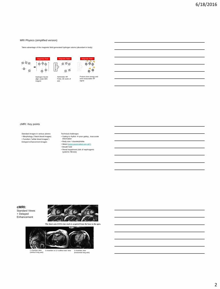
MRI Physics (simplified version)
Takes advantage of the magnetic field generated hydrogen atoms (abundant in body)
Magnetic Field
RF
Pu
lse
Administer RF
Pulse, tilt nuclei of
axis
h
h
h
h
h
h
Magnetic Field
Hydrogen Nuclei
align under MRI
magnet
h
h
h
h
h
Magnetic Field
Protons lose energy and
emit measurable RF
signal
cMRI: Key points
Standard images in various planes
• Morphology (“black blood images)
• Function (“white blood images”)
Delayed enhancement images
Technical challenges
• Gating to rhythm poor gating , inaccurate
information
• Body size / clausterphobia
• Metal (some pacemakers are ok!!)
• Breath hold
• Renal impairment (risk of nephrogenic
systemic fibrosis)
cMRI: Standard Views
+ Delayed
Enhancement
The short-axis (SAX) cine stack is acquired from the base to the apex.
2 chamber view
(vertical long axis)3 chamber or LV outflow tract view 4 chamber view
(horizontal long axis)
6/18/2016
3
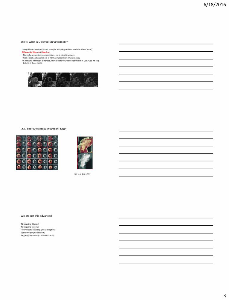
cMRI: What is Delayed Enhancement?
Late gadolinium enhancement (LGE) or delayed gadolinium enhancement (DGE)
Differential Washout Kinetics:
• Normally accumulates in interstitium, not in intact myocytes
• Gad enters and washes out of normal myocardium synchronously
• Cell injury, infiltration or fibrosis, increase the volume of distribution of Gad; Gad will lag
behind in these areas
LGE after Myocardial Infarction: Scar
Kim et al, Circ 1999
We are not this advanced
T1 Mapping (fibrosis)
T2 Mapping (edema)
Flow velocity encoding (measuring flow)
Spectroscopy (metablolism)
Tagging (regional myocardial function)
6/18/2016
4
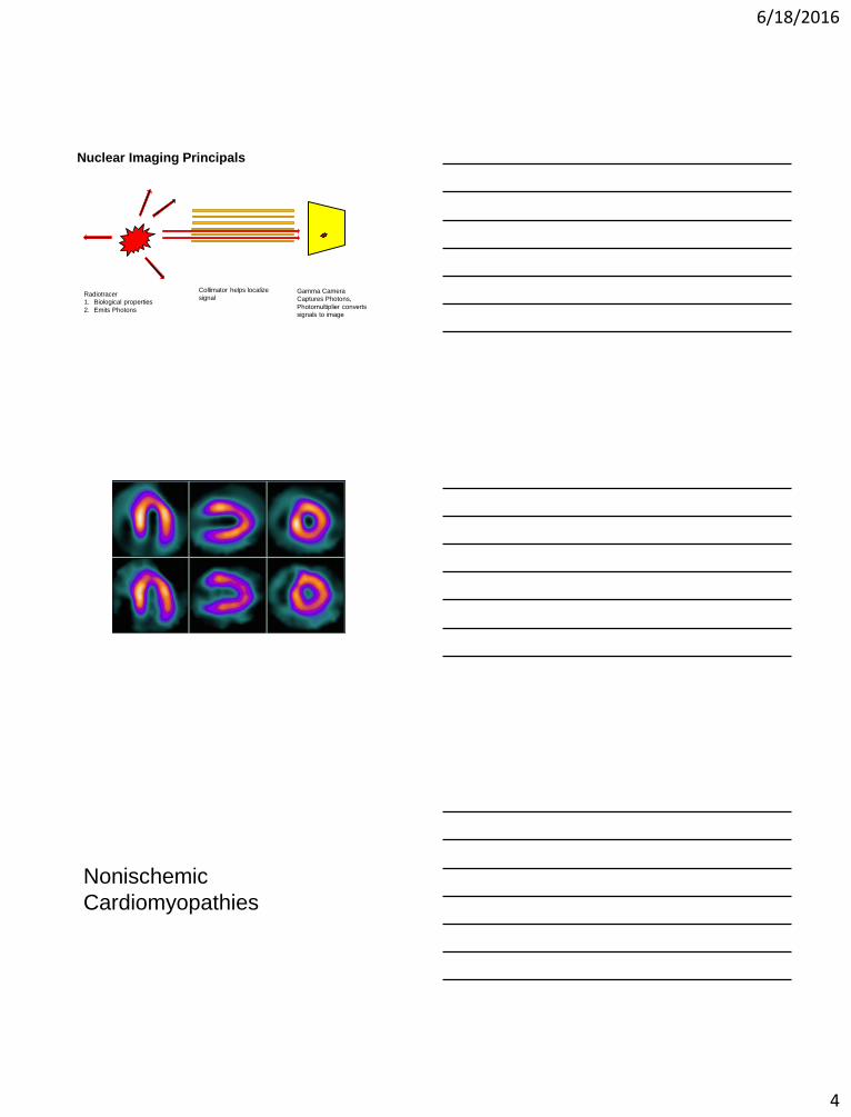
Nuclear Imaging Principals
Radiotracer
1. Biological properties
2. Emits Photons
Gamma Camera
Captures Photons,
Photomultiplier converts
signals to image
Collimator helps localize
signal
Nonischemic
Cardiomyopathies
6/18/2016
5
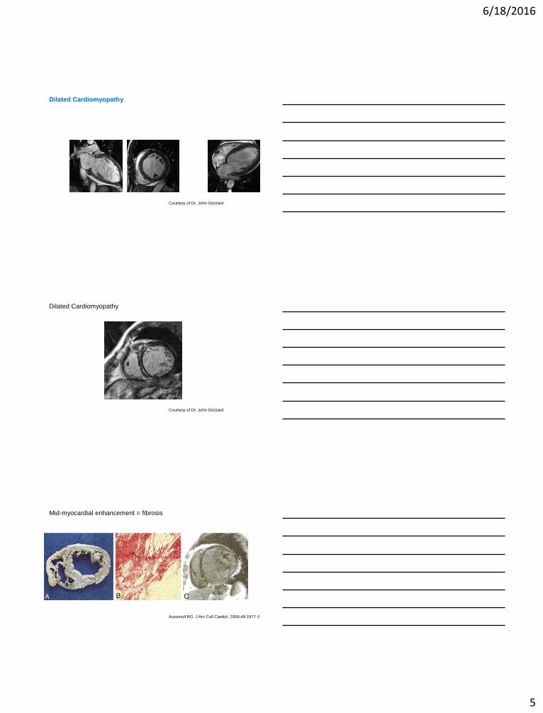
Dilated Cardiomyopathy
Courtesy of Dr. John Grizzard
Dilated Cardiomyopathy
Courtesy of Dr. John Grizzard
Mid-myocardial enhancement = fibrosis
Assomull RG. J Am Coll Cardiol. 2006;48:1977-1985.
6/18/2016
6
Midwall fibrosis:
• Larger ventricles
• Late stage disease
• Worse survival
• More SCD / VT
• More hospitalization
Assomull RG. J Am Coll Cardiol. 2006;48:1977-1985.
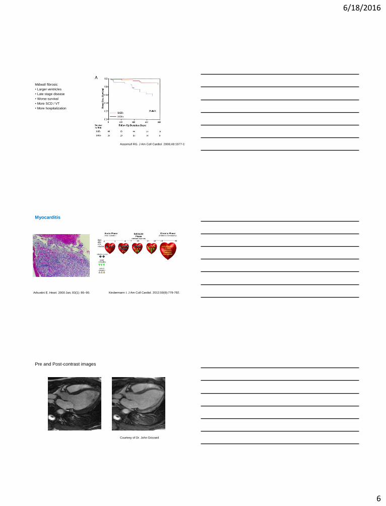
Myocarditis
Kindermann I. J Am Coll Cardiol. 2012;59(9):779-792.Arbustini E. Heart. 2000 Jan; 83(1): 86–90.
Pre and Post-contrast images
Courtesy of Dr. John Grizzard
6/18/2016
7
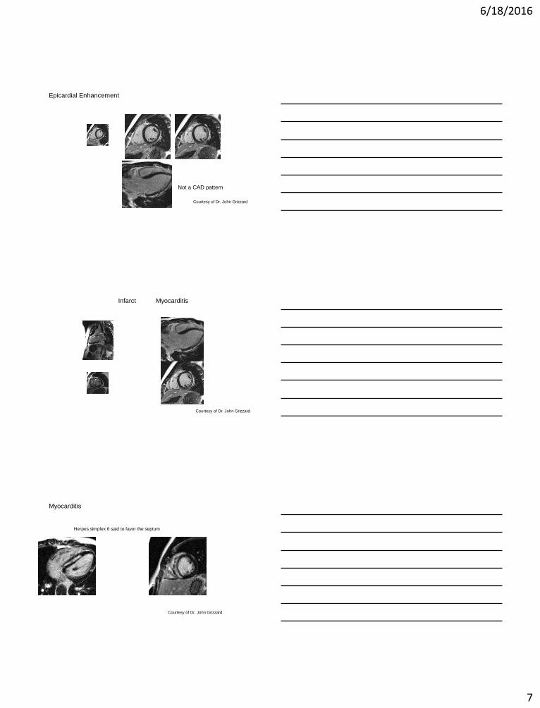
Epicardial Enhancement
Not a CAD pattern
Courtesy of Dr. John Grizzard
Infarct Myocarditis
Courtesy of Dr. John Grizzard
Myocarditis
Herpes simplex 6 said to favor the septum
Courtesy of Dr. John Grizzard
6/18/2016
8
Pre and Post-contrast images
Courtesy of Dr. John Grizzard
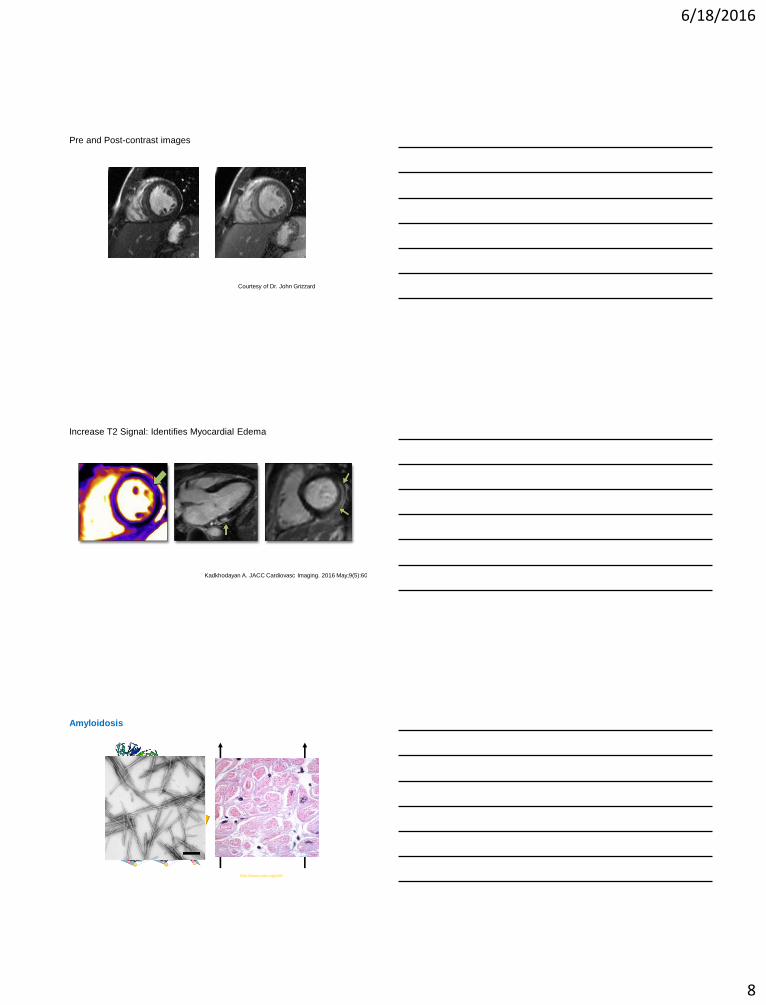
Increase T2 Signal: Identifies Myocardial Edema
Kadkhodayan A. JACC Cardiovasc Imaging. 2016 May;9(5):603-17.
http://www.rcsb.org/pdb/
Protofibrils
Β-pleated sheat
Protein
Amyloidosis
6/18/2016
9
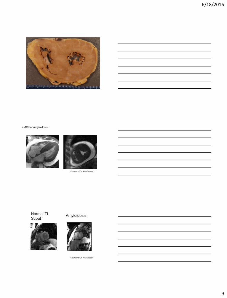
cMRI for Amyloidosis
Courtesy of Dr. John Grizzard
Normal TI
Scout Amyloid
Courtesy of Dr. John Grizzard
Amyloidosis
6/18/2016
10
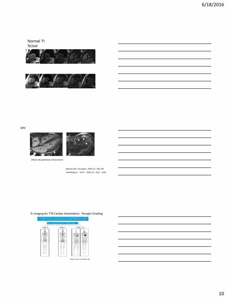
Normal TI
Scout
The blood pool “nulls” before the
myocardium
Amyloid
MRI
Diffuse late gadolinium enhancement
Maceira AM. Circulation. 2005;111: 186-193.
Vokelsberg H. . JACC. 2008; 51:: 1022 - 1030.
Tc imaging for TTR Cardiac Amyloidosis: Perugini Grading
Images courtesy of: James Gillmore, MD25
6/18/2016
11
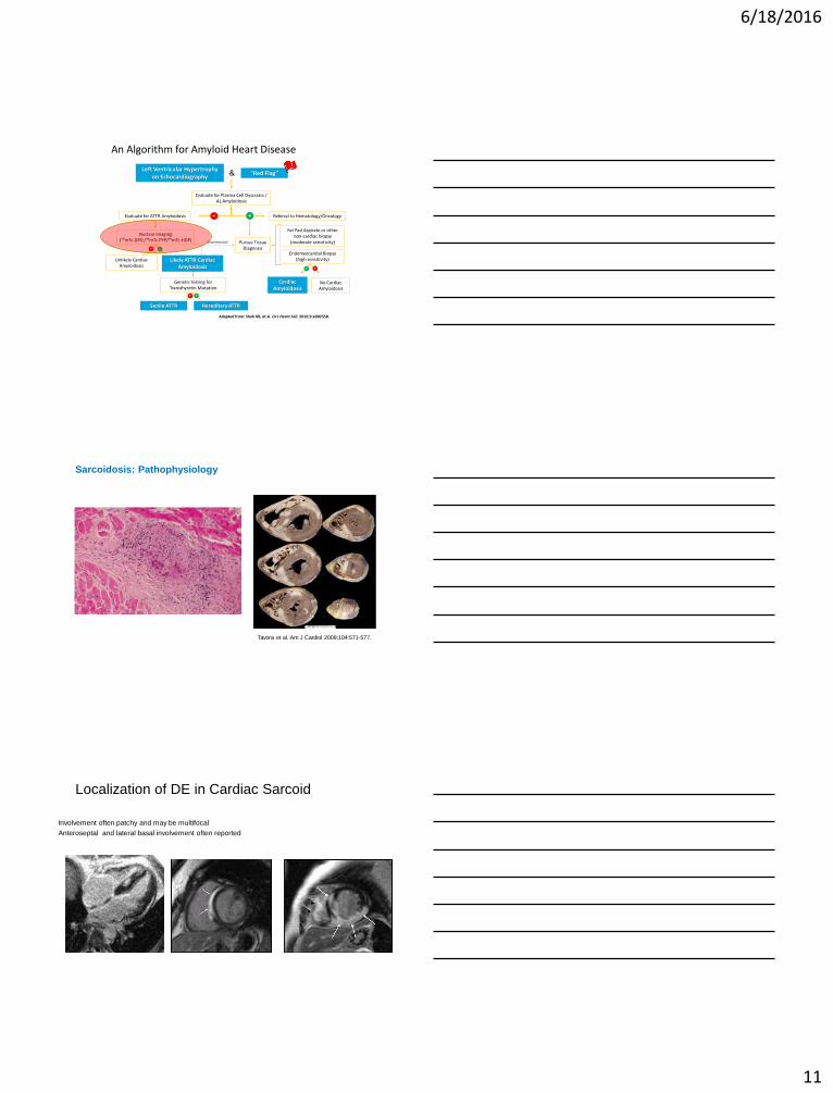
An Algorithm for Amyloid Heart Disease
Adapted from: Shah KB, et al. Circ Heart Fail. 2016;9:e002558.
Left Ventricular Hypertrophy on Echocardiography
Evaluate for Plasma Cell Dyscrasia / AL Amyloidosis
Evaluate for ATTR Amyloidosis
Unlikely Cardiac Amyloidosis
Nuclear Imaging(99mTc-DPD /99mTc-PYP/99mTc-HDP)
“Red Flag”
Referral to Hematology/Oncology
Likely ATTR Cardiac Amyloidosis
Genetic Testing for Transthyretin Mutation
Senile ATTR Hereditary ATTR
Cardiac Amyloidosis
No Cardiac Amyloidosis
(Discretionary) Pursue Tissue Diagnosis
Fat Pad Aspirate or other non-cardiac biopsy
(moderate sensitivity)
Endomyocardial Biopsy(high sensitivity)
&
- +
+-
-
-+
+
23
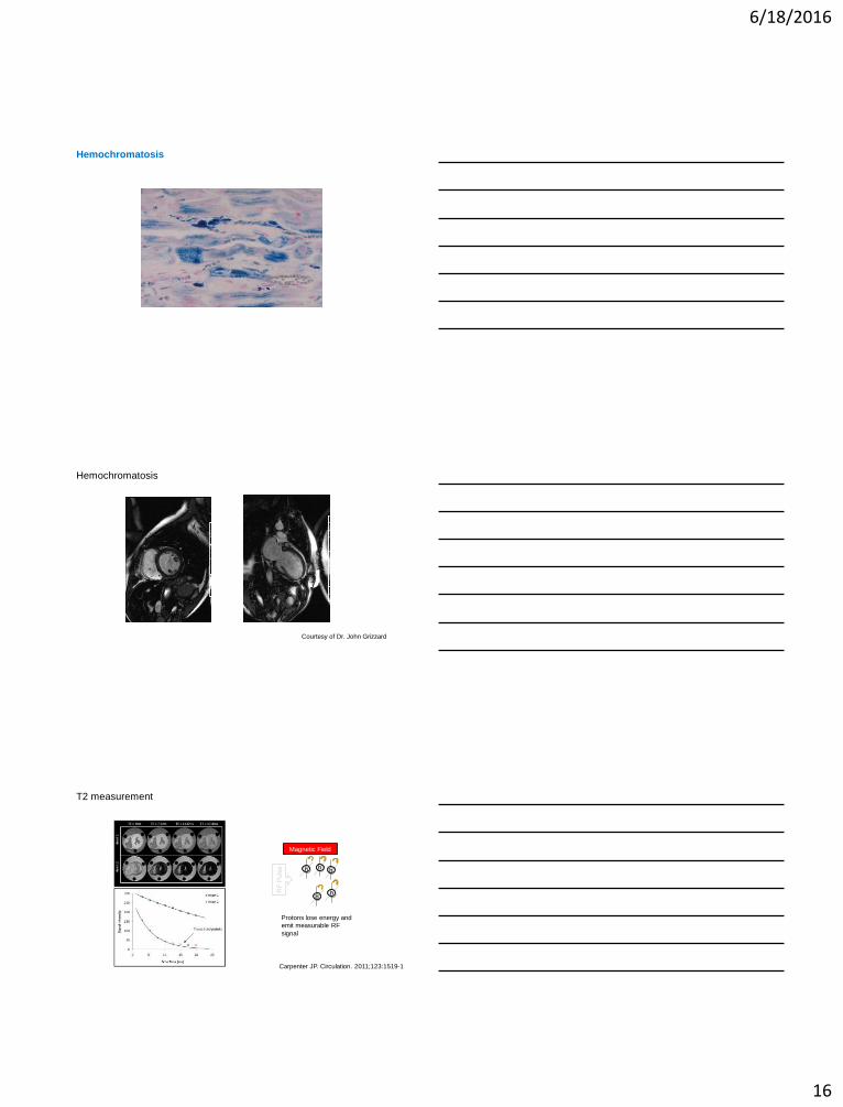
Sarcoidosis: Pathophysiology
Tavora et al. Am J Cardiol 2009;104:571-577.
Localization of DE in Cardiac Sarcoid
Involvement often patchy and may be multifocal
Anteroseptal and lateral basal involvement often reported
6/18/2016
12
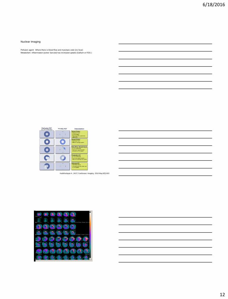
Nuclear Imaging
Pefusion agent: Where there is blood flow and myoctyes exist (no Scar)
Metabolism: Inflammation (active Sarcoid) has increased uptake (Gallium or FDG )
Kadkhodayan A. JACC Cardiovasc Imaging. 2016 May;9(5):603-17.
6/18/2016
13
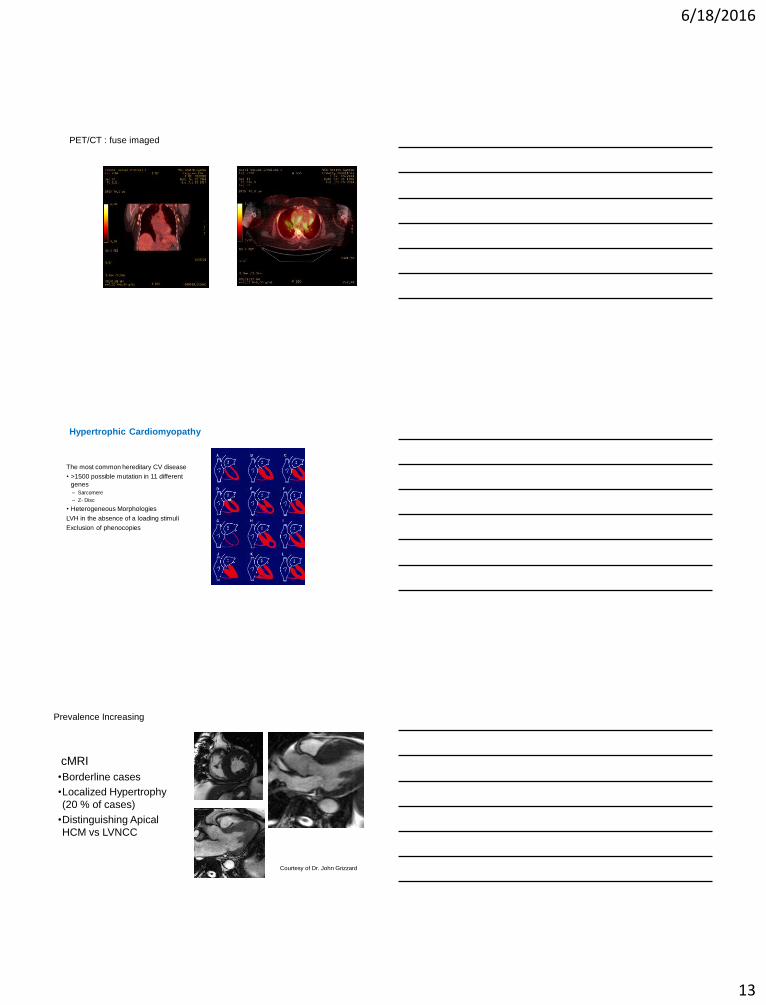
PET/CT : fuse imaged
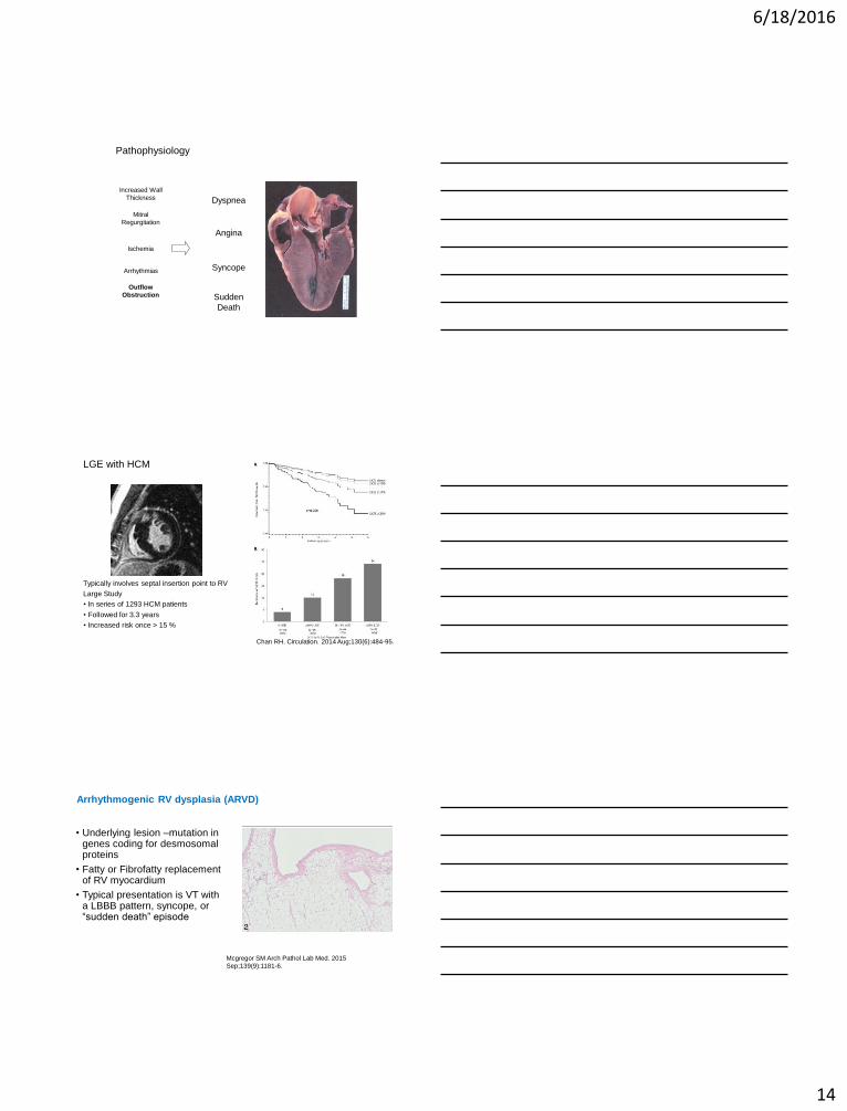
Hypertrophic Cardiomyopathy
The most common hereditary CV disease
• >1500 possible mutation in 11 different
genes
– Sarcomere
– Z- Disc
• Heterogeneous Morphologies
LVH in the absence of a loading stimuli
Exclusion of phenocopies
Prevalence Increasing
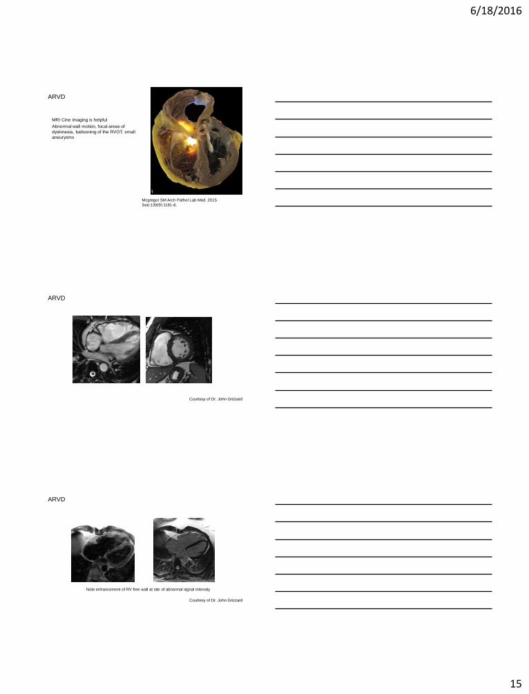
cMRI
•Borderline cases
•Localized Hypertrophy
(20 % of cases)
•Distinguishing Apical
HCM vs LVNCC
Courtesy of Dr. John Grizzard
6/18/2016
14
Pathophysiology
Dyspnea
Syncope
Sudden
Death
Increased Wall
Thickness
Outflow
Obstruction
Mitral
Regurgitation
Arrhythmias
Ischemia
Angina
LGE with HCM
Typically involves septal insertion point to RV
Large Study
• In series of 1293 HCM patients
• Followed for 3.3 years
• Increased risk once > 15 %
Chan RH. Circulation. 2014 Aug;130(6):484-95.
Arrhythmogenic RV dysplasia (ARVD)
• Underlying lesion –mutation in genes coding for desmosomalproteins
• Fatty or Fibrofatty replacement of RV myocardium
• Typical presentation is VT with a LBBB pattern, syncope, or “sudden death” episode
Mcgregor SM Arch Pathol Lab Med. 2015
Sep;139(9):1181-6.
6/18/2016
15
ARVD
MRI Cine imaging is helpful
Abnormal wall motion, focal areas of
dyskinesia, ballooning of the RVOT, small
aneurysms
Mcgregor SM Arch Pathol Lab Med. 2015
Sep;139(9):1181-6.
ARVD
Courtesy of Dr. John Grizzard
ARVD
Note enhancement of RV free wall at site of abnormal signal intensity
Courtesy of Dr. John Grizzard
6/18/2016
16
Hemochromatosis
Hemochromatosis
Courtesy of Dr. John Grizzard
T2 measurement
Carpenter JP. Circulation. 2011;123:1519-1528
h
h
h
h
h
Magnetic Field
Protons lose energy and
emit measurable RF
signal

6/18/2016
17
Thank you!!Questions?