potentially bioactive organotin(iv) compounds: synthesis, characterization, in vitro bioactivities...
TRANSCRIPT

lable at ScienceDirect
European Journal of Medicinal Chemistry 84 (2014) 343e363
Contents lists avai
European Journal of Medicinal Chemistry
journal homepage: http: / /www.elsevier .com/locate/ejmech
Original article
Potentially bioactive organotin(IV) compounds: Synthesis,characterization, in vitro bioactivities and interaction with SS-DNA
Muhammad Sirajuddin a, Saqib Ali a, *, Vickie McKee b, Manzar Sohail c, Hammad Pasha d
a Department of Chemistry, Quaid-i-Azam University, Islamabad 45320, Pakistanb Department of Chemistry, Loughborough University, Loughborough, Leics LE11 3TU, UKc Center of Excellence for Nanotechnology, King Fahd University of Petroleum and Minerals, Dhahran 31261, Saudi Arabiad Department of Biochemistry, Quaid-i-Azam University, Islamabad 45320, Pakistan
a r t i c l e i n f o
Article history:Received 14 December 2013Received in revised form1 July 2014Accepted 8 July 2014Available online 9 July 2014
Keywords:Organotin(IV) compoundDNA interactionAntitumor activityAntimicrobial activityCytotoxicityAntileishmanial activity
* Corresponding author.E-mail addresses: [email protected], msiraj09
[email protected] (S. Ali).
http://dx.doi.org/10.1016/j.ejmech.2014.07.0280223-5234/© 2014 Elsevier Masson SAS. All rights re
a b s t r a c t
Fourteen new organotin(IV) complexes with general formula R2SnL2 or R3SnL where R ¼ CH3, C2H5, C4H9,C6H5, C6H11, CH2eC6H5, C(CH3)3, C8H17 and L ¼ N-[(2-methoxyphenyl)]-4-oxo-4-[oxy]butanamide weresynthesized and characterized by elemental analyses, FT-IR, NMR (1H, 13C and 119Sn), mass spectrometryand single crystal X-ray structural analysis. Crystallographic data for four triorganotin(IV) complexes(R3SnL, R ¼ CH3, C2H5, C4H9, CH2eC6H5) showed the tin has approximate trigonal bipyramidal geometrywith the R groups in the trigonal plane. The carboxylate groups of ligands L bridge adjacent tin atoms,resulting in polymeric chains. In case of the diorganotin(IV) derivatives a six-coordinate geometry at thetin atom is proposed from spectroscopic evidence. The MeeSneMe bond angle in complex 7 wasdetermined from the 2J[119Sne1H] value as 166.3� that falls in the range of six-coordinate geometry. Theligand and its complexes (1e14) were screened for their antimicrobial, antitumor, cytotoxic and anti-leishmanial activities and found to be biologically active. The ligand and its complexes bind to DNAvia intercalative interactions resulting in hypochromism and minor bathochromic shifts as confirmed byUVevisible spectroscopy. Based on in vitro studies such as the potato disc method, the synthesizedcompounds were found to possess significant antitumor activity. Also, from cytotoxicity and DNAinteraction studies, these compounds can also be used for the prevention and treatment of cancer. Gelelectrophoresis assay was used to investigate the damage to double stranded super coiled plasmidpBR322 DNA by the synthesized compounds and compounds 1 and 7 were found to cause the maximumdamage. All the synthesized compounds exhibit strong antileishmanial activity that was even higherthan that of Amphotericin B, with significant cytotoxicity. This study, therefore, demonstrated the po-tential use of these compounds as source of novel agents for the treatment of leishmaniasis.
© 2014 Elsevier Masson SAS. All rights reserved.
1. Introduction and to avoid serious side-effects caused by platinum chemothera-
The chemistry of organotin compounds is gaining attention onaccount of their interesting structural features, schizonticidal,antimalarial, fungicidal activities and their potential as agriculturalbiocides [1]. Organotin(IV) compounds of carboxylic acids are beingextensively studied with special reference to their methods ofsynthesis, structural elucidation, and biological activity [2,3].Encouraged by the initial success of platinum chemotherapeuticmetallopharmaceuticals, concentration was first shifted to non-platinum chemotherapeutics starting from the basic cis-platinframework, with the aim to optimize the competence of such drugs
@gmail.com (M. Sirajuddin),
served.
peutics. Among these, organotins have appeared as biologicallyactive metallopharmaceuticals [4]. It has well been established thatorganotin(IV) compounds are very important in cancer chemo-therapy because of their apoptosis inducing character, while duringthe last few years it is noticeable that organotin compounds occupyan important place in cancer chemotherapy reports [5]. Recently,Blower described thirty interesting inorganic pharmaceuticals, fourof which are tin compounds [6]. The organotin compounds havealso received considerable attention as antiproliferative, antitumorand anticancer drugs. The activity is due to dissociated organo-tin(IV) moieties. The biological activity usually associates with thenature of organic ligand, since the organic ligand assists thetransportation of the complexes across the cell membrane [5,7e9].
The biological activity of organotin compounds is basicallydetermined by the number and nature of the organic groups bound
M. Sirajuddin et al. / European Journal of Medicinal Chemistry 84 (2014) 343e363344
to the central tin atom. The [R3Sn(IV)]þ and [Ar3Sn(IV)]þ derivativesexert powerful toxic action on the central nervous system. Withinthe series of [R3Sn(IV)]þ compounds, the lower homologues (Me,Et) are the most toxic when administrated orally, and the toxicityreduces progressively from propyl to octyl, the latter not being toxicat all [10]. The presence of easily hydrolysable groups (easilydissociable chelating ligands) producing intermediates such asRnSn(4�n)þ (n¼ 2 or 3) moieties, whichmay bind with DNA or high-affinity site of ATPase (histidine only), the low-affinity site ofATPase, and haemoglobins (histidine and cystine), play an impor-tant role in the determination of biological activity of the organotincompounds. Therefore, substantial attempts have been made tocharacterize model organotin compounds of ligands having heterodonor atoms (O, N and S), and simultaneously several studies havebeen focused on structureeactivity correlations during the last twodecades [11].
Keeping in view their biological applications, we are reporting aseries of 14 new organotin(IV) carboxylates of N-[(2-methoxyphenyl)]-4-oxo-4-[oxy]butanamide. These compoundswere characterized successfully by elemental analysis, FT-IR, NMR(1H, 13C, 119Sn) mass and single crystal analysis. They were screenedfor biological applications including interaction with SS-DNA,antimicrobial, antitumor, antileishmanial and cytotoxic activities.
2. Experimental section
2.1. Materials and methods
Reagents Me3SnCl, Bu3SnCl, Ph3SnCl, Cy3SnCl, Me2SnCl2,Bu2SnCl2, tert-Bu2SnCl2, Ph2SnCl2, n-Oct2SnO, o-anisidine, succinicanhydride were obtained from Aldrich (USA) and were usedwithout further purification. All the solvents purchased from E.Merck (Germany) were dried before use according to literatureprocedures [12]. Dibenzyltin dichloride (Bz2SnCl2) and tribenzyltinchloride (Bz3SnCl) were prepared according to the reportedmethod [13]. Sodium salt of Salmon fish sperm DNA (SS-DNA)(Arcos) was used as received. The melting points were determinedin a capillary tube using a Gallenkamp (UK) electrothermal meltingpoint apparatus. IR spectra in the range of 4000e100 cm�1 wereobtained on a Thermo Nicolet-6700 FT-IR Spectrophotometer.Elemental analysis was done using a CE-440 Elemental Analyzer(Exeter Analytical, Inc) and the experimentally found values aregiven in parenthesis in experimental part. 1H, 13C and 119Sn NMRwere recorded on a 400 MHz JEOL ECS instrument, using DMSO asan internal reference [1H (DMSO-d6) ¼ 2.50 and 13C (DMSO-d6) ¼ 39.5 ppm]. Tetramethylsilane (for 1H and 13C NMR) andMe4Sn (for 119Sn NMR) were used as external standards. For 119SnNMR the measurement was recorded at a working frequency of37.29 MHz and the chemical shift was referenced to Me4Sn as anexternal standard. Chemical shifts are given in ppm and couplingconstants (J) values are given in Hz. The multiplicities of signals in1H NMR are given with chemical shifts; (s ¼ singlet, d ¼ doublet,t¼ triplet, q¼ quartet, m¼multiplet). The absorption spectraweremeasured on a Shimadzu 1800 UVevisible Spectrophotometer. X-ray data for HL and complex 6 were collected at room temperaturewhile complexes 1 and 2 at 150 (2) K on a Bruker Apex II CCDdiffractometer. Complex 3 was collected at 190 (2) K on AgilentSuper Nova (Dual, Cu at zero, Eos) diffractometer. Details are givenin Table 2. All the non-hydrogen atoms were refined using aniso-tropic atomic displacement parameters, and hydrogen atomsbonded to carbon were inserted at calculated positions using ariding model. Hydrogen atoms bonded to O or N were located fromdifference maps and their coordinates refined. SHELXS-97 [14] wasused to solve and SHELX2012 [15] to refine the structures. Themassspectra were recorded on a Thermo Scientific executive (orbitrap)
utilizing an Advion TriVersa ™NanoMate sample introductionsystem. Them/z values were evaluated assuming that H¼ 1, C¼ 12,N ¼ 14, O ¼ 16, Cl ¼ 35, and Sn ¼ 120.
2.2. Synthesis
2.2.1. Synthesis of ligand: N-[(2-methoxyphenyl)]-4-oxo-4-[oxy]butanamide (HL)
Stoichiometric amounts of o-anisidine (2-methoxyaniline) andsuccinic anhydride were dissolved separately in glacial acetic acid.The o-anisidine solution was added slowly to the solution of suc-cinic anhydride and precipitate appeared [16] (Scheme 1). Theprecipitates were filtered, washed thoroughly with distilled water(to remove the unreacted succinic acid and succinic anhydride) andHCl (to remove unreacted aniline) and air dried. Prism-like whitesingle crystals of the HL used in X-ray diffraction studies weregrown in an acetone solution by slow evaporation of solvent atroom temperature.
Yield: 90%: M.p. 141e142 �C: Mol. Wt.: 223.23: Anal. Calc. forC11H13NO4: C, 59.2 (59.1); H, 5.9 (5.8); N, 6.3 (6.6): IR(4000e100 cm�1): 3200 n (OH); 3368 n (NH); 1708 n (amide C]O);1595 n (COOasym); 1289 n (COOsym); 306 (Dn); 1H NMR (DMSO-d6,400 MHz) d (ppm): 12.12 (s, 1H; OH); 2.67 (t, 2H, H2, 3J[1H,1H] ¼ 6.6 Hz); 2.53 (t, 2H, H3, 3J[1H, 1H] ¼ 6.6 Hz); 9.12 (s, 1H NH);7.98 (d, 1H, H6, 3J[1H, 1H] ¼ 7.8 Hz); 6.91 (dd, 1H, H8, 4J[1H,1H] ¼ 2.8 Hz); 7.08 (m, 2H, H7 and H9); 3.82 (s, 3H, H11): 13C NMR(DMSO-d6, 100 MHz) d (ppm): 174.4 (C1); 31.4 (C2); 29.4 (C3); 170.7(C4); 127.9 (C5); 122.1 (C6); 120.6 (C7); 124.5 (C8); 111.5 (C9); 149.8(C10); 56.1 (C11); ESI-MS, m/z (%): [C11H13NO4]þ 223 (30.1);[C11H11NO3]þ 205 (11.3); [C7H9NO]þ 123 (100); [C7H8O]þ 108(65.8); [C6H6N]þ 92 (5.5); [C5H5]þ 65 (7.1); [C3H3O]þ 55 (10.4).
2.2.2. Synthesis of sodium salt of ligand: sodium N-[(2-methoxyphenyl)]-4-oxo-4-[oxy]butanamide (NaL)
An aqueous solution of sodium hydrogen carbonate (NaHCO3)was added to a suspended solution of HL in distilled water. Themixture was stirred at room temperature to get a clear solutionwhich was then rotary evaporated to get the desired sodium salt ofthe ligand. The chemical reaction is shown in Scheme 1.
2.2.3. Synthesis of organotin(IV) complexes R3SnL with R ¼ Me (1),Bu (2), Ph (3), and [R2SnL2] with R ¼ Me (4), Bu (5)
Organotin(IV) carboxylates were synthesized by refluxing amixture of R3SnCl (5 mmol) or R2SnCl2 (2.5 mmol) and the sodiumsalt of ligand NaL (5 mmol) in dry toluene for 8 h (Scheme 2). Therefluxed solution was kept overnight at room temperature. TheNaCl precipitate was removed by filtration and the solvent wasremoved under reduced pressure. The product was purified byrecrystallization from chloroform at room temperature. Thenumbering of HL and alkyl groups attached to Sn is given inScheme 3.
2.2.4. N-[(2-methoxyphenyl)]-4-oxo-4-[(trimethylstannyl)oxy]butanamide (1)
Yield: 83%: M.p. 115e117 �C: Mol. Wt.: 386.03: Anal. Calc. forC14H21NO4Sn: C, 43.6 (43.2); H, 5.5 (5.2); N, 3.6 (3.7): IR(4000e100 cm�1): 3418 n (NH); 1689 n (amide C]O); 1557 n
(COOasym); 1394 n (COOsym); 146 (Dn); 546 n (SneC); 455 n (SneO):1H NMR (DMSO-d6, 400 MHz) d (ppm): 2.55 (t, 2H, H2, 3J[1H,1H]¼ 6.8Hz); 2.36 (t, 2H,H3, 3J[1H,1H]¼ 6.8Hz); 9.04 (s,1HNH); 8.02(d, 1H, H6, 3J[1H, 1H] ¼ 7.8 Hz); 6.90 (dd, 1H, H8, 4J[1H, 1H] ¼ 2.8 Hz);7.03 (m, 2H, H7 and H9); 3.81 (s, 3H, H11); 0.38 (s, 3H, Ha, 2J[119/117Sne1Ha] ¼ 70, 68 Hz): 13C NMR (DMSO-d6, 100 MHz) d (ppm):176.5 (C1);33.1 (C2); 31.7 (C3); 171.4 (C4); 128.1 (C5);121.7 (C6); 120.6(C7); 124.2 (C8); 111.4 (C9); 149.6 (C10); 56.0 (C11); 0.7 (Ca, 1J[119/
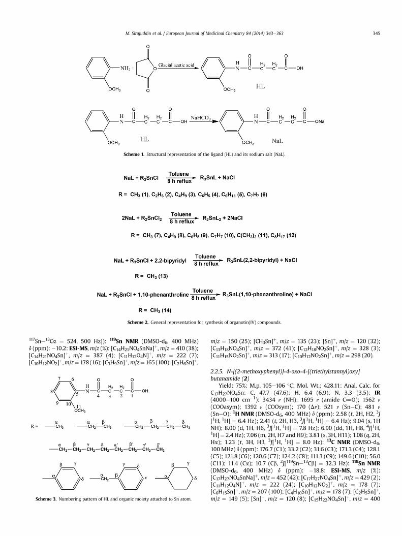
Scheme 1. Structural representation of the ligand (HL) and its sodium salt (NaL).
Scheme 2. General representation for synthesis of organotin(IV) compounds.
M. Sirajuddin et al. / European Journal of Medicinal Chemistry 84 (2014) 343e363 345
117Sne13Ca ¼ 524, 500 Hz]): 119Sn NMR (DMSO-d6, 400 MHz)d (ppm):�10.2: ESI-MS,m/z (%): [C14H21NO4SnNa]þ,m/z¼ 410 (38);[C14H21NO4Sn]þ, m/z ¼ 387 (4); [C11H12O4N]þ, m/z ¼ 222 (7);[C10H12NO2]þ,m/z¼ 178 (16); [C3H9Sn]þ,m/z¼ 165 (100); [C2H6Sn]þ,
Scheme 3. Numbering pattern of HL and organic moiety attached to Sn atom.
m/z ¼ 150 (25); [CH3Sn]þ, m/z ¼ 135 (23); [Sn]þ, m/z ¼ 120 (32);[C13H18NO4Sn]þ, m/z ¼ 372 (41); [C12H18NO2Sn]þ, m/z ¼ 328 (3);[C11H15NO2Sn]þ,m/z ¼ 313 (17); [C10H12NO2Sn]þ,m/z ¼ 298 (20).
2.2.5. N-[(2-methoxyphenyl)]-4-oxo-4-[(triethylstannyl)oxy]butanamide (2)
Yield: 75%: M.p. 105e106 �C: Mol. Wt.: 428.11: Anal. Calc. forC17H27NO4Sn: C, 47.7 (47.6); H, 6.4 (6.9); N, 3.3 (3.5): IR(4000e100 cm�1): 3434 n (NH); 1695 n (amide C]O); 1562 n
(COOasym); 1392 n (COOsym); 170 (Dn); 521 n (SneC); 481 n
(SneO): 1H NMR (DMSO-d6, 400 MHz) d (ppm): 2.58 (t, 2H, H2, 3J[1H, 1H] ¼ 6.4 Hz); 2.41 (t, 2H, H3, 3J[1H, 1H] ¼ 6.4 Hz); 9.04 (s, 1HNH); 8.00 (d, 1H, H6, 3J[1H, 1H] ¼ 7.8 Hz); 6.90 (dd, 1H, H8, 4J[1H,1H]¼ 2.4 Hz); 7.06 (m, 2H, H7 and H9); 3.81 (s, 3H, H11); 1.08 (q, 2H,Ha); 1.23 (t, 3H, Hb, 3J[1H, 1H] ¼ 8.0 Hz): 13C NMR (DMSO-d6,100 MHz) d (ppm): 176.7 (C1); 33.2 (C2); 31.6 (C3); 171.3 (C4); 128.1(C5); 121.8 (C6); 120.6 (C7); 124.2 (C8); 111.3 (C9); 149.6 (C10); 56.0(C11); 11.4 (Ca); 10.7 (Cb, 2J[119Sne13Cb] ¼ 32.3 Hz): 119Sn NMR(DMSO-d6, 400 MHz) d (ppm): �18.8: ESI-MS, m/z (%):[C17H27NO4SnNa]þ,m/z¼ 452 (42); [C17H27NO4Sn]þ,m/z¼ 429 (2);[C11H12O4N]þ, m/z ¼ 222 (24); [C10H12NO2]þ, m/z ¼ 178 (7);[C6H15Sn]þ,m/z ¼ 207 (100); [C4H10Sn]þ,m/z ¼ 178 (7); [C2H5Sn]þ,m/z ¼ 149 (5); [Sn]þ, m/z ¼ 120 (8); [C15H22NO4Sn]þ, m/z ¼ 400

M. Sirajuddin et al. / European Journal of Medicinal Chemistry 84 (2014) 343e363346
(98); [C14H22NO2Sn]þ, m/z ¼ 356 (4); [C12H17NO2Sn]þ, m/z ¼ 327(2); [C10H12NO2Sn]þ, m/z ¼ 298 (15).
2.2.6. N-[(2-methoxyphenyl)]-4-oxo-4-[(tributylstannyl)oxy]butanamide (3)
Yield: 76%: M.p. 52e54 �C: C23H39NO4Sn Mol. Wt.: 512.27: Anal.Calc. for C17H27NO4Sn: C, 53.9 (53.8); H, 7.7 (7.4); N, 2.7 (2.9): IR(4000e100 cm�1): 3426 n (NH); 1694 n (amide C]O); 1552 n
(COOasym); 1372 n (COOsym); 180 (Dn); 569 n (SneC); 455 n
(SneO): 1H NMR (DMSO-d6, 400 MHz) d (ppm): 2.55 (t, 2H, H2, 3J[1H, 1H] ¼ 6.8 Hz); 2.38 (t, 2H, H3, 3J[1H, 1H] ¼ 6.8 Hz); 9.00 (s, 1HNH); 8.02 (d, 1H, H6, 3J[1H, 1H] ¼ 7.8 Hz); 6.88 (dd, 1H, H8, 4J[1H,1H]¼ 2.4 Hz); 7.02 (m, 2H, H7 and H9); 3.81 (s, 3H, H11); 1.07 (t, 3H,Ha, 3J[1H, 1H] ¼ 8.0 Hz); 1.56 (m, 2H, Hb); 1.33 (m, 2H, Hg); 0.87 (t,3H, Hd, 3J[1H, 1H]¼ 6.8 Hz): 13C NMR (DMSO-d6, 100 MHz) d (ppm):176.4 (C1); 33.2 (C2); 31.7 (C3); 171.3 (C4); 128.2 (C5); 121.6 (C6);120.5 (C7); 124.1 (C8); 111.3 (C9); 149.5 (C10); 56.0 (C11); 19.1 (Ca);28.1 (Cb, 2J[119Sne13Cb] ¼ 27 Hz) 26.9 (Cg, 3J[119Sne13Cg] ¼ 75 Hz);14.1 (Cd): 119Sn NMR (DMSO-d6, 400 MHz) d (ppm): �17.6. ESI-MS,m/z (%): [C23H39NO4SnNa]þ, m/z ¼ 536 (27); [C23H39NO4Sn]þ, m/z¼ 513 (20); [C11H12NO4]þ,m/z¼ 222 (8); [C10H12NO2]þ,m/z ¼ 178(11); [C12H27Sn]þ, m/z ¼ 291 (100); [C8H18Sn]þ, m/z ¼ 234 (26);[C4H9Sn]þ, m/z ¼ 177 (20); [Sn]þ, m/z ¼ 120 (12); [C19H30NO4Sn]þ,m/z¼ 456 (22); [C18H30NO2Sn]þ,m/z¼ 412 (7); [C14H21NO2Sn]þ,m/z ¼ 355 (12); [C10H12NO2Sn]þ, m/z ¼ 298 (3).
2.2.7. N-[(2-methoxyphenyl)]-4-oxo-4-[(triphenylstannyl)oxy]butanamide (4)
Yield: 87%: M.p. 102e103 �C: Mol. Wt.: 572.24: Anal. Calc. forC29H27NO4Sn: C, 60.9 (60.5); H, 4.8 (4.2); N, 2.5 (2.8): IR(4000e100 cm�1): 3425 n (NH); 1645 n (amide C]O); 1545 n
(COOasym); 1382 n (COOsym); 163 (Dn); 267 n (SneC); 444 n
(SneO): 1H NMR (DMSO-d6, 400 MHz) d (ppm): 2.51 (t, 2H, H2, 3J[1H, 1H] ¼ 6.4 Hz); 2.39 (t, 2H, H3, 3J[1H, 1H] ¼ 6.4 Hz); 9.02 (s, 1HNH); 8.00 (d, 1H, H6, 3J[1H, 1H] ¼ 7.8 Hz); 6.91 (dd, 1H, H8, 4J[1H,1H]¼ 2.4 Hz); 7.05 (m, 2H, H7 and H9); 3.77 (s, 3H, H11); 7.91 (m,15H, SnePh): 13C NMR (DMSO-d6, 100 MHz) d (ppm): 176.3 (C1); 32.9(C2); 31.6 (C3); 171.2 (C4); 128.1 (C5); 121.8 (C6); 120.6 (C7); 124.3(C8); 111.4 (C9); 149.6 (C10); 56.0 (C11); 143.5 (Ca); 136.7 (Cb, 2J[119Sne13Cb] ¼ 46 Hz); 128.6 (Cg, 3J[119Sne13Cd] ¼ 63 Hz); 129.4(Cd): 119Sn NMR (DMSO-d6, 400MHz) d (ppm):�246.9: ESI-MS,m/z(%): [C29H27NO4SnNa]þ,m/z¼ 596 (15); [C29H27NO4Sn]þ,m/z¼ 573(13); [C11H12NO4]þ, m/z ¼ 222 (9); [C10H12NO2]þ, m/z ¼ 178 (10);[C18H15Sn]þ, m/z ¼ 351 (100); [C12H10Sn]þ, m/z ¼ 274 (25);[C4H9Sn]þ, m/z ¼ 197 (23); [Sn]þ, m/z ¼ 120 (10); [C23H22NO4Sn]þ,m/z ¼ 496 (12); [C22H22NO2Sn]þ, m/z ¼ 452 (24); [C16H17NO2Sn]þ,m/z ¼ 375 (21); [C10H12NO2Sn]þ, m/z ¼ 298 (35).
2.2.8. N-[(2-methoxyphenyl)]-4-oxo-4-[(tricyclohexylstannyl)oxy]butanamide (5)
Yield: 84%: M.p. 85e87 �C: Mol. Wt.: 590.38: Anal. Calc.C29H45NO4Sn: C, 59.0 (58.7); H, 7.7 (7.5); N, 2.4 (2.6): IR(4000e100 cm�1): 3426 n (NH); 1694 n (amide C]O); 1577 n
(COOasym); 1393 n (COOsym); 184 (Dn); 569 n (SneC); 455 n
(SneO): 1H NMR (DMSO-d6, 400 MHz) d (ppm): 2.58 (t, 2H, H2, 3J[1H, 1H] ¼ 6.4); 2.46 (t, 2H, H3, 3J[1H, 1H] ¼ 6.4); 9.05 (s, 1H NH);8.04 (d, 1H, H6, 3J[1H, 1H] ¼ 7.8 Hz); 6.88 (dd, 1H, H8, 4J[1H,1H] ¼ 2.4 Hz); 7.01 (m, 2H, H7 and, H9); 3.82 (s, 3H, H11); 1.19 (m,1H, Ha); 1.84 (m, 2H, Hb); 1.62 (m, 2H, Hg); 1.58 (m, 2H, Hd): 13CNMR (DMSO-d6, 100 MHz) d (ppm): 176.8 (C1); 33.2 (C2); 31.4 (C3);171.2 (C4); 128.2 (C5); 121.6 (C6); 120.5 (C7); 124.1 (C8); 111.3 (C9);149.5 (C10); 56.0 (C11); 27.1 (Ca, 1J[119Sne13Ca]¼ 512 Hz); 29.2 (Cb,2J[119Sne13Cb] ¼ 72 Hz) 36.1 (Cg) 30.9 (Cd, 4J[119Sne13Cg] ¼ 47 Hz):119Sn NMR (DMSO-d6, 400 MHz) d (ppm): �250.4: ESI-MS,m/z (%):[C29H45NO4SnNa]þ, m/z ¼ 614 (22); [C29H45NO4Sn]þ, m/z ¼ 591
(15); [C11H12NO4]þ, m/z ¼ 222 (27); [C10H12NO2]þ, m/z ¼ 178 (18);[C18H33Sn]þ, m/z ¼ 369 (100); [C12H22Sn]þ, m/z ¼ 287 (15);[C6H11Sn]þ,m/z ¼ 197 (29); [Sn]þ,m/z ¼ 120 (15); [C23H34NO4Sn]þ,m/z ¼ 508 (23); [C22H34NO2Sn]þ, m/z ¼ 464 (21); [C16H23NO2Sn]þ,m/z ¼ 381 (14); [C10H12NO2Sn]þ, m/z ¼ 298 (18).
2.2.9. N-[(2-methoxyphenyl)]-4-oxo-4-[(tribenzylstannyl)oxy]butanamide (6)
Yield: 81%: M.p. 112e114 �C: Anal. Calc. C32H33NO4Sn: Mol. Wt.:614.3: C, 62.6; H, 5.4 (5.3); N, 2.3 (2.3): IR (4000e100 cm�1): 3426 n
(NH); 1685 n (amide C]O); 1559 n (COOasym); 1357 n (COOsym);202 (Dn); 542 n (SneC); 447 n (SneO): 1H NMR (DMSO-d6,400 MHz) d (ppm): 2.46 (t, 2H, H2, 3J[1H, 1H] ¼ 6.8 Hz); 2.31 (t, 2H,H3, 3J[1H, 1H] ¼ 6.8 Hz); 8.96 (s, 1H NH); 7.94 (d, 1H, H6, 3J[1H,1H] ¼ 8.0 Hz); 6.97 (dd, 1H, H8, 4J[1H, 1H]¼ 3.6 Hz); 7.44 (m, 2H, H7and H9); 3.73 (s, 3H, H11); 2.05 (s, 2H, Ha, 2J[119Sne1H] ¼ 61); 7.14(d, 1H, Hg, 3J[1H, 1H] ¼ 7.6 Hz); 7.21 (t, 2H, Hd, 3J[1H, 1H] ¼ 8.0 Hz);6.84 (t, 1H, Hε, 3J[1H, 1H] ¼ 8.0 Hz): 13C NMR (DMSO-d6, 100 MHz)d (ppm): 176.3 (C1); 31.5 (C2); 30.5 (C3); 171.2 (C4); 128.2 (C5);121.9 (C6); 120.7 (C7); 124.3 (C8); 111.5 (C9); 149.7 (C10); 56.1(C11); 21.6 (Ca, 1J[119Sne13Ca] ¼ 483 Hz); 143.7 (Cb, 2J[119Sne13Cb]¼ 72 Hz), 128.7 (Cg, 3J[119Sne13Cg]¼ 62 Hz), 136.8 (Cd,4J[119Sne13Cd] ¼ 46 Hz), 125.9 (Cε): 119Sn NMR (DMSO-d6,400MHz) d (ppm):�256.9: ESI-MS,m/z (%): [C32H33NO4SnNa]þ,m/z ¼ 638 (60); [C32H33NO4Sn]þ, m/z ¼ 615 (15); [C32H33NO4Sn]þ, m/z¼ 616 (98); [C11H12NO4]þ,m/z¼ 222 (16); [C9H12NO2]þ,m/z¼ 178(23); [C32H32Sn]þ, m/z ¼ 393 (100); [C12H22Sn]þ, m/z ¼ 302 (23);[C6H11Sn]þ,m/z¼ 211 (9); [Sn]þ,m/z¼ 120 (5); [C25H26NO4Sn]þ,m/z ¼ 524 (21); [C25H26NO2Sn]þ, m/z ¼ 480 (26); [C16H17NO2Sn]þ, m/z ¼ 389 (3); [C10H11N2O4Sn]þ, m/z ¼ 343 (21).
2.2.10. Dimethylstannanediyl bis(4-(2-methoxyphenylamino)-4-oxobutanoate) (7)
Yield: 870%: M.p. 120e122 �C: Mol. Wt.: 593.21: Anal. Calc. forC24H30N2O8Sn: C, 48.6 (62.6); H, 5.1 (5.3); N, 4.7 (4.4): IR(4000e100 cm�1): 3253 n (NH); 1664 n (amide C]O); 1526 n
(COOasym); 1386 n (COOsym); 140 (Dn); 577 n (SneC); 450 n (SneO):1H NMR (DMSO-d6, 400 MHz) d (ppm): 2.66 (t, 2H, H2, 3J[1H,1H]¼ 6.6); 2.50 (t, 2H, H3, 3J[1H, 1H]¼ 6.6); 9.11 (s, 1H NH); 8.0 (d, 3J[1H, 1H]¼ 7.8 Hz, 1H, H6); 6.91 (dd, 1H, H8, 4J[1H, 1H]¼ 2.4 Hz); 7.07(m, 2H, H7 and H9); 3.82 (s, 3H, H11); 0.76 (s, 3H, Ha) [119/117Sne1H] ¼ 102, 100 Hz): 13C NMR (DMSO-d6, 100 MHz) d (ppm):178.8 (C1); 32.1 (C2); 30.1 (C3); 170.8 (C4); 128.0 (C5); 121.9 (C6);120.6 (C7); 124.4 (C8); 111.4 (C9); 149.7 (C10); 56.0 (C11); 12.7 (Ca):119Sn NMR (DMSO-d6, 400 MHz) d (ppm): �281.7: ESI-MS, m/z (%):[C24H30N2O8SnNa]þ, m/z ¼ 617 (34); [C24H30N2O8Sn]þ, m/z ¼ 594(20); [C23H27N2O8Sn]þ, m/z ¼ 579 (18); [C12H15NO4Sn]þ, m/z ¼ 357(19); [C11H12NO4Sn]þ,m/z¼ 342 (20); [C11H12NO4]þ,m/z¼ 222 (11);[C9H12NO2]þ,m/z¼178 (13); [C2H6Sn]þ,m/z¼150 (10); [CH3Sn]þ,m/z¼ 135 (14); [Sn]þ,m/z¼ 120 (4); [C13H18NO4Sn]þ,m/z¼ 372 (100);[C12H18NO2Sn]þ, m/z ¼ 328 (21); [C11H15NO2Sn]þ, m/z ¼ 313 (27);[C10H12NO2Sn]þ, m/z ¼ 298 (25); [C22H27N2O6Sn]þ, m/z ¼ 535 (12).
2.2.11. Dibutylstannanediyl bis(4-(2-methoxyphenylamino)-4-oxobutanoate) (8)
Yield: 80%: M.p. 115e117 �C: Mol. Wt.: 677.37: Anal. Calc. forC30H42N2O8Sn: C, 53.2 (53.5); H, 6.3 (6.6); N, 4.1 (4.5): IR(4000e100 cm�1): 3312 n (NH); 1698 n (amide C]O); 1525 n
(COOasym); 1391 n (COOsym); 134 (Dn); 571 n (SneC); 477 n (SneO):1H NMR (DMSO-d6, 400 MHz) d (ppm): 2.65 (t, 2H, H2, 3J[1H,1H]¼ 6.4); 2.52 (t, 2H, H3, 3J[1H, 1H]¼ 6.4); 9.10 (s,1HNH); 8.04 (d, 3J[1H, 1H]¼ 7.5 Hz,1H, H6); 6.89 (dd, 1H, H8, 4J[1H, 1H]¼ 2.4 Hz); 7.06(m, 2H, H7 and H9); 3.82 (s, 3H, H11); 1.20 (t, 3H, Ha, 3J(1H,1H)¼ 7.2Hz); 1.46 (m, 2H,Hb); 1.35 (m, 2H,Hg); 0.77 (t, 3H,Hd, 3J[1H,1H]¼7.2Hz): 13CNMR (DMSO-d6,100MHz) d (ppm): 179.5 (C1); 32.2

M. Sirajuddin et al. / European Journal of Medicinal Chemistry 84 (2014) 343e363 347
(C2); 29.9 (C3); 170.8 (C4); 128.0 (C5); 121.8 (C6); 120.5 (C7); 124.3(C8); 111.3 (C9); 149.6 (C10); 56.0 (C11): 21.4 (Ca); 28.1 (Cb, 2J[119Sne13Cb ¼ 27 Hz); 26.2 (Cg, 3J[119Sne13Cg] ¼ 75 Hz) 14.1 (Cd):119Sn NMR (DMSO-d6, 400 MHz) d (ppm): �296.7: ESI-MS, m/z (%):[C30H42N2O8SnNa]þ, m/z ¼ 701 (54); [C30H42N2O8Sn]þ, m/z ¼ 678(12); [C26H33N2O8Sn]þ, m/z ¼ 621 (7); [C15H21NO4Sn]þ, m/z ¼ 399(8); [C11H12NO4Sn]þ, m/z ¼ 342 (4); [C11H12NO4]þ, m/z ¼ 222 (11);[C10H12NO2]þ,m/z¼ 178 (12); [C8H18Sn]þ,m/z¼ 234 (7); [C4H9Sn]þ,m/z ¼ 177 (100); [Sn]þ, m/z ¼ 120 (12); [C19H30NO4Sn]þ, m/z ¼ 456(24); [C18H30NO2Sn]þ,m/z¼ 412 (6); [C14H21NO2Sn]þ,m/z¼ 355 (5);[C10H12NO2Sn]þ,m/z ¼ 298 (5); [C25H33N2O6Sn]þ,m/z ¼ 577 (4).
2.2.12. Diphenylstannanediyl bis(4-(2-methoxyphenylamino)-4-oxobutanoate) (9)
Yield: 78%: M.p. 121e123 �C: Mol. Wt.: 717.35: Anal. Calc. forC34H34N2O8Sn: C, 56.9 (56.2); H, 4.8 (4.4); N, 3.9 (3.6): IR(4000e100 cm�1): 3277 n (NH); 1646 n (amide C]O); 1541 n
(COOasym); 1381 n (COOsym); 160 (Dn); 280 n (SneC); 451 n
(SneO): 1H NMR (DMSO-d6, 400 MHz) d (ppm): 2.50 (t, 2H, H2, 3J[1H, 1H] ¼ 6.6); 2.36 (t, 2H, H3, 3J[1H, 1H] ¼ 6.6); 9.02 (s, 1H NH);8.00 (d, 3J[1H, 1H] ¼ 7.5 Hz, 1H, H6); 6.91 (dd, 1H, H8, 4J[1H,1H]¼ 2.2); 7.07 (m, 2H, H7 and H9); 3.77 (s, 3H, H11); 7.90 (m, 10 H,SnePh): 13C NMR (DMSO-d6, 100 MHz) d (ppm): 176.3 (C1); 32.9(C2); 31.7 (C3); 171.2 (C4); 128.0 (C5); 121.9 (C6); 120.6 (C7); 124.3(C8); 111.4 (C9); 149.6 (C10); 56.0 (C11): 143.6 (Ca), 136.7 (Cb, 2J[119Sne13Cb] ¼ 45.8 Hz), 128.6 (Cg, 3J[119/117Sne13Cg] ¼ 74, 68),129.4 (Cd, 4J[119Sne13Cd] ¼ 63 Hz]): 119Sn NMR (DMSO-d6,400 MHz) d (ppm): �256.7: ESI-MS, m/z (%): [C34H34N2O8SnNa]þ,m/z¼ 741 (20); [C34H34N2O8Sn]þ,m/z¼ 718 (12); [C28H29N2O8Sn]þ,m/z¼ 641 (4); [C17H17NO4Sn]þ,m/z ¼ 419 (10); [C11H12NO4Sn]þ,m/z ¼ 342 (11); [C11H12NO4]þ, m/z ¼ 222 (10); [C10H12NO2]þ, m/z ¼ 178 (12); [C12H10Sn]þ, m/z ¼ 274 (13); [C6H5Sn]þ, m/z ¼ 197(100); [Sn]þ, m/z ¼ 120 (9); [C23H22NO4Sn]þ, m/z ¼ 496 (12);[C22H22NO2Sn]þ, m/z ¼ 452 (9); [C16H17NO2Sn]þ, m/z ¼ 375 (7);[C10H12NO2Sn]þ, m/z ¼ 298 (3); [C27H29N2O6Sn]þ, m/z ¼ 597 (19).
2.2.13. Dibenzylstannanediyl bis[4-(2-methoxyphenylamino)-4-oxobutanoate] (10)
Yield: 70%: M.p. 131e133 �C: Mol. Wt.: 745.41: Anal. Calc. forC36H38N2O8Sn: C, 58.0 (58.2); H, 5.1 (4.9); N, 3.8 (3.6): IR(4000e100 cm�1): 3299 n (NH); 1665 n (amide C]O); 1530 n
(COOasym); 1371 n (COOsym); 159 (Dn); 538 n (SneC); 444 n
(SneO): 1H NMR (DMSO-d6, 400 MHz) d (ppm): 2.71 (t, 2H, H2, 3J[1H, 1H] ¼ 6.8 Hz); 2.60 (t, 2H, H3, 3J[1H, 1H] ¼ 6.8 Hz); 9.01 (s, 1HNH); 8.00 (d, 3J[1H, 1H] ¼ 7.6 Hz, 1H, H6); 6.91 (dd, 1H, H8, 4J[1H,1H]¼ 1.6 Hz); 7.01 (m, 2H, H7 and H9); 3.83 (s, 3H, H11); 1.18 (s, 2H,Ha); 7.95 (d, 1H, Hg, 3J(1H, 1H) ¼ 8.0 Hz); 7.23 (t, 2H, Hd, 3J[1H,1H] ¼ 7.8 Hz); 6.80 (t, 1H, Hε, 3J[1H, 1H] ¼ 7.8 Hz): 13C NMR (DMSO-d6, 100 MHz) d (ppm): 172.3 (C1); 32.2 (C2); 31.0 (C3); 169.7 (C4);127.5 (C5); 121.2 (C6); 120.8 (C7); 123.5 (C8); 110.4 (C9); 148.6 (C10);55.0 (C11): 21.6 (Ca); 143.5 (Cb), 127.0 (Cg), 136.6 (Cd), 126.8 (Cε):119Sn NMR (DMSO-d6, 400 MHz) d (ppm): �319.8: ESI-MS, m/z (%):[C36H38N2O8SnNa]þ,m/z¼ 769 (9); [C36H38N2O8Sn]þ,m/z¼ 746 (7);[C29H31N2O8Sn]þ, m/z ¼ 655 (14); [C18H19NO4Sn]þ, m/z ¼ 433 (15);[C11H12NO6Sn]þ, m/z ¼ 342 (4); [C11H12NO4]þ, m/z ¼ 222 (10);[C11H13NO4]þ, m/z ¼ 224 (100); [C10H12NO2]þ, m/z ¼ 178 (12);[C14H14Sn]þ,m/z¼ 302 (17); [C7H7Sn]þ,m/z¼ 211 (17); [CH3Sn]þ,m/z ¼ 135 (45); [Sn]þ,m/z ¼ 120 (2); [C25H26NO4Sn]þ,m/z ¼ 524 (44);[C25H26NO2Sn]þ, m/z ¼ 480 (12); [C17H19NO2Sn]þ, m/z ¼ 389 (5);[C10H12NO2Sn]þ, m/z ¼ 298 (9); [C28H31N2O6Sn]þ, m/z ¼ 611 (5).
2.2.14. Di-tert-butylstannanediyl bis[4-(2-methoxyphenylamino)-4-oxobutanoate] (11)
Yield: 85%: M.p. 106e108 �C: Mol. Wt.: 677.37: Anal. Calc. forC30H42N2O8Sn: C, 53.2 (53.5); H, 6.3 (6.0); N, 4.1 (4.6): IR
(4000e100 cm�1): 3425 n (NH); 1687 n (amide C]O); 1526 n
(COOasym); 1397 n (COOsym); 129 (Dn); 586 n (SneC); 455 n
(SneO): 1H NMR (DMSO-d6, 400 MHz) d (ppm): 2.66 (t, 2H, H2, 3J[1H, 1H]¼ 6.4); 2.52 (t, 2H, H3, 3J[1H, 1H]¼ 6.4); 9.10 (s, 1H NH); 7.90(d, 3J[1H, 1H] ¼ 8.0 Hz, 1H, H6); 6.85 (dd, 1H, H8, 4J[1H, 1H] ¼ 2.4);7.02 (m, 2H, H7 and H9); 3.77 (s, 3H, H11); 1.23 (s, 9H, Hb, 3J[119Sne1Hb]¼ 31 Hz): 13C NMR (DMSO-d6,100MHz) d (ppm): 180.9(C1); 32.0 (C2); 29.4 (C3); 170.6 (C4); 128.0 (C5); 122.1 (C6); 120.7(C7); 124.6 (C8); 111.6 (C9); 149.9 (C10); 56.1 (C11): 30.6 (Ca); 29.9(Cb): 119Sn NMR (DMSO-d6, 400 MHz) d (ppm): �215.8: ESI-MS,m/z (%):[C30H42N2O8SnNa]þ, m/z ¼ 701 (100); [C30H42N2O8Sn]þ, m/z ¼ 678 (20); [C30H43N2O8Sn]þ, m/z ¼ 679 (80); [C26H33N2O8Sn]þ,m/z¼ 621 (2); [C15H21NO4Sn]þ,m/z¼ 399 (12); [C11H12NO4Sn]þ,m/z¼ 342 (1); [C11H12NO4]þ,m/z¼ 222 (10); [C10H12NO2]þ,m/z¼ 178(8); [C8H18Sn]þ, m/z ¼ 234 (7); [C4H9Sn]þ, m/z ¼ 177 (77);[C3H6Sn]þ, m/z ¼ 162 (12); [C2H3Sn]þ, m/z ¼ 147 (7); [Sn]þ, m/z ¼ 120 (12); [C19H30NO4Sn]þ, m/z ¼ 456 (34); [C18H30NO2Sn]þ, m/z ¼ 412 (16); [C14H21NO2Sn]þ, m/z ¼ 355 (13); [C10H12NO2Sn]þ, m/z ¼ 298 (4); [C25H33N2O6Sn]þ, m/z ¼ 577 (15).
2.2.15. Dioctylstannanediyl bis(4-(2-methoxyphenylamino)-4-oxobutanoate) (12)
Yield: 73%: M.p. 81e83 �C: Mol. Wt.: 789.59: Anal. Calc. forC38H58N2O8Sn: C, 57.8 (57.4); H, 7.4 (7.1); N, 3.5 (3.8): IR(4000e100 cm�1): 3322 n (NH); 1690 n (amide C]O); 1521 n
(COOasym); 1376 n (COOsym); 145 (Dn); 575 n (SneC); 474 n
(SneO): 1H NMR (DMSO-d6, 400 MHz) d (ppm): 2.65 (t, 2H, H2, 3J[1H, 1H] ¼ 6.4); 2.52 (t, 2H, H3, 3J[1H, 1H] ¼ 6.4 Hz); 9.10 (s, 1H NH);8.04 (d, 3J[1H, 1H] ¼ 7.5 Hz, 1H, H6); 6.89 (dd, 1H, H8, 3J[1H,1H]¼ 2.4 Hz); 7.06 (m, 2H, H7 and H9); 3.82 (s, 3H, H11); 1.33 (t, 2H,Ha, 3J[1H, 1H] ¼ 6.8 Hz); 1.40 (bs, 6H, Hb, g, d); 1.50 (bs, 6H, Ha0, b0,g0); 0.83 (t, 3H, Hd0, 3J[1H, 1H] ¼ 6.8 Hz): 13C NMR (DMSO-d6,100 MHz) d (ppm): 172.5 (C1); 32.0 (C2); 30.9 (C3); 169.8 (C4);128.1 (C5); 121.9 (C6); 119.6 (C7); 124.0 (C8); 110.4 (C9); 148.7(C10); 55.0 (C11): 24.6 (Ca); 29.5 (Cb); 34.1 (Cg); 29.3 (Cd); 29.2(Ca0); 31.9 (Cb0); 22.8 (Cg0); 13.7 (Cd0): 119Sn NMR (DMSO-d6,400 MHz) d (ppm): �298.7: ESI-MS, m/z (%): [C38H58N2O8SnNa]þ,m/z ¼ 813 (43); [C38H58N2O8Sn]þ, m/z ¼ 790 (26);[C30H41N2O8Sn]þ, m/z ¼ 677 (14); [C19H29NO4Sn]þ, m/z ¼ 455 (7);[C11H12NO4Sn]þ, m/z ¼ 342 (7); [C11H12NO4]þ, m/z ¼ 222 (9);[C11H12NO4]þ, m/z ¼ 224 (100); [C10H12NO2]þ, m/z ¼ 178 (18);[C16H34Sn]þ, m/z ¼ 346 (4); [C8H17Sn]þ, m/z ¼ 233 (5); [Sn]þ, m/z ¼ 120 (8); [C27H46NO4Sn]þ, m/z ¼ 568 (47); [C26H46NO2Sn]þ, m/z ¼ 524 (32); [C18H29NO2Sn]þ, m/z ¼ 411 (3); [C10H12N2O4Sn]þ, m/z ¼ 298 (1); [C29H41N2O6Sn]þ, m/z ¼ 633 (2).
2.2.16. (2,20-Bipyridine) N-[(2-methoxyphenyl)]-4-oxo-4-[(trimethylstannyl)oxy]butanamide (13)
Yield: 75%: M.p. 110e112 �C: Mol. Wt.: 542.2: Anal. Calc. forC24H29N3O4Sn: C, 53.2 (53.5); H, 5.4 (5.3); N, 7.8 (7.9): IR(4000e100 cm�1): 3416 n (NH); 1690 n (amide C]O); 1522 n
(COOasym); 1392 n (COOsym); 130 (Dn); 546 n (SneC); 452 n
(SneO): 1H NMR (DMSO-d6, 400 MHz) d (ppm): 2.63 (t, 2H, H2, 3J[1H, 1H] ¼ 6.8 Hz); 2.29 (t, 2H, H3, 3J[1H, 1H] ¼ 6.8 Hz); 8.98 (s, 1HNH); 8.36 (d, 3J[1H, 1H] ¼ 7.6 Hz, 1H, H6); 6.85 (dd, 1H, H8, 3J[1H,1H]¼ 2.4); 7.01 (m, 2H, H7 and H9); 3.77 (s, 3H, H11); {8.66 (d,1H, 3J[1H, 1H] ¼ 5.2 Hz); 7.93 (m, 1H); 7.70 (m, 1H); 9.2 (d, 1H, 3J[1H,1H] ¼ 5.2 Hz) (2,20-Bipy H)}; 0.33 (s, 3H, Ha, 2J[119/117/115Sne1Ha] ¼ 70, 68, 30 Hz): 13C NMR (DMSO-d6, 100 MHz)d (ppm): 176.5 (C1); 33.2 (C2); 31.8 (C3); 171.5 (C4); 128.2 (C5);121.8 (C6); 120.7 (C7); 124.3 (C8); 111.5 (C9), 149.7 (C10), 56.1 (C11);{149.8, 124.7, 137.8, 121.0, 155.8 (2,20-Bipy C)}, 0.7 (Ca, 1J [119/117Sne13Ca] ¼ 521, 500 Hz); 119Sn NMR (DMSO-d6, 400 MHz)d (ppm): �10.1: ESI-MS, m/z (%): [C24H29N3O4SnNa]þ, m/z ¼ 566(15); [C24H29N3O4Sn]þ, m/z ¼ 543 (31), [C14H21NO4Sn]þ, m/z ¼ 387

M. Sirajuddin et al. / European Journal of Medicinal Chemistry 84 (2014) 343e363348
(32); [C11H12NO4]þ, m/z ¼ 222 (11); [C10H12NO2]þ, m/z ¼ 178 (12);[C3H9Sn]þ,m/z¼ 165 (43); [C2H6Sn]þ,m/z¼ 150 (21); [CH3Sn]þ,m/z ¼ 135 (24); [Sn]þ, m/z ¼ 120 (5); [C23H26N3O4Sn]þ, m/z ¼ 528(13); [C22H26N3O2Sn]þ, m/z ¼ 484 (10); [C21H23N3O2Sn]þ, m/z ¼ 469 (8); [C11H15NO2Sn]þ, m/z ¼ 313 (20); [C10H12NO2Sn]þ, m/z ¼ 298 (10); [C10H8N2]þ, m/z ¼ 157 (100).
2.2.17. (1,10-Phenanthroline) N-[(2-methoxyphenyl)]-4-oxo-4-[(trimethylstannyl)oxy]butanamide (14)
Yield: 70%: M.p. 125e127 �C: Mol. Wt.: 566.2: Anal. Calc. forC26H29N3O4Sn: C, 43.6 (43.7); H, 5.5 (5.1); N, 3.6 (3.2): IR(4000e100 cm�1): 3417 n (NH); 1688 n (amide C]O); 1519 n
(COOasym); 1393 n (COOsym); 126 (Dn); 546 n (SneC); 486 n
(SneO): 1H NMR (DMSO-d6, 400 MHz) d (ppm): 2.49 (t, 2H, H2, 3J[1H, 1H] ¼ 6.8 Hz); 2.31 (t, 2H, H3, 3J[1H, 1H] ¼ 6.8 Hz); 9.0 (s, 1HNH); 8.18 (d, 3J[1H, 1H] ¼ 8.4 Hz, 1H, H6); 6.83 (dd, 1H, H8, 3J[1H,1H]¼ 2.4 Hz); 7.04 (m, 2H, H7 and H9); 3.82 (s, 3H, H11); 0.40 (s, 3H,Ha, 2J[119Sne1Ha] ¼ 69 Hz); {9.08 (d, 1H, 3J[1H, 1H] ¼ 5.4 Hz); 7.76(m, 1H); 8.48 (m, 1H); 7.96 (d, 1H, 3J[1H, 1H] ¼ 5.4 Hz) (1,10-phenH)}: 13C NMR (DMSO-d6, 100 MHz) d (ppm): 173.5 (C1); 33.0(C2); 31.7 (C3); 171.4 (C4); 128.2 (C5); 121.9 (C6); 120.7 (C7); 124.3(C8); 111.5 (C9), 149.7 (C10), 56.1 (C11): 0.4 (Ca, 1J[119Sne13Ca] ¼ 525 Hz); {150.5, 123.9, 136.8, 129.0, 145.9, 127.2(1,10-phen C)}; 119Sn NMR (DMSO-d6, 400 MHz) d (ppm): �11.0:ESI-MS, m/z (%): [C26H29N3O4SnNa]þ, m/z ¼ 590 (5);[C26H29N3O4Sn]þ,m/z ¼ 567 (23); [C14H21NO4Sn]þ,m/z ¼ 387 (22);[C11H12NO4]þ, m/z ¼ 222 (6); [C10H12NO2]þ, m/z ¼ 178 (8);[C3H9Sn]þ,m/z ¼ 165 (33); [C2H6Sn]þ,m/z¼ 150 (13); [CH3Sn]þ,m/z¼ 135 (17); [Sn]þ,m/z¼ 120 (2); [C25H26N3O4Sn]þ,m/z¼ 552 (12);[C24H26N3O2Sn]þ, m/z ¼ 508 (4); [C23H23N3O2Sn]þ, m/z ¼ 493 (5);[C11H15NO2Sn]þ, m/z ¼ 313 (13); [C10H12NO2Sn]þ, m/z ¼ 298 (6);[C12H7N2]þ,m/z¼ 179 (100); [C12H8N2]þ,m/z¼ 180 (9); [C12H9N2]þ,m/z ¼ 181 (15).
2.3. DNA interaction study assay by UVevisible spectroscopy
SS-DNA (50 mg) was dissolved by overnight stirring in distilledwater (pH ¼ 7.0) and kept at 4 �C. 20 mM Phosphate buffer(NaH2PO4eNa2HPO4, pH ¼ 7.2) was prepared in distilled water. Asolution of (SS-DNA) in the buffer gave a ratio of UV absorbance at260 and 280 nm (A260/A280) of 1.8, indicating that the DNA wassufficiently free of protein [17]. The DNA concentration was deter-mined via absorption spectroscopy using the molar absorptioncoefficient of 6600 M�1 cm�1 (260 nm) for SS-DNA [18] and wasfound to be 1.4 � 10�4 M. The compound was dissolved in 60%DMSO at a concentration of 1 mM. The UV absorption titrationswere performed by keeping the concentration of the compoundfixed while varying the SS-DNA concentration. Equivalent solutionsof SS-DNA were added to the complex and reference solutions toeliminate the absorbance of DNA itself. Compound-DNA solutionswere allowed to incubate for about 10 min at room temperaturebefore measurements were made. Absorption spectra were recor-ded using cuvettes of 1 cm path length at room temperature(25 ± 1 �C).
2.4. Antibacterial assay
The disc diffusion method [19] was used to check the antibac-terial activity of the synthesized compounds against six bacterialstrains, namely: Staphylococcus aureus (ATCC 6538), Bacillus subtilis(ATCC 6633), Escherichia coli (ATCC15224), Bordetella bronchiseptica(ATCC 4617), Salmonella typhimurium (ATCC 14028) and Enter-obacter aerogenes (ATCC 13048). The organisms were cultured innutrient broth at 37 �C for 24 h. 1% broth culture containing approx.106 colony-forming units (CFU/mL) of test strain was added to
nutrient agar medium at 45 �C and poured into sterile petri plates.The medium was allowed to solidify. 5 mL of the test compound(40 mg/mL in DMSO) was poured on 4 mm sterile paper discs andplaced on nutrient agar plates. In each plate DMSO and standardantibacterial drugs (Roxithromycin and Cefixim) served as negativeand positive controls, respectively. Test samples were checked at200 mg/mL, 100 mg/mL and 50 mg/mL as final concentrations andtriplicate plates of each bacterial strain were prepared. The plateswere incubated at 37 �C for 24 h and the antibacterial activity wasdetermined by measuring the diameter of zones showing completeinhibition (mm). Growth inhibition was calculated with referenceto negative control [20].
2.4.1. Minimum inhibitory concentration (MIC) determinationTheMIC (minimum inhibitory concentration) of a bacterium to a
certain antimicrobial agent (test compounds) can be determinedand today gives the best quantitative estimate for susceptibility.MIC is defined as the lowest concentration of antimicrobial agentrequired to inhibit growth of the bacteria. The MIC tells about thedegree of resistance and might give important information aboutthe resistance mechanism and the resistance genes involved. Theprinciple of MIC determination employing the microdilutionmethod is that after an agar plate is inoculated with the bacteria, adisk or paper strip with antimicrobial agent is placed on the sur-face. During incubation the antimicrobial agent diffuses into theagar and inhibits growth of the bacteria if sensitive. Diffusion testsare cheap compared to most MIC-determination methods.
Serial twofold dilutions were performed by addition of culturebroth to reach concentrations ranging from 200, 100, 50, and 25 mg/mL. The concentration of 100 mg/mL was chosen because it yieldedinhibition zones that are wide enough to be measured and suffi-ciently narrow to be accurately determined. 20 mL of each dilutionwere distributed in 96-well plates, as well as a sterility control anda growth control (containing culture broth plus DMSO, withoutantimicrobial substance). Each test and growth control well wasinoculated with 5 mL of a bacterial suspension (108 CFU/mL or105 CFU/well). All experiments were performed in triplicate andthemicrodilution trays were incubated at 36 �C for 18 h. The resultswere expressed in micrograms per milliliters [21].
2.5. Antifungal assay
Antifungal activity of the compound was tested against fivefungal strains; Mucor species (FCBP 0400), Aspergillus niger (FCBP0198), Aspergillus fumigates (FCBP 66), Aspergillus flavous (FCBP0064) and Fusarium solani (FCBP0291) using the disc diffusionmethod [21]. The organisms were cultured on SDA at 28 �C for 24 h.Autoclaved broth culture (3 mL) was allowed to cool down to roomtemperature, poured into sterile Petri plates and the medium wasallowed to solidify. Then 5 mL of the test compound (40 mg/mL inDMSO) was poured on 4 mm sterile paper discs and placed on SDAplates. The discs treated with DMSO and Terbinafine were used asnegative and positive controls, respectively. Plates were incubatedat 28 �C for 7 days and fungal growthwas determined bymeasuringgrowth diameter (mm) around each well. Growth inhibition wascalculated with reference to the negative control. The MIC valuewas noted as lowest concentration at which no growth wasobserved.
2.6. Brine shrimp lethality assay
The shrimp's lethality bioassay was performed as developed byMichael et al. [22], and later modified by Sleet and Brendel [23]. Itwas based on the ability to kill laboratory-cultured Artemia nauplii(brine shrimp, Artemia salina). Doxorubicin was used as standard

M. Sirajuddin et al. / European Journal of Medicinal Chemistry 84 (2014) 343e363 349
drug. A stock solution of 40,000 mg/mL of each test compound wasprepared by dissolving 40 mg of test compound in 1 mL of DMSO.From this stock solution, further dilutions (0.1 mg/mL, 1 mg/mL and10 mg/mL) were made by the serial dilution method.
Artificial sea water was prepared using sea salt 34 g/L underconstant aeration for 48 h at room temperature. Brine shrimp eggs(Ocean Star Inc., USA) were hatched in shallow rectangular dish(22 � 32 cm) filled with prepared sea water, having a plasticseparator of 2 mm inwhich numerous holes were punched tomaketwo unequal sections in the dish. The eggs (about 25 mg) weresprinkled in the larger section, which was darkened (covered withaluminum foil) whereas the smaller section was illuminated. After24 h of hatching, phototropic nauplii (brine shrimp larvae) werecollected by pipette from the lightened side. From each stock so-lution (0.1 mg/mL, 1 mg/mL and 10 mg/mL) 25 mL of the test com-pound was placed in glass vials (25 mL) and 2 mL sea water wasadded. 15 shrimps were transferred to each vial after countingagainst light background, using Pasteur pipette, and volume wasraised up to 5 mL. The vials were kept under illumination at roomtemperature (25e28 �C). After 24 h of incubation, survivingshrimps were observedwith the help of 3�magnifying glasses. Theexperiment was performed in duplicate. In case, where controldeath occurred, the datawas corrected using Abbott's formula [24]:
Percentage death ¼ control� samplecontrol
� 100
Then LD50 (Lethal Dose that killed 50% of shrimps) was calcu-lated by using Finny software [25].
2.7. Potato disc antitumor assay
Antitumor activity of the synthesized compounds was tested byperforming modified potato disc antitumor assay [26,27]. A 48 h oldbacterial culture of At-10 strain ofAgrobacterium tumefaciens, a Gramnegative soil bacterium, was used in this experiment. This assay wasperformed with inoculums at four concentrations of test samples(1000, 100, 10 and 1 mg/mL), containing bacterial culture and 1.5 mLautoclaved distilled water. Red-skinned potatoes were surface ster-ilized in 0.1% HgCl2 solution, washed three times with autoclaveddistilledwater, and potato discs of size 8mm� 4mmwere preparedusing a sterilized cork borer. Autoclaved plain agar solution (1.5%)was poured into Petri plates and allowed to solidify. Ten discs wereplaced on the agar surface of each Petri plate, and 50 mL of inoculumwas placed on the surface of each disc. The plates were sealed withparafilm to avoid contamination and moisture loss and were incu-bated at 28 �C in dark. The experiment was carried out in triplicateand in strict aseptic conditions. After 21 days of incubation, discswere stained with Lugol's solution (10% KI and 5% I2) and tumorswere counted on each disc using dissecting microscope. Percent tu-mor inhibition was calculated by the following equation [28]:
Percent inhibition ¼ 100��
Average number of tumors in sampleAverage number of tumors in negative control
�� 100
2.8. Gel electrophoresis assay
This assay was carried out to check the DNA protection potentialof the compound against (OH�) free radical. The pBR322 plasmidDNA strand breaks were measured by the conversion of super coiled
plasmid double-stranded DNA to open circular and linear forms[29,30]. Briefly, the pBR322 plasmid DNA was incubated with thesynthesized compounds, FeSO4 and H2O2 at 37 �C for 60 min. FeSO4and H2O2 were used as a positive control. The assay was performedat four different concentrations: 1, 10, 100 and 1000 mg/mL.Following incubation, the samples were immediately loaded on a0.9% agarose gel (used for analysis of intensity of bands), and elec-trophoresed for 60 min. After electrophoresis, the gels were visu-alized and photographed. Quantification of closed-circular andnicked DNA was performed using the UVP software. The measure-ment of intensity of DNA protection of compound against hydroxylfree radical can be explained with the help of following scale.
� (�) sign means DNA damage. No super coiled DNA found� (þ) sign means low DNA protection. Super coiled DNA < opencircular DNA with or without linear band.
� (þþ) sign means mild DNA protection. Super coiled DNA > opencircular DNA with or without linear band.
� (þþþ) sign means good DNA protection. Super coiledDNA > open circular with no linear band.
2.9. Antileishmanial assay
Leishmanial major strain kwh 23 were used for in vitro anti-leishmanial activity. The strainswere incubated at 24 �C as previouslydescribedbyNabi et al.,with abitmodification [31]. The strainwas leftfor 7days in199mediumcontaining10%Fetal BovineSerum(PAALab,Gmbh). Before starting the activity, stock solutions of 10,000 ppm ofthe samples were prepared in DMSO. The culture containing about100 promastigote were transferred to 96 well plates. First row of the96wellplates contained198mLof culturewhile the remainingwellsofthe plate contained 180 mL of culture. About 2 mL of the stock solutionof each samples was transferred to first row of the plate to obtain a200ppmconcentration. The concentrationswere then serially dilutedby transferring 20 mL to eachwell and then 20 mLwere discarded fromthe last wells. The DMSO concentrationswere kept<5%which has notoxic affect on the culture [32]. Negative and positive controls weremaintained containing DMSO and drug Amphotericin B, respectively.The experiments were repeated 3 times and kept at 24 �C for 72 h.After required time the mortality rates were recorded by transferring20 mL of the culture to the improved neobar counting chamber underlight microscope. For IC50 calculation the data were statisticallyanalyzed by Probit regression analysis on SPSS version 16.
3. Results and discussion
3.1. FT-IR spectra
FT-IR data of organotin(IV) derivatives reveal valuable infor-mation about the structures of the complexes in the solid state.
Comparing the FT-IR spectrum of the free ligands with those of thecomplexes showed the disappearance of the hydrogen from hy-droxyl group at 3200 cm�1 indicating the complexation throughthe oxygen atom. The carboxylate groups in the organotin(IV) de-rivatives generally adopt a bridged structure in the solid state

Table 1(CeSneC) angles (�) based on NMR parameters of selected organotin(IV) derivatives.
Comp. no 1J(119Sn, 13C)(Hz)
2J(119Sn, 1H)(Hz)
Angle (�)
1J 2J
1 524 70 123 1182 490 e 120 e
3 488 e 1234 629 e 115 e
5 512 e 121.7 e
6 483 61 119.1 1007 e 102 e 16613 521 70 122.5 11814 525 70 122.9 118
M. Sirajuddin et al. / European Journal of Medicinal Chemistry 84 (2014) 343e363350
unless the organic substituents at tin are bulky or the carboxylategroup is branched at the a-carbon [33e35]. The values of Dn [Dn¼ n
(COOasym) � n (COOsym)] can be divided into 3 groups; (a) Incompounds where Dn > 350 cm�1, the carboxylate group binds in amonodentate fashion. However, other very weak intra- and inter-molecular interactions cannot be excluded. (b) WhenDn < 200 cm�1, the carboxylate groups of these compounds can beconsidered to be bidentate. (c) In compounds where Dn > 200 cm�1
and <350 cm�1 an intermediate state between monodentate andbidentate (anisobidentate) occurs. It has also been suggested thatthe Dn value in the chelating mode is less than that in a bridgingmode [36,37]. It has been found in literature that when the struc-ture changes from four to five or higher coordinated symmetry, then (COOsym) frequencies shift to lower and n (COOasym) to higherfrequencies which cause decrease in the Dn value in compounds[38]. The magnitude of Dn for the reported complexes is in therange of 126e202 cm�1 which reflects either chelating or bidentatenature of the ligand. Thus, a decrease in the Dn values are observedin this complex compared to the corresponding free acid. The Dnvalue was also calculated from single crystal XRD data for HL,complexes 1, 2, 3 and 6 by using the following equation [39]:
Dn ¼ 1818:1dr þ 16:47ðqOCO e 120Þ þ 66:8
where dr is difference between the two CeO bond lengths (Å) andqOCO is the OeCeO angle (◦).
A good correlation was found between the Dn value calculatedfrom FT-IR data (306, 141, 170, 180 and 202 for HL, complexes 1, 2, 3and 6, respectively) and single crystal XRD data (317, 146, 177, 180and 208 for HL, complex 2, 3 and 6, respectively).
Similarly the appearance of new peaks for n (SneC) in the rangeof 586e521 cm�1 (for alkyl-Sn), 280e267 cm�1 (for phenyl-Sn) andfor n (SneO) in the range of 481e444 cm�1, respectively confirmsthe synthesis of the new complexes [40]. The n (NeH) band in thespectrum of the ligand at 3368 cm�1 is also present in the spectra ofthe complexes in the range of 3434e3368 cm�1.
3.2. Multinuclear (1H, 13C and 119Sn) NMR spectroscopy
3.2.1. 1H and 13C NMR spectroscopyThe 1H NMR studies of compounds provide further support for
the formation of compounds. The characteristic peak for the OHbond in the spectrum of the ligand at 12.12 ppm is not present inspectra of the complexes consistent with complexation of thedeprotonated ligand to organotin(IV) species through oxygens ofcarboxylate group. There is no significant change in the position ofthe NH signal in the spectra of the complexes indicating that ni-trogen is not involved in coordination to tin. The phenyl proton atposition 6 (H6) appears as a doublet (d) at 7.98 ppm (HL) and from8.00 to 8.04 ppm in complexes with 3J(1H, 1H) ¼ 7.8 Hz. Proton atposition 8 (H8) appears as a doublet of doublets (dd) at 6.91 ppm(HL) and from 6.88 to 6.91 ppm in complexes. Protons at positions 7and 9 (H7 and H9) appear as a multiplet in the range of7.08e7.00 ppm. A small upfield shift is observed for protons H7 andH9 which is probably due to the ring current effect.
According to literature, coordination patterns of tin(IV) in di-and tri-methyltin(IV) derivatives with the 1J[119Sne13C] and 2J[119Sne1H] coupling constant values are as follows: in tetra-coordinated tin compounds 1J values are predicted to be smallerthan about 400 Hz, whereas 2J values should be below 59 Hz; forpentacoordinated tin, 1J values fall in the range of 450e670 Hz and2J values fall in the range of 65e80 Hz; finally, for hexacoordinatedtin 1J and 2J values are generally larger than 670 and 83 Hz,respectively [41]. Another important parameter that decides thegeometry around the tin atom is q (CeSneC angle). Its values are:
for tetracoordinated tin compounds, q� 112�, for pentacoordinatedtin, q ¼ 115e130� and for hexacoordinated tin, q ¼ 129e176� [41].The methyl protons in trimethyltin(IV) complex (1) give a charac-teristic signal at 0.38 ppm with 2J[119/117Sne1H] ¼ 70, 68 Hz whichindicates 5-coordinate geometry (trigonal bipyramidal geometry)around the tin atom in solution [41]. The protons of triethyltin(IV)complex (2) give two peaks: a quartet for Ha and a triplet for Hb. Inthe case of butyl groups, a clear triplet is observed for terminalmethyl groups at 0.87 and 0.84 ppm for complexes (3) and (8),respectively while the protons of other carbon atoms appear as acomplex multiplet pattern. The aromatic protons in PheSn appearin the range 7.91e7.15 and 7.90e7.37 ppm, respectively in com-pounds 4 and 9 [40]. The protons of the cyclohexyl derivatives(complex 5) givemultiplets in aliphatic regions. Themethyl protonsin the dimethyltin(IV) complex (7) give a signal at 0.73 ppm and 2J[119Sne1H] for dimethyltin(IV) derivatives were found to be 100 Hzthat falls in the range of 6-coordinated octahedral geometry. Ha ofthe benzyl group (complexes 6 and 10) gives a singlet while thearomatic protons appear in their respective regions. The methylprotons of the tertiary butyl group in complex 11, {C(CH3)3}, appearas a singlet at 1.23 ppm. In case of octyltin(IV) complex (12), asomewhat different and complex pattern is observed for themethylene protons. The terminal protons give a triplet while theremaining protons give broad and complex signals.
The 13C NMR data of the ligand and their complexes were inagreement with the 1H NMR and FT-IR data for the formation ofcomplexes. The C1 and C4 of the complexes were shifted downfieldcomparedwith the position in the free ligand due to the decrease ofelectron density at carbon atoms when oxygen is bonded to anelectropositive tin atom [40]. This observation provides furtherevidence that the complexation occurred through the oxygenatoms of the carboxylate group. However, the signals for eCeNe,and eCH2eCH2e peaks for the alkyl as well as aryl groups attachedto the tin atom appeared in their specific regions [42]. The positionsof the phenyl carbons of ligand undergo minor changes in thecomplex as compared to those observed in free ligand. In the tri-methyltin complex (1), the coupling constants 1J[119/117Sne13C]were observed at 523, 400 Hz that fall in the range of 5-coordinatetrigonal bipyramidal geometry. This is further supported by theCeSneC bond angle (Table 1) calculated from 1J[119Sne13C] valueusing the Lockhart equation [43] which is for complex 1 is 122.7�
that falls in range of 5-coordinated geometry around the tin(q ¼ 115e130�) [44]. For complex 6 the CeSneC bond angle(Table 1) calculated from 1J[119Sne13C] value using the Lockhartequation [43] is 119.2� that is comparable to that observed in singlecrystal X-ray (119.6�). In case of complex 7, the CeSneC valuecalculated from 2J[119/117Sne1H] coupling is 166.2 consistent with 6-coordinate geometry in solution state for diorganotin(IV) de-rivatives [45]. The ipso carbon (Ca) of the phenyl group attached totin(IV) (complexes 5 and 9) appears in the range of 5-coordinate
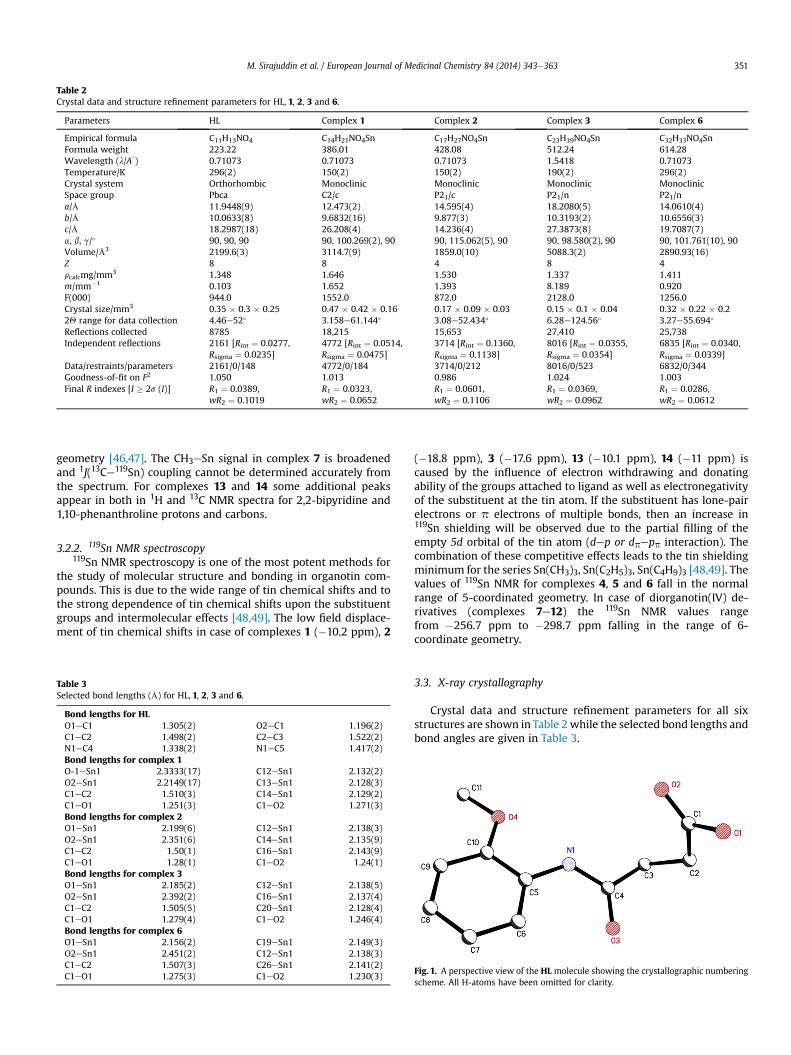
Table 2Crystal data and structure refinement parameters for HL, 1, 2, 3 and 6.
Parameters HL Complex 1 Complex 2 Complex 3 Complex 6
Empirical formula C11H13NO4 C14H21NO4Sn C17H27NO4Sn C23H39NO4Sn C32H33NO4SnFormula weight 223.22 386.01 428.08 512.24 614.28Wavelength (l/A
�) 0.71073 0.71073 0.71073 1.5418 0.71073
Temperature/K 296(2) 150(2) 150(2) 190(2) 296(2)Crystal system Orthorhombic Monoclinic Monoclinic Monoclinic MonoclinicSpace group Pbca C2/c P21/c P21/n P21/na/Å 11.9448(9) 12.473(2) 14.595(4) 18.2080(5) 14.0610(4)b/Å 10.0633(8) 9.6832(16) 9.877(3) 10.3193(2) 10.6556(3)c/Å 18.2987(18) 26.208(4) 14.236(4) 27.3873(8) 19.7087(7)a, b, g/� 90, 90, 90 90, 100.269(2), 90 90, 115.062(5), 90 90, 98.580(2), 90 90, 101.761(10), 90Volume/Å3 2199.6(3) 3114.7(9) 1859.0(10) 5088.3(2) 2890.93(16)Z 8 8 4 8 4rcalcmg/mm3 1.348 1.646 1.530 1.337 1.411m/mm�1 0.103 1.652 1.393 8.189 0.920F(000) 944.0 1552.0 872.0 2128.0 1256.0Crystal size/mm3 0.35 � 0.3 � 0.25 0.47 � 0.42 � 0.16 0.17 � 0.09 � 0.03 0.15 � 0.1 � 0.04 0.32 � 0.22 � 0.22Q range for data collection 4.46e52� 3.158e61.144� 3.08e52.434� 6.28e124.56� 3.27e55.694�
Reflections collected 8785 18,215 15,653 27,410 25,738Independent reflections 2161 [Rint ¼ 0.0277,
Rsigma ¼ 0.0235]4772 [Rint ¼ 0.0514,Rsigma ¼ 0.0475]
3714 [Rint ¼ 0.1360,Rsigma ¼ 0.1138]
8016 [Rint ¼ 0.0355,Rsigma ¼ 0.0354]
6835 [Rint ¼ 0.0340,Rsigma ¼ 0.0339]
Data/restraints/parameters 2161/0/148 4772/0/184 3714/0/212 8016/0/523 6832/0/344Goodness-of-fit on F2 1.050 1.013 0.986 1.024 1.003Final R indexes [I � 2s (I)] R1 ¼ 0.0389,
wR2 ¼ 0.1019R1 ¼ 0.0323,wR2 ¼ 0.0652
R1 ¼ 0.0601,wR2 ¼ 0.1106
R1 ¼ 0.0369,wR2 ¼ 0.0962
R1 ¼ 0.0286,wR2 ¼ 0.0612
M. Sirajuddin et al. / European Journal of Medicinal Chemistry 84 (2014) 343e363 351
geometry [46,47]. The CH3eSn signal in complex 7 is broadenedand 1J(13Ce119Sn) coupling cannot be determined accurately fromthe spectrum. For complexes 13 and 14 some additional peaksappear in both in 1H and 13C NMR spectra for 2,2-bipyridine and1,10-phenanthroline protons and carbons.
3.2.2. 119Sn NMR spectroscopy119Sn NMR spectroscopy is one of the most potent methods for
the study of molecular structure and bonding in organotin com-pounds. This is due to the wide range of tin chemical shifts and tothe strong dependence of tin chemical shifts upon the substituentgroups and intermolecular effects [48,49]. The low field displace-ment of tin chemical shifts in case of complexes 1 (�10.2 ppm), 2
Table 3Selected bond lengths (Å) for HL, 1, 2, 3 and 6.
Bond lengths for HLO1eC1 1.305(2) O2eC1 1.196(2)C1eC2 1.498(2) C2eC3 1.522(2)N1eC4 1.338(2) N1eC5 1.417(2)Bond lengths for complex 1O-1eSn1 2.3333(17) C12eSn1 2.132(2)O2eSn1 2.2149(17) C13eSn1 2.128(3)C1eC2 1.510(3) C14eSn1 2.129(2)C1eO1 1.251(3) C1eO2 1.271(3)Bond lengths for complex 2O1eSn1 2.199(6) C12eSn1 2.138(3)O2eSn1 2.351(6) C14eSn1 2.135(9)C1eC2 1.50(1) C16eSn1 2.143(9)C1eO1 1.28(1) C1eO2 1.24(1)Bond lengths for complex 3O1eSn1 2.185(2) C12eSn1 2.138(5)O2eSn1 2.392(2) C16eSn1 2.137(4)C1eC2 1.505(5) C20eSn1 2.128(4)C1eO1 1.279(4) C1eO2 1.246(4)Bond lengths for complex 6O1eSn1 2.156(2) C19eSn1 2.149(3)O2eSn1 2.451(2) C12eSn1 2.138(3)C1eC2 1.507(3) C26eSn1 2.141(2)C1eO1 1.275(3) C1eO2 1.230(3)
(�18.8 ppm), 3 (�17.6 ppm), 13 (�10.1 ppm), 14 (�11 ppm) iscaused by the influence of electron withdrawing and donatingability of the groups attached to ligand as well as electronegativityof the substituent at the tin atom. If the substituent has lone-pairelectrons or p electrons of multiple bonds, then an increase in119Sn shielding will be observed due to the partial filling of theempty 5d orbital of the tin atom (dep or dpepp interaction). Thecombination of these competitive effects leads to the tin shieldingminimum for the series Sn(CH3)3, Sn(C2H5)3, Sn(C4H9)3 [48,49]. Thevalues of 119Sn NMR for complexes 4, 5 and 6 fall in the normalrange of 5-coordinated geometry. In case of diorganotin(IV) de-rivatives (complexes 7e12) the 119Sn NMR values rangefrom �256.7 ppm to �298.7 ppm falling in the range of 6-coordinate geometry.
3.3. X-ray crystallography
Crystal data and structure refinement parameters for all sixstructures are shown in Table 2 while the selected bond lengths andbond angles are given in Table 3.
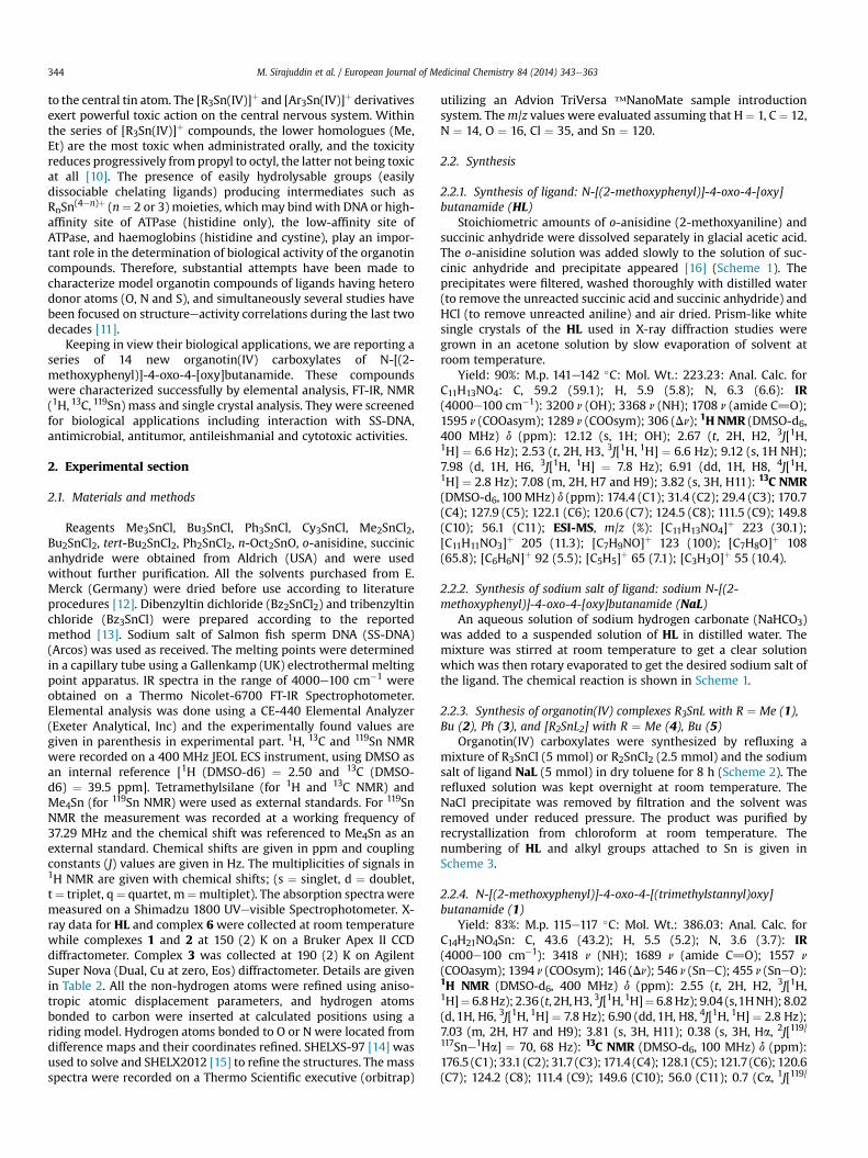
Fig. 1. A perspective view of the HL molecule showing the crystallographic numberingscheme. All H-atoms have been omitted for clarity.
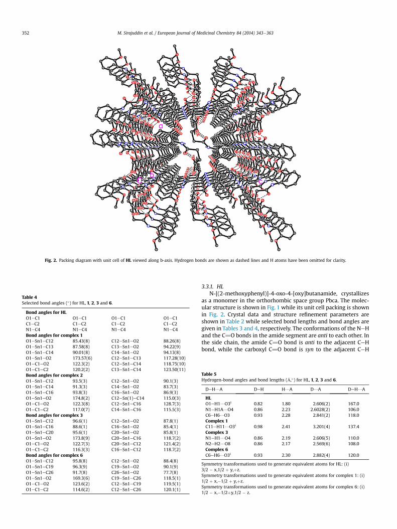
Table 4Selected bond angles (�) for HL, 1, 2, 3 and 6.
Bond angles for HLO1eC1 O1eC1 O1eC1 O1eC1C1eC2 C1eC2 C1eC2 C1eC2N1eC4 N1eC4 N1eC4 N1eC4Bond angles for complex 1O1eSn1eC12 85.43(8) C12eSn1eO2 88.26(8)O1eSn1eC13 87.58(8) C13eSn1eO2 94.22(9)O1eSn1eC14 90.01(8) C14eSn1eO2 94.13(8)O1eSn1eO2 173.57(6) C12eSn1eC13 117.28(10)O1eC1eO2 122.3(2) C12eSn1eC14 118.75(10)O1eC1eC2 120.2(2) C13eSn1eC14 123.50(11)Bond angles for complex 2O1eSn1eC12 93.5(3) C12eSn1eO2 90.1(3)O1eSn1eC14 91.3(3) C14eSn1eO2 83.7(3)O1eSn1eC16 93.8(3) C16eSn1eO2 86.9(3)O1eSn1eO2 174.8(2) C12eSn(1)eC14 115.0(3)O1eC1eO2 122.3(8) C12eSn1eC16 128.7(3)O1eC1eC2 117.0(7) C14eSn1eC16 115.5(3)Bond angles for complex 3O1eSn1eC12 96.6(1) C12eSn1eO2 87.8(1)O1eSn1eC16 88.6(1) C16eSn1eO2 85.4(1)O1eSn1eC20 95.6(1) C20eSn1eO2 85.8(1)O1eSn1eO2 173.8(9) C20eSn1eC16 118.7(2)O1eC1eO2 122.7(3) C20eSn1eC12 121.4(2)O1eC1eC2 116.3(3) C16eSn1eC12 118.7(2)Bond angles for complex 6O1eSn1eC12 95.8(8) C12eSn1eO2 88.4(8)O1eSn1eC19 96.3(9) C19eSn1eO2 90.1(9)O1eSn1eC26 91.7(8) C26eSn1eO2 77.7(8)O1eSn1eO2 169.3(6) C19eSn1eC26 118.5(1)O1eC1eO2 123.6(2) C12eSn1eC19 119.5(1)O1eC1eC2 114.6(2) C12eSn1eC26 120.1(1)
Fig. 2. Packing diagram with unit cell of HL viewed along b-axis. Hydrogen bonds are shown as dashed lines and H atoms have been omitted for clarity.
M. Sirajuddin et al. / European Journal of Medicinal Chemistry 84 (2014) 343e363352
3.3.1. HLN-[(2-methoxyphenyl)]-4-oxo-4-[oxy]butanamide, crystallizes
as a monomer in the orthorhombic space group Pbca. The molec-ular structure is shown in Fig. 1 while its unit cell packing is shownin Fig. 2. Crystal data and structure refinement parameters areshown in Table 2 while selected bond lengths and bond angles aregiven in Tables 3 and 4, respectively. The conformations of the NeHand the C]O bonds in the amide segment are anti to each other. Inthe side chain, the amide C]O bond is anti to the adjacent CeHbond, while the carboxyl C]O bond is syn to the adjacent CeH
Table 5Hydrogen-bond angles and bond lengths (Å,�) for HL, 1, 2, 3 and 6.
DeH/A DeH H/A D/A DeH/A
HLO1eH1/O3i 0.82 1.80 2.606(2) 167.0N1eH1A/O4 0.86 2.23 2.6028(2) 106.0C6eH6/O3 0.93 2.28 2.841(2) 118.0Complex 1C11eH11/O3i 0.98 2.41 3.201(4) 137.4Complex 3N1eH1/O4 0.86 2.19 2.606(5) 110.0N2eH2/O8 0.86 2.17 2.569(6) 108.0Complex 6C6eH6/O3i 0.93 2.30 2.882(4) 120.0
Symmetry transformations used to generate equivalent atoms for HL: (i)3/2 � x,1/2 þ y,þz.Symmetry transformations used to generate equivalent atoms for complex 1: (i)1/2 þ x,�1/2 þ y,þz.Symmetry transformations used to generate equivalent atoms for complex 6: (i)1/2 � x,�1/2þy,1/2 � z.

Fig. 3. Polymeric structure of R3SnL.
M. Sirajuddin et al. / European Journal of Medicinal Chemistry 84 (2014) 343e363 353
bond. The observed rare anti conformation of the C]O and OeHbonds of the acid group is similar to that examined in N-(4-Methoxyphenyl)maleamic acid [50]. There is an intramolecularCeH—O and NeH—O interaction within the polymeric chain of theHL. An intermolecular CeH—O interaction along b-axis links thepolymeric chains in zigzag form (Fig. 2) which are linked into athree-dimensional network via CeH—p interactions. The detail ofintramolecular H-bonds is given in Table 5.
3.3.2. Complexes 1, 2, 3 and 6The general polymeric structure of R3SnL is shown in Fig. 3. The
perspective diagrams of the complexes 1, 2, 3 and 6 are given inFigs. 4, 6, 8 and 10 while their unit cell packing diagrams are shownin Figs. 5, 7, 9 and 11, respectively. Crystal data and structurerefinement parameters are shown inTable 2while the selected bondlengths andbond angles are given inTables 3 and4, respectively. Thepresence of a bidendate ligand, 4-(2-methoxyphenylamino)-4-oxobutanoate, leads to the formation of the polymeric structure.In other words, the central R3Sn (R ¼ CH3, C2H5, C4H9 and C7H7)group bridges the two neighboring 4-(2-methoxyphenylamino)-4-oxobutanoate ligands via carboxylate (Fig. 3) moieties to form
Fig. 4. A perspective view of the complex 1 showing the crystallographic numberingscheme. All H-atoms have been omitted for clarity.
one-dimensional polymeric chain [51]. The geometry around the tinatom is distorted trigonal bipyramidal. The values of t (t ¼ (b � a)/60, where b is the largest basal angle around the tin atomwhile a isthe next largest angle around the tin atom) for the complexes 1, 2, 3,and 6 are 0.83, 0.80, 0.87 and 0.82, respectively that are typical fordistorted trigonal bipyramidal geometry [52]. The geometry aroundthe tin atom is defined by three R groups and two oxygen atoms. Thethree R groups occupy the equatorial positions with essentiallyidentical bond distances [SneC ¼ 2.125e2.149 Å]. The OeSneOangle is approximately linear [OeSneO ¼ 169.33e174.8�]. TheCeSneCandOeSneC angles arewithin the expected rangeof values[CeSneC ¼ 115.5(18)�e121.43(19)� and OeSneC ¼ 85.40e96.34�][53]. The sum of the CeSneC angles in the equatorial plane equals359.5�, 359.5�, 359.3� and 358.1�, respectively for complexes 1, 2, 3,6, indicating slight distortion. This distortion from ideal trigonalbipyramidal geometry is found in the axial angle [OeSneO]. Theintramolecular SneO1 separation of 2.333(17) Å, 2.199(6) Å,2.184(3) Å, 2.156(2) Å, respectively for complexes 1, 2, 3, 6 issignificantly shorter than the intermolecular SneO2 distance of2.215(17) Å, 2.351(6) Å, 2.392(3) Å, 2.451(2) Å, respectively forcomplexes 1, 2, 3, 6. The asymmetric SneO separations are reflectedin the associated CeO1 andCeO2distances of 1.251(3)Å,1.280(1)Å,1.278(5) Å, 1.257(3) Å and 1.271(3) Å, 1.24(1) Å, 1.246(5) Å,1.230(3) Å, respectively for complexes 1, 2, 3, 6 [54]. The Sn1eO1,Sn1eO2 (another Sn atombonded byO2 of the bridging carboxylateligand) bond distances involving the bridging carboxylate ligand areexactly same indicating a symmetrical bridge. The anisobidentatebidentate carboxylate has a difference of 0.058 Å between its CeObonds while for the bidentate carboxylate this difference is only0.021 Å; the variations in the CeO bond distances suggest chargedelocalization over the carboxylate group COO. The differentmodesof bonding of the acetates, i.e., bridging or chelating, are thus easilydifferentiated by the relevant bond lengths [55]. There is an intra-molecular CeH—O (O of amide carbonyl group) interaction withinthepolymeric chain. An intermolecular CeH—O interaction links thepolymeric chains into sheets form (Figs. 7 and 9) or zigzag form(Figs. 5 and 11) which are linked into a three-dimensional networkvia CeH—p interactions. The details of intramolecular H-bondsexisting within the structure are given in Table 5.
3.4. Mass spectrometry
The general fragmentation patterns for HL, tri- and di-organo-tin(IV) complexes are shown in Schemes 4e6, respectively. In themass spectrum of HL the molecular ion peak undergoes the elim-ination of a water molecule generating a mass fragment of m/
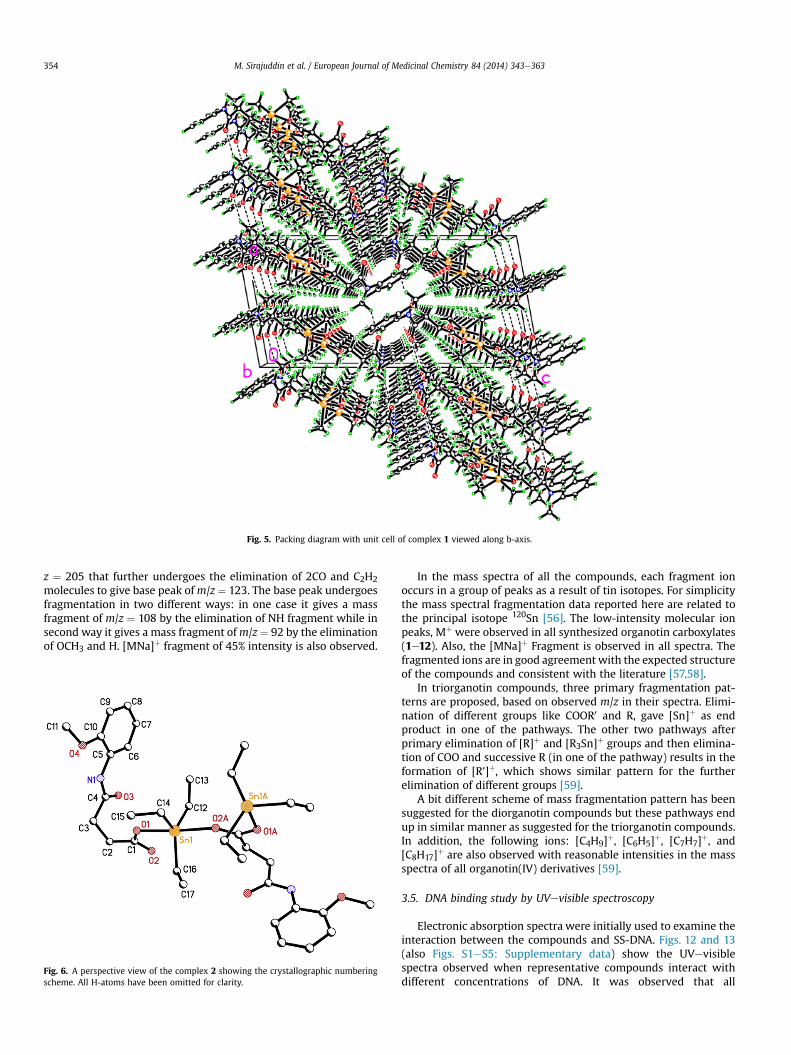
Fig. 5. Packing diagram with unit cell of complex 1 viewed along b-axis.
M. Sirajuddin et al. / European Journal of Medicinal Chemistry 84 (2014) 343e363354
z ¼ 205 that further undergoes the elimination of 2CO and C2H2molecules to give base peak ofm/z ¼ 123. The base peak undergoesfragmentation in two different ways: in one case it gives a massfragment of m/z ¼ 108 by the elimination of NH fragment while insecond way it gives a mass fragment ofm/z¼ 92 by the eliminationof OCH3 and H. [MNa]þ fragment of 45% intensity is also observed.
Fig. 6. A perspective view of the complex 2 showing the crystallographic numberingscheme. All H-atoms have been omitted for clarity.
In the mass spectra of all the compounds, each fragment ionoccurs in a group of peaks as a result of tin isotopes. For simplicitythe mass spectral fragmentation data reported here are related tothe principal isotope 120Sn [56]. The low-intensity molecular ionpeaks, Mþ were observed in all synthesized organotin carboxylates(1e12). Also, the [MNa]þ Fragment is observed in all spectra. Thefragmented ions are in good agreement with the expected structureof the compounds and consistent with the literature [57,58].
In triorganotin compounds, three primary fragmentation pat-terns are proposed, based on observed m/z in their spectra. Elimi-nation of different groups like COOR0 and R, gave [Sn]þ as endproduct in one of the pathways. The other two pathways afterprimary elimination of [R]þ and [R3Sn]þ groups and then elimina-tion of COO and successive R (in one of the pathway) results in theformation of [R']þ, which shows similar pattern for the furtherelimination of different groups [59].
A bit different scheme of mass fragmentation pattern has beensuggested for the diorganotin compounds but these pathways endup in similar manner as suggested for the triorganotin compounds.In addition, the following ions: [C4H9]þ, [C6H5]þ, [C7H7]þ, and[C8H17]þ are also observed with reasonable intensities in the massspectra of all organotin(IV) derivatives [59].
3.5. DNA binding study by UVevisible spectroscopy
Electronic absorption spectra were initially used to examine theinteraction between the compounds and SS-DNA. Figs. 12 and 13(also Figs. S1eS5: Supplementary data) show the UVevisiblespectra observed when representative compounds interact withdifferent concentrations of DNA. It was observed that all

Fig. 7. Packing diagram with unit cell of complex 2 viewed along b-axis.
Fig. 8. A perspective view of the complex 3 showing the crystallographic numberingscheme. All H-atoms have been omitted for clarity.
M. Sirajuddin et al. / European Journal of Medicinal Chemistry 84 (2014) 343e363 355
compounds have one strong absorption peak at 280e283 nm. Incase of compound 4 a shoulder at 267.40 nm also appeared. Afterinteraction with increasing amounts of DNA, all the peaksdecreased gradually and there was a minor red shift of upto 1 nmfor all compounds. Long et al. [60], have pointed out that the peakshift of the small molecules after they interacted with DNA could beclues to judge the binding mode between the small molecules andDNA. If the binding involves a typical intercalative mode, anhypochromism (decrease in absorption or molar absorptivity) ef-fect coupled with obvious bathochromism (shift of wavelengthtoward longer wavelength side, i.e. red shift) for the characteristicpeaks of the small molecules will be found due to the strongstacking between the chromophore and the base pairs of DNA[61,62]. Therefore, based on this viewpoint, the interaction betweencompounds and SS-DNA could be noncovalent intercalative bind-ing. After intercalating the base pairs of DNA, the p* orbital of theintercalated ligand could couple with p orbital of base pairs, thusdecreasing the pep* transition energy, and further resulting in thebathochromism. On the other hand, the coupling of a p orbital withpartially filled electrons decreases the transition probabilities
hence results hypochromic shift. Since hypochromism due to pep*stacking interactions may appear in the case of the intercalativebinding mode, while bathochromism may be observed when theDNA duplex is stabilized [1,63].
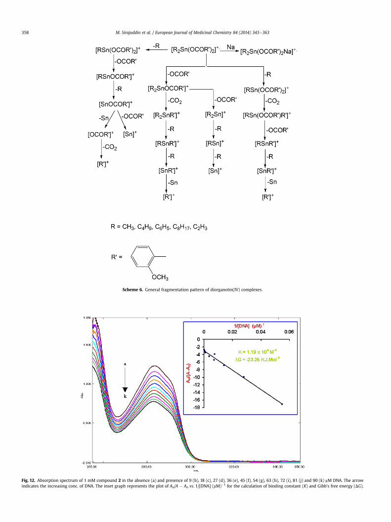
Based upon the variation in absorbance, the intrinsic bindingconstant of the compound with DNAwere determined according toBenesieHildebrand equation [64]:
A0
A� A0¼ εG
εH�G � εGþ εG
εH�G � εG� 1K½DNA�
where K is the association/binding constant, Ao and A are the ab-sorbances of the compound and its complex with DNA, respec-tively, and εG and εH�G are the absorption coefficients of thecompound and the compound-DNA complex, respectively. Theassociation constants were obtained from the intercept-to-sloperatios of Ao/(A � Ao) vs. 1/[DNA] plots. The Gibb's free energy (DG)was determined from the equation:
DG ¼ �RT ln K
where R is general gas constant (8.314 JK�1 mol�1) and T is thetemperature (298 K). The binding constants and calculated Gibb'sfree energies are given in Table 6.
From the interaction study with SS-DNA it is concluded that thesynthesized compounds could be used as an anticancer drug.
3.6. Biological activities
3.6.1. Antibacterial activityInvitro antibacterial screening tests of the synthesized ligand and
its organotin(IV) derivatives were carried out against 6 bacterialstrains; 2 g-positive [B. subtilis, and S. aureus] and 4 g-negative [E.coli, B. bronchiseptica, S. typhimurium, Enterobacter aerogens]. Thedisc diffusion method [21,22] was used in this assay and eachexperiment was performed in triplicate. Readings of the zone ofinhibition represent the mean value of 3 readings, which are shownin Table 7. Roxithromycin and Cefiximewere used as standard drugsin these assays. The data obtained show that the synthesized com-pounds have antibacterial activity. Compounds 1, 2, 7,11 and 13havemaximum antibacterial activity against all 6 strains. Their activity is
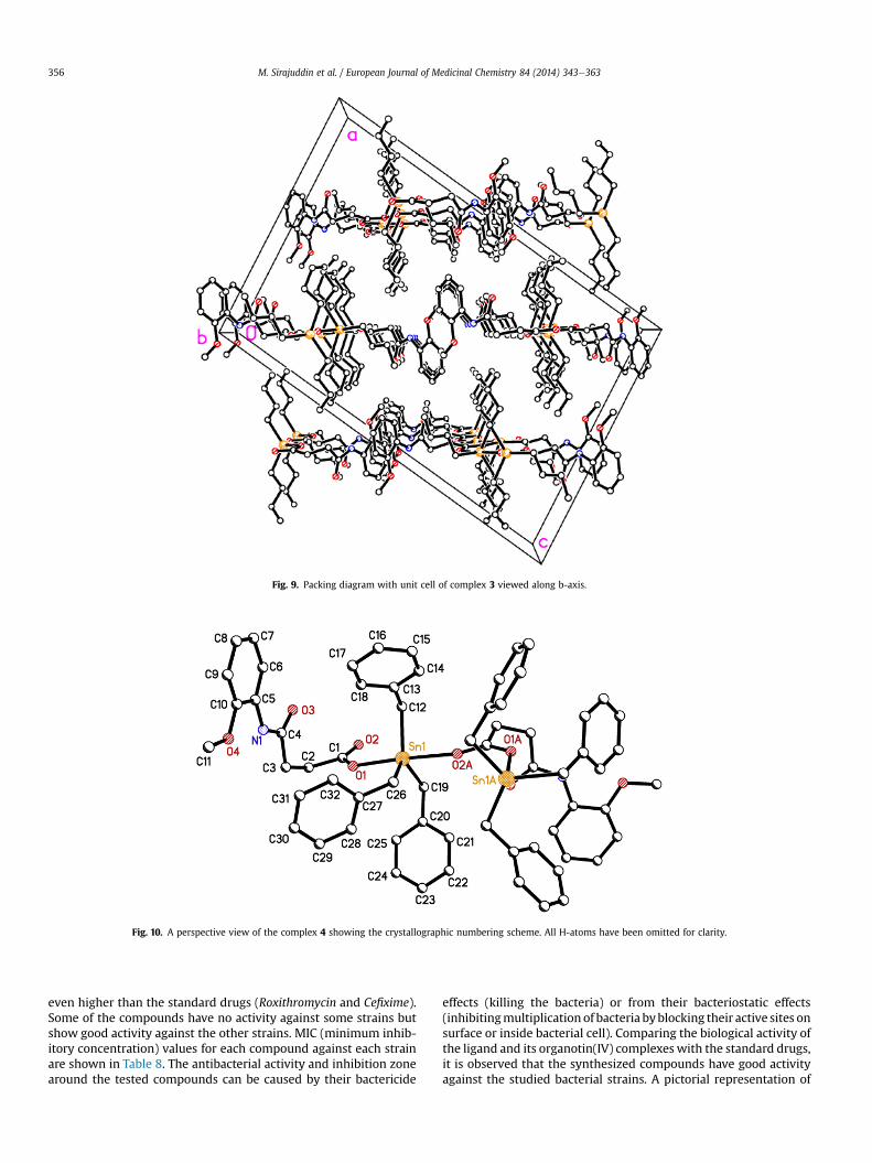
Fig. 9. Packing diagram with unit cell of complex 3 viewed along b-axis.
Fig. 10. A perspective view of the complex 4 showing the crystallographic numbering scheme. All H-atoms have been omitted for clarity.
M. Sirajuddin et al. / European Journal of Medicinal Chemistry 84 (2014) 343e363356
even higher than the standard drugs (Roxithromycin and Cefixime).Some of the compounds have no activity against some strains butshow good activity against the other strains. MIC (minimum inhib-itory concentration) values for each compound against each strainare shown in Table 8. The antibacterial activity and inhibition zonearound the tested compounds can be caused by their bactericide
effects (killing the bacteria) or from their bacteriostatic effects(inhibitingmultiplication of bacteria byblocking their active sites onsurface or inside bacterial cell). Comparing the biological activity ofthe ligand and its organotin(IV) complexeswith the standard drugs,it is observed that the synthesized compounds have good activityagainst the studied bacterial strains. A pictorial representation of

Fig. 11. Packing diagram with unit cell of complex 6 viewed along c-axis.
Scheme 4. General fragmentation pattern of HL. Scheme 5. General fragmentation pattern of triorganotin(IV) complexes.
M. Sirajuddin et al. / European Journal of Medicinal Chemistry 84 (2014) 343e363 357
Scheme 6. General fragmentation pattern of diorganotin(IV) complexes.
Fig. 12. Absorption spectrum of 1 mM compound 2 in the absence (a) and presence of 9 (b), 18 (c), 27 (d), 36 (e), 45 (f), 54 (g), 63 (h), 72 (i), 81 (j) and 90 (k) mM DNA. The arrowindicates the increasing conc. of DNA. The inset graph represents the plot of Ao/A � Ao vs. 1/[DNA] (mM)�1 for the calculation of binding constant (K) and Gibb's free energy (DG).
M. Sirajuddin et al. / European Journal of Medicinal Chemistry 84 (2014) 343e363358

Fig. 13. Absorption spectrum of 1 mM compound 5 in the absence (a) and presence of 9 (b), 18 (c), 27 (d), 36 (e), 45 (f), 54 (g), 63 (h), 72 (i), 81 (j) and 90 (k) mM DNA. The arrowindicates the increasing conc. of DNA. The inset graph represents the plot of Ao/A � Ao vs. 1/[DNA] (mM)�1 for the calculation of binding constant (K) and Gibb's free energy (DG).
M. Sirajuddin et al. / European Journal of Medicinal Chemistry 84 (2014) 343e363 359
zone of inhibition of antibacterial activity of one the representativecompound is shown in Fig. S6 (supplementary data).
3.6.2. Antifungal activityAll synthesized compounds were also subjected to in vitro
antifungal activity testing against 4 fungal strains [Mucor species, A.niger, Aspergillus flavus and Aspergillus fumigatus] using the diskdilution method [65]. The results are shown in Table 9. Turbinafinewas used as the reference drug. All the compounds, except HL and12, have shown antifungal activity against the various strains.Compound 3 shows higher activity than the reference drug. TheirMIC values against each strain are given in Table 10. A pictorialrepresentation of zone of inhibition of antifungal activity of one therepresentative compound is shown in Fig. S7 (supplementary data).Percentage inhibition and MIC value of biological active sampleswas evaluated by following formula:
Percent inhibition of fungal growth
¼�Growth diameter in test compoundðmmÞ
Growth diameter in controlðmmÞ�� 100
Table 6Overall binding constant and Gibb's free energy data of the selected compounds.a
Compound no. K (M�1) �DG (KJ mol�1)
HL 8.26 � 103 22.341 9.9 � 103 22.802 1.19 � 104 23.253 1.24 � 104 23.354 1.25 � 104 23.375 1.81 � 104 24.39 8.7 � 103 22.612 3.85 � 103 20.5
a Numbering of compounds is in accordance to Scheme 2.
It is concluded from the antibacterial and antifungal activitiesthat some of the synthesized compounds show good antibacterialand antifungal activities even higher than the standards so they canbe used as a potent antimicrobial drug.
3.6.3. Cytotoxic activityThis assay is used for preliminary screening which shows
cytotoxic effects as well as wide range of pharmacological activ-ities, i.e., pesticidal, anticancer, and antifungal activities [9]. Thecytotoxic activity of ligand HL and its organotin(IV) complexes(1e14) were studied in vitro against the brine shrimp lethalitymethod by using Doxorubicin as a standard drug and the resultsare summarized in Table 11. The data is based on mean value of 2duplicates. The mechanism of toxic effect of organotin(IV) com-pounds is quite complicated, and it cannot be regarded as thor-oughly studied. It is assumed that these compounds are capable ofreacting with cell membranes, finally leading to their decay, speedup the ion exchange processes, and inhibiting oxidative andphotochemical phosphorylation. Generally, among RnSnX4-n com-pounds, the most toxic are the R3SnX [66]. Toxicity in the R3Snseries is related to total molecular surface area and to the octa-nol:water partition coefficient, KOW, which is a measure of hy-drophobicity. The high lipid solubility of organotins ensures cellpenetration and association with intracellular sites, while cell wallcomponents also play an important role [67]. Also thedonoreacceptor interactions between the organotin species andthe target organism played an important role in the toxicitymechanism [68]. The brine shrimp cytotoxicity assay is consideredas a useful tool for preliminary assessment of toxicity. Cytotoxicitycan be considered significant if the LD50 value is less than20e30 mg/mL [69]. Cytotoxic drugs work best in cancers where thecancer cells are rapidly dividing and multiplying. In the presentstudy the LD50 value for the tested compounds is less than 4 mg/mL(except compound 12) so they can be consider as cytotoxic. Thusthese compounds can also used for the treatment of cancer.
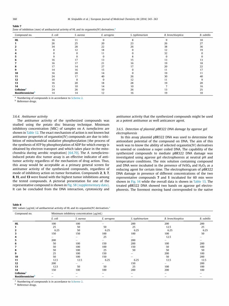
Table 7Zone of inhibition (mm) of antibacterial activity of HL and its organotin(IV) derivatives.a
Compound no. E. coli S. aureus E. aerogens S. typhimurium B. bronchiseptica B. subtilis
HL 16 11 0 8 9 101 26 25 20 23 26 272 34 28 22 26 38 363 0 12 18 0 12 194 0 0 11 0 32 05 9 0 0 8 0 06 16 17 13 15 13 137 16 17 21 16 19 188 17 14 27 17 20 229 14 16 15 0 11 1710 16 20 14 0 19 1111 24 17 40 26 30 4012 10 0 0 12 11 913 16 20 18 28 19 2014 13 10 14 10 7 16Cefiximeb 24 26 10 26 13 25Roxithromicineb 14 14 12 16 19 10
a Numbering of compounds is in accordance to Scheme 2.b Reference drugs.
M. Sirajuddin et al. / European Journal of Medicinal Chemistry 84 (2014) 343e363360
3.6.4. Antitumor activityThe antitumor activity of the synthesized compounds was
studied using the potato disc bioassay technique. Minimuminhibitory concentration (MIC) of samples on A. tumefaciens areshown in Table 12. The exact mechanism of action is not known butantitumor properties of organotin(IV) compounds are due to inhi-bition of mitochondrial oxidative phosphorylation (the process ofthe synthesis of ATP by phosphorylation of ADP for which energy isobtained by electron transport and which takes place in the mito-chondria during aerobic respiration) [64,70]. The A. tumefaciens-induced potato disc tumor assay is an effective indicator of anti-tumor activity regardless of the mechanism of drug action. Thus,this assay would be acceptable as a primary general screen forantitumor activity of the synthesized compounds, regardless ofmode of inhibitory action on tumor formation. Compounds 2, 3, 7,8, 11, and 13 were found with the highest tumor inhibitions amongthe tested compounds. A pictorial presentation for one of therepresentative compound is shown in Fig. S8 (supplementary data).It can be concluded from the DNA interaction, cytotoxicity and
Table 8MIC values (mg/mL) of antibacterial activity of HL and its organotin(IV) derivatives.a
Compound no. Minimum inhibitory concentration (mg/mL)
E. coli S. aureus E. aerogens
HL 100 100 e
1 25 50 502 6.25 50 6.253 150 150 1004 e e 255 200 e e
6 50 100 1507 50 100 1008 50 100 259 25 100 15010 50 100 15011 12.5 12.5 6.2512 200 e e
13 12.5 50 5014 150 100 100Cefiximeb e e e
Roxithromicineb e e e
a Numbering of compounds is in accordance to Scheme 2.b Reference drugs.
antitumor activity that the synthesized compounds might be usedas a potent antitumor as well anticancer agent.
3.6.5. Detection of plasmid pBR322 DNA damage by agarose gelelectrophoresis
In this assay plasmid pBR322 DNA was used to determine theantioxidant potential of the compound on DNA. The aim of thiswork was to know the ability of selected organotin(IV) derivativesto unwind or condense a super coiled DNA. The capability of thesynthesized compounds to mediate pBR322 DNA damage wasinvestigated using agarose gel electrophoresis at neutral pH andtemperature conditions. The mix solution containing compoundand DNA were incubated in the presence of FeSO4 and H2O2 as areducing agent for certain time. The electrophorogram of pBR322DNA damage in presence of different concentrations of the tworepresentative compounds 7 and 1 incubated for 60 min wereshown in Fig. 14 while the overall data is shown in Table 13. Thetreated pBR322 DNA showed two bands on agarose gel electro-phoresis. The foremost moving band corresponded to the native
S. typhimurium B. bronchiseptica B. subtilis
200 200 20025 12.5 256.25 6.25 6.25
100 100 50e 12.5 e
200 e e
200 100 200150 50 10050 50 50
e 200 100e 50 200
6.25 12.5 12.5150 e 20025 100 100
200 200 100e e e
e e e
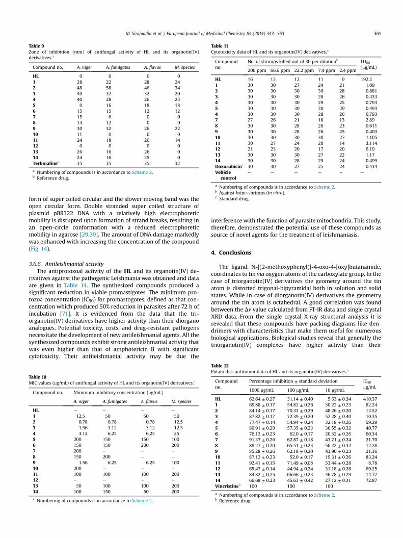
Table 9Zone of inhibition (mm) of antifungal activity of HL and its organotin(IV)derivatives.a
Compound no. A. niger A. fumigates A. flavus M. species
HL 0 0 0 01 28 22 28 242 48 58 46 343 40 32 32 204 40 28 26 235 0 16 18 186 15 15 12 127 15 0 0 08 14 12 0 09 30 32 26 2210 11 0 0 011 24 18 20 1412 0 0 0 013 26 16 26 014 24 16 25 0Terbinafineb 35 35 35 32
a Numbering of compounds is in accordance to Scheme 2.b Reference drug.
Table 11Cytotoxicity data of HL and its organotin(IV) derivatives.a
Compoundno.
No. of shrimps killed out of 30 per dilutionb LD50
(mg/mL)200 ppm 66.6 ppm 22.2 ppm 7.4 ppm 2.4 ppm
HL 16 13 12 11 9 192.21 30 30 27 24 21 1.092 30 30 30 30 28 0.8813 30 30 30 28 26 0.4334 30 30 30 29 25 0.7935 30 30 30 30 29 0.4934 30 30 30 28 26 0.7937 27 26 21 18 13 2.898 30 30 28 26 23 0.6119 30 30 28 26 25 0.40310 30 30 30 30 27 1.10511 30 27 24 20 14 3.11412 21 23 20 17 20 6.1913 30 30 30 27 22 1.1714 30 30 28 25 24 0.499Doxorubicinc 30 30 27 25 24 0.434Vehicle
controle e e e e e
a Numbering of compounds is in accordance to Scheme 2.b Against brine-shrimps (in vitro).c Standard drug.
M. Sirajuddin et al. / European Journal of Medicinal Chemistry 84 (2014) 343e363 361
form of super coiled circular and the slower moving band was theopen circular form. Double stranded super coiled structure ofplasmid pBR322 DNA with a relatively high electrophoreticmobility is disrupted upon formation of strand breaks, resulting inan open-circle conformation with a reduced electrophoreticmobility in agarose [29,30]. The amount of DNA damage markedlywas enhanced with increasing the concentration of the compound(Fig. 14).
3.6.6. Antileishmanial activityThe antiprotozoal activity of the HL and its organotin(IV) de-
rivatives against the pathogenic Leishmania was obtained and dataare given in Table 14. The synthesized compounds produced asignificant reduction in viable promastigotes. The minimum pro-tozoa concentration (IC50) for promastogotes, defined as that con-centration which produced 50% reduction in parasites after 72 h ofincubation [71]. It is evidenced from the data that the tri-organotin(IV) derivatives have higher activity than their diorganoanalogues. Potential toxicity, costs, and drug-resistant pathogensnecessitate the development of new antileishmanial agents. All thesynthesized compounds exhibit strong antileishmanial activity thatwas even higher than that of amphotericin B with significantcytotoxicity. Their antileishmanial activity may be due the
Table 10MIC values (mg/mL) of antifungal activity of HL and its organotin(IV) derivatives.a
Compound no. Minimum inhibitory concentration (mg/mL)
A. niger A. fumigates A. flavus M. species
HL e e e e
1 12.5 50 50 502 0.78 0.78 0.78 12.53 1.56 3.12 3.12 12.54 3.12 6.25 6.25 255 200 150 150 1006 150 150 200 2007 200 e e e
8 150 200 e e
9 1.56 6.25 6.25 10010 200 e e e
11 100 100 100 20012 e e e e
13 50 100 100 20014 100 150 50 200
a Numbering of compounds is in accordance to Scheme 2.
interference with the function of parasite mitochondria. This study,therefore, demonstrated the potential use of these compounds assource of novel agents for the treatment of leishmaniasis.
4. Conclusions
The ligand, N-[(2-methoxyphenyl)]-4-oxo-4-[oxy]butanamide,coordinates to tin via oxygen atoms of the carboxylate group. In thecase of triorganotin(IV) derivatives the geometry around the tinatom is distorted trigonal-bipyramidal both in solution and solidstates. While in case of diorganotin(IV) derivatives the geometryaround the tin atom is octahedral. A good correlation was foundbetween the Dn value calculated from FT-IR data and single crystalXRD data. From the single crystal X-ray structural analysis it isrevealed that these compounds have packing diagrams like den-drimers with characteristics that make them useful for numerousbiological applications. Biological studies reveal that generally thetriorganotin(IV) complexes have higher activity than their
Table 12Potato disc antitumor data of HL and its organotin(IV) derivatives.a
Compoundno.
Percentage inhibition ± standard deviation IC50
mg/mL1000 mg/mL 100 mg/mL 10 mg/mL
HL 62.64 ± 0.27 31.14 ± 0.40 5.63 ± 0.24 410.371 69.88 ± 0.17 54.82 ± 0.26 30.22 ± 0.23 82.242 84.14 ± 0.17 70.33 ± 0.29 48.26 ± 0.20 13.523 87.82 ± 0.17 72.39 ± 0.20 52.28 ± 0.40 10.354 77.47 ± 0.14 54.94 ± 0.24 32.18 ± 0.26 59.295 80.91 ± 0.29 57.35 ± 0.23 36.55 ± 0.32 40.776 76.12 ± 0.23 62.0 ± 0.17 20.32 ± 0.26 60.347 91.37 ± 0.26 62.87 ± 0.18 43.21 ± 0.24 21.708 88.27 ± 0.20 65.51 ± 0.23 50.22 ± 0.32 12.289 85.28 ± 0.26 62.18 ± 0.20 43.90 ± 0.23 21.3610 87.12 ± 0.23 52.0 ± 0.17 19.31 ± 0.26 83.2411 92.41 ± 0.15 71.49 ± 0.08 53.44 ± 0.28 8.7812 65.47 ± 0.14 44.94 ± 0.24 31.18 ± 0.26 69.2513 84.82 ± 0.25 66.66 ± 0.23 46.78 ± 0.29 14.7714 66.68 ± 0.23 45.63 ± 0.42 27.12 ± 0.31 72.87Vincristineb 100 100 100
a Numbering of compounds is in accordance to Scheme 2.b Reference drug.

Fig. 14. Effects of compounds 7 and 1 on plasmid (pBR322) DNA.
Table 13Plasmid pBR322 DNA damage by agarose Gel Electrophoresis data.a
Compound no. DNA protection activity at different concentration
1000 mg/mL 100 mg/mL 10 mg/mL 1 mg/mL
HL � � � �1 þþþ þþþ þþ þþ2 þ þ þ þ3 � � � �4 þþþ þþ þ þ5 � � � �6 þþ þþ þþ þ7 þþþ þþþ þþþ þþ8 � � � �9 þþ þþ þ þ10 þ þ þþ þþ11 þþ þþ þ þ13 � � � �14 þþþ þþ þþ þ
(�) ¼ no protection, (þ) ¼ Slight protection, (þþ) ¼ Moderate protection,(þþþ) ¼ Good protection.
a Numbering of compounds is in accordance to Scheme 2.
Table 14Antileishmanial data of HL and its organotin(IV) derivativesa.
Compound no. IC50 (mg/mL)
HL 3.42 � 10�3
1 7.59 � 10�5
2 9.19 � 10�5
3 4.17 � 10�5
4 8.70 � 10�5
5 3.90 � 10�5
6 9.96 � 10�5
7 5.89 � 10�4
8 6.65 � 10�4
9 6.60 � 10�4
10 7.96 � 10�4
11 1.92 � 10�3
12 1.04 � 10�3
13 1.56 � 10�2
14 9.22 � 10�2
Amphotericin Bb 0.56
a Numbering of compounds is in accordance to Scheme 2.b Reference drug.
M. Sirajuddin et al. / European Journal of Medicinal Chemistry 84 (2014) 343e363362
corresponding diorganotin(IV) complexes, possibly due to theirgreater lipophilicity and permeability through the cell membrane.The high lipid solubility of organotins ensures cell penetration andassociation with intracellular sites. In solution, triorganotin com-pounds may undergo spontaneous disproportionation into thecorresponding diorganotin and tetraorganotin derivatives while,in vivo, the loss of one alkyl or aryl group may occur through theintervention of enzymes such as aromatase. UVeVis spectroscopicresults show that the synthesized compounds bind to DNA viaintercalative mode of interaction resulting in hypochromism andminor red shift. The synthesized compounds may represent a newline of antitumor agents for prevention and treatment of cancer andalso serve as a potential source of chemoprotective agent(s). Theoxidative DNA damage by the synthesized compounds in thepresence of FeSO4 and H2O2 (used as a positive control) wasexplored using agarose gel electrophoresis. The extent of pBR322DNA damage was enhanced significantly with increasing concen-tration of the compounds. The antileishmanial data clearly indicatethat these compounds have a strong antileishmanial activity andmight be developed a new antileishmanial drug. Their anti-leishmanial activity may be due the interference with the functionof parasite mitochondria.
Acknowledgment
Muhammad Sirajuddin gratefully acknowledges the HigherEducation Commission (HEC) Islamabad, Pakistan, for financialsupport (PIN#117-7338-PS7-017).
Appendix A. Supplementary data
Supplementary data related to this article can be found at http://dx.doi.org/10.1016/j.ejmech.2014.07.028.
References
[1] M. Sirajuddin, S. Ali, A. Haider, N.A. Shah, A. Shah, M.R. Khan, Polyhedron 40(2012) 19e31.
[2] M.S. Ahmad, M. Hussain, M. Hanif, S. Ali, M. Qayyum, B. Mirza, Chem. Biol.Drug. Des. 71 (2008) 568e576.
[3] M. Sirajuddin, S. Ali, F.A. Shah, M. Ahmad, M.N. Tahir, J. Iran. Chem. Soc. 11(2013) 297e313.
[4] S.K. Hadjikakou, N. Hadjiliadis, Coord. Chem. Rev. 253 (2009) 235e249.

M. Sirajuddin et al. / European Journal of Medicinal Chemistry 84 (2014) 343e363 363
[5] C. Pellerito, P.D. Agati, T. Fiore, C. Mansueto, V. Mansueto, G. Stocco, L. Nagy,Pellerito, J. Inorg. Biochem. 99 (2005) 1294e1305.
[6] P.J. Blower, Annu. Rep. Prog. Chem. Sect. A 100 (2004) 633e658.[7] F. Barbieri, M. Viale, F. Sparatore, G. Schettini, A. Favre, C. Bruzzo, F. Novelli,
A. Alama, Anti-cancer Drugs 13 (2002) 599e604.[8] X. Shang, X. Meng, E.C.B.A. Alegria, Q. Li, M.F.C. Guedes da Silva,
M.L. Kuznetsov, A.J.L. Pombeiro, Inorg. Chem. 50 (2011) 8158e8167.[9] M. Tariq, N. Muhammad, M. Sirajuddin, S. Ali, N.A. Shah, N. Khalid, M.R. Khan,
M.N. Tahir, J. Organomet. Chem. 723 (2013) 79e89.[10] C. Pellerito, L. Nagy, L. Pellerito, A. Szorcsik, Chem. 691 (2006) 1733e1747.[11] M. Nath, S. Pokharia, G. Eng, X. Song, A. Kumar, J. Organomet. Chem. 669
(2003) 109e123.[12] W.L.F. Armarego, C.L.L. Chai, Purification of Laboratory Chemicals, fifth ed.,
Butterworth-Heinemann, London, New York, 2003.[13] K. Sisido, Y. Takeda, Z. Kingawa, J. Am. Chem. Soc. 83 (1961) 538e541.[14] G.M. Sheldrick, Acta Crystallogr. A64 (2008) 112e122.[15] G.M. Sheldrick, University of Gӧttingen, Germany, 2012.[16] F.A. Shah, M. Sirajuddin, S. Ali, S.M. Abbas, M.N. Tahir, C. Rizzoli, Inorgan.
Chim. Aca 400 (2013) 159e168.[17] M. Sirajuddin, S. Ali, N.A. Shah, M.R. Khan, M.N. Tahir, Spectrochim. Acta Part A
94 (2012) 134e142.[18] M. Sirajuddin, S. Ali, A. Badshah, J. Photochem. Photobiol. B 124 (2013) 1e19.[19] A. Felten, B. Grandry, P.H. Langrange, I. Casin, J. Clin. Microbiol. 40 (2002)
2766e2771.[20] L. Jafri, F.L. Ansari, M. Jamil, S. Kalsoom, S. Qureishi, B. Mirza, Chem. Biol. Drug.
Des. 79 (2012) 950e959.[21] S. Arikan, V. Paetznick, J.H. Rex, Antimicrob. Agents Chemother. 46 (2002)
3084.[22] A.S. Michael, C.G. Thompson, M. Abramovitz, Science 123 (1956) 464.[23] R.B. Sleet, K. Brendel, Ecotoxicol. Environ. Saf. 7 (1983) 435e446.[24] W.S. Abbott, J. Econ. Entomol. 18 (1925) 265e267.[25] D.J. Finney, Probit Analysis, third ed., Cambridge University Press, Cambridege,
1971, p. 333.[26] A. Ullah, F.L. Ansari, I. Haq, S. Nazir, B. Mirza, Chem. Biodivers. 4 (2007)
203e214.[27] J.L. McLaughlin, Methods in Plant Biochemistry, Academic Press, London,
1991, p. 1.[28] S. Kanwal, N. Ullah, I. Haq, I. Afzal, B. Mirza, Pak. J. Bot. 43 (2011) 85e89.[29] Y. Wang, X. Zhang, Q. Zhang, Z. Yang, Biometals 23 (2010) 265e273.[30] B. Tian, Y. Hua, Food Chem. 91 (2005) 413e418.[31] S. Nabi, N. Ahmed, M.J. Khan, Z. Bazai, M. Yasinzai, Y.M.S.A. Al-Kahraman,
World Appl. Sci. J. 19 (2012) 1495e1500.[32] L. Zhai, M. Chen, J. Blom, T.G. Theander, S.B. Christensen, A. Kharazmi,
J. Antimicrob. Chemother. 43 (1999) 793e803.[33] M. Vornefeld, F. Huber, H. Preut, G. Ruisi, R. Barbieri, Appl. Organomet. Chem.
6 (1992) 75e82.[34] K. Shahid, S. Shahzadi, S. Ali, J. Serbian Chem. Soc. 74 (2009) 141e154.[35] A. Hameed, T. Mohamad, E. Saad, Y. Farina, A. Graisa, E. Yousif, Eur. J. Sci. Res.
34 (2009) 212e217.[36] G. Eng, X. Song, A. Zapata, A.C. de Dios, L. Casabianca, R.D. Pike, J. Organomet.
Chem. 692 (2007) 1398e1404.[37] G.K. Sandhu, R. Hunda, E.R.T. Tiekink, J. Organomet. Chem. 430 (1992)
15e23.[38] D. Kovala-Demertzi, V.N. Dokorou, J.P. Jasinski, A. Opolski, J. Wiecek,
M. Zervou, M.A. Demertzis, J. Organomet. Chem. 690 (2005) 1800e1806.
[39] V. Zelenak, Z. Vargova, K. Gyoryova, Spectrochim. Acta Part A 66 (2007)262e272.
[40] Y. Farina, A. Graisa, E. Yousif, Mod. Appl. Sci. 3 (2009) 215e218.[41] T.P. Lockhart, W.F. Manders, Inorg. Chem. 25 (1986) 892e895.[42] B. Wrackmeyer, Annu. Rep. NMR Spectrosc. 38 (1999) 203e264.[43] T.P. Lockhart, W.F. Manders, E.M. Holts, J. Am. Chem. Soc. 108 (1986)
6611e6616.[44] G. Casella, F. Ferrante, G. Saielli, Inorg. Chem. 47 (2008) 4796e4807.[45] H. Jankovics, L. Nagy, Z. Kele, C. Pettinari, P. D'Agati, C. Mansueto, C. Pellerito,
L. Pellerito, J. Organomet. Chem. 668 (2003) 129e139.[46] A. Lycka, M. Nadvornik, K. Handlir, J. Holecek, Collect. Czech. Chem. Commun.
49 (1984) 2903e2911.[47] R. Willem, A. Bouhdid, M. Biesemans, J.C. Martins, D. Vos, E.R.T. Tiekink,
M. Gielen, J. Organomet. Chem. 514 (1996) 203e212.[48] V.N. Torocheshnikov, A.P. Tupciauskas, A.Y.A. Ustynyuk, J. Organomet. Chem.
81 (1974) 351e356.[49] J.J. Burke, P.C. Lauterbur, J. Am. Chem. Soc. 83 (1961) 326e331.[50] B.T. Gowda, M. Tokarcik, K. Shakuntala, J. Kozisek, H. Fuess, Acta Crystallogr.
E66 (2010) O1529.[51] A.W. Addison, R.T. Nageswara, J. Reedijk, J. Van Rijn, G.C. Verschoor, J. Chem.
Soc. Dalt. Trans. (1984) 1349e1356.[52] M.N. Tahir, D. Ulku, M. Danish, S. Ali, A. Badshah, M. Mazhar, Acta Crystallogr.
C53 (1997) 183e185.[53] Sadiq-ur-Rehman, S. Ali, M. Mazhar, M. Parvez, Acta Crystallogr. E60 (2004)
m1394em1396.[54] T.S.B. Baul, E.R.T. Tiekink, Acta Crystallogr. C52 (1996) 1428e1430.[55] V. Dokorou, M.A. Demertzis, J.P. Jasinski, D. Kovala-Demertzi, J. Organomet.
Chem. 689 (2004) 317e325.[56] Sadiq-ur-Rehman, K. Shahid, S. Ali, M.H. Bhatti, M. Parvez, J. Organomet.
Chem. 690 (2005) 1396e1408.[57] M.H. Bhatti, S. Ali, M. Mazhar, M. Danish, M.A. Choudhary, Turk. J. Chem. 23
(1999) 329e337.[58] S. Shahzadi, K. Shahid, S. Ali, M.H. Bhatti, J. Chem. Soc. Pak. 26 (2004)
395e399.[59] Sadiq-ur-Rehman, M.A. Choudhary, M.H. Bhatti, S. Ali, J. Iran. Chem. Soc. 9
(2012) 35e45.[60] E.C. Long, J.K. Barton, Chem. Res. 23 (1990) 271e273.[61] Q. Wang, X. Wang, Z. Yu, X. Yuan, K. Jiao, Int. J. Electrochem. Sci. 6 (2011)
5470e5481.[62] M. Hossain, M.F. Aziz, R. Ahmed, M. Hossain, A. Mahmuda, T. Ahmed,
M.H. Mazumder, Int. J. Pharm. Pharm. Sci. 2 (2010) 60e63.[63] M. Sirajuddin, N. Uddin, S. Ali, M.N. Tahir, Spectrochim. Acta Part A 116 (2013)
111e121.[64] H.A. Benesi, J.H. Hildebrand, J. Am. Chem. Soc. 71 (1949) 2703e2707.[65] A. Rehman, M.I. Choudhary, W.J. Thomsen, Bioassay Techniques for Drug
Development, Harwood Academic Publishers, Amsterdam, The Netherlands,2001, p. 9.
[66] S. Tabassum, C. Pettinari, J. Organomet. Chem. 691 (2006) 1761e1766.[67] A.K. Saxena, Appl. Organomet. Chem. 1 (1987) 39e56.[68] Y. Baravalia, Y. Vaghasiya, S. Chanda, Iran. J. Pharm. Res. 11 (2012) 851e861.[69] A.J. Crowe, P.J. Smith, G. Atassi, Inorg. Chim. Acta 93 (1984) 179e184.[70] F. Hadizadeh, A. Moradi, G. Naghibi, M. Vojdani, J. Behravan, M. Ramezani, Int.
J. Biomed. Sci. 3 (2007) 60e64.[71] S. Shahzadi, S. Ali, K. Shahid, M. Yousaf, S.K. Sharma, K. Qanungo, J. Chin.
Chem. Soc. 57 (2010) 659e670.