potential toxicity of egyptian ashwagandha: significance ... · volume 4 issue 2, february 2015 www...
TRANSCRIPT
International Journal of Science and Research (IJSR) ISSN (Online): 2319-7064
Index Copernicus Value (2013): 6.14 | Impact Factor (2013): 4.438
Volume 4 Issue 2, February 2015
www.ijsr.net Licensed Under Creative Commons Attribution CC BY
Potential Toxicity of Egyptian Ashwagandha:
Significance for their Therapeutic Bioactivity and
Anticancer Properties
Wafaa Abdallah Ahmed1, Mohamed A.
2, Nasser E. A.
3, Doaa E.
4
1, 2, 4 Cancer Biology Department, Biochemistry and Molecular Biology Unit, National Cancer Institute, Cairo University, Egypt
3Toxicology and Micro-analytical Research Unit, Faculty of Science, Suez Canal University, Egypt.
Abstract: Chemotherapy is usually given early after diagnosis in several cancer subtypes to offer best results. However,
chemotherapeutic drugs are often associated with some degree of toxicities, which are caused by reactive metabolites generated by the
biotransformation of anticancer drugs in the liver. Phytochemicals are dietary phytoestrogens that may play a role in cancer prevention
and treatment. Forty percent of Americans use complementary and alternative medicines (CAM) for disease prevention and therapy.
Ashwagandha (Withania somnifera) contains flavonoids and active ingredients like alkaloids and steroidal lactones which are called
‘Withanolides’. We hypothesize that the immune-modulatory and anti-inflammatory properties of Ashwagandha might contribute to its
overall effectiveness as an anti-carcinogenic agent. Ashwagandha is one of the most versatile plants used in the traditional Indian
medicine system (Ayurvedic).The goal of this study is evaluate the therapeutic bioactivity and cytotoxic effect of Egyptian
Ashwagandha on hepatocellular carcinoma cell line (HepG2).The HepG2 cells treated by different doses of Ashwagandha root extract.
The viability and cytotoxicity were measured by trypan blue and MTT assay. In conclusion: Ashwagandh can inhibit cell proliferation
and induce cytotoxic effect on hepatocellular cancer cells.
Keywords: Egyptian Ashwagandha , Hepatocellular Carcinoma Cell Line Antitumor activity
Abbreviation: HCC, Hepatocellular Carcinoma; HepG2, Human Hepatocellular Carcinoma Cell Line; W.S, Withania Somminera; IC50,
inhibition concentration dose; WA, Withaferin A; HeLa, Carcinoma of the Cervix; MCF-7, Breast Adenocarcinoma; EAW Ex ,Egyptian
Ashwagandha Water Extraction.
1. Introduction
Hepatocellular carcinoma (HCC), which are the most
health-threatening conditions drawing considerable attention
from medical professionals and scientists1.
Chemotherapeutic drugs are often associated with some
degree of toxicities, which are caused by reactive
metabolites generated by the biotransformation of
anticancer drugs in the liver2.Genotoxicity and cytotoxicity
of anticancer drugs to normal cells is a major problem in
cancer therapy and engender a risk of inducing secondary
malignancies3,4
. A dose of anticancer drug sufficient to kill
tumor cells is often toxic to the normal tissue and lead to
many side effects, which in turn, limits their treatment
efficacy5.However, HCC is well known to be a highly
chemo resistant tumor, and the response is very poor 6. To
this end, alternative medicines are being actively sought
from other sources with hopes to halt the disease's
progression or even eliminate the tumors.6,7
. Plant and plant
products have utilized with varying success to cure and
prevent diseases throughout history. Due to side effects of
synthetic products, herbal products are gaining popularity in
the world market7. Many active principles produced by
animals, plants and microorganisms have been employed in
the development of new drugs to treat diseases such as
cancer8.Medicinal plants are popular in indigenous system
of medicine like Ayurveda, siddha, unani and homoeopathy
and are used for its hepato-protective, antitumor,
antihypertensive, analgesic, anti-inflammatory and
antimicrobial properties9.
Historically, the medicinal plant Ashwagandha {Withania
somnifera (W.s.)} has been used for over centuries in Indian
Ayurvedic Medicine to treat a wide spectrum of disorders10
.
Ashwagandha found throughout the drier parts of India,
Baluchistan, Pakistan, Afghanistan, Sri Lanka, Congo,
South Africa, Egypt, Morocco and Jordan11
, the plant has
been used as an antioxidant, adaptogen, aphrodisiac, liver
tonic, anti-inflammatory agent, astringent and new to treat
ulcers, bacterial infection, venom toxins and senile
dementia. The major biochemical constituents of
Ashwagandha. are steroidal alkaloids and lactones, a class
of constituents together known as withanolides (steroidal
lactones with ergostane skeleton)12
.Further, it was able to
induce dose dependent DNA fragmentation in treated
cells13
.It promotes physical and mental health, rejuvenates
the body in debilitated conditions, and increase longevity14
.
Ashwagandha is known to have anti-inflammatory15
,
antitumor16
, antidiabetic17
, antioxidant18
, cardioprotective19
and anti-stress effect20
. The aim of this work is the study of
antitumor activity and cytotoxic effect of Ashwagandha on
hepatocellular carcinoma.
2. Materials and Methods
Plant: Withania Somnifera (Egyptian Ashwaghandha),
roots were harvested from Rafah, El-Arish, North Sinai,
Egypt in September 2008.
Paper ID: SUB14803 2170

International Journal of Science and Research (IJSR) ISSN (Online): 2319-7064
Index Copernicus Value (2013): 6.14 | Impact Factor (2013): 4.438
Volume 4 Issue 2, February 2015
www.ijsr.net Licensed Under Creative Commons Attribution CC BY
Figure 1: leaves and root of Egyptian Ashwagandha
1) Tested Compounds
Dry powder of Egyptian Ashwaghandha Water Extraction
(EAW Ex ) roots was prepared by suspending 10 g of dry
powder in 100 ml of distilled water and stirring it overnight
at 45±5oC, followed by filtration under sterile conditions.
The filtrate thus obtained was treated as 100% EAW Ex. It
was stored at –20oC in 1 ml aliquots until further use.
2) Cell Culture
Hep-G2, a human hepatocellular carcinoma cell line was
obtained frozen in liquid nitrogen (-180oC) from the
American Type Culture Collection. The tumor cell line was
maintained in the National Cancer Institute, Cairo, Egypt,
by serial sub-culturing. Cells were cultured in RPMI-1640
medium (RPMI-1640, Sigma–Aldrich, USA). The medium
was supplemented with antibiotic-free 10% fetal bovine
serum (FBS, Sigma, USA), 100 U/ml penicillin and 2-
mg/ml streptomycin. The cells were sub cultivated after
trypsinization (Trypsin-EDTA, Cambrex Bioscience
Verviers, Belgium) once or twice per week and re-
suspended in complete medium in a 1:5 split ratio. Cell line
was maintained as monolayer in T75 cell culture flasks with
filter screw caps (TPP, Trasadingen, Switzerland) at 37 °C
in a humidified 5% CO2 incubator.
3) Cell treatment
Cells were treated with EAW Ex at concentration range
from 0.65 to 1.00 %. The percentage of viable cells was
determined by trypan blue exclusion. Hep-G2 cells were
used when 90% confluence was reached in T25 flasks,
adherent cell lines were harvested with 0.025% trypsin.
Then cells were exposed to different concentrations of EAW
Ex. The cellular viability was determined using light
microscope employing 0.4% trypan blue dye exclusion
technique at 12 h and 24 h time intervals.
4) MTT cytotoxic assay
Anti-proliferative activity against liver tumor cell line was
estimated by the 3-[4,5-dimethyl-2-thiazolyl]-2,5-diphenyl-
2H-tetrazolium bromide (MTT) assay, which is based on the
cleavage of the tetrazolium salt by mitochondrial
dehydrogenases in viable cells21. Cells were treated for 24
h with various concentrations of EAW Ex before submitted
to the MTT assay. The relative cell viability was expressed
as the mean percentage of viable cells comparing to control
0 treated cells and the half maximal growth inhibitory
concentration (IC50) was calculated by the trend line
equation.
5) Flow cytometric cell cycle analysis
Hep-G2 cells (5x10⁵cells/well) were plated in 6 well micro
plates. Then collected after treatment with IC50
concentration of EAW Ex for 6h, washed two times with
PBS, re-suspended in 300µl of PBS, and fixed with 4 ml of
ice-cold 70% ethanol. When ready to stain with propidium
iodide (PI), cells were centrifuged; the ethanol was removed
and washed once in PBS. The cell pellets were then re-
suspended in one ml of PI/Triton X-100 staining solution
(0.1% Triton X-100 in PBS, 0.2 mg/ml RNase A, and 10
mg/ml PI) and incubated for 30 min at room temperature.
The stained cells were analyzed using a MoFlo flow
cytometer (Dako Cytomation, Glostrup, Denmark) 22.
6) Trypan Blue Exclusion
The dose response curve of viable cells was determined by
Trypan blue exclusion. The method was carried out
according to that of Sheldon and Preskorn, (1996)23 . (in
brief); Cells were cultured in 24-well plate and incubated for
24hr. Drug was added and incubated for another 24hr.
Medium was collected and cells were released by
trypsinization in falcon tubes. Supernatant was removed and
pellets were re-suspended. Cells were counted to determine
viability using Trypan blue dye.
7) Counting of the Viable Cells:
Cell suspension was prepared at a high concentration (~106
cells / ml) by trypsinization. A volume of 50 μl of 0.05%
Trypan blue solution was added to 50 μl of the single cell
suspension and left 1-2 min. The cells were examined under
the inverted microscope using the hemo cytometer. Non
stained (viable) cells were counted and the following
equation was used to calculate the cell count/ml of cell
suspension.
Viable cells / ml = Sum of viable cells Χ No. of counted
squares (5) Χ 104 Χ dilution factor.
The cells were then diluted to give the concentration of
single cell suspension required for each experiment.
8) Morphological Studies
Cells were cultured in 6-well plates and incubated for 24hr
then extract was added and incubated for 24hr. After that
cells were examined under inverted microscope and
morphological changes were recorded. Cells were
photographed using digital camera.
3. Statistical Analysis
The experimental data were expressed as mean ± standard
deviation (SD). A p value less than 0.05 was considered
significant. Microsoft Excel and computer program package
(SPSS version 15) was used for all statistical testing and
management of the database.
4. Results
Dose response curve:Hep-G2 cells were treated with graded
concentrations (0.6 – 1%) of EAWEx roots. The viability
was monitored. The data indicated that treatment of cells
with resulted in significant inhibition of viability of the cells
Paper ID: SUB14803 2171
International Journal of Science and Research (IJSR) ISSN (Online): 2319-7064
Index Copernicus Value (2013): 6.14 | Impact Factor (2013): 4.438
Volume 4 Issue 2, February 2015
www.ijsr.net Licensed Under Creative Commons Attribution CC BY
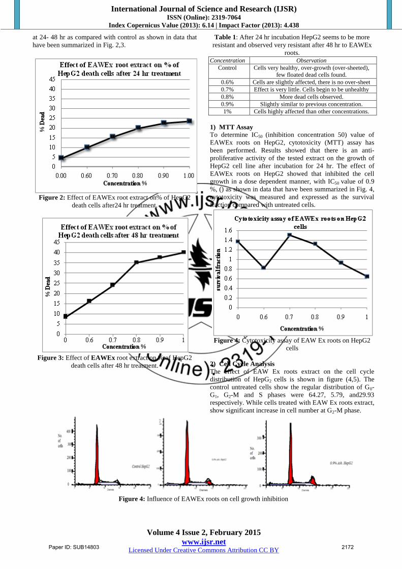
at 24- 48 hr as compared with control as shown in data that
have been summarized in Fig. 2,3.
Figure 2: Effect of EAWEx root extract on% of HepG2
death cells after24 hr treatment.
Figure 3: Effect of EAWEx root extract on % of HepG2
death cells after 48 hr treatment.
Table 1: After 24 hr incubation HepG2 seems to be more
resistant and observed very resistant after 48 hr to EAWEx
roots. Concentration Observation
Control Cells very healthy, over-growth (over-sheeted),
few floated dead cells found.
0.6% Cells are slightly affected, there is no over-sheet
0.7% Effect is very little. Cells begin to be unhealthy
0.8% More dead cells observed.
0.9% Slightly similar to previous concentration.
1% Cells highly affected than other concentrations.
1) MTT Assay
To determine IC50 (inhibition concentration 50) value of
EAWEx roots on HepG2, cytotoxicity (MTT) assay has
been performed. Results showed that there is an anti-
proliferative activity of the tested extract on the growth of
HepG2 cell line after incubation for 24 hr. The effect of
EAWEx roots on HepG2 showed that inhibited the cell
growth in a dose dependent manner, with IC50 value of 0.9
%, () as shown in data that have been summarized in Fig. 4,
cytotoxicity was measured and expressed as the survival
fraction compared with untreated cells.
Figure 4: Cytotoxicity assay of EAW Ex roots on HepG2
cells
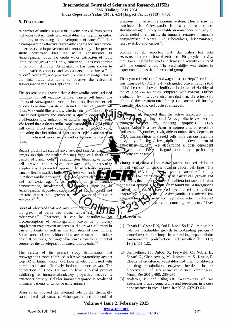
2) Cell Cycle Analysis
The effect of EAW Ex roots extract on the cell cycle
distribution of HepG2 cells is shown in figure (4,5). The
control untreated cells show the regular distribution of G0-
G1, G2-M and S phases were 64.27, 5.79, and29.93
respectively. While cells treated with EAW Ex roots extract,
show significant increase in cell number at G2-M phase.
Figure 4: Influence of EAWEx roots on cell growth inhibition
Paper ID: SUB14803 2172

International Journal of Science and Research (IJSR) ISSN (Online): 2319-7064
Index Copernicus Value (2013): 6.14 | Impact Factor (2013): 4.438
Volume 4 Issue 2, February 2015
www.ijsr.net Licensed Under Creative Commons Attribution CC BY
Figure 5: Flow cytometric analysis of EAWEx roots induced apoptosis in HepG2 cells.
3) Cell death assessment by DNA fragmentation
Treatment of HepG2 cells with different concentration of
EAW Ex roots extract (0.5, 1.0, 5.0 and 10%) for 48hrs
induced significant DNA ladder formation, suggesting
apoptotic cell death.
Figure 6: Cell death assessment by DNA fragmentation
showing effect of EAW Ex roots extract on DNA of HepG2
cells.
C: Control; SFC: Serum free control; M: DNA marker
4) Morphological Studies
Morphological studies were carried out to evaluate the
effect of EAW Ex roots extract on cell morphology in
relation to concentration and time of incubation. HepG2
cells were cultured in the presence of different extract
concentration for 24 and 48 hrs, and morphological changes
were examined under microscope. As shown in Fig.7,
incubation for 24 hrs mainly showed no significant effect on
cells. But at higher concentration (1%) extract treatment
caused cytotoxicity and cell death. Cells rounded up in
about 24 hr sand seemed to undergo apoptotic cell death.
However, at lower concentration cells appeared to be
growth arrested and showed morphology that appeared
similar to control cells. Treatment for 48 hrs showed
significant morphological changes; they showed condensed,
shrank and aggregated shapes.
Figure 7: After 24 and 48 hr incubation of HepG2 with
EAWEx roots it seems to be very resistant and indicate
apoptotic cells
Paper ID: SUB14803 2173
International Journal of Science and Research (IJSR) ISSN (Online): 2319-7064
Index Copernicus Value (2013): 6.14 | Impact Factor (2013): 4.438
Volume 4 Issue 2, February 2015
www.ijsr.net Licensed Under Creative Commons Attribution CC BY
5. Discussion
A number of studies suggest that agents derived from plants
including dietary fruits and vegetables are helpful in either
inhibiting or reversing the development of cancer24-27
. The
development of effective therapeutic agents for liver cancer
is necessary to improve current chemotherapy. The present
study confirmed that the active constituents in
Ashwagandha roots, the crude water extraction of roots
inhibited the growth of HepG2 cancer cell lines comparable
to control. Although Ashwagandha has been shown to
inhibit various cancers such as cancers of the breast28, 29
,
colon30
, ovarian31
, and prostate32
, To our knowledge, this is
the first study that done to observe the effect of
Ashwagandha roots on HepG2 cell line.
The present study showed that Ashwagandha roots induced
inhibition of cell viability in liver cancer cell lines. The
effects of Ashwagandha roots in inhibiting liver cancer cell
colony formation was demonstrated in HepG2 cancer cell
lines. We would like to know whether the inhibition of liver
cancer cell growth and viability is due to decreased cell
proliferation rate, induction of cellular apoptosis, or both.
We found that Ashwagandha roots caused both G2/M phase
cell cycle arrest and cellular apoptosis in HepG2 cells,
indicating that inhibition of liver cancer cells is attributed to
both induction of apoptosis and cell cycle arrest in these cell
lines.
Recent preclinical studies have revealed that Ashwagandha
targets multiple molecules for mediating cell death in a
variety of cancer cells32
. Simultaneous blocking of cancer
cell growth and survival pathways while activating
apoptosis is a powerful approach to effectively suppress
cancer. Recent studies indicate the involvement of apoptosis
in Ashwagandha dependent inhibition potential for this safe
and non-toxic agent32
. A number of prior studies
demonstrating involvement of apoptosis signaling in
Ashwagandha dependent suppression of human breast and
prostate cancer cell growth as well as the soft tissue
sarcomas33–35
.
Su et al. observed that WA was more effective in inhibiting
the growth of colon and breast cancer cell lines than
Adriamycin36
. Therefore, it can be postulated that
theconsumption of Ashwagandha leaves as a dietary
supplement may prevent or decrease the growth of tumors in
cancer patients as well as the formation of new tumors.
Since some of the withanolides are reported to induce
phase-II enzymes, Ashwagandha leaves may be a potential
source for the development of cancer therapeutics36
.
The results of the present study demonstrate that
Ashwagandha roots exhibited selective cytotoxicity against
Hep G2 of human cancer cell lines in vitro compared with
normal cells and effectively inhibited tumor growth. The
preparation of EAW Ex was to have a herbal product
exhibiting its immune-stimulatory properties besides its
anticancer activity. Cellular immune-response is weakened
in cancer patients or tumor bearing animals37
.
Khan et al., showed the potential role of the chemically
standardized leaf extract of Ashwagandha and its identified
component in activating immune system. Thus it may be
concluded that Ashwagandha is also a potent immune-
stimulatory agent easily available in abundance and may be
found useful in enhancing the immune response in immune
compromised diseases like tuberculosis, leishmaniases,
leprosy AIDS and cancer40
.
Sharma et al., reported that, the fishes fed with
Ashwagandha root showed enhanced Phagocytic activity,
total Immunoglobulin level and lysozyme activity compared
with the control group. The survivability was higher in
experimental diets than the control group42
.
The cytotoxic effect of Ashwagandha on HepG2 cell line
was measured by MTT test with graded concentrations (0.6
– 1%) the result showed significant inhibition of viability of
the cells at 24- 48 hr as compared with control. Further
evaluation by flow cytometry revealed that, the effectively
inhibited the proliferation of Hep G2 cancer cell line by
generally blocking cell cycle at all stages.
Serena el al, reported that, the active ingredient in the
chloroform extract fraction of Ashwagandha leaves exert its
cytotoxic effect by inducing apoptosis13
. DNA
fragmentation is a late event in apoptosis as observed by
Collins et al43
. Further, it was able to induce dose dependent
DNA fragmentation in treated cells; this demonstrates the
possibility of using Ashwagandha in the development of
anti-cancer drugs13
. We also found a dose dependent
increase in DNA fragmentation by performing
Diphenylamine test.
Zhang et al, showed that Ashwagandha induced inhibition
of cell viability in various ovarian cancer cell lines. The
effects of WA in inhibiting ovarian cancer cell colony
formation the inhibition of ovarian cancer cell growth and
viability is due to decreased cell proliferation rate, induction
of cellular apoptosis, or both. They found that Ashwagandha
caused both G2/M phase cell cycle arrest and cellular
apoptosis30
. In conclusion Ashwagandha considered has
anti-proliferative activity and cytotoxic effect on HepG2
cell line and can be used as a promising treatment of liver
cancer .
References
[1] Huynh H, Chow P K, Ooi L L and So K C. A possible
role for insulin-like growth factor-binding protein 3
autocrine/paracrine loops in controlling hepatocellular
carcinoma cell proliferation. Cell Growth differ. 2002;
13(3): 115-122.
[2] Steinkellner, H., Rabot, S., Freywald, C., Nobis, E.,
Scharf, G., Chabicovsky, M., Knasmuller, S., Kassie, F.
Effects of cruciferous vegetables and their constituents
on drug metabolizing enzymes involved in the
bioactivation of DNA-reactive dietary carcinogens.
Mutat. Res.2001; 480: 285–297.
[3] Aydemir, N. and Bilaglu,R: Genotoxicity of two
anticancer drugs , gemcitabine and topotecan, in mouse
bone marrow in vivo, Mutat. Res2003; 537: 43-51.
Paper ID: SUB14803 2174

International Journal of Science and Research (IJSR) ISSN (Online): 2319-7064
Index Copernicus Value (2013): 6.14 | Impact Factor (2013): 4.438
Volume 4 Issue 2, February 2015
www.ijsr.net Licensed Under Creative Commons Attribution CC BY
[4] Blasiak,J. and Kowalik,J.; protective action of vitamin
C against DNA damage induced by selenium cisplatin
conjugate. Acta.Biochim.Pol.2001 ;48: 233-240.
[5] Dai ZJ, Wang XJ, Li ZF, Ji ZZ, Ren HT, Tang W, Liu
XX, Kang HF, Guan HT, Song LQ. Scutellaria barbate
extract induces apoptosis of hepatoma H22 cells via the
mitochondrial pathway involving caspase-3. World J
Gastroenterol. 2008;14:7321-7328.
[6] Kamaljit,R. ,Nashi,W.,Avinash, N. ,Sunil,C. and
Renu,W.: Sensitization of human cancer cells to
anticancer drugs by leave extract of ashagandha .
Tiss.Cult.Res.Commun2007; 26: 193-199.
[7] Alok S.; C. Shanker; Lalit K. T.; Mahendra S. and
RaoCh.V. Herbal Medicine for Market Potential in
India, Academic Journal of Plant Sciences2008; 1, 2:
26-36.
[8] Heinen, T.E. and Gorini da Veiga, A.BArthropod
venoms and cancer. Toxicon2011; 57: 497–511.
[9] Joseph, B and S. Justin Raj A Comparative study on
various properties of five medicinally important plants.
International Journal of Pharmacology2011; 7 (2): 206-
211.
[10] Yang H, Wang Y, Cheryan VT, Wu W, Cui CQ, Polin
LA, Pass HI, DouQP, Rishi AK, WaliA.Withaferin a
inhibits the proteasome activity inmesothelioma in vitro
and in vivo. pLoS One 2012;7(8): 1-10.
[11] Bhatia P., S.I.S. Rattan, J. Cavallius and B.F.C.Clark
W.S. (Ashwagandha) a so Called Rejuvenator Inhibits
Growth and Macromolecular Synthesis of Human
Cells. Med Sci Res1987; 15: 515–516.
[12] Elaskka M., E. Grigorescuu, U. Stnescuu and V.
Dorneau. New data referring to Chemistry of W.
Somnifera Species. Rev Med ChirSoc Med Nat
Iasi1990; 94: 385–387.
[13] Serena D’S. , Abraham Nidhi , Asha Abraham. Leaf
Extract Of Withaniasomnifera (L.) Dunal Induces
Apoptosis in Ehrlich Ascites Carcinoma Cell Lines In
Vitro. Asian J Pharm Biol Res2011; 1(2): 201-209.
[14] Kulkarni, S.K.,Dhir,A.Withaniasomnifera:
AnIndianginseng.Prog. Neuropsychopharmacol.
Biol.Psychiatry2008; 32,1093–1105.
[15] Alhindawi,M.K.,Alkhafaji,S.H.,Abdulnabi,M.H.Antigr
anulomaactivityof Iraqi Withaniasomnifera.
J.Ethnopharmacol1992; 37(2).113-11.
[16] Widodo, N., Priyandoko, D., Shah, N., Wadhwa, R.,
Kaul, S.C. Selective killing of cancer cells by
ashwagandha leaf extract and its component withanone
involves ROS signaling. Plos One 2010; 5 (10): 1–10.
[17] Prasad, S.K., Kumar, R., Patel, D.K., Hemalatha, S.
Wound healing activity of Withaniacoagulans in
streptozotocin-induced diabetic rats. Pharm. Biol. 2010;
48 (12): 1397–1404.
[18] Das, K., Samanta, T.T., Samanta, P., Nandi, D.K.
Effect of extract of WithaniaSomnifera on dehydration-
induced oxidative stress-related uremia in male rats.
Saudi J. Kidney Dis. Transpl. 2010; 21(1): 75–80.
[19] Deocaris, C.C., Widodo, N., Wadhwa, R., Kaul, S.C.
Merger of ayurveda and tissue culture-based functional
genomics: inspirations from systems biology. J. Transl
Med. 2008;6: 14–19.
[20] Udayakumar,R.,Kasthurirengan,S.,Mariashibu,T.S.,Raj
esh,M.,Anbazhagan,V.R.,Kim,S.C.,Ganapathi,A.,Cho,
C.W.Hypoglycaemicandhypolipidaemic effects of
Withaniasomnifera root andleafextractsonalloxan-
induceddiabetic rats. Int.J.Mol.Sci.2009; 10(5): 2367–
2382.
[21] Hansen, M. B., Nielsen, S. E., & Berg, K. Re-
examination and further development of a precise and
rapid dye method for measuring cell growth/cell kill.
Journal of Immunological Methods1989; 119(2): 203–
210.
[22] Givan, A. L. (2001). Flow Cytometry: First Principles
(2nd ed.). New York: John Wiley and Sons, Inc.
[23] Sheldon and Preskorn,. trypan blue exclusion assay. Cancer, 26 (3) ( 1996), pp. 325–335.
[24] Wang Y, Rishi AK, Puliyappadamba VT, Sharma S,
Yang H et al. Targeted proteasome inhibition by
Velcade induces apoptosis in human mesothelioma and
breast cancer cell lines. Cancer ChemotherPharmacol
2010; 66:455–466.
[25] Dou QP, Smith DM, Daniel KG, Kazi A Interruption of
tumor cell cycle progression through proteasome
inhibition: implications for cancer therapy. Progress in
cell cycle research 2003; 5:441–6.
[26] Yang H, Landis-Piwowar KR, Chen D, Milacic V, Dou
QP Natural compounds with proteasome inhibitory
activity for cancer prevention and treatment. Curr
Protein PeptSci 2008; 9(3):227–39.
[27] Stan SD, Hahm ER, Warin R, Singh SV. Withaferin A
causes FOXO3a- and Bim-dependent apoptosis and
inhibits growth of human breast cancer cells in vivo.
Cancer Res. 2008; 68:7661–7669.
[28] Stan SD, Zeng Y, Singh SV. Ayurvedic medicine
constituent withaferin a causes G2 and M phase cell
cycle arrest in human breast cancer cells. Nutr Cancer.
2008; 60:51–60.
[29] Koduru S, Kumar R, Srinivasan S, Evers MB,
Damodaran C. Notch-1 inhibition by Withaferin-A: a
therapeutic target against colon carcinogenesis. Mol
Cancer Ther. 2010; 9:202–210.
[30] Zhang X, Samadi S.K, Roby K. F, Timmermann B, and
Cohen M. S. Inhibition of cell growth and induction of
apoptosis in ovarian carcinoma cell lines CaOV3 and
SKOV3 by natural withanolideWithaferin A.
GynecolOncol. 2012; 124(3): 606–612.
[31] Srinivasan S, Ranga RS, Burikhanov R, HanS SS,
Chendil D. Par-4-dependent apoptosis by the dietary
compound withaferin A in prostate cancer cells. Cancer
Res. 2007; 67:246–253.
[32] Yang H, Wang Y, Cheryan VT, Wu W, Cui CQ, et al.
(2012) Withaferin A Inhibits the Proteasome Activity in
Mesothelioma In Vitro and In Vivo. PLoS ONE 7(8):
e41214.
[33] Stan SD, Hahm ER, Warin R, Singh SV. Withaferin A
causes FOXO3a and Bim-dependent apoptosis and
inhibits growth of human breast cancer cells in vivo.
Cancer Res 2008; 68: 7661–7669.
[34] Srinivasan S, Ranga RS, Burikhanov R, Han SS,
Chendil D. Par-4- dependent apoptosis by the dietary
compound withaferin A in prostate cancer cells. Cancer
Res 2007; 67: 246–253.
[35] Lahat G, Zhu Q-S, Huang K-L, Wang S, Bolshakov S,
et al. Vimentin is a novel anti-cancer therapeutic target;
insights from in vitro and in vivo mice xenograft
studies. PLoS One 2010; 5(4) e10105z
Paper ID: SUB14803 2175

International Journal of Science and Research (IJSR) ISSN (Online): 2319-7064
Index Copernicus Value (2013): 6.14 | Impact Factor (2013): 4.438
Volume 4 Issue 2, February 2015
www.ijsr.net Licensed Under Creative Commons Attribution CC BY
[36] Su, B.N., Misico, R., Park, E.J., Santarsiero, B.D.,
Mesecar, A.D.,Fong, H.S.H., Pezzuto, J.M., Kinghorn,
A.D. Isolation andcharacterization of bioactive
principles of the leaves and stems
ofPhysalisphiladelphica. Tetrahedron 2002;58
(17):3453–3466.
[37] Sredni B, Tickler T, Shani A, etal. Predominance of
Th1Response in tumor bearing mice and cancer patients
treated with AS101.JNatlCancerInst 1996;88:1276–84.
[38] Ghosh P, Komschlies K L, Cippitelli M, et al. Loss of
T-helper1Populations in spleen of mice during
progressive tumor growth. JNatlCancerInst1995;
87:1478–83.
[39] L.Moretta.NK cell mediated immune response against
cancer. SurgOncol. 2007; 16:53–5.
[40] Malik F., A. Kumara, S. Bhushana, D. M. Mondhea, H.
C. Pala, R. Sharmaa, A. Khajuriaa, S. Singha, G.
Singha, A. K. Saxenaa, K. A. Surib, G. N. Qazia, J.
Singha. Immune modulation and apoptosis induction:
Two sides of antitumoural activity of a standardised
herbal formulation of Withaniasomnifera. european
journal of cancer 2009; 45: 1494 –1509.
[41] Khana, S.; F. Malika, K. A. Surib and J.Singha.
Molecularinsight into the immune up-regulatory
properties of the leaf extract ofAshwagandha and
identification of Th1 immunostimulatory chemical
entity.Vaccine 2009;27: 6080–6087.
[42] Sharma, A. ; A.D. Deo , S.T. lRiteshkumar, T.I. Chanu
and A. Das. Effect of Withaniasomnifera (L. Dunal)
root as a feedadditive on immunological parameters and
disease resistance toAeromon as hydrophila in
Labeorohita (Hamilton) finger lings. Fish& Shell fish
Immunology2010;29(3):508-12.
[43] Collins JA, Schandl CA, Young KK, Vesely Jand
Willingham MC. Major DNAfragmentation is a late
event inapoptosis. J. Histochem. Cytochem 1997(7);45
923 - 934-.
Paper ID: SUB14803 2176