potential role of nitrite for abiotic fe(ii) oxidation and cell...
TRANSCRIPT

1
Potential role of nitrite for abiotic Fe(II) oxidation and cell 1
encrustation during nitrate reduction by denitrifying bacteria 2
3
4
Nicole Klueglein1, Fabian Zeitvogel
2, York-Dieter Stierhof
3 , Matthias Floetenmeyer
4, 5
Kurt O. Konhauser5, Andreas Kappler
1*, Martin Obst
2 6
7
Geomicrobiology1, Environmental Analytical Microscopy2- Center for Applied Geosciences 8
University of Tuebingen, Germany; Center for Plant Molecular Biology3 - University of 9
Tuebingen, Germany; Max Planck Institute for Developmental Biology4, Tuebingen, 10
Germany; Department of Earth and Atmospheric Sciences, University of Alberta, Canada5 11
12
13
14
*To whom correspondence should be sent: 15
Andreas Kappler, Geomicrobiology, Center for Applied Geosciences 16
University of Tuebingen, Sigwartstrasse 10, D-72076 Tuebingen, Germany 17
Phone: +49-7071-2974992, Fax: +49-7071-29-295059 18
Email: [email protected] 19
20
21
22
FOR SUBMISSION TO AEM 23
24
AEM Accepts, published online ahead of print on 22 November 2013Appl. Environ. Microbiol. doi:10.1128/AEM.03277-13Copyright © 2013, American Society for Microbiology. All Rights Reserved.
on May 28, 2018 by guest
http://aem.asm
.org/D
ownloaded from

2
Abstract 25
Microorganisms have been described to oxidize Fe(II) at neutral pH under anoxic or microoxic 26
conditions. While most of the mixotrophic nitrate-reducing Fe(II)-oxidizing bacteria become encrusted 27
in Fe(III)-rich minerals, photoautotrophic and microaerophilic Fe(II)-oxidizers avoid cell encrustation. 28
The Fe(II) oxidation mechanisms and the reasons for encrustation remain largely unresolved. Here we 29
used cultivation-based methods and electron-microscopy to compare two previously described nitrate-30
reducing Fe(II)-oxidizers (Acidovorax sp. BoFeN1 and Pseudogulbenkiania sp. 2002) and two 31
heterotrophic nitrate-reducers (Paracoccus denitrificans sp. ATCC19367 and P. denitrificans 32
PD1222). All four strains oxidized ~8 mM Fe(II) within 5 days in the presence of 5 mM acetate and 33
accumulated nitrite (maximum concentrations of 0.8-1.0 mM) in the culture media. Iron(III) minerals, 34
mainly goethite, formed and precipitated extracellularly in close proximity to the cell surface. 35
Interestingly, mineral formation was also observed within the periplasm and cytoplasm; intracellular 36
mineralization is expected to be physiologically disadvantageous yet acetate consumption continued to 37
be observed even at an advanced stage of Fe(II) oxidation. Extracellular polymeric substances (EPS) 38
were detected by lectin-staining with fluorescence microscopy in particular in the presence of Fe(II) 39
suggesting that EPS production is a response to Fe(II)-toxicity or a strategy to decrease encrustation. 40
Based on the presented data we propose a nitrite-driven, indirect mechanism of cell encrustation 41
whereby nitrite forms during heterotrophic denitrification and abiotically oxidizes Fe(II). This work 42
adds to the known assemblage of Fe(II)-oxidizing bacteria in nature, and complicates our ability to 43
delineate microbial Fe(II) oxidation in ancient microbes preserved as fossils in the geological record. 44
45
on May 28, 2018 by guest
http://aem.asm
.org/D
ownloaded from
3
Introduction 46
Iron(II)-oxidizing bacteria play a significant role in geochemical element cycling and are involved in 47
iron redox transformation under oxic, microoxic and anoxic conditions in the environment (1-4). Their 48
use of Fe(II) as electron donor at neutral pH leads to the formation of Fe(III) and rapid precipitation of 49
poorly soluble Fe(III) (oxyhydr)oxide minerals. Besides affecting the iron cycle, Fe(III) minerals are 50
important for transformation and immobilization of pollutants, heavy metals and toxic metalloids (5-51
7). One physiological group of Fe(II)-oxidizers, which has been studied extensively since its discovery 52
in 1996, are nitrate-reducing Fe(II)-oxidizing bacteria (8). They use nitrate (NO3-) as well as 53
intermediates or end-products of denitrification and of dissimilatory nitrate-reduction (NO2-, NO, N2O) 54
(equation 1) as electron acceptors. 55
NO3- ĺ NO2
- ĺ NO ĺ N2O ĺ N2 (1) 56
Most of the isolated strains grow mixotrophically and thus need an organic co-substrate (e.g., acetate) 57
for continuous Fe(II) oxidation and growth. Nitrate-reducing Fe(II)-oxidizing organisms have recently 58
received a lot of attention because they seem to be widespread and abundant at neutral-pH in anoxic 59
habitats and belong to a variety of different bacterial phyla (2, 9-12). Nitrate-reducing iron-oxidizing 60
bacteria and all other neutrophilic Fe(II)-oxidizers face the problem of disposing their poorly soluble 61
metabolic product, i.e., Fe(III), at neutral pH. Rapid precipitation of Fe(III) minerals in the periplasm, 62
on the cell surface, and in the direct vicinity of the cell has been observed in microscopy studies with 63
these Fe(II)-oxidizing microbes (13, 14). This can lead to the formation of a mineral crust around the 64
cells that will probably hinder nutrient uptake and metabolite efflux and thus metabolism, and may 65
finally lead to cell death and lysis. In contrast, it has been shown that photoautotrophic and 66
microaerophilic Fe(II)-oxidizers are capable of avoiding encrustation. Different mechanisms to 67
prevent encrustation have been postulated (15). Firstly, low pH microenvironments around the cells 68
have been shown for phototrophic Fe(II)-oxidizing bacteria (16). Lowering the pH will increase the 69
solubility of the formed Fe(III), which will be able to diffuse away from the cell surface before 70
precipitating. Secondly, microaerophilic bacteria, such as Gallionella and Leptotrix species, are known 71
on May 28, 2018 by guest
http://aem.asm
.org/D
ownloaded from

4
for their production of organic structures that are attached to the outer cell surface. These so-called 72
twisted stalks and sheaths capture and bind the Fe(III) as soon as it is produced, allowing the cell 73
surface, periplasm and cytoplasm to remain free of mineral encrustation (17-19). The production of 74
organic fibers, which act as a template for mineral precipitation, have also been shown with 75
phototrophic bacteria, such as Rhodobacter sp. strain SW2 (20). Thirdly, Saini and Chan (21) showed 76
a near-neutral cell surface charge and hydrophobicity for Mariprofundus ferrooxydans and Gallionella 77
sp., which will decrease binding and precipitation of positively charged iron(III) ions on the cell 78
surface. Fourthly, soluble organic ligands which can complex and solublize Fe(III) have been 79
proposed (15, 22). However, until now no such molecules have been detected and identified in 80
cultures of non-encrusting Fe(II)-oxidizing bacteria. 81
By contrast, nitrate-reducing Fe(II)-oxidizers have as yet not been shown to prevent ferric iron 82
encrustation in substrate-rich batch systems. Chakraborty et al., however, showed that Acidovorax sp. 83
strain 2AN does not encrust when cultured in an advective system at low Fe(II) concentrations (50-84
250 µM) or with EDTA-chelated Fe(II) (12, 23). Cell encrustation has been demonstrated thoroughly 85
with the mixotrophic strain Acidovorax sp. BoFeN1 (10), for which minerals were shown to 86
precipitate at the cell wall and within the periplasm (13-15, 24, 25). For instance, Miot et al. (24) 87
observed formation of Fe(III)-phosphate minerals within 30 minutes after inoculation of the Fe(II)-88
containing medium with BoFeN1 cells, ultimately leading to a thick mineral crust around the cells 89
within three days. These differences observed for nitrate-reducing Fe(II)-oxidizers compared to 90
phototrophic and microaerophilic Fe(II)-oxidizers raise the question of why some Fe(II)-oxidizing 91
bacteria seem to have evolved efficient strategies to prevent encrustation while some have not despite 92
living in similar geochemical conditions (26). One potential reason for the observed differences in 93
Fe(III) mineral precipitation could be the role of abiotic vs. biotic (enzymatic) Fe(II) oxidation under 94
different incubation conditions. For the nitrate-reducing Fe(II)-oxidizers there have recently been 95
doubts cast regarding the capability of anaerobic, enzymatic Fe(II) oxidation, suggesting an important 96
contribution of abiotic Fe(II) oxidation by nitrite and NO (equations 2 to 5, upward arrows indicate 97
gaseous products) (27-29) and even questioning the capability of enzymatic Fe(II) oxidation (30). 98
on May 28, 2018 by guest
http://aem.asm
.org/D
ownloaded from

5
2 NO2- + 2 H+ ҡ2 HNO2 ѧ NO2Ĺ + NOĹ + H2O (2) 99
NO2 + 2 Fe2+ + 2H+ ĺ 2 Fe3+ + NOĹ + H2O (3) 100
NO + Fe2+ + H+ ĺ Fe3+ + HNOĹ (4) 101
2 HNO ĺ N2OĹ + H2O (5) 102
103
In a recent study (31), we have demonstrated experimentally that reactive nitrite, which is produced, 104
for example, by strain BoFeN1 during nitrate reduction with electrons from acetate oxidation, plays a 105
major role in abiotic Fe(II) oxidation in these cultures. This raises the question whether the strains 106
isolated originally as nitrate-reducing Fe(II)-oxidizers do have a specific enzymatic machinery for 107
Fe(II) oxidation at all, or whether at least some if not all of the observed Fe(II) oxidation is abiotically 108
driven. 109
Nitrate-reducing bacteria are often very abundant in soils and sediments, and they belong to a 110
wide range of different phylogenetic groups (32). It is currently unknown whether abiotic Fe(II) 111
oxidation as a consequence of bacterial nitrite production (in the presence of organic electron donors 112
and ferrous iron) is a common phenomenon for denitrifiers. It is also unknown whether this abiotic 113
Fe(II) oxidation process may lead to encrustation of these denitrifying cells comparable to that 114
observed for the described nitrate-reducing Fe(II)-oxidizers. Therefore, in this study we compared 115
Fe(II) oxidation and cell encrustation for four different bacterial strains capable of heterotrophic nitrate 116
reduction. Two strains were isolated and described as nitrate-reducing Fe(II)-oxidizers: the 117
mixotrophic Acidovorax strain BoFeN1 and Pseudogulbenkiania strain 2002, a strain that was 118
described to be able to grow lithoautotrophically with Fe(II) and nitrate (11, 33). The two other strains 119
are well characterized, heterotrophic, denitrifying bacteria: Paracoccus denitrificans strain P. 120
denitrificans PD 1222 (34) and ATCC 19367 (35). We cultivated all four strains in the presence of 121
nitrate, acetate as organic electron donor and ferrous iron to determine whether there is Fe(II) 122
oxidation, nitrite accumulation and cell encrustation. 123
on May 28, 2018 by guest
http://aem.asm
.org/D
ownloaded from
6
Material and methods 124
Source of microorganisms. Strain BoFeN1 was isolated from Lake Constance sediments (10) and 125
was kept in the authors’ laboratory since its original isolation. Pseudogulbenkiania strain 2002 was 126
isolated from a freshwater lake in Illinois (11) and was purchased from the DSMZ, Germany. 127
Paracoccus denitrificans was first isolated by Beijerinck in 1910 from soil (36). Paracoccus 128
denitrificans strains ATCC 19367 and P. denitrificans Pd 1222 were generously provided by Sebastian 129
Kopf, Caltech. 130
Microbial growth media and growth conditions. For routine cultivation, all strains were grown in 131
22 mM bicarbonate-buffered low phosphate mineral medium (pH 7.1), which was prepared anoxically 132
as described in detail by Hegler et al. (37). Acidovorax BoFeN1 and Pseudogulbenkiania 2002 were 133
cultivated with 10 mM Na-nitrate and 5 mM Na-acetate. Both Paracoccus denitrificans strains were 134
cultivated with 10 mM Na-nitrate and 5 mM Na-succinate. 135
Experimental setup. For oxidation experiments, 8 mM Fe(II)-containing medium was prepared as 136
described by Klueglein and Kappler (31). Bottles were amended with anoxic Na-nitrate (10 mM) and 137
Na-acetate (5 mM) and inoculated with 5% (v/v) of a four day old pre-culture grown on 5 mM acetate 138
and 10 mM nitrate (OD600 = 0.22). Sterile set-ups were used as control. All cultures were incubated at 139
28°C in the dark. Growth experiments were conducted in duplicates; both values are presented to 140
illustrate the variation of the data. 141
Analytical methods. For quantification of Fe(II) and Fe(III) we used the revised ferrozine protocol for 142
nitrite-containing samples provided by Klueglein & Kappler (31). Briefly, 100 µL of culture 143
suspension were withdrawn anoxically with a syringe and dissolved in 900 µL of 40 mM sulfamic acid 144
for 1 hour at room temperature. Sulfamic acid reacts with the nitrite present and therefore prevents 145
abiotic oxidation of Fe(II) by reactive N-species formed during sample acidification. The purple 146
ferrozine–Fe(II) complex was quantified at 562 nm using a microtiter plate reader (FlashScan 550, 147
Analytik Jena, Germany). Ferrozine measurements were done in triplicates. Maximum rates of 148
microbial iron oxidation for the cultures were calculated from the steepest slope between two 149
subsequent data points of Fe(II) concentrations. Acetate was quantified by HPLC (Class vp with RID 150
on May 28, 2018 by guest
http://aem.asm
.org/D
ownloaded from
7
10 A & DAD SPM 10A vp detectors, Shimadzu, Japan; pre-column: Micro guard cation H cartridge; 151
main column: Aminex HPX-87H Ion exclusion column 300 mm x 7.8 mm, Bio-Rad, Austria; eluent: 5 152
mM H2SO4 in MQ water). For quantification of cell growth in Fe(II)-free cultures (acetate/nitrate 153
only), optical density (OD) was quantified at 600 nm (SPEKOL 1300, Analytik Jena, Germany). 154
Nitrate and nitrite were quantified by a continuous flow analyzer system containing a dialysis 155
membrane for iron removal to prevent side reactions during analysis (Seal Analytical, Norderstedt, 156
Germany). In this automated system, nitrate is reduced to nitrite with hydrazine sulfate and quantified 157
photometrically with N-1-naphtylethylendiamin at 550 nm. Minerals were identified with a µ-XRD-158
device (Bruker D8 Discover X-ray diffraction instrument, Bruker AXS GmbH, Germany) equipped 159
with a Co KĮ X-ray tube and operating at 30 kV/30 mA (38). The EVA® 10.0.1.0 software was used to 160
identify the containing mineral phases using the PDF-database licensed by ICDD (International Centre 161
for Diffraction Data). 162
Electron and fluorescence microscopy. After Fe(II)-oxidizing experiments had run to completion 163
(6 days), the cells were prepared for electron microscopy imaging. For scanning electron microscopy 164
(SEM), cells from iron-containing cultures were fixed with 2.5% glutaraldehyde at 4°C over night. 165
The samples were applied to poly-l-lysine coated glass cover-slips, washed with PBS and successively 166
dehydrated using a series of ethanol dilutions (30%, 70%, 95%, 2 × 100% dried on molecular sieve). 167
After critical point drying in CO2, the samples were mounted on SEM-stubs using conductive carbon 168
pads. Since the thin samples were sufficiently conductive to avoid major charge effects, the samples 169
were not coated to preserve maximum Z-contrast. The samples were examined with a LEO 1450 VP at 170
5 kV for secondary electron (SE) contrast showing the cell surfaces or 12 kV for backscattered 171
electron contrast (BSE), showing the distribution of Fe at the surface of and within the bacteria. Being 172
dominated by topography contrast, SE images show the topographic structure of the samples with only 173
minor influence of their chemical composition. In contrast, BSE images are characterized by Z-174
number contrast, resulting in higher brightness from areas with a higher average atomic number. This 175
allowed for the identification of Fe accumulations within the organic samples. Thus it was possible to 176
on May 28, 2018 by guest
http://aem.asm
.org/D
ownloaded from

8
obtain information on both the surface of the samples that was shown in SE images and the 177
distribution of iron visible in BSE images. 178
For transmission electron microscopy (TEM), cells from six day old iron-containing cultures 179
were centrifuged and picked up with cellulose capillaries with a diameter of 200 µm. These were 180
immediately submersed in 1-hexadecene. The capillaries were cut into segments of approximately 181
2 mm length that fit precisely into the cavity of standard aluminum platelets (depth 150 µm) for high 182
pressure freezing filled with 1-hexadecene. The platelets were sandwiched with a second platelet 183
without cavity and high-pressure frozen using a HPM 010 high-pressure freezer (Bal-Tec, 184
Lichtenstein). The frozen samples were freeze-substituted in 1.25% glutaraldehyde in acetone using a 185
Leica EMAFS freeze-substitution unit. The temperature was kept at -90° C for 40 h, raised to -60° C 186
over 6 h, kept at -60° C for 4 h, raised to -40° C over 4 h, raised to 0°C over 2 h, and kept at 0° C for 1 187
h. Subsequently, they were washed five times in acetone and embedded in Epon, infiltrating them with 188
10% Epon resin in acetone for 2 h, 25% Epon overnight, 50% Epon for 8 h, 75% Epon overnight, 189
100% Epon for 8 h, 100% Epon for 20 h before being placed in embedding moulds containing fresh 190
resin and polymerized at 60°C for two days. Ultrathin sections with a nominal thickness of 70 nm 191
were prepared on an Om U3 ultramicrotome (C. Reichert, Austria) using a diamond knife (DuPont 192
Instruments) and placed on Formvar-coated 300 mesh Cu TEM grids (Plano, Wetzlar, Germany). 193
TEM images were acquired using a FEI Tecnai Spirit G2 TEM equipped with the biotwin lens and 194
operated at 120 kV. For measurement of the width of the EPS envelope the distance between cell 195
surface and iron minerals was measured at three different positions per cell in at least ten individual 196
cells. To show and compare the variability of the data, the standard deviation of all measurements is 197
presented. 198
For fluorescence microscopy, samples were stained with wheat germ agglutinin (WGA) Alexa 199
Fluor® 633 conjugate, targeting extracellular polymeric substances (EPS) (39) (Invitrogen, Carlsbad, 200
CA) for 20 min and rinsed three times. Images were acquired using a Leica DM 6000 Epifluorescence 201
microscope equipped with a 63x air lens with a numerical aperture (NA) of 0.9 and a DFC360 FX 202
camera. The Alexa dye was excited with red light while far-red fluorescence was recorded. Confocal 203
on May 28, 2018 by guest
http://aem.asm
.org/D
ownloaded from
9
laser scanning microscopy (CLSM) images were acquired using a Leica SPE system and an ACS APO 204
63x lens (NA=1.15). For CLSM imaging, DNA was stained with Syto 9 (Invitrogen, Carlsbad, CA), 205
lipids were stained with FM4-64 (Invitrogen, Carlsbad, CA) and EPS was stained with a WGA-Alexa 206
Fluor® 555 conjugate (Invitrogen, Carlsbad, CA). The excitation laser wavelengths were 488nm, 207
561nm and 561nm, whereas the recorded emission ranges were 492-520nm, 570-650nm and 710-800 208
nm respectively. Minerals were recorded using their reflection signal of the 488 nm laser. 209
210
Results 211
Fe(II) oxidation, nitrate reduction and nitrite accumulation 212
The four strains Acidovorax strain BoFeN1, Pseudogulbenkiania strain 2002, Paracoccus denitrificans 213
ATCC 19367 & Paracoccus denitrificans Pd 1222 were cultivated in the presence of ~8 mM dissolved 214
Fe(II), 10 mM nitrate (NO3-) and 5 mM acetate. All strains oxidized Fe(II) to completion within 4 days 215
at similar maximum rates (BoFeN1 = 3.6 ± 0.0 mM/day; strain 2002 = 3.6 ± 0.1 mM/day; ATCC 216
19367 = 4.3 ± 0.1 mM/day; Pd 1222 = 4.5 ± 0.3 mM/day, respectively) (Fig.1 A). (Similar Fe(II) 217
oxidation rates for strain BoFeN1 and strain 2002 were published before already (11, 31)). Although 218
the four strains showed very similar Fe(II) oxidation trends over time, nitrate reduction and nitrite 219
accumulation differed slightly between the strains. BoFeN1 accumulated up to 1 mM nitrite at day 2 220
and consumed all available nitrate and all available acetate within 3-4 days (Fig. 1 B and Fig. 2). 221
Strain 2002 accumulated slightly less nitrite (~0.4 mM) at day 2, also consumed all provided acetate 222
but did not reduce all available nitrate (remaining nitrate concentration ca. 1.4 mM after 5 days when 223
all Fe(II) and acetate was already consumed. Incomplete nitrate reduction was also observed by Weber 224
et al., 2006 (11)). Both Paracoccus denitrificans strains showed very similar and complete nitrate 225
consumption and nitrite production of up to 0.8 mM. Nitrite accumulation in both cultures started at 226
day 1 and reached the maximum concentration at day 3. Acetate was already consumed completely at 227
day 1, similar to cultures of strain BoFeN1 and strain 2002 (Fig. 2). 228
229
on May 28, 2018 by guest
http://aem.asm
.org/D
ownloaded from

10
Encrustation of cells during Fe(II) oxidation 230
Using both secondary electron (SE) and backscattered electron (BSE) imaging on the SEM, we 231
characterized the distribution of Fe(III) minerals at the cell surface and within the periplasm. 232
Fig. 3A&B show cells of strain BoFeN1 grown in the presence of Fe(II) and, therefore, associated 233
with iron(III) minerals. Some cells have minerals with needle-like structures on their surfaces 234
(Fig. 3A, arrow 2 & insert), whereas other cells seem to retain smooth surfaces. Employing BSE the 235
iron minerals can be clearly distinguished from the cells, which shows some iron inside the periplasm, 236
resulting in a bright rim structure around the cells on the BSE images (Fig.3B, arrow 3). Non-237
encrusted cells (Fig. 3A&B, arrow 1) were almost invisible in BSE mode. Compared to BoFeN1, 238
Pseudogulbenkiania strain 2002 showed smaller mineral agglomerates of globular shape (~40 nm), but 239
no needle-shaped minerals (Fig. 3C). One of the cells appears very bright in the corresponding BSE 240
image (Fig. 3D) indicating a strong internal accumulation of iron. The cells of Paracoccus 241
denitrificans strain ATCC 19367 appear smooth in SE mode but show at least some iron accumulation 242
inside the periplasm (Fig. 3E&F). Some of the visible structures can be interpreted as fully encrusted 243
cells (Fig. 3E, arrow 4). Strain Pd 1222 shows needle- and plate-shaped minerals on the surface along 244
with a strong BSE signal that can be attributed to strong periplasmic or cytoplasmic Fe accumulations 245
(Fig. 3G&H). 246
To confirm these observations made by SEM, we prepared high-pressure frozen and freeze-247
substituted samples from fully oxidized cultures for TEM imaging without addition of heavy metal 248
stains to allow for undisturbed imaging of the iron mineral distribution and to minimize preparation 249
artifacts. As a consequence, contrast in the TEM is mainly caused by iron(III) minerals while organic 250
structures create minimal to no contrast. Acidovorax strain BoFeN1 cells showed several encrustation 251
patterns (Fig. 4A&B and S1). Firstly, one fraction of cells showed cauliflower-like structures in direct 252
contact or at distant to the cells (Fig. 4A), suggesting that these cells excreted extracellular compounds 253
which bound Fe(III) or acted as nucleation sites. Secondly, some cells not only had a mineral layer 254
within their periplasm but their cytoplasm was to a large extent also filled with iron minerals (Fig. S1). 255
Thirdly, in some cells encrustation was characterized by mineral plates of several 100 nm in size on 256
on May 28, 2018 by guest
http://aem.asm
.org/D
ownloaded from

11
the cell surface, sometimes surrounding the entire cell (Fig. 4B, arrow 1). The cytoplasm did not 257
appear fully mineralized despite the mineral plates seeming to originate from inside of the 258
periplasmatic membrane. Fourthly, some cells were free of mineral precipitates except for their 259
periplasm (Fig. 4B, arrow 2). In contrast to the variety of encrustation patterns observed for BoFeN1, 260
the other three strains showed a more uniform behavior. The cells of Pseudogulbenkiania strain 2002 261
were mostly filled with regularly shaped Fe(III) minerals (Fig. 4C&D) which were in most cases 262
smaller than those observed for BoFeN1 (Fig. 4A&B). All cells of strain 2002 were enclosed by a 263
capsule of very fine-grained Fe(III) minerals that were either in direct contact to the cell surface or in 264
close vicinity (Fig. 4C). Very few cells showed a slightly encrusted periplasm without internal or 265
external minerals (Fig. 4D, arrow 3). Paracoccus denitrificans ATCC 19367 (Fig. 4E&F) showed 266
encrustation patterns similar to those of Pd 1222 (Fig. 4G&H). For a large number of cells, the 267
cytoplasm was completely filled with large, crystalline Fe(III) mineral particles. Many cells were 268
enclosed by shells composed of Fe(III) mineral plates of ~100 nm size that were not in direct contact 269
with the cell surface but separated by a thick, non electron-dense layer. This is probably the result of 270
excretion of EPS, which also seems responsible for connecting the cells into chains and aggregates 271
(Fig. 4F). XRD analysis of the mineral products of all tested strains showed diffraction reflexes 272
indicative for goethite (Suppl. Figure S2). 273
274
EPS formation by nitrate-reducing cells in the presence and absence of Fe(II) 275
To identify EPS in the surrounding of the cells, we stained ultrathin sections of all four strains on 276
TEM grids with the EPS-binding lectin WGA conjugated with Alexa Fluor® 633. Using 277
epifluorescence we observed bright fluorescence in the cell rims for all Fe(II)-grown cultures, clearly 278
indicting the presence of EPS (Fig. 5A & Suppl. Fig. S3). The strongest signal was detected for the 279
Paracoccus denitrificans 1222 cells (Fig. 5A). Even larger amounts of EPS might have been present in 280
native samples but could potentially have been extracted before analysis as a consequence of exposure 281
to acetone during TEM sample preparation. When analyzing wet samples of BoFeN1 by CLSM, we 282
on May 28, 2018 by guest
http://aem.asm
.org/D
ownloaded from

12
observed a very pronounced EPS-envelope around the cells when cultured with Fe(II) but with the 283
same lectin, no fluorescence was detectable in iron-free samples (Fig. 6). 284
Visualizing a cell profile with additional staining of cell membrane lipids clearly shows that 285
the Fe(II)-grown cells are actually surrounded by EPS (Suppl. Figure S4). Analysis of the thickness of 286
the surrounding layers from TEM images showed that Pd 1222 has the most pronounced EPS 287
envelope (253 ± 53 nm) compared to strain 2002 which showed the thinnest EPS-envelope of all 288
strains (109 ± 28 nm). In the EPS layers of Pd 1222 and ATCC 19367, electron-dense filamentous 289
structures can be observed (Fig. 4G, insert & 4H, arrow 4), which are likely accumulations of Fe(III). 290
At the outer layer of the EPS, particles in the nm-size range are forming which might act as nucleation 291
sites leading to bigger iron mineral crystals. In contrast to the cytoplasm that is filled with large and 292
very crystalline mineral particles, in the Paracoccus cultures we observed very small and poorly 293
crystalline Fe(III) mineral particles inside the EPS-rich layer where growth of larger crystals seems to 294
be inhibited by the EPS (Fig. 4G, insert). In contrast to strain BoFeN1 and the Paracoccus strains, 295
strain 2002 seems to form much smaller Fe(III) mineral particles (in the nm-range), which are visible 296
as a very electron dense and compact layer around the cell (Fig. 4 C-D). At the outer edge of the EPS-297
iron layer of strain 2002, slightly larger Fe(III) mineral particles are visible (Fig. 4C, insert). 298
299
Acetate consumption by encrusted cells after complete Fe(II) oxidation 300
The formation of large amounts of poorly soluble Fe(III) minerals at the cell surface and in the cell 301
interior raises the question of whether these cells can actually maintain their metabolic activity or 302
whether the cell encrustation slows down metabolic activity or even leads to cell lysis. To answer this 303
question we followed acetate consumption of all four cultures over time (Fig. 2). We found that all 304
four strains consumed the initially present 5 mM acetate within approximately 2 days when 8-23% 305
(0.8-2.3 mM) of the provided nitrate was still in solution. After complete consumption of acetate and 306
complete Fe(II) oxidation, we spiked the cultures again with 5 mM acetate and 10 mM nitrate at day 7. 307
We found that all four strains were able to resume acetate consumption (strain BoFeN1 = 1.6 ± 0.1 308
mM/day; strain 2002 = 2.3 ± 0.0 mM/day; P. denitrificans ATCC 19367 = 3.5 ± 0.1 mM/day; Pd 1222 309
on May 28, 2018 by guest
http://aem.asm
.org/D
ownloaded from

13
= 3.5 ± 0.1 mM/day respectively) even at this late stage of encrustation, indicating that at least some 310
cells were still metabolically active. 311
312
Discussion 313
Of the four strains used in our study, two were isolated as nitrate-reducing Fe(II)-oxidizers, while the 314
other two were known as ordinary denitrifiers. All four strains did not only cause oxidation of Fe(II), 315
but were encrusted in Fe(III) minerals during Fe(II) oxidation. Although the observed encrustation 316
patterns differed slightly, the four strains behaved similarly regarding Fe(II) oxidation rates and extent, 317
nitrate consumption, nitrite accumulation, identity and localization of Fe(III) minerals. Overall our 318
data shows that Fe(II) oxidation under denitrifying conditions in the presence of high substrate 319
concentrations can be caused by many more bacteria than previously thought. In particular Fe(II) 320
oxidation can be induced also by bacteria that probably do not contain an enzymatic system for Fe(II) 321
oxidation. With this study, we also want to draw attention to the possible abiotic side reactions of 322
nitrite in these cultures, which were often not taken into consideration, and also the fact under anoxic 323
conditions Fe2+ seems to be toxic to the bacteria. This will help to better understand and evaluate 324
anaerobic Fe(II) oxidation in natural environments. 325
326
Indirect abiotic Fe(II) oxidation by heterotrophic nitrate-reducing bacteria 327
Our data showed that the two denitrifying Paracoccus strains investigated efficiently oxidize Fe(II) 328
when provided with nitrate, Fe(II) and acetate. Oxidation of Fe(II) by heterotrophic denitrifying 329
Paracoccus strains has been tested before with slightly different results. While Muehe et al. (40) found 330
that Paracoccus denitrificans strain ATCC 17741 was also able to oxidize Fe(II) at low rates when the 331
Fe(II) was added as FeSO4 to the medium, Kumaraswamy et al. (41) observed no oxidation of 332
dissolved Fe(II) but oxidation of chelated Fe(II) with ethanol as carbon source by Paracoccus 333
denitrificans strain NCCB 80056. To potentially resolve this discrepancy, we tested two additional P. 334
denitrificans strains, i.e. Paracoccus denitrificans ATCC 19367 (35) and Paracoccus denitrificans PD 335
1222 (a genetic modification of DSM 413T) (34). We found that the two Paracoccus strains used in 336
on May 28, 2018 by guest
http://aem.asm
.org/D
ownloaded from

14
our study behaved very similar to each other and oxidation of dissolved, non-chelated Fe(II) was 337
comparable to Fe(II) oxidation by the Fe(II)-oxidizers Acidovorax strain BoFeN1 (10) and 338
Pseudogulbenkiania strain 2002 (11, 33) (Fig. 1). Possible explanations for the differences observed in 339
our study compared to the previous studies with Paracoccus strains that did not show Fe(II) oxidation 340
could either be the presence or absence of enzymatic Fe(II) oxidation in the different Paracoccus 341
strains used or, what we believe is more likely, strain-specific physiological differences. Specifically, 342
acetate oxidation and nitrate reduction resulted in differences in nitrite formation and thus different 343
extents of abiotic Fe(II) oxidation and cell encrustation. The intricacies of this metabolism is 344
illustrated by the available data for Fe(II) oxidation, nitrate consumption and nitrite formation for 345
strain BoFeN1 from several recent publications (10, 31, 40). These studies showed that small 346
variations in cultivation conditions (e.g., pre-culture conditions, small pH differences or different 347
nitrate/acetate/Fe(II) concentrations and ratios) can lead to major differences in cell growth, substrate 348
consumption, nitrite formation and Fe(II) oxidation. Since in our study the concentrations of nitrate, 349
carbon source and Fe(II) were similar for all four strains tested, our study allowed for a direct 350
comparison of Fe(II) oxidation by four nitrate-reducing strains. 351
Nitrite accumulated in all four cultures (0.8–1 mM), although it has to be considered that this 352
is only the remaining nitrite and does not take into account nitrite which had probably already reacted 353
with Fe(II) or was further reduced to NO/N2O/N2 and is, therefore, not included in the analysis. Nitrite 354
accumulation, nitrate consumption and concentrations of reacted nitrogen (Fig. S5) differed slightly in 355
the four tested strains. Despite these small differences, overall the Fe(II) oxidation rates were very 356
similar in the four bacterial cultures (3.6-4.5 mM/day respectively) when cultivated with an additional 357
organic electron donor (Fig. 1A). These rates are similar or slightly higher as compared to abiotic 358
Fe(II) oxidation rates (0.2-3.7 mM/day) (31). Cell surface catalysis was ruled out in a control 359
experiment (Suppl. Fig. S6). Our data in combination with the recent literature, provides strong 360
evidence for the role of abiotic oxidation of Fe(II) by nitrite and NO in our cultures even at neutral pH 361
(42-45). This reaction can be enhanced by reactive surfaces of the minerals formed, i.e., goethite and 362
green rust which was shown to be the main precursor mineral for goethite formation via a reaction 363
on May 28, 2018 by guest
http://aem.asm
.org/D
ownloaded from

15
with nitrite (46-49). Furthermore it has been shown that strain BoFeN1 cannot couple N2O reduction 364
to Fe(II) oxidation suggesting the absence of an enzymatic system for Fe(II) oxidation at least for 365
strain BoFeN1 (31). Evidence for an enzymatic Fe(II) oxidation was provided by Chakraborty and 366
Picardal (23) who found that Fe(II) oxidation is inducible at an enzymatic level for Acidovorax strain 367
2AN. In contrast, Carlson et al. (50) did not see inducible Fe(II) oxidation for strain Acidovorax 368
ebreus, and in a proteomic study with the same strain, these same authors did not find any specific 369
proteins responsible for Fe(II) oxidation. Although the main question therefore still remains whether in 370
these strains Fe(II) oxidation may be caused partly or even completely by an abiotic reaction with 371
nitrite/NO, our present study showed that ordinary nitrate-reducing bacteria show similar nitrite 372
accumulation and Fe(II) oxidation rates as two nitrate-reducing strains that were isolated as Fe(II)-373
oxidizers. This suggests that under our chosen experimental conditions with relatively high substrate 374
concentrations the observed Fe(II) oxidation is – at least to certain extent – indirect and caused by 375
reactive nitrite/NO produced biologically from nitrate reduction with electrons stemming from 376
provided or cell-stored acetate. Of course this does not rule out the possibility that the used 377
Paracoccus strains or other nitrate-reducing Fe(II)-oxidizers do have proteins for enzymatic Fe(II) 378
oxidation and that at least some fraction of the observed oxidation could be enzymatically driven. This 379
is in particular true for strain 2002 since this organism was described to be able to oxidize Fe(II) 380
lithoautotrophically. However, although strain 2002 is capable of growing with Fe(II) as the sole 381
electron donor and fixing CO2 into organic carbon under nitrate reducing conditions, it cannot be 382
indefinitely cultivated under lithoautotrophic conditions with Fe(II) and the metabolic pathway 383
remains unknown (Karrie Weber, personal communication). 384
385
Variation in iron(III) mineral encrustation for different nitrate-reducing strains and for 386
different cells within the same culture 387
The formation of mineral crusts around the cells during Fe(II) oxidation by nitrate-reducing Fe(II)-388
oxidizers has been recognized before (10, 51) and was studied intensively with the mixotrophic 389
on May 28, 2018 by guest
http://aem.asm
.org/D
ownloaded from
16
bacterial strain Acidovorax strain BoFeN1 (10, 13-15, 24, 25). In the present study we used SEM 390
(Fig.3) and TEM (Fig.4 & Suppl. Fig.1) and identified different patterns of encrustation ranging from 391
cells that had only a mineral-filled periplasm (arrow 3 and Fig. 4B, arrow 2) to cells that showed 392
additional encrustation of the cell surface (Fig. 4B, arrow 1) to cells that had a fully mineralized 393
cytoplasm (e.g. Suppl. Fig.1). After complete Fe(II) oxidation, these different mineralization stages 394
were found simultaneously in the same BoFeN1 culture. Previous studies with BoFeN1 described 395
similar encrustation but reported additional globular structures rich in organic carbon on the cell 396
surface (14, 15) while we observed needle- or plate-shaped goethite minerals (Fig. 3A and Fig. 4B) 397
(For comparison to abiotic synthesized goethite see Cornell & Schwertmann, 2003 (52)). We interpret 398
these different patterns as a continuum of progressive mineralization stages resulting from different 399
efficiencies (i.e., rates and time course) in nitrate reduction and nitrite formation of single cells 400
depending on the iron, acetate, nitrate and nutrient content of the cultures and thus the metabolic state 401
of the cells over time (53). 402
No data regarding encrustation have previously been published for the nitrate-reducing Fe(II)-403
oxidizer Pseudogulbenkiania strain 2002 and for heterotrophic denitrifiers such as Paracoccus 404
denitrificans. These three strains investigated here showed a similar and homogeneous encrustation 405
behavior, i.e. massive encrustation and many of the cells were completely filled with Fe(III) minerals 406
(Fig. 4C-H) similar to some BoFeN1 cells. The Fe(III) mineral particles in the culture of strain 2002, 407
however, remained smaller (only few nm) (Fig. 4C&D) compared to BoFeN1, while the Fe(III) 408
mineral particles in the Paracoccus cultures had a size of hundreds of nm both inside and outside of 409
the cells (Fig. 4E-H). This difference in particle size might hint to a different oxidation behavior in 410
strain 2002. This could be caused by a different nitrite production rate (see Fig.1B) or transport, or a 411
difference in produced EPS, but also the involvement of enzymatic Fe(II) oxidation cannot be ruled 412
out in particular since strain 2002 can grow lithoautotrophically on Fe(II) at least for some 413
generations. However the encrustation of all tested nitrate-reducing bacteria during growth in Fe(II)-414
rich media further supports our hypothesis that nitrite is probably an important (abiotic) oxidant for 415
Fe(II) in these tested strains and was responsible for the observed mineral formation and cell 416
on May 28, 2018 by guest
http://aem.asm
.org/D
ownloaded from

17
encrustation. We want to note that cell encrustation might be also caused by the relatively high 417
substrate concentrations typically used in batch cultures for Fe(II)-oxidizers favoring heterotrophic 418
nitrate reduction (24, 50, 54). It is possible that in the environment, where the concentrations are 419
mostly much lower, cell encrustation is not as pronounced or even absent as proposed by Chakraborty 420
& Picardal, 2011 (12). Future studies with continuous-flow-experiments with low, and thus more 421
environmentally relevant Fe(II) and nitrate concentrations, will show whether cell encrustation in these 422
strains is mainly an artifact caused by batch experiments with high substrate concentrations. 423
424
EPS formation and possible influence on mineral formation 425
A noticeable feature in all four strains was the presence of shells of different diameters containing 426
small mineral particles that enclosed the cells, forming a rim of up to 250 nm between the larger 427
mineral particles and the cell surfaces (Fig. 4G). The WGA-binding study in the fluorescence 428
microscope confirmed the presence of EPS that resembled the shape of those shells (Fig. 5A & Suppl. 429
Figure S3). The EPS could be secreted by the cells either (i) in an attempt to prevent the cell surface 430
from encrustation or (ii) to bind toxic Fe2+. The large mineral particles outside of this EPS layer, 431
especially in cultures of Pd 1222 and ATCC 19367 (Fig. 4E-H), seem to originate from a thin, 432
electron-dense and nm-size grained mineral layer at the outer rim of the EPS capsule. The organic EPS 433
polymers likely block kinks and steps of the initially formed mineral nuclei and thus prevent the 434
formation of larger crystals within the EPS layer (55). We indeed observed that the minerals within the 435
capsule, visible as filamentous structures, were generally much smaller-grained than those on the 436
surface. This feature is most notable for Pd 1222 and ATCC 19367 (Fig. 4G, insert & 4H, arrow 4), 437
where minerals outside the EPS layer achieve sizes of several hundreds of nm while those within the 438
EPS layer are merely nm-sized; similar observations have been made previously for other iron 439
minerals nucleating within EPS in environmental samples (56). Strain 2002 differs in its encrustation 440
pattern from the other strains used in our study because there is a very dense layer visible around the 441
cells (Fig. 4C&D). The iron mineral particles bound to the EPS are much smaller, compared to the 442
on May 28, 2018 by guest
http://aem.asm
.org/D
ownloaded from

18
minerals surrounding the Paracoccus strains (Fig. 4E-H), and may be a result of a difference in EPS 443
composition and/or density either binding Fe(III) more strongly or in higher amounts. A denser EPS 444
layer could also decrease diffusion of nitrate and nitrite explaining the slower and incomplete nitrate 445
consumption by strain 2002 (Fig. 1B). 446
Investigating wet samples of BoFeN1 by CLSM showed that larger amounts of EPS were 447
produced when the strain was cultured in presence of Fe(II) than in the absence of Fe(II) (Fig. 6 & 448
Suppl. Fig. S4) suggesting that the EPS could be produced in order to lower the toxicity of Fe2+. It is 449
worth noting though that we cannot exclude the possibility that EPS of different chemical composition 450
was produced and present but could not be stained with the used lectin WGA. Nevertheless, EPS, 451
which is typically composed of protein and mainly polysaccharide polymers, is often produced by 452
cells in response to the presence of toxic chemicals (57). Several studies showed that EPS production 453
is enhanced in the presence of toxic metals such as cadmium or copper (58, 59) and functions as a 454
protective barrier. The binding of several metals including iron was shown for bacterial EPS (60-63). 455
Fe2+ is known to be toxic due to the formation of oxygen radicals in the Fenton reaction under oxic 456
conditions (64). There are only few studies concerning the toxicity of Fe2+ under anoxic conditions 457
(65, 66). Some nitrate-reducing Fe(II)-oxidizing strains are not able to grow on dissolved Fe(II) but 458
instead require chelated Fe(II), for example Paracoccus ferrooxidans (41) or Pseudogulbenkiania 459
strain MAI-1 (67), and it was suggested by those authors that this was due to Fe2+ toxicity. Whether 460
these strains cannot produce EPS in the presence of toxic Fe(II), and whether this is the reason for the 461
absence of Fe(II) oxidation, is currently unknown. 462
463
Cell encrustation by Fe(II) oxidation via nitrite 464
The fact that heterotrophic denitrifiers encrust in a similar way as described before for the nitrate-465
reducing Fe(II)-oxidizer BoFeN1 suggests the possibility of abiotic Fe(II) oxidation and cell 466
encrustation by biologically produced nitrite for nitrate-reducing strains under heterotrophic growth 467
on May 28, 2018 by guest
http://aem.asm
.org/D
ownloaded from
19
conditions (see Fig. 7). Nitrite and NO are present in the periplasm during denitrification with 468
electrons stemming from oxidation of organic carbon (68) and can react abiotically with dissolved or 469
solid-phase Fe(II) to Fe(III) (29, 48), leading to Fe(III) mineral precipitation. In case of BoFeN1, the 470
Fe(III) mineral precipitation starts in the periplasm, continues on the cell surface, and then terminates 471
in the cytoplasm (13). In all cultures mineral precipitation at the cell surface was probably initially 472
avoided by a protective EPS layer surrounding the cells, which potentially complexed Fe(III) and/or 473
inhibited crystal nucleation and crystal growth because of binding of Fe-ions by the organics (55). 474
When nitrite diffuses through this EPS layer, green rust and goethite minerals will form on the outside 475
of the EPS layer catalyzing Fe(II) oxidation and Fe(III) mineral formation (31, 46, 69). This 476
mechanism would also explain the observations of (i) Fe(III) mineral formation without direct contact 477
to the cells despite the low solubility of Fe(III) (14), (ii) the oxidation of Fe(II) by sterile-filtered 478
supernatants (67), and (iii) the formation of an Fe(III) mineral coating on Shewanella putrefaciens 479
cells when incubated with Fe2+ and NO2- (69). 480
481
Is cell encrustation deleterious? 482
To determine whether cultures are actually dead after Fe(II) oxidation accompanied by encrustation of 483
all cells, we monitored consumption of acetate from solution as a measure for bacterial activity. We 484
found that all four strains tested were able to consume acetate even at a late stage of Fe(II) oxidation 485
when many, or even most, of the cells were encrusted in Fe(III) minerals. This observation might have 486
several explanations. Firstly, there is the possibility that some encrusted cells could grow out of, or 487
discard, the encrustation and resume metabolic activity; this has been demonstrated previously with 488
the formation of gypsum on cyanobacteria (70). Secondly, encrusted cells might still be able to 489
consume some acetate despite encrustation. Thirdly, there may still have been some non-encrusted and 490
thus metabolically healthy and active cells in the cultures. 491
on May 28, 2018 by guest
http://aem.asm
.org/D
ownloaded from
20
When comparing the maximum acetate consumption rate of 2.9 ± 0.5 mM/day for strain 492
BoFeN1 in the beginning of the experiment (first acetate amendment) to the rate observed when 493
acetate was added after complete Fe(II) oxidation (1.6 ± 0.05 mM/day), and considering cell numbers 494
published by Muehe et al. (40) (inoculum of 5E+06 cells/ml and final cell number of 1.2E+08 495
cells/ml), it is clear that if only a fraction of ~2% of the cells present at the end of Fe(II) oxidation 496
remained metabolically active, this cell number could be responsible for the measured acetate 497
consumption rates. When analyzing samples from strain BoFeN1 and ATCC 19367 after complete 498
Fe(II) oxidation by SEM and CLSM, we indeed detected low numbers of non-encrusted cells. This is 499
supported by the presence of living cells at the end of Fe(II) oxidation, demonstrated by dead-live 500
staining by Kappler et al., 2005 (10). As the samples were centrifuged for microscopy analysis it is 501
possible that due to the higher density of iron minerals, the pellets were enriched in encrusted cells and 502
the actual number of non-encrusted cells in the cultures might have been even higher. To confirm 503
viability, we tested samples from fully oxidized cultures for aerobic growth on LB-plates (data not 504
shown) and found that all four strains formed colonies and grew although both Paracoccus strains 505
showed only very poor growth and the strains BoFeN1 and 2002 diminished growth. Nevertheless, 506
this suggests that a fraction of cells is indeed not encrusted even at a late stage of oxidation and are 507
able to continue metabolizing acetate and nitrate, whereas the rest of the culture is probably dead. This 508
is underlined by the fact that we saw DNA-free, encrusted cells in BoFeN1 cultures indicating cell 509
lysis (Suppl. Fig. S3). Chakraborty and Picardal (71) did similar experiments with a newly isolated 510
Dechloromonas strain which was, however, not able to metabolize acetate that was added after 511
complete Fe(II) oxidation. Whether the reason for this difference is that in our tested strains the 512
formation of EPS already sufficiently protected at least some of the cells to survive Fe(III) formation, 513
whether the lower concentration of acetate in their study lead to the absence of non-/low-encrusted and 514
thus still metabolically active cells, or whether there is another reason remains unclear. Based on our 515
data it seems that at least a certain fraction of cells of nitrate-reducing bacteria found a way to survive 516
encrustation in the Fe(II)-rich cultures. 517
518
on May 28, 2018 by guest
http://aem.asm
.org/D
ownloaded from

21
Environmental significance and implications for preservation and mineral studies 519
Our study suggests that the Fe(III) minerals encrusting the nitrate-reducers are probably a metabolic 520
byproduct of heterotrophic nitrite formation. Partly and fully mineralized cells have been detected in 521
Fe(II)-rich rivers and springs (72, 73), even with the very unique formation of an iron-organic rich 522
layer similar to the ones observed in our study (Figure 5 B & Suppl. Information 1) (74). We would 523
like to note, however, that the similarity of encrustation patterns observed in the environment to the 524
structures observed with the four strains used in our study has to be interpreted with caution because in 525
contrast to our study, most TEM studies use heavy metal stains, such as uranyl-acetate or lead-citrate. 526
These heavy metals are adsorbed by functional groups of biological structures and thus generate 527
similar contrast even without iron being present. Because nitrate reduction is a very widespread 528
microbial metabolism, the observed Fe(II) oxidation by normal denitrifiers has implications for 529
cultivation-based studies focusing on quantification and isolation of nitrate-reducing Fe(II)-oxidizers 530
from the environment since normal denitrifiers will also be recognized and counted as Fe(II)-oxidizers 531
in such experiments when additional organic electron donors are added. Furthermore, the encrustation 532
and preservation of denitrifying cells by Fe(III) minerals is of importance since there are many studies 533
focusing on the preservation of microbial cells as biosignatures or microfossils to draw conclusions 534
about the presence of microbial activity in modern and ancient environments (51, 75-77). And finally, 535
the formation of biogenic iron minerals as a microbial byproduct of denitrification also affects other 536
geochemical cycles in nature. Biogenically produced Fe(III) (oxyhydr)oxides are highly reactive 537
towards other metal(loid)s such as arsenic and uranium, organic pollutants (5, 6, 25, 45) or nutrients 538
such as phosphate (78). The abiotically driven formation of Fe(III) (oxyhydr)oxide minerals by nitrite 539
that is produced by nitrate-reducing bacteria shows that under specific conditions these very abundant 540
bacteria may influence and facilitate immobilization and transformation of organic and inorganic 541
pollutants, which until now was often mainly attributed to specific Fe(II)-oxidizers and Fe(III)-542
reducers (79). The fact that denitrifying bacteria are also able to cause Fe(II) oxidation will help to 543
better understand anaerobic Fe(II) oxidation in the environment. 544
on May 28, 2018 by guest
http://aem.asm
.org/D
ownloaded from

22
Acknowledgements 545
We thank Ellen Struve for HPLC and Marc Zelder for nitrate/nitrite measurements. James Byrne and 546
Christoph Berthold are acknowledged for XRD analyses. We also thank Sebastian Kopf for providing 547
the Paracoccus strains. This work was supported by a research grant from the German Research 548
Foundation (DFG) to AK, by the Emmy-Noether program of the DFG to MO (OB 362/1-1), and the 549
Natural Sciences and Engineering Research Council of Canada (NSERC) to KOK. 550
551
References 552
1. Kappler A, Straub KL. 2005. Geomicrobiological cycling of iron. Rev. Mineral. Geochem. 553
59:85-108. 554
2. Hedrich S, Schlomann M, Johnson DB. 2011. The iron-oxidizing proteobacteria. Microbiology 555
157:1551-1564. 556
3. Kendall B, Anbar AD, Kappler A, Konhauser KO. 2012. The global iron cycle, p. 65-92, 557
Fundamentals of Geobiology. John Wiley & Sons, Ltd. 558
4. Konhauser KO, Kappler A, Roden EE. 2011. Iron in microbial metabolisms. Elements 7:89-93. 559
5. Vaughan DJ, Lloyd JR. 2011. Mineral-organic-microbe interactions: Environmental impacts 560
from molecular to macroscopic scales. C. R. Geosci. 343:140-159. 561
6. Borch T, Kretzschmar R, Kappler A, Van Cappellen P, Ginder-Vogel M, Voegelin A, Campbell 562
K. 2010. Biogeochemical redox processes and their impact on contaminant dynamics. 563
Environ. Sci. Technol. 44:15-23. 564
7. Clarke WA, Konhauser KO, Thomas JC, Bottrell SH. 1997. Ferric hydroxide and ferric 565
hydroxysulfate precipitation by bacteria in an acid mine drainage lagoon. FEMS Microbiol. 566
Rev. 20:351-361. 567
8. Straub KL, Benz M, Schink B, Widdel F. 1996. Anaerobic, nitrate-dependent microbial 568
oxidation of ferrous iron. Appl. Environ. Microbiol. 62:1458-1460. 569
9. Benz M, Brune A, Schink B. 1998. Anaerobic and aerobic oxidation of ferrous iron at neutral 570
pH by chemoheterotrophic nitrate-reducing bacteria. Arch. Microbiol. 169:159-165. 571
10. Kappler A, Schink B, Newman DK. 2005. Fe(III) mineral formation and cell encrustation by 572
the nitrate-dependent Fe(II)-oxidizer strain BoFeN1. Geobiol. 3:235-245. 573
11. Weber KA, Pollock J, Cole KA, O'Connor SM, Achenbach LA, Coates JD. 2006. Anaerobic 574
nitrate-dependent iron(II) bio-oxidation by a novel lithoautotrophic betaproteobacterium, 575
strain 2002. Appl. Environ. Microbiol. 72:686-694. 576
12. Chakraborty A, Roden EE, Schieber J, Picardal F. 2011. Enhanced growth of Acidovorax sp. 577
strain 2AN during nitrate-dependent Fe(II) oxidation in batch and continuous-flow systems. 578
Appl. Environ. Microbiol. 77:8548-8556. 579
13. Miot J, Maclellan K, Benzerara K, Boisset N. 2011. Preservation of protein globules and 580
peptidoglycan in the mineralized cell wall of nitrate-reducing, iron(II)-oxidizing bacteria: a 581
cryo-electron microscopy study. Geobiol. 9:459-470. 582
14. Miot J, Benzerara K, Morin G, Kappler A, Bernard S, Obst M, Ferard C, Skouri-Panet F, 583
Guigner JM, Posth N, Galvez M, Brown GE, Guyot F. 2009. Iron biomineralization by 584
anaerobic neutrophilic iron-oxidizing bacteria. Geochim. Cosmochim. Acta 73:696-711. 585
15. Schaedler S, Burkhardt C, Hegler F, Straub KL, Miot J, Benzerara K, Kappler A. 2009. 586
Formation of cell-iron-mineral aggregates by phototrophic and nitrate-reducing anaerobic 587
Fe(II)-oxidizing bacteria. Geomicrobiol. J. 26:93-103. 588
on May 28, 2018 by guest
http://aem.asm
.org/D
ownloaded from

23
16. Hegler F, Schmidt C, Schwarz H, Kappler A. 2010. Does a low-pH microenvironment around 589
phototrophic FeII-oxidizing bacteria prevent cell encrustation by FeIII minerals? FEMS 590
Microbiol. Ecol. 74:592-600. 591
17. Chan CS, Fakra SC, Edwards DC, Emerson D, Banfield JF. 2009. Iron oxyhydroxide 592
mineralization on microbial extracellular polysaccharides. Geochim. Cosmochim. Acta 593
73:3807-3818. 594
18. Chan CS, Fakra SC, Emerson D, Fleming EJ, Edwards KJ. 2011. Lithotrophic iron-oxidizing 595
bacteria produce organic stalks to control mineral growth: implications for biosignature 596
formation. ISME J. 5:717-727. 597
19. Comolli LR, Luef B, Chan CS. 2011. High-resolution 2D and 3D cryo-TEM reveals structural 598
adaptations of two stalk-forming bacteria to an Fe-oxidizing lifestyle. Environ. Microbiol. 599
13:2915-2929. 600
20. Miot J, Benzerara K, Obst M, Kappler A, Hegler F, Schadler S, Bouchez C, Guyot F, Morin G. 601
2009. Extracellular iron biomineralization by photoautotrophic iron-oxidizing bacteria. Appl. 602
Environ. Microbiol. 75:5586-5591. 603
21. Saini G, Chan CS. 2013. Near-neutral surface charge and hydrophilicity prevent mineral 604
encrustation of Fe-oxidizing micro-organisms. Geobiol.:191-200. 605
22. Kappler A, Newman DK. 2004. Formation of Fe(III)-minerals by Fe(II)-oxidizing 606
photoautotrophic bacteria. Geochim. Cosmochim. Acta 68:1217-1226. 607
23. Chakraborty A, Picardal F. 2013. Induction of nitrate-dependent Fe(II) oxidation by Fe(II) in 608
Dechloromonas sp strain UWNR4 and Acidovorax sp strain 2AN. Appl. Environ. Microbiol. 609
79:748-752. 610
24. Miot J, Benzerara K, Morin G, Bernard S, Beyssac O, Larquet E, Kappler A, Guyot F. 2009. 611
Transformation of vivianite by anaerobic nitrate-reducing iron-oxidizing bacteria. Geobiol. 612
7:373-384. 613
25. Hitchcock AP, Obst M, Wang J, Lu YS, Tyliszczak T. 2012. Advances in the detection of As in 614
environmental samples using low energy X-ray fluorescence in a scanning transmission X-ray 615
microscope: arsenic immobilization by an Fe(II)-oxidizing freshwater bacteria. Environ. Sci. 616
Technol. 46:2821-2829. 617
26. Melton ED, Schmidt C, Kappler A. 2012. Microbial Iron(II) Oxidation in Littoral Freshwater 618
Lake Sediment: The Potential for Competition between Phototrophic vs. Nitrate-Reducing 619
Iron(II)-Oxidizers. Front. Microbiol. 3:197. 620
27. Van Cleemput O, Samater AH. 1996. Nitrite in soils: Accumulation and role in the formation 621
of gaseous N compounds. Fert. Res. 45:81-89. 622
28. Nelson DW, Bremner JM. 1970. Role of soil minerals and metallic cations in nitrite 623
decomposition and chemo-denitrification in soils. Soil Biol. Biochem. 2:1-8. 624
29. Bonner FT, Pearsall KA. 1982. Aqueous Nitrosyliron(I1) Chemistry. 1. Reduction of Nitrite and 625
Nitric Oxide by Iron(I1) and (Trioxodinitrato)iron(II) in Acetate Buffer. Intermediacy of 626
Nitrosyl Hydride. Inorg. Chem. 21:1973-1978. 627
30. Picardal F. 2012. Abiotic and microbial interactions during anaerobic transformations of Fe(II) 628
and NOx-. Front. Microbiol. 3:112. 629
31. Klueglein N, Kappler A. 2013. Abiotic oxidation of Fe(II) by reactive nitrogen species in 630
cultures of the nitrate-reducing Fe(II) oxidizer Acidovorax sp. BoFeN1 – questioning the 631
existence of enzymatic Fe(II) oxidation. Geobiol. 11:180-190. 632
32. Rosch C, Mergel A, Bothe H. 2002. Biodiversity of denitrifying and dinitrogen-fixing bacteria 633
in an acid forest soil. Appl. Environ. Microbiol. 68:3818-3829. 634
33. Weber KA, Hedrick DB, Peacock AD, Thrash JC, White DC, Achenbach LA, Coates JD. 2009. 635
Physiological and taxonomic description of the novel autotrophic, metal oxidizing bacterium, 636
Pseudogulbenkiania sp strain 2002. Appl. Microbiol. Biotechnol. 83:555-565. 637
on May 28, 2018 by guest
http://aem.asm
.org/D
ownloaded from
24
34. Vries G, Harms N, Hoogendijk J, Stouthamer A. 1989. Isolation and characterization of 638
Paracoccus denitrificans mutants with increased conjugation frequencies and pleiotropic loss 639
of a (nGATCn) DNA-modifying property. Arch. Microbiol. 152:52-57. 640
35. Carlson CA, Ingraham JL. 1983. Comparison of denitrification by Pseudomonas stutzeri, 641
Pseudomonas aeruginosa, and Paracoccus denitrificans. Appl. Environ. Microbiol. 45:1247-642
1253. 643
36. Rainey FA, Kelly DP, Stackebrandt E, Burghardt J, Hiraishi A, Katayama Y, Wood AP. 1999. A 644
re-evaluation of the taxonomy of Paracoccus denitrificans and a proposal for the 645
combination Paracoccus pantotrophus comb. nov. Int J Syst Bacteriol 2:645-651. 646
37. Hegler F, Posth NR, Jiang J, Kappler A. 2008. Physiology of phototrophic iron(II)-oxidizing 647
bacteria: implications for modern and ancient environments. Fems Microbiology Ecology 648
66:250-260. 649
38. Berthold C, Bjeoumikhov A, Bruegamann L. 2009. Fast XRD2 Microdiffraction with Focusing 650
X-Ray Microlenses. Part. Part. Syst. Charact. 26:107-111. 651
39. Lawrence JR, Swerhone GDW, Kuhlicke U, Neu TR. 2007. In situ evidence for microdomains 652
in the polymer matrix of bacterial microcolonies. Can. J. Microbiol. 53:450-458. 653
40. Muehe EM, Gerhardt S, Schink B, Kappler A. 2009. Ecophysiology and the energetic benefit 654
of mixotrophicFe(II) oxidation by various strains of nitrate-reducing bacteria. FEMS Microbiol. 655
Ecol. 70:335-343. 656
41. Kumaraswamy R, Sjollema K, Kuenen G, van Loosdrecht M, Muyzer G. 2006. Nitrate-657
dependent Fe(II)EDTA (2-) oxidation by Paracoccus ferrooxidans sp nov., isolated from a 658
denitrifying bioreactor. Syst. Appl. Microbiol. 29:276-286. 659
42. Sorensen J, Thorling L. 1991. Stimulation by lepidocrocite (gamme-FeOOH) of Fe(II)-660
dependent nitrite reduction. Geochim. Cosmochim. Acta 55:1289-1294. 661
43. Wullstein LH, Gilmour CM. 1966. Non-enzymatic formation of nitrogen gas. Nature 662
210:1150-1151. 663
44. Van Cleemput O, Baert L. 1983. Nitrite stability influenced by iron compounds. Soil Biol. 664
Biochem. 15:137-140. 665
45. Weber KA, Picardal FW, Roden EE. 2001. Microbially catalyzed nitrate-dependent oxidation 666
of biogenic solid-phase Fe(II) compounds. Environ. Sci. Technol. 35:1644-1650. 667
46. Pantke C, Obst M, Benzerara K, Morin G, Ona-Nguema G, Dippon U, Kappler A. 2012. Green 668
rust formation during Fe(II) oxidation by the nitrate-reducing Acidovorax sp. strain BoFeN1. 669
Environ. Sci. Technol. 46:1439-1446. 670
47. Hansen HCB, Borggaard OK, Sorensen J. 1994. Evaluation of the free-energy of formation of 671
Fe(II)-Fe(III) hydroxide-sulfate (green rust) and its reduction by nitrite. Geochim. Cosmochim. 672
Acta 58:2599-2608. 673
48. Tai YL, Dempsey BA. 2009. Nitrite reduction with hydrous ferric oxide and Fe(II): 674
Stoichiometry, rate, and mechanism. Water Res. 43:546-552. 675
49. Kampschreur MJ, Kleerebezem R, de Vet W, van Loosdrecht MCM. 2011. Reduced iron 676
induced nitric oxide and nitrous oxide emission. Water Res. 45:5945-5952. 677
50. Carlson HK, Clark IC, Blazewicz SJ, Iavarone AT, Coates JD. 2013. Fe(II) oxidation is an innate 678
capability of nitrate-reducing bacteria that involves abiotic and biotic reactions. J. Bacteriol. 679
195:3260-3268. 680
51. Glasauer S, Mattes A, Gehring A. 2013. Constraints on the preservation of ferriferous 681
microfossils. Geomicrobiol. J. 30:479-489. 682
52. Cornell RM, Schwertmann U. 2003. The iron oxides: structure, properties, reactions, 683
occurrences and uses, p.71, vol. 2. Wiley-VCH. 684
53. Larese-Casanova P, Haderlein SB, Kappler A. 2010. Biomineralization of lepidocrocite and 685
goethite by nitrate-reducing Fe(II)-oxidizing bacteria: Effect of pH, bicarbonate, phosphate, 686
and humic acids. Geochim. Cosmochim. Acta 74:3721-3734. 687
on May 28, 2018 by guest
http://aem.asm
.org/D
ownloaded from
25
54. Hauck S, Benz M, Brune A, Schink B. 2001. Ferrous iron oxidation by denitrifying bacteria in 688
profundal sediments of a deep lake (Lake Constance). FEMS Microbiol. Ecol. 37:127-134. 689
55. Hao L. LJ, A. K, M. O. 2013. Mapping of heavy metal ions in biofilms, cell-mineral aggregates 690
and environmental samples using metal-selective fluorescence probes and CLSM. Appl. 691
Environ. Microbiol. accepted. 692
56. Konhauser KO, Fisher QJ, Fyfe WS, Longstaffe FJ, Powell MA. 1998. Authigenic 693
mineralization and detrital clay binding by freshwater biofilms: The Brahmani River, India. 694
Geomicrobiol. J. 15:209-222. 695
57. Aislabie J, Loutit MW. 1986. Accumulation of Cr(III) by bacteria isolated from polluted 696
sediment. Mar. Environ. Res. 20:221-232. 697
58. Henriques IDS, Love NG. 2007. The role of extracellular polymeric substances in the toxicity 698
response of activated sludge bacteria to chemical toxins. Water Res. 41:4177-4185. 699
59. Fang HHP, Xu LC, Chan KY. 2002. Effects of toxic metals and chemicals on biofilm and 700
biocorrosion. Water Res. 36:4709-4716. 701
60. Mikutta R, Baumgärtner A, Schippers A, Haumaier L, Guggenberger G. 2012. Extracellular 702
polymeric substances from Bacillus subtilis associated with minerals modify the extent and 703
rate of heavy metal sorption. Environ. Sci. Technol. 46:3866-3873. 704
61. Ferris FG, Schultze S, Witten TC, Fyfe WS, Beveridge TJ. 1989. Metal interactions with 705
microbial biofilms in acidic and neutral pH environments. Appl. Environ. Microbiol. 55:1249-706
1257. 707
62. Beveridge TJ, Koval SF. 1981. Binding of metals to cell envelopes of Escherichia coli K-12. 708
Appl. Environ. Microbiol. 42:325-335. 709
63. Konhauser KO, Urrutia MM. 1999. Bacterial clay authigenesis: a common biogeochemical 710
process. Chem. Geol. 161:399-413. 711
64. Cornelis P, Wei Q, Andrews SC, Vinckx T. 2011. Iron homeostasis and management of 712
oxidative stress response in bacteria. Metallomics 3:540-549. 713
65. Bird LJ, Coleman ML, Newman DK. 2013. Iron and copper act synergistically to delay 714
anaerobic growth of bacteria. Appl. Environ. Microbiol. 79:3619-3627. 715
66. Poulain AJ, Newman DK. 2009. Rhodobacter capsulatus catalyzes light-dependent Fe(II) 716
oxidation under anaerobic conditions as a potential detoxification mechanism. Appl. Environ. 717
Microbiol. 75:6639-6646. 718
67. Kopf SH, Henny C, Newman DK. 2013. Ligand-enhanced abiotic iron oxidation and the 719
effects of chemical versus biological iron cycling in anoxic environments. Environ. Sci. 720
Technol. 47:2602-2611. 721
68. Philippot L. 2002. Denitrifying genes in bacterial and Archaeal genomes. Biochimica Et 722
Biophysica Acta-Gene Structure and Expression 1577:355-376. 723
69. Coby AJ, Picardal FW. 2005. Inhibition of NO3- and NO2- reduction by microbial Fe(III) 724
reduction: Evidence of a reaction between NO2- and cell surface-bound Fe2+. Appl. Environ. 725
Microbiol. 71:5267-5274. 726
70. Schultze-Lam S, Harauz G, Beveridge TJ. 1992. Participation of a cyanobacterial S layer in 727
fine-grained mineral formation. J. Bacteriol. 174:7971-7981. 728
71. Chakraborty A, Picardal F. 2013. Neutrophilic, nitrate-dependent, Fe(II) oxidation by a 729
Dechloromonas species. World J. Microbiol. Biotechnol. 29:617-623. 730
72. Benzerara K, Morin G, Yoon TH, Miot J, Tyliszczak T, Casiot C, Bruneel O, Farges F, Brown 731
GE. 2008. Nanoscale study of As biomineralization in an acid mine drainage system. 732
Geochim. Cosmochim. Acta 72:3949-3963. 733
73. Preston LJ, Shuster J, Fernandez-Remolar D, Banerjee NR, Osinski GR, Southam G. 2011. 734
The preservation and degradation of filamentous bacteria and biomolecules within iron oxide 735
deposits at Rio Tinto, Spain. Geobiol. 9:233-249. 736
on May 28, 2018 by guest
http://aem.asm
.org/D
ownloaded from
26
74. Konhauser KO. 1998. Diversity of bacterial iron mineralization. Earth-Science Reviews 43:91-737
121. 738
75. Banfield JF, Moreau JW, Chan CS, Welch SA, Little B. 2001. Mineralogical biosignatures and 739
the search for life on Mars. Astrobiology 1:447-465. 740
76. Posth NR, Hegler F, Konhauser KO, Kappler A. 2008. Alternating Si and Fe deposition caused 741
by temperature fluctuations in Precambrian oceans. Nature Geosci. 1:703-708. 742
77. Cosmidis J, Benzerara K, Gheerbrant E, Esteve I, Bouya B, Amaghzaz M. 2013. Nanometer-743
scale characterization of exceptionally preserved bacterial fossils in Paleocene phosphorites 744
from Ouled Abdoun (Morocco). Geobiol. 11:139-153. 745
78. Konhauser KO, Fyfe WS, Schultzelam S, Ferris FG, Beveridge TJ. 1994. Iron phosphate 746
precipitation by epilithic microbial biofilms in arctic Canada. Can. J. Earth. Sci. 31:1320-1324. 747
79. Hohmann C, Morin G, Ona-Nguema G, Guigner J-M, Brown GE, Jr., Kappler A. 2011. 748
Molecular-level modes of As binding to Fe(III) (oxyhydr)oxides precipitated by the anaerobic 749
nitrate-reducing Fe(II)-oxidizing Acidovorax sp strain BoFeN1. Geochim. Cosmochim. Acta 750
75:4699-4712. 751
752
753
on May 28, 2018 by guest
http://aem.asm
.org/D
ownloaded from

27
Figure Legends 754
755
Figure 1: Oxidation of Fe(II), nitrate consumption and nitrite formation by four nitrate-reducing 756
strains. (A) Total Fe(II) concentrations over time for Acidovorax strain BoFeN1 (ڗ), 757
Pseudogulbenkiania strain 2002 (٩), Paracoccus denitrificans ATCC 19367 (ڐ), Paracoccus 758
denitrificans Pd 1222 (ټ), and a sterile control (ړ). (B) Dissolved concentrations of nitrate (solid 759
lines) and nitrite (dashed lines) for the four tested strains. Sterile controls neither show a decrease in 760
nitrate nor an increase in nitrite (not shown). Error bars indicate the range of concentrations of two 761
parallels. The absence of error bars indicates that the error bar is smaller than the symbol size. 762
763
Figure 2: Acetate consumption during incubation with 10 mM nitrate and ~8 mM Fe(II) over time for 764
Acidovorax strain BoFeN1 (ڗ), Pseudogulbenkiania strain 2002 (٩), Paracoccus denitrificans ATCC 765
Arrow indicates spike 766 .(ړ) and a sterile control ,(ټ) Paracoccus denitrificans Pd 1222 ,(ڐ) 19367
with ~5 mM acetate and ~10 mM nitrate at day 7 after all acetate has been consumed. Error bars 767
indicate the range of values of two parallels. The absence of error bars indicates that the range was 768
smaller than the symbol size. 769
770
Figure 3: SEM images of four nitrate-reducing strains cultured in the presence of 10 mM nitrate, 771
5 mM acetate and ~8 mM Fe(II). SE (5 kV) (left column) and BSE (10-12 kV) (right column) images 772
of samples of bacterial strains Acidovorax strain BoFeN1 (A & B), Pseudogulbenkiania strain 2002 (C 773
& D), Paracoccus denitrificans ATCC 19367 (E & F) and Paracoccus denitrificans Pd 1222 (G & H). 774
Arrows 1 points to a non-encrusted cell of strain BoFeN1. Arrow 2 points to needle-like minerals. 775
Arrow 3 points to the encrusted periplasm of a cell. Arrow 4 points to a complete encrusted cell. Scale 776
bars are 500 nm. 777
778
on May 28, 2018 by guest
http://aem.asm
.org/D
ownloaded from

28
Figure 4: Resin sections (TEM images) of cryofixed and freeze-substituted Acidovorax strain BoFeN1 779
(A & B), Pseudogulbenkiania strain 2002 (C & D), Paracoccus denitrificans ATCC 19367 (E & F), 780
and Paracoccus denitrificans 1222 (G & H). Cells were cultivated in the presence of 10 mM nitrate, 5 781
mM acetate and ~8 mM Fe(II). Arrow 1 points to mineral plates surrounding a BoFeN1 cell. Arrow 2 782
points to a BoFeN1 cell with encrusted periplasm. Arrow 3 points to a 2002 cell with encrusted 783
periplasm. Arrow 4 points to filamentous structures inside the EPS. Cells are not stained. Scale bars 784
are 500 nm, unless stated otherwise. 785
786
Figure 5: A) Fluorescence image of Paracoccus denitrificans Pd 1222 grown in the presence of Fe(II). 787
EPS was stained with WGA-Alexa Fluor® 633 conjugate directly on TEM grids. Bright color 788
indicates the fluorescing EPS-shells. B) TEM image of iron oxide precipitation around an 789
encapsulated bacterium in a thin section collected at 432 m underground at the Äspö Hard Rock 790
Laboratory near Oskarshamn, Sweden. The sample was stained with uranyl acetate to enhance the 791
electron contrast of the biological material. 792
793
Figure 6: CLSM images of Acidovorax strain BoFeN1 incubated for seven days with 10 mM nitrate, 5 794
mM acetate, without Fe(II) (upper row) and with ~8 mM Fe(II) (lower row). DNA was stained with 795
Syto9 (green), EPS was stained with WGA-Alexa Fluor® 555 conjugate (red). Images on the right 796
show the overlay of both images. Scale bars are 5 µm. 797
798
Figure 7: Proposed nitrite-driven mechanism of encrustation during heterotrophic denitrification in an 799
Fe(II)-rich environment. For simplicity only the first three enzymes of the denitrification pathway are 800
shown. Phase 1: Nitrate reduction by nitrate reductase (NAR) to nitrite with electrons stemming from 801
organic carbon oxidation (a) and nitrite transport out of the cytoplasm to the periplasm by a transporter 802
(there is also the possibility of separate nitrate/nitrite transporters) (T)(b), leading to an accumulation 803
on May 28, 2018 by guest
http://aem.asm
.org/D
ownloaded from

29
of nitrite in the periplasm (c) and outside of the cell (f). Nitrite will be further reduced by the 804
periplasmic nitrite reductase (NIR) to NO (d). Possible oxidation of dissolved Fe(II) by nitrite or NO 805
(c,e,f,g) forming Fe(III) that will rapidly hydrolyze and precipitate as Fe(II)/Fe(III) minerals, such as 806
green rust, and Fe(III) minerals such as goethite. Phase 2: Parts of the periplasm and enzymes become 807
encrusted (h). As soon as nitrite transport and/or the periplasm is blocked by minerals, nitrite will be 808
accumulating inside the cell (i), thus leading to Fe(III) mineral formation inside the cell. Phase 3: 809
Fe(III) minerals have filled the periplasm and the inside of the cell, probably leading to cell death. 810
Minerals have also grown at the cell surface and associated with the EPS. 811
on May 28, 2018 by guest
http://aem.asm
.org/D
ownloaded from

0
2
4
6
8
10
0 2 4
Fe
(II)
[m
M]
Time [days]
0
0.2
0.4
0.6
0.8
1
1.2
0
2
4
6
8
10
12
0 2 4
nitrite
[mM
]nit
rate
[m
M]
Time [days]
BoFeN1
strain 2002
ATCC 19367
Pd 1222
A B
on May 28, 2018 by guest
http://aem.asm
.org/D
ownloaded from

0
2
4
6
8
10
12
14
16
0 2 4 6 8 10 12
ac
eta
te [
mM
]
Time [days]
on May 28, 2018 by guest
http://aem.asm
.org/D
ownloaded from
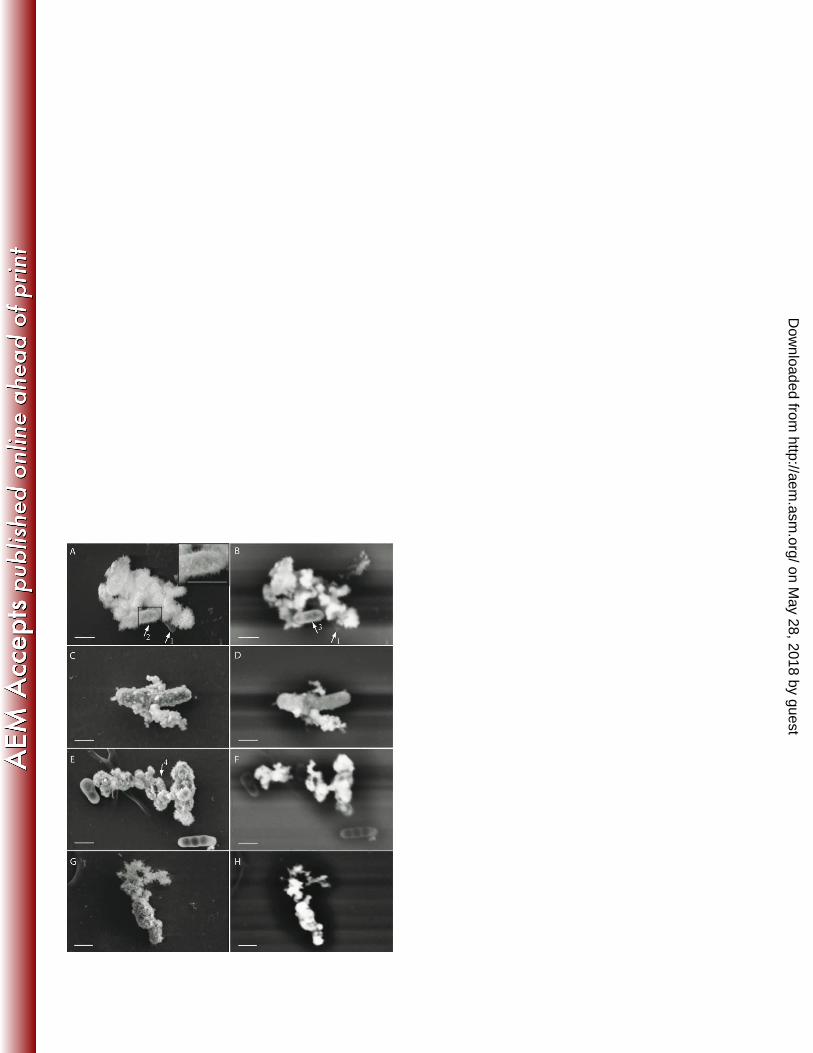


NO2-
NO3- NO2
-
Fe2+ Fe2+
NO2-
Fe2+
Fe3+
Fe2+
NO2-
NO2-
Fe2+
Fe3+
NO
NO2-
Fe2+ Fe3+
NO3-
NO2-
NO2-
Fe2+
Fe3+
Fe2+
Fe2+
NIR T
EPS
1
2
3
P
NAR
H+ H+
a
bc
de
f
g
H+
H+
h
Fe3+
i
Corg
CO2
e- a
cytoplasmperiplasmoutside/cell surface
on May 28, 2018 by guest
http://aem.asm
.org/D
ownloaded from