potential of tomosynthesis as a new modality for ... · each step, we performed an induced pain...
TRANSCRIPT
MEDICAL NOW No.76 (2014.8)
R/F Potential of tomosynthesis as a new modality for evaluating and treating painful shoulders
Satoshi Fujita, M.D.
Department of Orthopedics, Mitsubishi Kyoto Hospital1
Department of Radiological Technology, Mitsubishi Kyoto Hospital2
Department of Orthopaedic Surgery, Kyoto University Hospital3
Satoshi Fujita1, Hiroshi Yamamoto
1, Yoshihiro Uchida
2, Atsushi Irie
2, and Ryuzo Arai
3
1. Introduction
Due to advances in modalities such as MRI and
ultrasound systems, the use of conventional
arthrography of the shoulder joint and the subacromial
bursae is becoming less common. This is presumably
due not only to the relatively low diagnostic accuracy
of conventional arthrography but also to its other
disadvantages, such as allergic reactions to the
use of contrast media and exposure to radiation.
However, arthrography is still often used as a
diagnostic and treatment procedure in clinical
settings because of its strong capability for dynamic
evaluation while simultaneously providing pain
relief through the use of local anesthesia. The most
important advantage of the SONIALVISION safire
series is its high-resolution tomosynthesis capability.
When used in combination with the T-smart function,
which eliminates metal artifacts, the SONIALVISION
safire series allows the use of arthrographic
examinations to perform higher quality pathological
evaluations than were possible previously and is
even capable of reducing artifacts caused by contrast
media. Tomosynthesis combined with contrast
medium enables the visualization of the anatomy
and pathology of bone, cartilage, tendons, and
synovial tissue simultaneously, which is not
possible with MRI and ultrasound systems. Assuming
that this would be especially useful for evaluating and
treating the shoulder joint, which has a ball-and-socket
structure, we started using this modality mainly for
patients with painful shoulders in 2013. The
following report describes our experience using
this system.
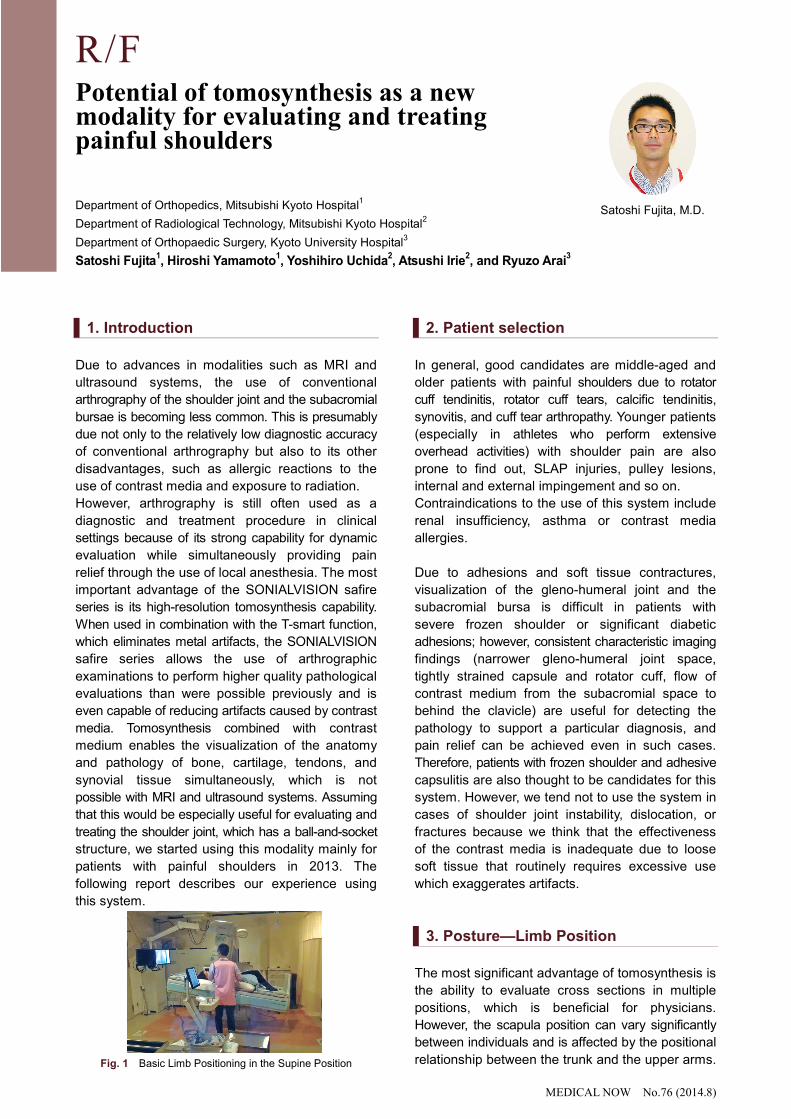
Fig. 1 Basic Limb Positioning in the Supine Position
2. Patient selection
In general, good candidates are middle-aged and
older patients with painful shoulders due to rotator
cuff tendinitis, rotator cuff tears, calcific tendinitis,
synovitis, and cuff tear arthropathy. Younger patients
(especially in athletes who perform extensive
overhead activities) with shoulder pain are also
prone to find out, SLAP injuries, pulley lesions,
internal and external impingement and so on.
Contraindications to the use of this system include
renal insufficiency, asthma or contrast media
allergies.
Due to adhesions and soft tissue contractures,
visualization of the gleno-humeral joint and the
subacromial bursa is difficult in patients with
severe frozen shoulder or significant diabetic
adhesions; however, consistent characteristic imaging
findings (narrower gleno-humeral joint space,
tightly strained capsule and rotator cuff, flow of
contrast medium from the subacromial space to
behind the clavicle) are useful for detecting the
pathology to support a particular diagnosis, and
pain relief can be achieved even in such cases.
Therefore, patients with frozen shoulder and adhesive
capsulitis are also thought to be candidates for this
system. However, we tend not to use the system in
cases of shoulder joint instability, dislocation, or
fractures because we think that the effectiveness
of the contrast media is inadequate due to loose
soft tissue that routinely requires excessive use
which exaggerates artifacts.
3. Posture—Limb Position
The most significant advantage of tomosynthesis is
the ability to evaluate cross sections in multiple
positions, which is beneficial for physicians.
However, the scapula position can vary significantly
between individuals and is affected by the positional
relationship between the trunk and the upper arms.
MEDICAL NOW No.76 (2014.8)
Therefore, even if good tomographic images are
obtained, it is sometimes difficult to determine the
anatomical position to evaluate the images.
Consequently, physicians must carefully determine
the scapula position during this procedure. First,
for basic limb positioning, coronal images in the
supine position are used; however, as mentioned,
because the scapular axis is not parallel to the
table, we insert a mat under the back and hips of
the patient and tilt the R/F table slightly so that the
patient's head is tilted down toward the table
before obtaining the images (Fig. 1). Second,
attention needs to be paid to the arm position
because soft tissues around the humerus move in
three dimensions as the axis of the humerus
rotates as the humeral head is rotated around its
center of the rotation. We think that a slightly
externally rotated arm position from a suspended
neutral position is appropriate as the basic position.
Minor shifts in the angle, especially the rotation
angle of the humerus can be addressed using
reconstruction functions in this system. To use the
advantage of tomosynthesis, we scan cross sections
of the shoulder in neutral, abducted with internal
rotation, or abducted with external rotation positions
or zero position in the supine position (Fig. 2).
Generally, the supraspinatus and infraspinatus
tendons can be evaluated based on the coronal
images in the supine position, and the long head of
the biceps can be evaluated by tracing the groove
with the upper arm in the raised (abducted and
slightly rotated outward, so called zero) position
(Fig. 3). Evaluating the subscapularis muscle can
be difficult in the supine position, so we obtain
images with the patient in the prone position with
the affected limb flexed and internally rotated, with
the unaffected side resting on a pillow followed by
confirming that the scapula is in the axial position
using fluoroscopy (Fig. 4).
(a) (c) (b)
(e) (d)
(g)
(f)
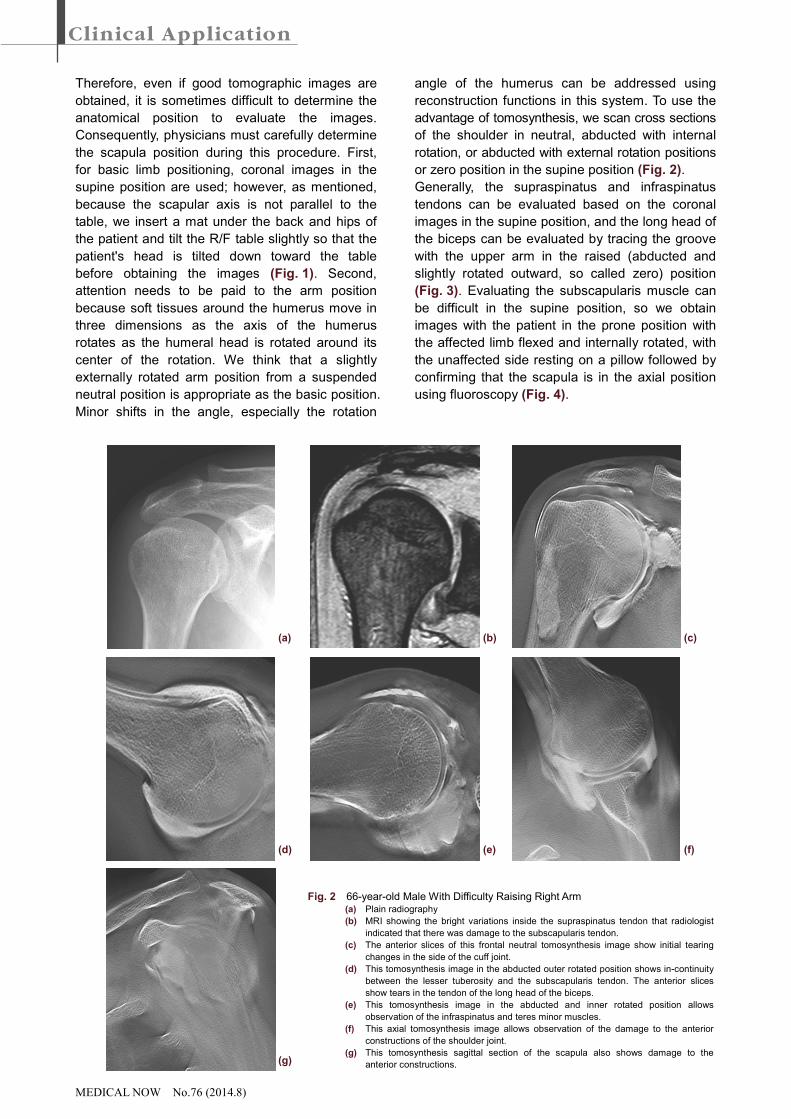
Fig. 2 66-year-old Male With Difficulty Raising Right Arm (a) Plain radiography
(b) MRI showing the bright variations inside the supraspinatus tendon that radiologist
indicated that there was damage to the subscapularis tendon.
(c) The anterior slices of this frontal neutral tomosynthesis image show initial tearing
changes in the side of the cuff joint.
(d) This tomosynthesis image in the abducted outer rotated position shows in-continuity
between the lesser tuberosity and the subscapularis tendon. The anterior slices
show tears in the tendon of the long head of the biceps.
(e) This tomosynthesis image in the abducted and inner rotated position allows
observation of the infraspinatus and teres minor muscles.
(f) This axial tomosynthesis image allows observation of the damage to the anterior
constructions of the shoulder joint.
(g) This tomosynthesis sagittal section of the scapula also shows damage to the
anterior constructions.
MEDICAL NOW No.76 (2014.8)
4. Procedure
Before the examination, a 10-mL syringe, 23-gauge
Cathelin needle, a local anesthetic (1 % Xylocaine),
and contrast medium (Urografin) were prepared.
The contrast medium was inserted in two steps:
into the gleno-humeral joint in the first step and
into the subacromial bursa as the second step. In
each step, we performed an induced pain test to
evaluate pain relief and measure the range of motion.
If the contrast medium flows into the subacromial
bursa after injection into the gleno-humeral joint (in
cases of full thickness tears), we do not perform
the second injection. We normally use a 1:1 ratio of
contrast medium to xylocaine. The quantity of the
contrast medium varies depending on the intra-articular
pressure of the gleno-humeral joint; typically, we
use approximately 8 mL for males and 6 mL for
females into the gleno-humeral joint, and approximately
4 mL for males and 3 mL for females into the
subacromial bursa.
In cases of frozen shoulders, the amount of the
contrast medium is decreased because of elevated
intra-capsular pressure of the gleno-humeral joint.
The contrast medium can be injected without
significant resistance in patients with supple soft
tissues, but injecting an over dose can cause
prominent overshooting (artifacts) in the tomosynthesis
images, even if a thin slice thickness is specified.
Therefore, we only use a maximum of 4 mL of
contrast medium. We do not pump or inject air for
double-contrast purposes because they can
reduce image quality or increase the risk of air
emboli. Care should be taken not to inject even
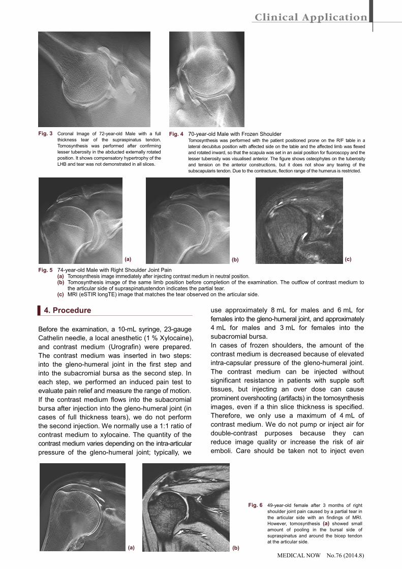
Fig. 3 Coronal Image of 72-year-old Male with a full
thickness tear of the supraspinatus tendon.
Tomosynthesis was performed after confirming
lesser tuberosity in the abducted externally rotated
position. It shows compensatory hypertrophy of the
LHB and tear was not demonstrated in all slices.
Fig. 4 70-year-old Male with Frozen Shoulder Tomosynthesis was performed with the patient positioned prone on the R/F table in a
lateral decubitus position with affected side on the table and the affected limb was flexed
and rotated inward, so that the scapula was set in an axial position for fluoroscopy and the
lesser tuberosity was visualised anterior. The figure shows osteophytes on the tuberosity
and tension on the anterior constructions, but it does not show any tearing of the
subscapularis tendon. Due to the contracture, flection range of the humerus is restricted.
Fig. 5 74-year-old Male with Right Shoulder Joint Pain (a) Tomosynthesis image immediately after injecting contrast medium in neutral position. (b) Tomosynthesis image of the same limb position before completion of the examination. The outflow of contrast medium to
the articular side of supraspinatustendon indicates the partial tear. (c) MRI (eSTIR longTE) image that matches the tear observed on the articular side.
(a) (c) (b)
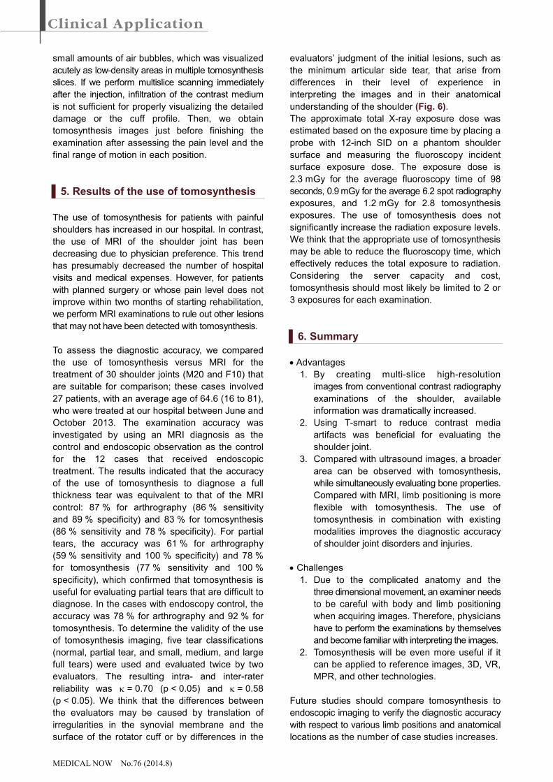
Fig. 6 49-year-old female after 3 months of right
shoulder joint pain caused by a partial tear in
the articular side with an findings of MRI.
However, tomosynthesis (a) showed small
amount of pooling in the bursal side of
supraspinatus and around the bicep tendon
at the articular side.
(a) (b)
MEDICAL NOW No.76 (2014.8)
small amounts of air bubbles, which was visualized
acutely as low-density areas in multiple tomosynthesis
slices. If we perform multislice scanning immediately
after the injection, infiltration of the contrast medium
is not sufficient for properly visualizing the detailed
damage or the cuff profile. Then, we obtain
tomosynthesis images just before finishing the
examination after assessing the pain level and the
final range of motion in each position.
5. Results of the use of tomosynthesis
The use of tomosynthesis for patients with painful
shoulders has increased in our hospital. In contrast,
the use of MRI of the shoulder joint has been
decreasing due to physician preference. This trend
has presumably decreased the number of hospital
visits and medical expenses. However, for patients
with planned surgery or whose pain level does not
improve within two months of starting rehabilitation,
we perform MRI examinations to rule out other lesions
that may not have been detected with tomosynthesis.
To assess the diagnostic accuracy, we compared
the use of tomosynthesis versus MRI for the
treatment of 30 shoulder joints (M20 and F10) that
are suitable for comparison; these cases involved
27 patients, with an average age of 64.6 (16 to 81),
who were treated at our hospital between June and
October 2013. The examination accuracy was
investigated by using an MRI diagnosis as the
control and endoscopic observation as the control
for the 12 cases that received endoscopic
treatment. The results indicated that the accuracy
of the use of tomosynthesis to diagnose a full
thickness tear was equivalent to that of the MRI
control: 87 % for arthrography (86 % sensitivity
and 89 % specificity) and 83 % for tomosynthesis
(86 % sensitivity and 78 % specificity). For partial
tears, the accuracy was 61 % for arthrography
(59 % sensitivity and 100 % specificity) and 78 %
for tomosynthesis (77 % sensitivity and 100 %
specificity), which confirmed that tomosynthesis is
useful for evaluating partial tears that are difficult to
diagnose. In the cases with endoscopy control, the
accuracy was 78 % for arthrography and 92 % for
tomosynthesis. To determine the validity of the use
of tomosynthesis imaging, five tear classifications
(normal, partial tear, and small, medium, and large
full tears) were used and evaluated twice by two
evaluators. The resulting intra- and inter-rater
reliability was κ = 0.70 (p < 0.05) and κ = 0.58
(p < 0.05). We think that the differences between
the evaluators may be caused by translation of
irregularities in the synovial membrane and the
surface of the rotator cuff or by differences in the
evaluators’ judgment of the initial lesions, such as
the minimum articular side tear, that arise from
differences in their level of experience in
interpreting the images and in their anatomical
understanding of the shoulder (Fig. 6).
The approximate total X-ray exposure dose was
estimated based on the exposure time by placing a
probe with 12-inch SID on a phantom shoulder
surface and measuring the fluoroscopy incident
surface exposure dose. The exposure dose is
2.3 mGy for the average fluoroscopy time of 98
seconds, 0.9 mGy for the average 6.2 spot radiography
exposures, and 1.2 mGy for 2.8 tomosynthesis
exposures. The use of tomosynthesis does not
significantly increase the radiation exposure levels.
We think that the appropriate use of tomosynthesis
may be able to reduce the fluoroscopy time, which
effectively reduces the total exposure to radiation.
Considering the server capacity and cost,
tomosynthesis should most likely be limited to 2 or
3 exposures for each examination.
6. Summary
• Advantages
1. By creating multi-slice high-resolution
images from conventional contrast radiography
examinations of the shoulder, available
information was dramatically increased.
2. Using T-smart to reduce contrast media
artifacts was beneficial for evaluating the
shoulder joint.
3. Compared with ultrasound images, a broader
area can be observed with tomosynthesis,
while simultaneously evaluating bone properties.
Compared with MRI, limb positioning is more
flexible with tomosynthesis. The use of
tomosynthesis in combination with existing
modalities improves the diagnostic accuracy
of shoulder joint disorders and injuries.
• Challenges
1. Due to the complicated anatomy and the
three dimensional movement, an examiner needs
to be careful with body and limb positioning
when acquiring images. Therefore, physicians
have to perform the examinations by themselves
and become familiar with interpreting the images.
2. Tomosynthesis will be even more useful if it
can be applied to reference images, 3D, VR,
MPR, and other technologies.
Future studies should compare tomosynthesis to
endoscopic imaging to verify the diagnostic accuracy
with respect to various limb positions and anatomical
locations as the number of case studies increases.