potential of environmental enrichment to prevent .../file/...potential of environmental enrichment...
TRANSCRIPT

Potential of Environmental Enrichment to PreventTransgenerational Effects of Paternal Trauma
Katharina Gapp1,3, Johannes Bohacek1, Jonas Grossmann2, Andrea M Brunner1,4, Francesca Manuella1,Paolo Nanni2 and Isabelle M Mansuy*,1
1Laboratory of Neuroepigenetics, University of Zurich and ETH Zurich, Brain Research Institute, Neuroscience Center Zürich, Zurich, Switzerland;2Functional Genomics Center Zurich (FGCZ), University Zurich/ETH Zurich, Zurich Switzerland
Adverse experiences in early life are risk factors for the development of behavioral and physiological symptoms that can lead to psychiatricand cognitive disorders later in life. Some of these symptoms can be transmitted to the offspring, in some cases by non-genomicmechanisms involving germ cells. Using a mouse model of unpredictable maternal separation and maternal stress, we show that postnataltrauma alters coping behaviors in adverse conditions in exposed males when adult and in their adult male progeny. The behavioral changesare accompanied by increased glucocorticoid receptor (GR) expression and decreased DNA methylation of the GR promoter in thehippocampus. DNA methylation is also decreased in sperm cells of exposed males when adult. Transgenerational transmission ofbehavioral symptoms is prevented by paternal environmental enrichment, an effect associated with the reversal of alterations in GR geneexpression and DNA methylation in the hippocampus of the male offspring. These findings highlight the influence of both negative andpositive environmental factors on behavior across generations and the plasticity of the epigenome across life.Neuropsychopharmacology advance online publication, 6 July 2016; doi:10.1038/npp.2016.87
��������������������������������������
INTRODUCTION
Exposure to traumatic and stressful events in early life canhave physiological and behavioral consequences that persistacross the lifespan. Such experiences often increase the riskto develop psychiatric and cognitive disorders in adulthood(Klengel and Binder, 2015). Further, their effects can betransmitted to subsequent generations and affect the off-spring similarly to the ancestors in the absence of any traumaor stress exposure (Bohacek and Mansuy, 2015; Metz et al,2015). In some cases, however, negative experiences canhave some benefits and lead to better adapted physiologicaland behavioral responses. Resilience is a form of adaptiveresponse manifested by active or passive coping in adverseconditions (Franklin et al, 2012) that can follow previoustrauma exposure. For instance, social avoidance is viewed asan active coping behavior that can be manifested by an adultindividual after being exposed to traumatic stress in early
postnatal life (Franklin et al, 2011) or chronic social defeat inadulthood (Kovalenko et al, 2014). In the case of earlytraumatic stress, social avoidance has been shown to beaccompanied by altered behavioral flexibility, and both aretransmitted to the progeny (Franklin et al, 2011; Gapp et al,2014b). Further, some maladaptive behaviors resulting fromearly trauma can be modulated by the environment; inparticular, they can be reversed by conditions such asenvironmental enrichment (EE) (Leshem and Schulkin,2012; Schloesser et al, 2010). This suggests that the effectsof early life trauma, whether detrimental or beneficial, andtheir transmission might be counteracted by environmentalfactors later in life.Mechanistically, the consequences of early exposure to
trauma are complex, and implicate a combination ofsignaling pathways that engage the hypothalamic–pitui-tary–adrenal (HPA) axis. In this axis, the glucocorticoidreceptor (GR) is a major component of stress responsesneeded for the rapid shut down of the stress response(de Kloet et al, 2005a) and for long-term adaptive processesafter chronic stress (Jankord and Herman, 2008; Liu et al,1997; Plotsky and Meaney, 1993), especially in the context ofactive avoidance behavior (Patacchioli et al, 1990). GR geneis subjected to epigenetic regulation (Weaver et al, 2004), andis particularly responsive to environmental factors in earlylife (Mueller and Bale, 2008).Here, we used a mouse model of unpredictable maternal
separation combined with unpredictable maternal stress(MSUS) to examine the consequences of traumatic stress on
*Correspondence: Professor IM Mansuy, Brain Research Institute,University Zurich/Swiss Federal Institute of Technology, Winterthur-erstrasse 190, Zurich 8057, Switzerland, Tel: +41 44 635 33 60, Fax:+41 44 635 33 03, E-mail: [email protected] address: Gurdon Institute, University of Cambridge, TennisCourt Road, Cambridge CB2 1QN, UK.4Current address: Biomolecular Mass Spectrometry and Proteomics,Bijvoet Center for Biomolecular Research and Utrecht Institute forPharmaceutical Sciences, University of Utrecht, Padualaan 8, 3584CHUtrecht, The Netherlands.Received 15 February 2016; revised 9 May 2016; accepted 28 May2016; accepted article preview online 9 June 2016
Neuropsychopharmacology (2016), 1–10© 2016 American College of Neuropsychopharmacology. All rights reserved 0893-133X/16
www.neuropsychopharmacology.org

coping behaviors in adulthood and across generations, andthe potential contribution of GR. We show that MSUS affectsavoidance behaviors and learning in aversive environmentsin exposed fathers and their male offspring. This isassociated with an increase in GR expression in thehippocampus, and with decreased DNA methylation of GRpromoter in the hippocampus and in germ cells. We showthat transmission of the effects of paternal trauma can beprevented by paternal EE, suggesting a reversibility of theseeffects.
MATERIALS AND METHODS
Animals
C57Bl/6J male and female mice were maintained under areverse light–dark cycle in a temperature- and humidity-controlled facility. Three to five mice per cage were housedwith water and food ad libitum and cages were cleaned oncea week. All experimental manipulations were performedduring the animals’ active cycle and in accordance withguidelines and regulations of the cantonal veterinary officeZurich under license 55/2012.
MSUS Treatment and Breeding
Unpredictable MSUS was conducted as previously described(Franklin et al, 2010). Briefly, adult males and females (F0)were mated, males were removed after a few days andfemales were maintained alone during gestation untildelivery. Newborn pups (F1) were separated from theirmother unpredictably (any time during the dark cycle) dailyfor 3 h. During separation, dams were exposed to anadditional stress consisting of either 20-min restraint in aPlexiglas tube or 5-min forced swim in cold water (18 °C)applied randomly any time during the 3 h of separation.MSUS was conducted from PND1 to PND14. Dams andpups were then left undisturbed from PND15 until weaningat PND21. About 20–30 females were bred for each MSUSexperiment and only dams giving birth less than a week apartof each other were used to obtain F1 pups. Litters with lessthan four pups were excluded. When adult, F1 control andMSUS males were bred with naive C57Bl/6J females togenerate an offspring (F2).
Behavioral Testing
The experimenter was blind to treatment for all behavioraltesting and tracking was performed manually and auto-matically (Viewpoint System, France). Testing started withthe least aversive task (light–dark box) when animals were atleast 3 months old. All behavioral tests were carried out inmales to avoid confounds related to the estrus cycle infemales.
Light–Dark Box
Mice were placed in the lit compartment of a plastic box(40 × 42 × 26 cm) separated from a dark compartment by adivider. The lit (aversive) compartment is large (2/3 of thebox), has white walls, and is brightly lit by an overhead
lightbulb. The neighboring dark (safe) compartment is small(1/3 of the box), has black walls, and is covered by a black lid.An opening in the divider (5 × 5 cm) allows the animal toescape from the lit compartment to the dark compartment.Each animal was tested for 10 min and the time spent in eachcompartment and the latency to enter the dark compartmentwere measured manually. Light dark box data from thestandard housed groups were reported before in Gapp et al(2014a).
Active Avoidance Task
Four identical operant conditioning chambers (15.9 × 16.5× 17.5 cm) with stainless steel grid floors (TSE Systems,Germany) were placed in sound-insulated boxes. Eachchamber is equipped with a shock grid floor, a nose-pokeresponse unit (2 cm diameter) fitted with a photocell sensor,and a yellow cue light in a hole. An additional green cue lightis located on the wall on top of the nose-poke module.A house light (2.8W) is placed on the ceiling, and is turnedon during each testing session. The nose-poke module islocated on the left part of one wall, which also had aphotocell sensor. Each mouse was habituated to the chamberone day for 30 min with the nose-poke module closed. Afterhabituation, animals were tested for active avoidance in a30-min session with 60 escape trials induced by 0.3 mA foot-shocks delivered every 30 s. Foot-shocks were terminatedwhen the animal had a nose-poke, and lasted a maximum of10 s. For each trial, the green light was turned on during non-shock periods, and the red light was turned on during shockperiods. The sensor located in the nose poke hole recordedthe timing of each nose poke with 1 s resolution. Latency toescape and number of escapes were measured in blocks offive trials.
Fixed Ratio Paradigm
Water deprived mice were tested 5 days a week on the fixedration paradigm and provided water access for 1 h per day.Each operant conditioning chamber was equipped asdescribed above, but further contained a liquid dispenserfitted with a photocell sensor, situated in the middle of thewall left to the nose-poke module. Mice were habituated tothe chamber for 30 min, during which 10 μl of water(= liquid reinforcer) were delivered to the liquid dipperevery 30 s. Mice were trained on a continuous fixed ratio(FR) schedule for which they had to nose-poke for thedelivery of a liquid reinforcer during daily 30-min sessionsacross 12 days. Activation of the nose-poke led to delivery of10 μl water in the liquid dipper. Learning was determined bythe ratio of total number of nose-pokes over total number ofcollected drink rewards.
Environmental Enrichment
Control and MSUS mice were placed in social groups(n= 12) in an enriched cage from weaning till adulthood.An enriched cage is a large box with two levels(55 × 36 × 19 cm bottom level, 55 × 36 × 11 cm upper level)(Marlau; Viewpoint). The bottom level is split into twocompartments, one containing food pellets and the otherproviding access to water and containing running wheels,
Effects of paternal trauma and environmental enrichmentK Gapp et al
2
Neuropsychopharmacology

and a covered/protected area. The upper level has a maze(35 × 36 × 11 cm) whose shape and configuration are chan-ged three times per week with a total of 12 different options.The box is organized such that animals have to go throughthe maze to reach the food compartment.
Real-Time Quantitative RT-PCR
DNaseI-treated RNA isolated from hippocampus (AllprepRNA/DNA kit; Qiagen) was reverse transcribed (RT) usingthe SuperScript First-Strand Synthesis System II for RTpolymerase chain reaction (PCR; Invitrogen Carlsbad,California). Quantitative RT-PCR (qRT-PCR) was performedin an ABI 7500 thermal cycler using TaqMan probes (AppliedBiosystems, Foster City, California; Mm00433832_m1) asdescribed previously(Franklin et al, 2010).
Bisulfite Pyrosequencing
Genomic DNA from the hippocampus, prefrontal cortex,and sperm was extracted with the Allprep (Qiagen) andDNeasy blood and tissue (Qiagen) kit, respectively, accord-ing to the manufacturer’s instructions. Bisulfite pyrosequen-cing of offspring hippocampus was performed by EpigenDx,USA. Universally methylated and unmethylated DNAsamples (Millipore Bioscience Research Regents) were usedas controls. Pyrosequencing of hippocampus, prefrontalcortex and sperm was performed as described previously(Bohacek et al, 2014). Amplicons containing the GR exon1-7promoter region were generated using a standard PCRprotocol, an unmodified forward primer (5′-GGTTTTGTAGGTTGGTTGTTATTT-3′), and a biotin-labeled reverse primer(5′-ATTTCTTTAATTTCTCTTCTCCCTAAC-3′). For high-resolution sequencing, the following sequencing primer wasused 5′-TTGTAGGTTGGTTGTTATTTTT-3′.
Samples Preparation, iTRAQ Labeling, MassSpectrometry Analyses and Protein Quantification
Hippocampi dissected from adult mice were homogenized in200 μl lysis buffer (100 mM triethyl ammonium bicarbonatepH ~ 8, 0.1% SDS, 2 M urea) by up-and-down strokes using a27 gauge syringe, sonicated for 2 min, and centrifuged at13 000 g for 10 min to remove insoluble material. Proteins(50 μg) were precipitated using six volumes ice-cold acetone,solubilized, reduced, and cysteines were blocked according tothe manufacturer’s protocol. Proteins were then digested intopeptides with trypsin overnight (1 : 10 enzyme : substrate).Peptides were differentially labeled with iTRAQ 8-plexreagents (Applied Biosystems) according to the manufac-turer’s protocol then combined. Peptide samples weredesalted and separated in-solution with an Agilent 3100OFFGEL fractionator (Agilent Technologies) according tothe manufacturer’s protocol. Fractions were acidified byadding 50% acetonitrile (ACN), 1% trifluoroacetic acid(1 : 10), and detergents were removed using ZipTip C18columns (Millipore). Samples were lyophilized then resus-pended in 3% ACN, 0.1% formic acid for LTQ-OrbitrapVelos analysis using collision-induced dissociation (CID),and higher energy collisional dissociation fragmentation asdescribed in Uzozie et al (2014). In brief, for the eight most
intense signals per cycle above a threshold of 1000, both CIDand higher-energy collisional dissociation spectra wereacquired in a data-dependent manner. Mascot Distiller2.4.3.3 (Matrix Science, Boston, MA) was used to generateMascot generic format peak lists. De-isotoping and peakpicking were not performed between 112.5 and 121.5m/z(range containing iTRAQ (isobaric tags for relative andabsolute quantitation) reporter ions), and the CID andhigher-energy collisional dissociation spectra were mergedby summing. Peptide and protein identification wasperformed with ProteinPilot software v4.5 (AB Sciex) andthe Paragon algorithm 4.5.0.0 (Shilov et al, 2007), bycomparison with the mouse Swissprot/TrEMBL database(downloaded in May 2009). OGE fractions obtained by F1analyses were searched separately from F2. Paragon methodparameters were peptide labeled with iTRAQ 8plex, fixedmodification of methyl methanethiosulfonate on Cys(+46 Da), digestion with trypsin, instrument Orbi/FT MS(1–3 ppm) LTQ MS/MS and ID focus on biologicalmodification with the thorough search effort. Proteins withat least one peptide above 90% confidence level asdetermined by Protein Pilot were recorded. For theestimation of protein abundance ratio, intensity of iTRAQreporter ion areas for each non-shared, non-discordant,quantifiable, and confidently identified MS/MS spectra wasexported from ProteinPilot. For each protein, the sum ofeach reporter channel was calculated across spectra matchedto the same protein. Data were normalized for loading errorby bias corrections using ProteinPilot. Statistical significanceof differences between means for each group (Control andMSUS mice) was determined on the transformed proteinsums (arc sin hyperbolic) using a two‐tailed t-test. Foldchanges were calculated using the group means of theprotein sums. Proteins were considered regulated if the P-value waso0.05 and fold-change between groups41.3-fold.Mass spectrometry proteomics data are deposited to theProteomeXchange Consortium via the PRIDE [1] partnerrepository (identifier PXD004073).
Statistical Analyses
Two-way ANOVAs with treatment (Control and MSUS) andhousing (standard and EE) as main factors were used toanalyze data of light dark test, qRT-PCR, and bisulfitepyrosequencing. Repeated measurements ANOVAs withtreatment and housing as main factors, and within factorof testing session (in blocks of 5 sessions), were used toanalyze data of the active avoidance task. Repeatedmeasurements ANOVAs with treatment as a main factorand testing session as within factor were used to analyze dataof the FR paradigm. Two-tailed Student’s t-tests were used toanalyze qRT-PCR and bisulfite pyrosequencing data whencomparing MSUS and control data in standard housingconditions only. All analyzed data matched the requirementsfor parametric statistical tests. If variance was not homo-geneous between groups, adjusted P-value, t-value, anddegree of freedom were reported. A value was consideredoutlier when deviating 42 SDs from the group mean, andoutlier exclusion criterion was pre-established. Significancewas set at Po0.05 for all tests. Error bars represent standarderror of the mean (SEM).
Effects of paternal trauma and environmental enrichmentK Gapp et al
3
Neuropsychopharmacology

RESULTS
Effects of MSUS on Coping Behaviors in AversiveConditions
We used a light–dark box to assess behavioral response inmildly aversive conditions in males exposed to MSUS (F1)and their male offspring (F2) (Supplementary Figure S1). Onthis task, an animal can escape from a brightly lit aversivecompartment of a box by moving to a dark compartment. F1MSUS males spent more time in the bright compartment ofthe box than controls (t(35)=− 2.14, Po0.05; SupplementaryFigure S2a). Similarly, the F2 MSUS males also spent moretime in the bright compartment (t(41.61)=� 3, Po0.01;Figure 1a), indicating reduced escape response suggestive of aform of resistance to aversive conditions in both generations.This effect was independent from the latency to first enter thedark compartment since it was similar in all groups (fathers:t(34)= 0.023, P40.05, Supplementary Figure S2b; offspring,t(63) = 0.03, P40.05; Figure 1b). We confirmed this effect bytesting the males in even more aversive conditions using anactive avoidance task. On this task, an animal is exposed to afoot-shock that can be terminated by a nose-poke into ahole. F2 MSUS males had shorter latency to nose-poke(F(1,23)= 8.50, P⩽ 0.01) and thus received fewer foot-shocksthan control offspring across time (F(1,26)= 7.74, P⩽ 0.01)(Figure 1c and d), suggesting more active coping response.This was not due to improved learning since F2 MSUS andcontrol males had similar nose-poke performance on a FRparadigm, a reward-based task for which a water-deprivedanimal learns to nose-poke into one hole to obtain a waterreward in a neighboring hole (F(1,26)= 1.33, P40.05)
(Supplementary Figure S3). These results suggest transmissionof altered coping behaviors across generations.
Reversibility of Altered Responses in AversiveConditions
EE has been reported to reverse some of the behavioralsymptoms induced by chronic stress (Hutchinson et al, 2012)or early life adversity (Leshem and Schulkin, 2012). UsingEE, we examined if the effects of MSUS on coping behaviorscan be reversed (Supplementary Figure S4). We exposedF1 MSUS males to EE from weaning till adulthood. After EE,F1 males were bred to control females to generate anF2 generation, and both F1 and F2 males were tested on thelight–dark box. When compared with F1 control malesraised in standard cages, F1 control males raised in EEspent more time in the lit compartment of the box(t(52)=− 0.9, Po0.05) and had shorter latency to enter thedark compartment (t(52)= -2.11; Po0.05, SupplementaryFigure S2), suggesting an effect of EE on response to adverseconditions. F1 MSUS males exposed to EE had similarperformance to control males exposed to EE; they spent acomparable amount of time in the lit compartment(t(15)=− 0.78, P40.05) and had similar latency to firstenter the dark compartment (t(16)=− 0.38), P40.05)(Supplementary Figure S2c and d). However, their F2 maleoffspring spent a similar amount of time in the litcompartment (t(20.85)= 1.94, P40.05) and had lowerlatency (t(17.89)= 2.33, Po0.05) than the F2 offspring ofcontrol EE fathers (Figure 2a and b), suggesting a reversal ofthe altered response to an adverse environment by paternalEE. Decreased latency may indicate more spontaneousescape behavior in the F2 offspring of MSUS males exposed
Figure 1 Aversive behavior in the offspring of males exposed to maternal separation combined with unpredictable maternal stress (MSUS) in postnatal life.Performance in a light–dark box shown by (a) time spent in the bright compartment in control and MSUS (Controls n= 33, MSUS n= 36) and (b) latency tofirst enter the dark compartment (Controls n= 34, MSUS n= 38). (c) The latency to escape a foot-shock and (d) the total number of foot-shocks on theactive avoidance task (Controls n= 15, MSUS n= 13). Data are presented as mean± SEM, *Po0.05. Part of the data were published in Gapp et al. (2014a).
Effects of paternal trauma and environmental enrichmentK Gapp et al
4
Neuropsychopharmacology
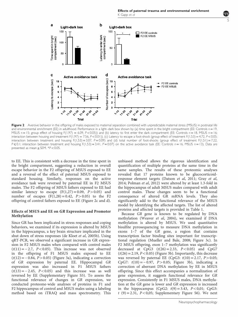
to EE. This is consistent with a decrease in the time spent inthe bright compartment, suggesting a reduction in overallescape behavior in the F2 offspring of MSUS exposed to EEand a reversal of the effect of paternal MSUS exposed tostandard housing. Similarly, responses on the activeavoidance task were reversed by paternal EE in F2 MSUSmales. The F2 offspring of MSUS fathers exposed to EE hadsimilar latency to escape (F(1,27)= 0.09, P40.05) andnumber of escapes (F(1,28)= 0.42, P40.05) to the F2offspring of control fathers exposed to EE (Figure 2c and d).
Effects of MSUS and EE on GR Expression and PromoterMethylation
Since GR has been implicated in stress responses and copingbehaviors, we examined if its expression is altered by MSUSin the hippocampus, a key brain structure implicated in theshut down of stress responses (de Kloet et al, 2005b). UsingqRT-PCR, we observed a significant increase in GR expres-sion in F2 MSUS males when compared with control males(t(11)=− 2.7, Po0.05). This increase was not observedin the offspring of F1 MSUS males exposed to EE(t(12)=− 0.84, P40.05) (Figure 3a), indicating a correctionof GR expression by paternal EE. Hippocampal GRexpression was also increased in F1 MSUS fathers(t(13)=− 2.45, Po0.05) and this increase was as wellreversed by EE (Supplementary Figure S5). To assess thefunctional relevance of changes in GR expression, weconducted proteome-wide analyses of proteins in F1 andF2 hippocampus of control and MSUS males using a labelingmethod based on iTRAQ and mass spectrometry. This
unbiased method allows the rigorous identification andquantification of multiple proteins at the same time in thesame samples. The results of these proteomic analysesrevealed that 17 proteins known to be glucocorticoid-response element targets (Datson et al, 2011; Gray et al,2014; Polman et al, 2012) were altered by at least 1.3-fold inthe hippocampus of adult MSUS males compared with adultcontrol males. These changes seem to be a functionalconsequence of altered GR mRNA levels. They alsosignificantly add to the functional relevance of the MSUSmodel by identifying the affected targets. The list of alteredproteins and affected targets is provided in Table 1.Because GR gene is known to be regulated by DNA
methylation (Weaver et al, 2004), we examined if DNAmethylation is altered by MSUS. We used quantitativebisulfite pyrosequencing to measure DNA methylation inexons 1–7 of the GR gene, a region that containstranscription factor binding sites important for transcrip-tional regulation (Mueller and Bale, 2008; Figure 3c). InF2 MSUS offspring, exon 1–7 methylation was significantlydecreased at CpG3 (t(26)= 2.33, Po0.05) and CpG7(t(26)= 2.19, Po0.05) (Figure 3b). Importantly, this decreasewas reversed by paternal EE (CpG3: t(10)= 2.17, P40.05;CpG7: t(10)=− 0.97, P40.05; Figure 3b), indicating acorrection of aberrant DNA methylation by EE in MSUSoffspring. Since this effect accompanies a normalization ofgene expression, it suggests functional relevance for GRexpression. Consistently in F1 MSUS males, DNA methyla-tion at the GR gene is lower and GR expression is increasedin the hippocampus (CpG2: t(9)= 3.43, Po0,01; CpG3:t (9)= 2.31, Po0,05; Supplementary Figure S6). We next
Figure 2 Aversive behavior in the offspring of males exposed to maternal separation combined with unpredictable maternal stress (MSUS) in postnatal lifeand environmental enrichment (EE) in adulthood. Performance in a light–dark box shown by (a) time spent in the bright compartment (EE: Controls n= 19,MSUS n= 15; group effect of housing F(1,97) = 6.09, Po0.05)) and (b) latency to first enter the dark compartment (EE: Controls n= 18, MSUS n= 16;interaction between housing and treatment F(1,97) = 7.56, Po0.01)). (c) Latency to escape a foot-shock (group effect of treatment F(1,53)= 4.72, Po0.05;interaction between treatment and housing F(1,53)= 3.07, P= 0.09) and (d) total number of foot-shocks (group effect of treatment F(1,51)= 7.22,P⩽ 0.1; interaction between treatment and housing F(1,53)= 3.41, P= 0.07) on the active avoidance task (EE: Controls n= 16, MSUS n= 13). Data arepresented as mean± SEM. *Po0.05.
Effects of paternal trauma and environmental enrichmentK Gapp et al
5
Neuropsychopharmacology

examined if the altered DNA methylation is passed to theoffspring from fathers by analyzing DNA methylation insperm cells of F1 males. DNA methylation was significantlydecreased at CpG3 (t (19.4)= 2.77, Po0.05, Po0.05) andCpG7 (t (20)= 2.3, Po0.05), consistent with hypomethyla-tion at the same CpG in the offspring’s hippocampus. Wefurther detected a decrease at CpG1 (t(14.03)= 3.47,Po0.01), CpG8 (t(14.07)= 3.48, Po0.01), and CpG9(t(14.15)= 3.5, Po0.01) (Figure 4). Importantly, these effectsdid not persist in the sperm of F1 fathers that had been
exposed to EE, suggesting a correction of the effects of MSUSby EE at the germline level (CpG1 t(8.03)=− 19, P40.05,Po0.05; CpG3 t((8.02)=− 2.09, P40.05, Po0.05; CpG7t(9)=− 2.26, P= 0.05, CpG8 t((−8.01)=− 2; P40.05,P40.05; CpG9 t(8.03)=− 2.12, P40.05; Figure 4).DNA methylation induced by MSUS in sperm may be
maintained after fertilization and during development, andthus be present in different tissues in the offspring. Weexamined this question by assessing DNA methylation inanother brain region, the prefrontal cortex, in adult
Figure 3 Molecular analysis in the F2 offspring of F1 fathers exposed to maternal separation combined with unpredictable maternal stress (MSUS) andenvironmental enrichment (EE). (a) Glucocorticoid receptor (GR) gene expression measured by quantitative reverse transcriptase-PCR (Standard housing(SH): Controls n= 6, MSUS n= 6; EE: Controls n= 7, MSUS n= 5; group effect of treatment F(1,21)= 7.74, P⩽ 0.01). (b) DNA methylation at GR exon 1–7promoter in the hippocampus assessed by bisulfite pyrosequencing (SH: Controls n= 14, MSUS n= 14; EE: Controls n= 7, MSUS n= 5; group effect oftreatment at CpG2 F(1,20.8)= 0.05 and at CpG3: F(1,61)= 7.94, P⩽ 0.01; a tendency for group effect of housing at CpG3: F(1,23.31)= 3.03 and aninteraction between housing and treatment at CpG4: F(1;43,9)= 3.97, P⩽ 0.05). (c) Schematic representation of the GR gene and the untranslated exon 1–7promoter sequence examined for DNA methylation by pyrosequencing assays including the transcription start site (+1). Predicted transcription factor bindingsites are underlined and in italic. Exons are shown as black boxes. Numbers represent individual CpG sites analyzed for DNA methylation (CpG 1–10). Dataare presented as mean± SEM, *Po0.05 t-test comparison.
Effects of paternal trauma and environmental enrichmentK Gapp et al
6
Neuropsychopharmacology

Table 1 Proteome-Wide Protein Expression in the Hippocampus of F1 MSUS Males and Their F2 Male Offspring
Generation Protein accessionnumber
Gene name Regulation ValidatedGR target
% Control Generation Protein accessionnumber
Gene name Regulation ValidatedGR target
% Control
F1 fathers Q69Z96 Atp13a1 Down 420.0 F2 offspring O08908 Pik3r2 Down 63.6
F1 fathers O55142 Rpl35a Down 65.9 F2 offspring O55142 Rpl35a Down 42.8
F1 fathers O35343 Kpna4 Down 62.1 F2 offspring Q3UKW2 Calm1 Down 44.5
F1 fathers Q9Z239 Fxyd1 Down 63.8 F2 offspring Q9Z239 Fxyd1 Down 62.4
F1 fathers B9EKC3 Arhgap5 Down 61.0 F2 offspring Q62178 Sema4a Down 47.5
F1 fathers P21995 Emb Down 64.9 F2 offspring Q71M36 Cspg5 Down 63.5
F1 fathers O35874 Slc1a4 Down X 69.5 F2 offspring Q9DCZ4 Apoo Down 61.6
F1 fathers Q80U23 Snph Down X 74.2 F2 offspring O08599 Stxbp1 Down 64.0
F1 fathers Q8JZK9 Hmgcs1 Down 70.4 F2 offspring P35802 Gpm6a Down 61.5
F1 fathers Q6GQX8 Gm15800 Down 70.9 F2 offspring P55096 Abcd3 Down 66.2
F1 fathers Q7TN99 Cpeb3 Down 73.6 F2 offspring Q8BGZ4 Cdc23 Down 60.1
F1 fathers Q9JMG7 Hdgfrp3 Down X 70.2 F2 offspring Q8K386 Rab15 Down 60.7
F1 fathers P62137 Ppp1ca Down 76.2 F2 offspring Q9CQE1 Nipsnap3b Down 57.0
F1 fathers Q9QZB1 Rgs20 Down 68.8 F2 offspring A2BI30 RP24-297H17.3 Down 64.2
F1 fathers Q08460 Kcnma1 Down X 71.2 F2 offspring Q8BH80 Vapb Down X 66.0
F1 fathers Q69ZP3 Pnkd Down X 72.9 F2 offspring Q9CPU4 Mgst3 Down 52.7
F1 fathers P39053 Dnm1 Down 74.7 F2 offspring P07724 Alb Down 65.7
F1 fathers Q99JR1 Sfxn1 Down 76.0 F2 offspring Q91W34 C16orf58 Down 64.5
F1 fathers Q9CW46 Raver1 Down 69.8 F2 offspring B9EKN8 Tnik Down 66.2
F1 fathers Q9D125 Mrps25 Down 74.2 F2 offspring Q9CPP6 Ndufa5 Down 56.1
F1 fathers B1ARB9 Ddx5 Down 76.2 F2 offspring Q9CZB4 Apool Down X 30.4
F1 fathers Q9D6D0 Slc25a27 Down 76.8 F2 offspring Q8K356 Ly6h Down 58.2
F1 fathers Q91W39 Ncoa5 Down 75.9 F2 offspring O70503 Hsd17b12 Down 58.4
F1 fathers Q8VCE2 Gpn1 Down 72.5 F2 offspring Q3U0D9 Hace1 Down 66.0
F1 fathers Q9D6U8 Fam162a Down 75.6 F2 offspring Q811S7 Ubp1 Down 53.8
F1 fathers Q8R1N4 Nudcd3 Down 70.1 F2 offspring Q9ES97 Rtn3 Down 65.6
F1 fathers Q9DC51 Gnai3 Down 72.4 F2 offspring Q9Z2W9 Gria3 Down 63.4
F1 fathers Q52KF3 Spire1 Up X 165.8 F2 offspring Q78IK2 Usmg5 Down 60.4
F1 fathers Q8C0W0 Tmsb15l Up 154.7 F2 offspring Q62277 Syp Down 67.3
F1 fathers P14069 S100a6 Up X 151.2 F2 offspring P52479 Usp10 Down X 71.0
F1 fathers Q9JIG8 Praf2 Up 169.2 F2 offspring Q9JHU9 Isyna1 Down 76.6
F1 fathers P59279 Rab2b Up 168.0 F2 offspring P21278 Gna11 Down 71.1
F1 fathers Q8BRF7 Scfd1 Up 150.4 F2 offspring Q8BGZ1 Hpcal4 Down 74.6
F1 fathers O88935 Syn1 Up 181.2 F2 offspring Q91VC3 Eif4a3 Down 73.2
F1 fathers A2AFJ1 Rbbp7 Up 136.4 F2 offspring Q921W4 Cryzl1 Down 66.7
F1 fathers Q62048 Pea15 Up 131.6 F2 offspring Q924M7 Mpi Down 75.8
F1 fathers Q3U409 Ablim2 Up 136.6 F2 offspring P63082 Atp6v0c Down 70.6
F1 fathers B1ASW5 Trappc1 Up 141.7 F2 offspring Q7JCY6 mt-Nd4 Down 68.6
F1 fathers Q9D3D9 Atp5d Up 136.2 F2 offspring P56564 Slc1a3 Down 72.8
F1 fathers Q99LY9 Ndufs5 Up 132.3 F2 offspring Q9D9M2 Usp12 Down 70.7
F1 fathers Q99M87 Dnaja3 Up X 148.6 F2 offspring Q9CR57 Rpl14 Down 72.1
F1 fathers P0C7L0 Wipf3 Up X 136.3 F2 offspring P39688 Fyn Down 72.7
F1 fathers B2RS21 Nudt19 Up 131.6 F2 offspring Q60930 Vdac2 Down X 71.6
F1 fathers P31786 Dbi Up 135.3 F2 offspring Q8CIP4 Mark4 Down 75.1
F1 fathers Q3TY60 Fam131b Up 138.2 F2 offspring Q9CQN6 Tmem14c Down 73.7
F1 fathers Q9JJU8 Sh3bgrl Up 131.8 F2 offspring Q3TCD4 Eci2 Down 69.8
F1 fathers O08529 Capn2 Up 131.4 F2 offspring Q8BG32 Psmd11 Down X 74.0
F1 fathers O35449 Prrt1 Up X 138.4 F2 offspring Q9Z1X2 Ptdss2 Down 66.8
F1 fathers P62274 Rps29 Up X 132.9 F2 offspring O70579 Slc25a17 Up 152.1
F1 fathers P62996 Tra2b Up 140.3 F2 offspring Q2PFD7 Psd3 Up 154.1
F1 fathers Q68ED7 Crtc1 Up 142.6 F2 offspring Q8VHH5 Agap3 Up 138.4
F1 fathers Q8BK30 Ndufv3 Up 147.1 F2 offspring Q60972 Rbbp4 Up 138.1
F1 fathers Q9D1Q6 Erp44 Up 133.0 F2 offspring Q8BGD9 Eif4b Up 131.1
F1 fathers Q3U7M5 Abhd4 Up 142.1 F2 offspring B1AWD9 Clta Up 133.2
F1 fathers Q9DBB8 Dhdh Up 141.7 F2 offspring Q9JLT4 Txnrd2 Up 141.5
F1 fathers B1AQE6 Ikzf3 Up 144.0 F2 offspring Q7TN29 Smap2 Up 146.8
F1 fathers Q3UH27 Asap2 Up 139.6 F2 offspring Q52KR3 Prune2 Up 133.5
F1 fathers P16390 Kcna3 Up X 131.7 F2 offspring Q61166 Mapre1 Up 144.9
F1 fathers Q52KR3 Prune2 Up 133.3 F2 offspring Q6R891 Ppp1r9b Up 130.5
F2 offspring A2AN47 Golga2 Up 131.7
F2 offspring P97930 Dtymk Up 140.0
Proteins with differential expression (Po0.05) in MSUS and controls and a minimal fold change of 1.3 measured by iTRAQ followed by mass spectrometry. Proteinswith significanlty different level (p⩽ 0.05) of at least 1.3-fold. Proteins significantly dysregulated in both F1 and F2 generations are in grey tint.
Effects of paternal trauma and environmental enrichmentK Gapp et al
7
Neuropsychopharmacology

offspring. Bisulfite pyrosequencing analyses in prefrontalcortex revealed that DNA methylation was not altered atCpG3 (t(14)=− 0.17; Supplementary Figure S7). Instead,DNA methylation was decreased at CpG2 (t(13.77)= 2.64,Po0.05, Po0.05) and CpG7 (t(14)= 1.94; Po0.1) in the F2offspring of MSUS males exposed to standard housing(Supplementary Figure S7). These effects were not observedin the F2 offspring of MSUS males exposed to EE at CpG2(t(13)=− 1.01; P40.05), and were reversed at CpG7(t(13)=− 2.26; Po0.05) (Supplementary Figure S7). Further,MSUS increased DNA methylation in the prefrontal cortex atCpG4, in both the F2 offspring of standard housed and EEfathers (Supplementary Figure S7). The observed changesdiffer from those in the hippocampus, suggesting thatchanges in DNA methylation in sperm cells are notsystematically passed to all cells in the offspring. Region/tissue-specific mechanisms likely mediate the transgenera-tional effects and the interplay of early life stress and housingconditions.
DISCUSSION
The present results show that the offspring of males exposedto traumatic stress during early postnatal life are better ableto appraise and respond to adversity when adult. At amolecular level, this effect is associated with GR and involvesincreased GR expression and decreased DNA methylation ofGR promoter in the hippocampus. Strikingly, the behavioralchanges are reversible and behavior in the offspring isnormalized when fathers are exposed to EE in adulthood.This is accompanied by a correction of DNA hypomethyla-tion at some CpGs of the GR gene in the sperm of fathers andthe hippocampus of the offspring.
In humans and primates, traumatic experiences oftenincrease the risk for psychopathology but in some condi-tions, they can lead to resilience (Lyons and Parker, 2007), aform of active coping when faced with a challengingenvironment, later in life (Zozulya et al, 2008). Similarly inrodents, successful adaptation after exposure to chronicstress in early life has been observed, and has been associatedwith changes in the HPA axis (Franklin et al, 2011; Gappet al, 2014b; Uchida et al, 2011). Thus, the response ofindividuals exposed to negative experiences in early life canbe pathological or adaptive depending on the context, aconcept theorized by the match/mismatch hypothesis(Daskalakis et al, 2012). The present results are in line withthis hypothesis because they show that the offspring of malesexposed to early trauma can cope better with a challengingsituation, similarly to that experienced by their father.Further, they extend previous observations that MSUSanimals have improved behavioral flexibility in aversiveconditions (Gapp et al, 2014b), suggesting that exposure toearly trauma can lead to various forms of resilience.Active avoidance is a form of adaptive behavior that
involves, in part, GR expression in the hippocampus(Korte, 2001). One of the known modes of regulation ofGR expression implicates DNA methylation at exons 1–7 ofthe GR gene. GR expression is known to be modulated bymaternal care (Weaver et al, 2004). Here, we show that GRexpression is also responsive to other factors, in particulartraumatic stress in postnatal life and EE in adulthood. GRexpression has been reported to be sex-specific at baseline(Elakovic et al, 2011) and has been linked to sex-specificresponses in changing environments (Lin et al, 2011).Interestingly, while GR expression is increased in MSUSmales, it is not altered in MSUS females (Gapp et al, 2014b),indicating sex-specific alteration by traumatic stress. Themechanisms for male-specific effects are not known butcould involve DNA methylation. For instance, severalalgorithms predict the existence of a binding site for thetranscription factor SP1 at CpG3, a CpG affected by MSUS(Tsunoda and Takagi, 1999). SP1 is known to regulate GRexpression in humans (Suehiro et al, 2004) and othermammals (Zou et al, 2013), and act by dimerizing with thesex hormone receptor ER (Klinge, 2001). SP1–ER inter-actions could differentially interfere with the hippocampalresponse to stress causing sex-specific differences inexpression.Our data on DNA methylation in F1 sperm and F2
hippocampus suggest the implication of this epigenetic markin the regulation of GR gene expression and the inheritanceof its alterations in our model. At some CpGs, DNAmethylation was reduced in the sperm of F1 fathers and thehippocampus of their offspring. The alterations werecorrected in sperm by paternal EE and DNA methylationwas normal at these sites in the offspring. However, at thisstage, it is not possible to determine whether and whichCpG(s) directly control the regulation of GR expression, andit is likely that other mechanisms are also implicated. Ourproteomic data indeed suggest that several proteins involvedin gene transcription, protein translation, peptide and amidebiosynthesis relevant to GR expression may also contributesince they were differentially expressed in the hippocampusof F1 or F2 MSUS males compared with controls. Inparticular, the nuclear receptor coactivator 5 (Ncoa5),
Figure 4 DNA methylation in sperm from males exposed to maternalseparation combined with unpredictable maternal stress (MSUS) andenvironmental enrichment (EE). GR exon 1–7 promoter methylation insperm cells of fathers assessed by bisulfite pyrosequencing (SH: Controlsn= 15, MSUS n= 10; EE: Controls n= 11, MSUS n= 9; interaction betweentreatment and housing at CpG1 F(1,6043.71)= 13.19, Po0.01; CpG3F(1,0.39)= 14, Po0.01; CpG7 F(1,173.42)= 14.18, Po0.01; CpG8F(1,3832.58)= 13.59, Po0.01; CpG9 F(1,3900.57)= 14.28, Po0.01). Dataare presented as mean± SEM, *P⩽ 0.05, **P⩽ 0.01, t-test comparison.
Effects of paternal trauma and environmental enrichmentK Gapp et al
8
Neuropsychopharmacology

a putative binding partner of steroid receptors like GR thatcan act as a transcriptional coactivator or repressor, wasfound to be downregulated. Further, the human growth andtransformation-dependent protein (HGTDP or Fam162a),involved in GR localization upon activation to the nucleus,are downregulated. In contrast, DNAJ3a a direct interactionpartner of GR (Hedman et al, 2006) is upregulated. DNAJ3astimulates Hsp70 leading to GR inactivation (Kirschke et al,2014) and impacting GR autoregulation. These resultstherefore suggest that, in the brain, GR expression is likelycontrolled by different mechanisms that may complementDNA methylation.EE is a non-invasive and natural paradigm that increases
the level of sensory, cognitive, and motor stimulation andpromotes brain plasticity. It has been used to restorehippocampal integrity after chronic stress (Hutchinsonet al, 2012) and may have some potential to counteractsymptoms of psychopathologies and neurodegenerativediseases (Hannan, 2014; Nithianantharajah and Hannan,2006). Our findings demonstrate that EE can reverse theeffects of early traumatic stress. The reversal might involveadaptive mechanisms resulting from the mismatch betweenfavorable EE conditions and adverse conditions in early life.These mechanisms may also involve epigenetic regulation ofGR gene in the hippocampus, consistent with the idea thatchronic stress and EE recruit similar pathways within thehippocampus (Hutchinson et al, 2012). Additional molecularpathways may also be involved. Besides regulating GR viaDNA methylation, EE might regulate GR at the transcrip-tional level. Since GR is an auto- and cross-regulated gene,initial hypermethylation observed in the sperm of envir-onmentally enriched MSUS males might first lead todecreased GR expression. This might release the negativeautofeedback by GR and thereby normalize GR levels whileat the same time, affect the recruitment of epigeneticenzymes such as DNA methyl transferases/demethylasesthat ultimately decrease DNA methylation.The mechanisms underlying germline-dependent epige-
netic inheritance remain not fully understood but have beenproposed to implicate one or more epigenetic marksincluding DNA methylation, histone/protamine post-translational modifications and RNA (Gapp et al, 2014c).Here, we show that DNA methylation likely contributes tothe transmission of alterations in GR expression in responseto environmental exposures such as MSUS and EE. Thissuggests that DNA methylation is modifiable at some CpGsduring postnatal life and adulthood. The data also show thatwhile some changes in sperm methylome are present in theoffspring, some are not maintained in the offspring,suggesting corrective mechanisms. These mechanisms arelikely tissue- and cell-specific. The causal involvement ofDNA methylation in epigenetic inheritance remains howeverto be demonstrated. But this is complicated by the largeinter-individual variability of the methylome in sperm,which is a strong confounding factor (Laurentino et al,2016). Our previous work demonstrated the involvement ofsperm RNA in the transmission of some behavioral andmetabolic traits in the MSUS model (Gapp et al, 2014a).Sperm non-coding RNAs like microRNAs could also beinvolved in the transgenerational regulation of GR gene butto date, no miRNA that can target GR have been identified inMSUS sperm (Gapp et al, 2014a).
In summary, our data provide new evidence that someforms of coping can be induced by trauma exposure inearly life and be transmitted to the offspring. They alsonewly demonstrate that behavior and the epigenome in thegermline and the brain are sensitive to the environmentand can be modified across life. These findings havepotential implications for the design of diagnostic andtherapeutic approaches for trauma-related psycho-pathologies.
FUNDING AND DISCLOSURE
The Mansuy laboratory is supported by the University ofZürich, the Swiss Federal Institute of Technology, the SwissNational Science Foundation, Roche. Katharina Gapp wassupported by a fFORTE fellowship by the Austrian Academyof Science. The authors declare no conflict of interest.
ACKNOWLEDGMENTS
We thank Tamara Franklin for help with samples prepara-tion, Megan Frugoli for help with behavioral testing, animalcare takers for their excellent work, and Carmen Sandi forcritical discussion of the results. KG, JB, and FM conductedMSUS and EE paradigms and collected tissue. KG andFM performed behavioral testing. KG conducted real-timeqRT-PCR. KG and JB performed pyrosequencing experi-ments. AMB carried out ITRAQ labeling and mass spectro-metry measurements with assistance of PN. Proteomic datawere analyzed by JG. KG and IMM designed the experi-ments, interpreted the results, and wrote the manuscript.
REFERENCES
Bohacek J, Farinelli M, Mirante O, Steiner G, Gapp K, Coiret G et al(2014). Pathological brain plasticity and cognition in the offspringof males subjected to postnatal traumatic stress. Mol Psychiatry20: 621–631.
Bohacek J, Mansuy IM (2015). Molecular insights into transgenera-tional non-genetic inheritance of acquired behaviours. Nat RevGenet 16: 641–652.
Daskalakis NP, Oitzl MS, Schachinger H, Champagne DL,de Kloet ER (2012). Testing the cumulative stress and mismatchhypotheses of psychopathology in a rat model of early-lifeadversity. Physiol Behav 106: 707–721.
Datson NA, Polman JA, de Jonge RT, van Boheemen PT,van Maanen EM, Welten J et al (2011). Specific regulatory motifspredict glucocorticoid responsiveness of hippocampal geneexpression. Endocrinology 152: 3749–3757.
de Kloet ER, Joels M, Holsboer F (2005a). Stress and the brain: fromadaptation to disease. Nat Rev Neurosci 6: 463–475.
de Kloet ER, Sibug RM, Helmerhorst FM, Schmidt MV (2005b).Stress, genes and the mechanism of programming the brain forlater life. Neurosci Biobehav Rev 29: 271–281.
Elakovic I, Djordjevic A, Adzic M, Djordjevic J, Radojcic M,Matic G (2011). Gender-specific response of brain corticosteroidreceptors to stress and fluoxetine. Brain Res 1384: 61–68.
Franklin TB, Linder N, Russig H, Thony B, Mansuy IM (2011).Influence of early stress on social abilities and serotonergicfunctions across generations in mice. PLoS One 6: e21842.
Franklin TB, Russig H, Weiss IC, Graff J, Linder N, Michalon Aet al (2010). Epigenetic transmission of the impact of early stressacross generations. Biol Psychiatry 68: 408–415.
Effects of paternal trauma and environmental enrichmentK Gapp et al
9
Neuropsychopharmacology

Franklin TB, Saab BJ, Mansuy IM (2012). Neural mechanisms ofstress resilience and vulnerability. Neuron 75: 747–761.
Gapp K, Jawaid A, Sarkies P, Bohacek J, Pelczar P, Prados Jet al (2014a). Implication of sperm RNAs in transgenerationalinheritance of the effects of early trauma in mice. Nat Neurosci17: 667–669.
Gapp K, Soldado-Magraner S, Alvarez-Sanchez M, Bohacek J,Vernaz G, Shu H et al (2014b). Early life stress in fathers improvesbehavioural flexibility in their offspring. Nat Commun 5: 5466.
Gapp K, von Ziegler L, Tweedie-Cullen RY, Mansuy IM (2014c).Early life epigenetic programming and transmission of stress-induced traits in mammals: how and when can environmentalfactors influence traits and their transgenerational inheritance?BioEssays 36: 491–502.
Gray JD, Rubin TG, Hunter RG, McEwen BS (2014). Hippocampalgene expression changes underlying stress sensitization andrecovery. Mol Psychiatry 19: 1171–1178.
Hannan AJ (2014). Environmental enrichment and brain repair:harnessing the therapeutic effects of cognitive stimulation andphysical activity to enhance experience-dependent plasticity.Neuropathol Appl Neurobiol 40: 13–25.
Hedman E, Widen C, Asadi A, Dinnetz I, Schroder WP, Gustafsson JAet al (2006). Proteomic identification of glucocorticoid receptorinteracting proteins. Proteomics 6: 3114–3126.
Hutchinson KM, McLaughlin KJ, Wright RL, Bryce Ortiz J,Anouti DP, Mika A et al (2012). Environmental enrichmentprotects against the effects of chronic stress on cognitive andmorphological measures of hippocampal integrity. NeurobiolLearn Mem 97: 250–260.
Jankord R, Herman JP (2008). Limbic regulation of hypothalamo-pituitary-adrenocortical function during acute and chronic stress.Ann NY Acad Sci 1148: 64–73.
Kirschke E, Goswami D, Southworth D, Griffin PR, Agard DA (2014).Glucocorticoid receptor function regulated by coordinated action ofthe Hsp90 and Hsp70 chaperone cycles. Cell 157: 1685–1697.
Klengel T, Binder EB (2015). Epigenetics of stress-related psychiatricdisorders and gene x environment interactions.Neuron 86: 1343–1357.
Klinge CM (2001). Estrogen receptor interaction with estrogenresponse elements. Nucleic Acids Res 29: 2905–2919.
Korte SM (2001). Corticosteroids in relation to fear, anxiety andpsychopathology. Neurosci Biobehav Rev 25: 117–142.
Kovalenko IL, Galyamina AG, Smagin DA, Michurina TV,Kudryavtseva NN, Enikolopov G (2014). Extended effect ofchronic social defeat stress in childhood on behaviors inadulthood. PLoS One 9: e91762.
Laurentino S, Borgmann J, Gromoll J (2016). On the origin ofsperm epigenetic heterogeneity. Reproduction 151: R71–R78.
Leshem M, Schulkin J (2012). Transgenerational effects of infantileadversity and enrichment in male and female rats. Dev Psychobiol54: 169–186.
Lin EJ, Choi E, Liu X, Martin A, During MJ (2011). Environmentalenrichment exerts sex-specific effects on emotionality in C57BL/6J mice. Behav Brain Res 216: 349–357.
Liu D, Diorio J, Tannenbaum B, Caldji C, Francis D, Freedman Aet al (1997). Maternal care, hippocampal glucocorticoid receptors,and hypothalamic-pituitary-adrenal responses to stress. Science277: 1659–1662.
Lyons DM, Parker KJ (2007). Stress inoculation-induced indicationsof resilience in monkeys. J TraumaStress 20: 423–433.
Metz GA, Ng JW, Kovalchuk I, Olson DM (2015). Ancestralexperience as a game changer in stress vulnerability and diseaseoutcomes. BioEssays 37: 602–611.
Mueller BR, Bale TL (2008). Sex-specific programming of offspringemotionality after stress early in pregnancy. J Neurosci 28:9055–9065.
Nithianantharajah J, Hannan AJ (2006). Enriched environments,experience-dependent plasticity and disorders of the nervous system.Nat Rev Neurosci 7: 697–709.
Patacchioli FR, Casolini P, Puglisi-Allegra S, Sadile AG, Angelucci L(1990). Hippocampal glucocorticoid receptor and behavior:a correlative study in rats and mice. J Steroid Biochem Mol Biol37: 405–409.
Plotsky PM, Meaney MJ (1993). Early, postnatal experience altershypothalamic corticotropin-releasing factor (CRF) mRNA,median eminence CRF content and stress-induced release inadult rats. Brain Res Mol Brain Res 18: 195–200.
Polman JA, Welten JE, Bosch DS, de Jonge RT, Balog J, van derMaarel SM et al (2012). A genome-wide signature of glucocorti-coid receptor binding in neuronal PC12 cells. BMC Neurosci13: 118.
Schloesser RJ, Lehmann M, Martinowich K, Manji HK,Herkenham M (2010). Environmental enrichment requires adultneurogenesis to facilitate the recovery from psychosocial stress.Mol Psychiatry 15: 1152–1163.
Shilov IV, Seymour SL, Patel AA, Loboda A, Tang WH, Keating SPet al (2007). The Paragon Algorithm, a next generation searchengine that uses sequence temperature values and featureprobabilities to identify peptides from tandem mass spectra.Mol Cell Proteomics 6: 1638–1655.
Suehiro T, Kaneda T, Ikeda Y, Arii K, Kumon Y, Hashimoto K(2004). Regulation of human glucocorticoid receptorgene transcription by Sp1 and p53. Mol Cell Endocrinol 222:33–40.
Tsunoda T, Takagi T (1999). Estimating transcription factorbindability on DNA. Bioinformatics 15: 622–630.
Uchida S, Hara K, Kobayashi A, Otsuki K, Yamagata H, Hobara Tet al (2011). Epigenetic status of Gdnf in the ventral striatumdetermines susceptibility and adaptation to daily stressful events.Neuron 69: 359–372.
Uzozie A, Nanni P, Staiano T, Grossmann J, Barkow-Oesterreicher S,Shay JW et al (2014). Sorbitol dehydrogenase overexpressionand other aspects of dysregulated protein expression in humanprecancerous colorectal neoplasms: a quantitative proteomics study.Mol Cell Proteomics 13: 1198–1218.
Weaver IC, Cervoni N, Champagne FA, D’Alessio AC, Sharma S,Seckl JR et al (2004). Epigenetic programming by maternalbehavior. Nat Neurosci 7: 847–854.
Zou H, Jiang Z, Li R, Jia Y, Yang X, Ni Y et al (2013). p53cooperates with Sp1 to regulate breed-dependent expression ofglucocorticoid receptor in the liver of preweaning piglets. PLoSOne 8: e70494.
Zozulya AA, Gabaeva MV, Sokolov OY, Surkina ID, Kost NV(2008). Personality, coping style, and constitutional neuroi-mmunology. J Immunotoxicol 5: 221–225.
Supplementary Information accompanies the paper on the Neuropsychopharmacology website (http://www.nature.com/npp)
Effects of paternal trauma and environmental enrichmentK Gapp et al
10
Neuropsychopharmacology