potential epigenetic mechanism in non-alcoholic … steatohepatitis (nash), which may develop into...
TRANSCRIPT

Int. J. Mol. Sci. 2015, 16, 5161-5179; doi:10.3390/ijms16035161
International Journal of
Molecular Sciences ISSN 1422-0067
www.mdpi.com/journal/ijms
Review
Potential Epigenetic Mechanism in Non-Alcoholic Fatty Liver Disease
Chao Sun 1, Jian-Gao Fan 1,* and Liang Qiao 2,*
1 Department of Gastroenterology, Xinhua Hospital, Shanghai Jiaotong University School of Medicine,
Shanghai 200092, China; E-Mail: [email protected] 2 Storr Liver Centre, Westmead Millennium Institute for Medical Research, University of Sydney,
the Westmead Clinical School, Westmead Hospital, Westmead, NSW 2145, Australia
* Authors to whom correspondence should be addressed;
E-Mails: [email protected] (J.-G.F.); [email protected] (L.Q.);
Tel.: +86-21-2507-7340 (J.-G.F.); +61-2-8627-3534 (L.Q.); Fax: +86-21-6503-0840 (J.-G.F.).
Academic Editor: Johannes Haybaeck
Received: 28 December 2014 / Accepted: 25 February 2015 / Published: 5 March 2015
Abstract: Non-alcoholic fatty liver disease (NAFLD) is characterized by excessive fat
accumulation in the liver. It ranges from simple steatosis to its more aggressive form,
non-alcoholic steatohepatitis (NASH), which may develop into hepatic fibrosis, cirrhosis,
or hepatocellular carcinoma (HCC) if it persists for a long time. However, the exact
pathogenesis of NAFLD and the related metabolic disorders remain unclear. Epigenetic
changes are stable alterations that take place at the transcriptional level without altering the
underlying DNA sequence. DNA methylation, histone modifications and microRNA are
among the most common forms of epigenetic modification. Epigenetic alterations are
involved in the regulation of hepatic lipid metabolism, insulin resistance, mitochondrial
damage, oxidative stress response, and the release of inflammatory cytokines, all of which
have been implicated in the development and progression of NAFLD. This review
summarizes the current advances in the potential epigenetic mechanism of NAFLD.
Elucidation of epigenetic factors may facilitate the identification of early diagnositic
biomarkers and development of therapeutic strategies for NAFLD.
Keywords: epigenetics; non-alcoholic fatty liver disease (NAFLD); non-alcoholic
steatohepatitis (NASH); DNA methylation; histone modifications; microRNA
OPEN ACCESS

Int. J. Mol. Sci. 2015, 16 5162
1. Introduction
Nonalcoholic fatty liver disease (NAFLD) is a common and complex liver disease, with the risk
factors including central obesity, dyslipidemia, hypertension, insulin resistance (IR), and type 2
diabetes mellitus (T2DM) [1,2]. Patients with NAFLD may develop non-alcoholic steatohepatitis
(NASH), which is defined as hepatic steatosis and inflammation or liver cell injury, with or without
fibrosis [3,4]. NASH may progresss to fibrosis or cirrhosis [5], leading to hepatocellular carcinoma
(HCC) [6] and even liver-related mortality [7]. Due to unhealthy lifestyles and dietary patterns,
the population of obese individuals has increased globally over the past few decades. NAFLD has
become the most common cause of liver disorders worldwide [8]. In China, the reported prevalence
of NAFLD is about 15% among the adults in the mainland and 27.3% in Hong Kong adults [8–10],
but the proportion of NAFLD patients with liver fibrosis is low (3.7%) [11]. Several studies have
shown that 26%–37% of patients with NASH will progress to hepatic fibrosis and 9% to cirrhosis over
the next six years [12–14]. Based on long term follow-up studies, the prevalence of HCC is 0%–0.5%
in NAFLD patients and 0%–2.8% in NASH patients [15,16].
The pathogenesis of NAFLD is multi-faceted and complicated. The liver plays a major role in lipid
and glucose metabolism and NAFLD represents impaired homeostasis of the metabolism [17]. In the
well-recognized double-hit theory of the pathogenesis of NAFLD, the first hit is the accumulation of
triglycerides (TG) in hepatocytes, followed by a second hit in which inflammatory mediators cause
hepatocellular injury, inflammation, and fibrosis [18–20]. Studies performed over the past few years
have described a new model in which multiple parallel hits might be responsible for the development
of NAFLD. Fatty acids and the metabolites facilitate the development of simple steatosis (SS) to NASH.
IR enhances the recruitment of free fatty acids from the circulation and hepatic deposition of free acids,
which activates endoplasmic reticulum (ER) and oxidative stress and ultimately apoptosis of hepatocytes.
Fatty acids also enhance hepatic IR, which induces a vicious cycle of lipid storage [21,22].
Generally, NAFLD is caused by interactions between many environmental and genetic factors.
However, the precise pathogenesis of NAFLD is not well understood. NAFLD is associated with
changes at the transcriptional level that influences gene expression and phenotype [23]. Although most
epigenetic aberrations are transient and non-heritable, some are transgenerational [24–26]. The most
thoroughly studied markers for epigenetic alterations are DNA methylation, histone modifications,
and the actions of microRNAs (miRs) [23]. These processes are regulated by the environmental factors
such as nutrition and diet [27], drugs [28], and stress [29]. Epigenetic modifiers are involved in lipid
metabolism, IR and ER stress, mitochondrial damage, oxidative stress, and inflammation. This can
induce hepatic lipid accumulation and eventually NAFLD [30–33]. Epigenetic dysregulation has
been found to trigger carcinogenesis of hepatocytes and facilitate the progression of HCC [34,35].
Evaluating epigenetic changes and their roles in the pathnogenesis of NAFLD would guide us to
develop novel approaches for the prevention or therapy for the metabolic liver diseases.
2. DNA Methylation
Aberrant DNA methylation represents one of the major epigenetic changes that contribute to
abnormal gene expression in the pathogenesis of NAFLD [36]. DNA methylation is catalyzed by DNA

Int. J. Mol. Sci. 2015, 16 5163
methyltransferases (DNMTs), which convert cytosine to 5-methylcytosine binding with guanine in
DNA [37]. DNA methylation is regarded as a key process involved in changing the liver phenotype
from normal liver through SS to NASH [38]. Usually, hypermethylation of CpG islands in gene
promoters induces gene silencing and inhibits gene expression, and hypermethylation of the global
DNA influences genomic stability [39]. It is essential to understand the role of DNA methylation in
nutrition and genetics.
Diet is one of the main elements that influence DNA methylation. DNA methylation relies on the
availability of S-adenosylmethionine (SAM), and the methyl donors from foods (including folate,
betaine, and choline) are associated with SAM synthesis [40,41]. Folate is a catalytic substrate for
transfering one-carbon units. Dysregulation of single-carbon metabolism may contribute to the hepatic
steatosis by DNA methylation reaction. Folate deficiency has been proved to induce TG accumulation
in the liver [42]. It has been reported that folate affects the expression of genes involved in fatty acid
synthesis [43]. Another important methyl donor is betaine, which has been found to alleviate
high-fat-diet-induced (HFD-induced) fatty livers [44]. Betaine supplementation has been showed to be
associated with significantly less methylation of the microsomal triglyceride transfer protein (MTTP)
promoter and more methylation of the genome. This promotes hepatic TG export and attenuates liver
steatosis in NAFLD [38]. High-fat-sucrose-induced (HFS-induced) liver fat accumulation was reverted
by the supplementation with methyl donors containing folic acid, choline, betaine, and Vitamin B12,
which decreased the liver global DNA methylation and changed the methylation levels of CpG sites
in the sterol regulatory element binding transcription factor 2 (Srebf2), 1-acylglycerol-3-phosphate
Oacyltransferase 3 (Agpat3), and estrogen receptor 1 (Esr1) promoter regions [45]. Similarly, the methyl
donor supplementation mediated fatty acid synthase (FASN) DNA hypermethylation, which may be
involved in the improvement of HFS-induced NAFLD [46]. Methyl-deficient diets were found to
reduce the concentration of hepatic SAM and resulted in CpG island methylation of 164 genes in
mouse livers. These genes were found to be involved in DNA damage and repair, lipid and glucose
metabolism, and the progression of fibrosis [47].
DNA methylation can be inherited from parents and passed to the next generation through markings
on the chromosome [48]. It has been reported that maternal HFD can result in hepatic dysfunction in
offspring during early postnatal life [49]. Cyclin-dependent kinase inhibitor 1A (CDKN1a), an inhibitor
of the hepatic cell cycle, was hypomethylated at first exon and CpG dinucleotides in maternal HFD
offspring, and the upregulated expression of CDKN1a was found to be correlated with hepatocyte
growth in pathological states [49]. A similar study showed maternal Western diet during prenatal and
post-weaning periods to increase the susceptibility of male offspring to NAFLD [50]. It has recently
been shown that melatonin can reverse the methylation of leptin and ameliorate glucocorticoid-induced
hepatic steatosis [51].
Peroxisome proliferator-activated receptors (PPARs) play a key role in adipogenesis and contribute
to liver steatosis [52]. The balance of PPARα and PPARγ activity is associated with the synthesis of
fatty acids and oxidation. PPARα modulates the activity of the proteins involved in protein transport
and liver cytosolic fatty acid-binding activity (e.g., fatty acid translocase). Results have shown that the
expression of PPARα is downregulated in liver steatosis [53], thereby favoring lipogenesis during
oxidation. However, PPARγ regulates differentiation and cytokine production and has been found to
reduce the expression of pro-inflammatory cytokines. One study reported that folic acid supplements

Int. J. Mol. Sci. 2015, 16 5164
could downregulate the methylation of PPARγ promoter and therefore upregulate the expression of
PPARγ [54]. In addition, epigenetic modifications of PPARγ in the liver of NAFLD patients contribute
to IR. Methylation levels of liver PPARγ coactivator 1α (PGC1α) and mitochondrial transcription
factor A (TFAM) were relevant to fasting insulin and homeostasis model assessment of IR (HOMA-IR).
Also, the hepatic level of mitochondrial DNA (mtDNA) is much higher in normal livers than in the
NAFLD livers and was found to be inversely associated with PGC1α methylation, fasting insulin,
and HOMA-IR [55].
Accumulating amounts of evidence have suggested that epigenetic variations of mtDNA methylation
may occur during the development of NAFLD [56,57]. Mitochondria are the major sources and
targets of reactive oxygen species (ROS). Oxidative stress can result in apoptosis due to impaired
proton translocation, electron transport, and ATP synthesis [58]. The mitochondrially encoded NADH
dehydrogenase 6 (MT-ND6) gene is a target of mitochondria methylation in NAFLD [32]. It has been
reported that MT-ND6 is highly methylated and that there is considerably less expression of MT-ND6
mRNA in NASH patients than in SS patients. In addition, liver methylation of MT-ND6 was found to
be related to the severity of NAFLD indicating that the epigenetic modification of mitochondrial gene
plays a critical role in the development and pathogenesis of NAFLD [32].
These data, which were collected in animal models of NAFLD, have also been demonstrated in
human NAFLD patients. Murphy and colleagues have found 69,247 methylated CpG sites in NAFLD
patients [59]. Methylation of these genes is involved in tissue repair and metabolic regulation [59].
Another study reported changes in the methylation of nine NAFLD-related genes coding for critical
enzymes in intermediate metabolism and insulin-like signaling [60]. These methylations can be
partially reversed after bariatric surgery [60]. Collectively, methylation modification with gene
expression profiles is a powerful approach to identifying the pathophysiological pathways related to
the development of NAFLD (Table 1).
Table 1. Target genes related to DNA methylations and histone modifications in NAFLD.
Mechanism Study Subject Target Gene(s) References
DNA methylation
Mouse MTTP Wang et al. [38]
Rat Srebf2, Agpat3, Esr1 Cordero et al. [45]
Rat FASN Cordero et al. [46]
Rat CDKN1a Dudley et al. [49]
Mouse PPARα, INSIG, and FASN Pruis et al. [50]
Rat Leptin Tiao et al. [51]
Rat PPARγ Sie et al. [54]
Human PGC1α, TFAM Sookoian et al. [55]
Human MT-ND6 Pirola et al. [32]
Human PC, ACLY, PLCG1, IGF1, IGFBP2,
PRKCE, GALNTL4, GRID1, IP6K3 Ahrens et al. [60]
Histone modificaitions
Macaques GPT2, DNAJA2, Rdh12, Npas2 Aagaard-Tillery et al.[61]
Mouse ChREBP Bricambert et al. [62]
Mouse CYP8B1 Pathak et al. [63]
Mouse TNFα, CCL2 Mikula et al. [64]
Mouse PPARα and related network genes Jun et al. [65]
Int. J. Mol. Sci. 2015, 16 5165
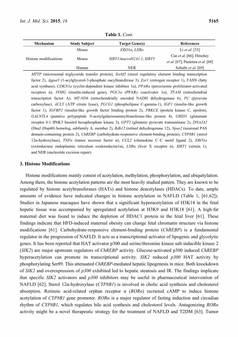
Table 1. Cont.
Mechanism Study Subject Target Gene(s) References
Histone modificaitions
Mouse ERO1α, LXRα Li et al. [33]
Mouse SIRT1/macroH2A1.1, SIRT3 Cao et al. [66]; Hirschey
et al. [67]; Pazienza et al. [68]
Human NER Schults et al. [69]
MTTP (microsomal triglyceride transfer protein), Srebf2 (sterol regulatory element binding transcription
factor 2), Agpat3 (1-acylglycerol-3-phosphate oacyltransferase 3), Esr1 (estrogen receptor 1), FASN (fatty
acid synthase), CDKN1a (cyclin-dependent kinase inhibitor 1a), PPARα (peroxisome proliferator-activated
receptors α), INSIG (insulin-induced gene), PGC1α (PPARγ coactivator 1α), TFAM (mitochondrial
transcription factor A), MT-ND6 (mitochondrially encoded NADH dehydrogenase 6), PC (pyruvate
carboxylase), ACLY (ATP citrate lyase), PLCG1 (phospholipase C-gamma-1), IGF1 (insulin-like growth
factor 1), IGFBP2 (insulin-like growth factor binding protein 2), PRKCE (protein kinase C, epsilon),
GALNTL4 (putative polypeptide N-acetylgalactosaminyltransferase-like protein 4), GRID1 (glutamate
receptor δ-1 IP6K3 Inositol hexaphosphate kinase 3), GPT2 (glutamic pyruvate transaminase 2), DNAJA2
(DnaJ (Hsp40) homolog, subfamily A, member 2), Rdh12 (retinol dehydrogenase 12), Npas2 (neuronal PAS
domain-containing protein 2), ChREBP (carbohydrate-responsive element-binding protein), CYP8B1 (sterol
12α-hydroxylase), TNFα (tumor necrosis factor α), CCL2 (chemokine C–C motif ligand 2), ERO1α
(oxireductase endoplasmic reticulum oxidoreductin1α), LXRα (liver X receptor α), SIRT1 (sirtuin 1),
and NER (nucleotide excision repair).
3. Histone Modifications
Histone modifications mainly consist of acetylation, methylation, phosphorylation, and ubiquitylation.
Among them, the histone acetylation patterns are the most heavily studied pattern. They are known to be
regulated by histone acetyltransferases (HATs) and histone deacetylases (HDACs). To date, ample
amounts of evidence have indicated changes in histone acetylation in NAFLD (Table 1, [61,62]).
Studies in Japanese macaques have shown that a significant hyperacetylation of H3K14 in the fetal
hepatic tissue was accompanied by upregulated acetylation at H3K9 and H3K18 [61]. A high-fat
maternal diet was found to induce the depletion of HDAC1 protein in the fetal liver [61]. These
findings indicate that HFD-induced maternal obesity can change fetal chromatin structure via histone
modifications [61]. Carbohydrate-responsive element-binding protein (ChREBP) is a fundamental
regulator in the progression of NAFLD. It acts as a transcriptional activator of lipogenic and glycolytic
genes. It has been reported that HAT activator p300 and serine/threonine kinase salt-inducible kinase 2
(SIK2) are major upstream regulators of ChREBP activity. Glucose-activated p300 induced ChREBP
hyperacetylation can promote its transcriptional activity. SIK2 reduced p300 HAT activity by
phosphorylating Ser89. This attenuated ChREBP-mediated hepatic lipogenesis in mice. Both knockdown
of SIK2 and overexpression of p300 exhibited led to hepatic steatosis and IR. The findings implicate
that specific SIK2 activators and p300 inhibitors may be useful in pharmaceutical intervention of
NAFLD [62]. Sterol 12α-hydroxylase (CYP8B1) is involved in cholic acid synthesis and cholesterol
absorption. Retinoic acid-related orphan receptor α (RORα) recruited cAMP to induce histone
acetylation of CYP8B1 gene promoter. RORα is a major regulator of fasting induction and circadian
rhythm of CYP8B1, which regulates bile acid synthesis and cholesterol levels. Antagonizing RORα
activity might be a novel therapeutic strategy for the treatment of NAFLD and T2DM [63]. Tumor

Int. J. Mol. Sci. 2015, 16 5166
necrosis factor α (TNFα) and chemokine C-C motif ligand 2 (CCL2) are the important inflammatory
mediators in the development of NAFLD. Chromatin immunoprecipitation assay showed an increase in
histone H3 lysine 9 and 18 acetylation at TNFα and CCL2 in mice with obesity [64]. These results
suggest that high levels of expression of TNFα and CCL2 at the chromatin level in fatty liver is
associated with the alterations in histone H3 acetylation [64].
Data have shown that NAFLD is correlated with changes in the transcriptome caused by histone
trimethylaton. Results have shown that hepatic lipid accumulation led to the aberrant histone H3K4
and H3K9 trimethylation in PPARα and lipid catabolism related genes, which increased the mRNA
expression of these genes in HFD-fed mice. Results suggested that histone H3K4 an H3K9
trimethylation may contribute to hepatic steatosis and disease progression [65]. Lipogenesis and ER
stress were promoted with the transgenerational alteration for oxireductase endoplasmic reticulum
oxidoreductin1α (ERO1α), liver X receptor α (LXRα), histone methylations, and H3K9 histone
methyltransferase. Low levels of accumulation of methylated histones in ERO1-α and LXRα gene
promoters render the offsprings of HFD-induced mothers susceptible to hepatic steatosis and obesity [33].
Sirtuins (SIRTs) are part of the silent information regulator-2 family. Among the seven different
sirtuins, SIRT1 has been studied the most. Studies revealed that the deacetylation of SIRT1 is
responsible for the regulation of a variety of proteins that are involved in the pathophysiology of
NAFLD [70]. SIRT1 is involved in the regulation of glucose homeostasis, antihyperlipidemic activity,
insulin sensitivity, oxidative stress, anti-inflammatory activity, and anti-aging activity [70]. Some studies
have revealed a significant reduction in the expression of SIRT1 in NAFLD animal models, and natural
SIRT1 activator showed protective effects on metabolic diseases [71]. Maternal HFD increased the
fetal acetylation of histone H3K14 and decreased SIRT1 expression in fetal livers. Maternal HFD
altered the expression of downstream effector in NAFLD by regulating SIRT1. SIRT1 maintains
deacetylase activity on the peptide substrate of H3K14 acetylation in vitro. This activity may be
abrogated due to the mutation of the catalytic domain of SIRT1. SIRT1 modulates the fetal metabolome
and epigenome as a molecular mediator under maternal HFD conditions [72]. Menin deficiency
enhanced liver steatosis in HFD-fed mice. The adaptor menin recruits SIRT1 to regulate CD36
expression and triglyceride accumulation via histone deacetylation [66]. Conversely, SIRT3 resides at
the mitochondria and modulates fatty acid oxidation. HFD feeding results in hepatic mitochondrial
protein hyperacetylation and decreases the SIRT3 expression. SIRT3 knockout mice developed hepatic
steatosis and IR in the mice fed with HFD [67]. In this way, SIRT1 and SIRT3 are involved in the
balance of metabolic and hepatic steatosis regulation through the epigenetic modification.
MacroH2A1, a target of SIRT1, is a variant of histone H2A and have two isoforms macroH2A1.1 and
macroH2A1.2. As an important transcriptional regulator, macroH2A1 is involved in cell senescence and
tumorigenic processes. The enhanced expression of macroH2A1.1 reduced lipid deposition in hepatocytes,
whereas macroH2A1.2 was unable to do so. MacroH2A1.1 ameliorated glucose metabolism and
downregulated the expression of lipidogenic genes. Thus, SIRT1/macroH2A1.1-specific epigenetic
regulation of lipid metabolism is associated with NAFLD development [68].
When NAFLD progressed to liver cancer, inflammation and oxidative stress played a critical role in
the process. Inflammation activated neutrophil-mediated oxidative stress, which led to the reduced
capacity of nucleotide excision repair (NER). Oxidative DNA damage further increased the risk of
carcinogenesis in inflammatory tissues. Schults and colleagues proposed an attractive mechanism of

Int. J. Mol. Sci. 2015, 16 5167
the development of liver cancer in NAFLD patients. They observed that the neutrophilic influx with
high expression of myeloperoxidase inhibited the damage recognition capacity, as indicated by
staining for histone 2AX phosphorylation, which was paralleled by a reduction in NER capacity under
hepatic inflammation conditions. Therefore, the reduction of NER capacity in the context of hepatic
inflammation may have been caused by the reduced damage recognition [69].
4. MicroRNAs
MiRs are small, highly conserved noncoding RNAs of approximately 18–25 nucleotides in length
that modulate translation and transcription of target genes. MiRs regulate a variety of biological
functions in animals and human [73]. It appears that most miRs are repressed by epigenetic
methylation of CpG islands [73]. MiRs perform a vital role in lipid metabolism and inflammation,
some of which has been reported to be epigenetically regulated in NAFLD [74–76]. The miR
alterations associated with NAFLD are summarized in Table 2. The miRs analysis showed a marked
decrease in miR-122, miR-451, and miR-27 and increase in miR-200a, miR-200b, and miR-429 in rats
fed HFD. These miRs are involved in the regulation of carbohydrate and lipid metabolism, signal
transduction and apoptosis [77]. HFD-induced animal models showed considerable dysregulation of
miRs and target genes. For instance, miR-467b expression was found to be markedly lower in the liver
tissues of mice with hepatic steatosis than in those of otherwise normal mice. This difference was
found to be inversely correlated with the expression of target gene of hepatic lipoprotein lipase (LPL).
The interplay between miR-467b and LPL was associated with IR [78]. Decreased expression of miR-216
and miR-302a and increased expression of miR-24 were observed in the liver tissues with NAFLD,
the target genes such as ATP-binding cassette transporter A1 (ABCA1), long-chain fatty acid elongation
6 (ELOVL6) and insulin-induced gene 1 (Insig1) are involved in hepatic fatty acid, cholesterol,
and glucose metabolism [79,80]. However, some miRs levels (miR-705, miR-1224, miR-182,
miR-183, miR-199a-3p, miR-200b, and miR-155) were upregulated in mice with methyl-deficient
diet-induced NAFLD [81,82]. The polycomb group protein enhancer of zeste homolog 2 (EZH2) has
been reported to regulate miRs by trimethylating Lys27 on histone H3. Inhibition of EZH2 upregulated
the inflammatory genes (TNF-α and TGF-β) and specific miRs (miR-200b and miR-155) and
subsequently favored hepatic steatosis in NAFLD [83]. One study that focused on NASH patients
showed 113 miRs expressed differentially. Among them, seven remained significant (miR-132,
miR-150, miR-433, miR-28-3p, miR-511, miR-517a, and miR-671). The target genes of these miRs
comprise obestatin gene, insulin receptor pathway genes, and inflammation-related genes. Additionally,
miR-197 and miR-99 levels were correlated with liver fibrosis in NASH patients. MiRs from visceral
adipose tissue (VAT) may be involved in the pathogenesis of NAFLD. These may serve as candidate
biomarkers for NASH [84].

Int. J. Mol. Sci. 2015, 16 5168
Table 2. MiRs alterations in NAFLD.
Study Subject Upregulated MiRs Downregulated MiRs References
Human
miR-10b, miR-16, miR19a/b,
miR-21, miR-27b-3p, miR-34a
miR-122, miR125b, miR-192-5p,
miR-451, miR-1290
miR-28-3p, miR-99a, miR-132,
miR-146b, miR-150, miR-181d,
miR-197, miR-296-5p, miR-433,
miR-511, miR-517a, miR-671
Estep et al. [84], Tan et al. [85],
Yamada et al. [86], Pirola et al. [87],
Celikbilek et al. [88], Cermelli et al. [89],
Clarke et al. [90], Miyaaki et al. [91],
Min et al. [92], Zheng et al. [93],
Cazanave et al. [94]
Mouse
miR-24, miR-33a, miR-34a,
miR-122, miR-155, miR-181a,
miR-182, miR-183, miR-192,
miR-199a-3p/5p, miR-200b,
miR-705, miR-1224
miR-92b-3p, miR-216, miR-302a,
miR-328-3p, miR-467b, miR-484,
miR-574-5p, miR-615-3p
Ahn et al. [78], Hoekstra et al. [79],
Ng et al. [80], Dolganiuc et al. [81],
Pogribny et al. [82], Tryndyak et al. [95],
Derdak et al. [96], Li et al. [97],
Li et al. [98], Miyamoto et al. [99]
Rat miR-15b, miR-155,
miR-200a/b, miR-429 miR-27, miR-122, miR-451
Alisi et al. [77], Vella et al. [83],
Zhang et al. [100]
Although liver biopsy is still the gold standard of histopathological diagnosis of NAFLD, its invasive
nature renders its performance difficult and unsatisfactory. A different technique, serum miR paneling,
is a non-invasive diagnostic biomarker of NAFLD. The study showed the serum levels of miRs to be
upregulated, including those of miR-122, miR-1290, miR-27b-3p, miR-192-5p, miR-21, miR-34a
miR-15b miR-16, miR-451, miR19a/b, and miR125b [85–87,100]. Those of other miRs were lower in
NAFLD patients. These included miR-146b, miR-99a, miR-181d, and miR-197 [88]. These miRs were
involved in cell proliferation, glucose consumption, and TG storage. Moreover, the levels of serum
miRs (miR-34a, miR-122, miR-181a, miR-192, and miR-200b) were strongly associated with
inflammatory activity, fibrosis stage, and liver enzyme levels [89,95]. These findings suggest that
the specific serum miRs may be used as noninvasive biomarkers of diagnosis and monitors of the
severity of NAFLD.
MiR-122 serves as a key regulator of the metabolism of glucose and lipids in adult livers.
The serum miR-122 levels remained high across the entire inductive time of NASH in mice, which
was correlated with the extent of NASH [90]. Mostly, miR-122 circulates in argonaute 2-free forms
and is expressed in the lipid-laden hepatocytes. MiR-122 increases alanine aminotransferase level by
activating the translation at multiple sites of the coding gene [87]. In NAFLD patients, the hepatic
miR-122 levels were lower in patients with mild steatosis than in those with severe steatosis.
Conversely, patients with mild fibrosis showed higher levels of serum and hepatic miR-122 levels than
patients with severe fibrosis. Consequently, the serum miR-122 level can be used as a predictive
circulating marker of hepatic fibrosis in NAFLD patients [91].
MiR-34a, a transcriptional target of p53, is involved in the pathogenesis of NAFLD. P53 inhibitor
pifithrin-α p-nitro (PFT) abrogated the HFD-induced overexpressing of miR-34a and activate the
SIRT/PGC1α/PPARα axis, which diminished hepatic TG deposition and ameliorated the liver steatosis [96].
NAFLD is also associated with free cholesterol and TG accumulation. NAFLD enhanced SREBP-2
maturation, and reduced phosphorylation of HMG CoA reductase (HMGCR). MiR-34a overexpression
repressed SIRT1 with adenosine monophosphate-activated protein kinase (AMPK) and HMGCR
dephosphorylation. HMGCR expression was related to free cholesterol, the histological extent of

Int. J. Mol. Sci. 2015, 16 5169
NAFLD and LDL-cholesterol. These findings indicate that the dysregulation of cholesterol metabolism
may contribute to NAFLD and cardiovascular risks [92].
Based on studies, miRs participate in the regulation of some metabolism-related signal pathways.
It has been proposed that cholesterol 7α-hydroxylase (CYP7A1)/steroid response element-binding
protein 2 (SREBP2)/miR-33a axis performs a crucial role in the regulation of the synthesis of hepatic
bile acid, cholesterol, and fatty acids. The overexpression of CYP7A1 incerased SREBP2-related
hepatic cholesterol synthesis and decreased hepatic fatty acid synthesis. Induction of SREBP2
subsequently activated miR-33a, which favored the lowered bile acid pool and elevated hepatic
cholesterol levels in mice [97]. MiR-10b was found to be correlated with the steatosis level. PPARα,
the direct target of miR-10b, showed markedly changed protein expression. The impact of miR-10b on
the regulation of hepatic steatosis may offer a novel explanation for the pathophysiology of NAFLD [93].
The overexpression of miR199a-5p exacerbated fatty acid accumulation and repressed ATP levels and
mtDNA content. Furthermore, miR199a-5p inhibited the expression of mitochondrial fatty acid
β-oxidation-related genes via suppression of caveolin1 (CAV1) and PPARα signal pathway in obese
mice and NAFLD patients. MiR199a-5p are involved in lipid metabolism and mitochondrial
β-oxidation in NAFLD. MiR199a-5p overexpression may impair fatty acid β-oxidation and induce
lipid deposition through the CAV1 and PPARα pathway [98].
Some studies have demonstrated that miRs regulate lipoapoptosis and ER stress. Lipoapoptosis is
associated with the upregulation of a pro-apoptotic protein p53-upregulated mediator of apoptosis
(PUMA). Inhibition of miR-296-5p expression was observed in the livers of NASH patients compared
with healthy controls. Moreover, miR-296-5p levels were inversely correlated with PUMA mRNA
expression in liver specimens. The data suggest that miR-296-5p regulates PUMA expression during
hepatic lipoapoptosis and increases in miR-296-5p expression may minimize apoptotic damage in
NAFLD patients [94]. Palmitate is a crucial pathogenic event in NAFLD and activates the ER stress
response through inducing the proapoptotic transcription factor C/EBP homologous protein (CHOP).
Additionally, loss of miRs regulates lipoapoptosis under the ER stress conditions. Five miRs were
downregulated under ER stress induced by palmitate in hepatocyte (miR-92b-3p, miR-328-3p,
miR-484, miR-574-5p, and miR-615-3p). Enhancement of miR-615-3p levels with a precursor
molecule decreased the CHOP expression and reduced the palmitate-induced hepatocyte apoptosis.
These findings suggest elevation of miR-615-3p levels brings therapeutic benefit by suppressing
palmitate-induced lipoapoptosis [99].
5. Potential Epigenetic Prevention or Therapy for NAFLD
Currently, NAFLD therapies are limited, and some studies have focused on identification of dietary
natural compounds to provide new strategies for NAFLD [101]. It has been proven that dietary
substances regulate fatty acid metabolism and are hypolipidemic agents [102]. Folate may affect
expression of miRs related to the severity of NAFLD. Folate affects miRs expression, possibly through
alterations in methylation levels in the genome [103]. The severity of NAFLD induced by folate-deficient
diet is associated with the alteration of hepatic miRs expression, including miR-181a, miR-34a, miR-200b,
and miR-221 [95]. Evidence has shown that the polyphenols decrease TG synthesis and oxidative
stress in the liver, increase fatty acid oxidation, and reduce the risk of metabolic diseases by regulating

Int. J. Mol. Sci. 2015, 16 5170
specific miRs such as miR-122 [104]. Lycopene decreased the expression of miR-21 and its direct
target fatty acid-binding protein 7 (FABP7) in mice fed stearic acid (SA), which blocked SA-evoked
lipid accumulation. Lycopene may be used as a functional compound for NAFLD treatment [105].
Especially, grape seed proanthocyanidins not only promoted liver cholesterol efflux to produce HDL
particles by imhibiting miR-33, but also repressed lipogenesis by decreasing miR-122. These agents
showed hepatoprotective effects through their regulation of miRs’ expression associated with lipid
metabolism [106].
Because it has been shown the overexpression of SIRT1 will reduce the degree of hepatic steatosis
in mice NAFLD models [72], activation of SIRT1 with resveratrol, a natural SIRT1 activator, protected
against ER and IR stress and had positive effects on metabolic diseases, which is mediated by
overexpression of oxygen-regulated protein 150. These findings indicated that SIRT1 attenuated
palmitate-induced ER and IR stress and the pharmacologic activation of SIRT1 may offer a potential
therapeutic strategy for NAFLD management [107]. It has been demonstrated that resveratrol
reduced liver lipid deposition, upregulated acyl-coenzyme A oxydase (ACO) and carnitine palmitoyl
transferase-Ia (CPT-Ia), downregulated acetyl-coenzyme A carboxylase (ACC) activity, which was
mediated by AMPK/SIRT1 pathway activation [102]. Recently, it has been reported that hepatic
steatosis could be ameliorated by drugs such as ursodeoxycholic acid (UDCA). UDCA repressed the
miR-34a/SIRT1/p53 pathway in the liver of NAFLD rats. UDCA prevented miR-34a-dependent SIRT1
suppression, p53 acetylation, and apoptosis. Consistently, the overexpression of p53 activated
miR-34a/SIRT1/p53, which was in turn repressed by UDCA [108]. Conclusively, these potential
modulators of NAFLD pathogenesis may provide new targets for therapeutic intervention.
6. Current Problems
Although the development of NAFLD is regulated by epigenetic alterations, including DNA
methylation, histone modification and miRs, the investigation is just beginning and still faces many
challenges. Clearly, various factors are involved in different stages of NAFLD, but the interactions
among different factors and how they affect metabolistic homeostasis have not been clarified [109].
In addition, so far, few effective diagnostic and therapeutic targets exist. The possible difficulty results
from the complicated biomolecular interactions and overlapping pathways in the setting of NAFLD [21].
It is important to exploit reliable biomolecules with high specificity and sensitivity based on the stage
of NAFLD. Because epigenetic changes undergo transgenerational inheritance, animal models need to
be developed to evaluate the influence of epigenetic modifications to the heritability of disease [110].
Nutritional cues regulated epigenetic alterations and affect the health in the offspring. Diet could
change the metabolic gene expression and increase disease susceptibility. More studies should be
undertaken to identify the optimal agents from diet for the prevention and intervention of NAFLD [37].
In recent years, the emergence of circulating miRs offers an option for using them as noninvasive
diagnostic and therapeutic biomarkers. Nevertheless, some problems still hinder research, such as the
identification of specific target miRs and the delivery mode, dosage, and period of treatment [111].
In addition, with regard to the observed potential biomolecules with hepatoprotective effect, further
clinical trials are needed in order to evaluate the effect and safety of the management.

Int. J. Mol. Sci. 2015, 16 5171
7. Conclusions and Perspectives
The accumulation of epigenetic evidence related to NAFLD has offered us a novel perspective into
the pathogenesis of the disease and may contribute to identifying attractive diagnostic biomarkers and
therapeutic strategies for NAFLD. Further research is needed to explore the role of epigenetics in the
mechanism of hepatic steatosis and steatohepatitis, the interaction of environmental factors, and epigenetic
modulation, in order to promote diagnostic and therapeutic approaches as well as reduce the morbidity
and mortality in NAFLD.
Acknowledgments
This work was supported by the State Key Development Program for Basic Research of China
(No. 2012CB517501), the Program of the Shanghai Committee of Science and Technology
(No. 10411956300), the National Natural Science Foundation of China (No. 81070322, No. 81270491,
No. 81000173). Liang Qiao’s work was supported by the Robert W. Storr Bequest to the Sydney
Medical Foundation, University of Sydney, and a Project Grant to Liang Qiao (APP1047417), and
a Cancer Council New South Wales grant to Liang Qiao (APP1070076).
Author Contributions
Chao Sun collected the data and wrote the manuscript; Jian-Gao Fan and Liang Qiao designed this
work, analyzed the data and edited the manuscript.
Abbreviations
NAFLD (non-alcoholic fatty liver disease), NASH (non-alcoholic steatohepatitis), HCC (hepatocellular
carcinoma), TG (triglycerides), SS (simple steatosis), IR (insulin resistance), ER (endoplasmic
reticulum), miRs (microRNAs), DNMTs (DNA methyltransferases), SAM (S-adenosylmethionine),
HFD (high fat diet), MTTP (microsomal triglyceride transfer protein), HFS (high-fat-sucrose),
Srebf2 (sterol regulatory element binding transcription factor 2), Agpat3 (1-acylglycerol-3-phosphate
Oacyltransferase 3), Esr1 (estrogen receptor 1), FASN (fatty acid synthase), Cdkn1a (cyclin-dependent
kinase inhibitor 1A), PPARs (Peroxisome proliferator-activated receptors), PGC1α (PPARγ
coactivator 1α), TFAM (mitochondrial transcription factor A), HOMA-IR (homeostasis model
assessment of IR), ROS (reactive oxygen species), MT-ND6 (mitochondrially encoded NADH
dehydrogenase 6), HATs (histone acetyltransferases), HDACs (histone deacetylases), ChREBP
(carbohydrate-responsive element-binding protein), SIK2 (serine/threonine kinase salt-inducible kinase 2),
CYP8B1 (sterol 12α-hydroxylase), RORα (retinoic acid-related orphan receptor α), TNFα (tumor necrosis
factor α), CCL2 (chemokine C-C motif ligand 2), ERO1α (endoplasmic reticulum oxidoreductin1α),
LXRα (liver X receptor α), SIRTs (sirtuins), NER (nucleotide excision repair), LPL (lipoprotein
lipase), Insig1 (insulin-induced gene 1), ABCA1 (ATP-binding cassette transporter A1), ELOVL6
(long-chain fatty acid elongation 6) EZH2 (enhancer of zeste homolog 2), VAT (visceral adipose
tissue), PFT (P53 inhibitor pifithrin-α p-nitro), HMGCR (HMG CoA reductase), AMPK (adenosine
monophosphate-activated protein kinase), CYP7A1 (cholesterol 7α-hydroxylase), SREBP2 (steroid
response element-binding protein 2), CAV1 (caveolin1), p53-upregulated mediator of apoptosis (PUMA),

Int. J. Mol. Sci. 2015, 16 5172
CHOP (C/EBP homologous protein), FABP7 (fatty acid-binding protein 7), SA (stearic acid), ACO
(acyl-coenzyme A oxydase), CPT-Ia (carnitine palmitoyl transferase-Ia), ACC (acetyl-coenzyme A
carboxylase), UDCA (ursodeoxycholic acid).
Conflicts of Interest
The authors declare no conflict of interest.
References
1. Marchesini, G.; Bugianesi, E.; Forlani, G.; Cerrelli, F.; Lenzi, M.; Manini, R.; Natale, S.; Vanni, E.;
Villanova, N.; Melchionda, N.; et al. Nonalcoholic fatty liver, steatohepatitis, and the metabolic
syndrome. Hepatology 2003, 37, 917–923.
2. Younossi, Z.M.; Stepanova, M.; Afendy, M.; Fang, Y.; Younossi, Y.; Mir, H.; Srishord, M.
Changes in the prevalence of the most common causes of chronic liver diseases in the United States
from 1988 to 2008. Clin. Gastroenterol. Hepatol. 2011, 9, 524–530.
3. Dowman, J.K.; Tomlinson, J.W.; Newsome, P.N. Pathogenesis of non-alcoholic fatty liver disease.
QJM 2010, 103, 71–83.
4. Liu, Q.; Bengmark, S.; Qu, S. The role of hepatic fat accumulation in pathogenesis of non-alcoholic
fatty liver disease. Lipids Health Dis. 2010, 9, 42–51.
5. Malik, S.M.; de Vera, M.E.; Fontes, P.; Shaikh, O.; Ahmad, J. Outcome after liver transplantation
for NASH cirrhosis. Am. J. Transplant. 2009, 7, 782–793.
6. Siegel, A.B.; Zhu, A.X. Metabolic syndrome and hepatocellular carcinoma. Cancer 2009, 115,
5651–5661.
7. Ong, J.P.; Pitts, A.; Younossi, Z.M. Increased overall mortality and liver-related mortality in
non-alcoholic fatty liver disease. J. Hepatol. 2008, 49, 608–612.
8. Fan, J.G. Epidemiology of alcoholic and nonalcoholic fatty liver disease in China.
J. Gastroenterol. Hepatol. 2013, 28, 11–17.
9. Amarapurkar, D.N.; Hashimoto, E.; Lesmana, L.A.; Sollano, J.D.; Chen, P.J.; Goh, K.L.
How common is non-alcoholic fatty liver disease in the Asia-Pacific region and are there local
differences? J. Gastroenterol. Hepatol. 2007, 22, 788–793.
10. Zhou, Y.J.; Li, Y.Y.; Nie, Y.Q.; Huang, C.M.; Cao, C.Y. Natural course of nonalcoholic fatty liver
disease in southern China: A prospective cohort study. J. Dig. Dis. 2012, 13, 153–160.
11. Wong, V.W.; Chu, W.C.; Wong, G.L.; Chan, R.S.; Chim, A.M.; Ong, A.; Yeung, D.K.; Yiu, K.K.;
Chu, S.H.; Woo, J.; et al. Prevalence of non-alcoholic fatty liver disease and advanced fibrosis in
Hong Kong Chinese: A population study using proton-magnetic resonance spectroscopy and
transient elastography. Gut 2012, 61, 409–415.
12. Adams, L.A.; Sanderson, S.; Lindor, K.D.; Angulo, P. The histological course of nonalcoholic fatty
liver disease: A longitudinal study of 103 patients with sequential liver biopsies. J. Hepatol. 2005,
42, 132–138.
13. Fassio, E.; Alvarez, E.; Dominguez, N.; Landeira, G.; Longo, C. Natural history of nonalcoholic
steatohepatitis: A longitudinal study of repeat liver biopsies. Hepatology 2004, 40, 820–826.

Int. J. Mol. Sci. 2015, 16 5173
14. Lindor, K.D.; Kowdley, K.V.; Heathcote, E.J.; Harrison, M.E.; Jorgensen, R.; Angulo, P.; Lymp, J.F.;
Burgart, L.; Colin, P. Ursodeoxycholic acid for treatment of nonalcoholic steatohepatitis: Results of
a randomized trial. Hepatology 2004, 39, 770–778.
15. Ekstedt, M.; Franzén, L.E.; Mathiesen, U.L.; Thorelius, L.; Holmqvist, M.; Bodemar, G.;
Kechagias, S. Long-term follow-up of patients with NAFLD and elevated liver enzymes. Hepatology
2006, 44, 865–873.
16. Rafiq, N.; Bai, C.; Fang, Y.; Srishord, M.; McCullough, A.; Gramlich, T. Long-term follow-up of
patients with nonalcoholic fatty liver. Clin. Gastroenterol. Hepatol. 2009, 7, 234–238.
17. Bechmann, L.P.; Hannivoort, R.A.; Gerken, G.; Hotamisligil, G.S.; Trauner, M.; Canbay, A.
The interaction of hepatic lipid and glucose metabolism in liver diseases. J. Hepatol. 2012, 56, 952–964.
18. Haider, D.G.; Schindler, K.; Schaller, G.; Prager, G.; Wolzt, M.; Ludvik, B. Increased plasma visfatin
concentrations in morbidly obese subjects are reduced after gastric banding. J. Clin. Endocrinol. Metab.
2006, 91, 1578–1581.
19. Farrell, G.C.; Larter, C.Z. Nonalcoholic fatty liver disease: From steatosis to cirrhosis. Hepatology
2006, 43, S99–S112.
20. Tacke, F.; Luedde, T.; Trautwein, C. Inflammatory pathways in liver homeo-stasis and liver injury.
Clin. Rev. Allergy Immunol. 2009, 36, 4–12.
21. Berlanga, A.; Guiu-Jurado, E.; Porras, J.A.; Auguet, T. Molecular pathways in non-alcoholic fatty
liver disease. Clin. Exp. Gastroenterol. 2014, 7, 221–239.
22. Neuschwander-Tetri, B.A. Hepatic lipotoxicity and the pathogenesis of nonalcoholic steatohepatitis:
The central role of nontriglyceride fatty acid metabolites. Hepatology 2010, 52, 774–788.
23. Lee, J.H.; Friso, S.; Choi, S.W. Epigenetic mechanisms underlying the link between non-alcoholic
fatty liver diseases and nutrition. Nutrients 2014, 6, 3303–3325.
24. Gu, S.G.; Pak, J.; Guang, S.; Maniar, J.M.; Kennedy, S.; Fire, A. Amplification of siRNA in
Caenorhabditis elegans generates a transgenerational sequence-targeted histone H3 lysine 9
methylation footprint. Nat. Genet. 2012, 44, 157–164.
25. Heijmans, B.T.; Tobi, E.W.; Stein, A.D.; Putter, H.; Blauw, G.J.; Susser, E.S.; Slagboom, P.E.;
Lumey, L.H. Persistent epigenetic differences associated with prenatal exposure to famine in humans.
Proc. Natl. Acad. Sci. USA 2008, 105, 17046–17049.
26. Hochberg, Z.; Feil, R.; Constancia, M.; Fraga, M.; Junien, C.; Carel, J.C.; Boileau, P.; Le Bouc, Y.;
Deal, C.L.; Lillycrop, K.; et al. Child health, developmental plasticity, and epigenetic programming.
Endocr. Rev. 2011, 32, 159–224.
27. Lomba, A.; Martinez, J.A.; Garcia-Diaz, D.F.; Paternain, L.; Marti, A.; Campion, J.; Milagro, F.I.
Weight gain induced by an isocaloric pair-fed high fat diet: A nutriepigenetic study on FASN and
NDUFB6 gene promoters. Mol. Genet. Metab. 2010, 101, 273–278.
28. Yoo, C.B.; Jones, P.A. Epigenetic therapy of cancer: Past, present and future. Nat. Rev. Drug Discov.
2006, 5, 37–50.
29. Paternain, L.; Garcia-Diaz, D.F.; Milagro, F.I.; Gonzalez-Muniesa, P.; Martinez, J.A.; Campion, J.
Regulation by chronic-mild stress of glucocorticoids, monocyte chemoattractant protein-1 and
adiposity in rats fed on a high-fat diet. Physiol. Behav. 2011, 103, 173–180.

Int. J. Mol. Sci. 2015, 16 5174
30. Jiang, M.; Zhang, Y.; Liu, M.; Lan, M.S.; Fei, J.; Fan, W.; Gao, X.; Lu, D. Hypermethylation of
hepatic glucokinase and L-type pyruvate kinase promoters in high-fat diet-induced obese rats.
Endocrinology 2011, 152, 1284–1289.
31. Sookoian, S.; Pirola, C.J. DNA methylation and hepatic insulin resistance and steatosis. Curr. Opin.
Clin. Nutr. Metab. Care 2012, 15, 350–356.
32. Pirola, C.J.; Gianotti, T.F.; Burgueño, A.L.; Rey-Funes, M.; Loidl, C.F.; Mallardi, P.; Martino, J.S.;
Castaño, G.O.; Sookoian, S. Epigenetic modification of liver mitochondrial DNA is associated with
histological severity of nonalcoholic fatty liver disease. Gut 2013, 62, 1356–1363.
33. Li, J.; Huang, J.; Li, J.S.; Chen, H.; Huang, K.; Zheng, L. Accumulation of endoplasmic reticulum
stress and lipogenesis in the liver through generational effects of high fat diets. J. Hepatol. 2012, 56,
900–907.
34. Tian, Y.; Wong, V.W.; Chan, H.L.; Cheng, A.S. Epigenetic regulation of hepatocellular carcinoma in
non-alcoholic fatty liver disease. Semin. Cancer Biol. 2013, 23, 471–482.
35. Dawson, M.A.; Kouzarides, T. Cancer epigenetics: From mechanism to therapy. Cell 2012, 150, 12–27.
36. Bruce, K.D.; Cagampang, F.R. Epigenetic priming of the metabolic syndrome. Toxicol. Mech. Methods
2011, 21, 353–361.
37. Iacobazzi, V.; Castegna, A.; Infantino, V.; Andria, G. Mitochondrial DNA methylation as a
next-generation biomarker and diagnostic tool. Mol. Genet. Metab. 2013, 110, 25–34.
38. Wang, L.J.; Zhang, H.W.; Zhou, J.Y.; Liu, Y.; Yang, Y.; Chen, X.L.; Zhu, C.H.; Zheng, R.D.;
Ling, W.H.; Zhu, H.L. Betaine attenuates hepatic steatosis by reducing methylation of the MTTP
promoter and elevating genomic methylation in mice fed a high-fat diet. J. Nutr. Biochem. 2014,
25, 329–336.
39. Li, Y.Y. Genetic and epigenetic variants influencing the development of nonalcoholic fatty liver
disease. World J. Gastroenterol. 2012, 18, 6546–6551.
40. Kalhan, S.C.; Edmison, J.; Marczewski, S.; Dasarathy, S.; Gruca, L.L.; Bennett, C.; Duenas, C.;
Lopez, R. Methionine and protein metabolism in non-alcoholic steatohepatitis: Evidence for lower
rate of transmethylation of methionine. Clin. Sci. (Lond.) 2011, 121, 179–189.
41. Niculescu, M.D.; Zeisel, S.H. Diet, methyl donors and DNA methylation: Interactions between
dietary folate, methionine and choline. J. Nutr. 2002, 132, 2333S–2335S.
42. Zivkovic, A.M.; Bruce German, J.; Esfandiari, F.; Halsted, C.H. Quantitative lipid metabolomic
changes in alcoholic micropigs with fatty liver disease. Alcohol. Clin. Exp. Res. 2009, 33, 751–758.
43. Da Silva, R.P.; Kelly, K.B.; Al Rajabi, A.; Jacobs, R.L. Novel insights on interactions between folate
and lipid metabolism. Biofactors 2014, 40, 277–283.
44. Chang, X.; Yan, H.; Fei, J.; Jiang, M.; Zhu, H.; Lu, D.; Gao, X. Berberine reduces methylation of the
MTTP promoter and alleviates fatty liver induced by a high-fat diet in rats. J. Lipid Res. 2010, 51,
2504–2515.
45. Cordero, P.; Campion, J.; Milagro, F.I.; Martinez, J.A. Transcriptomic and epigenetic changes
in early liver steatosis associated to obesity: Effect of dietary methyl donor supplementation.
Mol. Genet. Metab. 2013, 110, 388–395.
46. Cordero, P.; Gomez-Uriz, A.M.; Campion, J.; Milagro, F.I.; Martinez, J.A. Dietary supplementation
with methyl donors reduces fatty liver and modifies the fatty acid synthase DNA methylation
profile in rats fed an obesogenic diet. Genes Nutr. 2013, 8, 105–113.

Int. J. Mol. Sci. 2015, 16 5175
47. Tryndyak, V.P.; Han, T.; Muskhelishvili, L.; Fuscoe, J.C.; Ross, S.A.; Beland, F.A.; Pogribny, I.P.
Coupling global methylation and gene expression profiles reveal key pathophysiological events in
liver injury induced by a methyl-deficient diet. Mol. Nutr. Food Res. 2011, 55, 411–418.
48. Wolff, G.L.; Kodell, R.L.; Moore, S.R.; Cooney, C.A. Maternal epigenetics and methyl
supplements affect agouti gene expression in Avy/a mice. FASEB J. 1998, 12, 949–957.
49. Dudley, K.J.; Sloboda, D.M.; Connor, K.L.; Beltrand, J.; Vickers, M.H. Offspring of mothers fed
a high fat diet display hepatic cell cycle inhibition and associated changes in gene expression and
DNA methylation. PLoS One 2011, 6, e21662.
50. Pruis, M.G.; Lendvai, A.; Bloks, V.W.; Zwier, M.V.; Baller, J.F.; de Bruin, A.; Groen, A.K.;
Plösch, T. Maternal western diet primes non-alcoholic fatty liver disease in adult mouse offspring.
Acta Physiol. (Oxf.) 2014, 210, 215–227.
51. Tiao, M.M.; Huang, L.T.; Chen, C.J.; Sheen, J.M.; Tain, Y.L.; Chen, C.C.; Kuo, H.C.; Huang, Y.H.;
Tang, K.S.; Chu, E.W.; et al. Melatonin in the regulation of liver steatosis following prenatal
glucocorticoid exposure. Biomed. Res. Int. 2014, 2014, 942172.
52. Gavrilova, O.; Haluzik, M.; Matsusue, K.; Cutson, J.J.; Johnson, L.; Dietz, K.R.; Nicol, C.J.;
Vinson, C.; Gonzalez, F.J.; Reitman, M.L. Liver peroxisome proliferator-activated receptor gamma
contributes to hepatic steatosis, triglyceride clearance, and regulation of body fat mass. J. Biol. Chem.
2003, 278, 34268–34276.
53. Tyagi, S.; Gupta, P.; Saini, A.S.; Kaushal, C.; Sharma, S. The peroxisome proliferator-activated
receptor: A family of nuclear receptors role in various diseases. J. Adv. Pharm. Technol. Res.
2011, 2, 236–240.
54. Sie, K.K.; Li, J.; Ly, A.; Sohn, K.J.; Croxford, R.; Kim, Y.I. Effect of maternal and postweaning
folic acid supplementation on global and gene-specific DNA methylation in the liver of the rat
offspring. Mol. Nutr. Food Res. 2013, 57, 677–685.
55. Sookoian, S.; Rosselli, M.S.; Gemma, C.; Burgueño, A.L.; Fernández Gianotti, T.; Castaño, G.O.;
Pirola, C.J. Epigenetic regulation of insulin resistance in nonalcoholic fatty liver disease: Impact
of liver methylation of the peroxisome proliferator-activated receptor γ coactivator 1α promoter.
Hepatology 2010, 52, 1992–2000.
56. Chen, G.; Broséus, J.; Hergalant, S.; Donnart, A.; Chevalier, C.; Bolaños-Jiménez, F.; Guéant, J.L.;
Houlgatte, R. Identification of master genes involved in liver key functions through transcriptomics
and epigenomics of methyl donor deficiency in rat: Relevance to nonalcoholic liver disease.
Mol. Nutr. Food Res. 2015, 59, 293–302.
57. Carabelli, J.; Burgueno, A.L.; Rosselli, M.S.; Gianotti, T.F.; Lago, N.R.; Pirola, C.J.; Sookoian, S.
High fat diet-induced liver steatosis promotes an increase in liver mitochondrial biogenesis in
response to hypoxia. J. Cell. Mol. Med. 2011, 15, 1329–1338.
58. Turrens, J.F. Mitochondrial formation of reactive oxygen species. J. Physiol. 2003, 552, 335–344.
59. Murphy, S.K.; Yang, H.; Moylan, C.A.; Pang, H.; Dellinger, A.; Abdelmalek, M.F.; Garrett, M.E.;
Ashley-Koch, A.; Suzuki, A.; Tillmann, H.L.; et al. Relationship between methylome and transcriptome
in patients with nonalcoholic fatty liver disease. Gastroenterology 2013, 145, 1076–1087.

Int. J. Mol. Sci. 2015, 16 5176
60. Ahrens, M.; Ammerpohl, O.; von Schönfels, W.; Kolarova, J.; Bens, S.; Itzel, T.; Teufel, A.;
Herrmann, A.; Brosch, M.; Hinrichsen, H.; et al. DNA methylation analysis in nonalcoholic fatty
liver disease suggests distinct disease-specific and remodeling signatures after bariatric surgery.
Cell Metab. 2013, 18, 296–302.
61. Aagaard-Tillery, K.M.; Grove, K.; Bishop, J.; Ke, X.; Fu, Q.; McKnight, R.; Lane, R.H.
Developmental origins of disease and determinants of chromatin structure: Maternal diet
modifies the primate fetal epigenome. J. Mol. Endocrinol. 2008, 41, 91–102.
62. Bricambert, J.; Miranda, J.; Benhamed, F.; Girard, J.; Postic, C.; Dentin, R. Salt-inducible kinase 2
links transcriptional coactivator P300 phosphorylation to the prevention of ChREBP-dependent
hepatic steatosis in mice. J. Clin. Investig. 2010, 120, 4316–4331.
63. Pathak, P.; Li, T.; Chiang, J.Y. Retinoic acid-related orphan receptor α regulates diurnal rhythm
and fasting induction of sterol 12α-hydroxylase in bile acid synthesis. J. Biol. Chem. 2013, 288,
37154–37165.
64. Mikula, M.; Majewska, A.; Ledwon, J.K.; Dzwonek, A.; Ostrowski, J. Obesity increases histone
H3 lysine 9 and 18 acetylation at TNFα and CCL2 genes in mouse liver. Int. J. Mol. Med. 2014,
34, 1647–1654.
65. Jun, H.J.; Kim, J.; Hoang, M.H.; Lee, S.J. Hepatic lipid accumulation alters global histone H3
lysine 9 and 4 trimethylation in the peroxisome proliferator-activated receptor α network. PLoS One
2012, 7, e44345.
66. Cao, Y.; Xue, Y.; Xue, L.; Jiang, X.; Wang, X.; Zhang, Z.; Yang, J.; Lu, J.; Zhang, C.; Wang, W.; et al.
Hepatic menin recruits SIRT1 to control liver steatosis through histone deacetylation. J. Hepatol.
2013, 59, 1299–1306.
67. Hirschey, M.D.; Shimazu, T.; Jing, E.; Grueter, C.A.; Collins, A.M.; Aouizerat, B.; Stančáková, A.;
Goetzman, E.; Lam, M.M.; Schwer, B.; et al. SIRT3 deficiency and mitochondrial protein
hyperacetylation accelerate the development of the metabolic syndrome. Mol. Cell 2011, 44, 177–190.
68. Pazienza, V.; Borghesan, M.; Mazza, T.; Sheedfar, F.; Panebianco, C.; Williams, R.; Mazzoccoli, G.;
Andriulli, A.; Nakanishi, T.; Vinciguerra, M. SIRT1-metabolite binding histone macroH2A1.1
protects hepatocytes against lipid accumulation. Aging (Albany NY) 2014, 6, 35–47.
69. Schults, M.A.; Nagle, P.W.; Rensen, S.S.; Godschalk, R.W.; Munnia, A.; Peluso, M.; Claessen, S.M.;
Greve, J.W.; Driessen, A.; Verdam, F.J.; et al. Decreased nucleotide excision repair in steatotic livers
associates with myeloperoxidase-immunoreactivity. Mutat. Res. 2012, 736, 75–81.
70. Colak, Y.; Yesil, A.; Mutlu, H.H.; Caklili, O.T.; Ulasoglu, C.; Senates, E.; Takir, M.; Kostek, O.;
Yilmaz, Y.; Enc, F.Y.; et al. A potential treatment of non-alcoholic fatty liver disease with SIRT1
activators. J. Gastrointest. Liver Dis. 2014, 23, 311–319.
71. Colak, Y.; Ozturk, O.; Senates, E.; Tuncer, I.; Yorulmaz, E.; Adali, G.; Doganay, L.; Enc, F.Y.
SIRT1 as a potential therapeutic target for treatment of nonalcoholic fatty liver disease. Med. Sci. Monit.
2011, 17, HY5–HY9.
72. Suter, M.A.; Chen, A.; Burdine, M.S.; Choudhury, M.; Harris, R.A.; Lane, R.H.; Friedman, J.E.;
Grove, K.L.; Tackett, A.J.; Aagaard, K.M. A maternal high-fat diet modulates fetal SIRT1 histone
and protein deacetylase activity in nonhuman primates. FASEB J. 2012, 26, 5106–5114.
73. Wang, Z.; Yao, H.; Lin, S.; Zhu, X.; Shen, Z.; Lu, G.; Poon, W.S.; Xie, D.; Lin, M.C.; Kung, H.F.
Transcriptional and epigenetic regulation of human microRNAs. Cancer Lett. 2013, 331, 1–10.

Int. J. Mol. Sci. 2015, 16 5177
74. Finch, M.L.; Marquardt, J.U.; Yeoh, G.C.; Callus, B.A. Regulation of microRNAs and their role in
liver development, regeneration and disease. Int. J. Biochem. Cell. Biol. 2014, 54, 288–303.
75. Ferreira, D.M.; Simão, A.L.; Rodrigues, C.M.; Castro, R.E. Revisiting the metabolic syndrome and
paving the way for microRNAs in non-alcoholic fatty liver disease. FEBS J. 2014, 281, 2503–2524.
76. Panera, N.; Gnani, D.; Crudele, A.; Ceccarelli, S.; Nobili, V.; Alisi, A. MicroRNAs as controlled
systems and controllers in non-alcoholic fatty liver disease. World J. Gastroenterol. 2014, 20,
15079–15086.
77. Alisi, A.; Da Sacco, L.; Bruscalupi, G.; Piemonte, F.; Panera, N.; de Vito, R.; Leoni, S.; Bottazzo, G.F.;
Masotti, A.; Nobili, V. Mirnome analysis reveals novel molecular determinants in the pathogenesis
of diet-induced nonalcoholic fatty liver disease. Lab. Investig. 2011, 91, 283–293.
78. Ahn, J.; Lee, H.; Chung, C.H.; Ha, T. High fat diet induced downregulation of microRNA-467b
increased lipoprotein lipase in hepatic steatosis. Biochem. Biophys. Res. Commun. 2011, 414,
664–669.
79. Hoekstra, M.; van der Sluis, R.J.; Kuiper, J.; van Berkel, T.J. Nonalcoholic fatty liver disease
is associated with an altered hepatocyte microRNA profile in LDL receptor knockout mice.
J. Nutr. Biochem. 2012, 23, 622–628.
80. Ng, R.; Wu, H.; Xiao, H.; Chen, X.; Willenbring, H.; Steer, C.J.; Song, G. Inhibition of
microRNA-24 expression in liver prevents hepatic lipid accumulation and hyperlipidemia.
Hepatology 2014, 60, 554–564.
81. Dolganiuc, A.; Petrasek, J.; Kodys, K.; Catalano, D.; Mandrekar, P.; Velayudham, A.; Szabo, G.
MicroRNA expression profile in Lieber-DeCarli diet-induced alcoholic and methionine choline
deficient diet-induced nonalcoholic steatohepatitis models in mice. Alcohol. Clin. Exp. Res. 2009, 33,
1704–1710.
82. Pogribny, I.P.; Starlard-Davenport, A.; Tryndyak, V.P.; Han, T.; Ross, S.A.; Rusyn, I.; Beland, F.A.
Difference in expression of hepatic microRNAs miR-29c, miR-34a, miR-155, and miR-200b is
associated with strain-specific susceptibility to dietary nonalcoholic steatohepatitis in mice.
Lab. Investig. 2010, 90, 1437–1446.
83. Vella, S.; Gnani, D.; Crudele, A.; Ceccarelli, S.; de Stefanis, C.; Gaspari, S.; Nobili, V.; Locatelli, F.;
Marquez, V.E.; Rota, R.; et al. EZH2 down-regulation exacerbates lipid accumulation and
inflammation in in vitro and in vivo NAFLD. Int. J. Mol. Sci. 2013, 14, 24154–24168.
84. Estep, M.; Armistead, D.; Hossain, N.; Elarainy, H.; Goodman, Z.; Baranova, A.; Chandhoke, V.;
Younossi, Z.M. Differential expression of miRNAs in the visceral adipose tissue of patients with
non-alcoholic fatty liver disease. Aliment Pharmacol. Ther. 2010, 32, 487–497.
85. Tan, Y.; Ge, G.; Pan, T.; Wen, D.; Gan, J. A pilot study of serum microRNAs panel as potential
biomarkers for diagnosis of nonalcoholic fatty liver disease. PLoS One 2014, 9, e105192.
86. Yamada, H.; Suzuki, K.; Ichino, N.; Ando, Y.; Sawada, A.; Osakabe, K.; Sugimoto, K.; Ohashi, K.;
Teradaira, R.; Inoue, T.; et al. Associations between circulating microRNAs (miR-21, miR-34a,
miR-122 and miR-451) and non-alcoholic fatty liver. Clin. Chim. Acta 2013, 424, 99–103.
87. Pirola, C.J.; Gianotti, T.F.; Castaño, G.O.; Mallardi, P.; Martino, J.S.; Gonzalez Lopez Ledesma, M.M.;
Flichman, D.; Mirshahi, F.; Sanyal, A.J.; Sookoian, S. Circulating microRNA signature in
non-alcoholic fatty liver disease: From serum non-coding RNAs to liver histology and disease
pathogenesis. Gut 2014, doi:10.1136/gutjnl-2014-306996.

Int. J. Mol. Sci. 2015, 16 5178
88. Celikbilek, M.; Baskol, M.; Taheri, S.; Deniz, K.; Dogan, S.; Zararsiz, G.; Gursoy, S.; Guven, K.;
Ozbakır, O.; Dundar, M.; et al. Circulating microRNAs in patients with non-alcoholic fatty liver
disease. World J. Hepatol. 2014, 6, 613–620.
89. Cermelli, S.; Ruggieri, A.; Marrero, J.A.; Ioannou, G.N.; Beretta, L. Circulating microRNAs in
patients with chronic hepatitis C and non-alcoholic fatty liver disease. PLoS One 2011, 6, e23937.
90. Clarke, J.D.; Sharapova, T.; Lake, A.D.; Blomme, E.; Maher, J.; Cherrington, N.J. Circulating
microRNA 122 in the methionine and choline-deficient mouse model of non-alcoholic steatohepatitis.
J. Appl. Toxicol. 2014, 34, 726–732.
91. Miyaaki, H.; Ichikawa, T.; Kamo, Y.; Taura, N.; Honda, T.; Shibata, H.; Milazzo, M.; Fornari, F.;
Gramantieri, L.; Bolondi, L.; et al. Significance of serum and hepatic microRNA-122 levels in
patients with non-alcoholic fatty liver disease. Liver Int. 2014, 34, e302–307.
92. Min, H.K.; Kapoor, A.; Fuchs, M.; Mirshahi, F.; Zhou, H.; Maher, J.; Kellum, J.; Warnick, R.;
Contos, M.J.; Sanyal, A.J. Increased hepatic synthesis and dysregulation of cholesterol metabolism is
associated with the severity of nonalcoholic fatty liver disease. Cell Metab. 2012, 15, 665–674.
93. Zheng, L.; Lv, G.C.; Sheng, J.; Yang, Y.D. Effect of miRNA-10b in regulating cellular steatosis
level by targeting PPAR-α expression, a novel mechanism for the pathogenesis of NAFLD.
J. Gastroenterol. Hepatol. 2010, 25, 156–163.
94. Cazanave, S.C.; Mott, J.L.; Elmi, N.A.; Bronk, S.F.; Masuoka, H.C.; Charlton, M.R.; Gores, G.J.
A role for miR-296 in the regulation of lipoapoptosis by targeting PUMA. J. Lipid Res. 2011, 52,
1517–1525.
95. Tryndyak, V.P.; Latendresse, J.R.; Montgomery, B.; Ross, S.A.; Beland, F.A.; Rusyn, I.;
Pogribny, I.P. Plasma microRNAs are sensitive indicators of inter-strain differences in the severity
of liver injury induced in mice by a choline- and folate-deficient diet. Toxicol. Appl. Pharmacol.
2012, 262, 52–59.
96. Derdak, Z.; Villegas, K.A.; Harb, R.; Wu, A.M.; Sousa, A.; Wands, J.R. Inhibition of p53 attenuates
steatosis and liver injury in a mouse model of non-alcoholic fatty liver disease. J. Hepatol. 2013, 58,
785–791.
97. Li, T.; Francl, J.M.; Boehme, S.; Chiang, J.Y. Regulation of cholesterol and bile acid homeostasis by
the cholesterol 7α-hydroxylase/steroid response element-binding protein 2/microRNA-33a axis in
mice. Hepatology 2013, 58, 1111–1121.
98. Li, B.; Zhang, Z.; Zhang, H.; Quan, K.; Lu, Y.; Cai, D.; Ning, G. Aberrant miR199a-5p/
caveolin1/PPARα axis in hepatic steatosis. J. Mol. Endocrinol. 2014, 53, 393–403.
99. Miyamoto, Y.; Mauer, A.S.; Kumar, S.; Mott, J.L.; Malhi, H. Mmu-miR-615–3p regulates
lipoapoptosis by inhibiting C/EBP homologous protein. PLoS One 2014, 9, e109637
100. Zhang, Y.; Cheng, X.; Lu, Z.; Wang, J.; Chen, H.; Fan, W.; Gao, X.; Lu, D. Upregulation of miR-15b
in NAFLD models and in the serum of patients with fatty liver disease. Diabetes Res. Clin. Pract.
2013, 99, 327–334.
101. Pan, M.H.; Lai, C.S.; Tsai, M.L.; Ho, C.T. Chemoprevention of nonalcoholic fatty liver disease by
dietary natural compounds. Mol. Nutr. Food Res. 2014, 58, 147–171.
102. Alberdi, G.; Rodriguez, V.M.; Macarulla, M.T.; Miranda, J.; Churruca, I.; Portillo, M.P. Hepatic
lipid metabolic pathways modified by resveratrol in rats fed an obesogenic diet. Nutrition 2013,
29, 562–567.

Int. J. Mol. Sci. 2015, 16 5179
103. Iizuka, K.; Horikawa, Y. ChREBP: A glucose-activated transcription factor involved in the
development of metabolic syndrome. Endocr. J. 2008, 55, 617–624.
104. Bladé, C.; Baselga-Escudero, L.; Salvadó, M.J.; Arola-Arnal, A. miRNAs, polyphenols, and chronic
disease. Mol. Nutr. Food Res. 2013, 57, 58–70.
105. Ahn, J.; Lee, H.; Jung, C.H.; Ha, T. Lycopene inhibits hepatic steatosis via microRNA-21-induced
downregulation of fatty acid-binding protein 7 in mice fed a high-fat diet. Mol. Nutr. Food Res. 2012,
56, 1665–1674.
106. Baselga-Escudero, L.; Blade, C.; Ribas-Latre, A.; Casanova, E.; Salvado, M.J.; Arola, L.;
Arola-Arnal, A. Grape seed proanthocyanidins repress the hepatic lipid regulators miR-33 and
miR-122 in rats. Mol. Nutr. Food Res. 2012, 56, 1636–1646.
107. Jung, T.W.; Lee, K.T.; Lee, M.W.; Ka, K.H. SIRT1 attenuates palmitate-induced endoplasmic
reticulum stress and insulin resistance in HepG2 cells via induction of oxygen-regulated protein 150.
Biochem. Biophys. Res. Commun. 2012, 422, 229–232.
108. Castro, R.E.; Ferreira, D.M.; Afonso, M,B.; Borralho, P.M.; Machado, M.V.; Cortez-Pinto, H.;
Rodrigues, C.M. miR-34a/SIRT1/p53 is suppressed by ursodeoxycholic acid in the rat liver and
activated by disease severity in human non-alcoholic fatty liver disease. J. Hepatol. 2013, 58, 119–125.
109. Gori, M.; Arciello, M.; Balsano, C. MicroRNAs in nonalcoholic fatty liver disease: Novel biomarkers
and prognostic tools during the transition from steatosis to hepatocarcinoma. Biomed. Res. Int. 2014,
2014, 741465.
110. Zimmer, V.; Lammert, F. Genetics and epigenetics in the fibrogenic evolution of chronic liver
diseases. Best Pract. Res. Clin. Gastroenterol. 2011, 25, 269–280.
111. Moore, K.J.; Rayner, K.J.; Suárez, Y.; Fernández-Hernando, C. microRNAs and cholesterol
metabolism. Trends Endocrinol. Metab. 2010, 21, 699–706.
© 2015 by the authors; licensee MDPI, Basel, Switzerland. This article is an open access article
distributed under the terms and conditions of the Creative Commons Attribution license
(http://creativecommons.org/licenses/by/4.0/).