potassium major electrolyte in intracellular fluid normal serum k+ is 3.5 to 5.0 meq/l influences...
TRANSCRIPT

Potassium
• Major electrolyte in intracellular fluid
• Normal serum K+ is 3.5 to 5.0 mEq/L
• Influences both skeletal and cardiac muscle activity
• 2% is in the ECF is important for neuromuscular function.
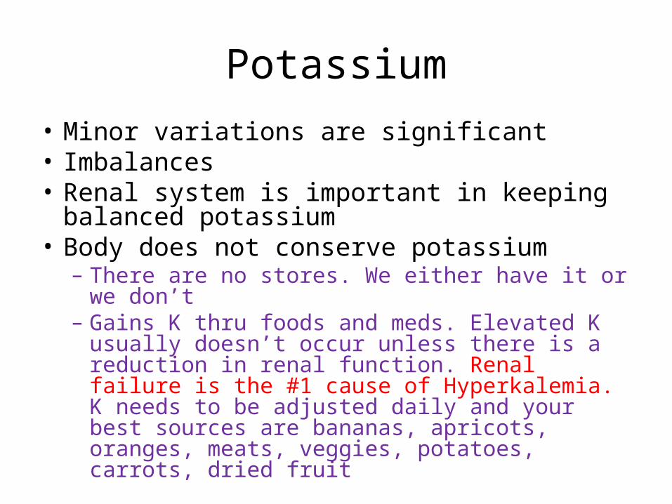
Potassium
• Minor variations are significant• Imbalances• Renal system is important in keeping
balanced potassium• Body does not conserve potassium
– There are no stores. We either have it or we don’t– Gains K thru foods and meds. Elevated K usually
doesn’t occur unless there is a reduction in renal function. Renal failure is the #1 cause of Hyperkalemia. K needs to be adjusted daily and your best sources are bananas, apricots, oranges, meats, veggies, potatoes, carrots, dried fruit

Hypokalemia• K+ < 3.5 mEq/L• Cause
– GI suction, vomiting, diarrhea– TPN or IVF without K+ replacement– Trauma– Diabetes – if it’s uncontrolled– Low oral intake of K+
– Sweat loss– Medications – diuretics, laxatives, insulin
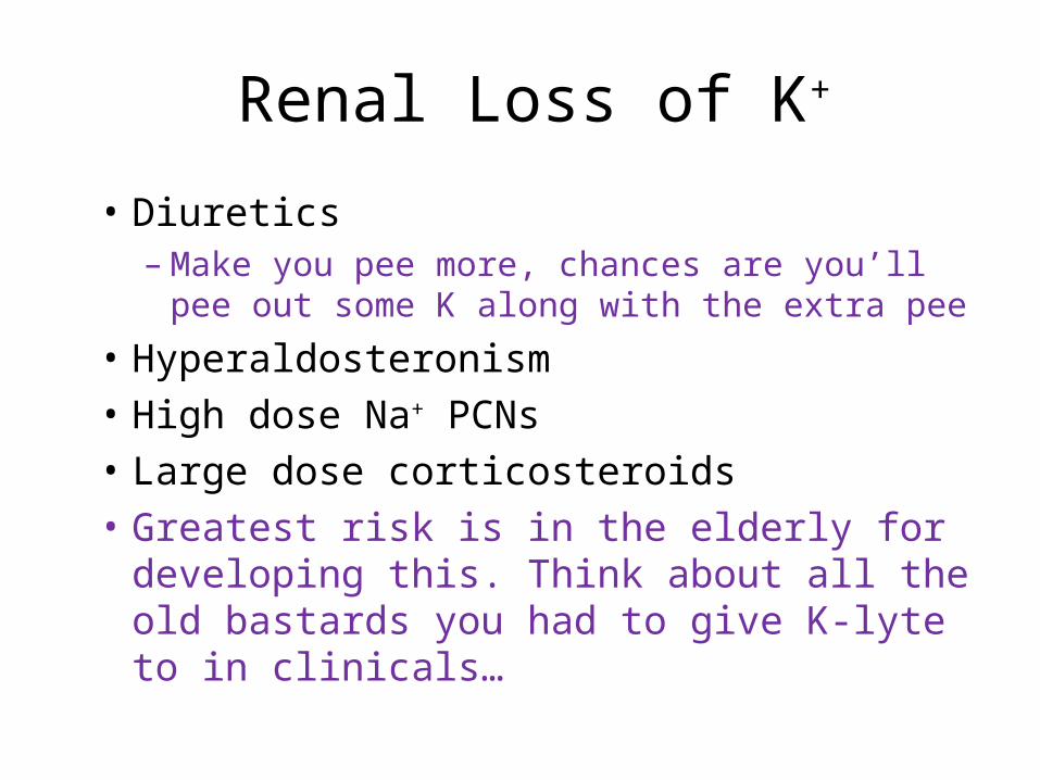
Renal Loss of K+
• Diuretics– Make you pee more, chances are you’ll pee out
some K along with the extra pee
• Hyperaldosteronism
• High dose Na+ PCNs
• Large dose corticosteroids
• Greatest risk is in the elderly for developing this. Think about all the old bastards you had to give K-lyte to in clinicals…
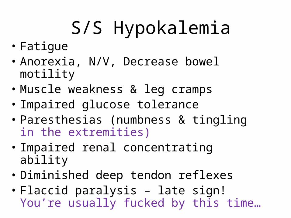
S/S Hypokalemia• Fatigue • Anorexia, N/V, Decrease bowel motility• Muscle weakness & leg cramps• Impaired glucose tolerance• Paresthesias (numbness & tingling in the
extremities)• Impaired renal concentrating ability• Diminished deep tendon reflexes • Flaccid paralysis – late sign! You’re
usually fucked by this time…

S/S Hypokalemia
• Increased sensitivity to digitalis
• Dysrhythmias
• Severe hypokalemia
• Hypokalemia commonly accompanies alkalosis.

Memory Jogger for Hypokalemia
• SUCTION
• S = Skeletal muscle weakness
• U = U wave (on the EKG)
• C = Constipation
• T = Toxicity of Digoxin
• I = Irregular or weak pulse
• O = Orthostatic Hypotension
• N = Numbness or parasthesia
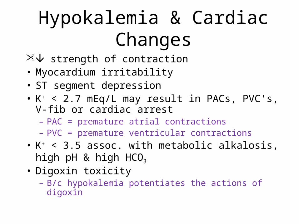
Hypokalemia & Cardiac Changes
strength of contraction• Myocardium irritability • ST segment depression• K+ < 2.7 mEq/L may result in PACs, PVC's, V-fib
or cardiac arrest– PAC = premature atrial contractions– PVC = premature ventricular contractions
• K+ < 3.5 assoc. with metabolic alkalosis, high pH & high HCO3
• Digoxin toxicity– B/c hypokalemia potentiates the actions of digoxin

Hypokalemia: Lab Results
• K+ deficit < 3.5 mEq/L• K+ < 3.5mEq/L often assoc. with
metabolic alkalosis, high pH, & high HCO3
• K+ < 2.7 may result in dangerous dysrhythmias
pH & HCO3
•Danger signs of low K are dyshrythmias, Cardiac arrest, digoxin toxicity, muscle paralysis (can lead to respiratory arrest)

Medical Treatment Hypokalemia
• K+ replacement (PO or IV)• Increase on a daily basis
– 40-80 mEq/day • At risk patient
– 50-100 mEq/day• K+ rich foods
– Green, leafy vegetable and what not• Treat the underlying cause• Is the patient’s magnesium low? Cause if it is,
it makes it harder for the kidneys to conserve K

Oral K+ Supplements• Minimize GI irritation
– Dilute liquid & effervescent supplement
– Give tabs & capsules w/ 8 oz. H2O– Give K+ with food
• Adverse reaction – N/V, diarrhea, GI bleed (sometimes)

Oral Potassium Supplements
• Avoid overdose (hyperkalemia) K+ dose if using K+ salt substitute
• Not used with K+ sparing diuretics
• K in the IV is VERY irritating to the vein! Can cause phlebitis very easily.
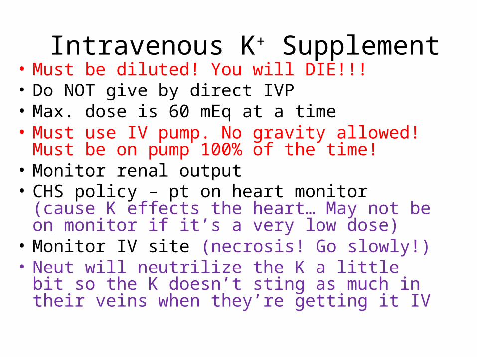
Intravenous K+ Supplement• Must be diluted! You will DIE!!!• Do NOT give by direct IVP• Max. dose is 60 mEq at a time• Must use IV pump. No gravity allowed! Must
be on pump 100% of the time! • Monitor renal output• CHS policy – pt on heart monitor (cause K
effects the heart… May not be on monitor if it’s a very low dose)
• Monitor IV site (necrosis! Go slowly!)• Neut will neutrilize the K a little bit so the K
doesn’t sting as much in their veins when they’re getting it IV

Nursing Interventions: Hypokalemia
• Identify pt at risk – esp. if on Digoxin
• Monitor EKG (or ECG) & BP
• Monitor serum K+
• Pt education – diuretics & laxatives
• Administer K+ supplements PO or IV dietary K+
• Monitor urine output

Hyperkalemia• Serum K+ > 5.5 mEq/L• Causes
– Renal failure– Release of K+ from damaged cells– Acidosis– Addison’s Disease– K+ sparing diuretics– High K+ intake– Medications

S/S Hyperkalemia• Main effects cardiac function• Muscle weakness and paralysis• Ventricular conduction is slowed• Paresthesias & irritability
– Respiratory and speech muscles• Flaccid muscular paralysis
– Legstrunkarms (including respiratory)• GI hyperactivity
– N/V, colic, & diarrhea

Hyperkalemia & Cardiac Changes
• Slows heart rate• ECG changes
– Tall, peaked T wave, short QT interval– Longer PR interval, widening QRS complex– Risk for Heart Block, A-fib, or, V-fib
• All of these are severe and we need to fix them as soon as we can, that is, if we can.
• The higher the K is the worse these are. Usually associated with a K higher than 7
• Severe K+ – Decreased heart contraction strength – Dilated & flaccid heart

ECG & Potassium

Hyperkalemia: Lab Data
• Serum potassium > 5.5 mEq/L
• ECG abnormalities
• Arterial blood gases – low pH indicating acidosis
• Metabolic acidosis is usually accompanied by hyperkalemia

Hyperkalemia Medical Treatment
• K+ restricted diet• Stop K containing medications• Monitor for “Digitalis toxicity”• Cation-exchange resins
– Kayexalate – PO or PR (in the rectum)• Fastest way to lower your potassium. It’s gross.
– 1Gm of resin removes 1 mEq K+ • Dialysis
– If conservative methods not suffice

Emergency Medical Treatment Hyperkalemia
• Ca Gluconate – IV– Does NOT K+ – Antagonizes K+ action on heart (keeps it from letting the
heart get flaccid. Works against the K but doesn’t lower the K levels! This is a quick thing to give them to prevent heart problems)
– Monitor ECG • Hypertonic Glucose & Insulin
– Insulin - facilitates K+ movement into cells• If you can get things out of the circulating volume and into the
cells, it doesn’t have an effect on the body. Unusable when it’s in the cells
– Glucose - insulin release from pancreas• NaHCO3
– K+ shifts into cells– May be the best thing to move K into the cells quickly!
On the test make sure to read the question and look for distracters and pick the appropriate answer

Nursing Interventions Hyperkalemia
• Be aware of pt at risk• Monitor for:
– Generalized weakness & dysrythmias– Irritability & GI symptoms– Nausea & intestinal colic– ECG or lab abnormalities
• Prevention of hyperkalemia• Educate pt: medication & diet• Do NOT draw blood above K+ infusion site
– Would have a very high rate of K if you do this.If they have hyperkalemia make sure you know foods
that are high in K. I missed the foods he was saying.

Calcium
• Serum Ca++ level 8.6 – 10.2 mg/dl (total) • 99% stored in bones (bones & teeth)• Found in three forms:
- bound: to proteins (less than 50%)- ionized: found in serum (50% of calcium and is most
important)- Children have high levels of this for bone growth- Old people have very low levels of this due to bone loss.- It’s important in muscle contraction, conduction of nerve
impulses, cardiac contractility, and helps in the formation of prothrombin
- complexed: combined with nonprotein anions: phosphate, citrate, and carbonate

Calcium and Phosphorus
• Ca and phosphorus have a reciprocal relationship– If the Ca is low, the phosphorus is high– If the phosphorus is low, the Ca will be high

Ionized Calcium
• Activate body chemical rxn
• Muscle contractions and relaxation
• Promote transmission of nerve impulse
• Cardiac contractility & automaticity
• Formation of prothrombin

Calcium Regulators
• Parathyroid Hormone (PTH) pulls– Releases Ca from the bone– Increases Ca absorption from GI– Increases Ca absorption from renal
tubules– When serum Ionized Ca is low, the
parathyroid gland releases PTH. Pulls Ca from the bone and promotes movement of Ca (with phosphorus) into the plasma
Calcium Regulators
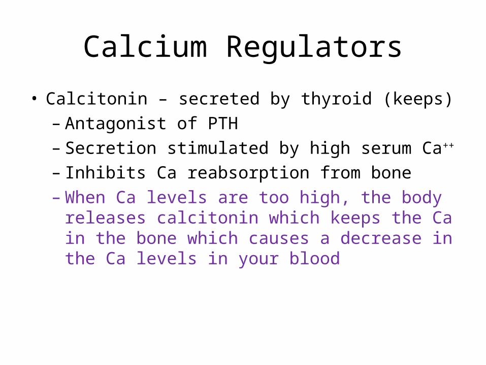
• Calcitonin – secreted by thyroid (keeps)– Antagonist of PTH– Secretion stimulated by high serum Ca++ – Inhibits Ca reabsorption from bone– When Ca levels are too high, the body
releases calcitonin which keeps the Ca in the bone which causes a decrease in the Ca levels in your blood

Calcium Regulators
• Phosphate
–Reciprocal relationship with Ca Ca = Phos
• Vitamin D
–Necessary for absorption & utilization of Ca
–We get Vitamin D from the sunshine

Hypocalcemia• Serum Ca++ < 8.5 mg/dl• Causes include:
- hypoparathyroidism & surgical hypoparathyroidism- malabsorption syndrome- vitamin D deficiency- prolonged admin. of Ca free IVF- acute pancreatitis (Affects PTH secretion, so you’re not able to absorb your Ca)
Ca absorption occurs primarily in the Small intestine. If you have Celiacs Disease or something like that, where you can’t absorb stuff, you’ll have low levels of Ca. Lack of Vit D decreases the absorption of Ca

Causes Hypocalcemia
- Excessive admin. of citrated blood- Alkalosis- Hyperphosphatemia- Hypomagnesemia- Thyroid cancer
- Causes excessive calcitonin secretion
- Low serum albumin- Cimetidine (Tagamet)
- Interferes with the PTH function
- Alcohol Abuse- Medications

S/S Hypocalcemia
• Tetany (# 1 sign)– Condition characterized by cramps, convulsions,
twitching of the muscles, and sharp flexion of the wrists and ankle joints. Think of tetanus. Tetanus is “Lock Jaw.” Your muscles get stiff and spastic.
• Vary with severity, duration & rate of development
• Numbness & tingling
• Spasms of muscles of extremities & face
• Pain

S/S Hypocalcemia
• Hyperactive deep tendon reflexes
• Abdominal muscle spasms
• Respiratory effects
• Altered mood & memory
• Convulsion/Seizures
– Seizures may occur b/c the hypocalemia
increases the irritability of the Central Nervous
System

S/S Hypocalcemia
• Laryngeal spasm
• + Trousseau’s
• + Chvostek’s
• Remember these bitches

+ Trousseau’s Sign• Carpopedal
spasm of hand when– Blood supply
– Pressure on nerve
• Occurs several minutes after BP cuff inflated > systolic BP
• Ischemia Indicates tetany and a good sign of hypocalcemia and hypomagnesium

+ Chvostek’s Sign
• Spasm of muscles innervated by facial nerve
• Tap facial nerve anterior to ear lobe below zygomatic process
• They close their eyes and their muscles kind of twitch

Hypocalcemia Cardiac Effects
• Prolonged QT interval
• Prolonged ST segment
cardiac contractility
sensitivity to Digoxin
Hypocalcemia: Lab Data
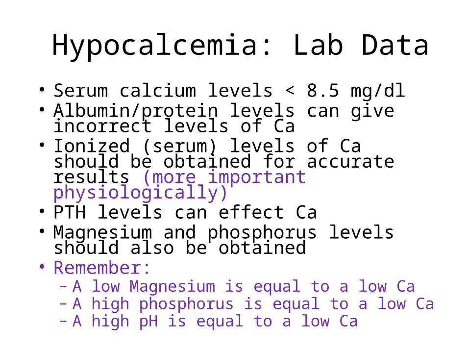
• Serum calcium levels < 8.5 mg/dl• Albumin/protein levels can give incorrect
levels of Ca• Ionized (serum) levels of Ca should be
obtained for accurate results (more important physiologically)
• PTH levels can effect Ca• Magnesium and phosphorus levels should
also be obtained• Remember:
– A low Magnesium is equal to a low Ca– A high phosphorus is equal to a low Ca– A high pH is equal to a low Ca

Hypocalcemia Medical Treatment
• Acute symptomatic Ca is emergency. – Requires prompt adm. of IV Calcium
• 10% Ca-Gluconate– For severe symptoms
• Has to be given IV and slowly! Never give IM!
• Ca-Chloride– Never give IM
• Oral Ca or Vitamin D

Nursing Interventions Hypocalcemia
• Identify pt at risk – Hx, labs, etc…
• Seizure precautions if severe Ca levels
• Monitor airway
• Monitor ECG
• Educate pt: Ca loss & risks & Ca rich foods– Ca rich foods are milk products, green leafy
veggies, canned salmon, sardines, and fresh oysters

Hypercalcemia• Calcium > 10.5 mg/dl• If severe – dangerous with mortality• Causes include:
– Hyperparathyroidism (most common cause of hypercalcemia!)
• Causes increased bone release of Ca and increased absorption of Ca from the intestines and kidneys
– malignant neoplastic disease and chemotherapies
• 2nd major cause of Hypercalcemia– thiazide diuretics– prolonged immobilization
• Causes and increase in loss of Ca from the bone, moves it into the circulatory system
– large doses Vit. D & Vit. A

S/S Hypercalcemia
• Decreased neuromuscular excitability:
Muscle weakness and incoordination
GI motility: anorexia, N/V,
constipation

S/S Hypercalcemia
• Altered memory, confusion, slurred
speech, lethargy, acute psychotic
behavior, & coma
• Depressed deep tendon reflexes

S/S Hypercalcemia
• Bone pain & abdominal pain
• Hypercalcemic crisis: severe polyuria &
polydipsia, intractable nausea (you have this
all the time and it won’t go away), abdominal
cramps, lethargy, coma and cardiac arrest
• Can cause kidney stones
– Increased urinary calcium concentrations
decreases the kidneys ability to concentrate urine.
This leads to polyuria and volume depletion

Hypercalcemia Cardiac Changes
• Calcium: inotropic effect on heart & reduces heart rate– Effects the contractility of the heart, it’s ability to
squeeze down. Because the heart and conduction system are effected by Ca you’ll get dysrhythmias and bradycardia which can lead to cardiac arrest
• Shorten ST segment & QT interval• Prolonged PR interval• Potentiate digoxin toxicity

Hypercalcemia: Lab Data
• Serum calcium > 10.2 mg/dl
• ECG-dysrythmias
• PTH- increased (which will throw all the Ca into the blood stream)
• X-ray-reveal osteoporosis (cause Ca is being tossed out from the bones into the blood)
• Urine (will be high in Ca)
Hypercalcemia Medical Treatment
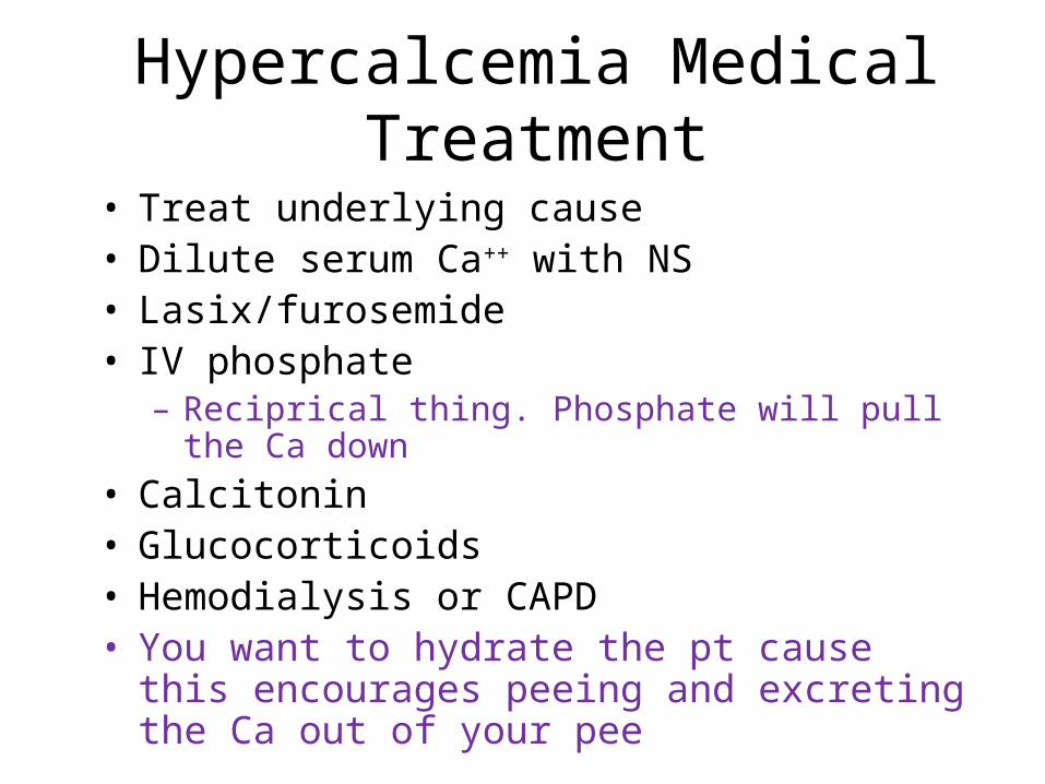
• Treat underlying cause• Dilute serum Ca++ with NS• Lasix/furosemide• IV phosphate
– Reciprical thing. Phosphate will pull the Ca down
• Calcitonin• Glucocorticoids• Hemodialysis or CAPD• You want to hydrate the pt cause this
encourages peeing and excreting the Ca out of your pee

Nursing Interventions Hypercalcemia
• Monitor for pt risk activity & fluids if possible Ca++ intake
• Safety measures for confusion
• Monitor ECG, I&O, breath sounds
• Monitor for Digoxin toxicity
• Prevent Ca++ renal stones– Whenever they get a kidney stone they usually
analyze them to see what they’re made of

Magnesium• Normal 1.3 – 2.3 mEq/L
• Second most important Ion in the ICF next to K
• Mg is important for neuromuscular function
• Activator for enzymes
• Carbohydrate & protein metabolism
• Vasodilation in peripheral arteries
• Found in bone and tissue
• Eliminated by kidneys
• The GI and urinary systems are the best regulator
systems for Magnesium
Hypomagnesemia
• Mg < 1.3 mEq/L• 1/3 Mg is bound to protein, 2/3 remains as free
cation• Causes include:• GI loss
-Alcoholism: decrease dietary intake, impairs renal conservation, intestinal malabsorption, intermittent diarrhea and vomiting
Kidney is the primary route of Mg excretion. Chronic alcoholism is the most common cause due to poor dietary intake of magnesium
Causes Hypomagnesemia
• Intestinal malabsorption syndromes
• Diarrhea
• Diuretics
• Prolonged admin. Mg free IVF/TPN
• NG Suction
• Renal or liver disease
• Diabetic ketoacidosis

S/S Hypomagnesemia
• Usually occur Mg < 1.0 mEq/L
• Most are neuromuscular: hyperexcitability with muscle weakness, tremors & athetoid movements (slow involuntary twisting motion, kind of constantly in motion and they don’t even know they are doing it. It’s reversible once you get the levels up)
• Tetany
• + Trouseau’s and Chvostek’s

S/S Hypomagnesemia
• Seizures• Laryngeal stridor
– You make a sound when you breathe. Like a high pitched squeek thing
• Signs of low hypocalcemia r/t low PTH• Alterations in mood: apathy, depression,
agitation, dizziness, insomnia, audio or visual hallucinations, psychoses, laryngeal strider (you make a sound when you breathe. Like a high pitched squeek thing…)
• Digoxin Toxicity

Memory Jogger for hypomagnesemia
• STARVED
• S = Seizures
• T = Tetany
• A = Anorexia
• R = Rapid Heart Rate
• V = Vomiting
• E = Emotional labillity (your emotions are all fucking crazy)
• D = Deep tendon reflexes increased

Hypomagnesemia Cardiac Changes
• Predisposes to dysrhythmias– PVC or V-fib
risk for digoxin toxicity• ECG:
– Prolonged PR & QT intervals– Widening QRS complex– depressed ST segment– Flattened T waves– Prominent U waves– Don’t need to know these guys

Hypomagnesemia: Lab Data
• Mg < 1.3 mEq/L
• Potassium
• Calcium
• ECG
• Urine Mg. level

Hypomagnesemia Medical Treatment
• Diet – Can be used alone for mild Mg– Green vegetables, meat, seafood,
nuts, seeds, legumes, whole grains, peanut butter, cocoa, and Spinach (probably one of the best sources)

HypomagnasemiaMedical Treatment (Cont)
• Mg replacement– Assess renal function – route of Mg
elimination– PO Slow-Mag
• Diarrhea possible side effect– IV or IM
• Because the kidneys are main route of excretion, make sure to watch BUN and Creatinine levels. Renal failure clients have problems with high Magnesium
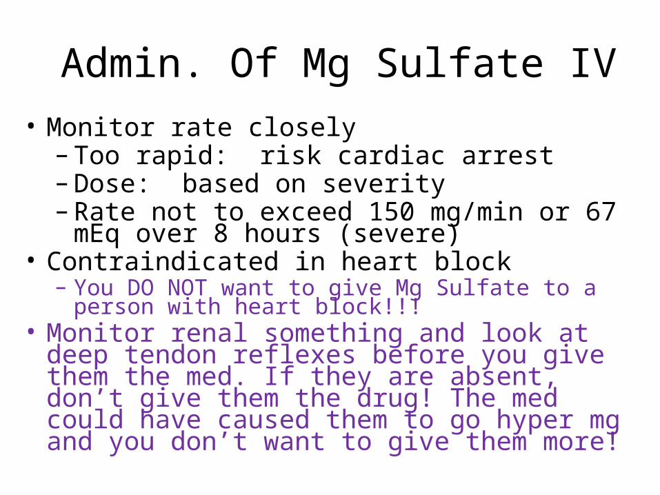
Admin. Of Mg Sulfate IV
• Monitor rate closely– Too rapid: risk cardiac arrest– Dose: based on severity – Rate not to exceed 150 mg/min or 67 mEq
over 8 hours (severe)• Contraindicated in heart block
– You DO NOT want to give Mg Sulfate to a person with heart block!!!
• Monitor renal something and look at deep tendon reflexes before you give them the med. If they are absent, don’t give them the drug! The med could have caused them to go hyper mg and you don’t want to give them more!
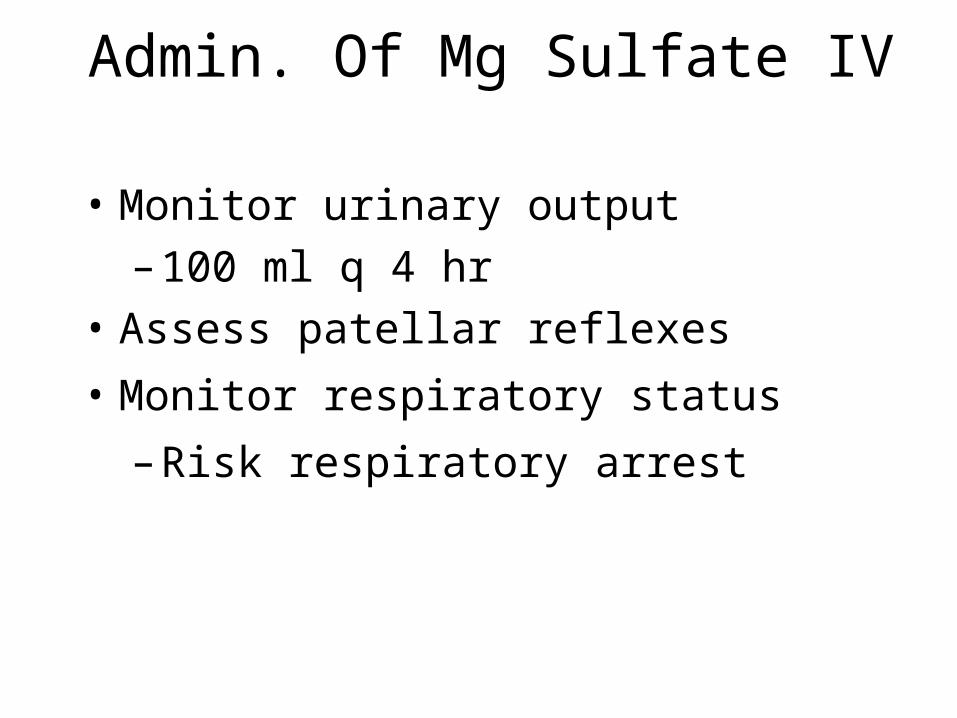
Admin. Of Mg Sulfate IV
• Monitor urinary output
– 100 ml q 4 hr
• Assess patellar reflexes
• Monitor respiratory status
– Risk respiratory arrest
Nursing Interventions Hypomagnesemia
• Identify & monitor pt at risk
• Asses of digoxin toxicity
• Seizure precautions
• Monitor airway
• Safety for confusion / psychosis– These guys you put them in bed and head
down the hall and before you get to the nurses station they’ve passed you on the way to the elevator trying to escape!

Nursing Interventions Hypomagnesemia
• Pt education: diuretics & laxative use
• Pt education: diet
Hypermagnesemia• Mg > 2.5 mEq/L (this abnormality is very rare)• Causes
– Hemolyzed blood samples• May show that you have hyper when you don’t. You’re
blood sample is fucked up and they look at it and it says you have this when you really don’t
– Renal failure• Most common reason for mypermagnesium
– Addison’s Disease– Excessive use antacids and laxatives– Untreated ketoacidosis– Excessive infusion– Hypothermia– Lithium toxicity

S/S Hypermagnesemia
• Acute elevations: depression of the CNS• Mild increases: (cause these things below)
-low blood pressure-N/V-facial flushing-sensations warmth
Primary symptoms occur as a result of peripheral and central nervous symptom depression

S/S Hypermagnesemia • Higher increases:
-lethargy-dysarthria (Difficult or poorly articulated speech caused by damage to central or peripheral motor nerves)
-drowsiness-loss of deep tendon reflexes-muscle weakness and paralysis-depressed respirations -coma

Hypermagnesemia Cardiac Changes
• Sinus Bradycardia
• Prolonged PR, & QT intervals
• Tall T waves
• Widened QRS
• Heart Block
• Cardiac arrest in diastole

Hypermagnesemia: Lab Data
• Mg > 2.5 mEq/L
• ECG
• K+ increased
• Ca- increased
• Creatinine clearance decreases to less than 3.0 ml/min.

Hypermagnesemia Medical Treatment
• Prevention is key!
• Avoid administration of Mg in renal failure
• Hemodialysis (sometimes this is necessary to get the Mg level down)

Hypermagnesemia Medical Treatment
• Emergency treatment if respiratory or cardiac problems develop– Ventilator support– Calcium Gluconate
• Direct antagonist to Magnesium• 5 – 10 mEq may reverse cardiac or
respiratory problems• Lasix• NaCL or LR

Nursing Interventions Hypermagnesemia
• Monitor pt at risk• Monitor vital signs
– Low BP – Shallow resp. with progressive apnea
• Assess patellar reflexes– Absent reflexes implies Mg > 7.0– When you go to hit the knee you get absolutely
nothing. This means it’s above 7.0• Monitor LOC
– Drowsy, lethargy, coma• When you have periods of low BP and signs of
apnea and lowered reflexes you need to call the dr. Have all your information ready before you call cause they’ll yell at you.

Phosphorus
• Normal 2.5-4.5 mg/dl (adult)
• Essential for fxn of muscle & RBCs
• Essential to nervous system
• Essential to metabolism of: – Carbohydrate– Protein– Fats– Crucial to cell membrane activity.
Phospholipids make up the cell membrane.

Phosphorus
• Aids in the formation of ATP and 2,3 diphosphoglycerate
• Maintenance in acid-base balance
• 85% is located in bones and teeth
• 14% located in soft tissue
• 1% in ECF
• Critical to nerve and muscle function

Hypophosphatemia• Phosphorus < 2.5 mg/dl
• Causes
– Severe protein – • calorie malnutrition
• Anorexia
• Alcoholism

Hypophosphatemia
• Overfeeding with simple carbohydrates
• Elderly debilated & unable to eat
• Hepatic encelopathy (can result in
hypophosphatemia)
Hypophosphatemia
• Prolonged intense hyperventilation– Alcohol withdrawal
– Diabetic ketoacidosis• You get Osmotic Diruesis and the insulin causes Ph to move
into cells
– Major thermal burns• Extensive diruesis of salt and water which typically occurs w/i
first couple of days or something like that. I missed it.
– Hyperventilation causes respiratory alkalosis which makes phoshporus move into cells (along with K)

S/S Hypophosphatemia
• Most signs & symptoms 2nd to deficiency
– Impaired cellular energy resources (ATP) Contractility of heart is decreased due to low amounts of ATP
– Impaired oxygen delivery to tissues
(2,3Diph) DPG

S/S Hypophosphatemia • Neurological
– Irritability, Apprehension, weakness,
– Numbness, confusion
– Seizure, fatigue, parasthesia, coma
– Without Ph the body can’t make enough ATP which is necessary for energy metabolism
• Hyperglycemia – 2nd to predisposed insulin resistance

S/S Hypophosphatemia • Muscle damage
– 2nd to ATP level in muscle tissue– Muscle weakness & pain– Acute rhabdomyolysis
• Disintegration of striated muscle• Skeletal muscle destruction that occurs
due to altered cell activity– Impaired ventilation
• 2nd to weakened respiratory muscles

Hypophophatemia: Lab Data
• Phos < 2.5 mg/dl
• Glucose/insulin admin.
• PTH
• Alkaline phosphatase
• X-ray

Medical Treatment Hypophosphatemia
• Prevention • TPN & TF should have adequate Phos.• Phosphorus – PO
– Aluminum Phosphate (Phosphojel)• Phosphorus < 1.0 mg/dl (severe)
– K-Phosphate or Na-Phosphate• 0.2 mMol /kg/hr is max. rate• Risk of hypocalcemia & tetany (when you’re
trying to correct this)
• Foods rich in Ph: eggs, nuts, whole grains, meat, fish, poultry and milk products.
Nursing Interventions Hypophosphatemia
• Identify & monitor pt at risk
• Gradual introduction of TPN & TF
– Avoid rapid shift of phosphorus
• Prevent infection
• Monitor serum phosphate levels
• Administer meds safely
• Teach about diet
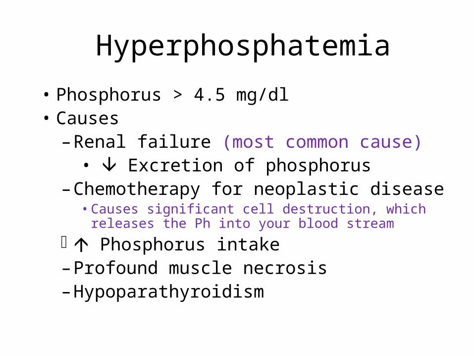
Hyperphosphatemia
• Phosphorus > 4.5 mg/dl• Causes
– Renal failure (most common cause)• Excretion of phosphorus
– Chemotherapy for neoplastic disease• Causes significant cell destruction, which releases the
Ph into your blood stream
Phosphorus intake– Profound muscle necrosis– Hypoparathyroidism

S/S Hyperphosphatemia• Similar to S/S of hypocalcemia (similar to
hypocalcemia)
• Tetany
– Tingling then numbness – fingertips & around mouth
– Spreads proximally to limbs & face severity
• Muscle spasm & pain
• Progressive renal impairment
• Remember the inverse relationship with Ph and Ca. When Ph is high, Ca is low and visa versa.

Hyperphosphatemia: Lab Data
• Phos > 4.5 mg/dl
• Calcium
• X-ray
• PTH
• Bun and creatinine (any time there is kidney involvement you are going to be checking these guys)

Medical Treatment Hyperphosphatemia
• Treat underlying disorder• If 2nd to tumor cell lysis
– Allopurinol – prevent urate nephropathy• If 2nd to renal failure
– Phosphate binding gels phosphate diet– Dialysis

Medical Treatment Hyperphosphatemia
• Acute hyperphosphatemia
– NS – IVF
• Promotes renal excretion
– Hypertonic dextrose & regular insulin
• Drive phosphorus into cells
– Hemodialysis or Peritoneal dialysis
– Surgery

Nursing Interventions Hyperphosphatemia
• Identify & monitor pt at risk• Monitor lab results• Pt education: Avoid meds with Phos.
– Laxatives & enemas• Change in urine output

Nursing Interventions Hyperphosphatemia
• Pt education: Avoid Phos. Foods– Dried fruit & vegetables– sardines– Hard cheeses,– Whole grain cereal– Nuts– All these are high in Ph
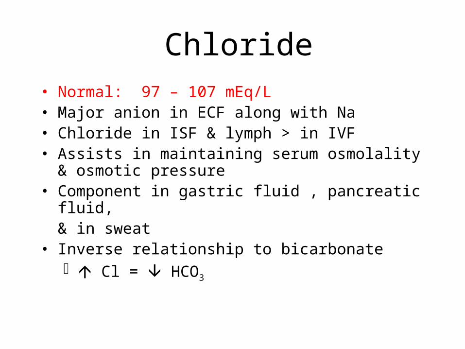
Chloride• Normal: 97 – 107 mEq/L• Major anion in ECF along with Na• Chloride in ISF & lymph > in IVF• Assists in maintaining serum osmolality & osmotic
pressure• Component in gastric fluid , pancreatic fluid,
& in sweat• Inverse relationship to bicarbonate
Cl = HCO3

Hypochloremia• Cl < 96 mEq/L
• Causes
– Prolonged vomiting
– Prolonged NG suctioning
– Prolonged diarrhea
– GI drainage
– Salt restricted diet
– Diuretics (can cause loss via the kidneys, you pee it out. No shit. Thanks Durbin, for that wonderful tidbit)

S/S Hypochloremia Bicarbonate level
Na level
• Hyperexcitability of muscles
– Tetany, twitching, weakness
• Hyperactive deep tendon reflexes
• Cardiac dysrhythmia
• Water excess

Hypochloremia: Lab Data
• Cl < 96 mEq/L
• Sodium (low)
• Potassium (low)
• Arterial Blood Gases: reveals metabolic alkalosis
• Urine chloride level (low)

Medical Treatment Hypochloremia
• Correct the cause
• IV therapy: NS or ½NS
• Ammonium chloride (to treat metabolic alkalosis)
– Dose calculated on chloride deficit
– 100mEq / 500ml NS – give slowly• You don’t want by gravity b/c the entire
amount could go in too quickly which could add to their problems (would add fluid overload)
– Foods high in chloride

Nursing Interventions Hypochloremia
• Monitor I&O• Monitor bicarbonate & sodium level• Assess LOC, muscle strength & movement
Avoid bottled water (doesn’t have any electrolytes, so large amts of chloride could be excreted in the kidneys. The water makes you pee, and in this case, peeing a lot is bad)
• Pt education: food in chloride– Tomato juice – canned vegetables– broth, fruit, processed meat

Hyperchloremia
• Cl > 107 mEq/L
• Causes:
– Loss of bicarbonate
• Kidney
• GI tract
• Remember Ch and bicarbonate inverse relationship, if you pee off Ch you will increase your bicarbonate levels…
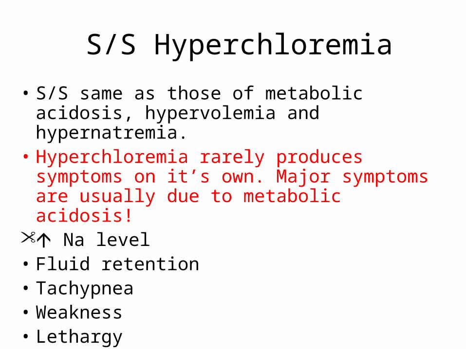
S/S Hyperchloremia
• S/S same as those of metabolic acidosis, hypervolemia and hypernatremia.
• Hyperchloremia rarely produces symptoms on it’s own. Major symptoms are usually due to metabolic acidosis!
Na level• Fluid retention• Tachypnea• Weakness• Lethargy

S/S Hyperchloremia
Cognitive ability• HTN• If Untreated
Cardiac output– Dysrhythmias– Coma

Medical Treatment Hyperchloremia
• IV fluid– Lactated Ringer’s – slowly – LR may be used to convert the lactate to
Bicarbonate, which increases the pH of your body, which in this case is good
• Diuretics• Restrict –
– Sodium– Chloride– Fluids other than LR until Cl level

Hyperchloremia: Lab Data
• Cl > 108 mEq/L
• Sodium >145
• pH <7.35
• Serum bicarbonate <22
• Urine chloride increased

Nursing Interventions Hyperchloremia
• Monitor for those pt at risk
• Monitor vital signs
• Monitor I&O
• Fluid restriction other than LR
• Monitor ABG
• Pt education: diet restrictions
• Remember, IV fluid for Hyperchloremia is LR.
And also remember, do this slowly!

Questions
??????