posttranscriptional control of microbe-induced rearrangement...
TRANSCRIPT

Posttranscriptional Control of Microbe-Induced Rearrangement ofHost Cell Actin
Charley C. Gruber, Vanessa SperandioDepartments of Microbiology and Biochemistry, University of Texas Southwestern Medical Center, Dallas, Texas, USA
ABSTRACT Remodeling of the host cytoskeleton is a common strategy employed by bacterial pathogens. Although there is vigor-ous investigation of the cell biology underlying these bacterially mediated cytoskeleton modifications, knowledge of the plastic-ity and dynamics of the bacterial signaling networks that regulate the expression of genes necessary for these phenotypes is lack-ing. Enterohemorrhagic Escherichia coli attaches to enterocytes, forming pedestal-like structures. Pedestal formation requiresthe expression of the locus-of-enterocyte-effacement (LEE) and espFu genes. The LEE encodes a molecular syringe, a type III se-cretion system (T3SS) used by pathogens to translocate effectors such as EspFu into the host cell. By using a combination of ge-netic, biochemical, and cell biology approaches, we show that pedestal formation relies on posttranscriptional regulation by twosmall RNAs (sRNAs), GlmY and GlmZ. The GlmY and GlmZ sRNAs are unique; they have extensive secondary structures andwork in concert. Although these sRNAs may offer unique insights into RNA and posttranscriptional biology, thus far, only onetarget and one mechanism of action (exposure of the ribosome binding site from the glmS gene to promote its translation) hasbeen described. Here we uncovered new targets and two different molecular mechanisms of action of these sRNAs. In the case ofEspFu expression, they promote translation by cleavage of the transcript, while in regard to the LEE, they promote destabiliza-tion of the mRNA. Our findings reveal that two unique sRNAs act in concert through different molecular mechanisms to coordi-nate bacterial attachment to mammalian cells.
IMPORTANCE Pathogens evolve by horizontal acquisition of pathogenicity islands. We describe here how two sRNAs, GlmY andGlmZ, involved in cellular metabolism and cellular architecture, through the posttranscriptional control of GlmS (the previ-ously only known target of GlmY and GlmZ), which controls amino sugar synthesis, have been coopted to modulate the expres-sion of virulence. These sRNAs quickly allow for plasticity in gene expression in order for enterohemorrhagic Escherichia coli tofine-tune the expression of its complex type III secretion machinery and its effectors to promote bacterial attachment and subse-quent actin rearrangement on host cells. Pedestal formation is a very dynamic process. Many of the genes necessary for pedestalformation are located within the same operon to evolutionarily guarantee that they are inherited together. However, it is worthnoting that within these operons, several genes need to yield more proteins than others and that these differences cannot be effi-ciently regulated at the transcriptional level.
Received 26 November 2013 Accepted 2 December 2013 Published 14 January 2014
Citation Gruber CC, Sperandio V. 2014. Posttranscriptional control of microbe-induced rearrangement of host cell actin. mBio 5(1):e01025-13. doi:10.1128/mBio.01025-13.
Editor Margaret McFall-Ngai, University of Wisconsin
Copyright © 2014 Gruber and Sperandio. This is an open-access article distributed under the terms of the Creative Commons Attribution-Noncommercial-ShareAlike 3.0Unported license, which permits unrestricted noncommercial use, distribution, and reproduction in any medium, provided the original author and source are credited.
Address correspondence to Vanessa Sperandio, [email protected].
Exploitation of the host cytoskeleton by bacterial pathogensis an essential feature of bacterium-host associations. Actin
remodeling promotes bacterial invasion of nonphagocytic cells,survival within cells, cell-to-cell spread and locomotion, and col-onization at the interface of the host epithelium (1). Enterohem-orrhagic Escherichia coli (EHEC) O157:H7 is a deadly pathogenthat attaches to enterocytes, forming attaching and effacing (AE)lesions characterized by the formation of a pedestal-like structurebeneath the bacterium (2). To induce pedestal formation on epi-thelial cells, EHEC employs a type III secretion system (T3SS), aneedle-like structure that translocates bacterial effectors directlyinto host cells. The genes for this T3SS and several other genesnecessary for AE lesion formation are located within a chromo-somal pathogenicity island named the locus of enterocyte efface-ment (LEE) (3, 4). The LEE region contains five major operons,LEE1 to LEE5 (5–7), which encode the components of the T3SS
(4), an adhesin (intimin) (8) and its receptor Tir, which is itselftranslocated through the T3SS to the host cell, where, upon itsinsertion into the cell membrane, it serves as a receptor for thebacterial adhesin intimin (9) and other effector proteins (10–14).The LEE-encoded T3SS also translocates effector proteins en-coded outside the LEE region, including EspFu/TccP, which isimportant for efficient AE lesion formation (15–18).
Expression of the LEE and espFu genes is governed throughcomplex multilayered signaling cascades in response to many en-vironmental cues, including human hormones (epinephrine [Epi]and norepinephrine [NE]), bacterial small signaling molecules(autoinducer-3 [AI-3], indole, acyl homoserine lactones), carbonand nitrogen sources, and stress responses, among others. Thisregulation occurs at both the transcriptional and posttranscrip-tional levels (19–29). In bacteria, there are many different mech-anisms of posttranscriptional regulation of genes (30). One of the
RESEARCH ARTICLE
January/February 2014 Volume 5 Issue 1 e01025-13 ® mbio.asm.org 1
on July 10, 2018 by guesthttp://m
bio.asm.org/
Dow
nloaded from

more abundant classes is that of trans-acting small RNAs (sRNAs).The majority of these sRNAs require the RNA chaperone Hfq andact by directly binding to mRNAs at the ribosome binding site(RBS) to repress translation, cause direct degradation of themRNA by recruitment of nucleases, or activate translation by re-lieving a hairpin that blocks the RBS. While genes of the LEE areknown to be posttranscriptionally regulated, no sRNAs responsi-ble for this have been identified to date.
The AI-3/Epi/NE interkingdom signaling cascade activates theexpression of virulence genes in EHEC (21, 31–33). The host hor-mones Epi and NE are specifically sensed by two membrane-bound histidine sensor kinases, QseC and QseE, which are the firstbacterial adrenergic receptors identified (34, 35). QseE is down-stream of QseC in this signaling cascade, given that the transcrip-tion of qseE is activated through QseC (36). In addition to sensingthese host hormones, QseC also senses the bacterial signal AI-3(32). QseE, however, does not sense AI-3, thereby discriminatingbetween host- and bacterium-derived signals (35). Upon sensingtheir respective signals, QseC and QseE autophosphorylate to ac-tivate virulence gene expression and pathogenesis in vitro and invivo in EHEC (32, 35, 37). QseC transfers its phosphate to threeresponse regulators (RRs), QseB, QseF, and KdpE, which, uponphosphorylation, are activated and function as transcription fac-tors (38). QseE transfers its phosphate only to QseF (39). Theconcerted action of these RRs activates the EHEC virulence rep-ertoire, including the LEE and espFu genes (Fig. 1A). The QseF RRis necessary for the expression of EspFu (36), and it is known toregulate the sRNA GlmY, located immediately upstream from theqseEGFglnB operon (Fig. 1B) (40). This sRNA is known to act as amolecular mimic (41), stabilizing another sRNA (GlmZ), whichdirectly binds to the mRNA of the gene encoding glucosaminesynthetase (glmS) and activates its translation by breaking a hair-pin loop and revealing the RBS (42).
Here we show that both the QseB and QseF RRs directly acti-vate the expression of glmY. GlmY and GlmZ coordinate LEE andEspFu expression posttranscriptionally through two differentmechanisms. GlmY and GlmZ posttranscriptional regulation ofthe LEE and EspFu ensures the correct timing and dynamics of AElesion formation by EHEC on epithelial cells. We propose thatsRNA-mediated posttranscriptional regulation is responsible forthe dynamic rewiring of the expression of different components ofbacterial complex machineries that allow successful interactionswith mammalian cells.
RESULTSTranscriptional regulation of glmY and glmZ. The QseC/QseEsignaling system controls a plethora of virulence genes in EHECthat have to be coordinately expressed to ensure optimal AE lesionformation on epithelial cells, leading to host infection (37, 38, 43,44). AE lesion formation is a dynamic process that requires plas-ticity and rapid adaptation of bacterial gene expression. Couplingof transcriptional and posttranscriptional regulation within a sig-naling transduction cascade in the bacterial cell is key to ensuringfine-tuning and rapid adaptation of gene expression toward theregulation of complex processes such as AE lesion formation. Up-stream of the qseEGFglnB operon is glmY (Fig. 1B). The glmY geneis known to have two overlapping promoters, one that is driven bya �70 RNA polymerase (the homeostatic form of this enzyme) andanother that is driven by a �54 RNA polymerase (Fig. 1B and C).Transcription of glmY is known to be regulated by the �54-
dependent transcriptional activator QseF (40). Additionally, a se-quence matching the known consensus sequence of QseB (45),another RR involved in interkingdom signaling, was identified insilico in this promoter region (Fig. 1C).
Transcriptional �-galactosidase reporters of the promoters ofboth glmY and glmZ were constructed. As previously reported, inthe qseF mutant, glmY expression is starkly decreased and almostablated. Meanwhile, the qseB mutant, while still expressing glmY,expressed significantly less than the wild type (WT) (Fig. 1D). Thisresult was confirmed by Northern blot analysis for the GlmY RNA(Fig. 1E). Neither RR had any effect on glmZ expression (Fig. 1F).The almost complete ablation of glmY expression in the �qseFmutant is due to the �54 RNA polymerase acting as a repressor inthe absence of QseF (40). The �54 RNA polymerase cannot pro-mote the formation of the DNA open complex to initiate tran-scription by itself; it requires a �54 RR, such as QseF, for thisprocess (46). Because the �54 promoter overlaps the �70 promoterand in the absence of QseF, the �54 RNA polymerase occupyingthe �54 promoter prevents access to the �70 promoter by the �70
RNA polymerase (40).Electrophoretic mobility shift assays (EMSAs) demonstrated
that both QseB and QseF directly bind to the glmY regulatoryregion (Fig. 1G). Interestingly, while the QseB consensus sequencewithin the glmY regulatory region differs slightly among EHECstrain 86-24, E. coli K-12 strain MG1655, and enteropathogenicE. coli (EPEC) strain E2348/69, all are capable of binding to QseB(see Fig. S1 in the supplemental material).
Insights into GlmY and GlmZ regulation of EspFu. Becausethe QseF RR controls AE lesion formation by indirectly promot-ing the expression of the EspFu T3SS effector (36), next we inves-tigated whether GlmY and/or GlmZ also play a role in EspFu ex-pression. EspFu interacts with another effector, Tir, throughIRTKS and acts as an Nck mimic to recruit N-WASP and Arp2/3to the site of bacterial attachment, causing the formation of thecharacteristic actin-rich pedestal (Fig. 2A) (15, 16, 47). The ex-pression of this effector is dependent on QseF, with the qseF mu-tant having the same phenotype as the �espFu mutant, which isthe almost complete lack of AE lesion formation on HeLa cells(Fig. 2B and 3A) (15, 16, 36).
Since the regulation of espFu by QseF is known to be indirect(36), we tested the ability of its known target, GlmY, as well as itsdownstream target, GlmZ, to complement a qseF mutant. TheglmY and glmZ genes were cloned under the control of an induc-ible promoter and transformed into the �qseF mutant strain.These strains were then used to infect HeLa cells to perform thefluorescein actin staining (FAS) test to visualize AE lesions. In theFAS assay, the HeLa cytoskeleton was stained green with fluores-cein isothiocyanate (FITC)-labeled phalloidin, the bacteria andnuclei were stained red with propidium iodide (PI), and pedestalswere visualized as brilliant patches of green underneath a red bac-terium. Both sRNAs were able to rescue AE lesion formation in theqseF mutant, indicating that these sRNAs are the intermediariesbetween QseF and espFu (Fig. 2B).
Since sRNAs act posttranscriptionally, we investigated espFumRNA levels by using Northern blot assays. EspFu is located out-side the LEE within a prophage. Upstream of the espFu gene is theespJ gene, which encodes another T3SS effector (Fig. 2E) (48). Inthe WT, there is a major band the size of the predicted espFutranscript (1,100 bp), as well as a much fainter upper band 2,100nucleotides in length. In the qseF mutant, the lower band is still
Gruber and Sperandio
2 ® mbio.asm.org January/February 2014 Volume 5 Issue 1 e01025-13
on July 10, 2018 by guesthttp://m
bio.asm.org/
Dow
nloaded from

FIG 1 QseF and QseB regulation of glmY. (A) Schematic representation of the QseC and QseE transduction signaling systems. QseC responds to AI-3,epinephrine, and norepinephrine (Nor), and QseE responds to epinephrine (Epi), sulfate (SO4), and phosphate (PO4). Upon sensing their signals, these histidinesensor kinases autophosphorylate and then transfer their phosphate to their response regulators. QseC phosphotransfers to KdpE (activates the LEE genes), QseB(regulates the flagellum regulon), and QseF (regulates espFu). QseE phosphotransfers only to QseF. (B) Schematic representation of glmY and the qseEGFglnBoperon depicting the locations of the �54 and �70 promoters. (C) Diagram showing the layout of the glmY regulatory region. QseF binding sites are red, QseBbinding sites are blue, and the �54 and �70 promoters are magenta and yellow, respectively. (D) �-Galactosidase assay of the glmY::lacZ transcriptional fusion inthe WT strain and the �qseB and �qseF mutant strains. The plasmid contains bp �250 to �20 from the transcription start site. (E) Northern blot assay with a
(Continued)
Control of Host Cell Actin Rearrangement
January/February 2014 Volume 5 Issue 1 e01025-13 ® mbio.asm.org 3
on July 10, 2018 by guesthttp://m
bio.asm.org/
Dow
nloaded from

present; however, the upper band is much more pronounced. Thisupper band corresponds to the expected size of an espJ-espFu tran-script (Fig. 2C and E). There are 320 bp between these two genes,and previous work suggested that they are not cotranscribed.However, since only RNA from WT bacteria was used in thoseexperiments, it is possible that the less abundant larger transcriptwas not detected (36). To confirm that this larger transcript isespJ-espFu, reverse transcription (RT)-PCR was performed withprimers spanning this region. While a very faint band correspond-ing to the espJ-espFu (2,100-bp) transcript was observed in WTEHEC, it was much more pronounced in the qseF mutant(Fig. 2D). Hence, the lack of EspFu expression in a qseF mutant isdue not to the absence of its transcript but to the lack of a process-ing event of the espJ-espFu transcript necessary for EspFu expres-sion.
Transcriptional and translational reporters of espFu were con-structed. As expected, the qseF mutant had no defect in espFutranscription (Fig. 2F); however, it is required for the translationof EspFu (Fig. 2G). To determine the regions of espJ-espFu re-quired for this regulation, various deletions in the intergenic re-gion between these genes were constructed. These deletions werecloned into a vector with a FLAG tag at the C terminus of espFu.The following four deletion mutants were constructed by usingthe previously identified 5= untranslated region (UTR) of theespFu transcript as a reference point (36): p1, which lacks theentire intergenic region; p2, which lacks the espJ 3= UTR; p3,which lacks the espFu 5=UTR; and p4, which does not have the espJgene but still has the intergenic region (Fig. 2H). Western blotassays with anti-FLAG antiserum were performed with whole-celllysates of EHEC expressing the WT espJ-espFu-FLAG plasmid andeach of these four deletion constructs. The p2 and p4 constructsexpressed levels of EspFu::FLAG similar to those of the WT plas-mid, while p1 expressed more protein and p3 did not expressEspFu (Fig. 2I). These data indicate that the 3= UTR of espJ actsnegatively on the translation of espFu and that this QseF/GlmY/GlmZ-mediated processing event is required for the translation ofespFu since the resulting transcript lacks the 3= UTR of espJ. Ad-ditionally, the presence of EspFu::FLAG from the p4 plasmid in-dicates that in addition to being cotranscribed with espJ, espFu alsohas its own promoter (Fig. 2I).
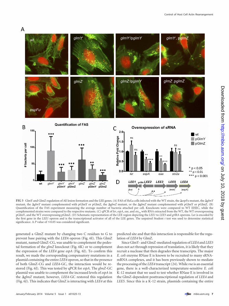
The roles of GlmY and GlmZ in pedestal formation. Giventhat EspFu is involved in pedestal formation, we further investi-gated the roles of GlmY and GlmZ in AE lesion formation byconstructing �glmY and �glmZ mutant strains and performingFAS assays with them. Since they were both capable of rescuing theqseF phenotype, we expected that both mutants would have a de-creased ability to form pedestals, similarly to the �qseF and�espFu mutant strains. Surprisingly, both sRNA mutants attachedto and formed pedestals on HeLa cells at levels far higher than thatof the WT strain. Both the glmY and glmZ plasmids were capableof complementing the glmY mutant, which is an expected result(Fig. 3A and B). The GlmY sRNA is known to stabilize the GlmZsRNA (42), and GlmZ, in an Hfq-dependent manner, exposes the
RBS of the glmS mRNA to promote its translation (42). Thus far,the only known target of GlmY and GlmZ regulation in E. coli wasglmS. The “effector” sRNA that base pairs with the glmS mRNA isGlmZ, and thus, the effect of GlmY on glmS is indirect and attrib-uted solely to its stabilization of GlmZ (42). However, the glmYplasmid was also capable of complementing the �glmZ mutant,which suggests that GlmY may have additional functions besidespreventing the degradation of GlmZ (Fig. 3A and B). Because, inaddition to espFu, AE lesion formation also requires the expres-sion of the LEE genes, we assessed LEE regulation by these sRNAs.The expression of the stx2a gene, which encodes Shiga toxin, and ofler (LEE1 operon), the master regulator of the LEE, was un-changed, but the expression of the LEE4 (espA) and LEE5 (eae)operons was decreased in the WT strain expressing GlmY or GlmZon a plasmid (Fig. 3C and D), suggesting that these sRNAs de-crease LEE4 and LEE5 expression posttranscriptionally. Thesedata offer an explanation for why the deletion of the genes encod-ing these sRNAs increases AE lesion formation. In the absence ofthese sRNAs, the transcripts of LEE4 and LEE5 operons (contain-ing many genes essential for pedestal formation) would be stabi-lized, increasing pedestal formation.
The roles of GlmY and GlmZ in the regulation of AE lesionformation by promoting EspFu translation and destabilizing LEEtranscripts (Fig. 2 and 3) seem to be initially confounding. How-ever, AE lesion formation is a dynamic process where pedestals areconstantly being formed and unformed during infection (seeMovie S1 in the supplemental material), and the precise modula-tion of the levels of LEE and EspFu expression is important for theefficiency of this phenotype. To better understand the dynamics ofpedestal formation responsible for the phenotype of the glmY andglmZ EHEC knockouts, we visualized the cells in real time. TheF-actin binding peptide Lifeact (49) proved to be the most effec-tive at visualizing the pedestals formed by EHEC. For ease of ex-perimentation, an HeLa cell line stably expressing Lifeact::greenfluorescent protein (GFP) was created. Bacteria were visualized bythe expression of mCherry (Fig. 4A to C; see Movies S1 to S3 in thesupplemental material). The �glmY and �glmZ mutant strainsattached to and formed pedestals much more efficiently and fasterthan the WT strain, suggesting that these sRNAs regulate theproper timing and amount of AE lesion formation on epithelialcells.
GlmY and GlmZ are known to promote the translation ofglmS, which encodes the glucosamine synthase enzyme in E. coliK-12 strain MC4100. GlmS is necessary for the synthesis ofN-acetylglucosamine-6-P, which is used for cell wall biosynthesis(see Fig. S2A in the supplemental material) (42). A glmS mutant islethal because it is defective in cell wall biosynthesis. However, theaddition of 1% N-acetylglucosamine (GlcNAC) to the mediumallows the survival of a glmS mutant because the GlcNAC sugarneeded for cell wall synthesis is being provided exogenously (50).To rule out the possibility that the AE lesion phenotype governedby GlmY and GlmZ is due to issues with cell wall synthesis in thesesRNA mutants because of the decreased expression of GlmS, FAS
Figure Legend Continued
glmY probe of RNAs from the WT strain and the �qseB and �qseF mutant strains (left) and Northern blot assay of a 5S rRNA probe of the same RNAs as a loadingcontrol (right). (F) �-Galactosidase assay of the glmZ::lacZ transcriptional fusion in the WT strain and the �qseB and �qseF mutant strains. (G) EMSAs of theglmY promoter with increasing amounts of QseF or QseB protein in the presence of acetyl phosphate (left) and EMSAs of the kan promoter with QseF and QseBas a negative control (right). The plasmid contains bp �246 to �20 from the transcription start site. The unpaired Student t test was used to determine statisticalsignificance. A P value of �0.05 was considered significant.
Gruber and Sperandio
4 ® mbio.asm.org January/February 2014 Volume 5 Issue 1 e01025-13
on July 10, 2018 by guesthttp://m
bio.asm.org/
Dow
nloaded from

FIG 2 Posttranscriptional regulation of EspFu. (A) Schematic representation of AE lesion formation. (B) FAS of HeLa cells infected with the WT strain, the�qseF mutant, or the �qseF mutant complemented with pGlmY or pGlmZ. (C) Northern blot assay with an espFu probe of RNA from the WT and �qseF and�espFu mutant strains (top) and Northern blot assay with a 5S rRNA probe with the same RNAs as a loading control (bottom). (D) RT-PCR of cDNA from theWT and the �qseF mutant with primer sets to the entire espJ-espFu region or just espFu. (E) Schematic representation of the espJ-espFu region and the 2,100-bpespJ-espFu and 1,100-bp espFu transcripts. (F) �-Galactosidase assay of the espFu::lacZ transcriptional fusion in the WT and �qseF mutant strains with theplasmid containing bp �461 to �48 from the translation start site. ns, no statistically significant difference. (G) �-Galactosidase assay of an EspFu::LacZ
(Continued)
Control of Host Cell Actin Rearrangement
January/February 2014 Volume 5 Issue 1 e01025-13 ® mbio.asm.org 5
on July 10, 2018 by guesthttp://m
bio.asm.org/
Dow
nloaded from

assays were repeated in medium containing 1% GlcNAC. The AElesion phenotypes of the WT and the glmY and glmZ mutants,higher AE lesion formation by both mutants than by the WT, wasthe same both in the absence and in the presence of GlcNAC (seeFig. S2B and C). Additionally, point mutations in glmZ that abol-ish the regulation of glmS by GlmZ were created as previouslyreported, and the mutant GlmZ sRNA was named GlmZ* (seeFig. S2D) (42). GlmZ* was still capable of complementing pedes-tal formation in the glmZ mutant, indicating that the pedestalformation phenotype is not mediated through downstream effectsof diminished GlmS expression or issues with cell wall biosynthe-sis (see Fig. S2E and F). To further assess the levels of GlmS ex-pression promoted by GlmY and GlmZ in EHEC, Northern blotassays of glmS were performed with RNA from WT EHEC andfrom WT EHEC expressing GlmY, GlmZ, and GlmZ* on a plas-mid. It has been previously reported that in E. coli K-12 strainMC4100, expression of the glmZ mRNA is increased by the ex-pression of these sRNAs on plasmids because of the more efficienttranslation of glmS (42). In EHEC, however, the expression ofboth of these sRNAs on plasmids did not affect the levels of theglmS transcript under the conditions we assayed (see Fig. S3A).We also constructed a translational reporter of GlmS. Previousstudies indicate that overexpression of either glmY or glmZ shouldlead to an increase in �-galactosidase activity with this reporterconstruct. However, in EHEC, overexpression of glmY or glmZ didnot change GlmS::LacZ expression (see Fig. S3B). This same re-porter plasmid was then assayed in MC4100, the E. coli K-12 strainused in previous studies of glmS (40, 42), and it behaved as previ-ously reported (see Fig. S3C). Since the sequences of both glmSand glmZ that interact are invariant between these two strains, it islikely that there is another level of regulation that is masking theregulation of glmS by GlmZ in EHEC 86-24 that is not present instrain MC4100.
Posttranscriptional regulation of LEE5 and LEE4 by GlmYand GlmZ. The LEE5 operon in EHEC consists of three genes thatencode the translocated intimin receptor (Tir), its chaperone(cesT), and the bacterial adhesin intimin (eae) with which Tir in-teracts (Fig. 2A and 5A) (5, 7). While this operon is transcribed bya single promoter upstream of tir (5, 51, 52), there is a processingevent that results in the separation of cesT-eae from tir (Fig. 5A toD). Inasmuch as GlmY and GlmZ overexpression decreased eaetranscript levels (Fig. 3A), we investigated the mRNA levels of eachgene in this operon by Northern blot assay (Fig. 5B to D). Over-expression of both sRNAs decreased the levels of the cesT-eae tran-script (3,300 bp) (Fig. 5C and D), while the tir transcript waslargely unaffected (1,600 bp) (Fig. 5B). The levels of transcriptionof the entire LEE5 operon (4,900 bp) were also decreased by thesesRNAs (Fig. 5B to D). Since the first gene of this operon is unaf-fected, GlmY and GlmZ must be acting posttranscriptionally. Oneof the primary ways in which sRNAs affect gene stability is block-ing of translation by binding to the RBS (53). An mRNA being
translated is largely protected from nucleases by the ribosomes, soblocking of translation can lead to degradation of the transcript.To test this possibility, translational LacZ reporters of all threegenes of LEE5 were constructed and �-galactosidase assays wereperformed (Fig. 5E to G). Neither the knockout of glmY and glmZnor their overexpression had any effect on the translation of any ofthe three reporter proteins, suggesting that they are not actingthrough this mechanism.
The LEE4 operon encodes the SepL regulator of effector trans-location into host cells; the EspA protein, which forms a filamentthat creates a sheath around the T3SS needle; EspBD, which createa pore through the eukaryotic cell membrane; a chaperone,CesD2; the EscF structural protein of the needle; an uncharacter-ized protein, Orf29; and the effector EspF (6, 7, 18, 54–56). TheLEE4 operon has only one promoter upstream of the sepL geneand no internal promoters (6, 51, 57–59). The first gene of thisoperon (sepL) is processed from this operon in an RNaseE-dependent manner (24), and there is a terminator in the cesD2gene that leads to lower expression of the last three genes (6)(Fig. 6A). Similarly to our studies concerning the posttranscrip-tional regulation of LEE5 (Fig. 5), Northern blot assays were per-formed for the LEE4 genes in the WT, the WT overexpressingglmY or glmZ, and the glmY and glmZ mutants (Fig. 6B). Similarlyto LEE5, the overexpression of both sRNAs led to lower levels ofthe espA-cesD2 transcript, while the sepL transcript was mostlyunaffected. The level of the espA-cesD2 transcript was noticeablyhigher in the sRNA mutants (Fig. 6B). The transcript of the lastthree genes of the LEE4 operon could not be detected by Northernblot assay because of their much lower expression due to the tran-scription terminator in cesD2, so a quantitative PCR (qPCR) wasperformed, and the results demonstrated that they were alsodownregulated by the overexpression of glmY and glmZ (Fig. 6C).Translational fusions of SepL, EspA, EspD, and EspB were con-structed and assayed, and again the expression of these reporterswas unaffected in the �glmY and �glmZ mutant strains and underthe overexpression of the sRNAs (Fig. 6D to G). In a scenariosimilar to the posttranscriptional regulation of LEE5 (Fig. 5),GlmY and GlmZ are not destabilizing the LEE4 transcripts byblocking translation (Fig. 6).
Some sRNAs bind directly to the coding region of their mRNAtarget transcript and cause its degradation through the recruit-ment of an RNase (53). The IntaRNA software (60) was used topredict potential sites of GlmY and GlmZ binding to the codingregions of LEE5 and LEE4, and then a point mutation that wouldaffect binding was created for each prediction. The resulting mu-tants were then assayed for the ability to complement the glmY andglmZ mutants in FAS tests and quantitative RT-PCR (qRT-PCR)assays of target genes. While we have been unable to find any site(either in silico or empirically) of direct GlmZ or GlmY binding tothe LEE5 transcript, one prediction of GlmZ binding to the orf29region of the LEE4 operon was promising (Fig. 6H and I). We
Figure Legend Continued
translation reporter plasmid in WT, the �qseF mutant, and the complemented �qseF mutant. The translational fusion contains the region from �170 bpdownstream of the translation start site to �48 bp upstream of it. (H) Diagram of the espJ-espFu WT and deletion constructs. To create p1, the region stretchingfrom 20 bp downstream of the espJ stop codon to 20 bp upstream of the espFu start codon was deleted. To create p2, the region stretching from 20 bp downstreamof the espJ stop codon to 170 bp upstream of the espFu start codon was deleted. To create p3, the region stretching from 170 bp upstream of the espFu start codonto 20 bp upstream of the espFu start codon was deleted. p4 contains the region stretching from 262 bp within the espJ gene to the end of espFu. (I) Western blotassays of WT EHEC expressing the espJ-espFu WT and FLAG-tagged deletion constructs probed with an anti-FLAG antibody. Western blot assays with anti-RpoAantibody were used as loading controls. The unpaired Student t test was used to determine statistical significance. A P value of �0.05 was considered significant.
Gruber and Sperandio
6 ® mbio.asm.org January/February 2014 Volume 5 Issue 1 e01025-13
on July 10, 2018 by guesthttp://m
bio.asm.org/
Dow
nloaded from
generated a GlmZ mutant by changing two C residues to G toprevent base pairing with the LEE4 operon (Fig. 6I). This GlmZmutant, named GlmZ-CG, was unable to complement the pedes-tal formation of the glmZ knockout (Fig. 6K) or to complementthe expression of the LEE4 gene espA (Fig. 6J). To confirm thisresult, we made the corresponding compensatory mutations in aplasmid containing the entire LEE4 operon, so that in the presenceof both GlmZ-CG and LEE4-GC, the interaction would be re-stored (Fig. 6J). This was tested by qPCR for espA. The glmZ-GCplasmid was unable to complement the increased levels of espA inthe �glmZ mutant; however, LEE4-GC restored this regulation(Fig. 6J). This indicates that GlmZ is interacting with LEE4 at this
predicted site and that this interaction is responsible for the regu-lation of LEE4 by GlmZ.
Since GlmY- and GlmZ-mediated regulation of LEE4 and LEE5does not act through repression of translation, it is likely that theyrecruit a nuclease that then degrades these transcripts. The majorE. coli enzyme RNase E is known to be recruited to many sRNA-mRNA complexes, and it has been previously shown to mediatethe processing of the LEE4 transcript (24). While rne is an essentialgene, there is a well-characterized temperature-sensitive E. coliK-12 mutant that we used to test whether RNase E is involved inthe GlmZ-dependent posttranscriptional regulation of LEE4 andLEE5. Since this is a K-12 strain, plasmids containing the entire
FIG 3 GlmY and GlmZ regulation of AE lesion formation and the LEE genes. (A) FAS of HeLa cells infected with the WT strain, the �espFu mutant, the �glmYmutant, the �glmY mutant complemented with pGlmY or pGlmZ, the �glmZ mutant, or the �glmZ mutant complemented with pGlmY or pGlmZ. (B)Quantification of the FAS experiment measuring the average number of bacteria attached per cell. Knockouts were compared to WT EHEC, while thecomplemented strains were compared to the respective mutants. (C) qPCR of ler, espA, eae, and stx2a with RNA extracted from the WT, the WT overexpressingpGlmY, and the WT overexpressing pGlmZ. (D) Schematic representation of the LEE region depicting the LEE1 to LEE5 and grlRA operons. Ler is encoded bythe first gene in the LEE1 operon and is the transcriptional activator of all of the LEE genes. The unpaired Student t test was used to determine statisticalsignificance. A P value of �0.05 was considered significant.
Control of Host Cell Actin Rearrangement
January/February 2014 Volume 5 Issue 1 e01025-13 ® mbio.asm.org 7
on July 10, 2018 by guesthttp://m
bio.asm.org/
Dow
nloaded from

LEE4 or LEE5 operon along with the glmZ overexpression plasmidwere transformed into the rneWT (WT) and rnets (temperature-sensitive mutant) strains. The bacteria were heat shocked for15 min before RNA was extracted. This was sufficient to stop theRNase E-mediated processing of sepL from espA as previously re-ported (24); however, it had no effect on the GlmZ downregula-tion of the espA-cesD2 transcript (see Fig. S4 in the supplementalmaterial). The processing of tir from cesT-eae is also RNase Edependent, but GlmZ again is able to function in the absence ofRNase E (see Fig. S4). These data show that, in both cases, LEE4and LEE5 are processed by RNase E; however, GlmZ does not actby recruiting this nuclease. Another potential nuclease known tobe recruited by sRNAs is RNase III. While the rnc gene is notessential, its knockout produces severe growth defects in EHEC,preventing its characterization.
DISCUSSION
By asking the question of how an extracellular bacterial pathogenrapidly and precisely coordinates the expression of its molecularcircuitry to engage the expression of an array of genes necessary toencode the molecular structures and effectors that rearrange hostactin dynamics, we uncovered three new targets and two differentmolecular mechanisms of action of the GlmY and GlmZ sRNAs.Our data establish that both the LEE and espFu genes that arenecessary for pedestal formation on epithelial cells are posttran-scriptionally regulated by the GlmY/GlmZ sRNAs through twodifferent mechanisms. Several previous reports recognized that
the LEE region is highly posttranscriptionally regulated (19, 23,24, 29, 61, 62). However, no sRNA has yet been shown to beresponsible for this regulation. One system known to be involvedis the RNA binding protein and global regulator CsrA, which di-rectly binds to the LEE4 operon and regulates a wide array ofvirulence factors through indirect means (19). Additionally, theRNA chaperone Hfq is required for the proper expression of manyvirulence genes (22, 23, 63), which suggested that trans-actingsRNAs are involved at some level of regulation. Here we describethe first sRNAs known to regulate the LEE, GlmY and GlmZ.Previous to this work, GlmY and GlmZ had only one target, theglmS mRNA, and one known molecular mechanism of action.GlmY was described as a molecular mimic of GlmZ, protectingGlmZ from degradation and allowing GlmZ to base pair with theglmS mRNA to expose the RBS and promote the translation of thisgene (41, 42). Our data unraveled two new mechanisms of actionfor these sRNAs. GlmY and GlmZ promote cleavage of the inter-genic region between espJ and espFu to allow the translation ofEspFu. We do not know whether this is a direct effect of thesesRNAs in the espJ-espFu transcript or an indirect effect throughQseF regulation of other regulatory elements controlling EspFuexpression (Fig. 2). Moreover, through destabilization of the LEE4and LEE5 transcripts, these sRNAs fine-tune LEE expression(Fig. 7). One of the key advantages of posttranscriptional regula-tion of the LEE by GlmY and GlmZ is that it also allows for thedifferential regulation of gene expression within the LEE4 andLEE5 operons. This decoupling of the regulation of the genes of a
FIG 4 GlmY and GlmZ regulation of AE lesion timing and dynamics. (A) Time-lapse microscopy of Lifeact::GFP-expressing HeLa cells being infected withmCherry-expressing WT EHEC. White arrows show clusters of EHEC AE lesions. (B) Time-lapse microscopy of Lifeact::GFP-expressing HeLa cells beinginfected with mCherry-expressing �glmY EHEC. (C) Time-lapse microscopy of Lifeact::GFP-expressing HeLa cells being infected with mCherry-expressing�glmZ EHEC.
Gruber and Sperandio
8 ® mbio.asm.org January/February 2014 Volume 5 Issue 1 e01025-13
on July 10, 2018 by guesthttp://m
bio.asm.org/
Dow
nloaded from

polycistronic mRNA from each other enables a much more variedpattern of gene expression. GlmZ specifically downregulates thedownstream portion of the LEE4 operon, including the filamentEspA, the pore EspDB, and the needle EscF, while leaving SepLunaffected (Fig. 6). SepL is an important regulator of the translo-cation of effectors (64) and is likely to be required for effectordelivery to host cells even when many of the structural proteins ofthe T3SS translocon are not. The posttranscriptional regulation ofLEE4 mediated by GlmZ enables EHEC to tightly regulate theprocess of AE lesion formation (Fig. 2 to 7). The role of GlmY andGlmZ in the regulation of AE lesion formation by promoting Es-pFu translation and destabilizing LEE transcripts, seems to be ini-tially confounding. However, AE lesion formation is a dynamicprocess, and the precise modulation of the levels of LEE and EspFuexpression are important for the efficiency of this phenotype.Coupling of transcriptional and posttranscriptional regulationwithin a signaling transduction cascade in the bacterial cell is keyto ensuring fine-tuning and rapid adaptation of gene expressiontoward the regulation of complex processes such as AE lesion for-mation.
Core chromosome-encoded sRNAs that regulate metabolicfunctions in bacteria have been shown to be coopted to regulatevirulence genes that are horizontally acquired by bacterial patho-
gens (65, 66). Pathogens evolve through the integration of hori-zontally acquired genetic material that is known to be integratedwithin existing transcriptional regulatory networks in the recipi-ent cell (67). EHEC integrates the transcription of horizontallyacquired virulence genes through the core QseC/QseE signalingsystem, which controls a plethora of virulence genes in EHEC thathave to be coordinately expressed to ensure optimal AE lesionformation on epithelial cells, leading to host infection (37, 38, 43,44). GlmZ is well characterized as the activator of glmS translation.This core metabolism-regulating sRNA was coopted to regulatethe LEE and espFu, both horizontally acquired islands, at somepoint in evolutionary history. Horizontal acquisition of pathoge-nicity islands contributes to the virulence of an organism, allowingexploitation of other niches and hosts for colonization (68). Ourresults suggest that the interplay between ancient and recent evo-lutionary acquisitions has shaped EHEC pathogenicity. An inverseexample of this phenomenon comes from the InvR sRNA fromSalmonella enterica (69), where a coopted sRNA that is adjacent tothe SPI-1 pathogenicity island regulates many core chromosomalgenes.
While we did not directly observe the regulation of glmS byGlmZ under the conditions we assayed in EHEC, we have evi-dence from the �qseF mutant transcriptome that suggests that
FIG 5 GlmY and GlmZ regulation of the LEE5 operon. (A) Schematic depiction of the LEE5 operon and its transcripts. (B) Northern blot assays with a probefor tir with RNA from WT bacteria, bacteria overexpressing glmY, and bacteria overexpressing glmZ with the 5S rRNA serving as a loading control. (C) Northernblot assays with a probe for cesT with RNA from WT bacteria, bacteria overexpressing glmY, and bacteria overexpressing glmZ with the 5S rRNA serving as aloading control. (D) Northern blot assays with a probe for eae with RNA from WT bacteria, bacteria overexpressing glmY, and bacteria overexpressing glmZ withthe 5S rRNA serving as a loading control. (E) �-Galactosidase assays of the Tir::LacZ translational fusion in WT, WT/pGlmY, WT/pGlmZ, �glmY mutant, and�glmZ mutant cells. The plasmid contained the region stretching from bp �137 to �90 from the translation start site. (F) �-Galactosidase assays of the Eae::LacZtranslational fusion in WT, WT/pGlmY, WT/pGlmZ, �glmY mutant, and �glmZ mutant cells. The plasmid contained the region stretching from bp �62 to �90from the translation start site. (G) �-Galactosidase assays of the CesT::LacZ translational fusion in WT, WT/pGlmY, WT/pGlmZ, �glmY mutant, and �glmZmutant cells. The plasmid contained the region stretching from bp �71 to �90 from the translation start site. The unpaired Student t test was used to determinestatistical significance. A P value of �0.05 was considered significant. ns, not statistically significant.
Control of Host Cell Actin Rearrangement
January/February 2014 Volume 5 Issue 1 e01025-13 ® mbio.asm.org 9
on July 10, 2018 by guesthttp://m
bio.asm.org/
Dow
nloaded from
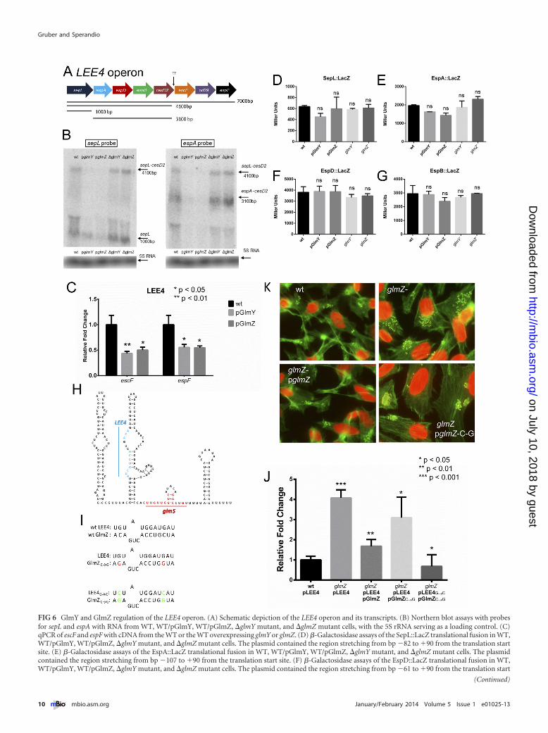
FIG 6 GlmY and GlmZ regulation of the LEE4 operon. (A) Schematic depiction of the LEE4 operon and its transcripts. (B) Northern blot assays with probesfor sepL and espA with RNA from WT, WT/pGlmY, WT/pGlmZ, �glmY mutant, and �glmZ mutant cells, with the 5S rRNA serving as a loading control. (C)qPCR of escF and espF with cDNA from the WT or the WT overexpressing glmY or glmZ. (D) �-Galactosidase assays of the SepL::LacZ translational fusion in WT,WT/pGlmY, WT/pGlmZ, �glmY mutant, and �glmZ mutant cells. The plasmid contained the region stretching from bp �82 to �90 from the translation startsite. (E) �-Galactosidase assays of the EspA::LacZ translational fusion in WT, WT/pGlmY, WT/pGlmZ, �glmY mutant, and �glmZ mutant cells. The plasmidcontained the region stretching from bp �107 to �90 from the translation start site. (F) �-Galactosidase assays of the EspD::LacZ translational fusion in WT,WT/pGlmY, WT/pGlmZ, �glmY mutant, and �glmZ mutant cells. The plasmid contained the region stretching from bp �61 to �90 from the translation start
(Continued)
Gruber and Sperandio
10 ® mbio.asm.org January/February 2014 Volume 5 Issue 1 e01025-13
on July 10, 2018 by guesthttp://m
bio.asm.org/
Dow
nloaded from

amino sugar metabolism and cell wall synthesis are still affected atsome level in this EHEC mutant (70). The tying of glucosaminesynthesis to interkingdom chemical signaling and pathogenesismay reflect the need to integrate both host and bacterial physio-logical cues to ensure a successful association between these or-ganisms. It is also noteworthy that the LEE-encoded T3SS mustspan the periplasm and pass through the peptidoglycan layer.Since EHEC creates dozens of these injectisomes, there is likelysignificant remodeling of the cell wall during host infection.
MATERIALS AND METHODSStrains and growth medium. For the bacterial strains and plasmids usedin this study, see Tables S1 and S2 in the supplemental material. Strainswere grown in Dulbecco’s modified Eagle’s medium (DMEM; Invitrogen)with low glucose or in Luria-Bertani medium (LB; Invitrogen) at 37°Cand 250 rpm unless otherwise stated. Where necessary, the mediumwas supplemented with antibiotics at the following concentrations: am-picillin, 100 �g ml�1; chloramphenicol, 50 �g ml�1; kanamycin, 50 �gml�1; streptomycin, 50 �g ml�1. When needed, the medium wasalso supplemented with 0.2% arabinose or 0.5 mM isopropyl-�-D-thiogalactopyranoside.
Plasmid construction. The glmY and glmZ genes were cloned intoplasmid pBAD33, and the LEE4 and LEE5 operons were cloned into pA-CYC177, disrupting the ampicillin gene. Site-directed mutagenesis wasperformed with the QuikChange site-directed mutagenesis kit (Strat-agene, La Jolla, CA). For transcriptional reporters, the promoter regionwas cloned into plasmid pRS551. For pCG48 (glmY::lacZ), bp �250 to�20 from the transcription start site were used. For pCG49 (glmZ::lacZ),bp �246 to �20 were used. For pCG50 (espFu::lacZ), bp �461 to �48from the translation start site were used. The translation reporter systemwas constructed by cloning lacZ from MG1655 into the vector pBAD24and then cloning the gene of interest from the transcription start site orprocessing site to a spot within the coding region, creating an in-framereporter protein under the control of the arabinose promoter. pCG57(EspFu::LacZ) contains bp �170 from the translation start site to bp �48.pCG59 (GlmS::LacZ) contains bp �160 to �117. pCG60 (Tir::LacZ) con-tains bp �137 to �90. pCG61 (CesT::LacZ) contains bp �71 to �90.pCG62 (Eae::LacZ) contains bp �62 to �90. pCG63 (SepL::LacZ) con-tains bp �82 to �90. pCG64 (EspA::LacZ) contains bp �107 to �90.pCG65 (EspD::LacZ) contains bp �61 to �90. pCG66 (EspB::LacZ) con-tains bp �68 to �90. The espJ-espFu constructs were created by firstcloning the region from the espJ promoter to espFu with a FLAG tag addedvia a primer and inserting it into the Zero Blunt TOPO vector (Invitro-gen). The deletion mutants were then created through sewing PCR. In p1,the region from 20 bp downstream of the espJ stop codon to 20 bp up-stream of the espFu start codon was deleted. In p2, the region from 20 bpdownstream of the espJ stop codon to 170 bp upstream of the espFu startcodon was deleted. In p3, the region from 170 bp upstream of the espFustart codon to 20 bp upstream of the espFu start codon was deleted. In p4,the region starting 262 bp within the espJ gene to the end of espFu wascloned. All plasmids were confirmed through sequencing. For the primersused, see Table S3 in the supplemental material.
Isogenic mutant construction. Nonpolar glmY and glmZ mutantswere constructed through the lambda Red system (71). Briefly, PCR prod-ucts (obtained with the primers listed in Table S3) were amplified from
plasmid pKD3 with flanking regions matching glmY or glmZ and weretransformed into EHEC expressing the Red genes from plasmid pKD46.After selection and confirmation, the resistance cassette was resolved withflippase from temperature-sensitive plasmid pCP20, which was thencured through growth at 37°C. This generated nonpolar mutants, whichwere confirmed by sequencing.
Reporter assays. Transcriptional and translational �-galactosidase as-says were performed by using the same protocol. Bacteria containing thereporter plasmids were grown overnight in LB with the appropriate anti-biotic and then diluted 1:100 in clear DMEM supplemented with 0.2%arabinose and grown to an optical density at 600 nm (OD600) of 0.8. Thesewere then assayed for �-galactosidase activity with o-nitrophenyl-�-D-galactopyranoside as previously described (72). For the experiments withthe GlmS::LacZ reporter plasmid, bacteria were grown overnight in LBwith 0.2% arabinose and specific activity (nM/min/mg protein) was mea-sured by determining protein concentrations by the assay of Lowry et al.(73). The unpaired Student t test was used to determine statistical signif-icance. A P value of �0.05 was considered significant.
EMSA. QseB and QseF proteins were His tagged and purified fromBL-21(DE3) and TOP10 cells, respectively, as previously described (36,45). The glmY promoter and the kanamycin resistance-encoding genewere amplified by PCR (for the primers used, see Table S3 in the supple-mental material) and radiolabeled with [�-32]ATP and T4 polynucleotidekinase (NEB). The labeled DNA was then incubated with increasingamounts of protein and run on a 6% polyacrylamide Tris-Bis gel, dried,and exposed to film overnight.
Operon analysis by RT-PCR. WT and �qseF mutant bacteria weregrown to an OD600 of 1.0 in low-glucose DMEM, and RNA was extractedwith the RiboPure kit (Ambion). RNA was reverse transcribed and am-plified by PCR with different sets of primers (see Table S3 in the supple-mental material).
Western blot assays. Bacteria were grown overnight in LB and thendiluted 1:100 in low-glucose DMEM and grown to an OD600 of 1.0. The
FIG 7 Proposed model of GlmY and GlmZ regulation of LEE4, LEE5, andespFu. Transcription of glmY is activated by both QseF and QseB. GlmY thenstabilizes GlmZ, which controls espFu expression at the translational level anddestabilizes the LEE4 and LEE5 mRNAs.
Figure Legend Continued
site. (G) �-Galactosidase assays of the EspB::LacZ translational fusion in WT, WT/pGlmY, WT/pGlmZ, �glmY mutant, and �glmZ mutant cells. The plasmidcontained the region stretching from bp �68 to �90 from the translation start site. (H) Sequence and secondary structure of the GlmZ sRNA depicting theregions that interact with the glmS and LEE4 mRNAs. (I) Schematics showing the predicted interaction between GlmZ and LEE4, the point mutations made inGlmZ to abolish binding, and the compensatory mutations made in LEE4. (J) qRT-PCR of espA with cDNA from WT/pLEE4, �glmZ/pLEE4, �glmZ/pGlmZ/pLEE4, �glmZ/pGlmZ¡CG/pLEE4, and �glmZ/pGlmZ¡CG/pLEE4¡GC cells. (K) FAS of HeLa cells infected with WT, �glmZ, �glmZ/pGlmZ, and �glmZ/pGlmZ¡CG cells. The unpaired Student t test was used to determine statistical significance. A P value of �0.05 was considered significant. ns, not statisticallysignificant.
Control of Host Cell Actin Rearrangement
January/February 2014 Volume 5 Issue 1 e01025-13 ® mbio.asm.org 11
on July 10, 2018 by guesthttp://m
bio.asm.org/
Dow
nloaded from

pellets were then lysed under denaturing conditions, subjected to 12%SDS-PAGE, and transferred to a polyvinylidene difluoride membrane.The membrane was probed with an ant-FLAG (Sigma) or anti-RpoA(Santa Cruz Biotechnology) antibody as the endogenous control with ahorseradish peroxidase-conjugated secondary antibody that was visual-ized by ECL (GE Bioscience) and exposed to film.
Cell culture and FAS. HeLa cells were maintained in high-glucoseDMEM supplemented with 10% fetal bovine serum (FBS) and penicillin-streptomycin-glutamine and grown at 37°C and 5% CO2. Cells were splitinto a 12-well plate, grown to confluence, washed, given low-glucoseDMEM supplemented with 10% FBS, and then infected with bacteriagrown overnight in LB statically at 37°C at a 1:100 dilution. FAS assayswere performed as described by Knutton et al. (36, 74) to examine pedes-tal formation. After 6 h of infection at 37°C in 5% CO2, the coverslips werewashed, fixed, and permeabilized. The samples were then treated withFITC-labeled phalloidin and PI to visualize actin accumulation and bac-teria, respectively. PI also stained HeLa nuclei red. The coverslips werethen mounted on slides and visualized with a Zeiss Axiovert microscope.Pedestal formation was quantified by randomly imaging different fields ofview and counting the first 100 cells while recording the number of bac-teria attached to each one. Replicate coverslips from multiple experimentswere quantified, and statistical analysis was performed with the Studentt test. Serially diluted samples of the original bacterial cultures were alsoplated to confirm that similar CFU ratios were used for infection.
Live-cell imaging. The Lifeact::GFP-expressing cell line was createdwith the Flip-In System (Invitrogen). HeLa cells were transfected withpLacZ::zeocin by using Fugene 6 (Promega) to create flippase recognitiontarget sites in the genome. Cells were then selected with 100 �g/ml zeocin.Resistant foci were grown and assayed by Southern blot assay for lacZ forsingle insertions, and then �-galactosidase assays were performed to mea-sure the expression of the inserted locus. High-expression single inser-tions were then transfected with the flippase helper plasmid pOD44 andLifeact::GFP cloned into plasmid pFRT. These transfected cells were thenselected in 100 �g/ml hygromycin, and resistant foci were visualized byfluorescence microscopy to measure levels of Lifeact::GFP expression.These cells were then maintained in 50 �g/ml hygromycin. When they aresplit before infection, hygromycin was not added. EHEC cells were trans-formed with mCherry-expressing plasmid pDP151 and grown overnightstatically in LB. The Lifeact::GFP-expressing cell medium was replacedwith low-glucose DMEM supplemented with 10% FBS, and the cells wereinfected with a 1:100 dilution of the overnight culture. Infection was al-lowed to continue for 2 h at 37°C and 5% CO2, and then the cells arewashed three times with DMEM and then visualized by live-cell imagingwith a Zeiss microscope. Images were taken every 2 min for 2 h.
Northern blot assays. Bacteria were grown aerobically in low-glucoseDMEM supplemented with 0.2% arabinose at 37°C to an OD600 of 1.0from a 1:100 dilution of an overnight culture grown in LB. RNA wasextracted with the RiboPure kit (Ambion), run on a 1% formaldehydeagarose gel, and transferred overnight to a Zeta-Probe membrane (Bio-Rad). RNA probes were created by PCR amplification of a segment of thegene of interest with the T7 promoter and in vitro transcription with theMAXIscript T7 kit (Ambion) with [�-32P]UTP. The oligoprobe for the 5Sendogenous control was labeled with [�-32P]ATP by using T4 polynucle-otide kinase (NEB). The membranes were then hybridized overnight withUltrahyb (Ambion) at 68ºC for the RNA probes and 37°C for the oligo-probes. The membranes were washed, exposed to a phosphorimagerscreen overnight, and then visualized with a STORM scanner (GE Health-care).
RNA extraction and qRT-PCR. Cultures were grown overnight in LB,diluted 1:100 in low-glucose DMEM with 0.2% arabinose on a 12-wellplate, and then grown for 6 h at 37°C under 5% CO2. RNA was extractedfrom three biological replicates with the RiboPure Bacteria isolation kitaccording to the manufacturer’s protocols (Ambion). qRT-PCR was per-formed as described previously (38). Briefly, diluted extracted RNA wasmixed with, validated primers (see Table S3 in the supplemental mate-
rial), RNase inhibitor, and reverse transcriptase (AB). The mixture wasused in a one-step reaction utilizing an ABI 7500 sequence detection sys-tem. Data were collected with ABI Sequence detection 1.2 software, nor-malized to endogenous rpoA levels, and analyzed by the comparative crit-ical threshold method. Analyzed data were presented as fold changes overthe WT levels. The unpaired Student t test was used to determine statisti-cal significance. A P value of �0.05 was considered significant.
Temperature-sensitive RNase E strain. rneWT strain NL3433 andrnets strain NL3431 were transformed with the pLEE4 or pLEE5 plasmidand pGlmZ. Both were grown overnight in LB at 30°C in the appropriateantibiotics and then grown to an OD600 of 1.0 from a 1:100 dilution in LBat 35°C. Once they reached this OD600, they were shifted to the nonper-missive temperature of 43°C for 15 min and then the samples were treatedas for the other Northern blot assays.
SUPPLEMENTAL MATERIALSupplemental material for this article may be found at http://mbio.asm.org/lookup/suppl/doi:10.1128/mBio.01025-13/-/DCSupplemental.
Table S1, DOCX file, 0.1 MB.Table S2, DOCX file, 0.1 MB.Table S3, DOCX file, 0.1 MB.Figure S1, PDF file, 0.7 MB.Figure S2, PDF file, 1.9 MB.Figure S3, PDF file, 0.2 MB.Figure S4, PDF file, 0.7 MB.Movie S1, AVI file, 2.8 MB.Movie S2, AVI file, 1.5 MB.Movie S3, AVI file, 2.7 MB.
ACKNOWLEDGMENTS
We thank Lora V. Hooper, and Jörg Vogel for reading of the manuscript.We also thank Neal M. Alto and Robert C. Orchard for help with thelive-cell imaging, as well as Nicholas K. Conrad and Sara H. Stubbs forhelp with creation of the Lifeact-expressing cell line. We also thank JamesB. Kaper for the rnets strain.
This work was supported by NIH grants AI053067 and AI077613 andby the Burroughs Wellcome Fund. C.C.G. was supported by grant T32-AI07520.
REFERENCES1. Hicks SW, Galán JE. 2013. Exploitation of eukaryotic subcellular target-
ing mechanisms by bacterial effectors. Nat. Rev. Microbiol. 11:316 –326.2. Kaper JB, Nataro JP, Mobley HL. 2004. Pathogenic Escherichia coli. Nat.
Rev. Microbiol. 2:123–140.3. McDaniel TK, Jarvis KG, Donnenberg MS, Kaper JB. 1995. A genetic
locus of enterocyte effacement conserved among diverse enterobacterialpathogens. Proc. Natl. Acad. Sci. U. S. A. 92:1664 –1668.
4. Jarvis KG, Girón JA, Jerse AE, McDaniel TK, Donnenberg MS, KaperJB. 1995. Enteropathogenic Escherichia coli contains a putative type IIIsecretion system necessary for the export of proteins involved in attachingand effacing lesion formation. Proc. Natl. Acad. Sci. U. S. A. 92:7996 – 8000.
5. Elliott SJ, Hutcheson SW, Dubois MS, Mellies JL, Wainwright LA,Batchelor M, Frankel G, Knutton S, Kaper JB. 1999. Identification ofCesT, a chaperone for the type III secretion of Tir in enteropathogenicEscherichia coli. Mol. Microbiol. 33:1176 –1189.
6. Mellies JL, Elliott SJ, Sperandio V, Donnenberg MS, Kaper JB. 1999.The per regulon of enteropathogenic Escherichia coli: identification of aregulatory cascade and a novel transcriptional activator, the locus of en-terocyte effacement (LEE)-encoded regulator (Ler). Mol. Microbiol. 33:296 –306.
7. Elliott SJ, Wainwright LA, McDaniel TK, Jarvis KG, Deng YK, Lai LC,McNamara BP, Donnenberg MS, Kaper JB. 1998. The complete se-quence of the locus of enterocyte effacement (LEE) from enteropatho-genic Escherichia coli E2348/69. Mol. Microbiol. 28:1– 4.
8. Jerse AE, Yu J, Tall BD, Kaper JB. 1990. A genetic locus of enteropatho-genic Escherichia coli necessary for the production of attaching and effac-ing lesions on tissue culture cells. Proc. Natl. Acad. Sci. U. S. A. 87:7839 –7843.
Gruber and Sperandio
12 ® mbio.asm.org January/February 2014 Volume 5 Issue 1 e01025-13
on July 10, 2018 by guesthttp://m
bio.asm.org/
Dow
nloaded from

9. Kenny B, DeVinney R, Stein M, Reinscheid DJ, Frey EA, Finlay BB.1997. Enteropathogenic E. coli (EPEC) transfers its receptor for intimateadherence into mammalian cells. Cell 91:511–520.
10. McNamara BP, Donnenberg MS. 1998. A novel proline-rich protein,EspF, is secreted from enteropathogenic Escherichia coli via the type IIIexport pathway. FEMS Microbiol. Lett. 166:71–78.
11. Kenny B, Jepson M. 2000. Targeting of an enteropathogenic Escherichiacoli (EPEC) effector protein to host mitochondria. Cell. Microbiol.2:579 –590.
12. Elliott SJ, Krejany EO, Mellies JL, Robins-Browne RM, Sasakawa C,Kaper JB. 2001. EspG, a novel type III system-secreted protein from en-teropathogenic Escherichia coli with similarities to VirA of Shigella flexneri.Infect. Immun. 69:4027– 4033.
13. Tu X, Nisan I, Yona C, Hanski E, Rosenshine I. 2003. EspH, a newcytoskeleton-modulating effector of enterohaemorrhagic and entero-pathogenic Escherichia coli. Mol. Microbiol. 47:595– 606.
14. Kanack KJ, Crawford JA, Tatsuno I, Karmali MA, Kaper JB. 2005.SepZ/EspZ is secreted and translocated into HeLa cells by the entero-pathogenic Escherichia coli type III secretion system. Infect. Immun. 73:4327– 4337.
15. Campellone KG, Robbins D, Leong JM. 2004. EspFU is a translocatedEHEC effector that interacts with Tir and N-WASP and promotes Nck-independent actin assembly. Dev. Cell 7:217–228.
16. Garmendia J, Phillips AD, Carlier MF, Chong Y, Schüller S, Marches O,Dahan S, Oswald E, Shaw RK, Knutton S, Frankel G. 2004. TccP is anenterohaemorrhagic Escherichia coli O157:H7 type III effector protein thatcouples Tir to the actin-cytoskeleton. Cell. Microbiol. 6:1167–1183.
17. Tobe T, Beatson SA, Taniguchi H, Abe H, Bailey CM, Fivian A, YounisR, Matthews S, Marches O, Frankel G, Hayashi T, Pallen MJ. 2006. Anextensive repertoire of type III secretion effectors in Escherichia coli O157and the role of lambdoid phages in their dissemination. Proc. Natl. Acad.Sci. U. S. A. 103:14941–14946.
18. Deng W, Puente JL, Gruenheid S, Li Y, Vallance BA, Vázquez A, BarbaJ, Ibarra JA, O’Donnell P, Metalnikov P, Ashman K, Lee S, Goode D,Pawson T, Finlay BB. 2004. Dissecting virulence: systematic and func-tional analyses of a pathogenicity island. Proc. Natl. Acad. Sci. U. S. A.101:3597–3602.
19. Bhatt S, Edwards AN, Nguyen HT, Merlin D, Romeo T, Kalman D.2009. The RNA binding protein CsrA is a pleiotropic regulator of the locusof enterocyte effacement pathogenicity island of enteropathogenic Esche-richia coli. Infect. Immun. 77:3552–3568.
20. Njoroge JW, Nguyen Y, Curtis MM, Moreira CG, Sperandio V. 2012.Virulence meets metabolism: Cra and KdpE gene regulation in enterohe-morrhagic Escherichia coli. mBio 3(5):e00280-00212. http://dx.doi.org/10.1128/mBio.00280-12.
21. Sperandio V, Torres AG, Jarvis B, Nataro JP, Kaper JB. 2003. Bacteria-host communication: the language of hormones. Proc. Natl. Acad. Sci.U. S. A. 100:8951– 8956.
22. Kendall MM, Gruber CC, Rasko DA, Hughes DT, Sperandio V. 2011.Hfq virulence regulation in enterohemorrhagic Escherichia coli O157:H7strain 86-24. J. Bacteriol. 193:6843– 6851.
23. Shakhnovich EA, Davis BM, Waldor MK. 2009. Hfq negatively regulatestype III secretion in EHEC and several other pathogens. Mol. Microbiol.74:347–363.
24. Lodato PB, Kaper JB. 2009. Post-transcriptional processing of the LEE4operon in enterohaemorrhagic Escherichia coli. Mol. Microbiol. 71:273–290.
25. Mellies JL, Barron AM, Carmona AM. 2007. Enteropathogenic andenterohemorrhagic Escherichia coli virulence gene regulation. Infect. Im-mun. 75:4199 – 4210.
26. Hughes DT, Terekhova DA, Liou L, Hovde CJ, Sahl JW, Patankar AV,Gonzalez JE, Edrington TS, Rasko DA, Sperandio V. 2010. Chemicalsensing in mammalian host-bacterial commensal associations. Proc. Natl.Acad. Sci. U. S. A. 107:9831–9836.
27. Kendall MM, Gruber CC, Parker CT, Sperandio V. 2012. Ethanolaminecontrols expression of genes encoding components involved in interking-dom signaling and virulence in enterohemorrhagic Escherichia coli O157:H7. mBio 3(3):e00050-12. http://dx.doi.org/10.1128/mBio.00050-12.
28. Pacheco AR, Curtis MM, Ritchie JM, Munera D, Waldor MK, MoreiraCG, Sperandio V. 2012. Fucose sensing regulates bacterial intestinal col-onization. Nature 492:113–117.
29. Bhatt S, Romeo T, Kalman D. 2011. Honing the message: post-
transcriptional and post-translational control in attaching and effacingpathogens. Trends Microbiol. 19:217–224.
30. Storz G, Vogel J, Wassarman KM. 2011. Regulation by small RNAs inbacteria: expanding frontiers. Mol. Cell 43:880 – 891.
31. Sperandio V, Torres AG, Giron JA, Kaper JB. 2001. Quorum sensing isa global regulatory mechanism in enterohemorrhagic Escherichia coliO157:H7. J. Bacteriol. 183:5187–5197.
32. Clarke MB, Hughes DT, Zhu C, Boedeker EC, Sperandio V. 2006. TheQseC sensor kinase: a bacterial adrenergic receptor. Proc. Natl. Acad. Sci.U. S. A. 103:10420 –10425.
33. Walters M, Sircili MP, Sperandio V. 2006. AI-3 synthesis is not depen-dent on luxS in Escherichia coli. J. Bacteriol. 188:5668 –5681.
34. Clarke MB, Hughes DT, Zhu C, Boedeker EC, Sperandio V. 2006. TheQseC sensor kinase: a bacterial adrenergic receptor. Proc. Natl. Acad. Sci.U. S. A. 103:10420 –10425.
35. Reading NC, Rasko DA, Torres AG, Sperandio V. 2009. The two-component system QseEF and the membrane protein QseG link adrener-gic and stress sensing to bacterial pathogenesis. Proc. Natl. Acad. Sci.U. S. A. 106:5889 –5894.
36. Reading NC, Torres AG, Kendall MM, Hughes DT, Yamamoto K,Sperandio V. 2007. A novel two-component signaling system that acti-vates transcription of an enterohemorrhagic Escherichia coli effector in-volved in remodeling of host actin. J. Bacteriol. 189:2468 –2476.
37. Rasko DA, Moreira CG, Li DR, Reading NC, Ritchie JM, Waldor MK,Williams N, Taussig R, Wei S, Roth M, Hughes DT, Huntley JF, FinaMW, Falck JR, Sperandio V. 2008. Targeting QseC signaling and viru-lence for antibiotic development. Science 321:1078 –1080.
38. Hughes DT, Clarke MB, Yamamoto K, Rasko DA, Sperandio V. 2009.The QseC adrenergic signaling cascade in enterohemorrhagic E. coli(EHEC). PLoS Pathog. 5:e1000553. http://dx.doi.org/10.1371/journal.ppat.1000553.
39. Yamamoto K, Hirao K, Oshima T, Aiba H, Utsumi R, Ishihama A.2005. Functional characterization in vitro of all two-component signaltransduction systems from Escherichia coli. J. Biol. Chem. 280:1448 –1456.
40. Reichenbach B, Göpel Y, Görke B. 2009. Dual control by perfectlyoverlapping sigma 54- and sigma 70-promoters adjusts small RNA GlmYexpression to different environmental signals. Mol. Microbiol. 74:1054 –1070.
41. Göpel Y, Papenfort K, Reichenbach B, Vogel J, Görke B. 2013. Targeteddecay of a regulatory small RNA by an adaptor protein for RNase E andcounteraction by an anti-adaptor RNA. Genes Dev. 27:552–564.
42. Urban JH, Vogel J. 2008. Two seemingly homologous noncoding RNAsact hierarchically to activate glmS mRNA translation. PLoS Biol. 6:e64.http://dx.doi.org/10.1371/journal.pbio.0060064.
43. Clarke MB, Hughes DT, Zhu C, Boedeker EC, Sperandio V. 2006. TheQseC sensor kinase: a bacterial adrenergic receptor. Proc. Natl. Acad. Sci.U. S. A. 103:10420 –10425.
44. Njoroge J, Sperandio V. 2012. Enterohemorrhagic Escherichia coli viru-lence regulation by two bacterial adrenergic kinases, QseC and QseE. In-fect. Immun. 80:688 –703.
45. Clarke MB, Sperandio V. 2005. Transcriptional regulation of flhDC byQseBC and sigma (FliA) in enterohaemorrhagic Escherichia coli. Mol. Mi-crobiol. 57:1734 –1749.
46. Gyaneshwar P, Paliy O, McAuliffe J, Jones A, Jordan MI, Kustu S. 2005.Lessons from Escherichia coli genes similarly regulated in response to ni-trogen and sulfur limitation. Proc. Natl. Acad. Sci. U. S. A. 102:3453–3458.
47. Vingadassalom D, Campellone KG, Brady MJ, Skehan B, Battle SE,Robbins D, Kapoor A, Hecht G, Snapper SB, Leong JM. 2010. Entero-hemorrhagic E. coli requires N-WASP for efficient type III translocationbut not for EspFU-mediated actin pedestal formation. PLoS Pathog.6:e1001056. http://dx.doi.org/10.1371/journal.ppat.1001056.
48. Marchès O, Covarelli V, Dahan S, Cougoule C, Bhatta P, Frankel G,Caron E. 2008. EspJ of enteropathogenic and enterohaemorrhagic Esch-erichia coli inhibits opsono-phagocytosis. Cell. Microbiol. 10:1104 –1115.
49. Riedl J, Crevenna AH, Kessenbrock K, Yu JH, Neukirchen D, Bista M,Bradke F, Jenne D, Holak TA, Werb Z, Sixt M, Wedlich-Soldner R.2008. Lifeact: a versatile marker to visualize F-actin. Nat. Methods5:605– 607.
50. Vogler AP, Trentmann S, Lengeler JW. 1989. Alternative route forbiosynthesis of amino sugars in Escherichia coli K-12 mutants by means ofa catabolic isomerase. J. Bacteriol. 171:6586 – 6592.
51. Sperandio V, Mellies JL, Nguyen W, Shin S, Kaper JB. 1999. Quorumsensing controls expression of the type III secretion gene transcription and
Control of Host Cell Actin Rearrangement
January/February 2014 Volume 5 Issue 1 e01025-13 ® mbio.asm.org 13
on July 10, 2018 by guesthttp://m
bio.asm.org/
Dow
nloaded from
protein secretion in enterohemorrhagic and enteropathogenic Escherichiacoli. Proc. Natl. Acad. Sci. U. S. A. 96:15196 –15201.
52. Abe A, de Grado M, Pfuetzner RA, Sánchez-Sanmartín C, Devinney R,Puente JL, Strynadka NC, Finlay BB. 1999. Enteropathogenic Escherichiacoli translocated intimin receptor, Tir, requires a specific chaperone forstable secretion. Mol. Microbiol. 33:1162–1175.
53. Waters LS, Storz G. 2009. Regulatory RNAs in bacteria. Cell 136:615– 628.
54. Sekiya K, Ohishi M, Ogino T, Tamano K, Sasakawa C, Abe A. 2001.Supermolecular structure of the enteropathogenic Escherichia coli type IIIsecretion system and its direct interaction with the EspA-sheath-likestructure. Proc. Natl. Acad. Sci. U. S. A. 98:11638 –11643.
55. Knutton S, Rosenshine I, Pallen MJ, Nisan I, Neves BC, Bain C, WolffC, Dougan G, Frankel G. 1998. A novel EspA-associated surface organelleof enteropathogenic Escherichia coli involved in protein translocation intoepithelial cells. EMBO J. 17:2166 –2176.
56. Crane JK, McNamara BP, Donnenberg MS. 2001. Role of EspF in hostcell death induced by enteropathogenic Escherichia coli. Cell. Microbiol.3:197–211.
57. Elliott SJ, Sperandio V, Girón JA, Shin S, Mellies JL, Wainwright L,Hutcheson SW, McDaniel TK, Kaper JB. 2000. The locus of enterocyteeffacement (LEE)-encoded regulator controls expression of both LEE-and non-LEE-encoded virulence factors in enteropathogenic and entero-hemorrhagic Escherichia coli. Infect. Immun. 68:6115– 6126.
58. McNally A, Roe AJ, Simpson S, Thomson-Carter FM, Hoey DE, CurrieC, Chakraborty T, Smith DG, Gally DL. 2001. Differences in levels ofsecreted locus of enterocyte effacement proteins between human disease-associated and bovine Escherichia coli O157. Infect. Immun. 69:5107–5114.
59. Roe AJ, Yull H, Naylor SW, Woodward MJ, Smith DG, Gally DL. 2003.Heterogeneous surface expression of EspA translocon filaments by Esch-erichia coli O157:H7 is controlled at the posttranscriptional level. Infect.Immun. 71:5900 –5909.
60. Busch A, Richter AS, Backofen R. 2008. IntaRNA: efficient prediction ofbacterial sRNA targets incorporating target site accessibility and seed re-gions. Bioinformatics 24:2849 –2856.
61. Lodato PB, Hsieh PK, Belasco JG, Kaper JB. 2012. The ribosome bindingsite of a mini-ORF protects a T3SS mRNA from degradation by RNase E.Mol. Microbiol. 86:1167–1182.
62. Campellone KG, Roe AJ, Lobner-Olesen A, Murphy KC, Magoun L,
Brady MJ, Donohue-Rolfe A, Tzipori S, Gally DL, Leong JM, MarinusMG. 2007. Increased adherence and actin pedestal formation by dam-deficient enterohaemorrhagic Escherichia coli O157:H7. Mol. Microbiol.63:1468 –1481.
63. Hansen AM, Kaper JB. 2009. Hfq affects the expression of the LEE patho-genicity island in enterohaemorrhagic Escherichia coli. Mol. Microbiol.73:446 – 465.
64. Deng W, Li Y, Hardwidge PR, Frey EA, Pfuetzner RA, Lee S, GruenheidS, Strynakda NC, Puente JL, Finlay BB. 2005. Regulation of type IIIsecretion hierarchy of translocators and effectors in attaching and effacingbacterial pathogens. Infect. Immun. 73:2135–2146.
65. Papenfort K, Podkaminski D, Hinton JC, Vogel J. 2012. The ancestralSgrS RNA discriminates horizontally acquired Salmonella mRNAsthrough a single G-U wobble pair. Proc. Natl. Acad. Sci. U. S. A. 109:E757–E764.
66. Papenfort K, Sun Y, Miyakoshi M, Vanderpool CK, Vogel J. 2013. SmallRNA-mediated activation of sugar phosphatase mRNA regulates glucosehomeostasis. Cell 153:426 – 437.
67. Navarre WW, Porwollik S, Wang Y, McClelland M, Rosen H, Libby SJ,Fang FC. 2006. Selective silencing of foreign DNA with low GC content bythe H-NS protein in Salmonella. Science 313:236 –238.
68. Ochman H, Lawrence JG, Groisman EA. 2000. Lateral gene transfer andthe nature of bacterial innovation. Nature 405:299 –304.
69. Vogel J. 2009. A rough guide to the non-coding RNA world of Salmonella.Mol. Microbiol. 71:1–11.
70. Reading NC, Rasko D, Torres AG, Sperandio V. 2010. A transcriptomestudy of the QseEF two-component system and the QseG membrane pro-tein in enterohaemorrhagic Escherichia coli O15:H7. Microbiology 156:1167–1175.
71. Datsenko KA, Wanner BL. 2000. One-step inactivation of chromosomalgenes in Escherichia coli K-12 using PCR products. Proc. Natl. Acad. Sci.U. S. A. 97:6640 – 6645.
72. Miller JH. 1972. Experiments in molecular genetics. Cold Spring HarborLaboratory Press, Cold Spring Harbor, NY.
73. Lowry OH, Rosebrough NJ, Farr AL, Randall RJ. 1951. Protein mea-surement with the Folin phenol reagent. J. Biol. Chem. 193:265–275.
74. Knutton S, Baldwin T, Williams PH, McNeish AS. 1989. Actin accumu-lation at sites of bacterial adhesion to tissue culture cells: basis of a newdiagnostic test for enteropathogenic and enterohemorrhagic Escherichiacoli. Infect. Immun. 57:1290 –1298.
Gruber and Sperandio
14 ® mbio.asm.org January/February 2014 Volume 5 Issue 1 e01025-13
on July 10, 2018 by guesthttp://m
bio.asm.org/
Dow
nloaded from