postioning the non comforming patient pt ii b #2.ppt€¦ · the base of attachment of the breast...
TRANSCRIPT
5/22/2017
1
Advanced Health Education Center
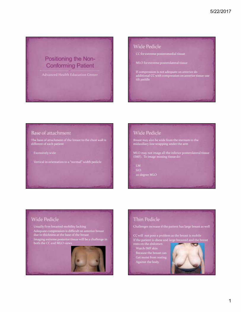
The base of attachment of the breast to the chest wall is different of each patient
� Excessively wide
� Vertical in orientation to a “normal” width pedicle
� Usually firm breasted-mobility lacking
� Adequate compression is difficult on anterior breast due to thickness at the base of the breast
� Imaging extreme posterior tissue will be a challenge in both the CC and MLO views
� CC for extreme posteromedial tissue
� MLO for extreme posterolateral tissue
� If compression is not adequate on anterior do additional CC with compression on anterior tissue-use tilt paddle
Breast may also be wide from the sternum to the midaxillary line wrapping under the arm
MLO may not image all the inferior posterolateral tissue (IMF). To image missing tissue do:
� LM
� SIO
� 20 degree MLO
Challenges increase if the patient has large breast as well
CC will not pose a problem as the breast is mobile
If the patient is obese and large breasted and the breast rests on the abdomen
� Watch IMF skin
� Because the breast can
� Get moist from resting
� Against the body.

5/22/2017
2
MLO
� Typically impossible to maintain the breast in a “out and up” position
� Prohibits proper representation of ductal structures
� Presenting the IMF open will be typically impossible
� Will need to add a LM or ML for proper visualization of anterior tissue
Breasts may also extend under the armpit which eliminates the lower-outer quadrant from view
� LM or SIO with arm up will access visualization of this tissue
Extremely small breasted patients also present additional difficulty if the breast are :
� Firm
� Wide base of attachment
� Extend more laterally then medially
May be impossible to image extreme posterior tissue both medial and lateral
� Raise the receptor to the correct IMF height for these patients if the receptor it too low you will miss most of the tissue
� CC for medial tissue
� CC for lateral tissue
CC view does not pose a positioning problem
MLO will be the challenge

5/22/2017
3
CC view does not pose a positioning problem
MLO will be the challenge
� Droops anteriorly from size and weight� DO NOT sacrifice posterior and lateral tissue in order to
make the breast not droop
� MLO view is to image posterior and upper outer quadrant
� May require a third view to demonstrate the anterior breast
� Mosaic imaging to demonstrate all of the tissue on several images
� The superimposition of tissue and distortion of the anterior breast tissue on the MLO makes it less than ideal for evaluating this area.
� Instead of repeating the MLO for anterior compression use a LM or ML
� These views will allow better compression and opening of the tissue giving the radiologist more information and a different superimposition of tissue
� Do not confuse obesity with breast size
� Choose the proper image receptor size or FOV
� Using the larger image receptor will make the positioning process more difficult
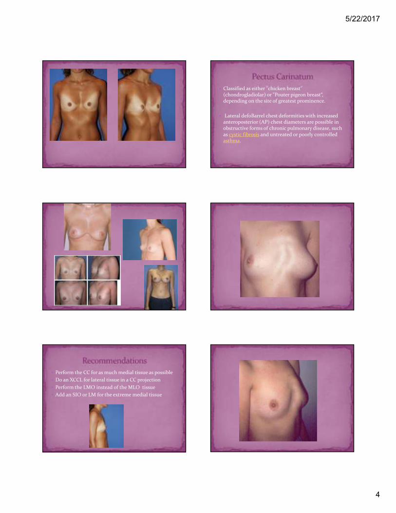
�Pectus Excavatum
� An abnormal development of the rib cage where the breastbone (sternum) caves in.
� Referred to as "funnel chest,“
� Often present at birth (congenital)
� Can be mild or severe
The cause of pectus excavatum is not well understood. Yet, researchers believe that the
deformity is caused by excessive growth of the connective tissue (cartilage) that joins the ribs
to the breastbone (also known as the costochondral region), which causes an inward
defect of the sternum.
5/22/2017
4
� Perform the CC for as much medial tissue as possible
� Do an XCCL for lateral tissue in a CC projection
� Perform the LMO instead of the MLO tissue
� Add an SIO or LM for the extreme medial tissue
� Classified as either "chicken breast" (chondrogladiolar) or "Pouter pigeon breast“, depending on the site of greatest prominence.
� Lateral defoBarrel chest deformities with increased anteroposterior (AP) chest diameters are possible in obstructive forms of chronic pulmonary disease, such as cystic fibrosis and untreated or poorly controlled asthma.

5/22/2017
5
� Perform the CC for the medial tissue (the nipple will fall laterally on the film)
� Perform the MLO to image posterolateral tissue and the upper-outer quadrant. In most cases, it will not be possible to also image posteromedial breast tissue
� Perform a 20° MLO or XCCL to image lateral tissue or repeat the MLO using a quadrant paddle to access the extreme posterolateral tissue excluded on the MLO
� The patients presents hunched over and may have many deformities of the rib cage. The anomalies are usually not symmetric from side to side. The patient may also present with either pectus excavatum or a barrel chest or a combination of the two.
� Sit the patient down for the CC view, her upper body will then be straight
� Perform the CC accessing as much medial breast tissue as possible
� If there is a combination of pectus and barrel chest perform the study as follows:� CC for medial tissue
� MLO for lateral tissue
� Add LM with arm up and over to image eliminated posterolateral and posteromedial tissue

5/22/2017
6
� Scoliosis is derived from a greek work meaning “crooked”. It is a medical condition in which a person’s spine is curved side to side.
� On an xray the spine may look more like an ”S” or a “C” shape instead of a straight line.
� Cause of scoliosis is classified as either congenital anomalies present at birth or idiopathic which is an onset occurrance at childhood or adulthood.
� On CC view have the patient lean in as much as possible maybe even turning their hips slightly to accommodate their curve in the spine to relieve pressure.
� On MLO view have the patient’s feet turned in as much as possible with them leaning in and down with the least amount of back pain. Sometimes the detector can be lowered some to not pull on the pectoralis muscle as much. I find a steep angle works best for these patients.
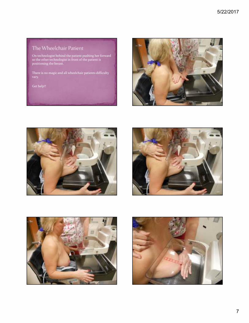
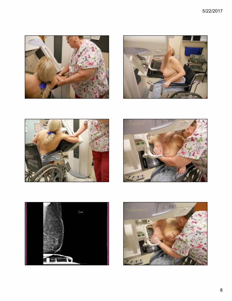
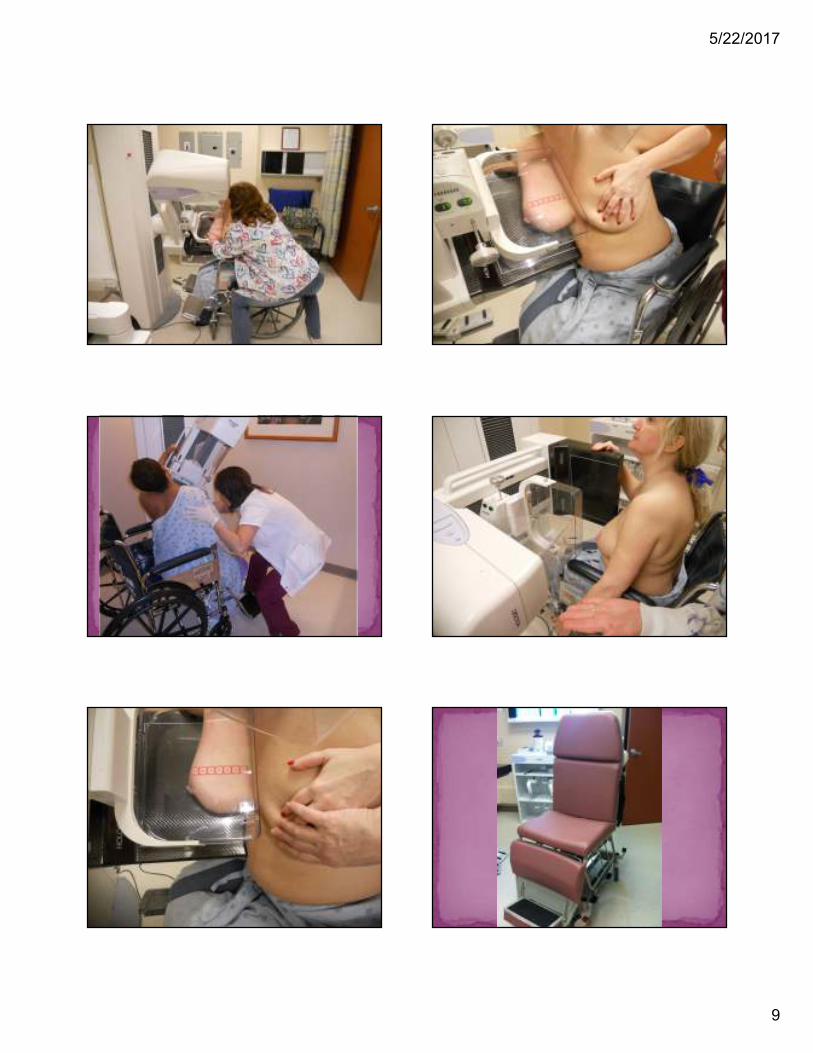
Bottom line is very simple….
It takes 2 technologists typically to do a wheelchair mammogram.
Not the walkie-talkie wheelchair patient
The REAL cannot move wheelchair patient.
5/22/2017
7
On technologist behind the patient pushing her forward so the other technologist in front of the patient is positioning the breast.
There is no magic and all wheelchair patients difficulty vary.
Get help!!
5/22/2017
8
5/22/2017
9
5/22/2017
10
5/22/2017
11
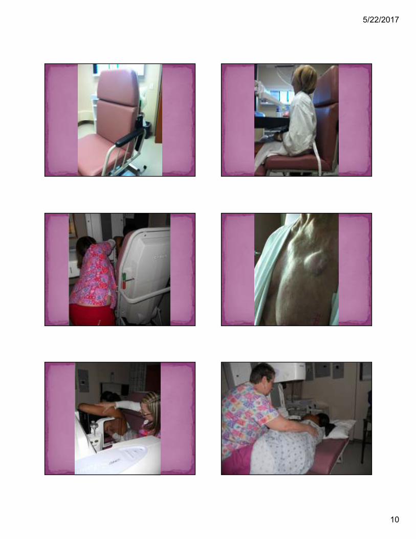
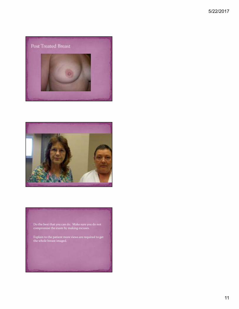
� Do the best that you can do. Make sure you do not compromise the exam by making excuses.
� Explain to the patient more views are required to get the whole breast imaged.