post-pleistocene changes in tooth root and jaw relationships
TRANSCRIPT
AMERICAN JOURNAL OF PHYSICAL ANTHROPOLOGY 70:339-348 (1986)
Post-Pleistocene Changes in Tooth Root and Jaw Relationships PATRICIA SMITH, YOCHANAN WAX, FANNY ADLER, URI SILBERMAN, AND GADY HEINIC Hebrew University Hadassah Faculty of Dental Medicine (EL, US., G.H.), Department of Statistics, Hebrew University, Hadassah, Ein Karem Jerusalem (y: W, FA.), Israel
ABSTRACT Mandibles of 126 sexed skeletal specimens, from Near Eastern sites dating over the past 12,000 years were radiographed. From the radio- graphs obtained, digitised tracings were made of crown length (mesiodistal), root height and width, and corpus height mesial to the premolars and first and second molars. The data obtained were analysed using rank transformation procedures. The significance of unidirectional trends in relation to periods sampled was examined graphically and analytically through linear regression analysis of the ranks on the chronological scale, and Spearman’s rank correla- tion was used to compare relationships between different parameters of indi- vidual teeth in different periods. Significant reduction was found in crown length between 12,000 B.P. and 6,000 B.P., but no further reduction was found between 6,000 B.P. and 1,000 B.P. Little change was observed in root size, but corpus height showed significant reduction over the past 6,000 years. The differences observed in the timing and extent of reduction in crown, root, and corpus height are associated with a low intrapopulation correlation between them.
Evolutionary studies of the teeth and jaws show a mosaic pattern of reduction over the past 40,000 years, with jaw size reducing more than tooth size (Hyrdlieka, 1930; Ly- sell, 1958; Goose, 1962; Moore et al., 1968; Lavelle, 1962; Carlson, 1976; Carlson and Van Gerven, 1977; Wolpoff, 1975). This has been attributed to the fact that selective pressures involved primarily affect jaw size, and only secondarily tooth size (Sofaer, 1973). Little attention has, however, been given in these studies to the rate of reduction in tooth roots and their relation to long-term changes in the supporting bone.
The teeth are attached to the bone by their roots through an interstitial plexus of con- nective tissue, the periodontal ligament. The shape and surface area of the roots are the main variables governing the direction and force per unit area of forces transmitted from the tooth to the bone (Brown, 1950; Nicholls et al., 1974; Levy and Wright, 1978; Des- peignes, 1979; Weine, 1982; Anderson et al., 1983). An unfavorable root-bone ratio may then result in root resorption if the roots are small, or conversely may predispose to devel- opmental defects and a tendency for resorp- tion of the alveolar bone if the roots are large.
The first has been described in Eskimos (Ped- erson, 1949; Sumner, 1965) and the second in some recent populations which show an in- creased frequency of dehiscences and fenes- trations (Silberman, 1983). Both dehiscences and fenestrations are associated with thin- ning of the buccal and lingual plates of alveo- lar bone overlying the roots, so that an increased frequency of either condition is presumably associated with either a less fa- vourable bone to root ratio, or excessive buc- cal or lingual tilting of roots.
Root size in living populations shows con- siderable inter- and intrapopulation varia- tion (Campbell, 1925; Shaw, 1931; Jacobson, 1978; Turner, 1971; Hochstetter, 1979; Levy and Wright, 1978) and only a low correlation with jaw and crown size (Anderson et al., 1977; Garn et al., 1978, 1979; Rosing, 1983). This is probably due to independent assort- ment of genes controlling root size and jaw size, and may be associated with diachronic changes in root, crown, and jaw relations.
In order to test this hypothesis we have examined the teeth and jaws of human skel-
Received May 19,1985; revision accepted October 28,1985.
0 1986 ALAN R. LISS, INC.
340 P. SMITH ET AL
eta1 remains from different periods covering the last 12,000 years, excavated from arche- ological sites in Israel and the West Bank (shown in Fig. 1). Periods sampled were the Natufian (12,000-10,500 B.P.); Prepottery Neolithic B (10,000-8,000 B.P.), Chalcolithic (6,000-5,100 B.P.), Middle Bronze Age (4,150- 3,500 B.P.); Late Bronze and Early Iron Age (3,500-2,536 B.P.), and the Hellenistic (circa 2,100 B.P.) and Early Arab (circa 1,000 B.P.). They cover the transition from hunting and gathering in the Natufan, with heavy de- pendence on wild cereals, through the early stages of plant and animal domestication in the Neolithic, to nomadic pastoralists and village farming communities in the Chalco- lithic, that continued to coexist with urban centres established by the end of this period. These three life styles have continued in this region until the present (Bar-Yosef, 1980).
Kovacs (1971) reviewed the problems asso- ciated with measurements of root size. In addition to length and angulation, width, surface area, volume, divergence, and num- ber of roots vary and affect the retention of the tooth in the jaws as well as its location in relation to basal bone. Among the meth- ods previously used for these measurements are tin foil, silicone, and PVC wrapped around roots and then peeled off and used to estimate surface area, as described by Brown (1950). Volume displacement, estimated from
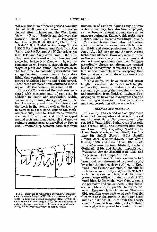
Fig. 1. Diagram of radiograph showing: (1) measure- ment of crown length (CW); (2) measurement of root width in first and second premolars (RW1, RW2); (3) measurement of root height (RH); (4) measurement of distance between root apices of first and second molars (RW3, RW4); (5) measurement of corpus height.
immersion of roots in liquids ranging from water to mercury, has also been employed, as has been wire bent around the root to measure perimeter. Radiographic techniques (Jepsen, 1963), ultrasonic recordings (Innou6, 19771, computerized simulation of surface area from serial cross sections (Nicholls et al., 19741, and stereo-photogrametry (Ander- son et al., 1983) are among the more recent techniques utilized. However, most of these methods are extremely time consuming and destructive of specimens examined. We have accordingly chosen an alternative method based on radiographs of molars and premo- lars in the mandible while accepting that this provides an estimate of cross-sectional diameters only.
In this study, we have measured crown length (mesiodistal diameter), root length, root width, interapical distance, and cross- sectional root area of the mandibular molars and premolars, from digitised tracings of ra- diographs. We have also investigated the rate and timing of changes in these parameters and their correlation with one another.
METHODS AND MATERIALS
This study was carried out on specimens from the following sites and periods in Israel and the West Bank: Natufian-Eynan (Per- rot, 1966; Valla, 1981), Nahal Oren (Steckelis and Ysraeli, 1963), and Hayonim (Bar-Yosef and Goren, 1973); Prepottery Neolithic B- Abou Gosh (Lechevallier, 1978); Chalcol- ithic-Bir Safadi (Perrot, 1961); Middle Bronze-Jebel Q’aaqir (Dever, 19721, Givon (Davis, 19791, and Sasa (Davis, 1976); Late Bronzelron-Achziv (unpublished), Shechem (Relimerl, 1976), and Jericho (unpublished); Hellenistic-Jericho (Hachlili et al., 1981) and Early Arab-Dor (Dauphin, 1979).
The age and sex of these specimens had been previously determined by one of us (PSI by using the methodology outlined in Krog- man (1972). From this sample, all mandibles, with two or more fully erupted cheek teeth with root apices complete, and the crown present were utilized, giving a total of 126 mandibles. Radiographs were then taken of the premolar and molar teeth, using dental occlusal films taped parallel to the dental arch in the premolar-molar region. The man- dible and film were positioned such that the film was at right angles to the x-ray beam and at a distance of 1.5 m from the energy source. Along each mandible, a 4-cm alumi- num wedge was placed to standardize den-
POST-PLEISTOCENE REDUCTION IN TEETH AND JAWS 341
sity and provide a scale. On the radiographs, digitized tracings were recorded of crown out- line, root outline, and corpus height.
The crown and root were demarcated by a line joining the cervical margin of the enamel on the mesial and distal aspect of each tooth. Mesiodistal crown length (CW) was traced parallel to this line and the maximum value was recorded, For the premolar roots the width midway along the root length was traced parallel to this line (RW1 and RW2) and root height (RH) was traced parallel to the long axis of the tooth to the root tip. For the molars, lines were constructed joining the root apices and used to measure distance between root apices (RW3 and RW4) and root height traced from these lines to the line demarcating the crown and root (Fig. 1). Cor- pus height was traced interproximally from the interdental crest to the base of the man- dible, mesial to each of the premolars and the first and second premolar. From these computerized tracings, we obtained crown height, crown length, cross-sectional root area, root height, root width, and corpus height for each tooth separately. On 15 spec- imens, duplicate radiographs were taken and redigitized and then compared with the orig- inal radiographs to determine experimental error. This was calculated after Dahlberg (1940) and found to average 2-3% for param- eters examined.
STATISTICAL ANALYSIS
Analyses were carried out separately for each of the measurements in males and fe- males of all periods studied. The Chalcolithic sample was very small; as values fell within the range observed for the Neolithic sample, these two groups were pooled for further analysis. Since we were interested in detect- ing monotonicity of microevolutionary changes rather than particular parametric relationships, and since sample size was rel- atively small, a nonparametric approach was utilized that is based on rank transformation of raw data by their ranks. This approach allowed more robust procedures than those provided by parametric tests since it stabi- lizes the variances and does not overweight outliers (Conover and Iman, 1981). It has been found to compare favorably with other procedures in general use for examining monotone trends rather than linear trends in the studied parameters. This was examined graphically and analytically through linear regression analysis of the ranks on the chro-
nological scale (Iman and Conover, 1979). Regression analysis was performed on each parameter, to test for the existence of unidi- rectional trends over the whole period from the Natufian to the Early Arab period. For those parameters where a significant reduc- tion over time was observed, a second regres- sion analysis was carried out, excluding the Natufian sample, to assess the magnitude of changes from the Neolithic-Chalcolithic to the Early Arab period.
Relative change between the Natufian mean values and those of other periods were evaluated by subtracting the means of all other periods combined from the Natufian means and dividing the figure obtained by the Natufian mean. Their approximate vari- ances were derived by using the Delta method (Elandt-Johnson and Johnson, 1980). Pairwise comparisons were made by using the corresponding Z test. Possible differences in age distribution of the samples (15-20,20- 40, 41+) were examined, and Spearman’s rank correlation was used to compare rela- tionships between different parameters of in- dividual teeth in different periods.
RESULTS
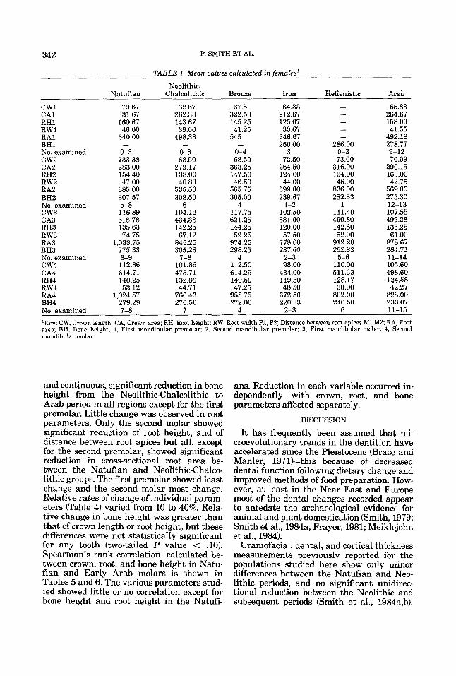
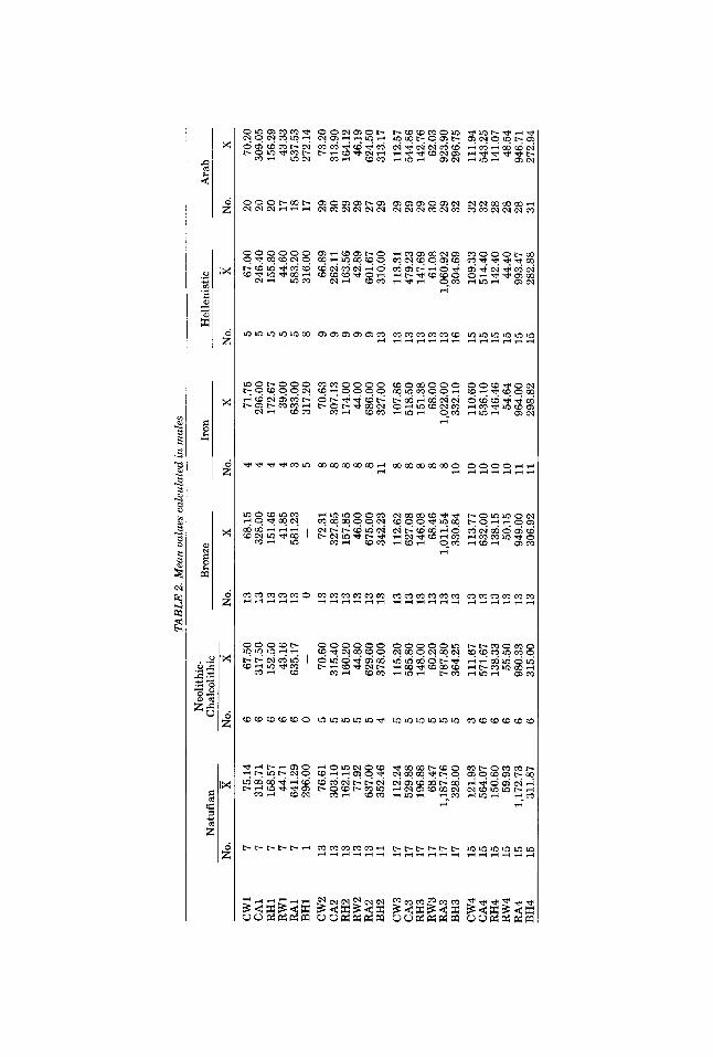
Initial analyses were carried out to deter- mine the effect of sex and age on values re- corded. In all periods studied, male values tended to be higher than those of females, and decreased over time while females showed no consistent trend (Tables 1, 2). As female sample sizes were very small, they were excluded from further analyses that were carried out on the male samples. Crown length of the first molar decreased with age in the Natufians, but not in the other groups. Bone height in the molar region increased in all samples between age groups 15-20 and 20-40, but showed no change in the 40+ group. Agecorrected scores derived by enter- ing age group as covarient were accordingly used in the regression analysis of diachronic change in bone height.
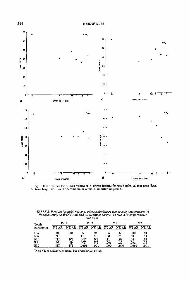
The pattern of reduction seen in those pa- rameters showing unidirectional change is shown in Figure 2 and the significance of change observed between the Natufian and all later groups and between the Neolithic- Chalcolithic group and the more recent groups is listed in Table 3. There was a sig- nificant reduction in crown length of all teeth except the first premolar between the Natu- fian and the Neolithic-Chalcolithic groups, but no significant reduction in later periods,
342 P. SMITH ET AL.
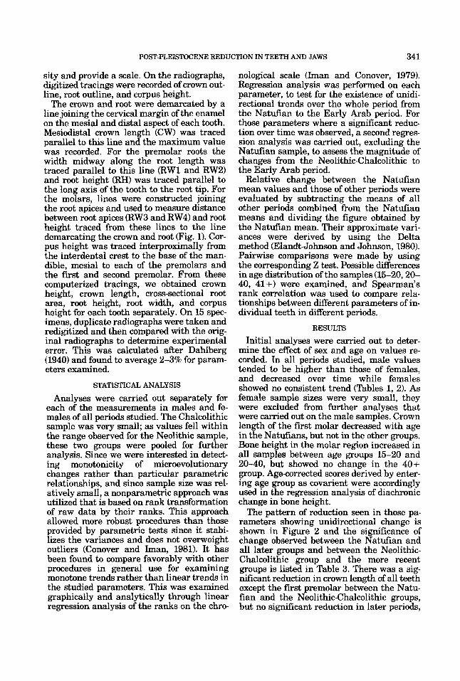
TABLE 1. Mean values calculated in females'
Neolithic- Natufian Chalcolithic Bronze Iron Hellenistic Arab
cw1 79.67 62.67 67.5 64.33 - 65.83 CA1 331.67 262.33 322.50 212.67 - 264.67
160.67 143.67 145.25 125.67 - 158.00 RW1 46.00 39.00 41.25 33.67 - 41.55 RH1
RA1 640.00 498.33 545 346.67 - 492.18 BH1 - - - 250.00 286.00 278.77 No. examined 0-3 0-3 0-4 3 0-3 9-12 c w 2 733.38 68.50 68.50 72.50 73.00 70.09 CA2 283.00 279.17 363.25 264.50 316.00 290.15 RH2 154.40 138.00 147.50 124.00 194.00 163.00 RW2 47.00 40.83 46.50 44.00 46.00 42.75 RA2 685.00 535.50 565.75 599.00 836.00 569.00 BH2 307.57 308.50 305.00 239.67 282.83 275.30 No. examined 5-8 6 4 1-2 1 12-13 c w 3 116.89 104.12 117.75 102.50 111.40 107.55 CA3 618.78 434.38 621.25 381.00 490.80 499.28 RH3 135.63 142.25 144.25 120.00 142.80 136.25 RW3 74.75 67.12 59.25 57.50 52.00 61.00 RA3 1,033.75 845.25 974.25 778.00 919.20 878.67 BH3 275.33 305.28 298.25 237.00 262.83 264.71 No. examined 8-9 7-8 4 2-3 5-6 11-14 c w 4 112.86 101.86 112.50 98.00 110.00 105.60 CA4 614.71 475.71 614.25 434.00 511.33 498.60 RH4 140.25 132.00 140.50 119.50 128.17 124.58 RW4 53.12 44.71 47.25 48.50 30.00 42.27 RA4 1,024.57 766.43 955.75 672.50 802.00 828.00 BH4 279.29 270.50 272.00 220.33 246.50 233.07 No. examined 7-8 7 4 2-3 6 11-15
'Key: CW, Crown length; CA, Crown area; RH, Root height; RW, Root width P1, P2; Distance between root apices M1,MZ; RA, Root area; BH, Bone height; 1, First mandibular premolar; 2, Second mandibular premolar; 3, First mandibular molar; 4, Second mandibular molar.
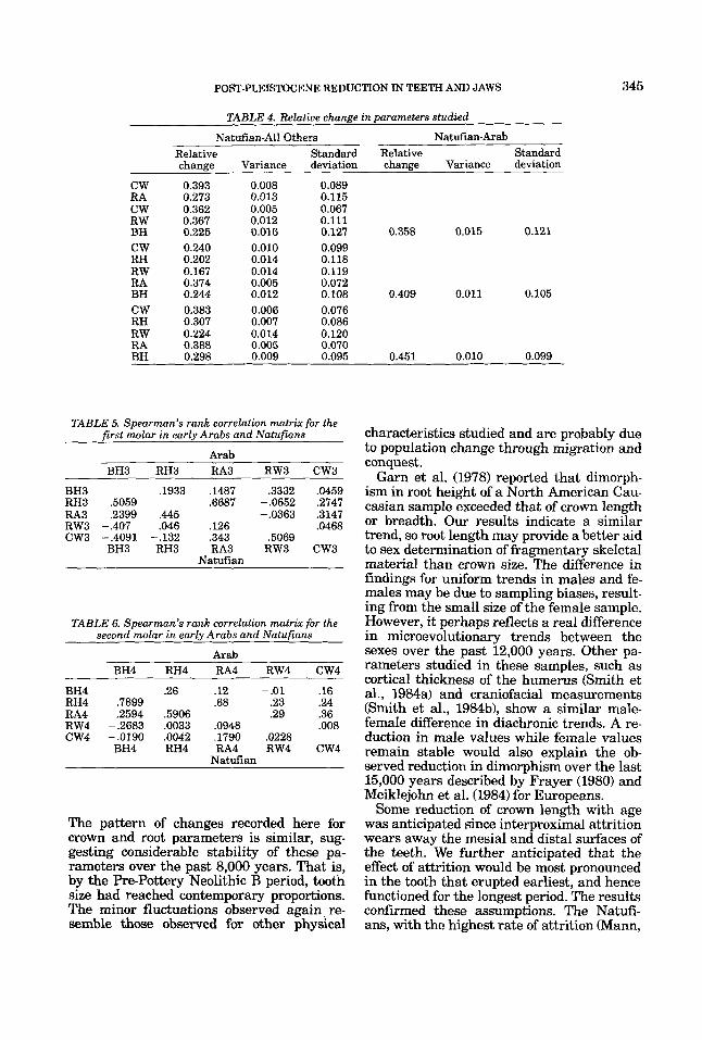
and continuous, significant reduction in bone height from the Neolithic-Chalcolithic to Arab period in all regions except for the first premolar. Little change was observed in root parameters. Only the second molar showed significant reduction of root height, and of distance between root apices but all, except for the second premolar, showed significant reduction in cross-sectional root area be- tween the Natufian and Neolithic-Chalco- lithic groups. The first premolar showed least change and the second molar most change. Relative rates of change of individual param- eters (Table 4) varied from 10 to 40%. Rela- tive change in bone height was greater than that of crown length or root height, but these differences were not statisticaIIy significant for any tooth (two-tailed P value < .lo). Spearman's rank correlation, calculated be- tween crown, root, and bone height in Natu- fian and Early Arab molars is shown in Tables 5 and 6. The various parameters stud- ied showed little or no correlation except for bone height and root height in the Natdi-
ans. Reduction in each variable occurred in- dependently, with crown, root, and bone parameters affected separately.
DISCUSSION
It has frequently been assumed that mi- croevolutionary trends in the dentition have accelerated since the Pleistocene (Brace and Mahler, 1971kthis because of decreased dental function following dietary change and improved methods of food preparation. How- ever, at least in the Near East and Europe most of the dental changes recorded appear to antedate the archaeological evidence for animal and plant domestication (Smith, 1979; Smith et al., 1984a; Frayer, 1981; Meiklejohn et aI., 1984).
Craniofacial, dental, and cortical thickness measurements previously reported for the populations studied here show only minor differences between the Natufan and Neo- lithic periods, and no significant unidirec- tional reduction between the Neolithic and subsequent periods (Smith et al., 1984a,b).
TAB
LE 2
. Mea
n va
lues
cal
cula
ted
in m
ales
Bro
nze
Iron
H
elle
nist
ic
Ara
b N
eolit
hic-
N
atuf
ian
Cha
lcol
ithic
-
-
-
-
-
-
No.
X
N
o.
X
No.
X
No.
X
N
o.
X N
o.
X
cw1
7 75
.14
6 67
.50
13
68.1
5 4
71.7
5 5
67.0
0 20
70
.20
CA
1 7
318.
71
6 31
7.50
13
32
8.00
4
296.
00
5 24
6.40
20
30
9.05
R
H1
7 15
8.57
6
152.
50
13
151.
46
4 17
2.67
5
155.
80
20
156.
29
RW
1 7
44.7
1 6
43.1
6 13
41
.85
4 39
.00
5 44
.60
17
43.3
3 R
A1
7 64
1.29
6
635.
17
13
581.
23
3 63
3.00
5
583.
20
18
537.
53
BH
1 1
29
6.00
0
-
0 -
5 31
7.20
8
316.
00
17
272.
14
cw
2
RA
2 B
H2
cw
3
CA
3 R
H3
RW
3 R
A3
BH
3 c
w4
C
A4
RH
4 R
W4
13
13
13
13
13
11
17
17
17
17
17
17
15
15
15
15
76.6
1 30
3.10
16
2.15
77
.92
637.
00
352.
46
112.
24
529.
88
196.
88
68.4
7 1,
187.
76
328.
00
121.
93
564.
07
150.
60
59.9
3
3 6 6 6
70.6
0 31
5.40
16
0.20
44
.80
629.
60
378.
00
115.
20
585.
80
148.
00
60.2
0 78
7.80
36
4.25
11
1.67
57
1.67
13
8.33
55
.50
13
13
13
13
13
13
13
13
~~ 13
13
13
13
13
13
13
13
72.3
1 32
7.85
15
7.85
46
.00
675.
00
342.
23
112.
62
627.
08
146.
08
68.4
6 1,
011.
54
330.
84
113.
77
632.
00
138.
15
50.1
5
8 8 8 8 8 11 8 8 8 8 8 10
10
10
10
10
70.6
3 30
7.13
17
4.00
44
.00
686.
00
327.
00
107.
86
518.
50
151.
38
68.0
0 1,
022.
00
332.
10
110.
60
536.
10
146.
46
54.6
4
9 9 9 9 9 13
13
13
13
13
13
16
15
15
15
15
66.8
9 26
2.11
16
3.56
42
.89
601.
67
310.
00
113.
31
479.
23
147.
69
61.0
8 1,
060.
92
304.
69
109.
33
514.
40
142.
40
44.4
0
29
30
29
29
27
29
29
29
29
30
29
32
32
32
28
28
73.2
0 31
3.90
16
4.12
46
.19
624.
50
313.
17
112.
57
544.
86
142.
76
62.0
3 92
3.90
29
6.75
11
1.94
54
3.25
14
1.07
48
.54
RA
4 15
1,
172.
73
6 98
0.33
13
94
9.00
11
96
4.00
15
99
3.47
28
94
6.71
B
H4
15
311.
87
6 31
5.00
13
30
6.92
11
29
8.82
15
28
2.88
31
27
2.94
70 -
60 -
50 -
I ' O -
ks 30-
20 -
10-
.
60-
50 -
40 -
38-
ks 20 -
10-
P. SMITH ET AL.
cw.
0 . .
70 -
60-
so -
8 40-
s t 30-
20 -
10-
.
70 7
60 -
so -
I ' O -
ks 30-
20 -
IO-
. . .
. . 0 . .
. . . .
O' ri 6 3 3 3 il 6 3 8 3 1 1
C TEms BPIXIOWI d TEms BllxlOOO1
Fig. 2. Mean values for ranked values of (a) crown length; 6) root height, (c) root area (3-4). (d) bone height (BH) in the second molar of males in different periods.
T A B U 3. P ualues for unidirectional microeuolutionary trends ouer time between (1) Natufian early Arab (NT-AR) and (2) Neolithicearly Arab (NE-AR) by parameter
and tooth'
T....CL Pml Pm2 M1 M2 I""*II
parameter NT-AR NE-AR NT-AR NEAR NT-AR NE-AR NT-AR NE-AR
cw .24 .19 .05 .24 .05 .58 .002 .94 R W NT .ll .78 .36 .70 .03 .14 RH NT NT NT NT .ll .63 .06 .57 RA .02 .09 NT NT .001 .20 .001 .79 BH NT NT .0001 ,001 ,000 ,000 .0002 ,001 'Key: NT, no unidirection trend; F'm, premolar; M, molar.
POST-PLEISTOCENE REDUCTION IN TEETH AND JAWS 345
TABLE 4. Relative change in parameters studied
Natufian-All Others Natuf an-Arab Relative Standard Relative Standard change Variance deviation change Variance deviation
CW 0.393 0.008 0.089 RA 0.273 0.013 0.115 CW 0.362 0.005 0.067 RW 0.367 0.012 0.111 BH 0.225 0.016 0.127 0.358 0.015 0.121 CW 0.240 0.010 0.099 RH 0.202 0.014 0.118 RW 0.167 0.014 0.119 RA 0.374 0.005 0.072 BH 0.244 0.012 0.108 0.409 0.011 0.105 CW 0.383 0.006 0.076 RH 0.307 0.007 0.086 RW 0.224 0.014 0.120 RA 0.388 0.005 0.070 BH 0.298 0.009 0.095 0.451 0.010 0.099
TABLE 5. Spearman’s rank correlation matrix for the first molar in early Arabs and Natufians
Arab BH3 RH3 RA3 RW3 CW3
BH3 .1933 .1487 .3332 .0459 RH3 ,5059 .6687 -.0652 ,2747 RA3 ,2399 ,445 -.0363 .3147 RW3 -.407 .046 .126 ,0468 CW3 -.4091 -.132 ,343 ,5069
BH3 RH3 RA3 RW3 CW3 Natufian
TABLE 6. Spearman’s rank correlation matrix for the second molar in early Arabs and Natufians
Arab BH4 RH4 RA4 RW4 CW4
BH4 .26 .12 -.01 .16 RH4 .7899 .68 .23 .24 RA4 ,2594 .5906 .29 .36 RW4 -.2683 ,0033 ,0948 .008
BH4 RH4 RA4 RW4 CW4 CW4 -.0190 ,0042 ,1790 ,0228
Natufian
The pattern of changes recorded here for crown and root parameters is similar, sug- gesting considerable stability of these pa- rameters over the past 8,000 years. That is, by the Pre-Pottery Neolithic B period, tooth size had reached contemporary proportions. The minor fluctuations observed again re- semble those observed for other physical
characteristics studied and are probably due to population change through migration and conquest.
Garn et al. (1978) reported that dimorph- ism in root height of a North American Cau- casian sample exceeded that of crown length or breadth. Our results indicate a similar trend, so root length may provide a better aid to sex determination of fragmentary skeletal material than crown size. The difference in findings for uniform trends in males and fe- males may be due to sampling biases, result- ing from the small size of the female sample. However, it perhaps reflects a real difference in microevolutionary trends between the sexes over the past 12,000 years. Other pa- rameters studied in these samples, such as cortical thickness of the humerus (Smith et al., 1984a) and craniofacial measurements (Smith et al., 1984b), show a similar male- female difference in diachronic trends. A re- duction in male values while female values remain stable would also explain the ob- served reduction in dimorphism over the last 15,000 years described by Frayer (1980) and Meiklejohn et al. (1984) for Europeans.
Some reduction of crown length with age was anticipated since interproximal attrition wears away the mesial and distal surfaces of the teeth. We further anticipated that the effect of attrition would be most pronounced in the tooth that erupted earliest, and hence functioned for the longest period. The results confirmed these assumptions. The Natuti- am, with the highest rate of attrition RJlann,
346 P. SMITH ET AL
1983), were the only group to show a signifi- cant reduction of crown length with age, and the first molar was the only tooth affected. It erupts at 6 years-some 4 years before the first premolar and 6 years before the second premolar and molar (Scott and Symons, 1980).
Crown length values recorded here, espe- cially of the first molar, may then underesti- mate the extent of dimensional changes in this variable between the Natufians and other groups examined. Root length, on the contrary, undergoes little or no dimensional change throughout life. While there may be continued deposition of cementum this is normally only some microns thick (Gustaf- son, 1950). Even so, in male second molars percentage change in crown length between Natufans and Arabs was 39% compared with 22% in the distance between root apices, and 31% in root height.
In order to obtain as large a sample as possible, adolescent specimens with fully formed premolar and second molar roots were included in this study. However, while the roots of these teeth are fully formed by the age of 14-15, the mandible continues grow- ing in males throughout the third decade, and surface apposition of basal bone contin- ues throughout life (Scott and Symons, 1980; Walker and Kowalski, 1971). Corpus height may, however, reduce in later life if periodon- tal disease is present, causing resorption of alveolar bone. The findings reported for males in this study confirm an increase in corpus height of the mandible in the premo- lar and molar region between the late teens and twenties in all periods studied. The ab- sence of reduction in corpus height indicates that periodontal disease was minimal in even the older individuals studied. Corpus height in the molar region was the only parameter to show significant uniform reduction over the past 8,000 years. Since age effects were corrected for in this analysis, it may be con- cluded that reduction in corpus height has continued to a greater extent than tooth re- duction. Observed reduction in male corpus height was 45% compared with 31% in root height, suggesting changing root-bone rela- tionships.
In addition to the differences found in pat- terns of diachronic change of crowns, root, and bone of individual teeth, differences were noted in the rate of reduction of analogous parameters in different teeth. The first pre-
molar and first molar showed less change than the second premolar and second molar- a finding consistent with the field theory (Butler, 1939; Dahlberg, 1968) attributing greater conservatism to the early developing key teeth.
Clinical interest in root size is mainly di- rected to establishing its relationship as measured by length and surface area to the progress of periodontal disease. Studies of the relation of bone mass to attachment area have demonstrated a large range of variation in the ratio of surface area to root length (Brown, 1950; Nicholls et al., 1974; Levy and Right, 1978; Despeignes, 1979; Anderson et al., 1983). Few population studies of root mor- phology include metric analysis; however, descriptions suggest that despite the wide range of intrapopulation variation, there are consistent interpopulation differences. Mon- goloids appear to have relatively short roots, but a high frequency of three rooted lower first molars (Pederson, 1949; Sumner, 1965; Turner, 1971; Hochstetter, 1979). Australian Aborigines have long sturdy roots with di- vergent apices (Campbell, 1925), and sub-Sa- haran African populations have long roots that are divergent but thin except for the upper canine (Shaw, 1931; Jacobson, 1978).
CONCLUSIONS
This study has shown that in populations of different periods, root size is poorly corre- lated with crown length and corpus height, and that there are temporal as well as geo- graphic differences in crown-root relation- ships. Over the past 12,000 years, populations in the Near East have undergone significant reduction in crown width and root area of the lower first and second molars, in crown width of the second premolars, and in corpus height distal to all three teeth, but not in root height. The reduction in tooth size took place between the Natufian and Neolithic-Chalco- lithic periods, and no significant change in these parameters has occurred over the past 7,000 years. Corpus height has, however, continued to reduce, while root height has not changed. This means that recent popula- tions have larger rooted teeth, relative to crown size or corpus height, than their pre- decessors during the late Pleistocene. Discus- sions of evolutionary trends in tooth and jaw size should then take into account root size and location.
POST-PLEISTOCENE REDUCTION IN TEETH AND JAWS 347
ACKNOWLEDGMENTS
This study was supported by a grant from the Israel Academy of Sciences.
LITERATURE CITED
Anderson, D, Thompson, L, and Popovich, F (1977) Tooth, chin, bone and body correlations. Am. J. Phys. Anthro- pol. 467-12.
Anderson, RW, McGarrah, HE, Lamb, RD, and Eick, J D (1983) Root surface measurements of mandibular mo- lars using stereophotogrammetry. J. Am. Dent. Assoc. 107613-615.
Bar-Yosef, 0 (1980) The prehistory of the Levant Ann. Rev. Anthropol. 9:lOl-133.
Bar-Yosef, 0, and Goren, N (1973) Natufian remains from Hayonim cave. Paleorient 1 :49-68.
Brace, CL, and Mahler, PE (1971) Post-Pleistocene changes in the human dentition. Am. J. Phys. Anthro- pol. 34:191-204.
Brown, R (1950) A method of measurement of root area. J . Can. Dent. Assoc. 16130-132.
Butler, PM (1939) Studies of the mammalian dentition. Differentiation of the post-canine dentition. Proc. Zool. Soc. Lond. [Biol.] 109:l-36.
Campbell, TD (1925) Dentition and palate of the Austra- lian Aborigine. Adelaide, South Australia: Keith Sher- idan Foundation Publ. 1.
Carlson, DS (1976) Temporal variation in prehistoric Nu- bian crania. Am. J. Phys. Anthropol. 45:467-484.
Carlson, DS, and Van Gerven, DP (1977) Masticatory function and post-Pleistocene evolution in Nubia. Am. J. Phys. Anthropol. 46495-506.
Conover, WJ, and Iman, RC (1981) Rank transforma- tions as a bridge between parametric and nonparame- tric statistics. Am. Statist. 35:124-133.
Dahlberg, G (1940) Statistical methods for Medical and Biological students. London: George Allen and Unwin, Ltd.
Dahlberg, AA (1968) On the teeth of early sapiens. In G Kurth (ed): Evolution and Hominisation, 2nd edit. Stuttgart, pp. 273-280.
Davis, D (1976) Sasa. Hadashot Archeologiot, Dept. An- tiquity, Jerusalem. (Hebrew) :9.
Davis, D (1979) Givon. Hadashot Archeologiot, Dept. Antiquity, Jerusalem. (Hebrew) :82.
Dauphin, CM (1979) Note on the 1978 excavations a t Dor. Israel Exploration J. 29236.
Despeignes, JR (1979) Variation in the area of intraper- iodontal surfaces of human teeth roots in relation to their depth. J . Periodontal. 50:630-635.
Dever, WG (1972) An MBI necropolis and settlement on the West Bank of the Jordan. Archaeology 25231-233.
Elandt-Johnson, RC, and Johnson, NL (1980) Survival models and data analysis. New York J. Wiley and Sons, pp. 69-72.
Frayer, DW (1980) Sexual dimorphism and cultural evo- lution in the late Pleistocene and Holocene of Europe. J. Human Evol. 9:399-415.
Garn, SM, Van Abstine, WL Jr, and Cole, PE (1978) Root length and crown size correlations in the mandible. J . Dent. Res. 57114.
Garn SM, Cole, PE, and Van Abstine, WL Jr (1979) Sex discriminatory effectiveness using combinations of root
lengths and crown diameters. Am. J. Phys. Anthropol. 50:115-118.
Goose, DG (1962) Reduction of palate size in modern populations. Arch. Oral. Biol. 7343-350.
Gustafson, G (1950) Age determination of teeth. J. Am. Dent. Assoc. 41:45-50.
Hachlili, R, Arensburg, B, Smith, P, and Killebrew, A (1981) The Jewish necropolis at Jericho. Curr. Anthro-
Hochstetter, RL (1979) Incidence of trifurcated mandi- bular first permanent molars in the population of Guam. J. Dent. Res. 54:1097.
Hyrdlieka, A (1930) The skeletal remains of early man. Smithsonian Collections Publ. 3075. Washington DC: Smithsonian Institute.
Iman, RC, and Conover, WJ (1979) The use of the rank transformation in regression. Technometrics 21 :499- 509.
Innout!, N (1977) A clinico-anatomical study for deter- mining root canal length by use of a novelty low fre- quency oscillation device. Bull. Tokyo Dent. Call. 18:71- 90.
Jacobson, A (1978) The dentition of the South African Negro. Birmingham, Alabama: U. of Alabama Press.
Jepsen, A (1963) Root surface measurement and a method for X-ray determination of root surface area. Acta. Odontol. Scan. 21:35-46.
Kovacs, I (1971) A systematic description of tooth roots. In AA Dahlberg (ed): Dental Morphology and Evolu- tion. Chicago U. Chicago Press, pp. 211-256.
Krogman, WM (1962) The human skeleton in forensic medicine. Springfield, Illinois: Charles C. Thomas.
Lavelle, CLB (1962) A comparison between the mandi- bles of Romano-British and 19th century periods. Am. J. Phys. Anthropol. 36213-220.
Lechevallier, M (1978) Abou Ghosh et Beisamoun. Deux gisements du VII millenaire avant l'ere Chretienne en Israel. Mem. et Trav. du CNRS, Jerusalem, Nu. 2.
Levy, AR, and Wright, WH (1978) The relationship be- tween attachment height and attachment area using a digitizer and a digital computer. J . Periodont. 49:483- 485.
Lysell, L (1958) A biometric study of occlusion and dental arches in a series of Medieval skulls from Northern Sweden. Acta. Odontol. Scand. 16267-292.
Mann, E (1983) Dental disease and jaw size in Natufian and Late Hellenistic populations. DMD Thesis, He- brew University (in Hebrew).
Meiklejohn, C, Shentag, C, Venema, A, and Key, P (1984) Socioeconomic change and patterns of pathology and variation in the Mesolithic and Neolithic of Western Europe: Some suggestions. In MH Cohen and GJ Ar- melagos (eds): Paleopathology at the Origins of Agr- culture. New York: Academic Press, pp. 75-100.
Moore, WJ, Lavelle, CLB, and Sperce, TF (1968) Changes in the size and shape of the human mandible in Brit- ain. Br. Dent. J. 125163-169.
Nicholls, JI, Daly, CH, Kydd, WL (1974) Root surface measurement using a digital computer. J. Dent. Res. 53:1338-1341.
Pederson, PO (1949) The East Greenland Eskimo denti- tion. Kobenhave CA Reitzels Forlag.
Perrot, J (1961) Une tombe a ossuaries du IVe millenaire a Azor pres de Tel Aviv. Atiquot 3:l-83.
Perrot, J (1966) Le gisement Natodien de Mallaha (Eynan), Israel. L'Anthropologie 70:437-584.
Rasing, FW (1983) Sexing immature human skeletons.
POI. 22:701-702.
348 P. SMITH ET AL.
J. Hum. Evol. 12:149-155. Scott, JH, and Symons, NBB (1980) Introduction to Den-
tal Anatomy 8th edit. London: Churchill Livingstone, p. 88.
Shaw, JCM (1931) The Beth, the bony palate and the mandible in Bantu races of South Africa. London: John Bull, Sons and Davidsson.
Silberman, U (1983) The Prevalence of Attrition and Periodontal Disease in Ancient Populations. DMD Thesis: Hebrew University (in Hebrew).
Smith, P (1979) Regional diversity in epipaleolithic pop- ulations. Int. J. Skel. Res. 6234-250.
Smith, P, Bar-Yosef, 0, and Sillen, A (1984a) Archaeolog- ical and skeletal evidence for dietary change during the Late Pleistocene/Early Holocene in the Levant. In M Cohen, and G Armelagos, (eds): Paleopathology at the Origins of Agriculture. New York Academic Press, pp. 101-136.
Smith, P, Bloom, RA, and Berkovitz, J (1984) Diachronic trends in humeral cortical thickness of Near Eastern population. J. Hum. Evol. 13:603-611.
Sofaer, J A (1973) A model relating developmental inter-
raction and differential evolutionary reduction of tooth size. Evolution 27427-434.
Stekelis, M, and Yisraeli, T (1963) Excavations at Nahal Oren. Isr. Exp. J. 13:l-12.
Sumner, R (1966) Dental abnormalities and caries prev- alence in British Columbia Indians. J. Can. Dent. As- soc. 31:379-385.
Turner, CG (1971) Three rooted mandibular first perma- nent molars and the question of American Indian origins. Am. J. Phys. Anthropol. 34:229-242.
Valla, F (1981) Les establissements Natoufiens dans le Nord d'Israe1. In: Colloques Int. de CNRS No. 598, Prehistoire du Levant. Paris: pp.409-419.
Walker, GF, and Kowalski, CJ (1971) On the growth of the mandible. Am. J. Phys. Anthropol. 36111-118.
Weine, FS (1982) Endodontic Therapy, 3rd Ed. CV Mosby
Wolpoff, MH (1975) Some aspects of human mandibular evolution. In: JA McNamara J r (ed): Determinants of mandibular form and growth. Ann Arbor: Michigan University Press, pp.1-64.
CO., pp.477-502.