portfolio - trg communicationsportfolio campaign development training materials and medical...
TRANSCRIPT

communicationsLLC
PortfolioCampaign Development
Training Materials and Medical Illustration
Marketing Programs and Slide Presentations
TRG Communications, LLC Rich Franco, President(m) 919.606.2908(e) [email protected]

TRG Communications, LLC Rich Franco, President(m) 919.606.2908(e) [email protected]
communicationsLLC
Campaign DevelopmentTESTOPEL® Turn It On
FAMILION Family
C-GAAP Gears
Hylenex® recombinant REVEAL...
communicationsLLC
www.trgcommunications.us | Rich Franco, President | (m) 919.606.2908 | [email protected]

www.trgcommunications.us | Rich Franco, President | (m) 919.606.2908 | [email protected]
communicationsLLC
Product:
Client:
Campaign:
Concept:
Creative:
Select Materials Developed:
TESTOPEL®
Slate Pharmaceuticals, a division of Actient Pharmaceuticals
Turn It On
TESTOPEL®, the only long-acting treatment for men with Low T. Once implanted (Turned On), men were spontaneously compliant with therapy. This feature would enable men to enjoy the benefits of testosterone therapy without the hassle of saw tooth T-levels while avoiding the daily hassle of administering a topical therapy.
We used the universal “On” symbol (as seen on a multitude of devices used today) as highlight recognizable inconography to denote the patient being “Powered On” for 3-6 months once TESTOPEL® is placed.
Core sales aid, patient education materials, posters
Campaign Development | TESTOPEL® Turn It On

communicationsLLC
Product:
Client:
Campaign:
Concept:
Creative:
Select Materials Developed:
FAMILION, family of genetic tests for inherited cardiac diseases
PGxHealth, Transgenomic
Family
Inherited cardiac diseases represent an unsuspecting risk to the families they affect. FAMILION represents an opportunity to make the unknown risk known so it can be treated and/or tracked through at-risk families.
Tie together real life family scenes with pedigree graphic to illustrate the existence of the unknown risk.
Test kit, core sales aid for channelopathies and cardiomyopathies, patient and family education
Campaign Development | FAMILION Family
www.trgcommunications.us | Rich Franco, President | (m) 919.606.2908 | [email protected]

communicationsLLC
Product:
Client:
Campaign:
Concept:
Creative:
Select Materials Developed:
C-GAAP Genetic Test for Clopidogrel Response
Transgenomic
Gears Working Together
The C-GAAP Clopidogrel Response Test was the only genetic test to include CYP2C19 and ABCB1, thus there were two genes that worked synergistically to predict a patient’s response to clopidogrel. We developed a well understood graphic to illustrate why it was important to genotype both genes as opposed to just CYP2C19.
Each gear represents one of the genes included as part of the test. Graphically this illustrated that both genes ultimately impact the metabolism or absorption of clopidogrel.
Core sales aid
Campaign Development | C-GAAP Gears
www.trgcommunications.us | Rich Franco, President | (m) 919.606.2908 | [email protected]
communicationsLLC
Product:
Client:
Campaign:
Concept:
Creative:
Select Materials Developed:
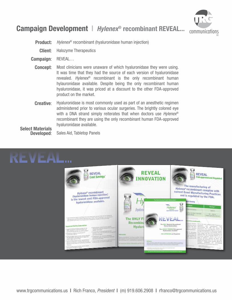
Hylenex® recombinant (hyaluronidase human injection)
Halozyme Therapeutics
REVEAL…
Most clinicians were unaware of which hyaluronidase they were using. It was time that they had the source of each version of hyaluronidase revealed. Hylenex® recombinant is the only recombinant human hylauronidase available. Despite being the only recombinant human hyaluronidase, it was priced at a discount to the other FDA-approved product on the market.
Hyaluronidase is most commonly used as part of an anesthetic regimen administered prior to various ocular surgeries. The brightly colored eye with a DNA strand simply reiterates that when doctors use Hylenex® recombinant they are using the only recombinant human FDA-approved hyaluronidase available.
Sales Aid, Tabletop Panels
Campaign Development | Hylenex® recombinant REVEAL...
www.trgcommunications.us | Rich Franco, President | (m) 919.606.2908 | [email protected]

Training Materials & Medical Illustration
Viibryd™ 7 Module Learning System
Hylenex® recombinant Infusion Illustrations
communicationsLLC
www.trgcommunications.us | Rich Franco, President | (m) 919.606.2908 | [email protected]

communicationsLLC
Product:
Client:
Material:
Background:
Viibryd™ (vilazadone HCl)
Trovis Pharmaceuticals
7 module learning system with original illustrations
Trovis was posed to launch a novel antidepressant into a very crowded market. We were asked to develop a written set of training materials that could then be incorporated into a web based training program. The materials developed included disease background, market background, competitors, About Viibryd and Objection Handling modules. All illustrations and copy were produced in-house.
Training Materials & Medical Illustration Viibryd™ 7 Module Learning System
Full module example included at the end of this PDF.
www.trgcommunications.us | Rich Franco, President | (m) 919.606.2908 | [email protected]

communicationsLLC
Product:
Client:
Material:
Background:
Hylenex® recombinant for use with insulin pumps
Halozyme
Patient Instruction sheets featuring original medical illustrations
As part of preparing for potential commercialization of Hylenex recombinant for use with insulin pumps, a series of patient instructions for use needed to be created. We were asked to develop original medical illustrations depicting several different infusion sets. Our goal was to be medically accurate but as patient-friendly as possible. All illustrations and copy were produced in-house.
Training Materials & Medical Illustration Hylenex® Recombinant Infusion Illustrations
www.trgcommunications.us | Rich Franco, President | (m) 919.606.2908 | [email protected]
Vial Vial Cap Syringe
PackageSyringePlunger
Cannula End of Tubing
TubingPackage
LuerLock
CannulaHousing
(not included)Vial
Adapter/Connector
Vial Adapter/
Connector Package
TESTOPEL® Patient Rebate Program
FAMILION Family Slide Presentation
Transgenomic Capital Campaign
Marketing Programs & Slide Presentations
communicationsLLC
www.trgcommunications.us | Rich Franco, President | (m) 919.606.2908 | [email protected]

www.trgcommunications.us | Rich Franco, President | (m) 919.606.2908 | [email protected]
communicationsLLC
Product:
Program:
Client:
Background:
Materials:
TESTOPEL®
TESTOPEL® Patient Rebate Program
Slate Pharmaceuticals, a division of Actient Pharmaceuticals
TESTOPEL is most commonly purchased and billed by clinicians, retail pharmacy visit as few. Slate Pharmaceuticals wanted to offer a rebate to patients. We developed the entire rebate process and oversaw the execution of the program with our fulfillment partner. The goal was to be compliant with all laws governing rebate programs and appropriate based upon regulatory/medical review.
Clinician/staff education materials, patient enrollment/education materials and all fulfillment materials (check and letter), weekly status reports, patient survey
Marketing Programs & Slide Presentations TESTOPEL® Patient Rebate Program
NO POSTAGENECESSARY
IF MAILEDIN THE
UNITED STATES
BUSINESS REPLY MAILFIRST-CLASS MAIL PERMIT NO. 626 CHAPEL HILL, NC
USPS BRM Template #10 Envelope 4-1/8" x 9-1/2" Working Layer
January 2001 File Name: b10.pdf
POSTAGE WILL BE PAID BY ADDRESSEE
TES-M1051 01/13
TESTOPEL Patient Rebate Program Headquartersc/o Beechwood AssociatesPO Box 1190Morrisville, NC 27560

www.trgcommunications.us | Rich Franco, President | (m) 919.606.2908 | [email protected]
communicationsLLC
Product:
Slide Presentation:
Client:
Background:
Materials:
FAMILION, family of genetic tests for inherited cardiac diseases
FAMILION Slides
PGxHealth, Transgenomic
We were asked to create a multi-module professional slide presentation that doctors or nurses could use to present to either other HCPs or patients. Shown here are the first several slides of one of the presentation decks.
Slide decks were assembled for each disease state for which there was a FAMILION test.
Marketing Programs & Slide Presentations FAMILION Family Slide Presentation

www.trgcommunications.us | Rich Franco, President | (m) 919.606.2908 | [email protected]
communicationsLLC
Product:
Slide Presentation:
Client:
Background:
Transgenomic Company Slide Presentation
Transgenomic Capital Campaign
PGxHealth, Transgenomic
We were asked to work with senior leadership at Transgenomic to assemble a slide presentation intended to raise capital. The presentation had to represent each one of Transgenomic’s business units and clearly articulate the unique value it represented. Shown here are the first several slides of the presentation deck.
Marketing Programs & Slide Presentations Transgenomic Capital Campaign

Module • 2 The Central Nervous System and Pathophysiology of
Major Depressive Disorder (MDD)
2CONFIDENTIALFor Internal Use Only. The information contained in this training module is for educational purposes only. It is designed to provide you with the information you need to be educated on the product, disease and competitive environment and is not to be distributed or used in detailing.

CONFIDENTIAL: For Internal Use Only.

1
22Table of Contents
Chapter 1: Theories About the Causes of MDD ...............................................3
1.1. Theories About the Causes of MDD ...................................................................................3
1.2. Select Emotions, Behaviors and Bodily Functions Regulated by Neurotransmitters .........................................................................................5
Chapter 2: The Central Nervous System .............................................................7
2.1. Regions of the Brain ..................................................................................................................7
2.2. Spinal Cord ................................................................................................................................10
2.3. Peripheral Nervous System .................................................................................................10
2.4. Summary of Central Nervous System..............................................................................11
Chapter 3: Neuroanatomy Related to MDD ................................................... 13
3.1. The Neuron ...............................................................................................................................13
3.2. Neurotransmission .................................................................................................................14
3.3. Neurotransmitters, Receptors and Ion Channels ........................................................16
3.3.1. Neurotransmitters ......................................................................................................16
3.3.2. Ion Channels.................................................................................................................17
3.3.3. Receptors .......................................................................................................................18
3.4. Biochemical Theory of Depression ...................................................................................19
Brain Booster .............................................................................................................. 21
Definitions .................................................................................................................... 23
References .................................................................................................................... 25
Learning Objectives Review theories of the causes of MDD.
Review the neurotransmission process.
Review neuroanatomic structures associated with MDD.
Describe different neurotransmitters associated with MDD.
Understand how the dysregulation of neurotransmitters may occur.

2CONFIDENTIAL: For Internal Use Only.
3
22
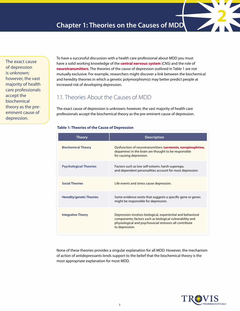
Theory Description
Biochemical Theory Dysfunction of neurotransmitters (serotonin, norepinephrine, dopamine) in the brain are thought to be responsible for causing depression.
Psychological Theories Factors such as low self-esteem, harsh superego, and dependent personalities account for most depression.
Social Theories Life events and stress cause depression.
Heredity/genetic Theories Some evidence exists that suggests a speci�c gene or genes might be responsible for depression.
Integrative Theory Depression involves biological, experiential and behavioral components; factors such as biological vulnerability and physiological and psychosocial stressors all contribute to depression.
Table 1: Theories of the Cause of Depression
Chapter 1: Theories on the Causes of MDD
To have a successful discussion with a health care professional about MDD you must have a solid working knowledge of the central nervous system (CNS) and the role of neurotransmitters. The theories of the cause of depression outlined in Table 1 are not mutually exclusive. For example, researchers might discover a link between the biochemical and heredity theories in which a genetic polymorphism(s) may better predict people at increased risk of developing depression.
1.1. Theories About the Causes of MDD
The exact cause of depression is unknown; however, the vast majority of health care professionals accept the biochemical theory as the pre-eminent cause of depression.
The exact cause of depression is unknown; however, the vast majority of health care professionals accept the biochemical theory as the pre-eminent cause of depression.
None of these theories provides a singular explanation for all MDD. However, the mechanism of action of antidepressants lends support to the belief that the biochemical theory is the most appropriate explanation for most MDD.

4CONFIDENTIAL: For Internal Use Only.
Figure 1: Dysregulation of Serotonin (5-HT) is Believed to be the Primary Cause of MDD1,2,3,4
Chapter 1: Theories on the Causes of MDD
The relationship between the effect of neurotransmitters on the brain and body is often referred to as the mind-body link. Figure 1 and several other sections of this module serve to explain the roles of neurotransmitters in depression.
Dysregulation of 5-HT (serotonin) is strongly associated with MDD.
5-HT (serotonin) and other neurotransmitters help modulate certain bodily functions via the descending pathway.
5
22
1.2. Select Emotions, Behaviors and Bodily Functions Regulated by Neurotransmitters
Neurotransmitters are chemical substances that transmit nerve impulses. Key neurotransmit-ters, like serotonin, originate in the midbrain and project to various locations throughout the human nervous system.
Within the brain, neurotransmitters help mediate numerous emotional and behavioral functions. A brief list of emotions and/or behaviors regulated by neurotransmitters is shown below. For MDD patients certain neurotransmitters may dysfunction and result in an inability to regulate emotions and/or behaviors.
Sadness
Anxiety
Irritability
Pleasure
Guilt
Concentration
Appetite
Sexual function
Projections from the brain stem descend down the spinal cord where they have a role in helping to regulate certain bodily functions. A brief list of these functions is shown below.
Vasoconstriction
Urethral sphincter contractions
Bladder wall relaxation
Gastrointestinal motility
Pilomotor contraction (goose bumps)
Normally, the sensations associated with the routine functioning of the body, such as digestion in the abdomen, urogenital function, and/or routine inputs to the musculoskeletal system throughout the body, are suppressed from our awareness so that attention can be paid to events occurring outside of the body and the central nervous system can react appropriately.
Neurotransmitters are chemical substances that transmit nerve impulses.

6CONFIDENTIAL: For Internal Use Only.

7
22
Cerebrum
Spinal Cord
Pons
Midbrain
Brainstem
Cerebellum
Medulla Oblongata
Diencephalon(thalamus & hypothalamus)
Figure 2: The Major Regions of the Human Brain
Together, the spinal cord and brain are known as the central nervous system (CNS). The CNS acts as the control center of the entire nervous system, interpreting incoming information and issuing responses.
2.1. Regions of the Brain
As shown in Figure 2, the brain has the following major regions:
Cerebrum
Cerebellum
Diencephalon (thalamus & hypothalamus)
Brainstem
Chapter 2: The Central Nervous System
The cerebrum is the largest region of the brain. It controls voluntary motor functions; coordinates physical, sensory, visual and auditory sensations; and integrates consciousness, memory, use of language, and emotions.

8CONFIDENTIAL: For Internal Use Only.
The surface of the cerebrum is called the cerebral cortex and is composed of six layers. It is made of gray matter, which is also found in the other parts of the nervous system. Because this gray matter enlarges more than the rest of the brain during development, it rolls and folds upon itself to produce convolutions (also known as gyri), fissures and sulci. The most prominent fissure separates the cerebrum into 2 halves—the right and left hemispheres of the brain.
The interior of the cerebrum, underneath the cerebral cortex, is known as the white matter. It consists mainly of the axons of neuronal cells connecting different regions of the cortex and the cortex with other parts of the brain and spinal cord.
The cerebellum, the second largest region of the brain, functions to control skeletal muscles, primarily in terms of coordination and balance.
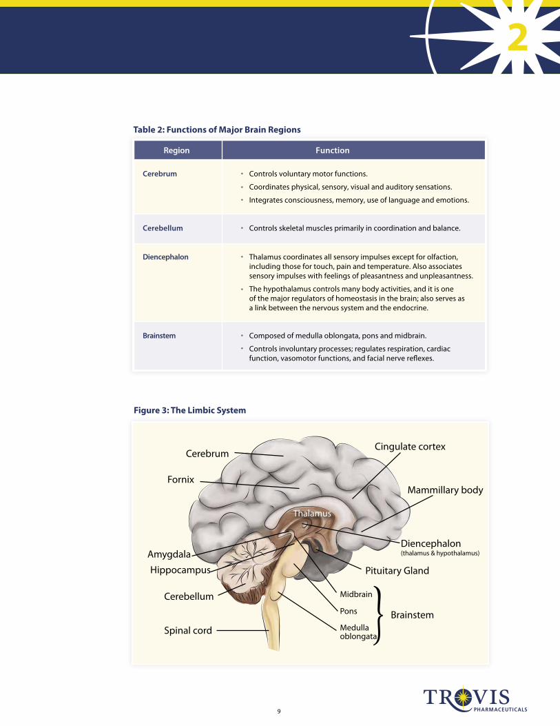
Human emotion involves the entire nervous system, but there are two parts of the CNS that are especially important: the limbic system and the autonomic nervous system. The limbic system is a complex set of structures that lies on both sides of the thalamus, just under the cerebrum. It includes the diencephalon (thalamus and hypothalamus), the hippocampus, the amygdala, and other nearby areas (see Figure 3). It is primarily responsible for our emotional life, and the formation of memories. The thalamus coordinates all sensory impulses, except for the sense of smell. It also plays a role in emotions by associating sensory impulses with feelings of pleasantness and unpleasantness. The hypothalamus controls many body activities, and it is one of the major regulators of homeostasis in the brain. It also serves as a link between the nervous system and the endocrine system. The hypothalamus modulates human urges, such as eating, drinking and sexual arousal, as well as emotional and behavioral patterns.
The brainstem includes the medulla oblongata, pons and midbrain. These areas regulate autonomic (involuntary) behaviors, including respiration, cardiac function and vasomotor functions.
Chapter 2: The Central Nervous System
Human emotion involves the entire nervous system, but there are two parts of the CNS that are especially important: the limbic system and the autonomic nervous system.
9
22
Region Function
Cerebrum Controls voluntary motor functions.
Coordinates physical, sensory, visual and auditory sensations.
Integrates consciousness, memory, use of language and emotions.
Cerebellum Controls skeletal muscles primarily in coordination and balance.
Diencephalon Thalamus coordinates all sensory impulses except for olfaction, including those for touch, pain and temperature. Also associates sensory impulses with feelings of pleasantness and unpleasantness.
The hypothalamus controls many body activities, and it is one of the major regulators of homeostasis in the brain; also serves as a link between the nervous system and the endocrine.
Brainstem Composed of medulla oblongata, pons and midbrain.
Controls involuntary processes; regulates respiration, cardiac function, vasomotor functions, and facial nerve re�exes.
···
·
·
·
··
Table 2: Functions of Major Brain Regions
Cerebrum
Spinal cord
Pons
Midbrain
Brainstem
Cerebellum
Medulla oblongata
Diencephalon(thalamus & hypothalamus)Amygdala
Hippocampus Pituitary Gland
Mammillary body
Cingulate cortex
Fornix
Thalamus
Figure 3: The Limbic System
10CONFIDENTIAL: For Internal Use Only.
2.2. Spinal Cord
The spinal cord is protected by the vertebral column as well as the meninges, which also surround the brain. The gray matter of the spinal cord is shaped like the letter H. It is made up of the cell bodies of neurons and unmyelinated axons and dendrites. Thirty-one pairs of spinal nerves emerge from the spinal cord, these serve as a means of neurotransmitter projection to the peripheral nervous system.
2.3. Peripheral Nervous System
The peripheral nervous system includes all nerves external to the CNS. These include the cranial and spinal nerves that emerge from and enter the CNS, and their terminal projections—the axon terminals that synapse with muscle fibers, glands, or other cells stimulated by nerves. These peripheral nerves relay impulses between the sense organs, the CNS, and muscles and glands throughout the body. Overall, the peripheral nervous system gathers information from the periphery, routes it to the CNS for interpretation, and then transmits a response from the CNS back to the periphery.
The peripheral nervous system may be further subdivided into:
The somatic (voluntary) nervous system
The autonomic (involuntary) nervous system, which is further divided into sympathetic and parasympathetic branches
Chapter 2: The Central Nervous System
Thirty-one pairs of spinal nerves emerge from the spinal cord, these serve as a means of neurotransmitter projection to the peripheral nervous system.

11
22
Figure 4: The Central Nervous System
Central nervous system (CNS)
Spinal Cord
Brain
Peripheral nervous system (PNS)
Input
Output
Somatic(voluntary)
Autonomic(involuntary)
Sympathetic Parasympathetic
Cardiac muscle, smooth muscle and glands.
Skeletal muscle, such as the facial muscles, and the muscles found in the limbs, such as the biceps and triceps.
Nervous system
2.4. Summary of the Central Nervous System
Figure 4 summarizes information presented to this point and provides an aggregate view of the CNS. Neurotransmitter regulation is essential to the central nervous system functioning properly. Malfunctioning neurotransmitters can cause illnesses like MDD, GI upset and urinary incontinence.
Malfunctioning neurotransmitters can cause illnesses like MDD, GI upset and urinary incontinence.

12CONFIDENTIAL: For Internal Use Only.
13
22Chapter 3: Neuroanatomy Related to MDD
3.1. The Neuron
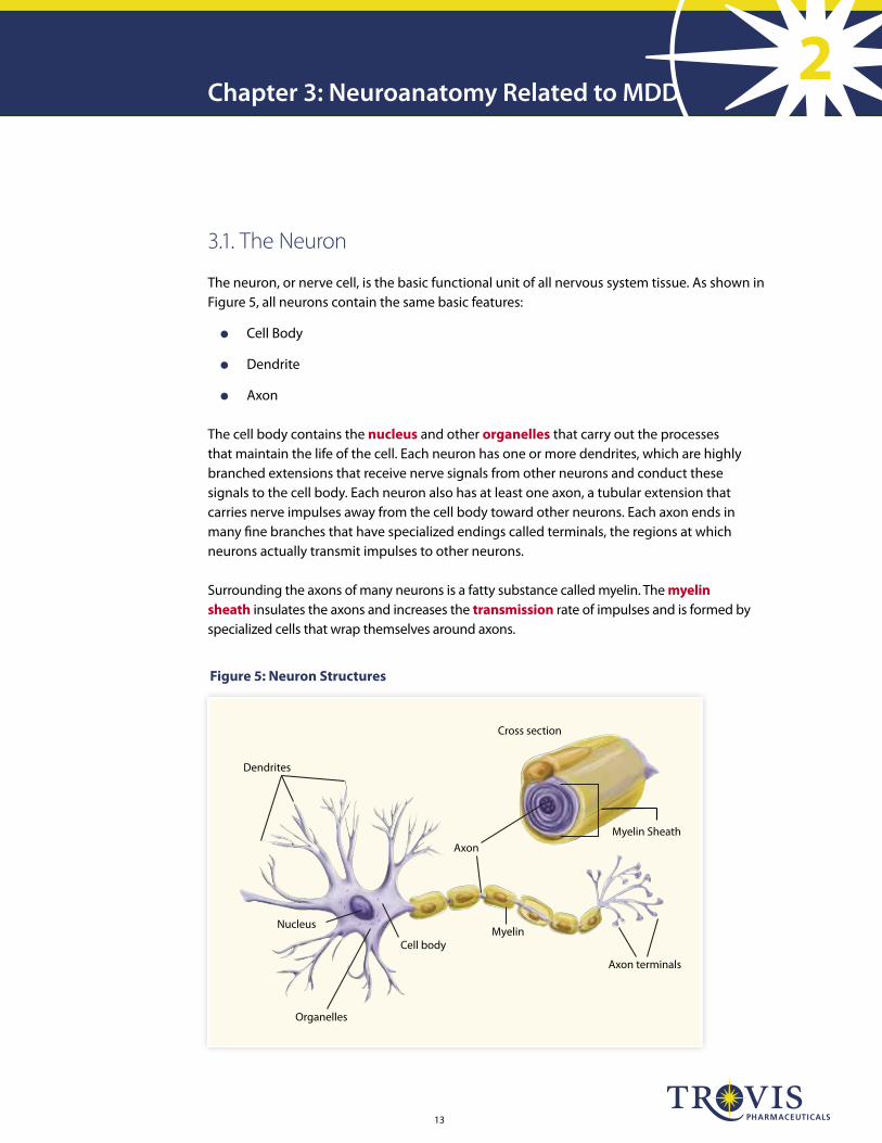
The neuron, or nerve cell, is the basic functional unit of all nervous system tissue. As shown in Figure 5, all neurons contain the same basic features:
Cell Body
Dendrite
Axon
The cell body contains the nucleus and other organelles that carry out the processes that maintain the life of the cell. Each neuron has one or more dendrites, which are highly branched extensions that receive nerve signals from other neurons and conduct these signals to the cell body. Each neuron also has at least one axon, a tubular extension that carries nerve impulses away from the cell body toward other neurons. Each axon ends in many fine branches that have specialized endings called terminals, the regions at which neurons actually transmit impulses to other neurons.
Surrounding the axons of many neurons is a fatty substance called myelin. The myelin sheath insulates the axons and increases the transmission rate of impulses and is formed by specialized cells that wrap themselves around axons.
Figure 5: Neuron Structures
Nucleus
Cell body
Dendrites
Axon
Organelles
Myelin
Cross section
Myelin Sheath
Axon terminals
14CONFIDENTIAL: For Internal Use Only.
3.2. Neurotransmission
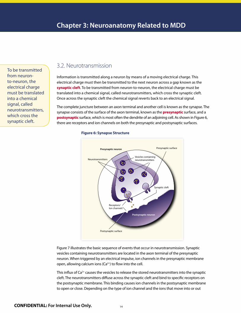
Information is transmitted along a neuron by means of a moving electrical charge. This electrical charge must then be transmitted to the next neuron across a gap known as the synaptic cleft. To be transmitted from neuron-to-neuron, the electrical charge must be translated into a chemical signal, called neurotransmitters, which cross the synaptic cleft. Once across the synaptic cleft the chemical signal reverts back to an electrical signal.
The complete juncture between an axon terminal and another cell is known as the synapse. The synapse consists of the surface of the axon terminal, known as the presynaptic surface, and a postsynaptic surface, which is most often the dendrite of an adjoining cell. As shown in Figure 6, there are receptors and ion channels on both the presynaptic and postsynaptic surfaces.
Chapter 3: Neuroanatomy Related to MDD
Figure 6: Synapse Structure
Presynaptic neuron
NeurotransmittersVesicles containingneurotransmitters
Synaptic cleft
Receptors/Ion channels
Postsynaptic surface
Postsynaptic neuron
Presynaptic surface
Figure 7 illustrates the basic sequence of events that occur in neurotransmission. Synaptic vesicles containing neurotransmitters are located in the axon terminal of the presynaptic neuron. When triggered by an electrical impulse, ion channels in the presynaptic membrane open, allowing calcium ions (Ca2+) to flow into the cell.
This influx of Ca2+ causes the vesicles to release the stored neurotransmitters into the synaptic cleft. The neurotransmitters diffuse across the synaptic cleft and bind to specific receptors on the postsynaptic membrane. This binding causes ion channels in the postsynaptic membrane to open or close. Depending on the type of ion channel and the ions that move into or out
To be transmitted from neuron-to-neuron, the electrical charge must be translated into a chemical signal, called neurotransmitters, which cross the synaptic cleft.

15
22
of the cell, the cell is either activated and propagates the electrical signal, or inhibited from propagating the signal. Each neuron may receive impulses from many other neurons, some excitatory and some inhibitory, therefore the behavior of a neuron is determined by the sum of the incoming impulses it receives.
The binding of a neurotransmitter to a receptor is reversible. When the complex formed between the neurotransmitter and the receptor dissociates, both neurotransmitter and receptor are free to function again.
Neurotransmitters crossing the synaptic cleft and binding to a postsynaptic receptor is not the only thing that can occur when neurotransmitters are released from the presynaptic neuron. For patients with MDD, neurotransmitters may be reabsorbed by the presynaptic terminal in a process known as reuptake. Specific pumps in the membrane of the presynaptic cell carry neurotransmitter molecules from the synaptic cleft back into the axon terminal. Once in the axon terminal, the neurotransmitters are either reincorporated into vesicles or broken down by enzymes. Two other alternatives are that the neurotransmitters released into the synaptic cleft may diffuse away or be enzymatically broken down. Many classes of antidepressants, including the SSRIs, are believed to relieve the symptoms of depression by blocking the reuptake of serotonin by the presynaptic neuron.
Figure 7: Neurotransmission
Presynapticaxon terminal
Receptor/ion channel
Postsynaptic cell
Neurotransmitters
Ions
Ca2+ Ca2+
Ca2+ Ca2+
Ca2+
Action potential innerve terminal opens
Ca2+ channels
1Ca2+ entry causes vesicles to release neurotransmitters
2 3Neurotransmitters
cross synaptic cleft and bind to
receptors
4Ion channels open,
ions enter the postsynaptic cell
Cell is either activated and
propagates signal or signal is inhibited
5
Each neuron may receive impulses from many other neurons, some excitatory and some inhibitory, therefore the behavior of a neuron is determined by the sum of the incoming impulses it receives.
For patients with MDD, neurotransmitters may be reabsorbed by the presynaptic terminal in a process known as reuptake.
Many classes of antidepressants, including the SSRIs, are believed to relieve the symptoms of depression by blocking the reuptake of serotonin by the presynaptic neuron.

16CONFIDENTIAL: For Internal Use Only.
Neurotransmitter Associated E�ects
Serotonin (5-HT) Mood, emotional behavior, sleep, memory, appetite, sexual functioning, painful physical symptoms
Norepinephrine (NE) Mood, emotions, cognition, attention, anxiety, lack of energy, vigilance, sweating, painful physical symptoms
Dopamine (DA) Body motion, motivational drive, muscle coordination
Origin
Raphe Nuclei
Locus Ceruleus
Midbrain
Table 3: Summary of Key Neurotransmitters in MDD
It was once believed that each neuron contained only a single type of neurotransmitter, and the neuron was named after the neurotransmitter it contained. For example, neurons containing serotonin would have been known as serotonergic neurons. Now it is known that many neurons may contain at least 2 neurotransmitters. Table 3 also shows the emotions or biological functions mediated by the three primary neurotransmitters associated with MDD.
Chapter 3: Neuroanatomy Related to MDD
3.3. Neurotransmitters, Receptors and Ion Channels
The roles and actions of neurotransmitters, the receptors they bind to, and the ion channels they activate are interrelated.
3.3.1. Neurotransmitters
As mentioned, functional neurotransmitters are chemical compounds carried through neurons in vesicles and are intended to bind to post-synaptic neuron receptors. There are several different groups of neurotransmitters, as listed in Table 3.
The roles and actions of neurotransmitters, the receptors they bind to, and the ion channels they activate are interrelated.
17
22
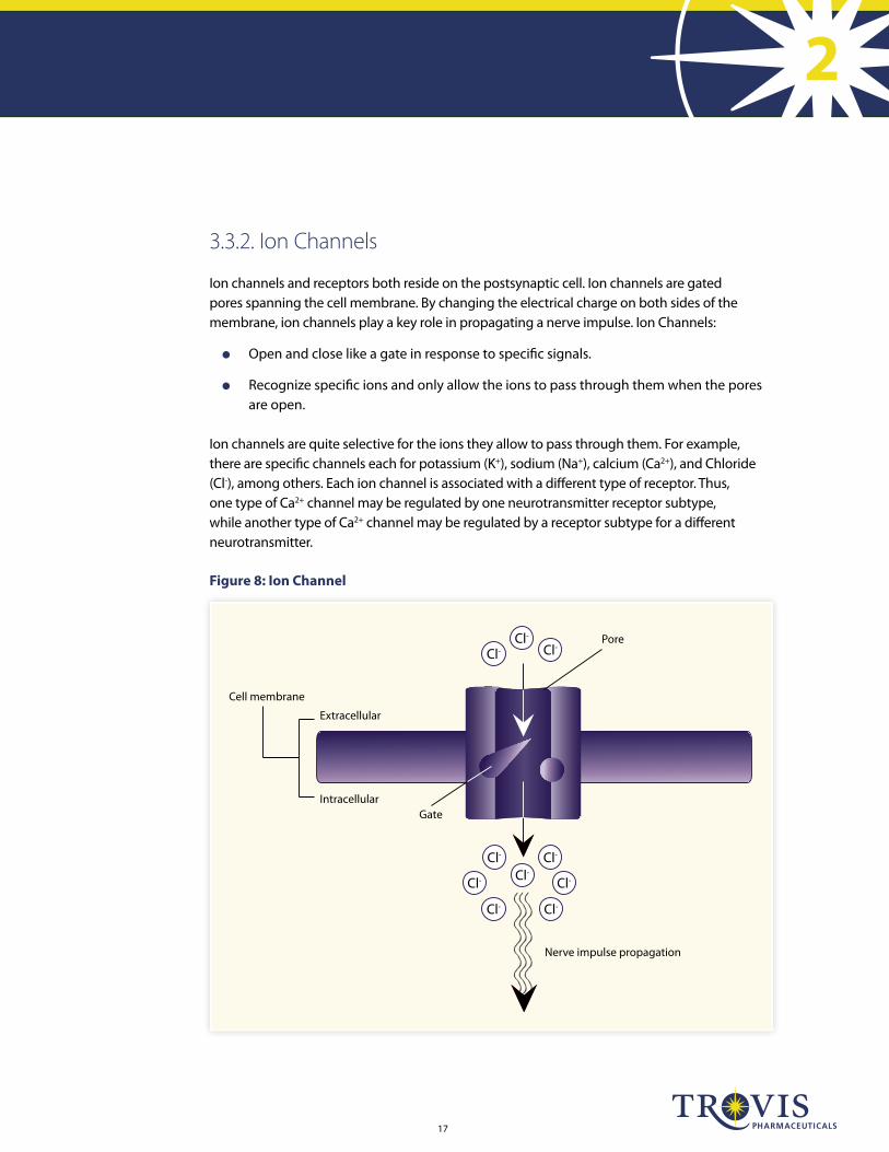
3.3.2. Ion Channels
Ion channels and receptors both reside on the postsynaptic cell. Ion channels are gated pores spanning the cell membrane. By changing the electrical charge on both sides of the membrane, ion channels play a key role in propagating a nerve impulse. Ion Channels:
Open and close like a gate in response to specific signals.
Recognize specific ions and only allow the ions to pass through them when the pores are open.
Ion channels are quite selective for the ions they allow to pass through them. For example, there are specific channels each for potassium (K+), sodium (Na+), calcium (Ca2+), and Chloride (Cl-), among others. Each ion channel is associated with a different type of receptor. Thus, one type of Ca2+ channel may be regulated by one neurotransmitter receptor subtype, while another type of Ca2+ channel may be regulated by a receptor subtype for a different neurotransmitter.
Figure 8: Ion Channel
Cell membrane
Extracellular
Intracellular
Pore
Gate
Nerve impulse propagation
Cl-Cl-
Cl-
Cl-Cl-
Cl-
Cl- Cl-
Cl- Cl-
18CONFIDENTIAL: For Internal Use Only.
Chapter 3: Neuroanatomy Related to MDD
3.3.3. Receptors
Receptors are complex proteins with special configurations that allow specific molecules, such as neurotransmitters or drugs, to bind with them. Understanding the interaction between neurotransmitter receptors and their biologic function upon drug binding is very important when discussing MDD and the various drug classes used to treat MDD. Most psychoactive are either receptor agonists or antagonists. These terms relate to their effect upon the receptor. This binding may modify the receptor’s behavior and leads to one of the following changes:
Receptor agonism – A drug or neurotransmitter binds to a specific cell receptor and triggers a response by the cell. The response triggered is often the cell’s natural biologic function.
Receptor antagonism – A drug binds to a specific cell receptor and blocks it from performing a specific biologic function.
Figure 7 in this module illustrates that neurotransmitter receptors are located on both the presynaptic and postsynaptic cell surfaces. Presynaptic receptors primarily act to regulate the release of neurotransmitters from that neuron. Postsynaptic receptors act to regulate the propagation or inhibition of the impulse in the postsynaptic cell.
Postsynaptic receptors act to regulate the propagation or inhibition of the impulse in the postsynaptic cell.

19
22
3.4. Biochemical Theory of Depression
While no single theory explains all MDD, the biochemical theory is widely accepted. The biochemical theory suggests that depression is the result of a neurochemical imbalance or a functional deficiency of one or more key neurotransmitters. While 5-HT is most commonly associated with MDD both norepinephrine and dopamine are also believed to influence depression.
The biochemical theory is based on the following observations:
Drugs that promote the augmentation of these key neurotransmitters, especially 5-HT, can alleviate depressive symptoms.
Drugs that impede the activity of these neurotransmitter systems (e.g., antihypertensives) can induce depression.
Autopsy data have shown low concentration of neurotransmitters in the brains of suicidal and/or depressed patients.
Concentrations of neurotransmitters also tend to be lower in the cerebrospinal fluid (CSF) and the peripheral blood of depressed patients.
The biochemical theory suggests that depression is the result of a neurochemical imbalance or a functional deficiency of one or more key neurotransmitters.

20CONFIDENTIAL: For Internal Use Only.

21
22Brain Booster – Prepare for your online quiz. Module 2 certification requires a score of ≥85%. These questions should help you prepare.
1. List the 5 theories of the cause of depression and the circle the one that is most accepted.
2. What is the name of the group of areas within the brain are responsible for our emotional health?
A. Cerebellum B. Cerebrum C. Limbic System D. Hypothalamus
3. What is a neurotransmitter carried within in the presynaptic neuron?
A. Lipoprotein B. Versicle C. Dopamine D. Receptor
4. List the neurotransmitters associated with depression.
5. Drugs have one of two effects on receptors, name their potential effects and describe each:
Brain Booster
22CONFIDENTIAL: For Internal Use Only.
6. What is the basic functional unit of the human nervous system?
A. Ion Channel B. Receptor C. Neuron D. None of these
7. The following process is described as an electrical charge being transmitted to the next neuron across synaptic cleft.
A. Antagonism B. Agonism C. Neurotransmission D. None of these
8. The diencephalon contains what two portions of the brain important to human emotion and memory creation?
A. Cerebellum and Hypothalamus
B. Brain Stem and Thalamus
C. Hypothalamus and Thalamus
D. None of these
Brain Booster

23
22Afferent Neuron: A neuron that carries impulse toward the CNS.
Autonomic Nervous System: Part of the nervous system that regulates activity of smooth muscle, cardiac muscle and glands.
Axon: A tubular extension that carries nerve impulses away from the cell body toward other neurons.
Brainstem: Stalklike portion of the brain connecting cerebral hemispheres with the spinal cord; controls involuntary process such as respiration.
Central Nervous System (CNS): One of the two main divisions of the nervous system, consisting of the brain and the spinal cord.
Cerebellum: The second largest region of the brain; functions to control skeletal muscles primarily in coordination and balance.
Cerebrum: Largest region of the brain; controls voluntary motor functions; coordinates physical, sensory, visual and auditory sensations; integrates consciousness, memory, use of language and emotions.
Cortex: The convoluted layer of gray matter covering each cerebral hemisphere.
Dendrite: Highly branched extensions that receive nerve signals from other neurons and conduct these signals to the cell body.
Diencephalon: Portion of the brain that contains the thalamus and the hypothalamus.
Dorsal Horn: Crescent-shaped projection of gray matter in spinal cord.
Efferent Neuron: Nerve fibers that carry impulses outward, away from the brain and spinal cord.
Fissures: The deep grooves between folds of brain tissue.
Gray Matter: Those regions of the brain and spinal cord that are made up primarily of the cell bodies and dendrites of nerve cells, rather than myelinated axons.
Gyri: Prominent rounded elevations of brain tissue that form the cerebral hemispheres.
Homeostasis: A state of equilibrium or a tendency to reach equilibrium, either metabolically within a cell or organism or socially and psychologically within an individual or group.
Hypothalamus: Brain region primarily involved in autonomic (involuntary) nervous system functions, hormone secretion, and mood; major regulatory of homeostasis.
Limbic System: A group of neuronal pathways that connect parts of the cerebrum, diencephalon and brainstem.
Midbrain: Also called mesencephalon; the short part of the brainstem just above the pons; the center for visual reflexes.
Medulla Oblongata: The base of the brain, which is formed by the enlarged top of the spinal cord; directly controls breathing, blood flow, and other essential functions.
Meninges: The 3 membranes enclosing the brain and the spinal cord, comprising of the dura matter, the pia matter and the arachnoid membrane.
Definitions

24CONFIDENTIAL: For Internal Use Only.
Myelin Sheath: A fatty substance that insulates the nerve fiber and helps to speed nerve impulse transmission.
Neurochemical: Pertaining to chemicals involved in nervous system function.
Neurotransmitter: Chemical substance that enables communication between nerve cells.
Noradrenergic: Using norepinephrine as a neurotransmitter, norepinephrine is also called noradrenaline.
Norepinephrine (NE): Neurotransmitter widely distributed throughout the nervous system; involved in regulating sleep, wakefulness, learning, memory and mood.
Nucleus: An organelle that is the location of the genetic material of the cell.
Organelles: A specialized part of a cell that has its own function .
Parasympathetic Nervous System: A branch of the autonomic nervous system; slows the heart rate, increases the intestinal and gland activity, and relaxes the sphincter muscle.
Pons: A prominence on the ventral (front) surface of the brainstem, between the medulla oblongata and the midbrain.
Presynaptic Neuron: Nerve cell that transmits the signal across the synaptic cleft by releasing neurotransmitters.
Postsynaptic Neuron: Nerve cell that receives the signal transmitted across the synaptic cleft.
Serotonergic: Using serotonin as a neurotransmitter.
Serotonin (5-HT): Neurotransmitter found in many regions of the brain and other parts of the body; involved in the regulation of mood, emotional behavior, sleep, memory and appetite, as well as modulation of the dopamine system.
Somatic Nervous System: Part of the peripheral nervous system that allows for interaction with the external environment; composed of afferent and efferent neurons.
Sulci: Grooves or furrows on the surface of the brain, bounding the several convolutions, or gyri.
Sympathetic Nervous System: Branch of the autonomic nervous system that accelerates the heart rate, constricts blood vessels, and raises blood pressure.
Synapse: The junction between an axon terminal and another neuron.
Synaptic Cleft: The space between the presynaptic and postsynaptic surfaces.
Thalamus: Brain region located above midbrain; relays sensory information to the cerebral cortex.
Transmission: The process of movement of nerve impulse along neural pathways in the body.
White Matter: Bundles of myelinated axons located in the brain and spinal cord.
Definitions

25
221. Fields H. Depression and pain: A neurobiological model. Neuropsychiatry Neuropsychol
Behav Neurol. 1991;4(1):83-92.
2. Bair MJ,Robinson RL,Katon W,Kroenke K. Depression and pain comorbidity. Arch Intern Med. 2003;163:2433-2445.
3. Blier P, Abbott FV. Putative mechanisms of action of antidepressant drugs in affective and anxiety disorders and pain. J Psychiatry Neurosci. 2001;26(1):37-43.
4. Nitti VW. Duloxetine: a new pharmacologic therapy for stress urinary incontinence. Rev Urol. 2004;6(suppl 3):S48-S55
References

Copyright © 2011 Trovis Pharmaceuticals. VIL-AMT-015 02/2011
2 CONFIDENTIALFor Internal Use Only. The information contained in this training module is for educational purposes only. It is designed to provide you with the information you need to be educated on the product, disease and competitive environment and is not to be distributed or used in detailing.