population genetic analysis and sub-structuring of … for use title population genetic analysis and...
TRANSCRIPT

Instructions for use
Title Population genetic analysis and sub-structuring of Theileria parva in the northern and eastern parts of Zambia
Author(s) Muleya, Walter; Namangala, Boniface; Simuunza, Martin; Nakao, Ryo; Inoue, Noboru; Kimura, Takashi; Ito, Kimihito;Sugimoto, Chihiro; Sawa, Hirofumi
Citation Parasites & Vectors, 5: 255
Issue Date 2012-11-12
Doc URL http://hdl.handle.net/2115/50994
Rights(URL) http://creativecommons.org/licenses/by/2.0/
Type article
File Information PV5_255.pdf
Hokkaido University Collection of Scholarly and Academic Papers : HUSCAP
RESEARCH Open Access
Population genetic analysis and sub-structuringof Theileria parva in the northern and easternparts of ZambiaWalter Muleya1,2†, Boniface Namangala3†, Martin Simuunza4†, Ryo Nakao5†, Noboru Inoue6†, Takashi Kimura1†,Kimihito Ito7,8†, Chihiro Sugimoto5,8† and Hirofumi Sawa1,8*
Abstract
Background: Theileriosis, caused by Theileria parva, is an economically important disease in Africa. It is a majorconstraint to the development of the livestock industry in some parts of eastern, central and southern Africa. InZambia, theileriosis causes losses of up to 10,000 cattle annually.
Methods: Cattle blood samples were collected for genetic analysis of Theileria parva from Isoka and Petaukedistricts in Zambia. Microsatellite analysis was then performed on all Theileria parva positive samples for PCR using apanel of 9 microsatellite markers. Microsatellite data was analyzed using microsatellite toolkit, GenAlEx ver. 6, Fstatver. 2.9.3.2, and LIAN computer softwares.
Results: The combined percentage of positive samples in both districts determined by PCR using the p104 geneprimers was 54.9% (95% CI: 46.7 – 63.1%, 78/142), while in each district, it was 44.8% (95% CI: 34.8 – 54.8%) and76.1% (95% CI = 63.9 – 88.4%) for Isoka and Petauke districts, respectively. We analyzed the population geneticstructure of Theileria parva from a total of 61 samples (33 from Isoka and 28 from Petauke) using a panel of 9microsatellite markers encompassing the 4 chromosomes of Theileria parva. Wright’s F index (FST = 0.178) showedsignificant differentiation between the Isoka and Petauke populations. Linkage disequilibrium was observed whenpopulations from both districts were treated as a single population. When analyzed separately, linkagedisequilibrium was observed in Kanyelele and Kalembe areas in Isoka district, Isoka district overall and in Petaukedistrict. Petauke district had a higher multiplicity of infection than Isoka district.
Conclusion: Population genetic analyses of Theileria parva from Isoka and Petauke districts showed a low level ofgenotype exchange between the districts, but a high level of genetic diversity within each district population,implying genetic and geographic sub-structuring between the districts. The sub-structuring observed, along withthe lack of panmixia in the populations, could have been due to low transmission levels at the time of sampling.However, the Isoka population was less diverse than the Petauke population.
Keywords: Theileria parva, Genetic diversity, Sub-structuring, Zambia
* Correspondence: [email protected]†Equal contributors1Division of Molecular Pathobiology, Research Center for Zoonosis Control,Hokkaido University, N20, W10, Kita-ku, Sapporo 001-0020, Japan8Global COE program, Research Center for Zoonosis Control, HokkaidoUniversity, N20, W10, Kita-ku, Sapporo 001-0020, JapanFull list of author information is available at the end of the article
© 2012 Muleya et al.; licensee BioMed Central Ltd. This is an Open Access article distributed under the terms of the CreativeCommons Attribution License (http://creativecommons.org/licenses/by/2.0), which permits unrestricted use, distribution, andreproduction in any medium, provided the original work is properly cited.
Muleya et al. Parasites & Vectors 2012, 5:255http://www.parasitesandvectors.com/content/5/1/255
BackgroundTheileriosis is an economically important disease of cat-tle in eastern, central and southern Africa. The disease,caused by the protozoan haemoparasite Theileria parva(T. parva), is transmitted by the 3-host tick Rhipicepha-lus appendiculatus [1]. The severity of the disease mani-fests differently in various breeds of cattle, with the zebucattle (Bos indicus) being more resistant than the exoticbreeds (Bos taurus) against both the parasite [2] and thevector [3,4]. Theileriosis causes high levels of mortalityin taurine breeds [5] and both high morbidity and mor-tality in indigenous 1–6 month old calves (Bos indicus).Full-scale epidemics affecting all age groups of indigen-ous breeds may occur [6], resulting in reduction in prod-uctivity. The high level of mortality in taurine breedsimpedes the introduction of highly productive exoticbreeds in endemic areas, further preventing the im-provement of cattle production in the affected areas.Theileriosis is thus a major constraint to the develop-ment of the livestock industry in the eastern, central andsouthern parts of Africa.The republic of Zambia, divided into 9 provinces
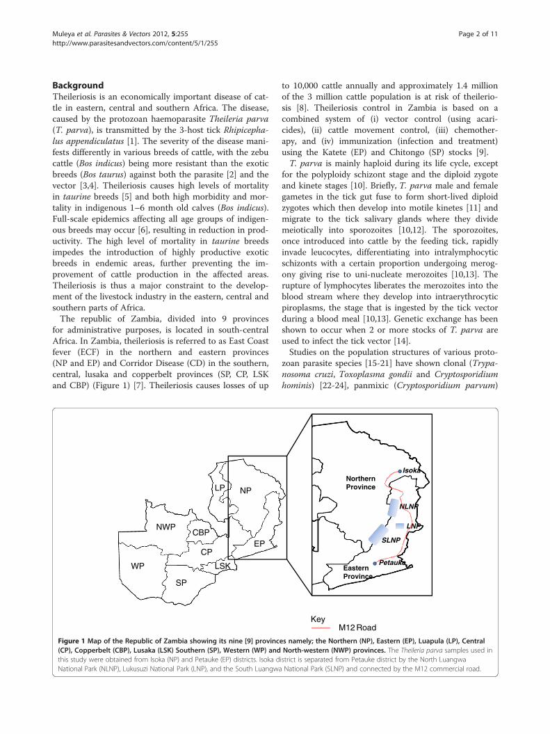
for administrative purposes, is located in south-centralAfrica. In Zambia, theileriosis is referred to as East Coastfever (ECF) in the northern and eastern provinces(NP and EP) and Corridor Disease (CD) in the southern,central, lusaka and copperbelt provinces (SP, CP, LSKand CBP) (Figure 1) [7]. Theileriosis causes losses of up
to 10,000 cattle annually and approximately 1.4 millionof the 3 million cattle population is at risk of theilerio-sis [8]. Theileriosis control in Zambia is based on acombined system of (i) vector control (using acari-cides), (ii) cattle movement control, (iii) chemother-apy, and (iv) immunization (infection and treatment)using the Katete (EP) and Chitongo (SP) stocks [9].T. parva is mainly haploid during its life cycle, except
for the polyploidy schizont stage and the diploid zygoteand kinete stages [10]. Briefly, T. parva male and femalegametes in the tick gut fuse to form short-lived diploidzygotes which then develop into motile kinetes [11] andmigrate to the tick salivary glands where they dividemeiotically into sporozoites [10,12]. The sporozoites,once introduced into cattle by the feeding tick, rapidlyinvade leucocytes, differentiating into intralymphocyticschizonts with a certain proportion undergoing merog-ony giving rise to uni-nucleate merozoites [10,13]. Therupture of lymphocytes liberates the merozoites into theblood stream where they develop into intraerythrocyticpiroplasms, the stage that is ingested by the tick vectorduring a blood meal [10,13]. Genetic exchange has beenshown to occur when 2 or more stocks of T. parva areused to infect the tick vector [14].Studies on the population structures of various proto-
zoan parasite species [15-21] have shown clonal (Trypa-nosoma cruzi, Toxoplasma gondii and Cryptosporidiumhominis) [22-24], panmixic (Cryptosporidium parvum)
EasternProvince
NorthernProvince
Petauke
Isoka
NLNP
SLNP
LNPNWP
WP
SP
CP
CBPEP
NPLP
LSK
KeyM12 Road
Figure 1 Map of the Republic of Zambia showing its nine [9] provinces namely; the Northern (NP), Eastern (EP), Luapula (LP), Central(CP), Copperbelt (CBP), Lusaka (LSK) Southern (SP), Western (WP) and North-western (NWP) provinces. The Theileria parva samples used inthis study were obtained from Isoka (NP) and Petauke (EP) districts. Isoka district is separated from Petauke district by the North LuangwaNational Park (NLNP), Lukusuzi National Park (LNP), and the South Luangwa National Park (SLNP) and connected by the M12 commercial road.
Muleya et al. Parasites & Vectors 2012, 5:255 Page 2 of 11http://www.parasitesandvectors.com/content/5/1/255
[24] and epidemic (Crytosporiduim parvum) [25] popu-lation structures. On the other hand, Trypanosomabrucei and Plasmodium falciparum population struc-tures have shown dependence on host specificity andtransmission intensity [26,27]. In general, the conclusionfrom these studies is that despite the presence of anobligatory sexual phase in their life cycle, a number offactors determine the population structures of differentprotozoan parasitic species.Micro- and mini-satellite markers have been used to
genotype several species of apicomplexan parasites andtheir vectors, revealing different population structures[18,21,26-30]. For example, a study on the populationstructure of T. parva in Uganda reported a mixture ofgenotypes in many isolates and linkage disequilibrium(LD) in 3 populations isolated from different areas [18].When multiple isolates with identical genotypes weretreated as a single isolate, the presence of an epidemicstructure was seen in 2 of the populations, suggestingan intermediate between extreme clonality and pan-mixia [18].A crucial and important aspect in population genetic
studies is to determine the effect of random and non-random mating on the population structures of disease-causing agents and consequently on the epidemiology ofthe diseases [31]. Information on the genetic exchangein T. parva populations has practical implications in dis-ease control and prevention. For instance, populationswith a high degree of genetic diversity arise when highlevels of recombination occur. This information is veryimportant in vaccine development as it is easier to de-velop a vaccine against a clonal pathogen than a highlydiverse pathogen. A panel of 9 polymorphic micro-satellite markers was used to genotype T. parva positivecattle blood DNA in order to answer the following ques-tions: (i) what is the population structure of the T. parvain eastern and northern Zambia? (ii) Does gene flowoccur between T. parva populations sampled from eacharea? (iii) Do the T. parva populations from the sam-pling areas consist of a single or multiple distinct popu-lations? To our knowledge, this is the first study on thepopulation genetics of T. parva in Zambia.
MethodsSample collection and DNA preparationThis study received ethical clearance for collection ofanimal blood from the Biomedical Research Ethics Com-mittee, University of Zambia and from the Departmentof Veterinary and Livestock Development, Eastern Prov-ince, Zambia. The recombinant DNA experiments wereapproved by Hokkaido University.About 10 mL of whole blood samples (n=142) were
collected in heparinized tubes from indigenous andmixed breeds of cattle from Kanyelele (n = 62) and
Kalembe (n=34) areas in Isoka district (n=96) of thenorthern province (NP) and from Saukani area inPetauke district (n=46) of the eastern province (EP)(Figure 1) of Zambia in May 2008, after the wet season.Kanyelele is located approximately 20 km from Kalembe.Isoka and Petauke districts are approximately 600 kmapart (straight line distance) and are separated by theNorth Luangwa, South Luangwa and Lukusuzi NationalParks (NLNP, SLNP and LNP, respectively, in Figure 1).Petauke and Isoka districts are connected by a commer-cial road (Figure 1) covering a distance of about 795 km.Whole genome DNA was extracted using DNAzol (Mo-lecular Research Center, Cincinnati, OH) following themanufacturer’s instructions and stored at −20°C. A panelof 16 polymorphic microsatellite markers [32-34], repre-senting the 4 chromosomes, was initially selected(Table 1) for genotyping of the samples. The followingT. parva parasite laboratory stocks were used for theinitial marker screening to determine which markersamplified more isolates and were sufficiently polymorphicfor use in the genotyping of field samples from Isoka andPetauke districts: Onderstepoort (South Africa, year ofisolation unknown), Serengeti (Tanzania, 1978), Muguga(Kenya, year of isolation unknown), Nyakizu (Rwanda,1979), Entebbe (Uganda, 1980), Katumba (Burundi, 1981),Kiambu Z464/C12 and B8 (Kenya, 1972), Katete B2(Zambia, 1989) and Buffalo Z5E5 (both origin and yearof isolation unknown). These stocks were also used asGenescan control samples during the analysis of fieldsamples.
T. parva screeningWhole blood DNA field samples were screened forT. parva DNA by using T. parva-specific p104 geneprimers [35]. Template DNA (1μL) was amplified in a20 μL reaction mixture as prescribed by the manufac-turer using ExTaq polymerase (Takara, Tokyo, Japan).The PCR conditions were as follows: denaturation at95°C for 5 minutes; 35 cycles at 95°C for 1 minute,63°C for 30 seconds and 72°C for 1 minute, followed bya final extension step of 5 minutes at 72°C. The ampli-fied products were analyzed on 2% ethidium bromidepre-stained agarose gel.
PCR amplification and analysis of microsatellite lociPCR was performed using primers (Table 1) designedto amplify each of the 16 repeat regions on each of theT. parva stocks isolated from different geographicalareas. The forward primer in each primer marker set wasfluorescently labeled. The 20 μL PCR mixture used com-prised of 10 ng of template DNA, 10 μL of 2x AmplitaqGold master mix (Invitrogen, CA), 0.25 μM of eachprimer and distilled water. The PCR conditions were asfollows: denaturation at 95°C for 5 minutes; 35 cycles
Muleya et al. Parasites & Vectors 2012, 5:255 Page 3 of 11http://www.parasitesandvectors.com/content/5/1/255
at 95°C for 15 seconds, 50 - 58°C for 60 seconds and72°C for 1 minute, followed by a final extension step of5 minutes at 72°C. The amplified products were observedon 1.5% ethidium bromide pre-stained agarose gels to de-termine the success of PCR amplification. To achieve highgenotyping resolution of field samples, microsatellite PCRproducts were denatured and then capillary electrophor-esed in an ABI 3130 genetic analyzer (Applied Biosystems,CA). DNA fragment sizes were analyzed relative to theROX-labeled GS600 LIZ size standard (Applied Biosys-tems) using Gene Mapper software (Applied Biosystems).This facilitated the resolution of multiple products with1 base pair (bp) difference in a single reaction. Multipleproducts from a single PCR reaction indicated the pres-ence of mixed genotypes. The output data from the gen-etic analyzer were provided as the area under the peakof each allele (quantitative measurement), with the pre-dominant allele possessing the greatest peak area. In thisway, the predominant allele at each locus was identifiedfor each sample, and this data was combined to gen-erate a multi-locus genotype (MLG) representing themost abundant genotype in each sample. Only the alleleswith the prescribed base pair range were used to generatethe MLG and samples from the same area were electro-phoresed and genescaned on the same plate. SeparatePCRs were also carried out for different regions.
Data analysisAn allele sharing co-efficient [36] in Excel microsatellitetoolkit (http://animalgenomics.ucd.i.e./sdepark/ms-toolkit/)was used for the similarity comparison of the MLGs
[37]. Principal component analysis (PCA) was used forsimilarity analysis. A similarity matrix was constructedand used to construct PCA using the Excel plug-in soft-ware GenAIEx6 (http://www.anu.edu.au/BoZo/GenAIEx/)[38]. FSTAT computer package version 2.9.3.2 was used tocalculate estimates of F statistics for population geneticanalysis (http://www2.unil.ch/popgen/softwares/fstat.htm).LIAN (http://adenine.biz.fh-weihenstephan.de/lian/) wasused to test the null hypothesis of linkage equilibriumby calculating a quantification of linkage equilibrium/linkage disequilibrium called the standardized index ofassociation (IA
S ) [39]. The statistical independence ofalleles at all pairwise combinations of loci under studycharacterizes linkage equilibrium (LE) and this inde-pendent assortment was initially tested by LIAN by de-termining the number of loci at which each pair of MLGsdiffers. The mis-match values from this distribution werethen used to calculate the variance (VD) which was thencompared to the variance expected (VE) for LE. MonteCarlo (MC) computer simulation was used to test thenull hypothesis that VD = VE. The computer software cal-culates a 95% confidence limit L (LMC). When VD isgreater than L, the null hypothesis of LE is discarded.
Multiplicity of infectionThe majority of the samples in this study possessed sev-eral alleles at one or more loci, representing a mixed in-fection. The mean number of alleles for the nine loci ineach sample was calculated and this index value repre-sented the multiplicity of infection within each sample.The overall mean for the index values for each sample
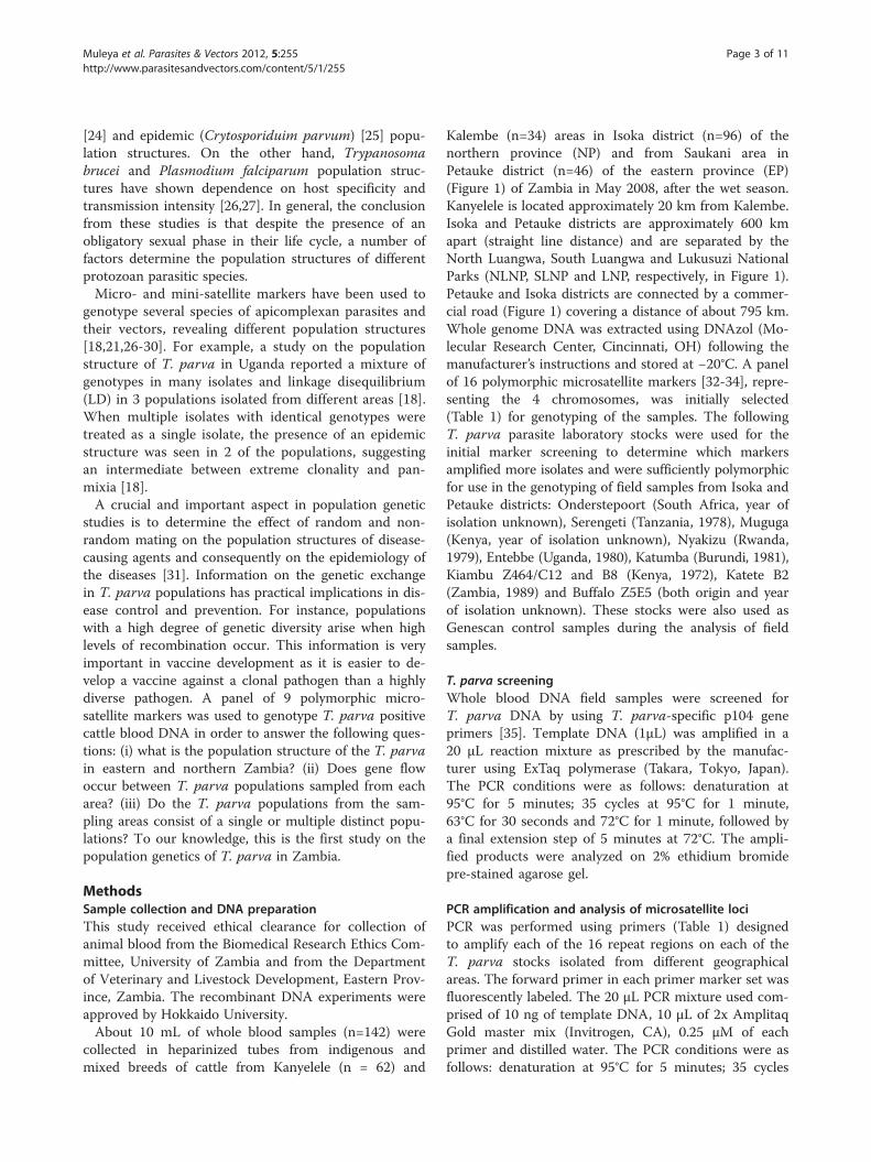
Table 1 Panel of microsatellite markers used to genotype Theileria parva samples from Isoka and Petauke districts
Marker Chromosome Amplicon size (bp) Used in final analysis Reference
MS1 1 235-368 No Oura et al., 2003 [32]
MS62 1 271 No Katzer et al., 2010 [34]
MS48 1 223 Yes Katzer et al., 2006 [33]
MS5 1 206-444 No Oura et al., 2003 [32]
MS66 1 266 No Katzer et al., 2010 [34]
MS67 1 245 No Katzer et al., 2010 [34]
MS77 2 270 No Katzer et al., 2010 [34]
MS71 2 252 Yes Katzer et al., 2010 [34]
MS74 2 246 Yes Katzer et al., 2010 [34]
MS75 2 244 Yes Katzer et al., 2010 [34]
MS72 2 230 Yes Katzer et al., 2010 [34]
MS51 3 161 Yes Katzer et al., 2006 [33]
MS53 3 208 Yes Katzer et al., 2006 [33]
MS57 4 118 Yes Katzer et al., 2006 [33]
MS58 4 300 No Katzer et al., 2006 [33]
MS59 4 111 Yes Katzer et al., 2006[33]
Muleya et al. Parasites & Vectors 2012, 5:255 Page 4 of 11http://www.parasitesandvectors.com/content/5/1/255
was then calculated to provide the multiplicity of infec-tion for each region.
ResultsPCR screeningThe screening of cattle blood samples (n=142) using thep104 gene primers showed a combined T. parva positivepercentage of 54.9% (95% CI: 46.7–63.1%, 78/142) fromboth districts. The percentage of positive samples withineach district was 44.8% (95% CI: 34.8–54.8%) (23 fromKanyelele and 20 from Kalembe, 43/96) and 76.1% (95%CI: 63.9–88.4%, 35/46) for Isoka and Petauke (Saukaniarea), respectively.
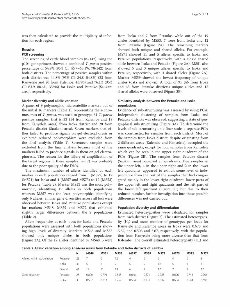
Marker diversity and allelic variationA panel of 9 polymorphic microsatellite markers out ofthe initial 16 markers (Table 1), representing the 4 chro-mosomes of T. parva, was used to genotype 61 T. parvapositive samples, that is 33 (14 from Kalembe and 19from Kanyelele areas) from Isoka district and 28 fromPetauke district (Saukani area). Seven markers that ei-ther failed to produce signals on gel electrophoresis orexhibited reduced polymorphism were excluded fromthe final analysis (Table 1). Seventeen samples wereexcluded from the final analysis because most of themarkers failed to produce signals in them on gel electro-phoresis. The reason for the failure of amplification ofthe target regions in these samples (n=17) was probablydue to the poor quality of the DNA.The maximum number of alleles identified by each
marker in each population ranged from 3 (MS72) to 12(MS71) for Isoka and 4 (MS57 and MS75) to 12 (MS53)for Petauke (Table 2). Marker MS53 was the most poly-morphic, identifying 19 alleles in both populationswhereas MS57 was the least polymorphic, identifyingonly 6 alleles. Similar gene diversities across all loci wereobserved between Isoka and Petauke populations exceptfor markers MS48, MS59 and MS72 that exhibitedslightly larger differences between the 2 populations(Table 2).Allele frequencies at each locus for Isoka and Petauke
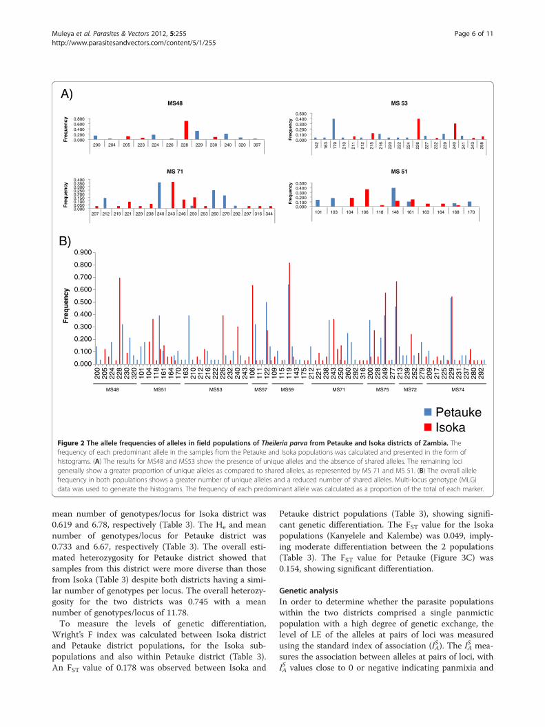
populations were assessed with both populations show-ing high levels of diversity. Markers MS48 and MS53showed only unique alleles in both populations(Figure 2A). Of the 12 alleles identified by MS48, 5 were
from Isoka and 7 from Petauke, while out of the 19alleles identified by MS53, 7 were from Isoka and 12from Petauke (Figure 2A). The remaining markersshowed both unique and shared alleles. For example,MS71 showed 11 and 5 alleles specific to Isoka andPetauke populations, respectively, with a single sharedallele between Isoka and Petauke (Figure 2A). MS51 alsoshowed 5 and 3 unique alleles specific to Isoka andPetauke, respectively, with 3 shared alleles (Figure 2A).Marker MS59 showed the lowest frequency of uniquealleles (data not shown). A total of 91 (46 from Isokaand 45 from Petauke districts) unique alleles and 15shared alleles were observed (Figure 2B).
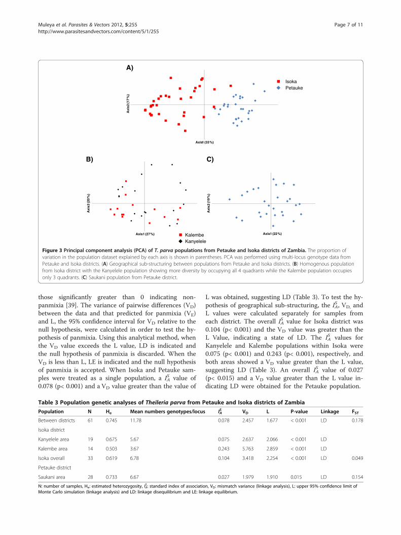
Similarity analysis between the Petauke and IsokapopulationsEvidence of sub-structuring was assessed by using PCA.Independent clustering of samples from Isoka andPetauke districts was observed, suggesting a state of geo-graphical sub-structuring (Figure 3A). To determine thelevels of sub-structuring on a finer scale, a separate PCAwas constructed for samples from each district. Most ofthe samples from Isoka district, despite originating from2 different areas (Kalembe and Kanyelele), occupied thesame quadrants, except for four samples from Kanyelelewhich can be seen in the upper right quadrant of thePCA (Figure 3B). The samples from Petauke district(Saukani area) occupied all quadrants. Five samples inthe upper left, 4 in the upper right and 2 in the lowerleft quadrants, appeared to exhibit some level of inde-pendence from the rest of the samples that had congre-gated mainly in the lower right quadrant, lower parts ofthe upper left and right quadrants and the left part ofthe lower left quadrant (Figure 3C) but due to theirreduced number, further investigation into these possibledifferences was not carried out.
Population diversity and differentiationEstimated heterozygosities were calculated for samplesfrom each district (Figure 3). The estimated heterozygos-ity (He) and mean number of genotypes per locus forKanyelele and Kalembe areas in Isoka were 0.675 and5.67, and 0.503 and 3.67, respectively, with the popula-tion from Kanyelele being more diverse than that fromKalembe. The overall estimated heterozygosity (He) and
Table 2 Allelic variation among Theileria parva from Petauke and Isoka districts of Zambia
N MS48 MS51 MS53 MS57 MS59 MS71 MS75 MS72 MS74
Alleles within population Petauke 28 7 6 12 4 6 6 4 6 9
Isoka 33 5 8 7 5 5 12 5 3 11
Overall 61 12 11 19 6 9 17 7 8 17
Gene diversity Petauke 28 0.820 0.794 0.833 0.648 0.571 0.783 0.696 0.743 0.706
Isoka 33 0.502 0.813 0.752 0.534 0.331 0.837 0.604 0.504 0.695
Muleya et al. Parasites & Vectors 2012, 5:255 Page 5 of 11http://www.parasitesandvectors.com/content/5/1/255
mean number of genotypes/locus for Isoka district was0.619 and 6.78, respectively (Table 3). The He and meannumber of genotypes/locus for Petauke district was0.733 and 6.67, respectively (Table 3). The overall esti-mated heterozygosity for Petauke district showed thatsamples from this district were more diverse than thosefrom Isoka (Table 3) despite both districts having a simi-lar number of genotypes per locus. The overall heterozy-gosity for the two districts was 0.745 with a meannumber of genotypes/locus of 11.78.To measure the levels of genetic differentiation,
Wright’s F index was calculated between Isoka districtand Petauke district populations, for the Isoka sub-populations and also within Petauke district (Table 3).An FST value of 0.178 was observed between Isoka and
Petauke district populations (Table 3), showing signifi-cant genetic differentiation. The FST value for the Isokapopulations (Kanyelele and Kalembe) was 0.049, imply-ing moderate differentiation between the 2 populations(Table 3). The FST value for Petauke (Figure 3C) was0.154, showing significant differentiation.
Genetic analysisIn order to determine whether the parasite populationswithin the two districts comprised a single panmicticpopulation with a high degree of genetic exchange, thelevel of LE of the alleles at pairs of loci was measuredusing the standard index of association (IA
S ). The IAS mea-
sures the association between alleles at pairs of loci, withIAS values close to 0 or negative indicating panmixia and
0.0000.2000.4000.6000.800
Fre
qu
ency
MS48
0.0000.1000.2000.3000.4000.500
Fre
qu
ency
MS 51
0.0000.1000.2000.3000.4000.500
142
163
179
210
211
212
215
216
220
222
224
226
227
232
239
240
241
243
298
Fre
qu
ency
MS 53
0.0000.0500.1000.1500.2000.2500.3000.3500.400
200 204 205 223 224 226 228 229 230 240 320 397
101 103 104 106 118 148 161 163 164 168 170207 212 219 221 229 238 240 243 246 250 253 260 279 292 297 316 344
Fre
qu
ency
MS 71
A)
B)
PetaukeIsoka
0.000
0.100
0.200
0.300
0.400
0.500
0.600
0.700
0.800
0.900
200
205
224
228
230
320
101
104
118
161
164
170
163
210
212
216
222
226
232
240
243
106
111
122
109
115
119
143
175
212
221
238
243
250
260
292
316
200
228
249
277
213
239
252
279
209
217
225
229
231
237
280
292
Fre
qu
ency
75SM35SM84SM 95SM15SM MS72MS75 47SM17SM
Figure 2 The allele frequencies of alleles in field populations of Theileria parva from Petauke and Isoka districts of Zambia. Thefrequency of each predominant allele in the samples from the Petauke and Isoka populations was calculated and presented in the form ofhistograms. (A) The results for MS48 and MS53 show the presence of unique alleles and the absence of shared alleles. The remaining locigenerally show a greater proportion of unique alleles as compared to shared alleles, as represented by MS 71 and MS 51. (B) The overall allelefrequency in both populations shows a greater number of unique alleles and a reduced number of shared alleles. Multi-locus genotype (MLG)data was used to generate the histograms. The frequency of each predominant allele was calculated as a proportion of the total of each marker.
Muleya et al. Parasites & Vectors 2012, 5:255 Page 6 of 11http://www.parasitesandvectors.com/content/5/1/255
those significantly greater than 0 indicating non-panmixia [39]. The variance of pairwise differences (VD)between the data and that predicted for panmixia (VE)and L, the 95% confidence interval for VD relative to thenull hypothesis, were calculated in order to test the hy-pothesis of panmixia. Using this analytical method, whenthe VD value exceeds the L value, LD is indicated andthe null hypothesis of panmixia is discarded. When theVD is less than L, LE is indicated and the null hypothesisof panmixia is accepted. When Isoka and Petauke sam-ples were treated as a single population, a IA
S value of0.078 (p< 0.001) and a VD value greater than the value of
L was obtained, suggesting LD (Table 3). To test the hy-pothesis of geographical sub-structuring, the IA
S , VD, andL values were calculated separately for samples fromeach district. The overall IA
S value for Isoka district was0.104 (p< 0.001) and the VD value was greater than theL Value, indicating a state of LD. The IA
S values forKanyelele and Kalembe populations within Isoka were0.075 (p< 0.001) and 0.243 (p< 0.001), respectively, andboth areas showed a VD value greater than the L value,suggesting LD (Table 3). An overall IA
S value of 0.027(p< 0.015) and a VD value greater than the L value in-dicating LD were obtained for the Petauke population.
Axi
s2(1
7%
)
Axis1 (33%)
Axi
s.2
(19
%)
Axis1 (22%)
Axi
s2
(20
%)
Axis1 (27%)
IsokaPetauke
A)
B) C)
KalembeKanyelele
Figure 3 Principal component analysis (PCA) of T. parva populations from Petauke and Isoka districts of Zambia. The proportion ofvariation in the population dataset explained by each axis is shown in parentheses. PCA was performed using multi-locus genotype data fromPetauke and Isoka districts. (A) Geographical sub-structuring between populations from Petauke and Isoka districts. (B) Homogenous populationfrom Isoka district with the Kanyelele population showing more diversity by occupying all 4 quadrants while the Kalembe population occupiesonly 3 quadrants. (C) Saukani population from Petauke district.
Table 3 Population genetic analyses of Theileria parva from Petauke and Isoka districts of Zambia
Population N He Mean numbers genotypes/locus IAS VD L P-value Linkage FST
Between districts 61 0.745 11.78 0.078 2.457 1.677 < 0.001 LD 0.178
Isoka district
Kanyelele area 19 0.675 5.67 0.075 2.637 2.066 < 0.001 LD
Kalembe area 14 0.503 3.67 0.243 5.763 2.859 < 0.001 LD
Isoka overall 33 0.619 6.78 0.104 3.418 2.254 < 0.001 LD 0.049
Petauke district
Saukani area 28 0.733 6.67 0.027 1.979 1.910 0.015 LD 0.154
N: number of samples, He: estimated heterozygosity, IAS : standard index of association, VD: mismatch variance (linkage analysis), L: upper 95% confidence limit of
Monte Carlo simulation (linkage analysis) and LD: linkage disequilibrium and LE: linkage equilibrium.
Muleya et al. Parasites & Vectors 2012, 5:255 Page 7 of 11http://www.parasitesandvectors.com/content/5/1/255
Multiplicity of infectionThe multiplicity of infection for Isoka district was 2.16while that of Petauke district was 2.44. Kanyelele andKalembe areas of Isoka district showed multiplicity ofinfection of 2.22 and 2.06, respectively (Table 4).
DiscussionIn order to establish effective control measures as wellas to assess the effectiveness of the current controlmeasures, information on the population structure ofT. parva with regard to its epidemiology is important.In this study, we analyzed samples from Kanyelele andKalembe areas in Isoka district (NP) and from Saukaniarea in Petauke district (EP) of Zambia, which are ap-proximately 600 km (straight line distance) apart and areseparated by the North Luangwa, South Luangwa andLukusuzi National Parks (Figure 1). To achieve this, weemployed micro-satellite analysis, an effective way ofstudying population structures of a wide range of species.Micro-satellite analysis enables direct genotyping ofparasite isolates directly from host blood samples byusing specific primers. Recently, a panel of polymorphicmicro- and mini-satellite markers for T. parva was iden-tified [32-34]. A panel of 16 of these microsatellite mar-kers was initially chosen and only 9 were used in the finalanalysis (Table 1). All the markers (locus pairs) on thesame chromosome were over 182 kbp apart. It is there-fore unlikely that any of the markers could have beenphysically linked. To perform the population geneticstudy of T. parva, a haploid organism, an MLG was con-structed for each sample, by assigning a single predomin-ant allele for each marker at each locus for each sample.This method of selecting the most predominant allele ateach locus from mixed infections is currently fairlystandard although it has limitations [18,21,25]. There areseveral potential short comings of selecting predominantalleles from mixed populations to form an MLG: (i) eachparticular marker does not always amplify the predomin-ant allele from all strains (ii) the same strain is not alwaysamplified by a particular marker in all the samples,(iii) target regions in different strains that have the high-est sequence homology with markers are easily amplified
whether they are representative of the predominantstrain or not and (iv) all markers in the selected panel ofmarkers most likely do not amplify the same strain indifferent samples. To avoid these short falls in mixedinfections would involve cloning out samples or to onlychose samples with only a single allele and then proceedwith analysis and generation of an MLG. In this studyhowever, we used a quantitative method to generate anMLG and by so doing, we assume that this MLG pre-sented the closest representative of the dominant strainsacross the whole sample size.Allele frequencies at each locus from the Isoka and
Petauke populations showed a high number of uniquealleles in each population (Figure 2). The data obtainedusing markers MS48 and MS53 indicated the possibility ofgenetic differences between Isoka and Petauke populationsas evidenced by the complete lack of shared alleles at these2 sites (Figure 2A). The separation of alleles on loci MS48and MS53 might also be due to artifacts and as such thedata on these loci should be treated with caution; howeveranalysis of the overall data with and without loci MS48 andMS53 produced similar results (data not shown). Theincreased number of unique alleles versus shared allelesobserved when Isoka and Petauke populations were treatedas a single population (Figure 2B) suggests a state of geneticand geographical sub-structuring. Genetic and geographicalsub-structuring was also observed on PCA where samplesfrom Isoka and Petauke districts clustered separately(Figure 3A). A few samples (n=3) from Isoka district occu-pied the same quadrants as those from Petauke(Figure 3A). This state of genetic and geographical sub-structuring indicated by allele frequencies (Figure 2C) andPCA (Figure 3A) was further confirmed by the significantdifferentiation (FST = 0.178) observed between the popula-tion from Isoka and Petauke districts. Furthermore, a stateof LD with a IA
S value greater than 0 was also observedwhen the Isoka and Petauke populations were treated as asingle population (Table 3), indicating the absence of ran-dom mating between T. parva from the 2 populations andconsolidating the state of genetic and geographical sub-structuring. The population from Isoka exhibited less diver-sity than that from Petauke. These observations therefore,
Table 4 Multiplicity of infection in Petauke and Isoka districts
N Mean SD Minimum Maximum
Petauke district
Petauke overall 28 2.44 0.44 1.22 3.77
Isoka district
Kanyelele 19 2.22 0.26 1.77 3.00
Kalembe 14 2.06 0.15 1.88 2.33
Isoka overall 33 2.16 0.23 1.77 3.00
Petauke and Isoka districts (overall) 61 2.28 0.36 1.22 3.77
SD: Standard deviation.
Muleya et al. Parasites & Vectors 2012, 5:255 Page 8 of 11http://www.parasitesandvectors.com/content/5/1/255
suggests that the parasite populations from these 2 areas,while comprising a similar number of genotypes per locus,exist as separate populations because there is little or nomovement of animals between these 2 districts, which inturn is caused by their separation by physical geographicalbarriers i.e. the national parks (Figure 1). These findings arein agreement with a study on a T. parva populationwherein geographical sub-structuring was observed be-tween the population from 2 areas separated by a distanceof 300 km and a lake [18].The population from Kanyelele occupied all four quad-
rants while that from Kalembe only occupied three quad-rants (Figure 3B). Although the Kanyelele population wasmore diverse than the Kalembe population in Isoka dis-trict, only moderate genetic differentiation (FST = 0.049)was observed between the 2 populations (Table 3). Thiswas indicative of the existence of similar genotypes ofT. parva in both areas. However, the ability of the para-sites to randomly mate was restricted as shown from thestate of LD, both at the district level and within each sub-population (Table 3). In the Petauke population, signifi-cant genetic differentiation (FST = 0.154) and a state of LDwas also observed. This state of LD in Isoka (overall andin sub-populations) and Petauke might be due to the lackof mixing or movement of animals resulting in a lack ofgene flow between the populations that can likely beattributed to the relative isolation of the sample areas fromeach other and the resulting restricted circulation of geno-types. Despite Kanyelele and Kalembe being far apart, ani-mals from the two areas tend to share grazing groundsduring the dry period because of the scarcity of pasturesand thus it can be hypothesized that the sharing of grazinggrounds allows the introduction of genotypes indigenousto one area into another area. However, due to the avail-ability and close proximity of moderate vegetation for ani-mal grazing during the month of May and the precedingmonths, it is unlikely that animals from Kanyelele andKalembe areas would have mixed or shared grazinggrounds as they will tend to graze pastures closer to theirvillages. This lack of mixing of animals would have pre-vented the introduction of genotypes from one area intoanother and vice-versa, hence the state of restrictive circu-lation. The tick vector population is also low at this par-ticular time of the year, thus reducing the challenge levelsof infection (i.e. low levels of transmission). It can furtherbe hypothesized that the low challenge levels of infectionis also likely to give rise to the presence of fewer genotypesof T. parva which may result in the state of LD and non-panmixia observed in the majority of areas/regions. This isin agreement with a report in which Plasmodium falcip-arum populations showed strong LD, extensive populationdifferentiation and low diversity in regions of low trans-mission [26]. The state of LD in these populations mightalso be due to the presence of epidemic strains [17]. A
large sample size, coupled with tick vector ecology data ofa wider geographical area in Petauke and Isoka districts, isrequired to completely validate the LD states observed inthis study.The majority of samples analyzed in this study com-
prised mixed infections, with Petauke district showing ahigher multiplicity of infection than Isoka district(Table 4). Within Isoka district, Kanyelele area alsoshowed a higher multiplicity of infection than Kalembearea (Table 4). Several reasons may be advanced for thissituation including (i) transmission intensity reflected bythe tick burden on cattle, (ii) the level of infection of theparasite in the ticks, (iii) cattle host factors such as ageand breed, (iv) farming systems and (v) the geography ofthe respective regions. However, this data was inconclu-sive and as such the effect of these factors could not befurther investigated.
ConclusionThe results of this study indicate that the T. parva popu-lation from Isoka district is distinctively different fromthat of Petauke district, with significant gene diversity ineach population. There was no evidence of genetic ex-change among the populations from the 2 districts. Add-itionally, these results show that the populations fromKanyelele and Kalembe areas in Isoka district comprisedsimilar genotypes although panmixia of parasites be-tween the 2 areas could not be demonstrated. The sub-structuring and population structure of the T. parvapopulations observed in each district could be due tothe restrictive circulation of parasites as a result of thelack of mixing of animals from different regions and/orthe low transmission levels of T. parva in the respectiveareas within each district, which may be due to thereduced population of tick vectors at the time ofsampling.
Competing interestsThe authors declare no competing interest.
Authors’ contributionsWM performed the molecular genetic analyses, data analysis, and statisticalanalysis and drafted the manuscript. BN was involved in field collection andhelped to draft the manuscript. RN purified DNA from the parasite referencestocks and helped in editing the manuscript. MS participated in analyzingdata and drafting the manuscript. TK and KI participated in drafting themanuscript. NI helped in collecting the samples and editing the manuscript.CS and HS helped to conceive the study, participate in its design andassisted in obtaining funding. All the authors read and approved the finalmanuscript.
AcknowledgementsWe thank Ms. Yamanouchi and Ms. Ohnuma, Research Center for ZoonosisControl, Hokkaido University, for technical assistance. This study wassupported in part by grants from the Ministry of Education, Culture, Sports,Science and Technology (MEXT); the Ministry of Health, Labor and Welfare,Japan; the Japan Initiative for Global Research Network on Infectious
Muleya et al. Parasites & Vectors 2012, 5:255 Page 9 of 11http://www.parasitesandvectors.com/content/5/1/255
Diseases (J-GRID), MEXT Japan. We also wish to thank the farmers and fieldveterinary officers in Isoka and Petauke including the Eastern provinceveterinary officer for their cooperation.
Author details1Division of Molecular Pathobiology, Research Center for Zoonosis Control,Hokkaido University, N20, W10, Kita-ku, Sapporo 001-0020, Japan.2Department of Biomedical Sciences, School of Veterinary Medicine,University of Zambia, P.O Box 32379, Lusaka 10101, Zambia. 3Department ofPara-clinical Studies, School of Veterinary Medicine, University of Zambia, P.OBox 32379, Lusaka 10101, Zambia. 4Department of Disease Control, School ofVeterinary Medicine, University of Zambia, P.O Box 32379, Lusaka 10101,Zambia. 5Division of Collaboration and Education, Research Center forZoonosis Control, Hokkaido University, N20, W10, Kita-ku, Sapporo 001-0020,Japan. 6National Research Center for Protozoan Diseases, Obihiro Universityof Agriculture and Veterinary Medicine, Inada-cho, Hokkaido 080-8555, Japan.7Division of Bioinformatics, Research Center for Zoonosis Control, HokkaidoUniversity, N20, W10, Kita-ku, Sapporo 001-0020, Japan. 8Global COEprogram, Research Center for Zoonosis Control, Hokkaido University, N20,W10, Kita-ku, Sapporo 001-0020, Japan.
Received: 5 May 2012 Accepted: 1 October 2012Published: 12 November 2012
References1. Waladde SM, Young AS, Ochieng SA, Mwaura SN, Mwakima FN:
Transmission of Theileria parva to cattle by Rhipicephalus appendiculatusadults fed as nymphae in vitro on infected blood through an artificialmembrane. Parasitol 1993, 107:249–56.
2. Guilbride PDL, Opwata B: Observations on the resistance of jersey/ngandacalves to east coast fever (theileria parva). Bull Epizoot Dis Afri 1993,11:289–298.
3. Jongejan F, Pegram RG, Zivkovic EJ: Monitoring of naturally acquired andartificially induced immunity to Amblyomma variegatum andRhipicephalus appendiculatus ticks under field and laboratory conditions.Exp Appl Acarol 1989, 7:181–199.
4. Fivaz BH, Norval RAI: Immunity of the ox to the brown ear tickRhipicephalus appendiculatus (neumann). Exp Appl Acarol 1990, 8:51–63.
5. Uilenberg G, International collaborative research: Significance of tick-borne haemoparasitic diseases to world animal health. Vet Parasitol1995, 57:19–41.
6. Berkvens DL: Re-assessment of tick control after immunization againsteast coast fever in the eastern province of Zambia. Ann Soc Belg MedTrop 1991, 71:87–94.
7. Nambota A, Nambota A: Immunisation against theileriosis in the southernprovince of Zambia. In Proceedings of a Workshop on East Coast FeverImmunisation, Lilongwe, Malawi 20–22 September 1988. Edited by Dolan TT.1991:87–89.
8. Nambota A, Samui K, Sugimoto C, Kakuta T, Onuma M: Theileriosis inZambia: etiology, epidemiology and control measures. Jap J Vet Res 1994,42(1):1–18.
9. Makala LH, Mangani P, Fujisaki K, Nagasawa H: The current status of majortick borne diseases in Zambia. Vet Res 2003, 34:27–45.
10. Gauer M, Mackenstedt U, Mehlhorn H, Schein E, Zapf F, Njenga E, Young A,Morzaria S: DNA measurements and ploidy determination ofdevelopmental stages in the life cycles of Theileria annulata and Theileriaparva. Parasitol Res 1995, 8:565–574.
11. Melhorn H, Schein E: Elekronenmikroskopische Untersuchengen anEntwicklungsstadien vonTheileria parva (Theiler 1904) im Darm derUbertragerzecke Hyalomma anatolicum excavatum (Koch 1844). Trop MedParasitol 1976, 27:182–191.
12. Konnai S, Imamura S, Nakajima C, Witola WH, Yamada S, Simuunza M,Nambota A, Yasuda J, Ohashi K, Onuma M: Acquisition and transmissionof Theileria parva by vector tick, Rhipicephalus appendiculatus. Acta Trop2006, 99:34–41.
13. Young AS, Grootenhius JG, Leitch BL, Schein E: The development ofTheileria= Cytauxzoon taurotragi (Martin and Brocklesby, 1960). Parasitol1980, 81:129–144.
14. Nene V, Morzaria S, Bishop R: Organization and informational content ofthe Theileria parva genome. Mol Biochem Parasitol 1998, 95:1–8.
15. Tibayrenc M, Kjellberg F, Ayala FJ: A clonal theory of parasitic protozoa—the population structures of Entamoeba, Giardia, Leishmania, Naegleria,Plasmodium, Trichomonas and Trypanosoma and their medical andtaxonomical consequences. Proc Natl Acad Sci USA 1990, 87:2414–2418.
16. Tibayrenc M, Kjellberg F, Arnaud J, Oury B, Brenière SF, Dardé ML, Ayala FJ:Are eukkaryotic microorganisms clonal or sexual? a population geneticvantage. Proc Natl Acad Sci USA 1991, 88:5129–5133.
17. Maynard SJ, Smith HN, O’Rourke M, Spratt BG: How clonal are bacteria?Proc Natl Acad Sci USA 1993, 90:4384–4388.
18. Oura CA, Asiimwe BB, Weir W, Lubega GW, Tait A: Populationgeneticanalysis and sub-structuring of Theileria parva in Uganda. MolBiochem Parasitol 2005, 140:229–239.
19. Weir W, Ben ML, Karagenc T, Katzer F, Darghouth MA, Shiels BR, Tait A:Genetic exchange and sub-structuring in Theileria annulata populations.Mol Biochem Parasitol 2007, 154:170–180.
20. Weir W, Karagenc T, Baird M, Tait A, Shiels BR: Evolution and diversityofsecretome genes in the apicomplexan parasite Theileria annulata. BMCGenomics 2010, 11:42.
21. Simuunza M, Bilgic H, Karagenc T, Syakalima M, Shiels B, Tait A, Weir W:Population genetic analysis and sub structuring of Babesia bovis. MolBiochem Parasitol 2011, 177:106–115.
22. Tibayrenc M, Ward P, Moya A, Ayala FJ: Natural populations ofTrypanasoma cruzi, the agent of Chagas disease, have a complexmulticlonal structure. Proc Natl Acad Sci USA 1986, 83:115–119.
23. Howe DK, Sibley LD: Toxoplasma gondii comprises three clonal lineages:correlation of parasite genotypes with human disease. J Infect Dis 1995,172:1561–1566.
24. Mallon M, MacLeod A, Wastling J, Smith H, Reilly B, Tait A: Populationstructure and the role of genetic exchange in the zoonotic pathogenCryptosporidium parvum. J Mol Evol 2003, 56:407–17.
25. Morrison JL, Mallon ME, Smith HW, Macleod A, Xiao L, Tait A: Thepopulation structure of the Cryptosporidium parvum population inScotland: a complex picture. Inf Gen Evol 2008, 8(2):121–129.
26. Macleod A, Tweedie A, Welburn SC, Maudlin I, Turner CMR, Tait A:Minisatellite marker analysis of Trypanosoma brucei: reconciliation ofclonal, panmyctic, and epidemic population genetic structures. Proc NatlAcad Sci USA 2000, 97:13442–13447.
27. Anderson TJC, Haubold B, Williams JT, Estrada-Franco JG, Richardson L,Mollinedo R, Bockarie M, Mokili J, Mharakurwa S, French N, Whitworth J,Velez ID, Brockman AH, Nosten F, Ferreira MU, Day KP: Microsatellitemarkers reveal a spectrum of population structures in the malariaparasite Plasmodium falciparum. Mol Biol Evol 2000, 17:1467–1482.
28. Samb B, Dia I, Konate L, Ayala D, Fontenille D, Cohuet A: Populationgenetic structure of the malaria vector Anopheles funestus, in a recentlyre-colonized area of the Senegal river basin and human-inducedenvironmental changes. Parasit vectors 2012, 5:188.
29. Melachio TT, Simo G, Ravel S, De Meeûs T, Causse S, Solano P, LutumbaP, Asonganyi T, Njiokou F: Population genetics of Glossina palpalispalpalis from central African sleeping sickness foci. Parasit vectors 2011,4:140.
30. Ouma JO, Beadell JS, Hyseni C, Okedi LM, Krafsur ES, Aksoy S, Caccone A:Genetic diversity and population structure of Glossina pallidipes inUganda and western Kenya. Parasit vectors 2011, 4:122.
31. Tibayrenc M, Ayala FJ: The clonal theory of parasitic protozoa: 12 yearson. Trends Parasitol 2002, 18:405–410.
32. Oura CAL, Odongo DO, Lubega GW, Spooner PR, Tait A, Bishop RP: A panelof microsatellite and minisatellite markers for the characterization offield isolates of Theileria parva. Int J Parasitol 2003, 33:1641–1653.
33. Katzer F, Daniel N, Chris O, Bishop RP, Evans LN, Taracha ARW, Declan JM:Extensive genotypic diversity in a recombining population of theampicomplexan parasite Theilria parva. Infect Immun 2006, 74(10):5456–5464.
34. Katzer F, Ngugi D, Walker AR, McKeever DJ: Genotypic diversity, a survivalstrategy for the apicomplexan parasite Theileria parva. Vet Parasitol 2010,167:236–243.
35. Skilton RA, Bishop RP, Katende JM, Mwaura S, Morzaria SP: The persistenceof Theileria parva infection in cattle immunized using two stocks whichdiffer in their ability to induce a carrier state: analysis using a novelblood spot PCR assay. Parasitol 2002, 124:265–276.
36. Bowcock AM, Ruiz-Linares A, Tomfohrde J, Minch E, Kidd JR, Cavalli-SforzaLL: High resolution of human evolutionary trees with polymorphicmicrosatellites. Nat 1994, 368:455–457.
Muleya et al. Parasites & Vectors 2012, 5:255 Page 10 of 11http://www.parasitesandvectors.com/content/5/1/255
37. Park KC: Trypanotolerance in West African cattle and the population geneticeffects of selection, Ph.D. Thesis. University of Dublin. 2001.
38. Peakall R, Smouse PE, GENALEX 6: Genetic analysis in excel. Populationgenetic software for teaching and research. Mol Ecol Notes 2006,6:288–295.
39. Haubold B, Hudson RR: LIAN 3.0: detecting linkage disequilibrium inmultilocus data. Linkage analysis. Bioinformatics 2000, 16:847–848.
doi:10.1186/1756-3305-5-255Cite this article as: Muleya et al.: Population genetic analysis and sub-structuring of Theileria parva in the northern and eastern parts ofZambia. Parasites & Vectors 2012 5:255.
Submit your next manuscript to BioMed Centraland take full advantage of:
• Convenient online submission
• Thorough peer review
• No space constraints or color figure charges
• Immediate publication on acceptance
• Inclusion in PubMed, CAS, Scopus and Google Scholar
• Research which is freely available for redistribution
Submit your manuscript at www.biomedcentral.com/submit
Muleya et al. Parasites & Vectors 2012, 5:255 Page 11 of 11http://www.parasitesandvectors.com/content/5/1/255