polyps – where do they come from and what do you do with them?! ron g. landmann, md grand rounds...
TRANSCRIPT

Polyps – Where do they Polyps – Where do they come from and what do come from and what do youyou
do with them?!do with them?!
Ron G. Landmann, MDRon G. Landmann, MDGrand RoundsGrand Rounds
Department of SurgeryDepartment of SurgerySt. Luke’s-Roosevelt Hospital CenterSt. Luke’s-Roosevelt Hospital Center
March 21, 2007March 21, 2007

PolypsPolyps Cancer epidemiologyCancer epidemiology Definition of the malignant polypDefinition of the malignant polyp Natural history of adenomatous polypsNatural history of adenomatous polyps Biology of polypsBiology of polyps The anatomy of the polypThe anatomy of the polyp Correlations with MalignancyCorrelations with Malignancy Endoscopic polypectomy alone???Endoscopic polypectomy alone??? Special considerationsSpecial considerations
* No discussion of technique* No discussion of technique

Colorectal Cancer – Colorectal Cancer – EpidemiologyEpidemiology
Incidence: Approx. 150,000 cases/yearIncidence: Approx. 150,000 cases/year Deaths: Approx. 50,000 deaths/yearDeaths: Approx. 50,000 deaths/year
At diagnosisAt diagnosis 10% in situ disease10% in situ disease 30% local disease30% local disease 30% regional disease30% regional disease 30% distant disease30% distant disease
5 year survival, all patients: 50%5 year survival, all patients: 50% local - 90%local - 90% regional - 60%regional - 60% distant - 5%distant - 5%
U.S. Cancer Statistics Working Group. United States Cancer Statistics: 2003 Incidence and Mortality (preliminary data). Atlanta (GA): Department of Health and Human Services, Centers for Disease Control and Prevention, and National Cancer Institute; 2006.

Incidence/Prevalence of Incidence/Prevalence of PolypsPolyps
Adenomatous polypsAdenomatous polyps 30% of Western population30% of Western population
Most cancers arise from polypsMost cancers arise from polyps
*excludes syndromes*excludes syndromes

Carcinoma in situ vs. Carcinoma in situ vs. cancercancer
ThinkThink Carcinoma Carcinoma in situ = in situ = high grade dysplasiahigh grade dysplasia Carcinoma Carcinoma in situin situ ≠ ≠ cancercancer
HistologyColorectal cancer is defined
by invasion of/through muscularis mucosa

Genetic model of colorectal tumorigenesis
Histology1. Colorectal cancer is
defined by invasion of muscularis mucosa
2. Lymphatics are located in submucosa

Colon Cancer StagingColon Cancer StagingT-stageT-stage
TisTis Intraepithelial or invasion of lamina Intraepithelial or invasion of lamina propriapropria
T1T1 Invades submucosaInvades submucosa
T2T2 Invades muscularis propriaInvades muscularis propria
T3T3 Invades subserosa or pericolic/rectal Invades subserosa or pericolic/rectal tissuestissues
T4T4 Into other organs/perforates visceral Into other organs/perforates visceral peritoneumperitoneum
N-N-stagestage
00 0 LN0 LN
11 1-3 positive LNs1-3 positive LNs
22 > 3 positive LNs> 3 positive LNs

Colon Cancer StagingColon Cancer StagingAJCC 5AJCC 5
StageStageTT NN MM 5 year DSS 5 year DSS
(%)(%)
ColoColonn
RectuRectumm
00 TisTis 00 00
II 1-21-2 00 00 7575 7070
IIII 3-43-4 00 00 6565 5555
IIIIII AnAnyy
1-21-2 00 4545 4040
IVIV AnAnyy
AnAnyy
11 55 55
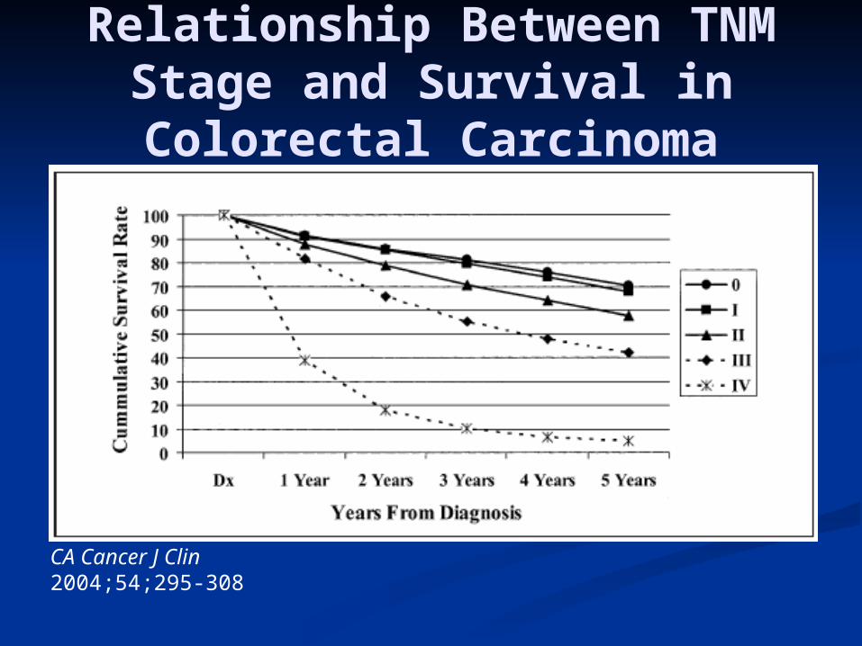
Relationship Between TNM Stage and Survival in Colorectal Carcinoma
CA Cancer J Clin 2004;54;295-308

Treatment of CRCTreatment of CRC
PolypectomyPolypectomy Colonic ResectionColonic Resection
Treatment depends on the risk of Treatment depends on the risk of lymph node metastasis.lymph node metastasis.
Pathology is key!1. Colorectal cancer is defined by
invasion of muscularis mucosa2. Lymphatics are located in
submucosa

Incidence of malignant Incidence of malignant polypspolyps
DefinitionDefinition Malignant polyps or T1 lesions (limited Malignant polyps or T1 lesions (limited
to the submucosa)to the submucosa) Represent 5% of all adenomasRepresent 5% of all adenomas
Colonoscopy polypectomy series: 2 – Colonoscopy polypectomy series: 2 – 12%12%
Colorectal resection series: 4 – 9%Colorectal resection series: 4 – 9%

Haggitt Level (1985)Haggitt Level (1985)Classification of polyps with Classification of polyps with
invasive cancerinvasive cancer
Haggitt RC, Glotzbach RE, Soffer EE, Wruble LD. Prognostic factors in colorectal carcinoma arising in adenomas: Implications for lesions removed by endoscopic polypectomy. Gastroenterology 89:328-36, 1985, p 330.
LevelLevel DefinitionDefinition Resected Resected (N)(N)
+ LN (N)+ LN (N)
00 Carcinoma Carcinoma in situin situ
11 Invasion of Invasion of headhead
66 0 (< 1%)0 (< 1%)
22 Invasion of Invasion of neckneck
33 0 (< 1%)0 (< 1%)
33 Invasion of Invasion of stalkstalk
44 0 (< 1%)0 (< 1%)
44 Invasion of Invasion of submucosa submucosa of bowel wall of bowel wall below polypbelow polyp
1313 4 (31%, 4 (31%, 12-25%)12-25%)
Villuous/sessile (flat) polyps with invasive cancer are by definition Haggitt 4.

Sessile PolypsSessile PolypsKudo, 1993Kudo, 1993
Risk of lymph node metastasis in each sessile Risk of lymph node metastasis in each sessile lesion is not the samelesion is not the same
Haggitt’s: no detail for sessile lesionsHaggitt’s: no detail for sessile lesions Classification of submucosal invasion:Classification of submucosal invasion:
Sm1—Invasion into the upper third of the submucosaSm1—Invasion into the upper third of the submucosa Sm2—Invasion into the middle third of the submucosaSm2—Invasion into the middle third of the submucosa Sm3—Invasion into the lower third of the submucosaSm3—Invasion into the lower third of the submucosa
High rate of LN metastasis: 12-25%High rate of LN metastasis: 12-25%

Sm systemSm system
Able to determine Sm1, Sm2, Sm3 in Able to determine Sm1, Sm2, Sm3 in 97% of cases97% of cases
Haggitt Level 1, 2, 3 = Sm1Haggitt Level 1, 2, 3 = Sm1 Haggitt Level 4 = Sm1, Sm2, or Sm3Haggitt Level 4 = Sm1, Sm2, or Sm3
Endoscopist must properly resect Endoscopist must properly resect and prepare specimenand prepare specimen
Pathologist must properly section Pathologist must properly section and examine all layersand examine all layers

Correlations with Correlations with MalignancyMalignancyMorphologyMorphology
MorphologMorphologyy
IncidenceIncidence % % MalignantMalignant
TubularTubular 7575 55
TubulovillouTubulovillouss
1515 2020
VillousVillous 1010 4040

Correlations with Correlations with MalignancyMalignancy
GradeGrade
DysplasiDysplasiaa
% % malignantmalignant
MildMild 55
ModeratModeratee
2020
SevereSevere 3030
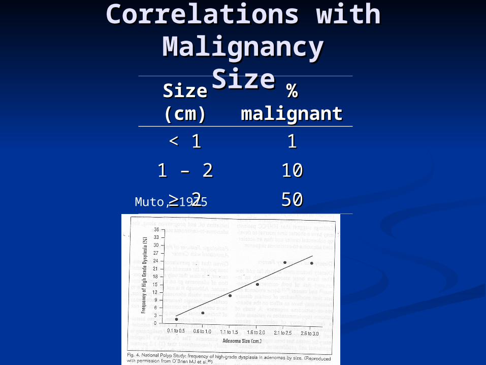
Correlations with Correlations with MalignancyMalignancy
SizeSizeSize Size (cm)(cm)
% % malignantmalignant
< 1< 1 11
1 – 21 – 2 1010
≥ ≥ 22 5050Muto, 1975

Correlations with Correlations with MalignancyMalignancy
SizeSize
Muto, 1975
Size Size (cm)(cm)
% % malignantmalignant
≤≤ 0.50.5 NegligibleNegligible
0.6 – 1.50.6 – 1.5 22
1.6 – 2.51.6 – 2.5 1919
2.6-3.52.6-3.5 4343
≥≥ 3.53.5 7676Nusco, 1997
Size Size (cm)(cm)
% % malignantmalignant
< 1< 1 11
1 – 21 – 2 1010
≥ ≥ 22 5050

Relationship betweenRelationship betweenSize and Morphology Size and Morphology
TubularTubular TubulovillTubulovillousous
VillousVillous
< 1 < 1 cmcm
76%76% 25%25% 14%14%
1-1-2cm2cm
20%20% 47%47% 26%26%
> 2 > 2 cmcm
4%4% 28%28% 60%60%
St. Mark’s Hospital Data

Increased risk of LN Increased risk of LN MetastasisMetastasis
Unfavorable pathologic features of Unfavorable pathologic features of malignant CR polypsmalignant CR polyps Poor differentiation (only on univariate)Poor differentiation (only on univariate) Lymphovascular invasion (P < 0.009)Lymphovascular invasion (P < 0.009) Invasion below submucosa (Haggitt Level 4)Invasion below submucosa (Haggitt Level 4) Depth of invasion in Sm3 (P < 0.001)Depth of invasion in Sm3 (P < 0.001) Site in lower 1/3 of the rectum (P < 0.001)Site in lower 1/3 of the rectum (P < 0.001)
Positive resection margin (< 1 mm or 1 HPF)Positive resection margin (< 1 mm or 1 HPF) Not really – this is inadequate treatment, not an Not really – this is inadequate treatment, not an
adverse risk factor!adverse risk factor!
P-values from Nascimbeni et al. N = 353 T1 colorectal sessile lesions

Management of Management of Pedunculated Malignant Pedunculated Malignant
PolypsPolyps Haggitt Level 1, 2, 3Haggitt Level 1, 2, 3
Complete excision or snaringComplete excision or snaring Risk of LN metastasis < 1%Risk of LN metastasis < 1%
Haggitt Level 4Haggitt Level 4 Treat as sessile lesionsTreat as sessile lesions

Management of Sessile Management of Sessile Malignant PolypsMalignant Polyps
< 2cm in diameter< 2cm in diameter Adequate snare in one piece via colonoscopyAdequate snare in one piece via colonoscopy Requires microscopic free margin of at least Requires microscopic free margin of at least
2mm2mm
Piecemeal removalPiecemeal removal Requires further excision/follow-up or resectionRequires further excision/follow-up or resection
High risk factors (LVI, Sm3, distal 1/3 High risk factors (LVI, Sm3, distal 1/3 rectum)rectum) Oncologic resectionOncologic resection Full thickness transanal excisionFull thickness transanal excision

Lesions amenable to Lesions amenable to colonoscopic polypectomycolonoscopic polypectomy
Pedunculated or sessile < 2cmPedunculated or sessile < 2cm Well/moderately differentiatedWell/moderately differentiated No lymphovascular invasionNo lymphovascular invasion Haggitt Level 1-3 or Sm1Haggitt Level 1-3 or Sm1 Close follow-up availableClose follow-up available
Endoscopically Endoscopically complete excisioncomplete excision Negative resection margins (2mm)Negative resection margins (2mm)
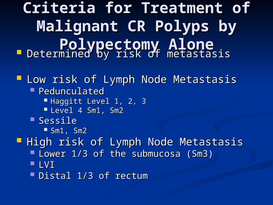
Criteria for Treatment of Criteria for Treatment of Malignant CR Polyps by Malignant CR Polyps by
Polypectomy AlonePolypectomy Alone Determined by risk of metastasisDetermined by risk of metastasis
Low risk of Lymph Node MetastasisLow risk of Lymph Node Metastasis PedunculatedPedunculated
Haggitt Level 1, 2, 3Haggitt Level 1, 2, 3 Level 4 Sm1, Sm2Level 4 Sm1, Sm2
SessileSessile Sm1, Sm2Sm1, Sm2
High risk of Lymph Node MetastasisHigh risk of Lymph Node Metastasis Lower 1/3 of the submucosa (Sm3)Lower 1/3 of the submucosa (Sm3) LVILVI Distal 1/3 of rectumDistal 1/3 of rectum

Malignant Colorectal Malignant Colorectal Polyps that Should have an Polyps that Should have an Oncologic Bowel ResectionOncologic Bowel Resection
Lesions in colonLesions in colon Pedunculated Haggitt Level 4 with invasion into distal Pedunculated Haggitt Level 4 with invasion into distal
third of submucosa (Sm3) or LVIthird of submucosa (Sm3) or LVI Sessile lesions removed with margin < 2mmSessile lesions removed with margin < 2mm Sessile lesions removed piecemealSessile lesions removed piecemeal Sessile lesions with depth of invasion into distal third of Sessile lesions with depth of invasion into distal third of
submucosa (Sm3)submucosa (Sm3) Sessile lesions with LVISessile lesions with LVI
Lesions in middle third and upper third rectumLesions in middle third and upper third rectum Same as lesions in colonSame as lesions in colon
Lesions in distal third rectumLesions in distal third rectum Pedunculated Haggitt Level 4 with invasion into distal Pedunculated Haggitt Level 4 with invasion into distal
third of submucosa (Sm3) or pedunculated lesions with third of submucosa (Sm3) or pedunculated lesions with LVILVI
All sessile lesionsAll sessile lesions

Why not just resect Why not just resect anyway?!anyway?!

What if ???What if ??? What if it’s clipped in ½?What if it’s clipped in ½?
PedunculatedPedunculated Repeat endoscopy.Repeat endoscopy. Require good resection with margin (2mm)Require good resection with margin (2mm)
SessileSessile Requires operative oncologic resection (even if Sm1, Sm2)Requires operative oncologic resection (even if Sm1, Sm2)
Unable to determine exact pathologic depthUnable to determine exact pathologic depth What if it’s shredded by forceps?What if it’s shredded by forceps?
Requires operative oncologic resectionRequires operative oncologic resection What if it’s a very small lesion?What if it’s a very small lesion?
Requires marking/tattoo CIRCUMFERENTIALLYRequires marking/tattoo CIRCUMFERENTIALLY What if it’s carcinoma in situ?What if it’s carcinoma in situ?
It’s not cancer. This is high grade dysplasia. Requires close It’s not cancer. This is high grade dysplasia. Requires close follow-up.follow-up.
Unless,Unless, poor margins: repeat endoscopy with good marginspoor margins: repeat endoscopy with good margins Piecemeal resection: discussion with pathologist and patientPiecemeal resection: discussion with pathologist and patient
What if it’s a large, non-endoscopically resectable polyp?What if it’s a large, non-endoscopically resectable polyp? Repeat endoscopy (2Repeat endoscopy (2ndnd MD?) MD?) Oncologic resectionOncologic resection

Other considerations…Other considerations…
When in doubtWhen in doubt Repeat colonoscopy Repeat colonoscopy
(endoscopy)(endoscopy) Personally review Personally review
pathologypathology Get a second opinionGet a second opinion Have a frank Have a frank
discussion with discussion with patientpatient

PolypsPolyps Natural history of adenomatous polypsNatural history of adenomatous polyps Biology of polypsBiology of polyps Cancer epidemiologyCancer epidemiology The anatomy of the polypThe anatomy of the polyp Correlations with MalignancyCorrelations with Malignancy Endoscopic polypectomy alone???Endoscopic polypectomy alone??? Special considerationsSpecial considerations Indications for PolypectomyIndications for Polypectomy
What if it’s clipped in ½What if it’s clipped in ½ What if it’s shredded by forceps?What if it’s shredded by forceps?
Pathology…Pathology… Marking/tattooMarking/tattoo Chances of Malignancy by histopath and size/morphologyChances of Malignancy by histopath and size/morphology * NO technique *** NO technique **