polyphosphazene design, synthesis, and characterization
TRANSCRIPT

The Pennsylvania State University
The Graduate School
Department of Chemistry
POLYPHOSPHAZENE DESIGN, SYNTHESIS, AND CHARACTERIZATION FOR
POTENTIAL LIGAMENT AND TENDON SCAFFOLDS
A Dissertation in
Chemistry
by
Jessica L. Nichol
2014 Jessica L. Nichol
Submitted in Partial Fulfillment
of the Requirements
for the Degree of
Doctor of Philosophy
December 2014

ii
The dissertation of Jessica L. Nichol was reviewed and approved* by the following:
Harry R. Allcock
Evan Pugh Professor of Chemistry
Dissertation Advisor
Chair of Committee
Philip Bevilacqua
Professor of Chemistry
Benjamin J. Lear
Assistant Professor of Chemistry
Mike Hickner
Associate Professor of Materials Science and Engineering, Chemical Engineering
Barbara J. Garrison
Shapiro Professor of Chemistry
Head of the Chemistry Department
*Signatures are on file in the Graduate School

iii
ABSTRACT
The work described in this thesis describes the progress towards developing
polyphosphazenes designed specifically for tissue engineering applications with an emphasis on
ligament and tendon repair and replacement. Chapter 1 outlines the fundamentals of polymer
chemistry in conjunction with the importance of polyphosphazene chemistry and its potential for
biomedical applications. Chapter 6 illustrates additional possibilities and considerations for
designing future polymers for tissue engineering scaffolds.
Chapter 2 discusses the design, synthesis, and characterization of new polyphosphazenes
to determine their potential as scaffolds for ligament and tendon tissue engineering. The
carboxylic acid moiety of the amino acids L-alanine and L-phenylalanine were protected with
alkyl esters with increasing chain length from 5 to 8 carbon atoms. This combined the hydrolytic
sensitivity of the amino acid ester polyphosphazenes with the elastomeric characteristics induced
by the long chain alkoxy polyphosphazenes. Test side group substitution reactions were
performed on the cyclic small molecule model, hexachlorocyclotriphosphazene (NPCl2)3, to
determine if steric hindrance would inhibit the degree of chlorine replacement by the amino acid
ester units. Counterpart polymers were then synthesized by replacement of the chlorine atoms in
poly(dichlorophosphazene) (NPCl2)n by the same amino acid esters. The glass transition
temperatures of the polymers decreased with increasing alkyl ester chain length, ranging from
11.6 to -24.2 °C. Polymer hydrolysis was studied for solid samples in deionized water at
physiological temperature for 12 weeks. The starting pH was 6.3 and the final pH ranged between
5.2 and 6.8. Polymer film mass decreased between ~8.7 and 26 percent during the 12 week
period, while the molecular weights decreased ~57 to 99 percent.
Chapter 3 describes the development of a possible polymer candidate that could
potentially serve as a ligament or tendon tissue engineering scaffold by meeting the requirements

iv
of biodegradability, biocompatibility, and elasticity. In an attempt to meet these requirements
novel citronellol-containing polyphosphazenes were synthesized, characterized, and crosslinked
to generate elastomers. Citronellol was chosen as a side group due to its anti-inflammatory
properties in addition to the presence of a double bond in its structure to permit polymer
crosslinking. Alanine ethyl ester was chosen as a co-substituent to tune hydrolysis rates without
severely affecting the glass transition temperatures of the final polymers. Hydrolysis of the
uncrosslinked polymers in the form of films in deionized water at 37 °C showed between a ~8 to
16% mass loss and between a ~28 to 88% molecular weight decline over 12 weeks. Polymers
were also crosslinked using UV radiation for increasing amounts of time. Preliminary mechanical
testing of the homo-citronellol polymer indicated increasing modulus and decreasing tensile
strength with increased crosslink density.
Chapter 4 outlines a different approach to attaching the anti-inflammatory molecule
citronellol to the polyphosphazene backbone. By contrast, in this work citronellol, was used as an
ester unit to the carboxylic acid moiety of the amino acids glycine, alanine, valine, and
phenylalanine that were in turn linked to the polymer through the amino functionality. This
method allowed the hydrolysis rate to be tuned via the steric hindrance generated by the amino
acid ester while still providing two crosslinkable sites per repeat unit from the citronellol units. A
hydrolysis study of the uncrosslinked polymers at physiological temperature showed between a
19.9 – 28.8% mass loss and between a 80.4 – 98.9% molecular weight decline after 12 weeks.
The double bond in the citronellol structure also allowed polymer crosslinking by UV radiation to
further tune the properties. Additionally, the mechanical properties of the alanine and
phenylalanine citronellol polymers were studied as a function of crosslinking.
In chapter 5 the field of ethoxyphosphaze polymers is considered for their potential as
biomedical materials. This was accomplished by determining the properties and hydrolytic
characteristics of poly(diethoxyphosphazene) and related derivatives with both ethoxy and

v
hydrophobic co-substitutent groups in a near 1:1 molar ratio. Co-substituents such as 2,2,2-
trifluoroethoxy, phenoxy, or p-methylphenoxy units were examined. These hydrophobic co-
substitutents serve as models for bioactive counterparts. The hydrolytic sensitivity of the
ethoxyphosphazene units was so pronounced that even hydrophobic or bulky O-linked co-
substitutents failed to counteract the hydrolysis behavior during a twelve-week hydrolysis study.
This work illustrates a pathway for the development of a new class of useful bioerodible
polymers.

vi
TABLE OF CONTENTS
List of Figures .......................................................................................................................... ix
List of Tables ........................................................................................................................... xi
Preface............... ...................................................................................................................... xii
Acknowledgements………………………………………..………………………………....xiii
Chapter 1 Introduction………………………………………………………………........………..1
1.1 History of Polymer Chemistry……………………………………………….........…..1
1.2 Polymer Definition……………………………….……………………….........……...2
1.3 Polyphosphazenes………………………………..…………………………........……3
1.3.1 Discovery.......................................................................................................3
1.3.2 Importance of Small Molecule Model Compounds.......................................5
1.3.3 Polymer Synthetic Challenges.......................................................................7
1.3.4 Characterization.............................................................................................9
1.3.5 Applications.................................................................................................10
1.4 Polymers for Tissue Engineering Applications...........................................................10
1.4.1 Ligament and Tendon Tissue Introduction..................................................11
1.4.2 Tissue Engineering Polymer Requirements.................................................13
1.4.3 Natural Polymers.........................................................................................13
1.4.4 Synthetic Polymers......................................................................................14
1.5 Polyphosphazenes for Ligament and Tendon Tissue Engineering..............................14
1.6 References....................................................................................................................16
Chapter 2 Biodegradable Alanine and Phenylalanine Alkyl Ester Polyphosphazenes as Potential
Ligament and Tendon Tissue Scaffolds..........................................................................................20
2.1 Introduction..................................................................................................................20
2.2 Experimental................................................................................................................22
2.2.1 Reagents and Equipment.............................................................................22
2.2.2 Synthesis of L-alanine and L-phenylalanine Alkyl Esters 1-8....................23
2.2.3 Synthesis of Cyclic Trimer Model Compounds 10 and 11..........................24
2.2.4 Synthesis of L-alanine and L-phenylalanine Alkyl Ester Polymers 13-20..25
2.2.5 Hydrolysis Study of Polymers 13-20...........................................................26
2.3 Results and Discussion................................................................................................26
2.3.1 Side Group Preparation and Synthetic Considerations................................26
2.3.2 Cyclic Trimer Model Synthesis and Characterization.................................27
2.3.3 Polymer Synthesis and Characterization.....................................................28
2.3.4 Testing for Residual Coordinated Hydrogen Chloride................................30
2.3.5 Thermal Behavior........................................................................................31
2.3.6 Hydrolysis Behavior....................................................................................32
2.4 Conclusions..................................................................................................................37

vii
2.5 Acknowledgements......................................................................................................37
2.6 References....................................................................................................................37
Chapter 3 Crosslinkable Citronellol Containing Polyphosphazenes and their Biomedical
Potential..........................................................................................................................................39
3.1 Introduction..................................................................................................................39
3.2 Results and Discussion................................................................................................41
3.2.1 Synthesis of Cyclic Trimer Model Compound (2)......................................41
3.2.2 Synthesis and Characterization of Polymers 4-8.........................................42
3.2.3 Uncrosslinked and Crosslinked Hydrolysis Behavior.................................45
3.2.4 Polymer Crosslinking and Swelling Studies................................................48
3.2.5 Thermal behavior of uncrosslinked and crosslinked polymers...................51
3.2.6 Poly[bis(citronellol)phosphazene] Mechanical Property Evaluation..........53
3.3 Experimental................................................................................................................54
3.3.1 Reagents and Equipment.............................................................................54
3.3.2 Synthesis of Hexa(citronellol)cyclotriphosphazene (2)...............................55
3.3.3 Synthesis of Poly[bis(citronellol)phosphazene] (4).....................................56
3.3.4 Synthesis of Poly[(citronellol)x(alanine ethyl ester)yphosphazenes] (5-8)..56
3.3.5 Hydrolysis Study of Polymers 4-8...............................................................57
3.3.6 UV-Crosslinking and Swelling Studies of Polymers 4-8............................57
3.3.7 Hydrolysis Study of Crosslinked Polymers 4-8...........................................58
3.3.8 Instron Tensile Testing of Crosslinked Poly[bis(citronellol)phosphazene
(4)..........................................................................................................................58
3.4 Conclusions..................................................................................................................59
3.5 Acknowledgements......................................................................................................59
3.6 References....................................................................................................................59
Chapter 4 Amino Acid Citronellol Ester Polymers for Biomedical Applications………………..62
4.1 Introduction………………………………………………………………..................62
4.2 Results and Discussion………………………………………………………………64
4.2.1 Synthesis of Amino Acid Citronellol Ester Side Groups............................64
4.2.2 Synthesis of the Cyclic Trimer as a Model System (6)…………………...65
4.2.3 Synthesis and Characterization of Polymers 8-11………………………...66
4.2.4 Hydrolysis Behavior of Uncrosslinked Polymers…………………………69
4.2.5 Polymer Crosslinking and Swelling Studies………………………………70
4.2.6 Thermal Behavior of Uncrosslinked and Crosslinked Polymers………….73
4.2.7 Polymer Mechanical Property Evaluation………………………………...73
4.3 Experimental Section………………………………………………………………...75
4.3.1 Reagents and Equipment………………………………………………….75
4.3.2 Synthesis of Amino Acid Citronellol Esters 1-4………………………….76
4.3.3 Synthesis of Hexa(phenylalanine citronellol ester)cyclotriphosphazene
(6)..........................................................................................................................77
4.3.4 Synthesis of Poly(amino acid citronellol ester)phosphazenes 8-11……….78
4.3.5 Hydrolysis of Polymers 8-11………………………………………...........78
4.3.6 UV-Crosslinking and Swelling Studies of Polymers 8-11………………..79
4.3.7 Instron Tensile Testing of Polymers 9 and 11.……………………………79
4.4 Conclusions…………………………………………………………………………..80

viii
4.5 Acknowledgements…………………………………………………………………..80
4.6 References……………………………………………………………………………80
Chapter 5 Ethoxyphosphazene Polymers and their Hydrolytic Behavior………………………...83
5.1 Introduction………………………………………………………………………..…83
5.2 Results and Discussion………………...…………………………………………….85
5.2.1 Polymer Synthesis…………………………………………………………85
5.2.2 Polymer Characterization and Properties…………………………………86
5.2.3 Thermal Characterization of Polymers 8-10………………………………89
5.2.4 Hydrolysis Behavior of Polymers 3-10…………………………………...90
5.2.5 Water Contact Angles of Polymers 3-10………………………………….92
5.3 Experimental Section………………………………………………………………...93
5.3.1 Reagents and Equipment………………………………………………….93
5.3.2 Synthesis of Poly(diethoxyphosphazene) (3)……………………………..95
5.3.3 Synthesis of Polyphosphazenes with 1-Propoxy, 1-Butoxy, 2,2,2-
Trifluoroethoxy, Phenoxy, P-methylphenoxy Side Groups (4-7)……………….95
5.3.4 Synthesis of Poly[(ethoxy)0.8(trifluoroethoxy)1.2phosphazene] (8)………..95
5.3.5 Synthesis of Poly[(ethoxy)1(phenoxy)1phosphazene] (9)…………………96
5.3.6 Synthesis of Poly[(ethoxy)1(p-methylphenoxy)1phosphazene] (10)……...96
5.3.7 Water Contact Angle Measurements……………………………………...97
5.3.8 Hydrolysis of Polymer Films……………………………………………...97
5.4 Conclusions…………………………………………………………………………..98
5.5 Acknowledgements…………………………………………………………………..98
5.6 References……………………………………………………………………………98
Chapter 6 Summary......................................................................................................................100
6.1 New Polyphosphazenes Developed for Tissue Engineering Applications................100
6.2 Future Considerations................................................................................................102
6.3 References..................................................................................................................103

ix
LIST OF FIGURES
Figure 1-1. Polymer architectures........................................................................................3
Figure 1-2. Synthetic pathway for polyphosphazenes.........................................................5
Figure 1-3. Features of a DSC curve..............................................................................................10
Figure 1-4. Hierarchical structure of a tendon................................................................................12
Figure 1-5. Typical stress-strain curve for a ligament or tendon....................................................12
Figure 1-6. Hydrolysis of poly(amino acid ethyl ester)phosphazenes............................................15
Figure 1-7. Proposed hydrolysis mechanisms of poly(amino acid ester)phosphazenes.................15
Figure 2-1. Synthesis of L-alanine and L-phenylalanine alkyl ester derivatives 1-8.....................27
Figure 2-2. Synthesis of [hexa(alanine octyl ester)cyclotriphosphazene] and [hexa(phenylalanine
octyl ester)cyclotriphosphazene].....................................................................................................28
Figure 2-3. Synthesis of L-alanine and L-phenylalanine alkyl ester polymers 13-20....................29
Figure 2-4. Polymer 16 1H NMR (top) and
31P NMR (bottom)......................................................29
Figure 2-5. pH of medium of polymers 13-16 (top) and 17-20 (bottom).......................................33
Figure 2-6. Percent film mass loss of polymers 13-16 (top) and 17-20 (bottom)..........................34
Figure 2-7. Molecular weight decline of polymers 13-16 (top) and 17-20 (bottom)......................35
Figure 3-1. Synthesis of hexa(citronellolcyclotriphosphazene)......................................................42
Figure 3-2. Synthesis of citronellol containing polymers...............................................................43
Figure 3-3. 1H NMR spectrum of polymer 8..................................................................................44
Figure 3-4. 31
P NMR spectra of polymer 8.....................................................................................45
Figure 3-5. Percent film mass loss of polymers 4-8.......................................................................46
Figure 3-6. Molecular weight decline of polymer 4 (top) and polymers 5-8 (bottom)...................47
Figure 3-7. Glass transition temperatures of polymers 4-8.............................................................52
Figure 3-8. Glass transition temperature change with increasing UV exposure for polymer 4......52

x
Figure 4-1. Synthesis of amino acid citronellol ester derivatives 1-4.............................................65
Figure 4-2. Synthesis of [hexa(phenylalanine citronellol ester)cyclotriphosphazene] (6).............65
Figure 4-3. Synthesis of amino acid citronellol ester polymers 8-11.............................................66
Figure 4-4. 1H NMR spectrum of polymer 8 (top) and
31P NMR (bottom)....................................68
Figure 4-5. Percent film mass loss of polymers 8-11.....................................................................69
Figure 4-6. Molecular weight decline of polymers 8-11................................................................70
Figure 5-1. Macromolecular substitution routes to poly(organophosphazenes).............................84
Figure 5-2. Single substituent polymers.........................................................................................85
Figure 5-3. Polymers 8-10 with both ethoxy and O-linked co-substitutents..................................86
Figure 5-4. 31
P NMR spectrums of polymers 8 (A), 9 (B), and 10 (C)...........................................89
Figure 5-5. UV-Vis concentration determination of p-cresol in hydrolysis media from polymer 10
during 12 week hydrolysis..............................................................................................................92

xi
LIST OF TABLES
Table 2-1. Characterization data of L-alanine and L-phenylalanine alkyl ester derivatives 1-
8.......................................................................................................................................................23
Table 2-2. Characterization of L-alanine and L-phenylalanine alkyl ester polymers 13-
20.....................................................................................................................................................31
Table 3-1. Characterization data of citronellol containing polymers 4-8.......................................44
Table 3-2. Group contribution parameters for polymers 4-8..........................................................50
Table 3-3. Calculated dispersion parameters and chi parameters for polymers 4-8.......................50
Table 3-4. Number of crosslinks for polymers 4-8 corresponding to UV exposure time in
minutes............................................................................................................................................51
Table 3-5. Mechanical properties of crosslinked polymer 4...........................................................53
Table 4-1. Characterization data of amino acid citronellol ester polymers 8-11............................67
Table 4-2. Group contribution parameters for polymers 8-11........................................................71
Table 4-3. Calculated dispersion parameters and chi parameters for polymers 8-11.....................72
Table 4-4. Number of crosslinks for polymers 8-11 corresponding to UV exposure time in
minutes............................................................................................................................................72
Table 4-5. Glass transition temperature comparison between the amino acid citronellol ester
polyphosphazenes (8-11) and the amino acid ethyl ester polyphosphazenes (°C).........................73
Table 4-6. Mechanical properties of poly(alanine citronellol ester)phosphazene (9).....................74
Table 4-7. Mechanical properties of poly(phenylalanine citronellol ester)phosphazene (11)........75
Table 4-8. Characterization data of amino acid citronellol ester side groups 1-4..........................77
Table 5-1. Characterization data of polymers 3 and 8-10...............................................................88
Table 5-2. Molecular weight decline and mass loss of polymers and corresponding polymer water
contact angles 3-10.........................................................................................................................91

xii
PREFACE
Portions of this thesis have been adapted for publication. Chapter 2 was adapted for
publication in Polymer Chemistry and coauthored by H. R. Allcock and N. L. Morozowich.
Chapter 3 was adapted for publication in Journal of Polymer Science: Polymer Chemistry and
was coauthored by H. R. Allcock, N. L. Morozowich, and T. D. Decker. Chapter 4 was submitted
for publication in European Polymer Journal and coauthored by H. R. Allcock. Chapter 5 was
adapted for publication in Polymer Degradation and Stability and was coauthored by H. R.
Allcock and I. T. Hotham.

xiii
ACKNOWLEDGEMENTS
Without many wonderful people and sequence of events the career and life path I am
gratefully following today would not have been possible. I would like to thank my graduate thesis
advisor, Professor Harry R. Allcock, for the opportunity to conduct research and expand my
knowledge throughout my graduate career. The structure and design of our research group I feel
has trained me well for my future career in industry and for that I am very grateful. I would like
to thank my graduate committee Dr. Ben Lear, Dr. Philip Bevilacqua, and Dr. Mike Hicker for
their help and guidance throughout my graduate work. I would also like to thank Noreen Allcock
for her help and encouragement during my time at Penn State and for our girls’ lunches. I am also
thankful to The Pennsylvania State University for accepting me into the Chemistry Department
and funding part of my research and also to the PN Leadership Endowment for funding part of
my research.
I would also like to express my gratitude toward several past and present group members
who have helped me during my graduate career, specifically: Dr. Nicole Morozowich, Tomasz
Modzelewski, Dr. Arlin Weikel, Dr. David Lee, Zhicheng Tian, Dr. Xiao Liu, Andrew Hess, Dr.
Chen Chen, Ian Hotham, Chris Fellin, Ryan J. Mondschein, and Thomas Decker. Many of these
group members were co-authors on papers with me and without their advice and insight many of
the projects would not have been possible. I would also like to acknowledge multiple
collaborators that have provided support along my journey including Brittany Banik and Dr.
Justin Brown (PSU Department of Bioengineering) and Peter Chhour and Dr. David Cormode
(Perelman School of Medicine, University of Pennsylvania). Other people I would like to thank
are Tim Tighe for his guidance and advice even after that particular project fell through, Dr. Scott
Showalter for his kindness and advice, James Miller for running all of the MS samples in this
dissertation, David Shelleman for his help with the Instron equipment, and Dr. Alan Benesi, Dr.

xiv
Wenbin Luo, and Dr. Emmanuel Hatzakis for their help with anything and everything NMR
related.
I would like to thank my undergraduate research advisor, Dr. Charles H. Lake, of Indiana
University of Pennsylvania. Without his guidance I would not have pursued a graduate career. I
would also like to thank my summer internship research advisor, Dr. Andrew L. Vance, of Sandia
National Laboratories who taught me a considerable amount about working in a laboratory and
for helping me choose a school for my graduate career. Further, I would like to thank my high
school chemistry teacher, William Smeltz for inspiring my love of chemistry and science.
Finally, I would like to thank my family and friends because without their love and
encouragement I would not be here. I would like to thank my parents, Tom and Valerie Nichol,
and my sister Rebecca Nichol for their support, advice, and love throughout my life and scientific
career. Especially my mom, for instilling a love of science and curiosity in my sister and I at a
young age by doing things like making my first polymer, silly putty. I would like to thank my
fiancé Marc Ferrington for helping me through the good times and the bad and for helping me
afford attending two American Chemical Society national meetings. I would also like to thank
Kaycee Quarles for her friendship and support. I also would like to thank Bailey for always being
there for me.

1
Chapter 1
Introduction
1.1 History of Polymer Chemistry
Polymers are as old as life itself, considering that proteins, RNA and DNA are all natural
macromolecules.1 The human race has always relied on natural polymers for nourishment,
clothing, and shelter.2 Nourishment was provided by animal or plant proteins, which are long
amino acid based polymers and plant starches. Clothing was made of materials such as wool,
cotton, and silk, which are very high molecular weight proteins or polysaccharides, and are the
most abundant natural polymers. Finally, shelters were made using materials such as wood,
bamboo, and reeds, all of which are primarily polymeric materials.1 However, it was not until the
1830s that people started to modify naturally occurring polymers for use in more specialized
applications.3 Charles Goodyear was the first to treat natural rubber with sulfur to produce
vulcanized rubber, a process which is still used widely today.4 Christian Fredrich Schonbein
treated cellulose with a combination of nitric and sulfuric acid resulting in the discovery of gun
cotton.1 Despite this wide usage, a complete understanding of the structure of polymers remained
elusive, with most investigators believing them to be comprised of conglomerations of individual
small molecules. It was not until the 1920s that Herman Staudinger proposed his
“macromolecular theory” which proposed that polymers are actually long molecules comprised of
individual units connected through covalent bonds.5 Initially his theory was not well accepted, but
later work proved his initial hypothesis for which he received the 1953 Nobel Prize in
Chemistry.6 Once these fundamental facts were accepted, the field of synthetic polymers

2
underwent a rapid expansion in the development of an ever-increasing number of materials for
increasingly specialized applications.7 This revolution is still going strong today.
1.2 Polymer Definition
A single polymer chain is a molecule comprised of a large number of individual repeating
units covalently linked together to form a long macromolecule.8 Macromolecules can be tailored
to have different architectures such as linear, branched, or interconnected to form three-
dimensional networks (Figure 1-1).9 Three main synthetic pathways exist to generate all of the
different polymers developed to date: step-growth, chain-growth, and ring-opening
polymerization.10
It is also possible to use a combination of these techniques to generate the final
product.10
Polymers can also exhibit multiple distinct physical phases, more specifically:
amorphous, rubbery, glassy, and semicrystalline.8 Combinations of all of these characteristics
govern the range of properties of all polymers and determine their final applications. Polymers
have become an integral part of society due to their ease of fabrication, corrosion resistance, light
weight, and cost effectiveness.11

3
Figure 1-1. Polymer architectures
1.3 Polyphosphazenes
1.3.1 Discovery
Although conventional polymers typically contain a carbon-based backbone, there exists
a unique family of polymers that have a phosphorus and nitrogen backbone, known as
polyphosphazenes.12
Small molecule phosphazenes were first discovered in 1834 by Liebig,
Wohler, and Rose, when it was noted that phosphorus pentachloride reacted with ammonium
chloride to yield a white crystalline compound.13, 14
In 1844 Gerhardt and Laurent examined this
compound and discovered that it had the empirical formula of NPCl2.15
Later work revealed the
molecular formula to be (NPCl2)3.16, 17
In the late 1800s it was shown by Stokes that when heated,
this molecule was transformed into a moisture-sensitive elastomeric material which was named
“inorganic rubber.”18
This material remained a curiosity until the mid-1960s when Allcock and
Kugel discovered a method to produce a soluble form of this polymer.19-21
At that time it was also

4
found that by controlling the polymerization in a vacuum sealed container, a reactive
macromolecular intermediate was formed that was soluble in various solvents which wasa a
crucial requirement for allowing chlorine replacement reactions. The high reactivity of the P-Cl
bonds allowed complete chlorine replacement to occur by exposure to alkoxides, aryloxides, and
primary or secondary amines as nucleophiles.12
Unlike the chlorophosphazenes, these
organophosphazene polymers were stable to moisture. This chemistry was developed during
subsequent decades during which time several hundred different side groups were linked to
phosphazene polymers to yield a wide variety of different polymers, often with unique properties
and potential uses.12
Thus, phosphazenes are a unique family of molecules with alternating phosphorus and
nitrogen atoms linked to form linear or ring structures.22
The cyclic trimer,
hexachlorocyclotriphosphazene (1), can be polymerized by a ring opening method at 250°C under
vacuum to form the reactive, high molecular weight intermediate, poly(dichlorophosphazene)
(2).23
This route yields polymers that typically contain 10,000 – 15,000 repeat units with
molecular weights ranging as high as 2 – 10 million after complete chlorine replacement.24
An
alternative route to the reactive intermediate poly(dichlorophosphazene) is through a living
cationic polymerization of a phosphoranimine in chloroform at room temperature, with PCl5 used
as the initiator.25
The benefits of the second method are the ability to better control the molecular
weight of the final polymer, which results in a much narrower polydispersity index because of the
“living” nature of the polymerization reaction.25
The key feature of polyphosphazene chemistry
is the fact that the intermediate, poly(dichlorophosphazene), is an excellent reactive substrate.
Both chlorine atoms attached to every phosphorus atom can be replaced by nucleophillic
substitution by reaction with various nucleophiles such as alkoxides (RONa), amines (RNH2,
R2NH), or organometallic reagents (RLi) (Figure 1-2).12

5
Figure 1-2. Synthetic pathway for polyphosphazenes
Compared to conventional polymers in which the monomers possess the required side
groups prior to polymerization, poly(organophosphazenes) are generated by post-polymerization
nucleophilic substitution reactions.26
This synthetic versatility, of both the cyclic and polymeric
phosphazenes, allows for a high degree of property optimization not inherent in most other
systems. The tunable properties include solubility, hydrophobicity or hydrophilicity, glass
transition temperature, mechanical properties, and biocompatibility. Thus, more than 700
polyphosphazenes and over 250 small-molecule phosphazenes have been synthesized to date.12
1.3.2 Importance of Small Molecule Model Compounds
It is a feature of the Penn State program that small molecule model reactions are usually
employed as a preliminary step before attempting the same reaction on a macromolecular level.
By using this proof of concept route, challenges that may make the high polymer system difficult
to synthesize can be identified and solutions developed. This methodology not only saves
expensive reagents but also yields a more fundamental understanding of the system being studied.
In our program, the cyclic trimer, hexacholorocyclotriphosphazene, acts as an excellent model
system for the high polymer.27
This small molecule has been utilized extensively as the first step

6
in polyphosphazene synthesis to understand how a new side group may react.28
Due to the small
molecule characteristics of the trimer, many characterization methods can be used that would be
difficult or impossible for the high polymer. These techniques include mass spectrometry and
nuclear magnetic resonance.29
When considering a side group for polymer substitution reactions many factors must be
considered. One consideration is the influence of steric hindrance involving possible side groups.
A large amount of research has shown that if the chosen side group is too bulky, complete
chlorine replacement may not be possible.30
This suggests that the initially introduced side groups
can be responsible for the physical exclusion of incoming side groups. This results in incomplete
nucleophilic substitution. When this occurs, a smaller co-substituent group can be employed to
complete the replacement of the remaining chlorine atoms.31
Another important use of the small
molecule model approach is to examine the protection and / or de-protection chemistry for a
particular substituent.30
Some side group candidates have multiple functionalities (i.e. several
hydroxyl or amino groups), each of which could act as attachment points and would lead to
crosslinking of the system during synthesis. Alternatively, prospective side groups may have
functionalities which are incompatible with poly(dichlorophosphazene), such as carboxylic acid
moieties, which will lead to polymer backbone degradation during macromolecular substitution.
To prevent these side reactions these functional groups must first be protected to ensure that only
one reactive site is present for attachment to the phosphazene. Once all of the chlorine atoms are
replaced the protecting groups can be removed to re-form the free functional moieties or they can
be left in place, depending on the final need of the system. One additional consideration must be
kept in mind when examining different protecting groups. This is that the polymer may be more
sensitive to the extremely acidic or basic conditions often employed in these macromolecular
substitution reactions.32
Therefore only protective groups which can be removed under relatively
mild conditions are appropriate. Optimization of these reactions at the cyclic trimer model level
7
allows ideal conditions to be determined. These conditions can then be utilized for attachment of
the side groups to the polyphosphazene chain while at the same time limiting polymer
degradation.
1.3.3 Polymer Synthetic Challenges
Although the cyclic trimer system provides an understanding of how a small molecule
may behave as a model for the high polymer, not all synthetic problems can be anticipated until
substitution is attempted at the high polymer level. In some cases solubility issues during
substitution may prevent complete chlorine replacement by premature precipitation of the
partially substituted product from solution. This issue can be overcome through co-substitution
reactions using a more soluble nucleophile either by introducting this group before or after the
desired side group depending on if it is oxygen or amine linked.33
In other cases the side group
may be too bulky to allow full substitution, even if it was able to achieve complete chlorine
replacement on the trimer model.
Another major challenge associated with the polymer scale reactions are the by-products
that are formed during side group introduction. In the case of the sodium salt of an oxygen-linked
nucleophile, such as a sodium alkoxide or aryloxide, the by-product is sodium chloride which is
removed from the reaction medium due to its insolubility. However, in the case of an amine-
linked side group, the by-product is hydrogen chloride (HCl) which would cause chain cleavage
of the polymer in solution. To help overcome this problem, triethylamine (TEA) is added to the
reaction solution to complex with the HCl and form an insoluble salt.34
Although this helps to
provide a driving force for the reaction, the TEA-HCl complex formed is not completely
insoluble in the reaction medium due to its equilibrium dissociation and can still cause polymer
degradation, especially when longer reaction times are needed.

8
Due to the high versatility of this polymer system, it is possible to link more than one side
group to the polymer backbone.12
The possibilities include introducing two different alkoxide or
amine substituents or a combination of the two. In some cases even three different side groups
have been employed.33
The challenge in using multiple substituents is obtaining either the desired
side group distribution (blocky or random), and the correct side group ratios. Methods to help
ensure random side group distribution along the polymer backbone include ensuring efficient
reagent mixing, or introducing the bulkier group first to use the steric effects of the group to
facilitate spacing between attachment points.31
The correct side group ratio is often more a
function of the types of side groups being employed. Challenges arise when the sodium salt of the
chosen side group has low solubility or low reactivity. By changing the order of the addition these
challenges can be overcome. Due to the generation of hydrogen chloride when using amine
linked substituents it is often advised to introduce an amino group second.
Despite precautions and reaction monitoring it is still possible that un-reacted chlorine
atoms remain along the backbone. These residual chlorine atoms can react with water to form P-
OH groups which would result in polymer condensation crosslinking or subsequent hydrolytic
cleavage of the backbone. Premature polymer precipitation from the reaction medium may also
cause incomplete substitution. Although the reactivity of P-Cl bonds is high, for a polymer chain
with 15,000 repeat units there exist 30,000 chlorine atoms to be replaced, which decreases the
possibility of 100% chlorine replacement. However, under favorable reaction conditions, all the
chlorine atoms can be replaced at least down to the limits of detectability using existing analytical
techniques.12

9
1.3.4 Characterization
Complete characterization of a polymer structure is important when examining the
structure-property relationships of macromolecules.35
Common characterization techniques
include nuclear magnetic resonance spectroscopy (NMR), gel permeation chromatography
(GPC), and differential scanning calorimetry (DSC). NMR spectroscopy is particularly helpful
for determining molecular level polymer structure. The unique backbone of polyphosphazenes
allows the use of 31
P NMR spectroscopy. This aspect is useful as it allows monitoring of the
progress of the substitution reaction without the need to isolate the product. It is also helpful
when multiple side groups are linked to the backbone because the 31
P shifts change with different
side group substitution patterns.12
In addition, 1H NMR spectroscopy can be used to further
confirm the presence and relative concentrations of individual side groups. GPC analysis is used
to estimate polymer molecular weights (Mw) and polydispersity indices (PDI). The molecular
weight of a polymer provides information about the number of repeat units it contains, while the
PDI discloses information about the distribution of the polymer masses in the sample. DSC
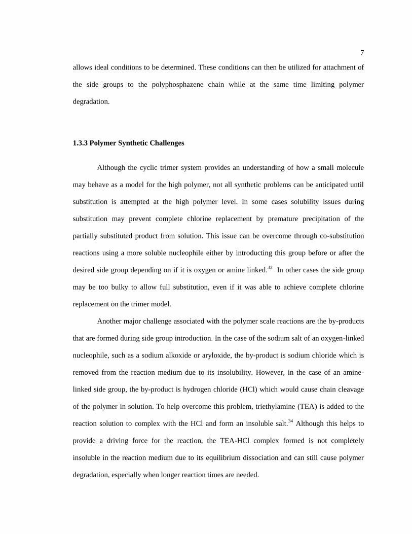
analysis identifies the polymer glass transition (Tg), melt (Tm) transitions, and crystallization
characteristics (Figure 1-3). At the glass transition temperature the polymer changes from a glassy
to a rubbery state. The high flexibility of the polyphosphazene backbone can give rise to
polymers with very low (~ -100 °C) glass transition temperatures.36
However, the incorporation
of bulky and torsional restrictive side groups has generated polymers with Tg’s as high as ~160
°C.37
A melt transition is indicative of crystalline regions in a polymer, although these may be
absent in mixed substituent polyphosphazenes.
10
Figure 1-3. Features of a DSC curve
1.3.5 Applications
The high synthetic tunability of the polyphosphazene platform makes them excellent
candidates for a wide variety of possible applications. They can be designed to function as ion
conductors by incorporation of etheric oxygen containing side groups.38-40
Due to the high
phosphorus content of phosphazenes, both the cyclic trimer and polymer based materials have fire
retardancy properties.41-43
Finally, by the incorporation of biologically compatible side groups, a
wide range of cyclic trimers and polymers have been developed for biomedical applications, such
as drug delivery systems and tissue engineering scaffolds.44-46
1.4 Polymers for Tissue Engineering Applications
Every year millions of bone, muscle, and skin injuries are reported and treated.47, 48
Current repair solutions rely on autografts, allografts, and synthetic materials.49
Autografts are
considered the gold-standard materials for replacement, in which tissue is harvested from the

11
patient and transplanted to the defect site.50
However, this requires two surgical procedures, thus
increasing both the healing time and risk of infection.51
Also, neither site ever regains its initial
mechanical properties. Another major disadvantage of this method is limited availability of living
tissue within the patient.52
An alternative is the use of allografts, where tissue is taken from
cadavers or donors and implanted into the site of injury.53
Although this method overcomes the
multiple surgical procedures and limited living tissue availability, there is a significant risk for
disease transmission and immunological rejection.54
Therefore the use of allografts is less
commonly used.53
Due to these shortcomings, scientists are investigating alternative materials to repair
damaged tissues, including metals, ceramics, and polymers.55
Among these, the use of
biodegradable polymers appears to be the most promising avenue.56
By combining expertise from
chemistry, biology, and engineering the ultimate goal of tissue engineering is to use a scaffold
which allows the body to slowly re-grow the damaged tissue as the support is hydrolyzed and
eliminated from the injury site.57
Utilization of this method would overcome most of the
disadvantages of the currently used systems.
1.4.1 Ligament and Tendon Tissue Introduction
Each year nearly 35 million musculoskeletal injuries occur in the United States, with half
involving ligaments and tendons, with an associated cost of tens of billions of dollars.47
Ligaments and tendons are fibrous connective tissues that attach either muscles to bones or bones
to other bones, respectively.58
Ligaments are mostly type I (90%) and type III (10%) collagen
whereas tendons are primarily type I collagen with smaller amounts of collagens type III, V, XII,
and XIV.59
These tissues are able to transmit load without substantial energy loss or
deformation.60
Although ligaments and tendons are primarily made up of collagen, researchers

12
have not been able to match the complexity inherent in these tissues for tissue engineering
applications.61
Their complex structure is depicted in Figure 1-4.
Figure 1-4. Hierarchical structure of a tendon
Tendons and ligaments are generally considered to be viscoelastic materials with a
typical stress strain curve shown in Figure 1-5.59
The “toe region” in the curve is due to the
crimped portion of the fiber bundle undergoing straightening.62
The “linear region” is
characteristic when the fibers become aligned and it is this portion of the curve which is used to
determine the elastic or Young’s modulus.62
Figure 1-5. Typical stress-strain curve for a ligament or tendon
Original investigations into the use of synthetic polymers for this application focused on
the use of bio-stable polymers.63
Due to the constant cyclic loads being applied to these grafts and

13
extensive scar tissue build-up they needed to be replaced often within 3 years of implantation and
have been discontinued.49
1.4.2 Tissue Engineering Polymer Requirements
In order for a polymer to be considered for tissue engineering applications it must meet
several requirements.64, 65
The most important of these is biodegradability into non-toxic
products.66
This requirement alone drastically decreases the number of potential polymers for this
application. Another key requirement is that the material must possess and maintain similar
mechanical properties to those of the parent tissue as the scaffold degrades and is replaced by
newly formed tissue.67
The scaffold must also have a high porosity with sufficient mechanical
stability to promote cell growth and proliferation during degradation.68
Thus, through tissue
engineering a scaffold seeded with cells and signaling molecules would be utilized to re-grow the
damaged tissue as the scaffold degrades.69
An elastomeric polymer would be an ideal matrix
material because the scaffold undergoes multiple cyclic loading cycles. Elastomers are flexible
low glass transition temperature (Tg) polymers with physical or chemical crosslinks.70
1.4.3 Natural Polymers
Of the naturally occurring polymers, silk and collagen have been studied extensively;
however their use is limited by batch to batch inconsistencies and uncontrollable enzymatic
degradation in the body.71-73
Chitosan has also been investigated as a potential scaffold material
because of its biocompatibility, however its lacks the necessary mechanical properties to perform
as a viable scaffolding material.71

14
1.4.4 Synthetic Polymers
Among the synthetic polymers studied, poly(lactic acid) (PLA), poly(glycolic acid)
(PGA), and poly(lactic acid-co-glycolic acid) (PLGA), are the most widely examined for
biomedical applications due to their biocompatibility, mechanical properties, and degradability.74
This led to approval by the Food and Drug Administration (FDA) in the 1960’s. Hence although
these systems typically have better mechanical properties than the natural polymers, their
drawbacks have limited their implementation as scaffolding materials. Their main limitation
being their degradation into acidic by-products (lactic and / or glycolic acid) during hydrolysis
which causes tissue necrosis at the implant site.75
1.5 Polyphosphazenes for Ligament and Tendon Tissue Engineering
Due to the ease with which the properties of polyphosphazenes can be tuned based on the
chosen side groups, this makes them an ideal platform for the development of new tissue
engineering materials.76
In the presence of specific side groups the polyphosphazene backbone
may be hydrolytically sensitive and this property is essential for a tissue regeneration scaffold.77
Previous research with poly(amino acid ethyl ester) phosphazenes has shown that
polyphosphazenes can be biocompatible.78, 79
Furthermore, unlike polyesters, these polymers
hydrolyze into the parent amino acid, ethanol, and a natural buffer of phosphate and ammonia,
resulting in a near neutral pH as shown in Figure 1-6.78, 79
Proposed hydrolysis mechanisms of
polyphosphazenes are shown in Figure 1-7.44

15
Figure 1-6. Hydrolysis of poly(amino acid ethyl ester)phosphazenes
Figure 1-7. Proposed hydrolysis mechanisms of poly(amino acid ester)phosphazenes
Since the discovery of the hydrolytic sensitivity of specifically designed
polyphosphazenes many related systems have been synthesized.80
These include
polyphosphazenes with dipeptides32
, depsipeptides81
, sugars82
, antioxidants83
and vitamins30
as

16
side units. Choice of different side groups allows properties including the hydrolysis rate, glass
transition temperature, and fabrication characteristics to be controlled.12
Earlier polyphosphazenes
exhibit elastomeric characteristics, which is another necessary requirement.84
Instead, of
synthesizing new polymers that meet the necessary requirements for ligament and tendon
scaffolds, researchers are engineering the natural and synthetic polymers using different scaffold
fabrication techniques such as knitting or braiding.85-88
By using the polyphosphazene platform,
polymers can be designed that meet all of the requirements for tissue engineering scaffolds.
1.6 References
1. Morris, P. J. T., Polymer Pioneers. The Center for History of Chemistry: Philadelphia,
PA, 1986.
2. Seymour, R. B., Pioneers in Polymer Science. Kluwer Academic Publishers: Norwell,
MA, 1989.
3. Allcock, H. R.; Lampe, F. W.; Mark, J. E., Contemporary Polymer Chemistry. 3rd ed.;
Pearson Education, Inc. : Upper Saddle River, NJ, 2003.
4. Brydson, J., Plastics Materials. 7th ed.; Butterworth-Heinemann: Woburn, MA, 1999.
5. Flory, P. J., Principles of Polymer Chemistry. Cornell University Press: Ithaca, NY, 1953.
6. Hiemenz, P. C., Polymer Chemistry: The Basic Concepts. Marcel Dekker, Inc. : New
York, 1968; p 738.
7. Campo, E. A., Industrial Polymers. Hanser Gardner Publications: Cincinnati, OH, 2008.
8. Peacock, A.; Calhoun, A., Polymer Chemistry: Properties and Applications. Hanser
Gardner Publishers: Cincinnati, OH, 2006.
9. Fred W. Billmeyer, J., Textbook of Polymer Science. 3rd ed.; John Wiley & Sons: New
York, 1984; p 578.
10. Arnold, L. K., Introduction to Plastics. The Iowa State University Press Ames, IA, 1968.
11. Allcock, H. R., Introduction to Materials Chemistry. John Wiley & Sons, Inc.: Hoboken,
NJ, 2008.
12. Allcock, H. R., Chemistry and Applications of Polyphosphazenes. John Wiley & Sons,
Inc.: Hoboken, NJ, 2003; Vol. 1, p 725.
13. Liebig, J. Ann. Chem. 1834, 11, 139.
14. Rose, H. Ann. Chem. 1834, 11, 131.
15. Gerhardt, C. Ann. Chim. Phys. 1846, 18, (3), 188.
16. Gladstone, J. H.; Holmes, J. D. Journal of the American Chemical Society 1864, 17, 225-
237.
17. Wichelhaus, H. Chemische Berichte 1870, 3, (1), 163-166.
18. Stokes, H. N. American Chemical Journal 1897, 19, 782-785.
19. Allcock, H. R.; Kugel, R. L. Journal of the American Chemical Society 1965, 87, (18),
4216-4217.

17
20. Allcock, H. R.; Kugel, R. L.; Valan, K. J. Inorganic Chemistry 1966, 5, (10), 1709-1715.
21. Allcock, H. R.; Kugel, R. L. Inorganic Chemistry 1966, 5, 1716-1718.
22. Allcock, H. R. Science 1976, 193, 1214-1219.
23. Allcock, H. R. Science 1992, 255, 1106-1112.
24. Allcock, H. R. Journal of Inorganic and Organometallic Polymers 1992, 2, (2), 197-211.
25. Allcock, H. R.; Crane, C. A.; Morrissey, C. T.; Nelson, J. M.; Reeves, S. D.; Honeyman,
C. H.; Manners, I. Macromolecules 1996, 29, 7740-7747.
26. Potin, P.; De Jaeger, R. European Polymer Journal 1991, 27, 341-348.
27. Allcock, H. R. Accounts of Chemical Research 1979, 12, (10), 351-358.
28. Schmutz, J. L.; Allcock, H. R. Inorganic Chemistry 1975, 14, (10), 2433-2438.
29. Rutt, J. S.; Parvez, M.; Allcock, H. R. Journal of the American Chemical Society 1986,
108, (19), 6089-6090.
30. Morozowich, N. L.; Weikel, A. L.; Nichol, J. L.; Chen, C.; Nair, L. S.; Laurencin, C. T.;
Allcock, H. R. Macromolecules 2011, 44, 1355-1364.
31. Nichol, J. L.; Hotham, I. T.; Allcock, H. R. Polymer Degradation and Stability 2014.
32. Weikel, A. L.; Krogman, N. R.; Nguyen, N. Q.; Nair, L. S.; Laurencin, C. T.; Allcock, H.
R. Macromolecules 2009, 42, 636-639.
33. Morozowich, N. L.; Modzelewski, T.; Allcock, H. R. Macromolecules 2012, 45, 7684-
7691.
34. Nichol, J. L.; Morozowich, N. L.; Allcock, H. R. Polymer Chemistry 2013, 4, 600-606.
35. Labarre, D. J.-P.; Ponchel, G.; Vauthier, C., Biomedical and Pharmaceutical Polymers.
Pharmaceutical Press: Gurnee, IL, 2011.
36. Allcock, H. R.; Connolly, M. S.; Sisko, J. T.; Al-Shali, S. Macromolecules 1988, 21, (2),
323-334.
37. Carriedo, G. A.; Fernandez-Catuxo, L.; Alonso, F. J. G.; Gomez-Elipe, P.; Gonzalez, P.
A. Macromolecules 1996, 29, 5320-5325.
38. Lee, D. K.; Allcock, H. R. Solid State Ionics 2010, 181, 1721-1726.
39. Allcock, H. R.; Wood, R. M. Journal of Polymer Science Part B: Polymer Physics 2006,
44, (16), 2358-2368.
40. Argun, A. A.; Ashcraft, J. N.; Herring, M. K.; Lee, D. K. Y.; Allcock, H. R.; Hammond,
P. T. Chemistry of Materials 2010, 22, (1), 226-232.
41. Chen, C.; Liu, Z.; Tian, Z.; Allcock, H. R. Macromolecules 2012, 45, 9085-9091.
42. Allcock, H. R.; Taylor, J. P. Polymer Engineering and Science 2000, 40, (5), 1177-1189.
43. Allcock, H. R.; Taylor, J. P. Polymer Engineering and Science 2004, 40, (5), 1177-1189.
44. Allcock, H. R.; Morozowich, N. L. Polymer Chemistry 2012, 3, 578-590.
45. Allcock, H. R. Annals of the New York Academy of Sciences 1997, 831, (1), 13-31.
46. Lakshmi, S.; Katti, D. S.; Laurencin, C. T. Advanced Drug Delivery Reviews 2003, 55,
467.
47. Caliari, S. R.; Ramirez, M. A.; Harley, B. A. C. Biomaterials 2011, 32, 8990-8998.
48. Laurencin, C. T.; Ambrosio, A. M. A.; Borden, M. D.; Cooper, J., J. A. Annu. Rev.
Biomed. Eng. 1999, 1, 19-46.
49. Vanjak-Novakovic, G.; Altman, G.; Horan, R.; Kaplan, D. L. Annu. Rev. Biomed. Eng.
2004, 6, 131-156.
50. Petrigliano, F. A.; McAllister, D. R.; Wu, B. M. Arthroscopy: The Journal of
Arthroscopic and Related Surgery 2006, 22, (4), 441-451.
51. McGuire, D. A.; Grinstead, G. L. Alaksa Medicine 1990, 32, (2), 101-105.
52. Freeman, J. W.; Kwansa, A. L. Recent Patents on Biomedical Engineering 2008, 1, 18-
23.

18
53. Marrale, J.; Morrissey, M. C.; Haddad, F. S. Knee Surgery, Sports Traumatology,
Arthroscopy 2007, 15, (6), 690-704.
54. Liu, C.-F.; Aschbacher-Smith, L.; Barthelery, N. J.; Dyment, N.; Butler, D.; Wylie, C.
Tissue Engineering: Part B 2011, 17, (3), 165-176.
55. Boccaccini, A. R.; Gough, J. E., Tissue engineering using ceramics and polymers. CRC
Press LLC: Boca Raton, FL, 2007.
56. Reis, R. L.; Roman, J. S., Biodegradable Systems in Tissue Engineering and
Regenerative Medicine. CRC Press: New York, 2005.
57. Saxena, A. K. Pediatric Surgury International 2010, 26, 557-573.
58. Hoffmann, A.; Gross, G. Regenerative Medicine 2006, 1, (4), 563-574.
59. Goh, J. C.-H.; Ouyang, H.-W.; Teoh, S.-H.; Chan, C. K. C.; Lee, E.-H. Tissue
Engineering 2003, 9, S-31-S-44
60. Hsu, S.-L.; Lang, R.; Woo, S. L. Sports Medicine, Arthroscopy, Rehabilitation, Therapy
and Technology 2010, 2, (12), 1-10.
61. Rodrigues, M. T.; Reis, R. L.; Gomes, M. E. Journal of Tissue Engineering and
Regenerative Medicine 2013, 7, 673-686.
62. Frank, C.; Amiel, D.; Woo, S. L.-Y.; Akeson, W. Clinical Orthopaedics & Related
Research 1985, 196, 15-25.
63. Leong, N. L.; Petrigliano, F. A.; McAllister, D. R. Journal of Biomedical Materials
Research Part A 2013, 102, (5), 1614-1624.
64. Jenkins, M., Biomedical Polymers. CRC Press LLC: Boca Raton, FL, 2007.
65. Meyer, U.; Handschel, J.; Wiesmann, H. P.; Meyer, T., Fundamentals of tissue
engineering and regenerative medicine. Springer Leipzig, Germany, 2009.
66. Chen, G.; Ushida, T.; Tateishi, T. Macromol. Biosci. 2002, 2, 67-77.
67. Liu, Y.; Ramanath, H. S.; Want, D.-A. Trends in Biotechnology 2008, 26, (4), 201-209.
68. Kuo, C. K.; Marturano, J. E.; Tuan, R. S. Sports Med. Arthrosc Rehabil. Ther. Technol.
2010, 2, (20), 1-14.
69. Hampson, K.; Forsyth, N. R.; Haj, A. E.; Maffulli, N., Tendon Tissue Engineering In
Topics in Tissue Engineering, 2008; Vol. 4 pp 1 - 21
70. Li, Y.; Thouas, G. A.; Chen, Q.-Z. RSC Advances 2012, 2, 8229-8242.
71. Shoichet, M. S. Macromolecules 2010, 43, 581-591.
72. Fan, H.; Liu, H.; Toh, S. L.; Goh, J. C. H. Biomaterials 2009, 30, (2009), 4967-4977.
73. Ge, Z.; Yang, F.; Goh, J. C. H.; Ramakrishna, S.; Lee, E.-H. Journal of Biomedical
Materials Research Part A 2006, 77, 639-652.
74. Laurencin, C. T.; Freeman, J. W. Biomaterials 2005, 26, (36), 7530-7536.
75. Bostman, O.; Pihlajamaki, H. Biomaterials 2000, 21, 2615-2621.
76. Andrianov, A. K., Polyphosphazenes for Biomedical Applications. John Wiley & Sons,
Inc.: Hoboken, NJ, 2009.
77. Deng, M.; Kumbar, S. G.; Wan, Y.; Toti, U. S.; Allcock, H. R.; Laurencin, C. T. Soft
Matter 2010, 6, 3119-3132.
78. Allcock, H. R.; Pucher, S. R. Macromolecules 1994, 27, (5), 1071-1075.
79. Allcock, H. R.; Pucher, S. R.; Scopelianos, A. G. Biomaterials 1994, 15, (8), 563-569.
80. Allcock, H. R.; Fuller, T. J.; Matsumura, K. Inorganic Chemistry 1982, 21, 515-521.
81. Crommen, J.; Vandorpe, J.; Schacht, E. Journal of Controlled Release 1993, 24, 167-180.
82. Heyde, M.; Moens, M.; Vaeck, L. V.; Shakesheff, K. M.; Davies, M. C.; Schacht, E. H.
Biomacromolecules 2007, 8, 1436-1445.
83. Morozowich, N. L.; Nichol, J. L.; Mondschein, R. J.; Allcock, H. R. Polymer Chemistry
2012, 3, 778-786.
84. Allcock, H. R. Soft Matter 2012, 8, 3521-3532.

19
85. Lu, H. H.; Jr., J. A. C.; Manuel, S.; Freeman, J. W.; Attawia, M. A.; Ko, F. K.; Laurencin,
C. T. Biomaterials 2005, 26, 4805-4816.
86. Cooper, J. A.; Lu, H. H.; Ko, F. K.; Freeman, J. W.; Laurencin, C. T. Biomaterials 2005,
26, 1523-1532.
87. Liu, H.; Fan, H.; Wang, Y.; Toh, S. L.; Goh, J. C. H. Biomaterials 2008, 29, (662-674).
88. Shen, W.; Chen, X.; Chen, J.; Yin, Z.; Heng, B. C.; Chen, W.; Ouyang, H.-W.
Biomaterials 2010, 31, 7239-7249.

20
Chapter 2
Biodegradable Alanine and Phenylalanine Alkyl Ester Polyphosphazenes as
Potential Ligament and Tendon Tissue Scaffolds
2.1 Introduction
Approximately 35 million musculoskeletal injuries occur annually: half involve
ligaments and tendons.1 These important connective tissues are necessary for optimal functioning
and mobility, and are found throughout the body.2 Limitations to their current repair and
replacement include restricted living tissue availability and risk of infection from allografts. This
has led to an increase in the demand for alternative materials.3, 4
The use of tissue engineering
techniques allows a polymer scaffold to be seeded with the patient’s cells and signaling molecules
for implantation into the defect site that will ultimately regenerate the parent tissue.5 The ideal
polymer scaffold must be biodegradable, biocompatible, have high porosity, and promote cell
growth and proliferation, while maintaining the mechanical properties (strength, elasticity) of the
parent tissue as the polymer degrades and is replaced by new tissue.6 Natural polymers such as
collagen and silk have been investigated extensively as potential scaffold materials. Collagen is
the main component of both ligaments and tendons and stimulates natural cellular adhesion.7 Silk
is biocompatible and has the high mechanical properties necessary for a scaffolding material.8
However, natural polymers often undergo unpredictable enzymatic degradation and suffer from
batch to batch inconsistency, which limits their use.9-12
The most extensively studied synthetic
polymer for this application is poly(lactic acid) (PLA) due to its general biocompatibility, tunable
mechanical properties, and hydrolytic sensitivity.13
However acidic products generated during the

21
hydrolysis of this polymer can cause inflammation and tissue necrosis at the implant site.14
These
drawbacks have led to an increased need for more suitable materials.
Polyphosphazenes are attractive candidates for scaffolding materials due to their
synthetic tunability and ability to degrade into non-toxic products with a near-neutral pH.15
Hybrid polymers in this class possess a backbone of alternating phosphorus and nitrogen atoms,
with two organic groups attached to each phosphorus atom.16, 17
Different side groups control the
final polymer properties and allow for a high degree of optimization.18
Properties and applications
range from elastomeric materials19
, bioerodible polymers for tissue engineering20
, and drug
delivery vehicles.21
Previous amino acid ester polyphosphazenes were examined primarily as
tissue engineering scaffolds for bone repair.20, 22
They were shown to hydrolyze to a near neutral
pH with non-toxic products consisting of the parent amino acid, alcohol, phosphates, and
ammonia.23, 24
However, these polymers are not elastomers. Other polyphosphazenes that contain
long carbon chain alkoxy side groups have elastomeric properties, but are not biodegradable.25
A
combination of these two properties in one polymer could generate an ideal scaffold material for
ligament and tendon tissue regeneration. The strategy utilized in the present work is to protect the
carboxylic acid moieties of L-alanine and L-phenylalanine with alkyl ester side chains that have
increasing chain length from 5 to 8 carbon atoms. Based on previous hydrolysis studies, the
corresponding non-toxic alcohols should be one of the main hydrolysis products.26
The potential
of these materials as scaffolding substrates was estimated by a 12 week hydrolysis study in which
pH, film mass loss, and molecular weights were monitored.

22
2.2 Experimental
2.2.1 Reagents and Equipment
All synthesis reactions reactions took place under a dry argon atmosphere using standard
Schlenk line techniques. Glassware was dried overnight in at oven at 125 °C before use.
Tetrahydrofuran (THF) and triethylamine (TEA) (EMD) were dried using solvent purification
columns. L-phenylalanine (Alfa Aesar), L-alanine (Alfa Aesar), 1-pentanol (Alfa Aesar), 1-
hexanol (Alfa Aesar), 1-heptanol (Alfa Aesar), 1-octanol (Alfa Aesar), p-toluenesulfonic acid
monohydrate (Fluka Analytical), toluene (EMD), methanol (EMD), dichloromethane (EMD),
sodium hydroxide (VWR), and hydrochloric acid (EMD) were used as received.
Poly(dichlorophosphazene) was prepared by the thermal ring-opening polymerization of
recrystallized and sublimed hexachlorocyclotriphosphazene (Fushimi Chemical Co., Japan or
Ningbo Chemical, Japan) in evacuated Pyrex tubes at 250 °C. 1H and
31P NMR spectra were
obtained using a Bruker 360 WM instrument operated at 145 and 360 MHz, respectively, with 31
P
shifts relative to a 85 % H3PO4 at 0 ppm reference. Glass transition temperatures were obtained
using a TA Instruments Q10 differential scanning calorimetry apparatus with a heating rate of 10
°C/ min and a sample size of ca. 10 mg. Gel permeation chromatography data was obtained using
a Hewlett-Packard 1047A refractive index detector and two Phenomenex Phenogel 10μm linear
columns with elution times calibrated using polystyrene standards. The samples were eluted at
1.0 mL/min with a 10 mM solution of tetra-n-butylammonium nitrate in THF with elution times
calibrated with polystyrene standards. Mass spectrometric data were collected using a Waters
LCT Premier time-of-flight (TOF) mass spectrometer in positive mode, using electrospray
ionization (ESI). The mobile phase was 90 % acetonitrile (LC-MS grade) and 10 % aqueous

23
ammonium acetate (10 mM) at a flow rate of 0.25 mL min-1
. pH data was measured using a
VWR Symphony SB70P pH meter.
2.2.2 Synthesis of L-alanine and L-phenylalanine Alkyl Esters 1 – 8
Amino acid alkyl ester derivatives 1 – 8 were synthesized following modification of a
previously published procedure.27
Derivative 4 is given as a representative example. L-alanine
(25.0 g, 0.209 mol), p-toluenesulfonic acid monohydrate (64.0 g, 0.337 mol), and 1-octanol (44.4
mL, 0.209 mol) were dissolved in toluene (450 mL). This mixture was refluxed for 24 h and the
water generated was collected by a Dean-Stark apparatus. Toluene was then removed under
reduced pressure to give the p-toluenesulfonic acid amino acid ester salt in quantitative yield.
This was then dissolved in dichloromethane and extracted three times with 5 % NaOH(aq) and
once with 5 % HCl(aq). Products 1 – 3 remained in the water layer after the 5 % HCl(aq) wash and
products 4 – 8 were in the organic layer (Table 2-1).
Table 0-1. Characterization data of L-alanine and L-phenylalanine alkyl ester derivatives 1 – 8
Side Group 1H NMR (ppm) Yield
1 8.7 (s, 3H, NH2HCl), 4.2 (m, 3H, NH2CHCH3 & OCH2CH2), 1.7 (s,
3H, CHCH3), 1.6 (m, 2H, OCH2CH2CH2), 1.3 (m, 4H, CH2(CH2)2CH3),
0.88 (t, 3H, CH2CH3)
73%
2 8.6 (s, 3H, NH2HCl), 4.1 (m, 3H, NH2CHCH3 & OCH2CH2), 1.7 (s, 3H,
CHCH3), 1.6 (m, 2H, OCH2CH2CH2), 1.3 (m, 6H, CH2(CH2)3CH3),
0.86 (t, 3H, CH2CH3)
94%
3 8.7 (s, 3H, NH2HCl), 4.2 (m, 3H, NH2CHCH3 & OCH2CH2), 1.7 (s, 3H,
CHCH3), 1.6 (m, 2H, OCH2CH2CH2), 1.3 (m, 8H, CH2(CH2)4CH3),
69%

24
0.86 (t, 3H, CH2CH3)
4 8.7 (s, 3H, NH2HCl), 4.2 (m, 3H, NH2CHCH3 & OCH2CH2), 1.7 (s, 3H,
CHCH3), 1.6 (m, 2H, OCH2CH2CH2), 1.3 (m, 10H, CH2(CH2)5CH3),
0.86 (t, 3H, CH2CH3)
83%
5 8.7 (s, 3H, NH2HCl), 7.2 (d, 5H, C6H5), 4.3 (t, 1H, NH2CHCH3), 4.0 (t,
2H, OCH2CH2), 3.3 (m, 2H, CH2CHNH2), 1.4 (t, 2H, OCH2CH2CH2),
1.2 (m, 2H, CH2CH2CH2), 1.1 (m, 2H, CH2CH3), 0.79 (t, 3H, CH2CH3)
86%
6 8.8 (s, 3H, NH2HCl), 7.2 (d, 5H, C6H5), 4.3 (t, 1H, NH2CHCH3), 4.0 (t,
2H, OCH2CH2), 3.4 (m, 2H, CH2CHNH2), 1.5 (t, 2H, OCH2CH2CH2),
1.1 (m, 6H, CH2(CH2)3CH3), 0.88 (t, 3H, CH2CH3)
97%
7 8.8 (s, 3H, NH2HCl), 7.2 (d, 5H, C6H5), 4.3 (t, 1H, NH2CHCH3), 4.0 (t,
2H, OCH2CH2), 3.4 (m, 2H, CH2CHNH2), 1.5 (t, 2H, OCH2CH2CH2),
1.2 (m, 8H, CH2(CH2)4CH3), 0.84 (t, 3H, CH2CH3)
94%
8 8.7 (s, 3H, NH2HCl), 7.2 (d, 5H, C6H5), 4.3 (t, 1H, NH2CHCH3), 4.0 (t,
2H, OCH2CH2), 3.3 (m, 2H, CH2CHNH2), 1.4 (t, 2H, OCH2CH2CH2),
1.2 (m, 10H, CH2(CH2)5CH3), 0.84 (t, 3H, CH2CH3)
95%
2.2.3 Synthesis of Cyclic Trimer Model Compounds 10 and 11
Hexachlorocyclotriphosphazene model reactions were all carried out using similar
reaction conditions. Compound 10 is given as a representative example.
Hexachlorocyclotriphosphazene (0.500 g, 1.44 mmol) was dissolved in THF (50 mL). L-alanine
octyl ester hydrochloride (3.42 g, 14.4 mmol) and triethylamine (2.41 mL, 17.3 mmol) were
dissolved in THF (50 mL). The mixture was then refluxed for 12 h and was then added via filter
addition funnel to the chlorophosphazene solution. The mixture was refluxed for 24 h after which

25
the appearance of a fully-substituted product was detected by mass spectrometric analysis (10:
m/z 1337 [M + H] and 11: m/z 1794 [M + H]). After reflux for an additional 12 d a major singlet
peak appeared at 15 ppm indicating complete full substitution. Solvent was removed under
reduced pressure and the product was dissolved in ethyl acetate or DCM and extracted twice with
water to yield the hexa substituted product. Compound 10: 31
P (145 MHz, CDCl3) δ 15.5 ppm. 1H
NMR (360 MHz, CDCl3); δ 4.0 (m, 3H, NHCHCH3 & OCH2CH2), 1.6 (m, 2H, OCH2CH2CH2),
1.2 (m, 10H, CHCH3 & CH2(CH2)5CH3), 0.82 (t, 3H, CH2CH3) Compound 11: 31
P (145 MHz,
CDCl3) δ 15.1 ppm. 1H NMR (360 MHz, CDCl3); δ 7.2 (m, 5H, C6H5), 4.1 (t, 2H, OCH2CH2), 4.0
(t, 1H, NH2CHCH3), 2.9 (m, 2H, CH2CHNH2), 1.6 (t, 2H, OCH2CH2CH2), 1.3 (m, 10H,
CH2(CH2)5CH3), 0.88 (t, 3H, CH2CH3).
2.2.4 Synthesis of L-alanine and L-phenylalanine Alkyl Ester Polymers 13 – 20
Synthesis of polymers 13 – 20 followed analogous procedures, with polymer 16
described as a representative example. Poly(dichlorophosphazene) (3.00 g, 25.9 mmol) was
dissolved in THF (300 mL). Alanine octyl ester hydrochloride (55.4 g, 233 mmol) was dissolved
in THF (200 mL), and TEA (65.0 mL, 466 mmol) was added. This mixture was refluxed for 10 h,
and was subsequently filtered and added to the polymer solution. For polymers 13 – 16 the
mixture was stirred for 1 hr, filtered, and refluxed for 10 h to achieve full substitution. For
polymers 17 – 20 the reaction mixture was stirred for 1 h, was filtered, then refluxed for 7 h,
filtered, and finally refluxed for an additional 64 h to achieve full substitution. In both cases the
mixture was allowed to cool to room temperature, filtered, concentrated, and precipitated once
into methanol. The polymer was then re-dissolved in THF, stirred with 5-10 mL triethylamine
(TEA), concentrated, and precipitated into methanol seven times. Finally the polymer was
dissolved in THF and precipitated into methanol twice to yield an off-white solid.

26
2.2.5 Hydrolysis Study of Polymers 13 – 20
Polymers 13 – 20 were dissolved in unstabilized THF (2.5 wt/v %) and were solution-
cast into square films (5 cm x 5 cm). These were air dried for 24 h and then vacuum dried for
several days. The films were divided into 24 samples ~ 10 mg each and were placed in vials each
with 5 mL deionized water with a pH of 6.3. These were then secured in a shaker bath at 37 °C
for 12 weeks. After weeks 2, 4, 6, 8, 9, 10, 11, and 12 three samples were removed for each
polymer. The aqueous medium was decanted and the pH was measured. The remaining solid
samples were dried under vacuum and weighed. After weighing, each solid sample was dissolved
in THF and the molecular weight was estimated by GPC.
2.3 Results and Discussion
2.3.1 Side Group Preparation and Synthetic Considerations
The synthesis of amino acid alkyl ester derivatives depicted in Figure 2-1 involved the
reaction of either L-alanine or L-phenylalanine with the corresponding primary alcohol in the
presence of p-toluenefulfonic acid (pTSA) monohydrate. This yielded the amino acid alkyl ester
as the pTSA salt. Initial attempts were made to utilize the side groups as the pTSA salt for cyclic
trimer and polymer synthesis reactions. With respect to the trimer reactions that utilized the side
groups as the pTSA salt, complete chlorine replacement was not detected by 31
P NMR
techniques. However, the high polymeric chlorine replacement reactions using the side groups as
the pTSA salts yielded fully substituted polymers, but extensive polymer chain cleavage was
detected. These synthetic difficulties were attributed to the fact that the pTSA-TEA complex is
soluble in the reaction medium allowing pTSA to induce phosphazene degradation. Previous
reports have demonstrated the sensitivity of some polyphosphazenes to acidic conditions.28
In

27
addition, previous syntheses involving amino acid ethyl ester polyphosphazenes were carried out
utilizing the hydrochloride (HCl) salt of the corresponding amino acid ethyl esters together with
triethylamine (TEA). Therefore it was decided to utilize the amino acid esters as the
hydrochloride salt for subsequent reactions. During chlorine replacement reactions triethylamine
forms an insoluble triethylammonium chloride salt, which essentially removes the HCl from the
reaction mixture.29
The equilibrium lies toward the TEA-HCl salt when the reaction takes place in
THF, and the formation of an insoluble hydrochloride salt helps to prevent the attack of hydrogen
chloride on the backbone. Therefore, all the amino acid alkyl ester side groups were utilized as
the HCl salts for both trimer and polymer reactions.
Figure 2-1. Synthesis of L-alanine and L-phenylalanine alkyl ester derivatives 1 – 8
2.3.2 Cyclic Trimer Model Synthesis and Characterization
Preliminary studies utilized the cyclic trimeric chlorophosphazene (9) to determine if
restrictions existed that would deter complete halogen replacement by the nucleophiles.30
Model
reactions were performed using each octyl ester amino acid. L-alanine octyl ester and L-
phenylalanine octyl ester were each allowed to react with hexachlorocyclotriphosphazene as
shown in Figure 2-2 in the presence of TEA. After 24 h at reflux, samples were removed and
examined by mass spectrometric analysis, which revealed that complete chlorine replacement had
occurred. This full substitution was also confirmed by the appearance of a primary singlet peak in

28
the 31
P NMR spectra after an additional 12 d at reflux which indicated that full substitution
occurred.
Figure 2-2. Synthesis of [hexa(alanine octyl ester)cyclotriphosphazene] and [hexa(phenylalanine
octyl ester)cyclotriphosphazene]
2.3.3 Polymer Synthesis and Characterization
The overall reaction process for polymer synthesis is shown in Figure 2-3. The
thermolysis of hexachlorocyclotriphosphazene (9) was used to generate the reactive intermediate,
poly(dichlorophosphazene) (12). This polymer was then allowed to react with nucleophiles 1 – 8.
The reactions were monitored by 31
P NMR spectroscopy to determine when all the chlorine atoms
in poly(dichlorophosphazene) had been replaced. For these reactions, regardless of the
nuclophile, a single broad peak centered around 0 ppm was formed as shown in Figure 2-4. This
represents phosphorus atoms that bear two amino acid ester side groups. Polymers 13 – 16
required shorter reaction times (11 h) than polymers 17 – 20 (72 h) probably due to the decreased
steric hindrance at the α-carbon of 13 – 16. The alkyl ester protecting groups did not appear to
affect the rate of reaction because the processes generating polymers 13 – 16 or 17 – 20 all were
complete within the same amount of time. The torsional mobility of the long alkyl chains is high
and can create considerable free volume but at a location distant from the main reaction sites.
Bulky phenyl rings close to the reaction site have the greatest influence on ease of substitution,
effectively decreasing the reactivity and increasing the reaction time.

29
Figure 2-3. Synthesis of L-alanine and L-phenylalanine alkyl ester polymers 13 – 20
4.0 3.5 3.0 2.5 2.0 1.5 1.0 0.5
Chemical Shift (ppm)
0
0.1
0.2
0.3
0.4
0.5
0.6
0.7
0.8
0.9
Norm
alized Inte
nsity
3.0111.812.844.031.161.53
4.1
1
3.9
5
1.6
1
1.4
2
1.2
5
0.8
8
8 6 4 2 0 -2 -4 -6 -8 -10
Chemical Shift (ppm)
0
0.1
0.2
0.3
0.4
0.5
0.6
0.7
0.8
0.9
Norm
alized Inte
nsity
-1.5
1
Figure 2-4. Polymer 16 1H NMR (top) and
31P NMR (bottom)

30
Analysis by GPC revealed that the increased times required for synthesis of the
phenylalanine-containing polymers led to lower molecular weights than in the case of their
alanine counterparts. A technique used to avoid excessive interactions between the hydrochloride
salt formed in the reaction and the polymer was to filter off the salt and drive the equilibrium in
favor of more TEA-HCl salt formation, thus decreasing exposure to the free HCl in solution.
After synthesis, the polymers were purified extensively by precipitation into polar solvents.
Traditional amino acid ester polyphosphazene purification involved precipitation into hexane
three times and pentane twice.23, 24
This method was ineffective for removing excess TEA-HCl
salt due to its poor solubility in non-polar solvents. The current purification protocol involved
using TEA to remove residual coordinated HCl.
2.3.4 Testing for Residual Coordinated Hydrogen Chloride
After purification, each polymer was cast into square films and dried. Before dividing the
films for hydrolysis analysis, each polymer was first examined for residual coordinated hydrogen
chloride. This was accomplished by using 30 mg of each polymer film divided into 3 samples ~
10 mg each. These were placed in vials each with 5 mL deionized water at a pH of 6.3. The
samples were agitated in a shaker bath at 37 °C for one week. The aqueous medium was decanted
and the pH measured. If the pH decreased dramatically, this was considered evidence that the
polymer contained residual coordinated HCl and needed further purification. If the pH remained
the same, the purification was considered complete.

31
2.3.5 Thermal Behavior
The glass transition temperatures (Tg) of the polymers decreased with increasing alkyl
chain length of the amino acid ester, as shown in Table 2-2. Overall, the phenylalanine alkyl ester
derivatives had higher Tg’s than their alanine counterparts due to increased steric hindrance at the
α-carbon, and the consequential decrease in the mobility of the backbone. In comparison to
poly(phenylalanine ethyl ester)phosphazene (Tg 41.6 °C),31
the phenylalanine long chain alkyl
ester polymers showed a decrease in their Tg’s by ~ 30-45 °C. A larger decrease in Tg was found
for phenylalanine-containing polymers, possibly a consequence of the flexibility of the long alkyl
chains counteracting the ability of the bulky phenylalanine side group to decrease the overall
torsional mobility. For example, alanine and phenylalanine ethyl ester polyphosphazenes have
Tg’s at -10 and 41.6 °C respectively, while alanine and phenylalanine octyl ester
polyphosphazenes have Tg’s at -24.4 and -3.7 °C respectively.31
These results may reflect the
inherent flexibility of the polyphosphazene backbone coupled with the high torsional mobility of
the alkyl chains.25
Polymers 13 – 16 also had a crystalline melting temperature (Tm) in the range
of -9 to 0 °C. However, this was not evident with polymers 17 – 20 possibly because the phenyl
rings on each phenylalanine unit preventing efficient chain packing.
Table 2-2. Characterization data of L-alanine and L-phenylalanine alkyl ester polymers 13 – 20
Polymer 1H NMR (ppm)
31P NMR
(ppm)
Mw (g
mol-1
)
Repeat
units
Tg (°C) Yield
13 4.1 (2H), 3.9 (1H), 1.6 (2H),
1.4 (3H), 1.3 (4H), 0.9 (3H)
-1.6 8.2x10⁵
2270 -9.59 [Tm
= 4.94]
55%
14 4.1 (2H), 3.9 (1H), 1.6 (2H),
1.4 (3H), 1.3 (6H), 0.9 (3H)
-1.6 4.7x10⁵
1200 -12.5 [Tm
= -0.18]
65%
15 4.1 (2H), 3.9 (1H), 1.6 (2H), -1.6 9.3x10⁵
2229 -16.2 [Tm 63%

32
1.4 (3H), 1.3 (8H), 0.9 (3H) = 4.60]
16 4.1 (2H), 3.9 (1H), 1.6 (2H),
1.4 (3H), 1.2 (10H), 0.9 (3H)
-1.5 5.5x10⁵
1230 -24.4 [Tm
= -9.3]
67%
17 7.0 (5H), 4.4 (1H), 3.7 (2H),
3.0 (2H), 1.1 (6H), 0.74 (3H)
-0.9 1.7x10⁵
338 11.6 66%
18 7.1 (5H), 4.3 (1H), 3.7 (2H),
3.1 (2H), 1.1 (8H), 0.80 (3H)
-0.9 1.2x10⁵
231 -2.0 65%
19 7.1 (5H), 4.3 (1H), 3.7 (2H),
3.0 (2H), 1.1 (10H), 0.86
(3H)
-1.4 1.2x10⁵
215 -3.3 66%
20 7.1 (5H), 4.3 (1H), 3.7 (2H),
3.1 (2H) 1.7 (2H), 1.2 (10H),
0.87 (3H)
-1.3 1.0x10⁵
172 -3.7 61%
2.3.6 Hydrolysis Behavior
A viable scaffolding material must maintain its mechanical properties for 6-8 weeks
before decomposing by hydrolysis.2 Thus, solid polymers 13 – 20 were examined for their
hydrolytic susceptibility over a 12 week period at 37 °C in deionized water with a starting pH of
6.3. All the polymers were hydrolytically sensitive. Figure 2-5 shows the pH of the medium for
the duration of the experiment. Figures 2-6 and 2-7 represent the polymer film mass loss and
molecular weight decline respectively. Overall, the order of increasing hydrolytic sensitivity
detected by molecular weight decline (GPC) ,from slowest to fastest was 18 < 17 ~ 19 ~ 15 < 13
< 20 < 14 ~ 16 and the order detected by weight loss was 13 < 15 ~ 18 ~ 20 ~ 19 ~ 17 < 16 < 14.

33
Figure 2-5. pH of medium of polymers 13 – 16 (top) and 17 – 20 (bottom)
3.5
4
4.5
5
5.5
6
6.5
7
7.5
0 2 4 6 8 10 12
pH
Weeks
13 14 15 16
3.5
4
4.5
5
5.5
6
6.5
7
7.5
0 2 4 6 8 10 12
pH
Weeks
17 18 19 20

34
Figure 2-6. Percent film mass loss of polymers 13 – 16 (top) and 17 – 20 (bottom)
65
70
75
80
85
90
95
100
0 2 4 6 8 10 12
% M
ass
Loss
Weeks
13
14
15
16
75
80
85
90
95
100
0 2 4 6 8 10 12
% M
ass
Loss
Weeks
17 18
19 20

35
Figure 2-7. Molecular weight decline of polymers 13 – 16 (top) and 17 – 20 (bottom)
Of the alanine alkyl ester derivatives (13 – 16) the molecular weight decline and solid
film mass loss indicated that polymers 13 and 15 are similar and are less sensitive to hydrolysis
than polymers 16 and 14 which were also comparable. The two polymers with the higher
1000
10000
100000
1000000
0 2 4 6 8 10 12
Mw
Weeks
13 14
15 16
20000
60000
100000
140000
180000
0 2 4 6 8 10 12
Mw
Weeks
17
18
19
20
36
molecular weights (13 and 15) also underwent the least loss of mass and molecular weight. This
also correlates with their final pH values at week 12 (6.2-6.7) which are higher than the other
polymers pH (5.2-5.6).
The phenylalanine alkyl ester polymers had molecular weight declines from slowest to
fastest of 18 < 17 ~ 19 < 20 and they all had the same approximate film mass loss 15 ~ 17 ~ 19 ~
20. As evident for the alanine-based polymers, the phenylalanine derivative with the lowest
molecular weight (20) degraded the fastest. Also, the pH change was found to correlate with the
molecular weight decline with the lowest pH solution (20) degrading the fastest.
In this study the steric hindrance at the α-carbon played a larger role in retarding polymer
hydrolysis than did the presence of long alkyl ester chains. Thus, in general, phenylalanine
polymers 17-20 had a higher percentage of molecular weight retention (25-33 %) than alanine
polymers 13 – 16 (1-4 %). This is evident from the indication that poly(alanine pentyl
ester)phosphazene (13) had an initial molecular weight of 820 000 g/mol and a final molecular
weight of 36 000 g/mol (4.4 % Mw retention after 12 weeks of hydrolysis). However,
poly(phenylalanine pentyl ester)phosphazene (17) had an initital molecular weight of 174 000
g/mol and a final molecular weight of 41 000 g/mol (24 % Mw retention). Rapid molecular weight
decrease despite high initial molecular weights suggests random chain cleavage along the
polymer backbone due to hydrolysis. This effect is more pronounced in polymers 13 – 16 because
the methyl group on the α-carbon does not shield the polymers from hydrolysis as much as the
phenyl group in polymers 17 – 20. With respect to film mass loss, although polymers 13 and 15
have a greater retention of weight, this is only about 4-6 % more than 17 – 20. It is possible that
polymers 13 and 15 retained more of their weight due to high polymer chain entanglements, a
consequence of their dramatically higher molecular weights. Since polymer degradation is caused
by random chain cleavage, the chain entanglements would help keep the film intact. For a
ligament and tendon tissue engineering scaffold, the phenylalanine containing polymers (17 – 20)

37
would be the best candidates due to their slower molecular weight decline and good film forming
capabilities.
2.4 Conclusions
Amino acid alkyl ester polyphosphazenes were synthesized and examined to determine
their potential as ligament and tendon tissue engineering scaffold materials. The carboxylic acid
of L-alanine and L-phenylalanine were protected with long aliphatic chains and the amino groups
were then used for nucleophilic substitution reactions with poly(dichlorophosphazene). Four
alanine and four phenylalanine derivatives were studied. The glass transition temperatures
decreased with increasing alkyl chain length and ranged from -24.2 to 11.6 °C. A pH-dependent
hydrolysis study revealed hydrolytic sensitivity in all polymers with an 8.7 to 26 % film mass loss
and 57 to 99 % molecular weight loss over 12 weeks. For ligament and tendon tissue engineering
the phenylalanine containing polymers 17 – 20 are currently the best candidates based on their
film forming abilities and molecular weight retention during hydrolysis.
2.5 Acknowledgements
The authors thank Dr. Chen Chen for his valuable discussions.
2.6 References
1. Caliari, S. R.; Ramirez, M. A.; Harley, B. A. C. Biomaterials 2011, 32, 8990-8998.
2. Goh, J. C.-H.; Ouyang, H.-W.; Teoh, S.-H.; Chan, C. K. C.; Lee, E.-H. Tissue
Engineering 2003, 9, S-31-S-44
3. Freeman, J. W.; Kwansa, A. L. Recent Patents on Biomedical Engineering 2008, 1, 18-
23.

38
4. Laurencin, C. T.; Ambrosio, A. M. A.; Borden, M. D.; Cooper, J., J. A. Annu. Rev.
Biomed. Eng. 1999, 1, 19-46.
5. Matthew, H. W. T., Polymers for Tissue Engineering Scaffolds. In Polymeric
Biomaterials, 2002; Vol. 2, pp 167-186.
6. Hampson, K.; Forsyth, N. R.; Haj, A. E.; Maffulli, N., Tendon Tissue Engineering In
Topics in Tissue Engineering, 2008; Vol. 4 pp 1 - 21
7. Shoichet, M. S. Macromolecules 2010, 43, 581-591.
8. Fan, H.; Liu, H.; Toh, S. L.; Goh, J. C. H. Biomaterials 2009, 30, (2009), 4967-4977.
9. Vanjak-Novakovic, G.; Altman, G.; Horan, R.; Kaplan, D. L. Annu. Rev. Biomed. Eng.
2004, 6, 131-156.
10. Liu, Y.; Ramanath, H. S.; Want, D.-A. Trends in Biotechnology 2008, 26, (4), 201-209.
11. Chen, G.; Ushida, T.; Tateishi, T. Macromol. Biosci. 2002, 2, 67-77.
12. Ge, Z.; Yang, F.; Goh, J. C. H.; Ramakrishna, S.; Lee, E.-H. Journal of Biomedical
Materials Research Part A 2006, 77, 639-652.
13. Laurencin, C. T.; Freeman, J. W. Biomaterials 2005, 26, (36), 7530-7536.
14. Bostman, O.; Pihlajamaki, H. Biomaterials 2000, 21, 2615-2621.
15. Allcock, H. R., Expanding Options in Polyphosphazene Biomedical Research In
Polyphosphazenes for Biomedical Applications Andrianov, A. K., Ed. John Wiley & Sons, Inc. :
Hoboken, NJ, 2009; pp 15-43.
16. Allcock, H. R. Journal of Inorganic and Organometallic Polymers 1992, 2, (2), 197-211.
17. Allcock, H. R. Science 1992, 255, 1106-1112.
18. Potin, P.; De Jaeger, R. European Polymer Journal 1991, 27, 341-348.
19. Allcock, H. R. Science 1976, 193, 1214-1219.
20. Allcock, H. R.; Morozowich, N. L. Polymer Chemistry 2012, 3, 578-590.
21. Lakshmi, S.; Katti, D. S.; Laurencin, C. T. Advanced Drug Delivery Reviews 2003, 55,
467-482.
22. Sethuraman, S.; Nair, L. S.; El-Amin, S.; Nguyen, M.-T.; Singh, A.; Krogman, N.;
Greish, Y. E.; Allcock, H. R.; Brown, P. W.; Laurencin, C. T. Acta Biomaterialia 2010, 6, 1931-
1937.
23. Allcock, H. R.; Pucher, S. R. Macromolecules 1994, 27, (5), 1071-1075.
24. Allcock, H. R.; Pucher, S. R.; Scopelianos, A. G. Biomaterials 1994, 15, (8), 563-569.
25. Allcock, H. R.; Connolly, M. S.; Sisko, J. T.; Al-Shali, S. Macromolecules 1988, 21, (2),
323-334.
26. Materials Safety Data Sheet.
27. Zielinski, T.; Achmatowicz, M.; Janusz, J. Tetrahedron: Asymmetry 2002, 13, 2053-
2059.
28. Weikel, A. L.; Krogman, N. R.; Nguyen, N. Q.; Nair, L. S.; Laurencin, C. T.; Allcock, H.
R. Macromolecules 2009, 42, 636-639.
29. Allcock, H. R.; Cook, W. J.; Mack, D. P. Inorganic Chemistry 1972, 11, (11), 2584-2590.
30. Morozowich, N. L.; Weikel, A. L.; Nichol, J. L.; Chen, C.; Nair, L. S.; Laurencin, C. T.;
Allcock, H. R. Macromolecules 2011, 44, 1355-1364.
31. Deng, M.; Kumbar, S. G.; Wan, Y.; Toti, U. S.; Allcock, H. R.; Laurencin, C. T. Soft
Matter 2010, 6, 3119-3132.

39
Chapter 3
Crosslinkable Citronellol Containing Polyphosphazenes and their Biomedical
Potential
3.1 Introduction
Tissue engineering has emerged as one of the most promising candidates for the repair
and regeneration of ligaments, tendons and other tissues.1 This process involves the seeding of a
biodegradable polymer scaffold with cells and signalling molecules, which is then inserted into a
defect site.2 Over time, the polymer degrades slowly into non-toxic products as the tissue is
slowly rebuilt by the body.3 In order for a polymer to be considered a potential scaffolding
material it must be biodegradable, biocompatible, porous, elastomeric, and promote cell growth
and proliferation while mimicking the mechanical properties of the original tissue.4, 5
The main
challenge in developing materials for this type of tissue regeneration results from the difficulty of
matching the mechanical properties of the scaffold, to those of the parent tissue.6 Natural
polymers including silk and collagen have been investigated for this purpose, but they undergo
unpredictable enzymatic degradation and suffer from batch to batch inconsistency. This limits
their use.7, 8
Synthetic polymers, most commonly poly(lactic acid) (PLA) and other polyesters
have also been investigated extensively. However, the acidic by-products generated during
degradation of polyesters cause tissue necrosis at the implant site.9, 10
These limitations
demonstrate a need for more suitable materials.
Polyphosphazenes are a unique class of highly tunable synthetic polymers that can be
designed with specific chemical and physical properties. This makes them attractive candidates
for scaffolding materials.11, 12
Synthesis of these polymers starts from poly(dichlorophosphazene),

40
a polymer with a flexible backbone of alternating nitrogen and phosphorus atoms, with each
phosphorus bearing two chlorine atoms.13
This polymer intermediate can undergo nucleophilic
substitution by various alkoxides and amines to replace the chlorine atoms with organic side
units.14
Several hundred different nucleophiles have been shown to participate in this reaction.
This high degree of synthetic tunability gives rise to many different polymers the properties of
which can be easily controlled by varying the side group chemistries.15
Previously polymers
examined for bone tissue engineering research utilized amino acid ethyl ester substituted
polyphosphazenes that hydrolyze to a non-toxic buffered medium.16-18
This property has also been
shown for polyphosphazenes substituted with amino acids protected with long alkyl ester chains,
examined for ligament and tendon tissue engineering.19
Separate research has also shown that
polyphosphazenes with long alkoxy side chains generate biostable elastomeric polymers.20
Because classical elastomers are low glass transition temperature (Tg) polymers with physical or
chemical crosslinks that prevent the chains from sliding past one another during extension, they
are logical candidates for ligament or tendon applications.21
However, individually these systems
accomplish only one of the two main requirements of ligament and tendon scaffolds, primarily
their need to be both biodegradable and elastomeric. By utilizing structurally similar side groups
to those used previously in elastomeric polyphosphazenes, together with a hydrolytically sensitive
component the possibility exists that polymers can be obtained that fulfil both requirements for
ligament and tendon tissue engineering scaffolds.
Citronellol is an acyclic monoterpenoid with beneficial antimicrobial and anti-
inflammatory properties.22-24
These properties are beneficial for wound healing applications
including ligament and tendon tissue engineering.25, 26
The structure of citronellol is similar to the
long alkoxy side chains that have previously been shown to generate elastomeric behavior when
linked to the polyphosphazene backbone. The double bond in the structure also provides a site for
crosslinking. Here we describe the synthesis of cyclic and polymeric phosphazenes with

41
citronellol side units. Polymers were also synthesized with alanine ethyl ester as a co-substituent
to tune the hydrolysis rate while not drastically affecting the glass transition temperatures.
Polymer crosslinking was carried out using UV radiation. Subsequent swelling studies were used
to determine crosslink density. The potential of these polymers to perform as scaffolding
materials was evaluated using a 12 week hydrolysis study of the solid materials in deionized
water at physiological temperature (37 °C). Both film mass loss and molecular weights were
monitored. In addition, preliminary mechanical tests were performed on crosslinked samples of
the homo-citronellol polymer to determine potential scaffold viability.
3.2 Results and Discussion
3.2.1 Synthesis of Cyclic Trimer Model Compound (2)
The synthetic challenges that arise when attempting chlorine replacement on high
molecular weight poly(dichlorophosphazene) (3) can often be identified and subsequently
overcome by first performing reactions with the small molecule cyclic trimeric
chlorophosphazene (1).27
Therefore small molecule model reactions were first attempted using
hexachlorocyclotriphosphazene to determine potential challenges that citronellol might pose as a
reactant and side group. The reaction is shown in Figure 3-1. After the reaction mixture had
refluxed for 24 h a major singlet peak appeared in the 31
P NMR spectrum indicating complete
chlorine replacement. The identity of the product was also confirmed by mass spectrometric
analysis (1067 [M+H]).

42
Figure 3-1. Synthesis of hexa(citronellolcyclotriphosphazene)
3.2.2 Synthesis and Characterization of Polymers 4 – 8
Macromolecular substitution reactions were carried out as shown in Figure 3-2. The
reactive intermediate, poly(dichlorophosphazene) (3), was first obtained by the thermolysis of
hexachlorocyclotriphosphazene (1) at 250 °C. The labile chlorine atoms were then replaced by
addition of the sodium salt of citronellol and the mixture was refluxed for 48 h to form polymer 4.
In order to control the polymer hydrolysis rate, polymers 5 – 8 were co-substituted with alanine
ethyl ester. The cyclic trimer studies modelled the homo-citronellol substituted polymer well.
However, when citronellol was used to obtain specific ratios of the two different side groups on
the polymer level, an excess of citronellol was needed to obtain the desired side group ratio. The
synthesis of each polymer followed similar procedures. First, the sodium salt of citronellol was
added slowly drop-wise to a solution of 3. This technique is often utilized for polymers with
multiple substituents to ensure random distribution of the side groups along the polymer
backbone. Moreover, if one of the selected side groups is to be attached through an oxygen
linkage it should be added first because this produces sodium chloride which precipitates from the
reaction mixture. Then, an excess of the second nucleophile (alanine ethyl ester), together with
triethylamine as a hydrochloride acceptor was added to complete the substitution. Triethylamine
is used in the reaction mixture to complex with free hydrogen chloride to form the insoluble
Et3NH+Cl
- complex.
19 This avoids the formation of free HCl which can cause chain cleavage.

43
Thus, the amine is added second to minimize this effect.28
All reactions were monitored by 31
P
spectrometry to determine reaction completion by the disappearance of the peaks that correspond
to P-Cl bonds. Polymers 5 – 8 were synthesized to obtain an increased percentage of alanine ethyl
ester (15 – 40%). Polymers 5 and 6 required shorter reaction times (~2-3 days) to reach full
substitution than polymers 7 and 8 (~4-5 days). This may be due to the citronellol units
temporarily blocking reaction sites along the polymer backbone after most of the chlorine atoms
have been replaced. In the case of the fully substituted citronellol polymer (4) this effect was not
detected because a large excess of the nucleophile was used. For polymers 5 – 8 roughly a 0.2
equivalent excess of citronellol was needed beyond the stoichiometry required for the desired side
group ratio.
Figure 3-2. Synthesis of citronellol containing polymers
The polymers were characterized further by 1H NMR,
31P NMR, GPC and DSC analysis
as shown in Table 3-1. The side group ratios in the polymers were determined using peaks a, k, l,
and g from 1H NMR spectroscopy (Figure 3-3).
31P NMR spectroscopy could not be used due to
peak overlap as shown in Figure 3-4. Chemical shifts -0.6 to -4.5 ppm are indicative of a
phosphorus atom linked to two amino acid moieties. Chemical shifts that ranged from -4.4 to -6.6
ppm indicate phosphorus atoms attached to one citronellol and one amino acid ester group.
Chemical shifts in the range of -7.8 to -8.3 ppm, indicate phosphorus attached to two citronellol
groups. The GPC results are consistent with a synthesis process in which some chain cleavage

44
occurs if the reaction mixture encounters prolonged exposure to traces of hydrogen chloride
released by dissociation of Et3NH+Cl
-.
Table 3-1. Characterization data of citronellol containing polymers 4 – 8
Polymer 1H NMR (ppm)
31P NMR (ppm) Mw (kDa) R.U.
4 5.0 (1H), 3.9 (2H), 1.9 (2H), 1.6 (3H),
1.5 (3H), 1.3 (4H), 1.1 (1H), 0.86 (3H)
-7.9 2990 16400
5 5.1 (1H), 3.9 (5H), 1.9 (2H), 1.7 (3H),
1.6 (3H), 1.2 (11H), 0.88 (3H)
-2.2, -5.3, -8.3 131 381
6 5.1 (1H), 3.9 (5H), 1.9 (2H), 1.7 (3H),
1.6 (3H), 1.3 (11H), 0.88 (3H)
-4.5, -6.6, -8.2 98 288
7 5.1 (1H), 3.9 (5H), 1.9 (2H), 1.7 (3H),
1.6 (3H), 1.3 (11H), 0.89 (3H)
-0.67, -4.4, -7.8 253 767
8 5.1 (1H), 4.0 (5H), 1.9 (2H), 1.7 (3H),
1.6 (3H), 1.2 (11H), 0.89 (3H)
-0.94, -4.5, -7.8 172 528
Figure 3-3. 1H NMR spectrum of polymer 8

45
Figure 3-4. 31
P NMR spectra of polymer 8
3.2.3 Uncrosslinked and Crosslinked Hydrolysis Behavior
A major requirement for polymers as potential tissue engineering scaffolds is they must
be biodegradable.29
Polymers 4 – 8 were all hydrolytically sensitive when examined over a 12
week period at 37 °C in deionized water. The pH of the medium for the duration of the
experiment did not change from that of the starting deionized water (pH 6.3). This is important
because a severe change in pH could cause tissue necrosis at the implant site.10, 30
Overall, the rate
of polymer hydrolysis increased with increasing amounts of alanine ethyl ester in the
macromolecule as shown in Figures 3-5 and 3-6. Polymer 4 showed the least mass loss (~8%)
followed by polymers 5 and 6 (~ 12%), and finally polymers 7 and 8 which underwent the most
mass loss (~ 16%). Alanine ethyl ester is known to induce hydrolytic instability when linked to a
polyphosphazene backbone.16, 17
This was supported by the hydrolysis behavior of 4 – 8. It is

46
speculated that alanine ethyl ester side groups are hydrolysis sensitizing because their small
dimensions provide only minimal backbone protection against hydrolytic attack. Citronellol side
groups also appear to provide minimal hydrolytic protection for the backbone of polymer 4. The
molecular weight decline of the polymers followed the same trend as the mass loss with the
exception of polymer 6. However, this discrepancy is within the error of the equipment. Polymer
4 showed the least molecular weight decline (27.7%) after 12 weeks and polymer 8 showed the
most molecular weight loss (88.0%).
Figure 3-5. Percent film mass loss of polymer 4 – 8
80
85
90
95
100
0 2 4 6 8 10 12
% M
ass
Loss
Weeks
4
5
6
7
8

47
Figure 3-6. Molecular weight decline of polymer 4 (top) and polymers 5 – 8 (bottom)
Both mass loss and molecular weight decline during hydrolysis are important properties
for a viable scaffolding material. The polymer must maintain its mechanical properties for at least
2000
2200
2400
2600
2800
3000
0 2 4 6 8 10 12
Mw
(kD
a)
Weeks
4
0
50
100
150
200
250
0 2 4 6 8 10 12
Mw
(kD
a)
Weeks
5
6
7
8

48
6-8 weeks. This is crucial because initially the scaffold is responsible for the mechanical integrity,
but over time the load-bearing character transfers to the newly formed tissue.1 In this study all the
uncrosslinked polymers underwent both a mass loss and a molecular weight decline. Both
characteristics were dependant on the amount of alanine in the polymer. The polymers were also
examined for film mass loss behavior after being crosslinked. Polymer 4 was crosslinked for 15
min, 1 hr, and 4 hr and then examined for its hydrolysis behavior. Polymers 5 – 8 were
crosslinked for 2 and 4 hr before hydrolysis testing. None of the crosslinked polymers underwent
a mass loss over a 12 week period. GPC analysis could not be performed for the crosslinked
hydrolysis samples. However, chain cleavage cannot be ruled out during this time. During
hydrolysis it is possible for a polymer to maintain its mass but undergo a significant decline in
molecular weight due to random chain cleavage.31
In order to fully determine the viability of the
crosslinked polymers as tissue engineering scaffold, large scale hydrolysis reactions coupled with
mechanical testing need to be performed.
3.2.4 Polymer Crosslinking and Swelling Studies
In order for a polymer to be considered a good ligament or tendon tissue engineering
scaffold it must have very similar mechanical properties to those of the tissue it replaces. For this
application, the polymers need to be elastomeric in order to withstand constant cyclic stress and
relaxation loads.32
Polymers 4 – 8 were irradiated with UV light at 400 watts to induce
crosslinking and impart elasticity. Before crosslinking the polymers were soft, adhesive gums.
With increased UV exposure the polymers became stretchable films. The highest levels of UV
exposure converted the films to non-elastic brittle materials. The shortest crosslinking times were
chosen based on the amount of UV exposure needed for the polymer to maintain its shape when
swollen in a suitable solvent. The maximum exposure times were determined by the point at

49
which the polymer films became brittle. Solvents were chosen based upon their ability to swell
the polymer. Dimethylformamide was found to be the most suitable solvent for polymers 4 – 8.
Polymers were allowed to equilibrate in the swelling solvent for 72 h at ambient temperature. The
polymers were then removed from the solvent and weighed in the swollen state. Swollen
polymers were then dried under vacuum for 48 h to obtain their dry mass.33
The Flory-Rehner
equation allows an estimate to be made of the crosslink density of a lightly crosslinked swollen
polymer.34
It allows calculation of the average molecular mass (chain length) between crosslinks
(Mc) as shown below:
(1/Mc) = [ln(1-Vp) + Vp + χ12Vp2]/[V1(Vp
0.33-(Vp/2))]
In this equation Vp is the volume of polymer per the volume of polymer in the swollen
gel which is found experimentally. The variable V1 is the molar volume of the solvent which is
available from the literature for the experimentally determined solvent.35
The chi parameter from
the above equation can be calculated using the following equation:
χ12 = β + [V1(δdp – δds)2]/RT
where β is an empirical constant (normally 0.3), R is the gas constant, and T is the absolute
temperature.35
The solvent dispersion parameter (δds) is available in the literature for the chosen
solvent.36
The polymer dispersion parameter (δdp) must be calculated for polyphosphazene
polymers by group contribution theory using the following equation:35
This methodology has
been used previously for polyphosphazene polymers.37, 38
δdp = Σi Fi / Σi Vi
The sum of the molar attraction constants of each group divided by the sum of the corresponding
molar volume constants for each group yields δdp. The group contribution parameters used are
shown in Table 3-2 and the calculated δdp and corresponding chi parameters are given for
polymers 4 – 8 in Table 3-3.39

50
Table 3-2. Group contribution parameters for polymers 4 – 839
Functional Group V (cm3/mol)
zFd (MPa)
0.5
Phosphorus 8.8 164
Nitrogen 4.0 164
-CH2- 16.6 270
-CH3 31.7 419
>CH- -1.0 80
=CH- 12.4 223
-O- (etheric) 3.6 235
=C< -5.7 45
-NH- 4.5 160
-COO- (ester) 8.2 667
Table 3-3. Calculated dispersion parameters and chi parameters for polymers 4 – 8
Polymer δdp (MPa0.5
) χ12
4 19.74 0.51
5 20.05 0.56
6 20.18 0.58
7 20.47 0.64
8 20.64 0.67
The number of crosslinks was determined by dividing the polymer molecular weight by
the average molecular weight between crosslinks (Mc). These data for polymers 4 – 8 are shown
in Table 3-4. The number of crosslinks increased with increasing UV exposure times for each

51
polymer. Also, the more alanine the polymer contained, the longer was the minimal ultraviolet
exposure time necessary for the polymer to maintain its shape when swollen. A cross-polymer
analysis for the number of crosslinks at a given time point is not possible because the data are
based on the molecular weight of the individual polymer.
Table 3-4. Number of crosslinks for polymers 4 – 8 corresponding to UV exposure time in
minutes
Time P4 P5 P6 P7 P8
15 3921
30 5523 39
60 6072 84 33
120 6367 139 43 12 78
240 164 55 95 93
360 277 208
480 222
3.2.5 Thermal Behavior of Uncrosslinked and Crosslinked Polymers
The glass transition temperatures (Tg) of the polymers must remain low to allow
elastomeric character to become manifest at normal temperature. Polymer 4, which contained
only citronellol as a side group, had a low Tg of -87.9 °C. The maximum amount of alanine ethyl
ester used as a co-substituent in this work increased the Tg from that of the homo-citronellol
polymer by about 16 °C. No melt transition temperatures (Tm) were detected for any of the
polymers in this study. The change in Tg with increasing % alanine ethyl ester is shown in Figure
3-7.

52
Figure 3-7. Glass transition temperatures of polymers 4 – 8
Polymer glass transition temperatures were also examined as a function of UV exposure
time. Every polymer followed the same trend wherein Tg increased and broadened with increased
UV exposure. Polymer 4 is shown as an example in Figure 3-8.
Figure 3-8. Glass transition temperature change with increasing UV exposure for polymer 4

53
3.2.6 Poly[bis(citronellol)phosphazene] Mechanical Property Evaluation
Poly[bis(citronellol)phosphazene] (4) was chosen as the model system to examine the
effects of crosslinking on the mechanical properties of the polymers studied in this work. Before
crosslinking, each sample was very adhesive and pliable which prevented its separation from the
Teflon substrate without major deformation. This limitation disappeared when samples were
crosslinked, as they became more elastomeric allowing them to be peeled from the Teflon support
without deformation.
Dogbone shaped specimens were cut from the uncrosslinked polymer film then placed in the
UV reactor for 15, 30, and 60 min. Samples were then removed from the Teflon backing and their
thickness measured. Differences in sample thickness were accounted for in the test calculations.
For testing, samples were placed in smooth gripped clamps and pulled to the break point on an
Instron machine with a fixed crosshead speed of 10 mm min-1
. Mechanical data are shown in
Table 3-5. The tensile strength decreased with increasing crosslink density. This could be a result
from crosslinking the sample beyond its optimal level. However, the modulus increased with
increasing crosslink density. Based on these results the crosslinking time can be used to control
the mechanical properties. The tunability of the system would allow it to be used for multiple
ligaments or tendons because of this range.
Table 3-5. Mechanical properties of crosslinked polymer 4
Time (min) Tensile Strength (MPa) Modulus
15 0.31 ± 0.09 0.31 ± 0.06
30 0.24 ± 0.08 0.40 ± 0.04
60 0.18 ± 0.03 0.44 ± 0.08

54
3.3 Experimental
3.3.1 Reagents and Equipment
All synthesis reactions were carried out under a dry argon atmosphere using standard
Schlenk line techniques. Glassware was dried overnight in an oven at 125 °C before use.
Tetrahydrofuran (THF), triethylamine (TEA) and diethyl ether (EMD) were dried using solvent
purification columns. Citronellol was distilled from sodium and stored under argon over
molecular sieves (4 Å, mesh beads, EMD). Sodium hydride was rinsed twice with ether to
remove mineral oil before use. Methanol (EMD), dicloromethane (EMD), hexanes (EMD), ethyl
acetate (EMD), alanine ethyl ester hydrochloride (Chem Impex) were all used as received.
Poly(dichlorophosphazene) was prepared by the thermal ring-opening polymerization of
recrystallized and sublimed hexachlorocyclotriphosphazene (Fushimi Chemical Co., Japan) in
evacuated Pyrex tubes at 250 °C. 1H and
31P NMR spectra were obtained using a Bruker 360 WM
instrument operated at 145 and 360 MHz, respectively, with 31
P shifts relative to 85 % H3PO4 at 0
ppm reference. Glass transition temperatures were obtained using a TA Instruments Q10
differential scanning calorimetry apparatus with a heating rate of 10 °C min-1
and a sample size of
ca. 10 mg. Gel permeation chromatography data were obtained using a Hewlett-Packard 1047A
refractive index detector and two Phenomenex Phenogel 10 μm linear columns with elution times
calibrated using polystyrene standards. The samples were eluted at 1.0 mL min-1
with a 10mM
solution of tetra-n-butylammonium nitrate in THF. Mass spectrometric data were collected using
a Waters LCT Premier time-of-flight (TOF) mass spectrometer in positive mode, using
electrospray ionization (ESI). The mobile phase was 90% acetonitrile (LC-MS grade) and 10%
aqueous ammonium acetate (10 mM) at a flow rate of 0.25 mL min-1
. Polymer crosslinking was
performed using a Rayonet Photochemical Reactor with 400 watt UV radiation bulbs (λ =
55
254nm). Polymer solution homogenization was achieved using a Black and Decker six cup
FusionBlade 550-Watt Blender for 8 – 10 minutes on the “whip speed”. Polymer mechanical
testing samples were cut using a Pioneer-Dietecs stainless steel die with dimensions specified in
ASTM D-1708. Mechanical properties were evaluated using an Instron 5866 tensile testing
equipment at a fized crosshead speed of 10 mm min-1
equipped with a 100 N load cell. BlueHill
software was used for data collection and analysis.
3.3.2 Synthesis of Hexa(citronellol)cyclotriphosphazene (2)
Hexachlorocyclotriphosphazene (0.50 g, 1.4 mmol) was dissolved in dry THF (50 mL).
Citronellol (3.2 mL, 17.3 mmol) was added to a suspension of sodium hydride (0.69 g, 17.3
mmol) in THF (50 mL). After the formation of the sodium salt during 4 days at 40 °C, this
solution was added to the hexachlorocyclotriphosphazene solution and the mixture was stirred at
reflux temperature for 24 h, after which time a major singlet peak appeared in 31
P NMR at 18
ppm indicating complete chlorine replacement. Solvent was removed under reduced pressure and
the product was dissolved in ethyl acetate and extracted twice with water. The ethyl acetate
solvent was then removed under reduced pressure. The residual yellow oil was dried under
vacuum for 48 h. Mass spectrometric analysis: m/z 1067 [M + H]. 31
P NMR (145 MHz, CDCl3) δ
18.3 ppm. 1H NMR (360 MHz, CDCl3); δ 5.0 (s, 1H, CH2CHC), 3.6 (m, 2H, OCH2), 1.9 (m, 2H,
CH2CH2CHC), 1.6 (s, 3H, CH3C), 1.5 (s, 3H, CH3C), 1.3 (m, 2H, OCH2CH2), 1.2 (m, 1H,
CH3CH), 1.1 (m, 2H, CH2CHCH2), 0.81 (m, 3H, CH3CH).

56
3.3.3 Synthesis of Poly[bis(citronellol)phosphazene] (4)
Poly(dichlorophosphazne) (3.00 g, 25.9 mmol) was dissolved in THF (300 mL).
Citronellol (18.9 mL, 104 mmol) was added to a suspension of sodium hydride (4.14 g, 104
mmol) in THF (200 mL). After the formation of the sodium salt during 4 days at 40 °C this
solution was added to the poly(dichlorophosphazene) solution. The mixture was refluxed for 48
h, concentrated, and precipitated into methanol 5 times. A white adhesive solid was obtained in
an 82% yield. Physical and structural characterization data are shown in Table 3-1.
3.3.4 Synthesis of Poly[(citronellol)x(alanine ethyl ester)yphosphazenes] (5 – 8)
Synthesis of polymers 5 – 8 followed similar procedures, with polymer 8 described as a
representative example. Poly(dichlorophosphazene) (2.00 g, 17.3 mmol) was dissolved in THF
(200 mL). Citronellol (5.03 mL, 27.6 mmol) was added to a suspension of sodium hydride (1.10
g, 27.6 mmol) in THF (100 mL). After the formation of the sodium salt during 4 days at 40 °C
this solution was added to the poly(dichlorophosphazene) solution. The mixture was heated at 40
°C for 4-6 days. In a separate vessel, alanine ethyl ester hydrochloride (2.65 g, 17.3 mmol) was
suspended in THF (100 mL) with triethylamine (4.81 mL, 34.5 mmol). This mixture was refluxed
for 24 h, filtered, and added to the polymer solution. The reaction mixture was refluxed for an
additional 2-5 days, concentrated, then dialyzed for 5 days versus 40% dichloromethane, 40%
methanol, and 20% hexanes. A white solid was obtained with yields between 60-80%. Physical
and structural characterization data are shown in Table 3-1.

57
3.3.5 Hydrolysis Study of Polymers 4 – 8
Polymers 4-8 were dissolved in unstabilized THF (2.5 w/v%) and were each solution-cast
into square 100 μm thick films (5 cm x 5 cm). These were air dried for 24 h and then vacuum
dried for several days. The films were divided into either 36 samples for polymer 4 or 27 samples
for polymers 5-8 (~10 mg each) and were placed in vials each with 5 mL deionized water with a
pH of 6.3. These were then agitated in a shaker bath at 37 °C for 12 weeks. Three samples of each
polymer were removed each week for polymer 4 and at weeks 1-6, 8, 10 and 12 for polymers 5-8.
The aqueous media were decanted and the pH was measured. The remaining solid samples were
dried under vacuum and weighed. After weighing, each solid sample was dissolved in THF and
the molecular weight was estimated by GPC.
3.3.6 UV-crosslinking and Swelling Studies of Polymers 4 – 8
Polymers 4-8 were solution-cast into films in a manner similar to the hydrolysis samples
and were dried. All the polymers were crosslinked using a 400 watt source of UV irradiation in a
Rayonet Photochemical Reactor. Samples of polymer 4 were UV irradiated for 15, 30, 60, 120,
and 240 minutes, polymer 5 for 30, 60, 120, and 240 minutes, polymer 6 for 60, 120, and 240
minutes, polymer 7 for 120, 240, and 360 minutes, polymer 8 for 120, 240, 360, and 480 minutes.
Polymers for each time point were then divided into four ~25 mg samples then covered with
10mL DMF (polymers 4-8). Like a previously published procedure33
, the polymers were allowed
to equilibrate at room temperature for 72 hr. After this time polymers were removed from the
liquid phase, blotted to remove excess solvent, and then weighed in their swollen state. The
polymers were then dried under vacuum and weighed again.
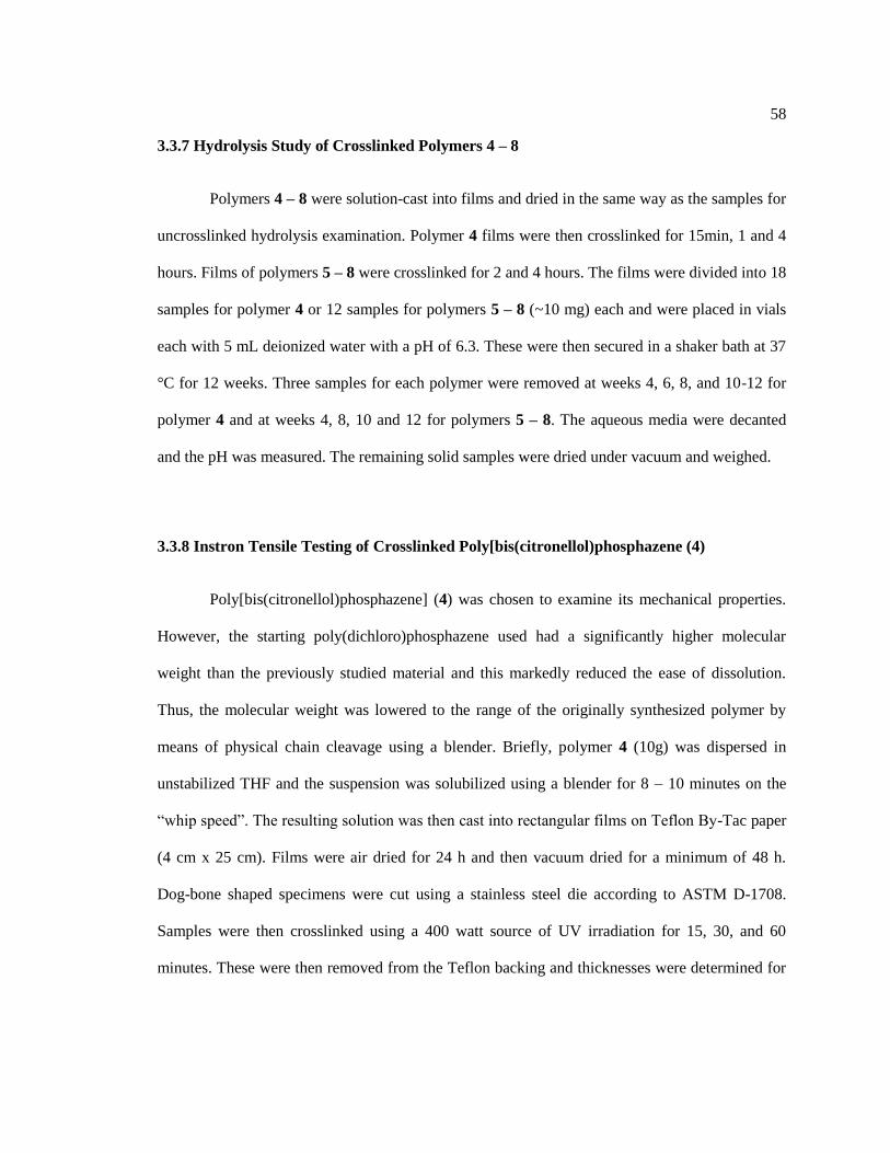
58
3.3.7 Hydrolysis Study of Crosslinked Polymers 4 – 8
Polymers 4 – 8 were solution-cast into films and dried in the same way as the samples for
uncrosslinked hydrolysis examination. Polymer 4 films were then crosslinked for 15min, 1 and 4
hours. Films of polymers 5 – 8 were crosslinked for 2 and 4 hours. The films were divided into 18
samples for polymer 4 or 12 samples for polymers 5 – 8 (~10 mg) each and were placed in vials
each with 5 mL deionized water with a pH of 6.3. These were then secured in a shaker bath at 37
°C for 12 weeks. Three samples for each polymer were removed at weeks 4, 6, 8, and 10-12 for
polymer 4 and at weeks 4, 8, 10 and 12 for polymers 5 – 8. The aqueous media were decanted
and the pH was measured. The remaining solid samples were dried under vacuum and weighed.
3.3.8 Instron Tensile Testing of Crosslinked Poly[bis(citronellol)phosphazene (4)
Poly[bis(citronellol)phosphazene] (4) was chosen to examine its mechanical properties.
However, the starting poly(dichloro)phosphazene used had a significantly higher molecular
weight than the previously studied material and this markedly reduced the ease of dissolution.
Thus, the molecular weight was lowered to the range of the originally synthesized polymer by
means of physical chain cleavage using a blender. Briefly, polymer 4 (10g) was dispersed in
unstabilized THF and the suspension was solubilized using a blender for 8 – 10 minutes on the
“whip speed”. The resulting solution was then cast into rectangular films on Teflon By-Tac paper
(4 cm x 25 cm). Films were air dried for 24 h and then vacuum dried for a minimum of 48 h.
Dog-bone shaped specimens were cut using a stainless steel die according to ASTM D-1708.
Samples were then crosslinked using a 400 watt source of UV irradiation for 15, 30, and 60
minutes. These were then removed from the Teflon backing and thicknesses were determined for

59
each sample. Tests were performed at a fixed crosshead speed of 10 mm min-1
to breaking using a
100 N load cell. At least 10 specimens were used for each test.
3.4 Conclusions
Citronellol-containing polyphosphazenes have been synthesized. The crosslink density of
these polymers can be controlled via their exposure to ultraviolet light. This also affects their
hydrolysis rate. The more alanine ethyl ester incorporated as a co-substituent in the polymer the
faster is the hydrolysis rate and the longer the UV exposure time needed to crosslink the
polymers. The final properties can therefore be controlled by tuning the amount of alanine ethyl
ester in the polymer and the level of UV exposure. This was also supported by the change in
mechanical properties with increased crosslink density. Due to the tunability of this system these
polymers are excellent candidates for further evaluation as tissue engineering scaffolds.
3.5 Acknowledgements
The authors thank Tomasz Modzelewski for his valuable discussions.
3.6 References
1. Goh, J. C.-H.; Ouyang, H.-W.; Teoh, S.-H.; Chan, C. K. C.; Lee, E.-H. Tissue
Engineering 2003, 9, S-31-S-44
2. Chen, G.; Ushida, T.; Tateishi, T. Macromol. Biosci. 2002, 2, 67-77.
3. Shoichet, M. S. Macromolecules 2010, 43, 581-591.
4. Gloria, A.; Santis, R. D.; Ambrosio, L. Journal of Applied Biomaterials & Biomechanics
2010, 8, 57-67.
5. Laurencin, C. T.; Ambrosio, A. M. A.; Borden, M. D.; Cooper, J., J. A. Annu. Rev.
Biomed. Eng. 1999, 1, 19-46.
6. Place, E. S.; Evans, N. D.; Stevens, M. M. Nature Materials 2009, 8, 457-470.

60
7. Ge, Z.; Yang, F.; Goh, J. C. H.; Ramakrishna, S.; Lee, E.-H. Journal of Biomedical
Materials Research Part A 2006, 77, 639-652.
8. Vanjak-Novakovic, G.; Altman, G.; Horan, R.; Kaplan, D. L. Annu. Rev. Biomed. Eng.
2004, 6, 131-156.
9. Laurencin, C. T.; Freeman, J. W. Biomaterials 2005, 26, (36), 7530-7536.
10. Bostman, O.; Pihlajamaki, H. Biomaterials 2000, 21, 2615-2621.
11. Allcock, H. R. Journal of Inorganic and Organometallic Polymers 1992, 2, (2), 197-211.
12. Allcock, H. R. Science 1992, 255, 1106-1112.
13. Allcock, H. R. Science 1976, 193, 1214-1219.
14. Allcock, H. R., Chemistry and Applications of Polyphosphazenes. John Wiley & Sons,
Inc.: Hoboken, NJ, 2003; Vol. 1, p 725.
15. Potin, P.; De Jaeger, R. European Polymer Journal 1991, 27, 341-348.
16. Allcock, H. R.; Pucher, S. R. Macromolecules 1994, 27, (5), 1071-1075.
17. Allcock, H. R.; Pucher, S. R.; Scopelianos, A. G. Biomaterials 1994, 15, (8), 563-569.
18. Allcock, H. R.; Morozowich, N. L. Polymer Chemistry 2012, 3, 578-590.
19. Nichol, J. L.; Morozowich, N. L.; Allcock, H. R. Polymer Chemistry 2013, 4, 600-606.
20. Allcock, H. R.; Connolly, M. S.; Sisko, J. T.; Al-Shali, S. Macromolecules 1988, 21, (2),
323-334.
21. Li, Y.; Thouas, G. A.; Chen, Q.-Z. RSC Advances 2012, 2, 8229-8242.
22. Brito, R. G.; Guimaraes, A. G.; Quintans, J. S. S.; Santos, M. R. V.; Sousa, D. P. D.; Jr.,
D. B.-P.; Jr., W. d. L.; Brito, F. A.; Barreto, E. O.; Oliveira, A. P.; Jr., L. J. Q. Journal of Natural
Medicine 2012, 66, 637-644.
23. Griffin, S. G.; Wyllie, S. G.; Markham, J. L.; Leach, D. N. Flavour and fragrance
journal 1999, 14, 322-332.
24. Paduch, R.; Kandefer-Szerszen, M.; Trytek, M.; Fiedurek, J. Arch. Immunol. Ther. Exp.
2007, 55, 315-327.
25. Hahn, S. K.; Jelacic, S.; Maier, R. V.; Stayton, P. S.; Hoffman, A. S. Journal of
Biomaterials Science, Polymer Edition 2004, 15, (9), 1111-1119.
26. Qureshi, A. T.; Terrell, L.; Monroe, W. T.; Dasa, V.; Janes, M. E.; Gimble, J. M.; Hayes,
D. J. Journal of Tissue Engineering and Regenerative Medicine 2014, 8, 386-395.
27. Morozowich, N. L.; Weikel, A. L.; Nichol, J. L.; Chen, C.; Nair, L. S.; Laurencin, C. T.;
Allcock, H. R. Macromolecules 2011, 44, 1355-1364.
28. Weikel, A. L.; Krogman, N. R.; Nguyen, N. Q.; Nair, L. S.; Laurencin, C. T.; Allcock, H.
R. Macromolecules 2009, 42, 636-639.
29. Dhandayuthapani, B.; Yoshida, Y.; Maekawa, T.; Kumar, D. S. International Journal of
Polymer Science 2011, 2011, 1-19.
30. Gunatillake, P. A.; Adhikari, R. European Cells and Materials 2003, 5, 1-16.
31. Singh, A.; Krogman, N. R.; Sethuraman, S.; Nair, L. S.; Sturgeon, J. L.; Brown, P. W.;
Laurencin, C. T.; Allcock, H. R. Biomacromolecules 2006, 7, 914-918.
32. Kuo, C. K.; Marturano, J. E.; Tuan, R. S. Sports Med. Arthrosc Rehabil. Ther. Technol.
2010, 2, (20), 1-14.
33. Collins, E. A.; Bares, J.; Billmeyer, F. W., Experiments in Polymer Science. John Wiley
& Sons New York, NY, 1973.
34. Barton, A. F. M., Handbook of Polymer-Liquid Interaction Parameters and Solubility
Parameters CRC Press: Boca Raton, FL 1990.
35. Hansen, C. M., Hansen Solubility Parameters. CRC Press New York, NY, 2007.
36. Barton, A. F. M. Pure & Appl. Chem. 1985, 57, (7), 905-912.
37. Orme, C. J.; Harrup, M. K.; McCoy, J. D.; Weinkauf, D. H.; Stewart, F. F. Journal of
Membrane Science 2002, 197, 89-101.

61
38. Allcock, H. R.; Kwon, S.; Riding, G. H.; Fitzpatrick, R. J.; Bennett, J. L. Biomaterials
1988, 9, 509-513.
39. Barton, A. F. M., CRC Handbook of Solubility Parameters and Other Cohesion
Parameters. 2nd ed.; CRC Press Boston, 1991.

62
Chapter 4
Amino Acid Citronellol Ester Polymers for Biomedical Applications
4.1 Introduction
Current solutions for repair of the millions of ligament and tendon injuries occurring
annually primarily utilize autografts and allografts. These are non-ideal.1 The shortcomings
include limited living tissue availability, increased healing times, and the risk of infection.2
Among the alternative solutions currently under investigation, the field of tissue engineering has
emerged as a prominent candidate.3-6
This approach uses a biodegradable polymer scaffold,
seeded with cells and signaling molecules, for implantation in place of the damaged ligament or
tendon.7, 8
As the polymer degrades slowly the incorporated signaling molecules induce the body
to slowly rebuild the damaged tissue.9, 10
A number of natural and synthetic polymers have been
investigated for this application as they meet some of the necessary scaffold characteristics such
as biocompatibility or biodegradability.11-14
Thus, silk and collagen have been studied
extensively; however, their use is limited by batch to batch inconsistencies and uncontrollable
enzymatic degradation in the body.15
One of the most commonly studied synthetic polymers is
poly(lactic acid), due to its good mechanical properties and FDA approval, but its hydrolysis into
acidic monomers and oligomers can cause tissue necrosis at the implant site and this has limited
its viability.16
Another key requirement for a scaffold material is that it must have similar
mechanical properties to those of the parent tissue.17
An elastomeric polymer would be an ideal
matrix material for tendons and ligaments because the scaffold would be subjected to numerous
loading and unloading cycles.18
Attempts are also being made to improve the performance of
63
natural and synthetic fibers using different scaffold fabrication techniques such as knitting or
braiding.19-22
In the current work we have explored a new method for polymer synthesis to favor the
properties required for ligament and tendon tissue engineering applications. For this, we used
polyphosphazenes due to their high degree of synthetic tunability.23, 24
These macromolecules
possess a backbone of alternating phosphorus and nitrogen atoms with two organic groups
attached to each phosphorus atom.25
They are synthesized from a reactive macromolecular
intermediate, poly(dichlorophosphazene), (NPCl2)n, by replacement of the chlorine atoms by
reactions with various alkoxide or amino nucleophiles.26
The properties of the resultant polymers
are controlled by both the properties of the skeleton and those of the organic side groups.27, 28
The
goal of this work was to utilize side groups that would facilitate degradation of the polymers to
non-toxic products, while also favoring the generation of elastomeric characteristics. Previous
research in our program demonstrated that amino acid ethyl ester polyphosphazenes hydrolyze
into the non-toxic by-products, specifically amino acid, ethanol, phosphates, and ammonia,
fulfilling the first requirement for a bioerodible biocompatible material.29, 30
The flexibility of the
polyphosphazene backbone makes it possible to design polymers with low glass transition
temperatures (as low as -100 °C).31, 32
The combination of flexible polymers with crosslinkable
side groups together with the additional ability of the system to break down slowly should
provide access to polymers that meet the requirements for ligament or tendon replacement or
repair.33
To accomplish this objective we used citronellol units connected to the polymer
backbone through an amino acid ester linker unit. These structures maintained a high constant
citronellol density along the polymer chain, but allowed the steric hindrance of different amino
acid units to control the hydrolytic degradation rate. This differs from earlier work in which
citronellol was attached directly to a polyphosphazene backbone, a situation in which the

64
hydrolysis rate could only be tuned by incorporating various ratios of a second type of side group,
an option that decreased the amount of citronellol that could be incorporated into the final
polymer.34
Thus, in this work we explore the use of citronellol as a carboxylic acid ester moiety
for the amino acids glycine, alanine, valine, and phenylalanine.
4.2 Results and Discussion
4.2.1 Synthesis of Amino Acid Citronellol Ester Side Groups
Every amino acid used for chlorine replacement in phosphazenes requires the carboxylic
acid moiety to be protected to avoid side reactions.35, 36
Traditionally this was accomplished by
using the ethyl ester of the amino acid.37
Other previous work has utilized longer chain alcohols
to esterify the carboxylic acid units in an attempt to determine the effect of the steric bulk of the
alcohol on the hydrolysis rate and physical properties of the final polymers.29, 30, 38
However, in
this present work citronellol was used to esterify the amino acid as a means to provide the
necessary unsaturated functionality which could act as a crosslink site. In addition, citronellol has
antimicrobial and anti-inflammatory properties, which may be beneficial for a tissue engineering
application.39-42
The syntheses for all side groups followed a similar synthetic protocol (Figure 4-
1). These yielded the side group unit as its HCl salt.38
The HCl salt was then converted to the
neutral form utilizing triethylamine as a base. Triethylamine also serves to capture the HCl
released when the amino functionality reacts with the P-Cl bond of the polymer. The insolubility
of the TEA-HCl complex provides a driving force for both reactions. This is important because
free HCl in the reaction medium could protonate the polymer backbone and cause skeletal
cleavage.
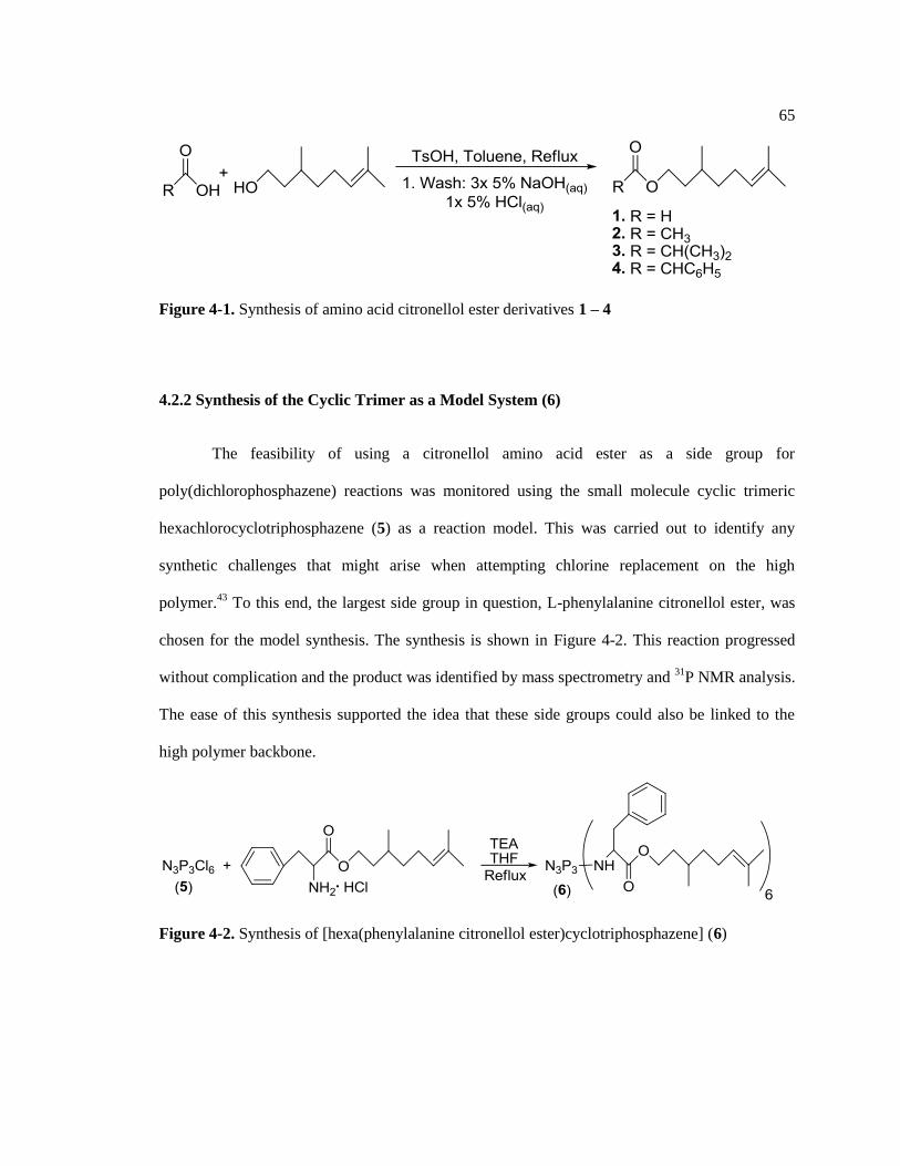
65
Figure 4-1. Synthesis of amino acid citronellol ester derivatives 1 – 4
4.2.2 Synthesis of the Cyclic Trimer as a Model System (6)
The feasibility of using a citronellol amino acid ester as a side group for
poly(dichlorophosphazene) reactions was monitored using the small molecule cyclic trimeric
hexachlorocyclotriphosphazene (5) as a reaction model. This was carried out to identify any
synthetic challenges that might arise when attempting chlorine replacement on the high
polymer.43
To this end, the largest side group in question, L-phenylalanine citronellol ester, was
chosen for the model synthesis. The synthesis is shown in Figure 4-2. This reaction progressed
without complication and the product was identified by mass spectrometry and 31
P NMR analysis.
The ease of this synthesis supported the idea that these side groups could also be linked to the
high polymer backbone.
Figure 4-2. Synthesis of [hexa(phenylalanine citronellol ester)cyclotriphosphazene] (6)

66
4.2.3 Synthesis and Characterization of Polymers 8 – 11
All the polymer substitution reactions were performed in a similar manner, as shown in
Figure 4-3. The reactive intermediate, poly(dichlorophosphazene) (7), was generated by the ring-
opening polymerization of hexachlorocyclotriphosphazene (5) in a sealed system at 250 °C. This
polymer was then treated with nucleophiles 1 – 4 for chlorine replacement as discussed.
Triethylamine (TEA) was added to the reaction mixture as an acid scavenger to sequester the
hydrogen chloride (HCl) generated during the substitution reaction as an insoluble complex.
However, the TEA-HCl complex is slightly soluble in the reaction mixture and is a potential
cause of backbone cleavage.44
Thus, the substitution reaction was monitored by 31
P NMR
spectroscopy and was promptly terminated and the polymer purified before skeletal cleavage
became serious. Specifically, the disappearance of the poly(dichlorophosphazene) 31
P peak at δ =
-17 ppm and the appearance of a new peak at δ = 0 ppm, representing a polyphosphazene unit
substituted with two amino acid ester side groups, and this was used to determine when the
substitution reaction was complete. The characterization data for the resultant amino acid
citronellol ester polymers are shown in Table 4-1. Representative 1H and
31P spectra are provided
for polymer 8 shown in Figure 4-4.
Figure 4-3. Synthesis of amino acid citronellol ester polymers 8 – 11

67
Table 4-1. Characterization data of amino acid citronellol ester polymers 8 – 11
Polymer 1H NMR (ppm)
*
31P NMR (ppm)
* Mw (kDa) R.U.
**
8 5.1 (1H), 4.0 (2H), 3.7 (2H), 2.0 (2H), 1.7
(3H), 1.6 (3H), 1.3 (3H), 1.2 (2H), 0.88
(3H)
2.5 3,421 7,248
9 5.0 (1H), 4.2 (1H), 4.0 (2H), 1.9 (2H), 1.6
(3H), 1.5 (3H), 1.4 (5H), 1.2 (3H), 0.87
(3H)
-1.1 1,111 2,221
10 5.0 (1H), 4.2 (1H), 4.0 (2H), 2.0 (2H), 1.6
(3H), 1.5 (3H), 1.3 (5H), 1.1 (1H), 0.88
(9H)
0.89 556 264
11 7.1 (5H), 5.0 (1H), 4.2 (1H), 3.8 (2H), 3.1
(2H), 1.8 (2H), 1.6 (3H), 1.5 (3H), 1.1
(5H), 0.66 (3H)
-1.5 126 192
* in CDCl3
** R.U. = Repeat Units

68
Figure 4-4. 1H NMR spectrum of polymer 8 (top) and
31P NMR (bottom)
The reaction time required for total chlorine replacement increased as the steric hindrance
at the α-carbon position of the amino acid increased. Glycine citronellol ester required the
shortest reaction time (24 h), whereas the phenylalanine citronellol ester needed the longest (9 d).
Analysis by GPC confirmed that the increasing reaction times correlated directly with decreased
polymer molecular weights presumably due to the prolonged exposure to free HCl in the reaction
media.

69
4.2.4 Hydrolysis Behavior of Uncrosslinked Polymers
Solid polymers 8 – 11 were examined for their hydrolytic susceptibility over a twelve
week period at 37 °C in deionized water, to determine their viability as scaffolding materials.
Figure 4-5 shows polymer film mass loss and Figure 4-6 illustrates the molecular weight decline.
The hydrolysis rate decreased with increasing steric hindrance at the α-carbon of the amino acid
ester and this was evident from both the mass loss and molecular weight decline. The order of
polymer hydrolytic sensitivity from fastest hydrolyzing to slowest was 8 > 9 > 10 > 11. These
results are similar to previous reports involving amino acid ester protected polyphosphazenes
(methyl, ethyl, octyl, benzyl, etc.).29, 30, 38
Figure 4-5. Percent film mass loss of polymers 8 – 11
70
75
80
85
90
95
100
0 2 4 6 8 10 12
% M
ass
Loss
Weeks of Hydrolysis
8
9
10
11

70
Figure 4-6. Molecular weight decline of polymers 8 – 11
4.2.5 Polymer Crosslinking and Swelling Studies
Polymers 8 – 11 were crosslinked by irradiation with UV light. The degree of
crosslinking was then estimated by solvent swelling studies. The crosslinking time was based on
the level of UV irradiation needed for the polymer to maintain its general shape when swollen.
The maximum irradiation time was chosen as the point at which the sample became brittle.
Although numerous solvents were examined, DMF was found to have the best ability to swell all
the polymers. Crosslinked polymers were allowed to equilibrate in DMF for 72 hr at room
temperature. They were then removed from the solvent and weighed in the swollen state. The
polymers were then dried under vacuum for 48 h to obtain their dry mass.45
The mass difference
0
500
1000
1500
2000
2500
3000
0 2 4 6 8 10 12
Mw
(kD
a)
Weeks
8
9
10
11

71
was then used to determine the ratio of the volume of the polymer compared to the volume of the
polymer in the swollen gel, or Vp. Using this value in the Flory-Rehner equation, shown below,
the average molecular mass between crosslinks (Mc) was determined by46
:
(1/Mc) = [ln(1-Vp) + Vp + χ12Vp2]/[V1(Vp
0.33-(Vp/2))]
In this equation, the variable V1 corresponds to the molar volume of the solvent with is
available in the literature for the chosen solvent (V1 = 77 for DMF)47
. The chi parameter was
calculated using the following equation:
χ12 = β + [V1(δd p – δd s)2]/RT
The dispersion parameter for the solvent (δd s = 17.4 for DMF) as well as the empirical
constant β (0.3) were found in the literature.47
The polymer dispersion parameter (δd p) was
calculated for the polyphosphazenes using the following equation48
:
δd p = Σi Fi / Σi Vi
Taking the sum of the molar attraction constants of each group and dividing them by the
sum of the corresponding molar volume constant for each group yields δ d p. The group
contribution factors used to calculate the dispersion parameters for polymers 8 – 11 are shown in
Table 4-2.49
The calculated δd p and corresponding chi parameters are shown in Table 4-3. This
approach has been used in previous work for other polyphosphazenes.50, 51
Table 4-2. Group contribution parameters for polymers 8 – 11
Functional Group V (cm3/mol)
zFd (MPa)
0.5
Phosphorus 8.8 164
Nitrogen 4.0 164
-CH2- 16.6 270
-CH3 31.7 419
>CH- -1.0 80

72
=CH- 12.4 223
=C< -5.7 45
-NH- 4.5 160
-COO- (ester) 8.2 667
Phenyl 75.4 1499
Table 4-3. Calculated dispersion parameters and chi parameters for polymers 8 – 11
Polymer δd p (MPa0.5
) χ12
8 19.45 0.47
9 19.24 0.45
10 18.87 0.41
11 19.92 0.54
The average number of crosslinks per polymer chain generated as a function of UV
exposure time for polymers 8 – 11 is shown in Table 4-4. These values were calculated by
dividing the polymer molecular weight by the average molecular weight between crosslinks (Mc).
The number of crosslinks increased as UV exposure time increased. However, this number cannot
be compared directly from one polymer to the next because each is based on the molecular weight
of the individual polymer.
Table 4-4. Number of crosslinks for polymers 8 – 11 corresponding to UV exposure time in
minutes
Time P8 P9 P10 P11
30 881 472 64 8
60 1600 789 97 18

73
120 2056 1355 123 27
240 3797 2005 177 38
4.2.6 Thermal Behavior of Uncrosslinked and Crosslinked Polymers
Differential scanning calorimetry was used to determine the thermal properties of the
polymers. The glass transition temperatures (Tg) of polymers 8 – 11 were significantly lower than
those of their ethyl ester counterparts (Table 4-5). Thus, the presence of the citronellol unit causes
a roughly 50 °C decrease in each polymers glass transition temperature presumably due to its
high degree of torsional freedom.29, 30
No melt transitions (Tm) were detected for any of the
polymers. The glass transition temperatures were also monitored as a function of UV exposure.
All the polymers followed the same trend, wherein the Tg increased and broadened with increased
UV exposure.52
Table 4-5. Glass transition temperature comparison between the amino acid citronellol ester
polyphosphazenes (8 – 11) and the amino acid ethyl ester polyphosphazenes (°C)29, 30
Polymer Amino Acid Citronellol Ester Ethyl Ester
8 Glycine -62.7 -20.0
9 Alanine -58.2 -10.0
10 Valine -23.0 24.8
11 Phenylalanine -10.5 41.6
4.2.7 Polymer Mechanical Property Evaluation
In addition to changing the thermal properties of the polymers, the crosslink density also
changes the polymer mechanical properties. This is important as it allows the development of

74
materials that have similar mechanical properties to those of the tissue being replaced. Two
polymers, poly(alanine citronellol ester)phosphazene and poly(phenylalanine citronellol
ester)phosphazene, were chosen for direct mechanical property evaluations as a function of
increasing UV exposure time. These polymers were also selected to determine the role that steric
hindrance at the α-carbon position plays on mechanical properties. Both uncrosslinked and
crosslinked dogbone shaped samples were examined for their mechanical properties. The
crosslinked samples were made by placing the precut specimens in the UV reactor for 30, 60,
120, and 240 minutes. Mechanical data for poly(alanine citronellol ester)phosphazene (9) are
shown in Table 4-6. For this example it was found that the modulus increased with increasing UV
exposure while the tensile strength decreased. This trend was also found for a
poly[bis(citronellol)phosphazene] described in a previous report.34
As speculated previously, this
could be due to over-crosslinking the polymer.52
Mechanical data for poly(phenylalanine
citronellol ester)phosphazene (10) are shown in Table 4-7. For this sample both the modulus and
tensile strength increased with increasing UV exposure. It was also found that the presence of
increased steric hindrance at the α-carbon position of the amino acid ester increased both the
modulus and tensile strength. This effect is evident from a comparison between the phenylalanine
containing polymers and the alanine containing derivatives. Thus, the high degree of tunability
associated with these polymers makes them excellent candidates for ligament and tendon tissue
engineering scaffolds. The properties of each polymer can be tuned to match the mechanical
requirements of the specific damaged ligament or tendon.
Table 4-6. Mechanical properties of poly(alanine citronellol ester)phosphazene (9)
UV Exposure (min) Modulus Tensile Strength (MPa)
0 0.073 ± 0.006 0.18 ± 0.03
30 0.10 ± 0.01 0.13 ± 0.01

75
60 0.14 ± 0.02 0.10 ± 0.07
120 0.50 ± 0.01 0.09 ± 0.01
240 1.00 ± 0.01 0.09 ± 0.01
Table 4-7. Mechanical properties of poly(phenylalanine citronellol ester)phosphazene (11)
UV Exposure (min) Modulus Tensile Strength (MPa)
0 3.0 ± 0.3 0.46 ± 0.02
30 8.6 ± 0.9 0.54 ± 0.01
60 10.0 ± 0.7 0.65 ± 0.06
120 11.8 ± 0.8 0.82 ± 0.07
240 22 ± 2 1.08 ± 0.07
4.3 Experimental Section
4.3.1 Reagents and Equipment
Syntheses were performed under a dry argon atmosphere using standard Schlenk line
techniques. Before use, glassware was dried overnight in an oven at 125 °C. Tetrahydrofuran
(THF) (EMD) and triethylamine (EMD) were dried using solvent purification columns. L-
Phenylalanine, L-valine, L-alanine, glycine (all Chem-Impex), sodium metal (Aldrich), sodium
hydroxide (BDH), hydrochloric acid (EMD), p-toluenesulfonic acid monohydrate (Sigma
Aldrich), toluene (EMD), dimethylformamide (EMD), dichloromethane (EMD), hexanes (EMD),
and methanol (EMD) were used as received. Citronellol (Alfa Aesar) was distilled from sodium
metal and stored over molecular sieves (EMD, 4 Å mesh beads) under an argon atmosphere.

76
Poly(dichlorophosphazene) was prepared by the thermal ring-opening polymerization of
recrystallized and sublimed hexachlorocyclotriphosphazene (Fushimi Chemical Co., Japan) in
evacuated Pyrex tubes at 250 °C. Spectra/Por molecular porous cellulose dialysis membranes
with molecular weight cut-off of 12,000-14,000 were employed for purification of the polymers.
1H and
31P NMR spectra were obtained using a Bruker 360 WM instrument operated at 145 and
360 MHz, respectively, with 31
P shifts relative to 85 % H3PO4 at 0 ppm as a reference. Glass
transition temperatures were obtained using a TA Instruments Q10 differential scanning
calorimetry apparatus with a heating rate of 10 °C/ min and a sample size of ca. 10 mg. Gel
permeation chromatography experiments were carried out using a Hewlett-Packard 1047A
refractive index detector and two Phenomenex Phenogel 10μm linear columns with elution times
calibrated using polystyrene standards. The samples were eluted using 1.0 mL/min with a 10 mM
solution of tetra-n-butylammonium nitrate (Alfa Aesar) in THF with elution times calibrated with
polystyrene standards. pH data were obtained using a VWR Symphony SB70P pH meter.
Polymer crosslinking was performed using a Rayonet Photochemical Reactor with 400 watt UV
radiation bulbs (λ = 254nm). Samples for mechanical testing samples were cut using a Pioneer-
Dietecs stainless steel die with dimensions specified in ASTM D-1708. Mechanical properties
were evaluated using an Instron 5866 tensile testing equipment at a fized crosshead speed of 10
mm min-1
equipped with a 100 N load cell. BlueHill software was used for data collection and
analysis.
4.3.2 Synthesis of Amino Acid Citronellol Esters 1 – 4
Amino Amino acid citronellol ester derivatives 1 – 4 were synthesized using a previously
published procedure.38, 53
L-glycine citronellol ester (1) is given as a representative example.
Glycine (20.0 g, 0.266 mol), p-toluenesulfonic acid monohydrate (60.8 g, 0.320 mol), and

77
citronellol (48.5 mL, 0.266 mol) were dissolved in toluene (250 mL). This mixture was refluxed
for 24 h and the water generated (~ 11 mL) was collected using a Dean-Stark apparatus. Toluene
was then removed under reduced pressure to give the p-toluenesulfonic acid amino acid
citronellol ester salt in quantitative yield. This solid was then dissolved in dichloromethane and
was extracted three times with 5 % NaOH(aq) and once with 5 % HCl(aq). The organic layer was
removed under reduced pressure and the resultant solid was dried under vacuum to give the final
product as the hydrochloride salt (Table 4-8).
Table 4-8. Characterization data of amino acid citronellol ester side groups 1 – 4
Side group 1H NMR (ppm in CDCl3) Yield
1 8.5 (3H), 5.1 (1H), 4.2 (2H), 4.0 (2H), 2.0 (2H), 1.7 (3H), 1.6
(3H), 1.3 (4H), 1.1 (1H), 0.88 (3H)
78%
2 8.7 (3H), 5.0 (1H), 4.2 (3H), 3.7 (1H), 2.0 (2H), 1.7 (3H), 1.6
(3H), 1.3 (4H), 1.2 (1H), 0.89 (3H)
89%
3 8.8 (3H), 5.0 (1H), 4.2 (2H), 3.9 (1H), 2.0 (2H), 1.7 (3H), 1.6
(3H), 1.5 (4H), 1.3 (1H), 1.1 (6H), 0.89 (3H)
86%
4 8.9 (3H), 7.4 (5H), 5.1 (1H), 4.4 (2H), 4.2 (2H), 2.0 (2H), 1.8
(3H), 1.7 (3H), 1.4 (4H), 1.2 (1H), 0.90 (3H)
90%
4.3.3 Synthesis of Hexa(phenylalanine citronellol ester) cyclotriphosphazene (6)
Hexachlorocyclotriphosphazene (0.50 g, 1.4 mmol) was dissolved in THF (25 mL). L-
Phenylalanine citronellol ester hydrochloride (4.9 g, 14.4 mmol) and triethylamine (4.0 mL, 28.8
mmol) were dissolved in THF (25 mL). This mixture was then refluxed for 12 h and was then
added via filter addition funnel to the trimer solution. The mixture was refluxed for 72 h after

78
which a fully-substituted product peak was detected by mass spectrometric analysis (m/z 1950
[M+H]). After an additional 20 days at reflux a singlet peak was detected, which indicated full
substitution. Solvent was removed under reduced pressure and the product was dissolved in
dichloromethane and extracted twice with water to yield the final product. 31
P (145 MHz, CDCl3)
δ 15.9 ppm. 1H NMR (360 MHz, CDCl3); δ 7.2 (5H), 5.0 (1H), 4.2 (1H), 3.8 (2H), 3.1 (2H), 1.8
(2H), 1.6 (3H), 1.5 (3H), 1.1 (5H), 0.68 (3H).
4.3.4 Synthesis of Poly(amino acid citronellol ester)polyphosphazenes 8 – 11
Synthesis of polymers 8 – 11 followed similar procedures, with polymer 8 described as a
representative example. Poly(dichlorophosphazene) (3.00 g, 25.9 mmol) was dissolved in THF
(300 mL). Glycine citronellol ester hydrochloride (39.0 g, 155 mmol) was dissolved in THF (150
mL), and triethylamine (43 mL, 311 mmol) was added. This mixture was refluxed for 12 h, and
was subsequently filtered and added to the poly(dichlorophosphazene) solution which was then
refluxed. Once complete, the reaction mixture was cooled to room temperature, filtered,
concentrated, and precipitated once into methanol. The polymer was re-dissolved in THF, stirred
with 15 mL of triethylamine, concentrated, and precipitated into methanol twice. Finally, the
polymer was dissolved in THF, concentrated and precipitated into methanol three times to yield
an off-white solid.
4.3.5 Hydrolysis of Polymers 8 – 11
Each of the polymers 8 – 11 was dissolved in 18 mL of unstabilized THF (2.5 w/v%) and
was then solution-cast into square 100 μm thick films (5 cm x 5 cm). These were air dried for 24
h and vacuum dried for 48 h. The films were divided into 36 samples (~10 mg each) and were
79
placed in vials each with 5 mL deionized water with a pH of 6.3. These were then agitated in a
shaker bath at 37 °C for 12 weeks. Each week three samples were removed for an individual
polymer. The aqueous media were decanted and the pH was measured to ensure that hydrolysis
did not cause a change in solution pH. The remaining solid samples were dried under vacuum and
weighed. After weighing, each solid sample was dissolved in THF and the molecular weight was
estimated by GPC.
4.3.6 UV-Crosslinking and Swelling Studies of Polymers 8 – 11
Polymers 8 – 11 were solution-cast into square films (in a similar manner to the
hydrolysis samples) and were dried under vacuum. The polymers were then crosslinked using a
400 watt source of UV irradiation in a Rayonet Photochemical Reactor (λ = 254nm). The
polymers were irradiated for 30, 60, 120, and 240 minutes. Polymers crosslinked at each time
point were then divided into four ~25 mg samples. These were placed in vials and covered with
10 mL DMF, similarly to a previously published procedure.45
The polymers were then allowed to
equilibrate at room temperature for 72 h. After this time, samples were removed from the DMF,
blotted to remove excess solvent, and then weighed in their swollen state. The samples were then
dried for several days under vacuum and weighed again.
4.3.7 Instron Tensile Testing of Polymers 9 and 11
Polymers films were prepared by dissolving 10 g of each polymer in unstabilized THF
and then casting the solution into rectangular films on Teflon By-Tac coated paper (4 cm x 25
cm). Films were air dried for 48 h and then vacuum dried for a minimum of 48 h. Dog-bone
shaped specimens were cut using a stainless steel die according to ASTM D-1708. All tests were

80
performed at a fixed crosshead speed of 10 mm min-1
to break using a 100 N load cell, with at
least 10 specimens analyzed for each test.
4.4 Conclusions
The linkage of citronellol esterified amino acid units to a polyphosphazene chain
provides access to a new series of bioerodible elastomers. The hydrolysis rates can be controlled
by the steric hindrance of the R group at the α-carbon position of the amino acid residue, in spite
of the presence of two citronellol residues per repeat unit. Short UV exposure times are needed to
introduce crosslinks and generate elastomeric character. Both the steric hindrance of the R group
and the UV exposure time can be used to tune the polymer mechanical properties. This range of
tunable properties makes these polymers excellent candidates for ligament or tendon tissue
engineering scaffolds.
4.5 Acknowledgements
The authors thank Tomasz Modzelewski for his valuable discussions.
4.6 References
1. Caliari, S. R.; Ramirez, M. A.; Harley, B. A. C. Biomaterials 2011, 32, 8990-8998.
2. Freeman, J. W.; Kwansa, A. L. Recent Patents on Biomedical Engineering 2008, 1, 18-
23.
3. Shoichet, M. S. Macromolecules 2010, 43, 581-591.
4. Laurencin, C. T.; Ambrosio, A. M. A.; Borden, M. D.; Cooper, J., J. A. Annu. Rev.
Biomed. Eng. 1999, 1, 19-46.
5. Nair, L. S.; Laurencin, C. T. Progress in Polymer Science 2007, 32, 762-798.
6. Langer, R.; Vacanti, J. P. Science 1993, 260, 920-926.
7. Goh, J. C.-H.; Ouyang, H.-W.; Teoh, S.-H.; Chan, C. K. C.; Lee, E.-H. Tissue
Engineering 2003, 9, S-31-S-44

81
8. Place, E. S.; Evans, N. D.; Stevens, M. M. Nature Materials 2009, 8, 457-470.
9. Hampson, K.; Forsyth, N. R.; Haj, A. E.; Maffulli, N., Tendon Tissue Engineering In
Topics in Tissue Engineering, 2008; Vol. 4 pp 1 - 21
10. Pallua, N.; Suschek, C. V., Tissue Engineering. Springer: Berlin, 2011.
11. Ge, Z.; Yang, F.; Goh, J. C. H.; Ramakrishna, S.; Lee, E.-H. Journal of Biomedical
Materials Research Part A 2006, 77, 639-652.
12. Leong, N. L.; Petrigliano, F. A.; McAllister, D. R. Journal of Biomedical Materials
Research Part A 2014, 102A, (5), 1614-1624.
13. Liu, Y.; Ramanath, H. S.; Want, D.-A. Trends in Biotechnology 2008, 26, (4), 201-209.
14. Reis, R. L.; Roman, J. S., Biodegradable Systems in Tissue Engineering and
Regenerative Medicine. CRC Press: New York, 2005.
15. Chen, G.; Ushida, T.; Tateishi, T. Macromol. Biosci. 2002, 2, 67-77.
16. Bostman, O.; Pihlajamaki, H. Biomaterials 2000, 21, 2615-2621.
17. Vanjak-Novakovic, G.; Altman, G.; Horan, R.; Kaplan, D. L. Annu. Rev. Biomed. Eng.
2004, 6, 131-156.
18. Kuo, C. K.; Marturano, J. E.; Tuan, R. S. Sports Med. Arthrosc Rehabil. Ther. Technol.
2010, 2, (20), 1-14.
19. Fan, H.; Liu, H.; Toh, S. L.; Goh, J. C. H. Biomaterials 2009, 30, (2009), 4967-4977.
20. Laurencin, C. T.; Freeman, J. W. Biomaterials 2005, 26, (36), 7530-7536.
21. Shen, W.; Chen, X.; Chen, J.; Yin, Z.; Heng, B. C.; Chen, W.; Ouyang, H.-W.
Biomaterials 2010, 31, 7239-7249.
22. Lu, H. H.; Jr., J. A. C.; Manuel, S.; Freeman, J. W.; Attawia, M. A.; Ko, F. K.; Laurencin,
C. T. Biomaterials 2005, 26, 4805-4816.
23. Allcock, H. R., Chemistry and Applications of Polyphosphazenes. John Wiley & Sons,
Inc.: Hoboken, NJ, 2003; Vol. 1, p 725.
24. Allcock, H. R. Annals of the New York Academy of Sciences 1997, 831, (1), 13-31.
25. Allcock, H. R. Journal of Inorganic and Organometallic Polymers 1992, 2, (2), 197-211.
26. Allcock, H. R. Science 1976, 193, 1214-1219.
27. Allcock, H. R. Science 1992, 255, 1106-1112.
28. Andrianov, A. K., Polyphosphazenes for Biomedical Applications. John Wiley & Sons,
Inc.: Hoboken, NJ, 2009.
29. Allcock, H. R.; Pucher, S. R. Macromolecules 1994, 27, (5), 1071-1075.
30. Allcock, H. R.; Pucher, S. R.; Scopelianos, A. G. Biomaterials 1994, 15, (8), 563-569.
31. Allcock, H. R.; Connolly, M. S.; Sisko, J. T.; Al-Shali, S. Macromolecules 1988, 21, (2),
323-334.
32. Allcock, H. R. Soft Matter 2012, 8, 3521-3532.
33. Allcock, H. R.; Morozowich, N. L. Polymer Chemistry 2012, 3, 578-590.
34. Nichol, J. L.; Morozowich, N. L.; Decker, T. E.; Allcock, H. R. Journal of Polymer
Science, Part A: Polymer Chemistry 2014, DOI: 10.1002/pola.27236.
35. Deng, M.; Kumbar, S. G.; Wan, Y.; Toti, U. S.; Allcock, H. R.; Laurencin, C. T. Soft
Matter 2010, 6, 3119-3132.
36. Lakshmi, S.; Katti, D. S.; Laurencin, C. T. Advanced Drug Delivery Reviews 2003, 55,
467-482.
37. Sethuraman, S.; Nair, L. S.; El-Amin, S.; Nguyen, M.-T.; Singh, A.; Krogman, N.;
Greish, Y. E.; Allcock, H. R.; Brown, P. W.; Laurencin, C. T. Acta Biomaterialia 2010, 6, 1931-
1937.
38. Nichol, J. L.; Morozowich, N. L.; Allcock, H. R. Polymer Chemistry 2013, 4, 600-606.

82
39. Brito, R. G.; Guimaraes, A. G.; Quintans, J. S. S.; Santos, M. R. V.; Sousa, D. P. D.; Jr.,
D. B.-P.; Jr., W. d. L.; Brito, F. A.; Barreto, E. O.; Oliveira, A. P.; Jr., L. J. Q. Journal of Natural
Medicine 2012, 66, 637-644.
40. Griffin, S. G.; Wyllie, S. G.; Markham, J. L.; Leach, D. N. Flavour and fragrance
journal 1999, 14, 322-332.
41. Paduch, R.; Kandefer-Szerszen, M.; Trytek, M.; Fiedurek, J. Arch. Immunol. Ther. Exp.
2007, 55, 315-327.
42. Abe, S.; Maruyama, N.; Hayama, K.; Ishibashi, H.; Inoue, S.; Oshima, H.; Yamaguchi,
H. Mediators of Inflammation 2003, 12, (6), 323-328.
43. Allcock, H. R. Accounts of Chemical Research 1979, 12, (10), 351-358.
44. Weikel, A. L.; Krogman, N. R.; Nguyen, N. Q.; Nair, L. S.; Laurencin, C. T.; Allcock, H.
R. Macromolecules 2009, 42, 636-639.
45. Collins, E. A.; Bares, J.; Billmeyer, F. W., Experiments in Polymer Science. John Wiley
& Sons New York, NY, 1973.
46. Barton, A. F. M., Handbook of Polymer-Liquid Interaction Parameters and Solubility
Parameters CRC Press: Boca Raton, FL 1990.
47. Hansen, C. M., Hansen Solubility Parameters. CRC Press New York, NY, 2007.
48. Barton, A. F. M. Pure & Appl. Chem. 1985, 57, (7), 905-912.
49. Barton, A. F. M., CRC Handbook of Solubility Parameters and Other Cohesion
Parameters. 2nd ed.; CRC Press Boston, 1991.
50. Orme, C. J.; Harrup, M. K.; McCoy, J. D.; Weinkauf, D. H.; Stewart, F. F. Journal of
Membrane Science 2002, 197, 89-101.
51. Allcock, H. R.; Kwon, S.; Riding, G. H.; Fitzpatrick, R. J.; Bennett, J. L. Biomaterials
1988, 9, 509-513.
52. Nielsen, L. E. Journal of Macromolecular Science, Part C: Polymer Reviews 1969, 3,
(1), 69-103.
53. Zielinski, T.; Achmatowicz, M.; Janusz, J. Tetrahedron: Asymmetry 2002, 13, 2053-
2059.

83
Chapter 5
Ethoxyphosphazene Polymers and their Hydrolytic Behavior
5.1 Introduction
The utility of hydrolytically sensitive polymers as biomedical materials is now well-
known, especially polymers that hydrolyze to harmless products and can be employed as tissue
engineering matrices or for controlled drug delivery.1 This attention to biomedical polymers has
generated a growing interest in hydrolytically sensitive polyphosphazenes.2 The introduction of
hydrolytic sensitivity into polyphosphazenes can be a challenge. Most poly(organophosphazenes)
are resistant to hydrolysis. However, the incorporation of N-linked amino acid ethyl ester side
groups sensitizes these polymers to hydrolytic breakdown to amino acid, ethanol, phosphate and
ammonia, with the last two products generating a near-pH 7 medium.3, 4
The two main challenges
with the synthesis of these polymers are the release of hydrogen chloride during the reaction of
the amino group of the amino acid ester with the precursor poly(dichlorophosphazene), and steric
hindrance effects that limit the coupling of bulky amino acid esters to the polyphosphazene
skeleton.5, 6
These problems can be overcome, but at the risk of lowering the chain length of the
final macromolecule.7 For this reason we have studied the use of ethoxy side groups as an
alternative to the amino acid esters used previously. The small dimensions of the ethoxy group
and the simplicity of the process for linkage of this unit to a polyphosphazene backbone offered
the promise of a useful series of high molecular weight biomedically useful polymers that might
hydrolyze to ethanol, phosphate and ammonia. Moreover, this provided an opportunity to widen
the range of polymers with potential biomedical utility, especially when ethoxy side groups are
accompanied by other biomedically interesting side units.8-10
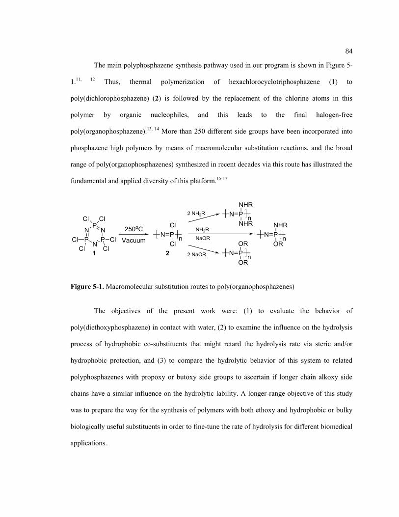
84
The main polyphosphazene synthesis pathway used in our program is shown in Figure 5-
1.11, 12
Thus, thermal polymerization of hexachlorocyclotriphosphazene (1) to
poly(dichlorophosphazene) (2) is followed by the replacement of the chlorine atoms in this
polymer by organic nucleophiles, and this leads to the final halogen-free
poly(organophosphazene).13, 14
More than 250 different side groups have been incorporated into
phosphazene high polymers by means of macromolecular substitution reactions, and the broad
range of poly(organophosphazenes) synthesized in recent decades via this route has illustrated the
fundamental and applied diversity of this platform.15-17
Figure 5-1. Macromolecular substitution routes to poly(organophosphazenes)
The objectives of the present work were: (1) to evaluate the behavior of
poly(diethoxyphosphazene) in contact with water, (2) to examine the influence on the hydrolysis
process of hydrophobic co-substituents that might retard the hydrolysis rate via steric and/or
hydrophobic protection, and (3) to compare the hydrolytic behavior of this system to related
polyphosphazenes with propoxy or butoxy side groups to ascertain if longer chain alkoxy side
chains have a similar influence on the hydrolytic lability. A longer-range objective of this study
was to prepare the way for the synthesis of polymers with both ethoxy and hydrophobic or bulky
biologically useful substituents in order to fine-tune the rate of hydrolysis for different biomedical
applications.

85
5.2 Results and Discussion
5.2.1 Polymer Synthesis
The reactive intermediate, poly(dichlorophosphazene) (2), was generated by an
uncatalyzed thermal ring-opening polymerization of hexachlorocyclotriphosphazene (1) at 250 °C
(Figure 5-1).15
Poly(diethoxyphosphazene) (3) was synthesized by the reaction of sodium
ethoxide with poly(dichlorophosphazene), which is a facile reaction in THF solvent at room
temperature (~25oC).
18 The relatively small size of this nucleophile facilitates the replacement of
all the chlorine atoms without the use of forcing conditions. The co-substituent polymers were
designed to contain a near 1:1 molar ratio of ethoxy and co-substituents trifluoroethoxy-,
phenoxy-, and p-methylphenoxy. Comparisons were also made with the single-substituent
polymers and those with butoxy, propoxy, trifluoroethoxy, and p-methylphenoxy side groups (4 –
7) prepared by previously reported methods.19-21
Thus, the single-substituent polymer structures
are summarized in Figure 5-2.
Figure 5-2. Single substituent polymers
The mixed-substituent polymers 8 – 10 were synthesized following the procedure shown
in Figure 5-3. Chlorine replacement was accomplished by introducing the non-ethoxy co-
substituent group first. For the side groups, trifluoroethoxy, phenoxy, or p-methylphenoxy, the
sodium salt was formed by treatment of the alcohol or phenols with sodium metal. The resultant

86
reagents were then added drop wise to the THF solution of poly(dichlorophosphazene) (2) to
favor a random distribution of the co-substituent groups along the polymer chains. The intention
was to replace half of the available chlorine atoms. Once the first nucleophile had reacted
completely (as detected by 31
P NMR) an excess of sodium ethoxide was then added to complete
the chlorine replacement sequence. This procedure of adding the co-substituent group first was
followed because the low solubility of sodium ethoxide in THF requires the salt to be generated
in the presence of excess of dry ethanol rather than THF. Chlorine replacement also occurs when
a THF solution of poly(dichlorophosphazene) is stirred at room temperature in the presence of
dry ethanol. Thus, if the sodium ethoxide / ethanol solution were to be added first, control of the
side group ratios would be compromised. Particular care was taken to ensure the absence of water
from the reaction mixtures, the presence of which would generate P-OH groups in place of P-OR
units, a result that would complicate the evaluation of the hydrolysis behavior.3
Figure 5-3. Polymers 8 – 10 with both ethoxy and O-linked co-substituents
For this project it was essential to ensure that all the chlorine atoms in
poly(dichlorophosphazene) had been replaced by the organic groups. Otherwise the hydrolysis of
residual P-Cl bonds would mask the sensitivity of the halogen-free polymers to hydrolysis. The
absence of P-Cl bonds was monitored by 31
P NMR spectroscopy, a technique that is sensitive to
less than 5% chlorine concentration. To ensure complete chlorine replacement, the reactions were
allowed to continue for an additional 24 – 48 h after appearing to be complete by 31
P NMR
analysis. After purification, there was no residual P-Cl peak visible in the 31
P NMR spectrum.

87
Additionally, the final products from all of these reactions were examined by elemental
microanalysis for sodium and chlorine content to determine if any detectable chlorine was present
due to incomplete substitution or to traces of sodium chloride. In all cases any detected chlorine
was due to the presence of traces of sodium chloride in the final polymer.
In the second substitution step, the time needed to completely replace the remaining
chlorine atoms by sodium ethoxide depended on the nature of the first substituent. Polymer 8,
with trifluoroethoxy as the first side group introduced, required a much shorter reaction time (4 d
at room temperature) than polymers 9 (15 d) and 10 (27 d) with bulky aryloxy cosubstituents.
This difference reflects the strong electron withdrawing influence by the trifluoroethoxy side
groups which sensitizes the nearby P-Cl bonds to nucleophilic substitution. There is also a
significant size difference between the bulky aryloxy side groups versus the trifluoroethoxy side
groups. This is another possible reason why the ethoxide nucleophile can complete the
replacement of the remaining chlorine atoms faster with the trifluoroethoxy-chloro polymer than
the aryloxy co-substituted counterpart.
5.2.2 Polymer Characterization and Properties
Poly(diethoxyphosphazene) is a rubbery elastomer at room temperature (Tg = -84oC).
Thus, it was of interest to examine the change in physical as well as hydrolytic properties that
result from the influence of the co-substituent groups. These polymers were also compared to
those of the known single-substituent polymers that contained the substituents shown in Figure 5-
2.
The polymers were characterized by 31
P and 1H NMR spectroscopy, GPC, and DSC. The
data are listed in Table 5-1. The mixed-substituent polymer side group ratios were estimated by

88
1H NMR, and the side group distributions were evaluated by
31P NMR spectroscopy (Figure 5-4).
For polymer 8 the final side group composition was calculated to be 60 % trifluoroethoxy and 40
% ethoxy based on the 1H NMR spectra. The
31P NMR spectra showed two overlapping signals at
-6.1 ppm and -7.8 ppm that represented phosphorus atoms with two trifluoroethoxy or ethoxy
groups. This suggests a relatively blocky substitution pattern and / or that the peaks associated
with one of each side group per phosphorus atom also overlap in this region. Polymers 9 and 10
were found by 1H NMR to have 1:1 side group ratios. Both polymers showed major peaks at -
13.8 ppm representing phosphorus atoms with one ethoxy and one aryloxy substituent. The small
peaks at -8 ppm and -19 ppm are from phosphorus atoms with either two ethoxy or two aryloxy
side groups respectively.
Table 5-1. Characterization data of polymers 3 and 8 – 10
1H NMR (ppm)
* 31P NMR (ppm)
* Mw (kDa) R.U.
** Tg (°C)
3 4.0 (2H, CH2), 1.2 (3H, CH3) -7.7 1,360 10,069 -84.0
8 4.5 (2H, CH2CF3), 4.1 (2H,
CH2CH3), 1.3 (3H, CH3)
-6.1, -7.8 5,192 27,461 -70.9
9 7.1 (5H, C6H5), 3.9 (2H, CH2), 1.0
(3H, CH3)
-8.3, -13.8, -19.8 1,364 7,450 -40.8
10 6.9 (4H,C6H4), 3.9 (2H, CH2), 2.2
(3H, C6H4CH3), 1.0 (3H, CH2CH3)
-8.4, -13.8, -19.5 1,658 15,474 -34.0
* NMR solvent used was CDCl3
**R.U.: Repeat Units

89
Figure 5-4. 31
P NMR Spectrums of Polymers 8 (A), 9 (B), and 10 (C)
5.2.3 Thermal Characterization of Polymers 8 – 10
The glass transition temperatures (Tg) of polymers 8 – 10 (Table 5-1) reflect the 1:1 or
near 1:1 ratios of two different side groups. The Tg of polymer 8 at -70.0 °C is roughly
intermediate between those of the ethoxy (-84 ºC) and trifluoroethoxy (-62 ºC) single-substituent
polymers. Poly[bis(phenoxy)phosphazene] and poly[bis(p-methylphenoxy)phosphazene] have
glass transition temperatures at -8 °C and -25 °C respectively.15
The Tg of polymer 9 is at -40 ºC
and polymer 10 at -34 ºC, thus demonstrating a logical influence by each side group on the
polymer morphology.

90
5.2.4 Hydrolysis Behavior of Polymers 3 – 10
Recent observations had suggested that ethoxy side groups linked to a polyphosphazene
chain are a source of hydrolytic instability in spite of the fact that long chain alkoxy groups, as
well as methoxyethoxyethoxy units do not sensitize the system to hydrolysis. A recent study of
polyphosphazenes with vitamin E and ethoxy or ethyl glycinate co-substituent groups, revealed
that the polymer with ethoxy co-substitutents underwent an unexpectedly faster hydrolysis than
the ethyl glycinate-containing polymer. This suggested a serious hydrolytic destabilization by the
ethoxy substituents.5 One purpose of this work was to establish conclusively whether ethoxy side
groups are unique in this respect.
Thus, in this work the hydrolysis behavior of poly(diethoxyphosphazene) was examined
via a heterogeneous hydrolysis study in which film mass loss and molecular weight declines were
monitored over a 12 week period. This provided a starting point to evaluate how this polymer
might perform in a biological environment. The co-substituent polymers 8 – 10 with ethoxy and
the O-linked trifluoroethoxy, phenoxy, and p-methylphenoxy groups were examined using the
same conditions to determine if the hydrolysis could be completely prevented. The single
substituent polymers 4 – 7 were also evaluated for comparison purposes.
The results are summarized in Table 5-2. Solid poly(diethoxyphosphazene) is
hydrolytically sensitive when suspended in water. It loses almost 14 % of its mass in 12 weeks
and undergoes a 76 % molecular weight decline during the same period. However, polymer 4
with n-propoxy side groups is less sensitive, although it too undergoes a slow breakdown that is
evident from the molecular weight decline of 28 % after 12 weeks. The n-butoxy-substituted
polymer (5) appears to be completely stable to water over this time period. This is probably a
consequence of steric and hydrophobic protection of the backbone by the butoxy groups. Single

91
substituent polymers 6 and 7, containing 2,2,2-trifluroethoxy or p-methylphenoxy side groups
respectively, were also found to be completely stable.
Table 5-2. Molecular weight decline and mass loss of polymers and corresponding polymer water
contact angles 3 – 10
Polymer 3 4 5 6 7 8 9 10
% Mass Loss*
13.8 0 0 0 0 12.4 0 0
% Mw Decline*
76 28 0 0 0 78 30 30
Water Contact Angle (°) 77 101 106 109 92 77 94 102
* After 12 week hydrolysis study
The mixed substituent polymers 8 – 10 were all hydrolytically sensitive. Poly(ethoxy)0.8
(trifluoroethoxy)1.2 phosphazene (8) showed a similar mass loss and molecular weight decline to
that of the ethoxy single substituent polymer (3). Thus, the ethoxy substituents govern the
hydrolysis characteristics, irrespective of the hydrophobicity of the cosubstituent. Mixed
substituent polymers that contain both ethoxy, and phenoxy, or p-methylphenoxy groups behave
in an intermediate way, undergoing a molecular weight decline from hydrolytic chain cleavage
but without loss of weight after 12 weeks. This suggests that the hydrolysis products from these
two polymers yield species that still retain a high enough molecular weight to be insoluble in
water, thus maintaining the original mass. The aqueous media from the hydrolysis of polymer 10
were examined for the presence of free p-cresol as shown in Figure 5-5. Although the
concentration of p-cresol increases linearly over the 12 week period, the degree of hydrolysis was
low enough that a corresponding mass loss change was not detected. Moreover, the linear
increase in concentration of p-cresol demonstrates that its detection by UV-Vis spectroscopy

92
cannot be attributed to residual p-cresol remaining in the polymer after synthesis. It seems clear
from this study that ethoxy side groups have a powerful hydrolytic sensitizing effect on
polyphosphazenes, a property that opens many opportunities for their use in tissue engineering or
controlled drug delivery.
Figure 5-5. UV-Vis concentration determination of p-cresol in hydrolysis media from Polymer
10 during 12 week hydrolysis
5.2.5 Water Contact Angles of Polymers 3 – 10
Is the hydrolysis behavior of these polymers connected with the surface hydrophilicity?
In other words, is the process controlled by surface effects rather than chemical reactivity? The
relative hydrophobicities of the O-linked substituent polymers were examined by static water
contact angles.15
All the polymers are relatively hydrophobic, with their contact angles exceeding
0
0.02
0.04
0.06
0.08
0.1
0.12
0.14
0.16
0.18
0.2
0 2 4 6 8 10 12
P-C
reso
l Co
nce
ntr
atio
n (
mM
)
Week

93
70º (Table 5-2). Only the hydrophobicity of polymer 8 showed no significant change when
compared to the single substituent polymer 3, and was primarily influenced by the ethoxy
substituent. This is because the ethoxy groups control the surface characteristics more than
trifluoroethoxy groups, such that the contact angle of mixed-substituent polymer 8 is more like
that of single-substituent polymer 3 than single-substituent polymer 6. The water contact angle of
polymers 9 and 10 are governed by the aryloxy substituents. Thus, the evidence suggests that the
differences in hydrolytic sensitivity are mainly a consequence of different chemical reactivities
rather than surface effects.
However, the mechanism of hydrolysis is still a subject for speculation. Certainly, access
by water to the phosphorus atoms of the backbone is important. However, it is known that the
hydrolytic displacement of a side group from phosphorus leaves a hydroxyl unit in its place, and
that this unit is capable of a rearrangement to protonate a nearby skeletal nitrogen atom and leave
a P=O side unit in its place. Based on traditional phosphate chemistry is seems reasonable to
assume that the resultant single-bonded skeletal site is then cleaved by water, although the
relative rates of side group displacement and skeletal cleavage are not known.
5.3 Experimental Section
5.3.1 Reagents and Equipment
Reproducible syntheses of poly(organophosphazenes) require the strict exclusion of
moisture. Thus, all synthesis reactions were executed under a dry argon atmosphere using
standard Schlenk line techniques. Before use, glassware was dried overnight in an oven at 125
°C. Tetrahydrofuran (THF) (EMD) was dried using solvent purification columns. Sodium

94
(Aldrich), dichloromethane (EMD), hexanes (EMD), methanol (EMD), and p-cresol (Aldrich)
were used as received. Phenol (Acros) was vacuum sublimed before use. Ethanol (Koptec), 1-
propanol (Aldrich), 1-butanol (Aldrich) and 2,2,2-trifluoroethanol (Sigma) were distilled from
and stored over molecular sieves (EMD, 4A mesh beads) under an argon atmosphere.
Poly(dichlorophosphazene) was prepared by the thermal ring-opening polymerization of
recrystallized and sublimed hexachlorocyclotriphosphazene (Fushimi Chemical Co., Japan) in
evacuated Pyrex tubes at 250 °C. Spectra/Por molecular porous cellulose dialysis membranes
with molecular weight cut-off of 12,000-14,000 were employed for purification of the polymers.
1H and
31P NMR spectra were obtained using a Bruker 360 WM instrument operated at 145 and
360 MHz, respectively, with 31
P shifts relative to 85 % H3PO4 at 0 ppm as a reference. Glass
transition temperatures were obtained using a TA Instruments Q10 differential scanning
calorimetry apparatus with a heating rate of 10 °C/ min and a sample size of ca. 10 mg. Gel
permeation chromatography data were obtained using a Hewlett-Packard 1047A refractive index
detector and two Phenomenex Phenogel 10μm linear columns with elution times calibrated using
polystyrene standards. The samples were eluted using 1.0 mL/min with a 10 mM solution of
tetra-n-butylammonium nitrate (Alfa Aesar) in THF with elution times calibrated with
polystyrene standards. pH data were obtained using a VWR Symphony SB70P pH meter. Water
contact angle measurements were performed using a Ramé-Hart Automated
Goniometer/Tensiometer with DROPimage Advanced v2.6 software. Elemental analyses for
residual sodium and chlorine were obtained by Intertek, with the error in chlorine content being
±0.3% absolute and the error in sodium a relative 10%. Ultraviolet spectra were obtained using a
double-beam spectrophotometer (Varian, Cary 500). Measurements were conducted using
distilled water.

95
5.3.2 Synthesis of Poly(diethoxyphosphazene) (3)
Synthesis of polymer 3 was similar to a previously published procedure.18
Briefly,
sodium ethoxide was generated in-situ by adding ethanol (25.1 mL, 0.43 mol) to sodium metal
(1.00 g, 43 mmol), before transferring this reagent to a solution of poly(dichlorophosphazene)
(1.00 g, 8.6 mmol) in THF (100 mL) followed by stirring the mixture at room temperature for 96
hr. When the substitution was complete, based on the 31
P NMR spectrum, the polymer was
concentrated and precipitated into hexane, followed by dialysis versus 1:1
dichloromethane:methanol for 3 days. Solvent was removed under reduced pressure to afford the
polymer in 67 % yield.
5.3.3. Synthesis of Polyphosphazenes with 1-propoxy, 1-butoxy, 2,2,2-trifluoroethoxy,
phenoxy, or p-methylphenoxy Side Groups (4 – 7)
These polymers were synthesized following previously published procedures.19-21
5.3.4 Synthesis of Poly[(ethoxy)0.8(trifluoroethoxy)1.2phosphazene] (8)
Poly(dichlorophosphazene) (1.00 g, 8.6 mmol) was dissolved in dry THF (100 mL). In a
separate vessel, THF (50 mL) was added to sodium metal (0.20 g, 8.6 mmol), and 2,2,2-
trifluoroethanol (0.86 g, 8.6 mmol) was added to form the sodium salt. This mixture was then
introduced into the polymer solution dropwise and allowed to react at room temperature for 24 h.
Sodium ethoxide was formed in a separate vessel and was then added to the polymer solution and
allowed to react at room temperature for 3 days. The reaction mixture was then concentrated and
precipitated into hexane five times and then dialyzed versus 1:1 dichloromethane:methanol for 4
days. Solvent was removed under reduced pressure to yield the polymer in a 71% yield.

96
5.3.5 Synthesis of Poly[(ethoxy)1(phenoxy)1phosphazene] (9)
Poly(dichlorophosphazene) (1.00 g, 8.6 mmol) was dissolved in dry THF (100 mL). In a
separate vessel, phenol (0.81 g, 8.6 mmol) was dissolved in THF (150 mL) then added to sodium
metal (0.198 g, 8.6 mmol) to form the sodium salt. This mixture was subsequently added to the
polymer solution dropwise and allowed to react at room temperature for 12 h. The sodium
ethoxide solution was prepared as described above and was then added to the polymer reaction
solution, and the mixture was stirred at room temperature for 4 d then at 40 °C for 8 d, then for an
additional 2 d to ensure complete chlorine replacement. The reaction mixture was then
concentrated, precipitated twice into methanol, then dialyzed for 8 d versus 60%
dicholoromethane, 20% methanol, 20% hexanes. Solvent was removed under reduced pressure to
give the polymer in a 79% yield.
5.3.6 Synthesis of Poly[(ethoxy)1(p-methoxyphenoxy)1phosphazene] (10)
Poly(dichlorophosphazene) (1.00 g, 8.6 mmol) was dissolved in THF (100 mL). In a
separate vessel p-cresol (0.933 g, 8.6 mmol) was dissolved in THF (150 mL) then added to
sodium metal (0.198 g, 8.6 mmol) to form the sodium salt which was subsequently added to the
polymer solution dropwise and allowed to react at room temperature for 12 h. The sodium
ethoxide solution, prepared as described above, was then added to the polymer solution and the
mixture was allowed to react at room temperature for 10 days after which time the solution was
heated to 40 °C for an additional 12 days and was stirred for an additional 4 days to at 40 °C to
ensure complete chlorine replacement. The reaction mixture was then concentrated and
precipitated into methanol three times, then dialyzed versus 40% dichloromethane, 40%

97
methanol, 20% hexane for 14 days. Solvent was removed under reduced pressure to give the
polymer in an 86% yield.
5.3.7 Water Contact Angle Measurements
The surface hydrophobicities of polymers 8 – 10 were examined by static water contact
angle measurements. Thin films (~ 100-200 µm thick) of each polymer (0.45 g) were cast onto
Bytac (Teflon) paper (5 cm x 5 cm) using stabilizer-free tetrahydrofuran (18 mL). After drying,
samples were mounted on glass slides before measurement. All measurements were made
immediately after the droplet was placed on the surface of the sample in air at ambient
temperature. Values were obtained in triplicate with the error in the measurements being ± 3°.
5.3.8 Hydrolysis of Polymer Films
Polymers 3 – 10 (0.45 g) were dissolved in stabilizer-free tetrahydrofuran (18 mL) and
were solution-cast into square films on Bytac paper (5 cm x 5 cm, ~ 100 μm thick). The samples
were air dried for 24 h followed by further drying under vacuum for 48 h. The films were divided
into 36 samples of ~ 10 mg each and were placed in vials with 5 mL deionized water with a
starting pH of 6.3. The sealed vials were maintained at 37 °C for 12 weeks, with samples
removed in triplicate each week for each polymer. The aqueous media were decanted and the pH
was measured. The undissolved products were dried under vacuum and re-weighed. After
weighing, each solid sample was dissolved in THF and the molecular weight was estimated by
GPC.

98
5.4 Conclusions
Ethoxy containing polyphosphazenes are hydrolytically sensitive, although the sensitivity
to hydrolysis can be reduced by the presence of bulky hydrophobic cosubstituent groups.
Moreover, the rate of hydrolysis can be tuned by changing co-substituent ratios. This result has
important implications for the development of new bioerodible polymers.
5.5 Acknowledgements
The authors thank Dr. Nicole L. Morozowich for her help with the UV-Vis measurements
and discussions, and Tomaz Modzelewski and Zicheng Tian for their assistance with water
contact angle measurements. Dr. Chen Chen and Andrew Hess provided previously synthesized
polymers, and Evan Thursby and Yuichi Tsuji initiated preliminary studies on this topic.
5.6 References
1. Nair, L. S.; Laurencin, C. T. Progress in Polymer Science 2007, 32, 762-798.
2. Allcock, H. R.; Morozowich, N. L. Polymer Chemistry 2012, 3, 578-590.
3. Allcock, H. R.; Pucher, S. R. Macromolecules 1994, 27, (5), 1071-1075.
4. Allcock, H. R.; Pucher, S. R.; Scopelianos, A. G. Biomaterials 1994, 15, (8), 563-569.
5. Morozowich, N. L.; Weikel, A. L.; Nichol, J. L.; Chen, C.; Nair, L. S.; Laurencin, C. T.;
Allcock, H. R. Macromolecules 2011, 44, 1355-1364.
6. Nichol, J. L.; Morozowich, N. L.; Allcock, H. R. Polymer Chemistry 2013, 4, 600-606.
7. Weikel, A. L.; Krogman, N. R.; Nguyen, N. Q.; Nair, L. S.; Laurencin, C. T.; Allcock, H.
R. Macromolecules 2009, 42, 636-639.
8. Allcock, H. R. Annals of the New York Academy of Sciences 1997, 831, (1), 13-31.
9. Lakshmi, S.; Katti, D. S.; Laurencin, C. T. Advanced Drug Delivery Reviews 2003, 55,
467-482.
10. Deng, M.; Kumbar, S. G.; Wan, Y.; Toti, U. S.; Allcock, H. R.; Laurencin, C. T. Soft
Matter 2010, 6, 3119-3132.
11. Allcock, H. R. Science 1976, 193, 1214-1219.
12. Allcock, H. R. Science 1992, 255, 1106-1112.
13. Allcock, H. R. Journal of Inorganic and Organometallic Polymers 1992, 2, (2), 197-211.
14. Potin, P.; De Jaeger, R. European Polymer Journal 1991, 27, 341-348.

99
15. Allcock, H. R., Chemistry and Applications of Polyphosphazenes. John Wiley & Sons,
Inc.: Hoboken, NJ, 2003; Vol. 1, p 725.
16. Allcock, H. R. Soft Matter 2012, 8, 3521-3532.
17. Allcock, H. R.; Crane, C. A.; Morrissey, C. T.; Nelson, J. M.; Reeves, S. D.; Honeyman,
C. H.; Manners, I. Macromolecules 1996, 29, 7740-7747.
18. Allcock, H. R.; Kugel, R. L.; Valan, K. J. Inorganic Chemistry 1966, 5, (10), 1709-1715.
19. Allcock, H. R.; Kugel, R. L. Journal of the American Chemical Society 1965, 87, (18),
4216-4217.
20. Singler, R. E.; Hagnauer, G. L.; Schneider, N. S.; Laliberte, B. R.; Sacher, R. E.; Matton,
R. W. Journal of Polymer Science 1974, 12, 433-444.
21. Allcock, H. R.; Connolly, M. S.; Sisko, J. T.; Al-Shali, S. Macromolecules 1988, 21, (2),
323-334.

100
Chapter 6
Summary
The rise in the number of ligament and tendon injuries continues to increase the need for
a better repair strategy such as tissue engineering. The importance and impact of tissue
engineering is not limited to ligaments and tendons. Research in this field may also benefit the
repair and replacement of many of the other living tissues in the human body. This dissertation is
an outline of the work done towards the development of a polyphosphazene polymer specifically
designed to function as a ligament or tendon tissue engineering scaffold.
6.1 New Polyphosphazenes Developed for Tissue Engineering Applications
Polyphosphazenes are a unique class of highly tunable synthetic polymers able to be
designed with specific chemical and physical properties, which allows them to be utilized as
potential ligament or tendon tissue engineering scaffolds.1 Several polymers have been
synthesized and studied in an attempt to generate the necessary scaffold properties. All the
polymers were characterized using 1H and
31P NMR spectroscopy, GPC, and DSC techniques.
The first approach utilized long alkyl chain (5-8 carbon atoms) alcohols to protect the carboxylic
acid group of alanine or phenylalanine which in turn, was linked to a polyphosphazene skeleton.
This was an attempt to combine the biodegradability of the amino acid derivative polymer with
the elastomeric characteristics generated by the long alkoxy chains.2 Although these polymers
have the necessary low glass transition temperatures and hydrolytic instability, they failed to
show elastomeric properties due to the absence of either physical or chemical crosslinks. To
overcome this shortcoming, a second group of polymers was developed which used citronellol as

101
a side group. This provided crosslink sites for the UV-induced 2+2 addition of the double bonds
present in citronellol. Two routes of attachment were examined to incorporate citronellol units
into the final polymer. The first involved direct linkage of citronellol to the polymer backbone
with varying amounts of alanine ethyl ester as a co-substituent.3 The second involved using
citronellol to esterify the carboxylic acid units of glycine, alanine, valine, and phenylalanine
followed by attachment of the amino acid to the phosphazene backbone through the free amine.
Both routes were successful, with the final properties dependent on how the citronellol was linked
on the polymer. It was also found that these polymers can be crosslinked by UV radiation in the
solid state, with the crosslink density increasing with increasing UV-exposure time. Additionally,
the crosslinking also allowed for tuning of the polymer mechanical properties.
An additional research focus was placed on the use of ethoxyphosphazene polymers for
biomedical applications. This work was spurred by the small size and low cost of ethanol which
should allow the development of a new class of useful biomedical polymers.4 Moreover, the
ability of the ethoxy side group to sensitize the backbone to hydrolytic breakdown, which has
been noted previously in the literature, was examined and fully characterized.5 This work
demonstrated that ethanol can be used to sensitize the phosphazene polymer backbone to
hydrolytic degradation without the disadvantages found for the more traditional amino acid based
approaches which often involve polymer molecular weight decline during synthesis.4
In summary, a series of biodegradable polyphosphazenes with increasing viability as
potential ligament and tendon scaffold materials has been developed. Of the systems studied, the
best scaffold candidates to date are the citronellol-containing polyphosphazenes due to their
tunable hydrolysis rates, ability to be easily crosslinked, and tunable mechanical properties.

102
6.2 Future Considerations
Future work should focus on the introduction of side groups that promote cellular
adhesion to promote cell growth and proliferation.6 A previous report has shown that the
incorporation of RGD, a cell adhesive tri-peptide, onto the polyphosphazene backbone, makes
this a potential strategy.7 Also, growth factors need to be incorporated into the fabricated scaffold
to promote wound healing. Another point, often ignored, are the bony insertion sites where the
properties of the tissue change significantly.6 One way to address this issue is to combine
polyphosphazenes designed specifically for bone tissue engineering that can coordinate
hydroxyapatite with materials optimized for strength and elasticity.8, 9
A hybrid system of this
type should increase the likelihood of the material incorporating at a bone insertion location.
Collaborative research to continue this project must include cell seeding studies followed by
animal model studies for the most promising candidates.6
6.3 References
1. Allcock, H. R., Chemistry and Applications of Polyphosphazenes. John Wiley & Sons,
Inc.: Hoboken, NJ, 2003; Vol. 1, p 725.
2. Nichol, J. L.; Morozowich, N. L.; Allcock, H. R. Polymer Chemistry 2013, 4, 600-606.
3. Nichol, J. L.; Morozowich, N. L.; Decker, T. E.; Allcock, H. R. Journal of Polymer
Science, Part A: Polymer Chemistry 2014, DOI: 10.1002/pola.27236.
4. Nichol, J. L.; Hotham, I. T.; Allcock, H. R. Polymer Degradation and Stability 2014,
DOI: 10.1016/j.polymdegradstab.2014.05.015.
5. Morozowich, N. L.; Weikel, A. L.; Nichol, J. L.; Chen, C.; Nair, L. S.; Laurencin, C. T.;
Allcock, H. R. Macromolecules 2011, 44, 1355-1364.
6. Rodrigues, M. T.; Reis, R. L.; Gomes, M. E. Journal of Tissue Engineering and
Regenerative Medicine 2013, 7, 673-686.
7. Chun, C.; Lim, H. J.; Hong, K.-Y.; Park, K.-H.; Song, S.-C. Biomaterials 2009, 30, 6295-
6308.
8. Morozowich, N. L.; Nichol, J. L.; Allcock, H. R. Chemistry of Materials 2012, 24, (17),
3500-3509.
9. Morozowich, N. L.; Modzelewski, T.; Allcock, H. R. Macromolecules 2012, 45, 7684-
7691.

VITA Jessica Lee Nichol
Jessica Lee Nichol was born on August 30, 1987 in Indiana, Pennsylvania. She was
raised on a farm by her parents Thomas and Valerie Nichol and attended Indiana University of
Pennsylvania in Indiana, Pennsylvania where she obtained her Bachelor of Science degree in
Chemistry in May 2009. Jessica began her graduate studies at The Pennsylvania State University
under the guidance of Evan Pugh Professor Harry R. Allcock in August of 2009. After
graduation, she will continue her career at Eastman Chemical Company in Kingsport, Tennessee.