polypeptide resurfacing method improves fibroblast's adhesion to hyaluronan strands
TRANSCRIPT

Polypeptide resurfacing method improves fibroblast’sadhesion to hyaluronan strands
Min Hu,1 Eric E. Sabelman,1,2 Susan Lai,1 Ewa K. Timek,2 Feng Zhang,1 Vincent R. Hentz,1,2
William C. Lineaweaver1
1Department of Functional Restoration, Stanford University, Palo Alto, California 943052RR&D Center, VA Hospital, Palo Alto, California 94306
Received 29 September 1998; accepted 1 April 1999
Abstract: Hyaluronic acid (hyaluronan, HyA) is a matrixcomponent that takes part in cell adhesion and growth innormal and repaired tissues. Since it is soluble in water,HyA has been of limited use in tissue engineering of artifi-cial matrices. Recent studies demonstrate that polypeptideshave the twin advantages of reducing solubility of HyA andimproving cellular attachment via cell surface adhesion mol-ecule receptors. This paper describes a new approach of us-ing a polypeptide resurfacing method to enhance the attach-ment of cells to HyA strands. HyA strands were crosslinkedby glutaraldehyde and then resurfaced with poly-D-lysine,poly-L-lysine, glycine, or glutamine. After inoculation withfibroblasts in vitro, modified HyA was evaluated with his-tological and immunohistochemical staining methods forcell adhesion and proliferation. Modified HyA with fibro-
blast cells also were implanted in vivo. The results show that(1) both polylysines enhanced fibroblast adhesion tocrosslinked HyA strands; (2) HyA strands were able to becrosslinked well by 3 days of treatment in glutaraldehyde,and as a biomaterial they could resist biodegradation; (3)modified HyA has good biocompatibility, both in vitro andin vivo. The results demonstrate that HyA material resur-faced by polypeptides has positive advantages for cellularadhesion. Resurfaced HyA has much potential as an im-proved biomaterial for clinical usage. © 1999 John Wiley &Sons, Inc. J Biomed Mater Res, 47, 79–84, 1999.
Key words: hyaluronic acid; polypeptide; glutaraldehyde;fibroblast; cell adhesion
INTRODUCTION
Hyaluronan (hyaluronic acid, HyA) is a high mo-lecular weight polysaccharide composed of chains ofrepeating disaccharide units of D-glucuronic acid andN-acetyl-D-glucosamine.1 Existing in all connective tis-sues as a matrix component,2 HyA has been found topromote cell motility and proliferation in addition toits previously known functions of supporting andmaintaining tissue or organ morphological features.3–5
These characteristics enable HyA to play an importantrole in morphogenesis, wound repair, inflammation,and metastasis.4,5 HyA has been demonstrated to me-diate these physiologic or pathologic events mainlythrough three HyA cell surface receptors, namelyCD44, RHAMM, and ICAM-1.6–8 In addition to thecell surface receptors, intracellular HyA binding pro-
teins have been described that suggest an additionalnovel mechanism by which HyA may regulate cellbehavior.6–9 Because of the nonadhesive feature of pu-rified HyA, it currently is employed to reduce the in-cidence of postoperative adhesions in intraocular sur-gery, and with synovial replacement devices, and it isused in various cosmetic applications as a noninflam-matory, viscoelastic biomaterial.10,11 These appliancestake advantage of only one of HyA’s many potentialfunctions. Furthermore, purified HyA is water solubleat room temperature; some surgical, pharmaceutical,and industrial applications require crosslinking or es-terification of HyA molecules to make them water in-soluble.7,12 Many attempts have been made to intro-duce crosslinks into HyA molecules, and it continuesto be a method under investigation and develop-ment.7–12
Glutaraldehyde (Glut) is a traditional crosslinkingagent broadly used in electron microscopy, proteinchemistry, and immunochemistry. As one of the mostwidely used linking agents for biomaterials, Glut alsohas been used to stabilize tissue heterografts, tostrengthen and develop tissue bioprostheses, such asheart valves, blood vessels, and tendons, during the
Correspondence to: M. Hu at 4483 Macbeth Circle, Fre-mont, CA 94555
Contract grant sponsor: Veterans Administration; Contractgrant number: B1839PA
© 1999 John Wiley & Sons, Inc. CCC 0021-9304/99/010079-06

past twenty years, and, recently, in the crosslinking ofcollagen.13–17 Although there are some concerns aboutthe cytotoxicity of aldehyde,18–20 renewed interest inthe use of short-term Glut-treated autologous graftwas encouraged by the observation that Glut-crosslinked pericardium appears not to calcify orshrink after implantation.21,22
Cell-surface adhesion receptors are a newly identi-fied mechanism associated with the secretion of cer-tain polypeptide growth factors and cytokines.23 Posi-tively charged coating materials, such as polylysine,commonly are used to improve cellular attachment tothe plastic or glass surface of culturing devices invitro.24 There is increasing interest in polylysine, andnew uses are continuing to be discovered,25–27 how-ever, direct coating of graft material has not yet beenattempted. In the previous study, HyA strands werecrosslinked by Glut and then coated with four differ-ent amino acids and polypeptides. After culturingwith fibroblasts, modified HyA strands were evalu-ated for adhesion and proliferation of fibroblast cellswith histological and immunohistochemical stainingmethods. Some physiobiological characteristics alsowere investigated. The purpose of this study was toexplore the possibility of cell adhesion to HyA by aresurfacing technique and further to expand the ma-terial’s use in both autografts and xenografts.
MATERIALS AND METHODS
Fabrication of HyA strand
Ion exchange of Na-hyaluronate
Samples of 0.15 g HyA (sodium hyaluronate, MW 1.6 ×10⁶ Daltons; Lifecore Biomedical Inc.) were put into auto-claved dialysis tubing and dialyzed against cation exchangecellulose (Dowex AG 50W-X4, from BIO-RAD) at 1 g/100mL of deionized distilled water and stirred by a magneticbar at 4°C for 2 days. The samples were rinsed with sterilewater, and liquefied HyA was transferred into DMSO. After2 days of further stirring, HyA was aspirated into 10-mLsyringes.
HyA strand fabrication and crosslinking
Prepared HyA was pushed out of the 10-mL syringesthrough a 25-gauge (0.5 mm) needle to form strands in abeaker filled with 100% alcohol and stored at 4°C overnight.The strands were placed on 5-mm filter paper and air dried.Then samples in the experimental group were immersed inbiological grade Glut (TED PELLA, Inc.) at concentrations of0.5, 5, 25, and 50% aqueous solution for 1, 2, 4, 6, 8, 12, 24, 48,or 72 h at 4°C. After extensive washing in distilled water, thestrands were dialyzed in PBS for 48 h in order to reduce
residual toxic Glut. The samples then were stored in PBSovernight.
Resurfacing with amino acids and polypeptides
Solutions of 1% L-glutamine (Sigma), 1% glycine (amino-acetic acid, BIO-RAD), 50 mg/1 mL of poly-L-lysine (Sigma),and 10 mg/mL of poly-D-lysine (Sigma) were freshly pre-pared. HyA strands were immersed in one of four solutionsfor 1 h. After having been resurfaced, the strands werewashed thoroughly with distilled water.
Fibroblast inoculation of HyA strands
Monolayers of rat fibroblasts in their growth phase, ap-proximately 3 days after splitting, were trypsinized for 15min, forming a suspension. Cells were pooled by centrifu-gation at 2000 rpm for 10 min and resuspended in DMEM,4–6 × 10⁵/cm3. A syringe with a cut-tip needle was used toaspirate and push out the cellular medium three to fivetimes to homogenize the clumps of cells. The fibroblastsgently were applied onto the surface of the HyA strands in35 × 10-mm culture dishes, followed by 1 mL of medium(DMEM + 10% fetal bovine serum). Cultures of cell-seededHyA strands were incubated at 37°C and observed at 24-hintervals for 7 days with an inverted microscope. One ex-perimental group of cell-seeded HyA strands remained inculture in vitro. The other experimental group was set asidefor implantation in vivo.
Implantation of fibroblast-seeded HyA strands
Five Wistar male rats weighing about 400 g were anesthe-tized with intraperitoneal injections of 4% chloral hydrate.NIH guidelines for the care and use of laboratory animals(NIH Publication #85-23, Rev. 1985) were observed. Foursegments (of 2 cm each) of the fibroblast-seeded HyAstrands were implanted in a subcutaneous pocket in theright chest of the rat. The incision was closed with a con-tinuous 4.0 nylon suture.
Extraction and preparation of specimens
After 2 or 4 weeks the rats were sacrificed, the implantswith surrounding tissue were removed, and the specimenswere fixed in buffered formaldehyde for 2 h. The specimensroutinely were processed, paraffin embedded, and cut into10 mm sections onto slides. The slides then were deparaf-finized and rehydrated in preparation for staining. Speci-mens in culture dishes were washed in PBS for 30 min, fixedin 10% neutral formaldehyde for 1 h, and washed again inPBS for 30 min prior to staining.
80 HU ET AL.
Immunohistochemical staining
Specimens were incubated in 0.3% H2O2 for 30 min andwashed in PBS. Then they were blocked with normal horseserum for 30 min, followed by overnight incubation withPCNA antibody (mouse anti-proliferative cell nuclear anti-gen, pc-10, DAKO) at 1:200 dilution. Afterwards specimenswere treated with the avidin-biotin-complex kit (Vector), de-veloped with DAB tablets (Sigma), and mounted with gela-tin. Other specimens routinely were stained with H&E orAlcian Blue. A Baxter cell counter was used to count cellsattached to a 1-cm segment of a HyA strand. Proliferatingcells were marked by PCNA-positive staining. The propor-tion of positively stained cells was assessed and graded on ascale of 0 to ++++ (0 = negative immunoreactivity; + = 1–25percent; ++ = 26–50 percent; +++ = 51–75 percent; and ++++= 76–100 percent).
RESULTS
Fabricated HyA strands crosslinked by 50% Glut for72 h were water insoluble and very stable compared tothe groups crosslinked with lower concentrations ofGlut or for shorter incubation times (Fig. 1). HyAstrands, crosslinked for 72 h or longer with 50% Glutmaintained their shape for at least 5 days to over 3months while the group crosslinked by 50% Glut for48 h dissolved in PBS or distilled water after 2 days.When lower concentrations of Glut were used, such as25% Glut, the strands dissolved in PBS in from 5 minto 12 h while all other experimental groupscrosslinked with concentrations of Glut less than 25%immediately dissolved in PBS or distilled water.
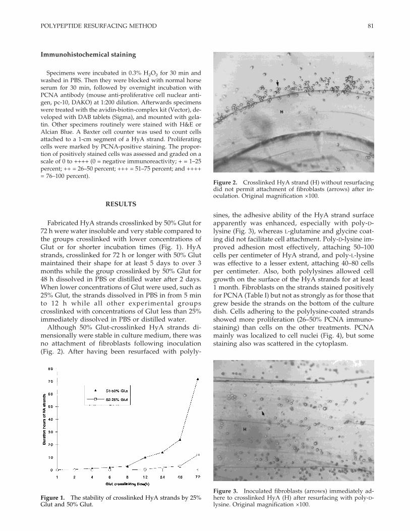
Although 50% Glut-crosslinked HyA strands di-mensionally were stable in culture medium, there wasno attachment of fibroblasts following inoculation(Fig. 2). After having been resurfaced with polyly-
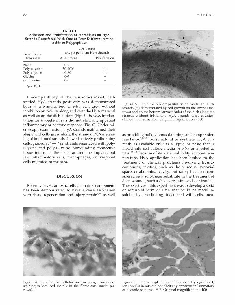
sines, the adhesive ability of the HyA strand surfaceapparently was enhanced, especially with poly-D-lysine (Fig. 3), whereas L-glutamine and glycine coat-ing did not facilitate cell attachment. Poly-D-lysine im-proved adhesion most effectively, attaching 50–100cells per centimeter of HyA strand, and poly-L-lysinewas effective to a lesser extent, attaching 40–80 cellsper centimeter. Also, both polylysines allowed cellgrowth on the surface of the HyA strands for at least1 month. Fibroblasts on the strands stained positivelyfor PCNA (Table I) but not as strongly as for those thatgrew beside the strands on the bottom of the culturedish. Cells adhering to the polylysine-coated strandsshowed more proliferation (26–50% PCNA immuno-staining) than cells on the other treatments. PCNAmainly was localized to cell nuclei (Fig. 4), but somestaining also was scattered in the cytoplasm.
Figure 3. Inoculated fibroblasts (arrows) immediately ad-here to crosslinked HyA (H) after resurfacing with poly-D-lysine. Original magnification ×100.
Figure 1. The stability of crosslinked HyA strands by 25%Glut and 50% Glut.
Figure 2. Crosslinked HyA strand (H) without resurfacingdid not permit attachment of fibroblasts (arrows) after in-oculation. Original magnification ×100.
81POLYPEPTIDE RESURFACING METHOD
Biocompatibility of the Glut-crosslinked, cell-seeded HyA strands positively was demonstratedboth in vitro and in vivo. In vitro, cells grew withoutinhibition or toxicity along and over the HyA materialas well as on the dish bottom (Fig. 5). In vivo, implan-tation for 4 weeks in rats did not elicit any apparentinflammatory or necrotic response (Fig. 6). Under mi-croscopic examination, HyA strands maintained theirshape and cells grew along the strands. PCNA stain-ing of implanted strands showed actively proliferatingcells, graded at “++,” on strands resurfaced with poly-L-lysine and poly-D-lysine. Surrounding connectivetissue infiltrated the space around the implant, butfew inflammatory cells, macrophages, or lymphoidcells migrated to the area.
DISCUSSION
Recently HyA, an extracellular matrix component,has been demonstrated to have a close associationwith tissue regeneration and injury repair6,28 as well
as providing bulk, viscous damping, and compressionresistance.7,28,29 Most natural or synthetic HyA cur-rently is available only as a liquid or paste that ismixed into cell culture media in vitro or injected invivo.30–32 Because of its water solubility at room tem-perature, HyA application has been limited to thetreatment of clinical problems involving liquid-containing cavities, such as the vitreous, synovialspace, or abdominal cavity, but rarely has been con-sidered as a soft-tissue substitute in the treatment ofdeep wounds, such as bed sores, sinusoids, or fistulae.The objective of this experiment was to develop a solidor semisolid form of HyA that could be made in-soluble by crosslinking, inoculated with cells, incu-
TABLE IAdhesion and Proliferation of Fibroblasts on HyA
Strands Resurfaced With One of Four Different AminoAcids or Polypeptides
ResurfacingTreatment
Cell Count(Avg # per 1 cm HyA Strand)
Attachment Proliferation
None 0–2 −Poly-D-lysine 50–100* ++Poly-L-lysine 40–80* ++Glycine 0–7 +L-glutamine 0–5 +
*p < 0.01.
Figure 4. Proliferative cellular nuclear antigen immuno-staining is localized mainly in the fibroblasts’ nuclei (ar-rows).
Figure 5. In vitro biocompatibility of modified HyAstrands (H) demonstrated by cell growth on the strands (ar-rows) and on the bottom (arrowheads) of the dish along thestrands without inhibition. HyA strands were counter-stained with Sirus Red. Original magnification ×100.
Figure 6. In vivo implantation of modified HyA grafts (H)for 4 weeks in rats did not elicit any apparent inflammatoryor necrotic response. H.E. Original magnification ×100.
82 HU ET AL.

bated in a culture dish, and later implanted as a graftin tissue gaps caused by disease, surgery, or injury.
In order for the HyA strands to maintain a consis-tent shape, fabricated strands were incubated in high-concentration Glut, a widely used crosslinking agentof many biomaterials.15–17 Previous studies on colla-gen demonstrated that increased Glut concentrationincreased collagen insolubility, implicating the forma-tion of intermolecular crosslinks in addition to intra-molecular links.14 In the present study, results dem-onstrate that high-concentration Glut effectivelycrosslinks HyA. The morphology of the strands wasvery stable after a 72-h immersion in 50% Glut solu-tion. At lower concentrations or shorter incubationtimes, crosslinked HyA strands readily dissolved inPBS. To avoid toxicity from Glut to the cells,13,25
crosslinked HyA strands were washed thoroughlyand dialyzed in PBS. The observations that fibroblastsgrew along 50% Glut-crosslinked HyA strands andthat HyA strands successfully were implanted in ratswithout evident tissue inflammation or abnormal re-sponse suggest that all or most of the Glut was re-moved by washing and dialysis. Any residual alde-hyde was insufficient to cause harm to living cells ortissues.
Polypeptide coating of microtitre plates and culturedishes has been used broadly in the biomedical field.In particular, positively charged coating materials,such as polylysine, improve cell attachment in vitro.33
Recent studies have noted that polypeptides havesome significant characteristics that promote cell at-tachment via adhesion molecule receptors, such asCD44 receptor, on the cell surface.34 This interactionmay participate in cell signal transduction throughprotein kinase cooperation.5,6,35 Other studies demon-strated that protein kinase CK2 activity could bestimulated by polylysine.36 Therefore the polypeptideitself and/or HyA, through these extracellular signal-regulated kinases and perhaps even through mitogen-activated protein kinases, can modulate cell activity,including cellular adhesion and proliferation.
Moreover, in some studies polypeptides demon-strated crosslinking function.7,31 Unlike somecrosslinking agents that form relatively weak hy-droxyl and carboxyl bonds, L-lysine or L-lysine methylester crosslink HyA molecules with a stronger amidebond that is more resistant to hydrolysis than theSchiff-base bond involved in Glut crosslinks.7 In thisstudy, HyA strands first were crosslinked by high-concentration Glut, giving the strands an initialcrosslinking formation that then was reinforced dur-ing polypeptide treatment. Therefore, not only didpolypeptide treatment resurface the strands, enhanc-ing the biocompatibility of the material by improvingcell attachment, but it also served to further crosslinkthe strands and retard biodegradation.
CONCLUSIONS
The results of this study demonstrate a significantincrease in cell adhesion to HyA strands after theywere resurfaced with a polypeptide whereas an in-crease in proliferation was not as dramatic. Poly-D-lysine is more effective than poly-L-lysine in promot-ing cellular adhesion. Although the mechanism of ad-hesion between HyA material and polypeptidesremains unclear, modified HyA strands are antici-pated to be a clinically useful biomaterial for implan-tation as either an autograft or xenograft and even asa drug carrier. Future work will involve the investi-gation of the mechanism of adhesion, the attachmentcapability of different cell types to HyA and collagenmatrices, and the clinical use of those modified matri-ces as better, more consistent biografts.
References
1. Hellstrom S, Laurent C. Hyaluronan and healing of tympanicmembrane perforations. An experimental study. Acta Oto-laryngol 1987;442(suppl):54–61.
2. Swann DA. Macromolecules of synovial fluid. In: Sokoloff L,(editor) The joints and synovial fluid, New York: AcademicPress, Inc.; 1978:407–435.
3. Longaker MT, Chiu ES, Harrison MR, Crombleholme TM,Langer JC, Duncan BW, Adzick NS, Verrier ED, Stern R. Stud-ies in fetal wound healing. IV. Hyaluronic acid-stimulatingactivity distinguishes fetal wound fluid from adult woundfluid. Ann Surg 1989;210:667–672.
4. Longaker MT, Chiu ES, Adzick NS, Stern M, Harrison MR,Stern R. Studies in fetal wound healing. V. A prolonged pres-ence of hyaluronic acid characterizes fetal wound fluid. AnnSurg 1991;213:292–296.
5. Lokeshwar VB, Iida N, Bourguignon LYW. The cell adhesionmolecule, GP116, is a new CD44 variant (ex14/v10) involved inhyaluronic acid binding and endothelial cell proliferation. JBiol Chem 1996;271:23853–23864.
6. Entwistle J, Hall CL, Turley EA. HA receptors: Regulators ofsignalling to the cytoskeleton. J Cell Biochem 1996;61:569–577.
7. Larsen NE, Pollak CT, Reiner K, Leshchiner E, Balazs EA. Hy-langel biomaterial: Dermal and immunologic compatibility. JBiomed Mater Res 1993;42:716–723.
8. McCourt P, Ek B, Forsberg N, Gustafson S. Intercellular adhe-sion molecule-1 is a cell surface receptor for hyaluronan. J BiolChem 1994;269:30081–30084.
9. Grammatikakis N, Grammatikakis A, Yoneda M, Yu Q, Baner-jee SD, Toole BP. A novel glycosaminoglycan-binding proteinis a vertebrate homologue of the cell cycle control proteinCdc37. J Biol Chem 1995;270:16198–16205.
10. Shyjan AM, Heldin P, Butcher EC. Functional cloning of thecDNA for a human hyaluronan synthase. J Biol Chem 1996;271:23395–23399.
11. Miller D, Stegmann R. Use of Na-hyaluronate in anterior seg-ment eye surgery. Am Intra-Ocular Impl Soc J 1980;6:13–15.
12. Kuo JW, Swann DA, Prestwich GD. Chemical modification ofhyaluronic acid by carbodiimides. Bioconjugate Chem 1991;2:232–241.
13. Huang LLH, Lee PC, Chen LW, Hsieh KH. Comparison ofepoxides on grafting collagen to polyurethane and their effectson cellular growth. J Biomed Mater Res 1998;39:630–636.
83POLYPEPTIDE RESURFACING METHOD

14. Cheung DT, Nimni ME. Mechanism of crosslinking of proteinsby glutaraldehyde. II. Reaction with monomeric and polymericcollagen. Connect Tissue Res 1982;10;201–216.
15. Love JW, Schoen FJ, Breznock EM, Shermer SP, Love CS. Ex-perimental evaluation of an autologous tissue heart valve. JHeart Valve Dis 1992;1:232–241.
16. Love CS, Love JW. The autogenous tissue heart valve: Currentstatus. J Cardiovasc Surg 1991;6:499–507.
17. Liao KX, Frater RWM, LaPietra A, Diuffo G, Ilardi CF, SeifterE. Time-dependent effect of glutaraldehyde on the tendency tocalcify both autografts and xenografts. Ann Thorac Surg 1995;60:S343–S347.
18. van Luyn MJ, van Wachem PB, Damink LO, Dijkstra PJ, FeijenJ, Nieuwenhius P. Relations between in vitro cytotoxicity andcrosslinked dermal sheep collgens. J Biomed Mater Res 1992;26:1091–1110.
19. Widdelkoop E, de Vries HJ, Ruuls L, Everts V, Wildevuur CH,Westerhof W. Adherence, proliferation and collagen sponges.Cell Tissue Res 1995;280:447–453.
20. Girardot JM, Girardot MN. Amide cross-linking: An alterna-tive to glutaraldehyde fixation. J Heart valve Dis 1996;5:518–525.
21. Harriger MD, Supp AP, Warden GD, Boyce ST. Glutaralde-hyde crosslinking of collagen substrates inhibits degradationin skin substitutes grafted to athymic mice. J Biomed Mater Res1997;35:137–145.
22. Melrose J, Numata Y, Ghosh P. Biotinylated hyaluronan: Aversatile and highly sensitive probe capable of detecting nano-gram levels of hyaluronan binding proteins (hyaladherins) onelectroblots by a novel affinity detection procedure. Electro-phoresis 1996;17:205–212.
23. Boensch C, Kuo MD, Connolly DT, Huang SS, Huang JS. Iden-tification, purification, and characterization of cell-surface re-tention sequence-binding proteins from human SK-Hep cellsand bovine liver plasma membranes. J Biol Chem 1995;270:1807–1816.
24. Walluscheck KP, Steinhoff G, Kelm S, Haverich A. Improvedendothelial cell attachment on ePTFE vascular grafts pre-treated with synthetic RGD-containing peptides. Eur J VascEndovasc Surg 1996;12:321–330.
25. Middelkoop E, de Vries HD, Ruuls L, Everts V, Wildevuur CH,
Westerhof W. Adherence, proliferation and collagen turnoverby human fibroblasts seeded into different types of collagensponges. Cell Tissue Res 1995;280:447–453.
26. Loomans EE, van Petersen EA, Bloemers HP, Schielen WJ. Di-rect coating of poly(lys) or acetyl-thio-acetyl peptides to poly-styrene: the effects in an enzyme-linked immunosorbent assay.Anal Biochem 1997;15:248,117–129.
27. Ozerdem B, Tozeren A. Physical response of collagen gels totensile strain. J Biomech Eng 1995;117:397–401.
28. Anmarkrud N, Bergaust B, Bulle T. The effect of Healon andtimolol on early postoperative intraocular pressure after extra-capsular cataract extraction with implantation of posteriorchamber lens. Acta Ophthalmol 1992;70:96–100.
29. Tomihata K, Ikada Y. Crosslinking of hyaluronic acid withwater-soluble carbodiimide. J Biomed Mater Res 1997;37:243–251.
30. Thomas SC, Jones LD, Hungerford DS. Hyaluronic acid and itseffect on postoperative adhesions in the rabbit flexor tendon: Apreliminary look. Clin Orthop 1986;206:281–289.
31. Iwata H. Pharmacologic and clinical aspects of intraarticularinjection of hyaluronate. Clin Orthop 1993;289:285–291.
32. Isdale AJ, Hordon LD, Bird HA, Wright V. Intra-articular hy-aluronate (Healon): A dose-ranging study in rheumatoid ar-thritis and osteoarthritis. J Drug Dev 1991;4:93–99.
33. Makohliso SA, Valentini RF, Aebischer P. Magnitude and po-larity of a fluoroethylene propylene electret substrate chargeinfluences neurite outgrowth in vitro. J Biomed Mater Res1993;27(8):1075–1085.
34. Szymanski PT, Ferguson DG, Paul RJ. Polylysine activatessmooth muscle myosin ATPase activity via induction of a 10Sto 6S transition. Am J Physiol 1993;265:C379–C386.
35. Yannariello BJ, Zhou B, Weigel PH. Identification of a 175 kDaprotein as the ligand-binding subunit of the rat liver sinusoidalendothelial cell hyaluronan receptor. Glycobiology 1997;7:15–21.
36. Nothias F, Vernier P, von Boxberg Y, Mirman S, Vincent JD.Modulation of NCAM polysialylation is associated with mor-phofunctional modifications in the hypothalamo–neurohypo-physical system during lactation. Eur J Neurosci 1997;9(8):1553–1565.
84 HU ET AL.