pneumothorax: a rare complication of colonoscopy. a ... · pneumothorax, eventually accompanied by...
TRANSCRIPT

Remedy Publications LLC., | http://clinicsinsurgery.com/
Clinics in Surgery
2018 | Volume 3 | Article 20181
Pneumothorax: A Rare Complication of Colonoscopy. A Systematic Review of Literature
OPEN ACCESS
*Correspondence:Giuseppe Cavallaro, Department of Surgery, “P. Valdoni” Sapienza
University of Rome Viale del Policlinico 155-00161 Rome, Italy, Tel: + 39-
0649972192;E-mail: [email protected]
Received Date: 14 Jun 2018Accepted Date: 11 Jul 2018
Published Date: 14 Jul 2018
Citation: Crocetti D, Fiori E, Costi U, Tarallo
M, De Gori A, Cavallaro G, et al. Pneumothorax: A Rare Complication of
Colonoscopy. A Systematic Review of Literature. Clin Surg. 2018; 3: 2018.
Copyright © 2018 Giuseppe Cavallaro. This is an open access
article distributed under the Creative Commons Attribution License, which permits unrestricted use, distribution,
and reproduction in any medium, provided the original work is properly
cited.
Research ArticlePublished: 14 Jul, 2018
AbstractIntroduction: Colonic perforations during diagnostic or therapeutic colonoscopy can rarely evolve into pneumothorax, due to the continuity between the visceral space of neck, thorax and abdomen via a fascial compartment. The purpose of this study is to determine the anatomical aspects, mechanisms, risk factors and appropriate management of development of pneumothorax during a routine colonoscopy, through a systematic review of literature.
Methods: The review has been carried out according to PRISMA statement. The literature search included PubMed and Scopus database. Hand searching of reference lists of relevant studies and previous review articles was also performed. No language restrictions were applied. The search string was “pneumothorax and colonoscopy”.
Results: 36 papers describing case reports met the inclusion, and 1 case directly described by Authors, with a total amount of 37 patients included in this review. In 28 out of 37 cases a chest tube was inserted to treat pneumothorax, while in 19 patients a surgical approach to the abdomen (laparoscopy or laparotomy) was needed. Interventional procedures during colonoscopy seem to be not related to higher risk of pneumothorax.
Conclusion: Pneumothorax, eventually accompanied by pneumomediastinum, pneumoperitoneum, pneumoretroperitoneum, and pneumoderma is a very rare complication of rectosigmoidoscopy. If the patient develops dyspnea and pneumoderma during or after this procedure, a chest radiogram or thoracoabdominal CT should be taken for diagnostic purposes. Urgent treatment, starting with chest tube insertion(s) and laparotomy or laparoscopy could be lifesaving.
Keywords: Colonoscopy; Pneumothorax; Colonic perforation
Daniele Crocetti, Enrico Fiori, Umberto Costi, Mariarita Tarallo, Antonietta De Gori, Giuseppe Cavallaro* and Giorgio De Toma
Department of Surgery, “Pietro Valdoni” Sapienza University of Rome, Italy
IntroductionColonoscopy is worldwide used for diagnosis and treatment of most colorectal diseases. As
the number of colonoscopies has increased over the years, the frequency of complications has increased too [1,2]. Bowel perforation is the most common complication, often requiring urgent surgery. The incidence of perforation after diagnostic colonoscopy has been reported to be 0.03% to 0.65%, while it rises up to 0.07% to 2.14% after interventional manoeuvers [3]. Perforations of the colon usually cause pneumoperitoneum and subsequent peritonitis. Rarely, colonic perforation can occur into the extraperitoneal space, thus leading to the passage and diffusion of air along the fascial planes and large vessels, possibly causing pneumoretroperitoneum, pneumomediastinum, pneumopericardium, pneumothorax, and subcutaneous emphysema [3,4]. The combination of intraperitoneal and extraperitoneal perforation has also been reported. The purpose of this study is to determine the anatomical aspects, mechanisms, risk factors and appropriate management of development of pneumothorax during a routine colonoscopy [3].
Materials and MethodsThe review has been carried out according to PRISMA statement [5]. The literature search
included PubMed and Scopus database. Hand searching of reference lists of relevant studies and previous review articles was also performed. No language restrictions were applied. The search string was “pneumothorax and colonoscopy”. Advanced search options including synonyms, partial word and combinations were used. Articles were included if they had enough information regarding symptoms, demographics characteristics, and type of procedure performed (screening, diagnostic or therapeutic colonoscopy). In case of duplicate publications, the latest and most complete one was
Giuseppe Cavallaro, et al., Clinics in Surgery - Gastroenterological Surgery
Remedy Publications LLC., | http://clinicsinsurgery.com/ 2018 | Volume 3 | Article 20182
included in this review.
Data extractionTwo independent reviewers (DC and ADG) extracted data from
each study using a predefined database form, which resulted in high inter observer agreement. Information’s included name of the
authors, title of the study, journal in which the study was published, country and year of the study, treatment regimen, performed procedure (screening, diagnostic or therapeutic colonoscopy), clinical signs and symptoms and procedure performed after colonoscopy. After completing data extraction from included papers, the reviewers discussed results of collected data and, if discrepancies were present, a
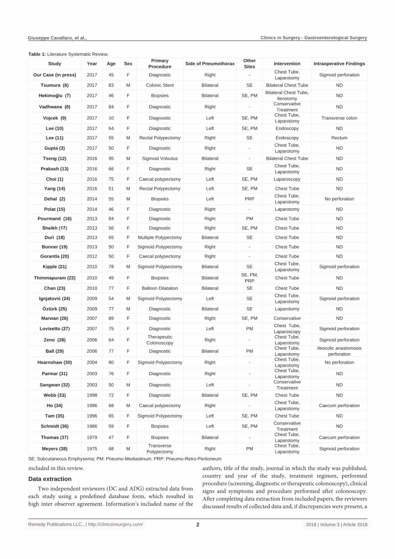
Study Year Age Sex PrimaryProcedure Side of Pneumothorax Other
Sites Intervention Intraoperative Findings
Our Case (in press) 2017 45 F Diagnostic Right - Chest Tube, Laparotomy Sigmoid perforation
Tsumura (6) 2017 83 M Colonic Stent Bilateral SE Bilateral Chest Tube ND
Hekimoğlu (7) 2017 46 F Biopsies Bilateral SE, PM Bilateral Chest Tube, Ileostomy ND
Vadhwana (8) 2017 84 F Diagnostic Right - Conservative Treatment ND
Vojcek (9) 2017 10 F Diagnostic Left SE, PM Chest Tube,Laparotomy Transverse colon
Lee (10) 2017 64 F Diagnostic Left SE, PM Endoscopy ND
Lee (11) 2017 55 M Rectal Polypectomy Right SE Endoscopy Rectum
Gupta (3) 2017 50 F Diagnostic Right - Chest Tube,Laparotomy ND
Tseng (12) 2016 95 M Sigmoid Volvulus Bilateral - Bilateral Chest Tube ND
Prakash (13) 2016 66 F Diagnostic Right SE Chest Tube,Laparotomy ND
Choi (1) 2016 75 F Caecal polypectomy Left SE, PM Laparoscopy ND
Yang (14) 2016 51 M Rectal Polypectomy Left SE, PM Chest Tube ND
Dehal (2) 2014 55 M Biopsies Left PRP Chest Tube, Laparotomy No perforation
Polat (15) 2014 46 F Diagnostic Right - Laparotomy ND
Pourmand (16) 2013 84 F Diagnostic Right PM Chest Tube ND
Sheikh (17) 2013 56 F Diagnostic Right SE, PM Chest Tube ND
Durì (18) 2013 65 F Multiple Polypectomy Bilateral SE Chest Tube ND
Bonner (19) 2013 50 F Sigmoid Polypectomy Right - Chest Tube ND
Gorantla (20) 2012 50 F Caecal polypectomy Right - Chest Tube ND
Kipple (21) 2010 78 M Sigmoid Polypectomy Bilateral SE Chest Tube, Laparotomy Sigmoid perforation
Thimmapuram (22) 2010 49 F Biopsies Bilateral SE, PM, PRP Chest Tube ND
Chan (23) 2010 77 F Balloon Dilatation Bilateral SE Chest Tube ND
Ignjatović (24) 2009 54 M Sigmoid Polypectomy Left SE Chest Tube, Laparotomy Sigmoid perforation
Öztürk (25) 2009 77 M Diagnostic Bilateral SE Laparotomy ND
Marwan (26) 2007 89 F Diagnostic Right SE, PM Conservative ND
Lovisetto (27) 2007 75 F Diagnostic Left PM Chest Tube,Laparoscopy Sigmoid perforation
Zeno (28) 2006 64 F Therapeutic Colonoscopy Right - Chest Tube,
Laparotomy Sigmoid perforation
Ball (29) 2006 77 F Diagnostic Bilateral PM Chest Tube, Laparotomy
Ileocolic anastomosis perforation
Hearnshaw (30) 2004 80 F Sigmoid Polypectomy Right - Chest Tube, Laparotomy No perforation
Parmar (31) 2003 76 F Diagnostic Right - Chest Tube, Laparotomy ND
Sangwan (32) 2003 50 M Diagnostic Left - Conservative Treatment ND
Webb (33) 1998 72 F Diagnostic Bilateral SE, PM Chest Tube ND
Ho (34) 1996 68 M Caecal polypectomy Right - Chest Tube, Laparotomy Caecum perforation
Tam (35) 1996 65 F Sigmoid Polypectomy Left SE, PM Chest Tube ND
Schmidt (36) 1986 59 F Biopsies Left SE, PM Conservative Treatment ND
Thomas (37) 1979 47 F Biopsies Bilateral - Chest Tube, Laparotomy Caecum perforation
Meyers (38) 1975 68 M Transverse Polypectomy Right PM Chest Tube,
Laparotomy Sigmoid perforation
Table 1: Literature Systematic Review.
SE: Subcutaneous Emphysema; PM: Pneumo-Mediastinum; PRP: Pneumo-Retro-Peritoneum
Giuseppe Cavallaro, et al., Clinics in Surgery - Gastroenterological Surgery
Remedy Publications LLC., | http://clinicsinsurgery.com/ 2018 | Volume 3 | Article 20183
consensus was reached by mutual agreement on the accuracy of data.
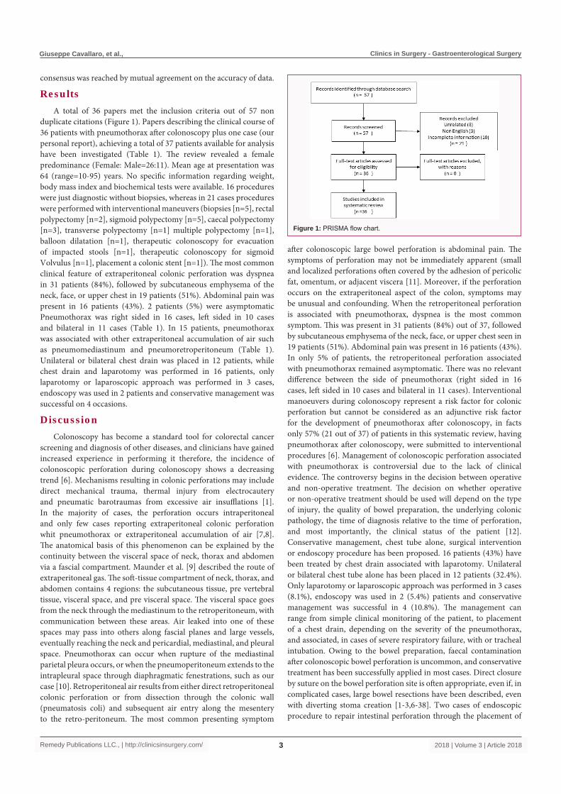
ResultsA total of 36 papers met the inclusion criteria out of 57 non
duplicate citations (Figure 1). Papers describing the clinical course of 36 patients with pneumothorax after colonoscopy plus one case (our personal report), achieving a total of 37 patients available for analysis have been investigated (Table 1). The review revealed a female predominance (Female: Male=26:11). Mean age at presentation was 64 (range=10-95) years. No specific information regarding weight, body mass index and biochemical tests were available. 16 procedures were just diagnostic without biopsies, whereas in 21 cases procedures were performed with interventional maneuvers (biopsies [n=5], rectal polypectomy [n=2], sigmoid polypectomy [n=5], caecal polypectomy [n=3], transverse polypectomy [n=1] multiple polypectomy [n=1], balloon dilatation [n=1], therapeutic colonoscopy for evacuation of impacted stools [n=1], therapeutic colonoscopy for sigmoid Volvulus [n=1], placement a colonic stent [n=1]). The most common clinical feature of extraperitoneal colonic perforation was dyspnea in 31 patients (84%), followed by subcutaneous emphysema of the neck, face, or upper chest in 19 patients (51%). Abdominal pain was present in 16 patients (43%). 2 patients (5%) were asymptomatic Pneumothorax was right sided in 16 cases, left sided in 10 cases and bilateral in 11 cases (Table 1). In 15 patients, pneumothorax was associated with other extraperitoneal accumulation of air such as pneumomediastinum and pneumoretroperitoneum (Table 1). Unilateral or bilateral chest drain was placed in 12 patients, while chest drain and laparotomy was performed in 16 patients, only laparotomy or laparoscopic approach was performed in 3 cases, endoscopy was used in 2 patients and conservative management was successful on 4 occasions.
DiscussionColonoscopy has become a standard tool for colorectal cancer
screening and diagnosis of other diseases, and clinicians have gained increased experience in performing it therefore, the incidence of colonoscopic perforation during colonoscopy shows a decreasing trend [6]. Mechanisms resulting in colonic perforations may include direct mechanical trauma, thermal injury from electrocautery and pneumatic barotraumas from excessive air insufflations [1]. In the majority of cases, the perforation occurs intraperitoneal and only few cases reporting extraperitoneal colonic perforation whit pneumothorax or extraperitoneal accumulation of air [7,8]. The anatomical basis of this phenomenon can be explained by the continuity between the visceral space of neck, thorax and abdomen via a fascial compartment. Maunder et al. [9] described the route of extraperitoneal gas. The soft-tissue compartment of neck, thorax, and abdomen contains 4 regions: the subcutaneous tissue, pre vertebral tissue, visceral space, and pre visceral space. The visceral space goes from the neck through the mediastinum to the retroperitoneum, with communication between these areas. Air leaked into one of these spaces may pass into others along fascial planes and large vessels, eventually reaching the neck and pericardial, mediastinal, and pleural space. Pneumothorax can occur when rupture of the mediastinal parietal pleura occurs, or when the pneumoperitoneum extends to the intrapleural space through diaphragmatic fenestrations, such as our case [10]. Retroperitoneal air results from either direct retroperitoneal colonic perforation or from dissection through the colonic wall (pneumatosis coli) and subsequent air entry along the mesentery to the retro-peritoneum. The most common presenting symptom
after colonoscopic large bowel perforation is abdominal pain. The symptoms of perforation may not be immediately apparent (small and localized perforations often covered by the adhesion of pericolic fat, omentum, or adjacent viscera [11]. Moreover, if the perforation occurs on the extraperitoneal aspect of the colon, symptoms may be unusual and confounding. When the retroperitoneal perforation is associated with pneumothorax, dyspnea is the most common symptom. This was present in 31 patients (84%) out of 37, followed by subcutaneous emphysema of the neck, face, or upper chest seen in 19 patients (51%). Abdominal pain was present in 16 patients (43%). In only 5% of patients, the retroperitoneal perforation associated with pneumothorax remained asymptomatic. There was no relevant difference between the side of pneumothorax (right sided in 16 cases, left sided in 10 cases and bilateral in 11 cases). Interventional manoeuvers during colonoscopy represent a risk factor for colonic perforation but cannot be considered as an adjunctive risk factor for the development of pneumothorax after colonoscopy, in facts only 57% (21 out of 37) of patients in this systematic review, having pneumothorax after colonoscopy, were submitted to interventional procedures [6]. Management of colonoscopic perforation associated with pneumothorax is controversial due to the lack of clinical evidence. The controversy begins in the decision between operative and non-operative treatment. The decision on whether operative or non-operative treatment should be used will depend on the type of injury, the quality of bowel preparation, the underlying colonic pathology, the time of diagnosis relative to the time of perforation, and most importantly, the clinical status of the patient [12]. Conservative management, chest tube alone, surgical intervention or endoscopy procedure has been proposed. 16 patients (43%) have been treated by chest drain associated with laparotomy. Unilateral or bilateral chest tube alone has been placed in 12 patients (32.4%). Only laparotomy or laparoscopic approach was performed in 3 cases (8.1%), endoscopy was used in 2 (5.4%) patients and conservative management was successful in 4 (10.8%). The management can range from simple clinical monitoring of the patient, to placement of a chest drain, depending on the severity of the pneumothorax, and associated, in cases of severe respiratory failure, with or tracheal intubation. Owing to the bowel preparation, faecal contamination after colonoscopic bowel perforation is uncommon, and conservative treatment has been successfully applied in most cases. Direct closure by suture on the bowel perforation site is often appropriate, even if, in complicated cases, large bowel resections have been described, even with diverting stoma creation [1-3,6-38]. Two cases of endoscopic procedure to repair intestinal perforation through the placement of
Figure 1: PRISMA flow chart.
Giuseppe Cavallaro, et al., Clinics in Surgery - Gastroenterological Surgery
Remedy Publications LLC., | http://clinicsinsurgery.com/ 2018 | Volume 3 | Article 20184
clips are reported in this review [15,16]. This treatment, made possible by the progress of operative endoscopy, appear appropriate to repair small and localized perforations following colonoscopy procedure.
ConclusionPneumothorax and tension pneumothorax following a
colonoscopy is an extremely rare but severe and often life threatening complication. Pneumothorax accompanied by pneumomediastinum, pneumoperitoneum, pneumoretroperitoneum, and pneumoderma is a very rare complication of rectosigmoidoscopy. If the patient develops dyspnea and pneumoderma during or after this procedure, a chest radiogram or thoracoabdominal CT should be taken for diagnostic purposes. Urgent treatment, starting with chest tube insertion(s) and laparotomy or laparoscopy could be life saving.
References1. Choi PW. Pneumomediastinum, Pneumothorax, and Subcutaneous
Emphysema Caused by Colonoscopic Perforation: A Report of Two Cases. J Emerg Med. 2017;52(4):117-22.
2. Dehal A, Tessier DJ. Intraperitoneal and extraperitoneal colonic perforation following diagnostic colonoscopy. JSLS. 2014;18(1):136-41.
3. Gupta A, Zaidi H, Habib K. Pneumothorax after Colonoscopy - A Review of Literature. Clin Endosc. 2017;50(5):446-50.
4. Tiwari A, Sharma H, Qamar K, Sodeman T, Nawras A. Recognition of Extraperitoneal Colonic Perforation following Colonoscopy: A Review of the Literature. Case Rep Gastroenterol. 2017;11(1):256-64.
5. Moher D, Liberati A, Tetzlaff J, Altman DG; PRISMA Group. Preferred reporting items for systematic reviews and meta-analyses: the PRISMA Statement. PLoS Med. 2009;6(7):e1000097.
6. Panteris V, Haringsma J, Kuipers EJ. Colonoscopy perforation rate, mechanisms and outcome: from diagnostic to therapeutic colonoscopy. Endoscopy. 2009;41(11):941-51.
7. Fornaro R, Caratto E, Caratto M, Fornaro F, Caristo G, Frascio M, et al. Post-operative recurrence in Crohn'sdisease. Critical analysis of potential risk factors. An update. Surgeon. 2015;13(6):330-47.
8. Fornaro R, Frascio M, Denegri A, Stabilini C, Impenatore M, Mandolfino F, et al. [Chron'sdisease and cancer]. Ann Ital Chir. 2009;80(2):119-25.
9. Maunder RJ, Pierson DJ, Hudson LD: Subcutaneous and mediastinal emphysema, pathophysiology diagnosis and management. Arch Intern Med. 1984;144(7):1447-53.
10. Zeno BR, Sahn SA. Colonoscopy-associated pneumothorax: a case of tension pneumothorax and review of the literature. Am J Med Sci. 2006;332(3):153-5.
11. Tsumura H. Bilateral Tension Pneumothoraces During Colonoscopy: A Case Report and Review of Literature. AANA Journal. 2017;85(3):171-7.
12. Hekimoğlu E, Turna A, Kara V, Demirkaya A, Kaynak K. Rectosigmoidoscopy complicated by bilateral pneumothoraces, pneumomediastinum, pneumoperitoneum, pneumoretroperitoneum, and pneumoderma. Ulus Travma Acil Cerrahi Derg. 2017;23(3):269-71.
13. Vadhwana B, Bell DJ, Nair MS. Not so simple: tension pneumothorax from caecal colonoscopic perforation. ANZ J Surg. 2017;87(4):314.
14. Vojcek E, Vatai B, Veres G, Vörös P, Szabó A, Lódi C. Pneumothorax Pneumomediastinum and Pneumoperitoneum in a 10- Year-Old Girl Following Colonoscopy. Pediatr Ther. 2017;7:2.
15. Lee HS, Park HH, Kim JS, Kang SH, Moon HS, Sung JK, et al. Pneumoretroperitoneum, Pneumomediastinum, Pneumothorax, and Subcutaneous Emphysema after Diagnostic Colonoscopy. Korean J Gastroenterol. 2017;70(3):145-9.
16. Lee MJ, Connelly TM. Head and neck subcutaneous emphysema, a rare complication of iatrogenic perforation during colonoscopy: management review of reported cases from 2000-2016. Expert Rev Gastroenterol Hepatol. 2017;11(9):849-56.
17. Tseng WC, Yeh CC, Jao SW, Wu ZF, Lin SL. Bilateral tension pneumothorax during colonoscopy in a patient with chronic obstructive pulmonary disease: a case report. J Clin Anesth. 2016;34:432-5.
18. Prakash K, Singh A, Sharma S, Pandey VK. Tension pneumothorax as a complication of colonic perforation during colonoscopy: An anesthesiologist's nightmare. Saudi J Anaesth. 2016;10(4):481-3.
19. Yang J, Liu WQ, Dong J, Wen ZQ, Zhu Z, Li WL. Pneumothorax, pneumomediastinum, pneumoperitoneum and extensive subcutaneous emphysema resulting from endoscopic mucosal resection secondary to colonoscopy: A case report. Oncol Lett. 2016;11(4):2763-7.
20. Polat N, Kesici U, Kesici S, Yılbaş S. Morgagni hernia detected due to pneumothorax development during diagnostic colonoscopy. Turk J Gastroenterol. 2014;25(4):448-9.
21. Pourmand A, Shokoohi H. Tension Pneumothorax, Pneumoperitoneum, and Cervical Emphysema following a Diagnostic Colonoscopy. Case Rep Emerg Med. 2013;2013:583287.
22. Sheikh R, Hou J. P-136 Case of Diffuse Air Leak Associated with Colonoscopy in a Patient with Perianal Crohn’s Disease. IBD. 2013;19:78.
23. Durì D, Toso F, De Monte A. Pneumoperitoneum and pneumothorax complicating colonoscopy. BMJ 2013;346:2516.
24. Bonner KP, Ramcharan A. Asymptomatic isolated right sided pneumothorax after screening colonoscopy with polypectomy. Surg Endosc. 2013;27:S479.
25. Gorantla S, Culpepper-Morgan J. Pneumothorax following colonoscopicpolypectomy. Am J Gastroenterol. 2012;107:S555.
26. Kipple JC. Bilateral tension pneumothoraces and subcutaneous emphysema following colonoscopic polypectomy: a case report and discussion of anesthesia considerations. AANA J. 2010;78(6):462-7.
27. Talluri S, Thimmapuram J, Panchwagh R, Manzella J. Colonoscopy and biopsy associated bilateral pneumothoraces, pneumomediastinum, pneumoperitoneum, pneumoretroperitoneum and subcutaneous emphysema. Indian J Med Case Rep. 2017;6(2):18-20.
28. Chan YC, Tsai YC, Fang SY. Subcutaneous emphysema, pneumothorax, pneumomediastinum, and pneumoperitoneum during colonoscopic balloon dilation: a case report. Kaohsiung J Med Sci. 2010;26(12):669-72.
29. Ignjatović M, Jović J. Tension pneumothorax, pneumoretroperitoneum, and subcutaneous emphysema after colonoscopicpolypectomy: a case report and review of the literature. Langenbecks Arch Surg. 2009;394(1):185-9.
30. Öztürk E, Yücel A, Turtay MG, Aydoğan MS, Tekdemir D, Ersoy MO. Pneumomediastinum, Pneumoperitoneum, Pneumothorax and Cervical Subcutaneous Emphysema Following Diagnostic Colonoscopy: A Case Report. İnönü Üniversitesi Tıp Fakültesi Dergisi. 2009;16(3):185-7.
31. Marwan K, Farmer KC, Varley C, Chapple KS. Pneumothorax, pneumomediastinum, pneumoperitoneum, pneumoretroperitoneum and subcutaneous emphysema following diagnostic colonoscopy. Ann R Coll Surg Engl. 2007;89(5):W20-1.
32. Lovisetto F, Zonta S, Rota E, Mazzilli M, Faillace G, Bianca A, et al. Left pneumothorax secondary to colonoscopic perforation of the sigmoid colon: a case report. Surg Laparosc Endosc Percutan Tech. 2007;17(1):62-4.
33. Ball CG, Kirkpatrick AW, Mackenzie S, Bagshaw SM, Peets AD, Temple WJ, et al. Tension pneumothorax secondary to colonic perforation during diagnostic colonoscopy: report of a case. Surg Today 2006;36(5):478-80.
34. Hearnshaw SA, Oppong K, Jaques B, Thompson NP. Tension
Giuseppe Cavallaro, et al., Clinics in Surgery - Gastroenterological Surgery
Remedy Publications LLC., | http://clinicsinsurgery.com/ 2018 | Volume 3 | Article 20185
pneumothorax as a complication of colonoscopy. Endoscopy 2004;36:190.
35. Parmar R, Abdullah M, Cappell M, Grosman I, Mejia JO. Tension pneumothorax as a complication of colonoscopy. AJG. 2003;171.
36. Sangwan S, Singh S, Shaikh N, Lichtenstein S. Pneumoperitoneum, pneumothorax and subcutaneous emphysema after colonoscopy in a patient with crohn’s disease. AJG. 2003;98:143.
37. Webb T. Pneumothorax and pneumomediastinum during colonoscopy. Anaesth Intensive Care. 1998;26(3):302-4.
38. Ho HC, Burchell S, Morris P, Yu M. Colon perforation, bilateral pneumothoraces, pneumopericardium, pneumomediastinum, and subcutaneous emphysema complicating endoscopic polypectomy: anatomic and management considerations. Am Surg. 1996;62(9):770-4.
39. Tam WC, Pollard I, Johnson RD. Case report: pneumomediastinum and pneumothorax complicating colonoscopy. J Gastroenterol Hepatol. 1996;11(8):789-92.
40. Schmidt G, Börsch G, Wegener M. Subcutaneous emphysema and pneumothorax complicating diagnostic colonoscopy. Dis Colon Rectum. 1986;29(2):136-8.
41. Thomas JH, Pierce GE, MacArthur RI. Bilateral pneumothoraces secondary to colonic endoscopy. J Natl Med Assoc. 1979;71(7):701-2.
42. Meyers MA, Ghahremani GG. Complications of fiberoptic endoscopy. II. Colonoscopy. Radiology. 1975;115(2):301-7.